


Abstract
Functional divergence among lectins can arise even in the absence of variation in carbohydrate recognition domains (CRDs), yet the underlying mechanisms remain poorly understood. Here, we investigated the structure–function relationships of hRTLs, a family of six prototype galectins from the sponge Chondrilla australiensis. Recombinant hRTLs lacking the predicted 23-amino-acid non-canonical signal peptide were expressed in Escherichia coli and analyzed for glycan-binding specificity using glycan arrays. Four members (hRTL-1, 2, 5, and 6) exhibited glycan-binding activity, whereas hRTL-3 and 4 showed markedly weaker activity. hRTL-1 and 2 displayed similar binding profiles, including recognition of TF-related glycans, while hRTL-5 preferentially bound type-1 LacNAc and hRTL-6 recognized blood group-related glycans. Structural analyses revealed distinct oligomeric states, with dimeric and tetrameric species supported by native mass spectrometry and size-exclusion chromatography, while additional higher-molecular-weight (HMW) species were observed by non-heated SDS–PAGE. Far-UV CD spectroscopy further showed that representative active and low-activity hRTLs retained β-sheet-rich secondary structures. Notably, thermal stability assays showed that multimer-forming hRTLs retained hemagglutination activity after prolonged incubation at 60 °C, whereas non-multimerizing members exhibited reduced stability. These findings show that distinct oligomeric organizations are associated with functional diversification among structurally conserved galectin CRDs and suggest that modulation of multimerization may have contributed to the evolution of early metazoan lectins.
1. Introduction
Galectins are β-galactoside-binding lectins widely distributed among metazoans and fungi. Despite significant diversity in their overall molecular structures, their carbohydrate-recognition domains (CRDs) adopt a highly conserved β-sandwich fold. Based on domain architecture, galectins are classified into three structural groups: prototype galectins, which possess a single CRD; chimera-type galectins, consisting of a single CRD fused to an N-terminal oligomerization domain; and tandem-repeat galectins, composed of multiple distinct CRDs linked by a peptide sequence [1]. These structural features underpin the functional diversity of galectins in processes such as cell adhesion, immunomodulation, host-microbe interactions, and development [2,3,4].
Accumulated research indicates that oligomerization is a key determinant of galectin function. Prototype galectins, exemplified by galectin-1, form non-covalent homodimers that cross-link cell-surface glycoconjugates, thereby regulating downstream signaling pathways. Chimera-type galectin-3 forms higher-order oligomers through self-association of its N-terminal region; in contrast, tandem-repeat galectins exist as monomers but achieve multivalency through their multiple CRDs connected by linkers. Such differences in oligomeric structure profoundly influence ligand valency, the formation of glycoprotein lattices, receptor clustering, signaling activity, and thermal stability. Here, we use the term “multimerization” to refer broadly to the assembly of galectin molecules into oligomeric states, including dimers, trimers, tetramers, and higher-order oligomers. Together, these studies establish oligomerization as a primary mechanism for regulating galectin function, independent of changes in carbohydrate-recognition specificity [5,6,7,8].
Despite extensive studies of vertebrate galectins, the evolutionary origin of functional diversification within the galectin family remains largely unresolved. In vertebrates, diversification has been accompanied by the emergence of distinct domain architectures, including prototype, chimera-type, and tandem-repeat galectins, as well as sophisticated mechanisms regulating oligomerization and multivalency. However, it remains unclear how galectin functions first diversified in early-animal lineages, where galectins are predominantly represented by structurally conserved prototype forms lacking these complex domain organizations. Whether modulation of oligomeric organization alone was sufficient to generate functional diversity before the evolution of additional domain architectures therefore remains an open question. Addressing this question is essential for understanding the evolutionary origin of galectin functional diversification [9,10].
To address this question, early-diverging metazoans provide an attractive evolutionary model because they predominantly possess prototype galectins while lacking the structural complexity characteristic of vertebrate galectin families. We previously identified six prototype galectin orthologs (hRTL-1 to hRTL-6) from the marine sponge Chondrilla australiensis (Order Chondrillida) [11]. These proteins share 55–95% sequence identity with one another but exhibit relatively low similarity to vertebrate galectins, consistent with previous observations in other poriferan galectins. Native sponge galectins have been reported to exhibit distinct glycan-binding specificities, including preferential recognition of β1-3-galactosides, suggesting that functional diversification had already emerged despite the conservation of prototype domain architecture. These features make C. australiensis an attractive model for investigating the structural basis and evolutionary origins of functional diversification among early metazoan galectins.
In the present study, we expressed recombinant hRTL-1 to hRTL-6 and systematically compared their glycan-binding specificities, oligomerization states, and thermal stabilities. By integrating biochemical, structural, and glycan-binding analyses, we investigated how oligomeric architecture and structural variation within the conserved CRD contribute to functional diversification among prototype galectins. Our findings provide a framework for understanding how galectin functions diversified before the emergence of complex domain architectures in later-evolving metazoans.
2. Results
2.1. Expression of Recombinant hRTLs from C. australiensis
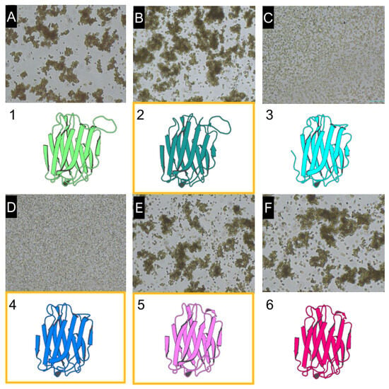
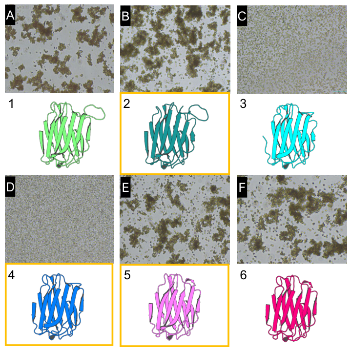
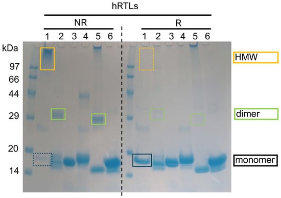
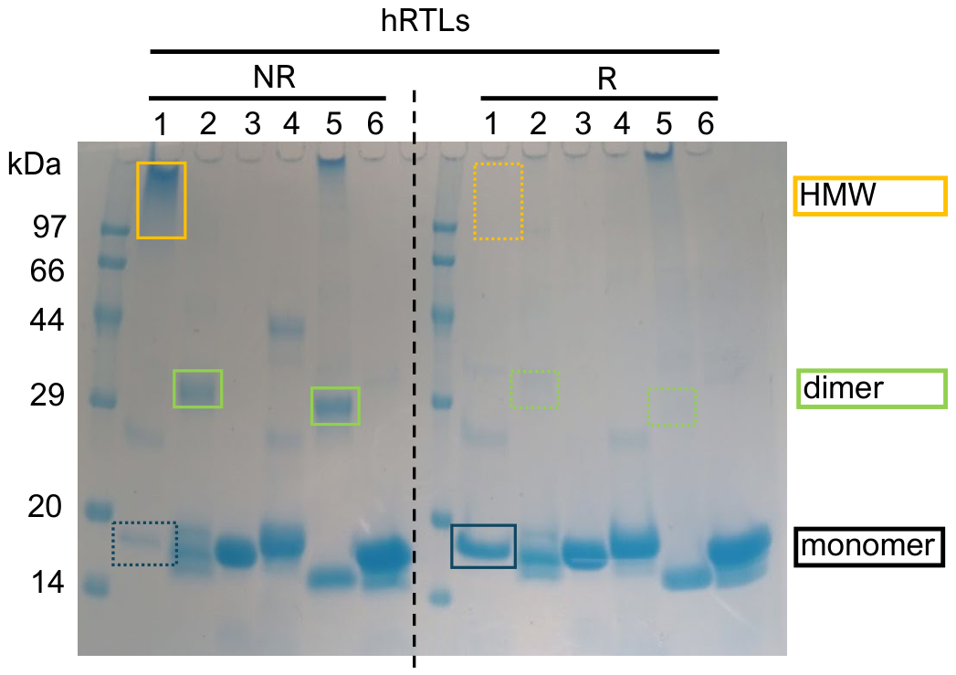
E. coli cells expressing hRTL-1 to hRTL-6 (Figure 1, 1-6; Supplementary Figure S1) were harvested, sonicated, and the supernatants were collected. Hemagglutination activity is a classical functional assay for lectins, based on their ability to cross-link glycans on erythrocyte surfaces. Therefore, it was used as an initial screen to assess the glycan-binding activity of recombinant hRTLs. Using this method, the activity was detected in extracts expressing hRTL-1, 2, 5, and 6 (Figure 1A,B,E,F), whereas hRTL-3 and 4 did not show hemagglutination (Figure 1C,D). Crude supernatants were first applied to a lactose-agarose column and eluted with a lactose-containing buffer. Proteins recovered in the lactose-eluting fractions retained binding to the lactose affinity matrix. hRTL-1, -2, -5, and -6 were successfully purified using this approach. While hRTL-3 and -4 showed barely visible bands in the lactose-eluting fractions, they were largely recovered in the flow-through fraction and subsequently purified using Ni-agarose columns and eluted with an imidazole-containing buffer. The six prototype hRTL galectins (heated at 80 °C for 15 min prior to the analysis) exhibited characteristic SDS-PAGE patterns (Figure 2). Lactose-agarose-purified hRTL-1, which lacked Cys residues, appeared as both HMW species and monomeric species under non-reducing (NR) and reducing (R) conditions (Figure 2, column 1).
Figure 1.
Hemagglutination activity of crude extracts of hRTL1–6 expressed in Escherichia coli. Bacterial cell lysates were incubated with 1% fixed rabbit erythrocytes ((A–F) correspond to hRTL-1 to hRTL-6, respectively). Numbers 1–6 below the panels indicate the corresponding hRTL proteins. Hemagglutination was observed for selected hRTLs. Orange boxes indicate hRTLs (hRTL-2, -4, and -5) containing a single cysteine residue in their polypeptide chain (see Supplementary Figure S1).
Figure 2.
Multimerization states of recombinant hRTLs. SDS–PAGE analysis under non-reducing (NR) and reducing (R) conditions. Molecular mass markers (kDa) are indicated on the left. Boxes indicate the observed molecular species of each hRTL. HMW, higher-molecular-weight species. The dashed vertical line separates the non-reducing (NR) and reducing (R) conditions.
In contrast, hRTL-2 and hRTL-5, both containing a single Cys residue, migrated predominantly as dimers under NR conditions and as monomers under R conditions (Figure 2, columns 2 NR and R, 5 NR and R). hRTL-6, which also lacked Cys residues, migrated exclusively as a monomer under both conditions (Figure 2, column 6). Ni-agarose-purified hRTL-3 (no Cys residue) and hRTL-4 (containing 1 Cys residue) were likewise detected only as monomers regardless of reducing conditions (Figure 2, columns 3 and 4).
2.2. Hemagglutination Activity in hRTLs
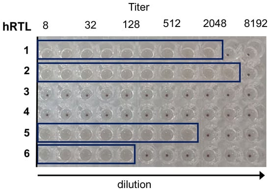
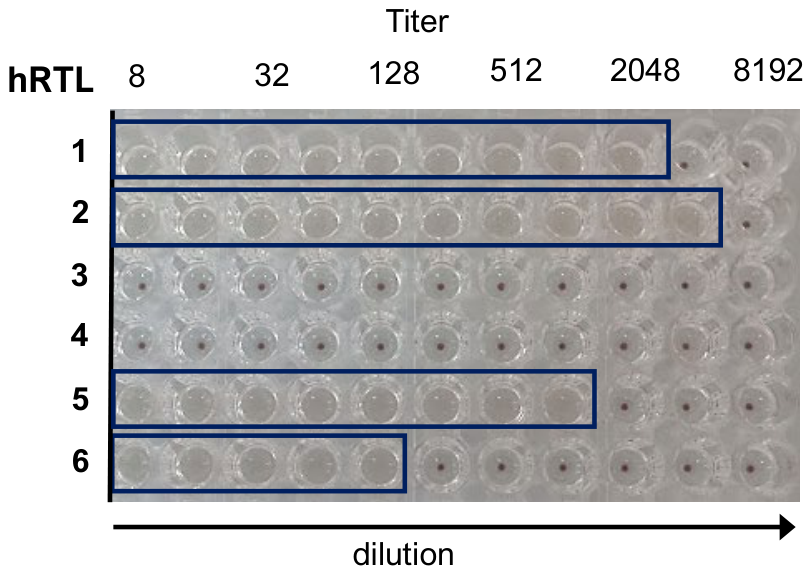
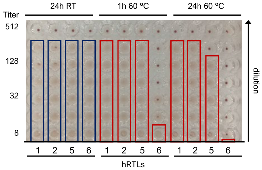
To compare the functional glycan-binding capacities of the six recombinant hRTLs, their hemagglutination activities were examined. At 1 μg/mL protein, hRTL-1, -2, and -5 exhibited hemagglutination activity approximately fivefold higher than hRTL-6 (Figure 3). In contrast, no detectable activity was observed for hRTL-3 and -4.
Figure 3.
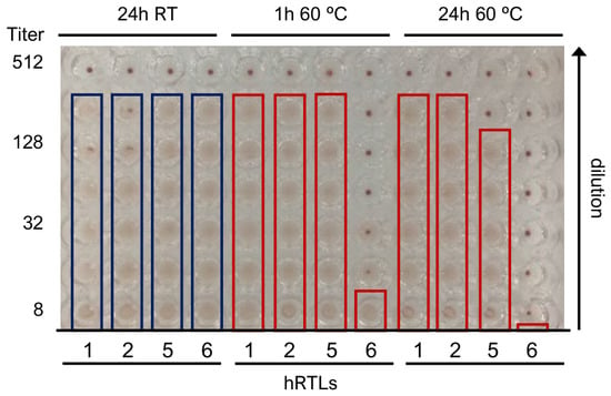
Comparative hemagglutination activities of hRTLs. Each hRTL-1 to -6 was serially diluted in V-bottom microtiter plates. Boxes indicate wells showing positive hemagglutination.
2.3. Glycan-Binding Properties of hRTLs
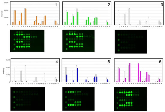
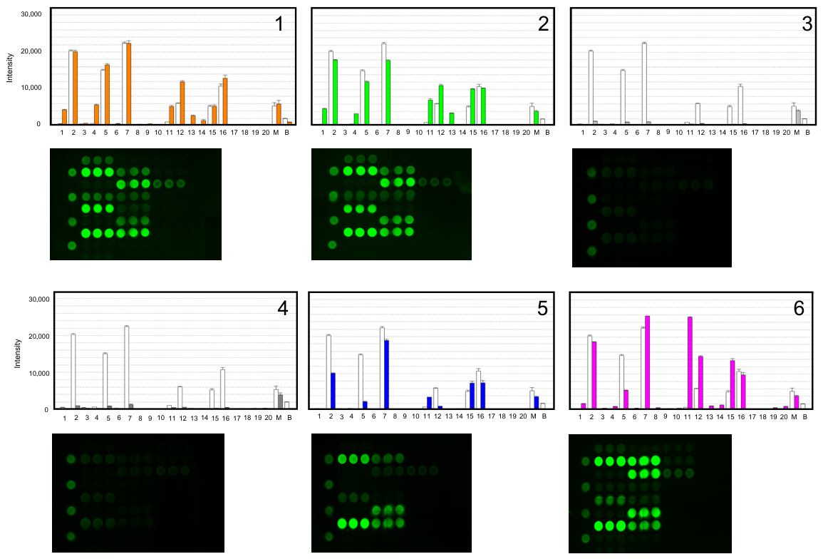
To characterize the glycan-binding specificity of individual hRTLs in greater detail, we performed glycan array analysis. Glycan array analysis revealed distinct binding profiles among hRTLs (Figure 4, Supplementary Table S1). hRTL-3 and -4 showed only very weak glycan-binding signals, close to the detection/background level (Figure 4, panels 3 and 4). The remaining hRTLs preferentially bound type-1 LacNAc (Galβ1-3GlcNAc) (7) over type-2 LacNAc (Galβ1-4GlcNAc) (15). In addition, they exhibited strong affinity for the Thomsen–Friedenreich (TF) antigen (Galβ1-3GalNAc) (2), a mucin-associated cancer-related glycan, and moderate binding to blood group-related disaccharides, including A (GalNAcα1-3Gal) (11) and B (Galα1-3Gal) (12) (Figure 4, panels 1, 2, 5, and 6).
Figure 4.
Glycan-binding profiles of hRTLs. Panels 1–6 correspond to hRTL-1 to hRTL-6, respectively. Cy3-labeled hRTLs were analyzed using a glycan array containing 20 immobilized glycans (X-axis; numbering corresponds to Supplementary Table S1). Binding of Cy3-hRTLs to glycans was detected as evanescent-field fluorescence and is represented as fluorescence intensity (Y-axis). White bars represent glycan-binding profiles of native hRTL mixtures purified from C. australiensis [11].
The binding characteristics of individual hRTLs are summarized below.
hRTL-1 exhibited a binding profile similar to that of hRTL-2, with moderate affinity for the Tn-antigen (GalNAc) (1) compared with other hRTLs (Figure 4, panel 1).
hRTL-2 showed a binding pattern nearly identical to that of hRTL-1 (Figure 4, panel 2).
hRTL-5 exhibited the highest affinity for type-1 LacNAc (Galβ1-3GlcNAc) (7) among the hRTLs, but showed relatively low affinity for the TF antigen (2) (Figure 4, panel 5).
hRTL-6, in addition to binding TF antigen (2) and type-1 LacNAc (7), displayed relatively high affinity for blood group A (GalNAcα1-3Gal) (11) and B (Galα1-3Gal) (12) antigens (Figure 4, panel 6). Quantitative glycan-array binding signals for all six hRTLs against the 20 glycans, expressed as relative fluorescence intensities (mean ± SD, n = 3), are provided in Supplementary Table S2.
2.4. Phylogenetic and Structural Comparison of hRTLs
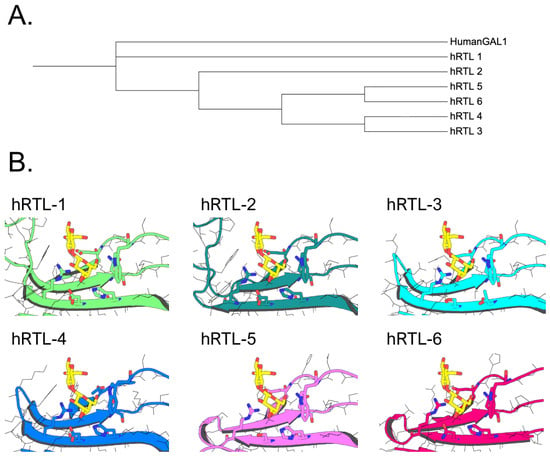
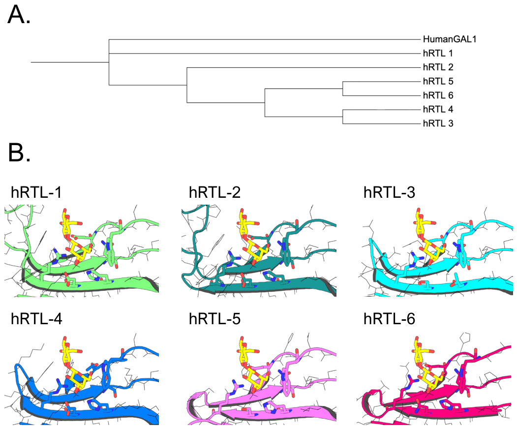
To examine the evolutionary relationships and structural features associated with the distinct glycan-binding profiles, we performed phylogenetic and structural comparisons of the six hRTLs. Phylogenetic analysis based on the primary amino acid sequences revealed that hRTL-1 and hRTL-2 formed a closely related pair, as did hRTL-5 and hRTL-6, whereas hRTL-3 and hRTL-4 occupied distinct phylogenetic positions (Figure 5A). Structural modeling further showed that all six hRTLs retained the characteristic β-sandwich fold of galectin carbohydrate-recognition domains (CRDs). However, comparison of the regions surrounding the predicted carbohydrate-binding sites revealed several substitutions among the hRTLs (Figure 5B).
Figure 5.
Phylogenetic and structural comparison of hRTLs. (A) Phylogenetic relationships of hRTLs based on their primary amino acid sequences. Human galectin-1 is included as a structural reference. (B) Structural comparison of regions surrounding the predicted carbohydrate-binding sites among hRTLs. The predicted carbohydrate-binding sites are shown in Supplementary Figure S2.
Using human galectin-1 (PDB 1W6O) as a structural reference, conserved residues surrounding the carbohydrate-binding site were compared among the six hRTLs. hRTL-1 and hRTL-2 showed similar residue arrangements in this region, consistent with their similar glycan-binding profiles. In hRTL-3, residues corresponding to His and Asp in the other hRTLs were replaced by Thr (T40) and Ser (S42), respectively, whereas in hRTL-4, residues corresponding to Asn and Glu were replaced by Ser (S57) and Thr (T68), respectively (Figure 5B; Supplementary Figure S2). hRTL-5 and hRTL-6 also differed in the configuration of residues surrounding the predicted carbohydrate-binding site, consistent with their distinct glycan-binding specificities.
2.5. Far-UV CD Spectroscopy Supports the Folded State of hRTL-3 and hRTL-4
Far-UV circular dichroism (CD) spectroscopy was performed to experimentally assess the secondary structures of hRTL-1, hRTL-3, and hRTL-4. All three proteins exhibited similar overall spectral features, including a pronounced negative band around 215–220 nm, consistent with predominantly β-sheet-rich structures characteristic of galectin CRDs (Supplementary Figure S6). The similar spectral profiles of hRTL-3 and hRTL-4 to that of hRTL-1 indicate that these proteins retain broadly similar overall secondary-structure characteristics despite their markedly reduced glycan-binding activities.
2.6. Native Mass Spectrometry and Size-Exclusion Chromatography Reveal Diverse Oligomerization States Among hRTLs
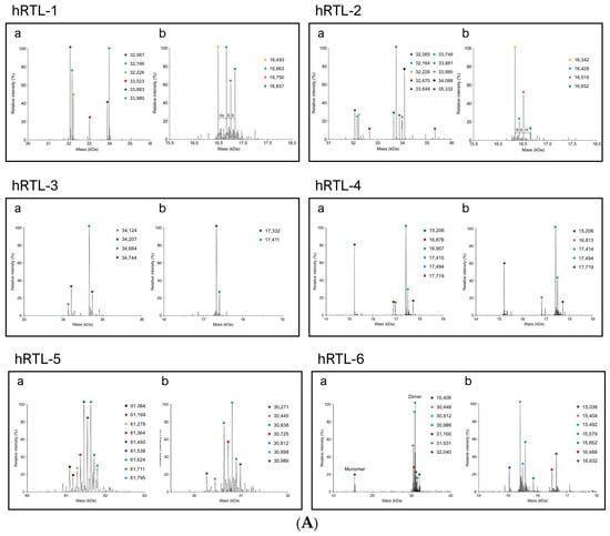
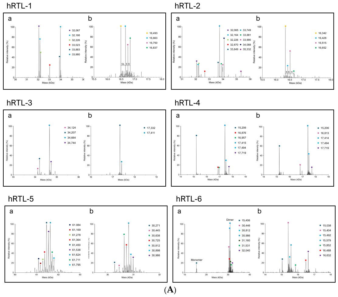
To characterize the oligomeric states of hRTLs in solution, purified recombinant proteins were analyzed by native mass spectrometry (Figure 6A, Supplementary Table S3) and, for selected hRTLs, by size-exclusion chromatography (Figure 7, Supplementary Figure S7). Each hRTL exhibited a distinct mass distribution, reflecting differences in oligomerization behavior rather than extensive post-translational modification. hRTL-1 predominantly formed noncovalent dimers (~32 kDa) (Figure 6A, hRTL-1, a), together with a broad unresolved distribution of higher-molecular-weight (HMW) species extending above the dimeric region. Monomeric species (~16.5–16.8 kDa) were also detected (Figure 6A, hRTL-1, b).
Figure 6.
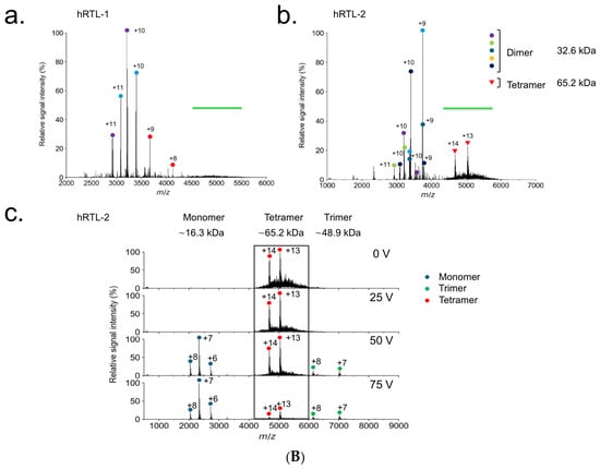
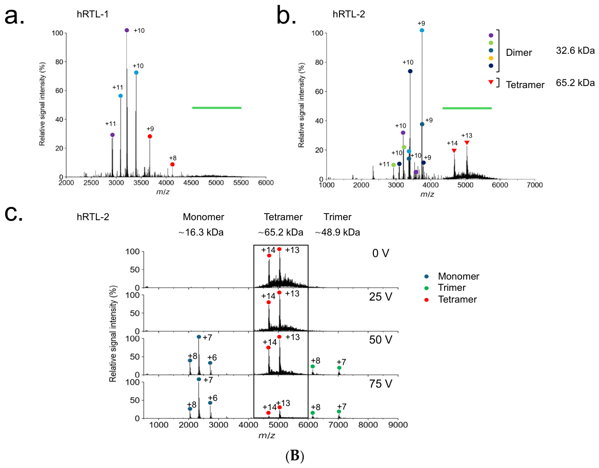
(A) Native mass spectrometry of hRTL-1 to hRTL-6. Panels a and b show spectra acquired under native and denaturing conditions, respectively, with denaturation induced by the addition of a denaturant. Charge states assigned to each oligomeric species are indicated in the spectra. Deconvoluted molecular masses are shown on the right of each panel. The experimentally observed molecular masses correspond well with the theoretical masses of the assigned monomeric and oligomeric species (Supplementary Table S3). Heterogeneous molecular species detected for some hRTLs are represented by multiple deconvoluted mass peaks (Supplementary Figure S3). Detailed analyses of hRTL-1 and hRTL-2 are presented in Figure 6B. (B) Native mass spectrometric characterization of hRTL-1 and hRTL-2 oligomeric species. (a) Raw native mass spectrum of hRTL-1. Charge states assigned to the dimeric species are indicated. A reproducible broad unresolved signal was observed around m/z 5000–6000 (green bar). (b) Raw native mass spectrum of hRTL-2. Charge states assigned to dimeric and tetrameric species are indicated. The broad signal around m/z 5000–6000 (green bar) contains tetrameric ions together with unresolved heterogeneous species. (c) Collision-induced dissociation (CID) analysis of the isolated tetrameric ions of hRTL-2. Increasing collision energy led to progressive dissociation of the tetrameric precursor into monomeric and trimeric ions, thereby confirming the assignment of the detected tetrameric species.
Figure 7.
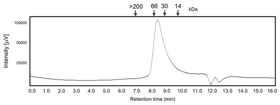
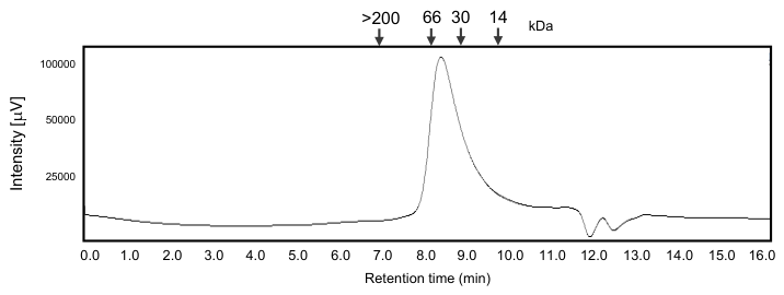
Size-exclusion chromatography of recombinant hRTL-1. Recombinant hRTL-1 was applied to a Shodex KW 402.5-4F column connected to an HPLC pump (JASCO PU-2089, Tokyo, Japan) at a flow rate of 0.33 mL/min. The molecular mass standards indicated above the chromatogram were blue dextran (>200 kDa), bovine serum albumin (66 kDa), carbonic anhydrase (30 kDa), and horse myoglobin (14 kDa). The absorbance was monitored at 220 nm (JASCO UV-2070 Plus, Tokyo, Japan). The major hRTL-1 peak eluted at a position corresponding approximately to the 66 kDa standard, consistent with a tetrameric assembly.
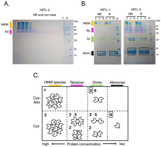
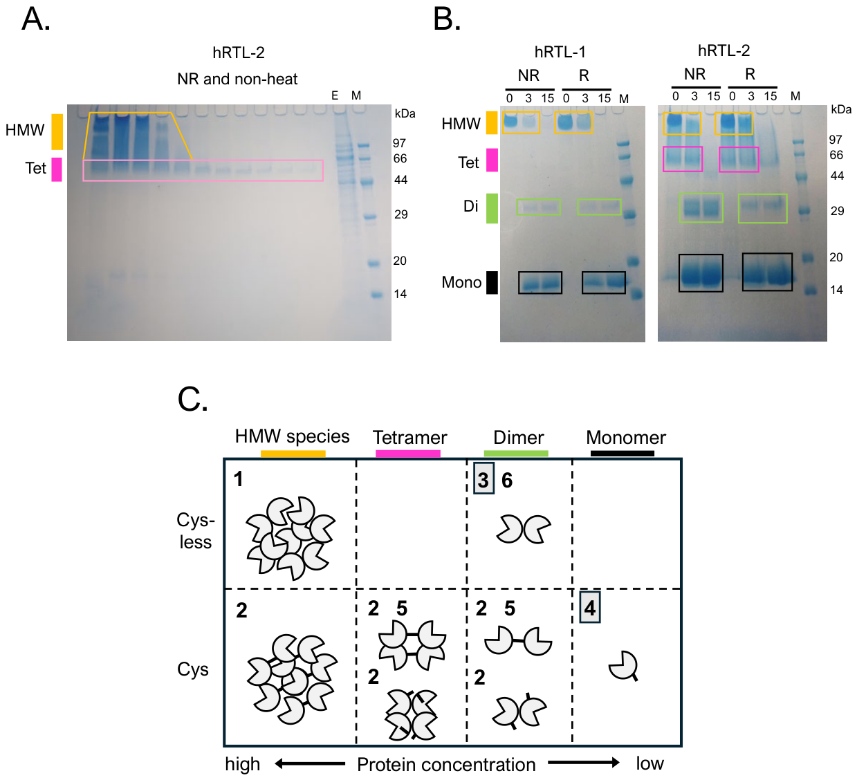
hRTL-2 exhibited a more complex oligomerization profile. Monomeric (~16.4 kDa) and noncovalent dimeric (~32–35 kDa) species coexisted with higher-mass species (Figure 6B). Collision-induced dissociation (CID) analysis demonstrated that the higher-mass species corresponded to tetramers (Figure 6B(b)), which dissociated into monomeric and trimeric ions, confirming tetramer formation in solution (Figure 6B(c)). Although native MS predominantly detected monomeric, noncovalent dimeric, and tetrameric species, non-heated SDS-PAGE revealed additional HMW species, including reduction-sensitive species in hRTL-2 (Figure 8A,B, Supplementary Figures S4 and S8). Native MS did not detect these HMW species (Figure 6A,B), possibly because their large size or heterogeneity reduced ionization and transmission efficiency under the MS conditions.
Figure 8.
Higher-molecular-weight species and proposed assembly model of hRTLs. (A) Eluting fractions of recombinant hRTL-2 from a lactose–agarose affinity column analyzed by SDS–PAGE under non-reducing (NR) and non-heated conditions. HMW species were detected in the eluted fractions. (B) SDS–PAGE analysis of recombinant hRTL-1 and hRTL-2 after heat treatment under non-reducing (NR) or reducing (R) conditions. Numbers indicate the heating duration at 80 °C (min). HMW, higher-molecular-weight species (orange); Tet, tetramer (pink); Di, dimer (green); Mono, monomer (black). (C) Proposed assembly model for multimerization-associated functional divergence of hRTLs. Numbers 1–6 indicate the corresponding hRTL proteins (hRTL-1–6), respectively. HMW species of hRTL-1 and hRTL-2 were observed by non-heated SDS–PAGE analyses (A,B). The non-covalent dimeric structures of hRTL-3 and hRTL-6 and the monomeric structure of hRTL-4 were supported by native mass spectrometry (Figure 6A). The tetrameric structure of hRTL-5, composed of two disulfide-linked dimers associated through non-covalent interactions, was supported by native mass spectrometry (Figure 6) and SDS–PAGE (Figure 2, Supplementary Figure S8). Thick bars represent disulfide bonds, whereas the absence of bars indicates free sulfhydryl groups of cysteine residues. Gray boxes indicate hRTL-3 and hRTL-4, which showed markedly reduced glycan-binding activity and no detectable hemagglutination.
hRTL-3 was detected almost exclusively as noncovalent dimers (~34 kDa) (Figure 6A, hRTL-3, a and b).
In contrast, hRTL-4 remained exclusively monomeric. The observed molecular masses ranged from approximately 15.2 to 17.7 kDa, most likely attributable to heterogeneous N-terminal processing during recombinant expression, including incomplete removal of the expression tag and the thrombin cleavage sequence, rather than to glycosylation or oligomerization (Figure 6A, hRTL-4, a and b).
hRTL-5 predominantly existed as disulfide-linked dimers (~30.5 kDa) (Figure 6A, hRTL-5, b) together with tetramers (~61.5 kDa) (Figure 6A, hRTL-5, a). The tetrameric species are consistent with the association of two covalent dimers through noncovalent interactions, suggesting hierarchical assembly of this lectin.
hRTL-6 mainly formed noncovalent dimers (~30.5–32.0 kDa), although a minor monomeric population (~15–16.6 kDa) was also detected (Figure 6A, hRTL-6, a and b).
Collectively, native MS demonstrated that the six hRTLs exhibit distinct oligomerization states. hRTL-1 showed noncovalent dimeric species together with an unresolved HMW distribution, whereas hRTL-2 showed monomeric, dimeric, and tetrameric species, with the tetrameric assignment confirmed by CID (Figure 6B).
Size-exclusion chromatography identified a major solution-phase species of hRTL-1 corresponding approximately to a tetramer, whereas non-heated SDS-PAGE revealed additional HMW species. Recombinant hRTL-1 eluted at a retention time corresponding approximately to the 66 kDa molecular mass standard (bovine serum albumin), consistent with a tetrameric state of the approximately 16–17 kDa monomer (Figure 7). In contrast, HMW species above the tetrameric range were detected by non-heated SDS–PAGE (Figure 8A,B), although their structural nature could not be resolved by this analysis. The difference between the predominant dimeric species detected by native mass spectrometry and the tetramer-sized major peak observed by SEC may reflect differences in the analytical conditions, including protein concentration and buffer composition. However, the present data do not establish which factors account for this difference.
HMW species were observed for hRTL-1 and hRTL-2 by non-heated SDS-PAGE and were also detected in native sponge-derived hRTL lectins (Supplementary Figure S5). hRTL-1 and hRTL-2 exhibited both glycan-binding and hemagglutinating activities (Figure 1, Figure 3 and Figure 4). The occurrence of HMW species in glycan-binding hRTLs provides additional evidence of structural heterogeneity among the recombinant proteins, although the functional significance of these species remains unresolved.
2.7. Structural Modeling of Oligomeric Assemblies
To further examine the structural basis of the oligomeric states identified experimentally, we generated predicted oligomeric models of hRTL-2 using AlphaFold3. hRTL-2 was selected as a representative because native mass spectrometry identified monomeric, dimeric, and tetrameric species, with the tetrameric assignment further confirmed by collision-induced dissociation (Figure 6B). The predicted dimeric model showed a plausible intermolecular interface between two hRTL-2 molecules, while the predicted tetrameric model formed a higher-order arrangement of the dimeric units (Supplementary Figure S9).
The predicted hRTL-2 tetramer was further compared with the experimentally determined tetrameric structure of CchG-1 from the sponge Cinachyrella sp. (PDB ID: 4AGV) [12]. Superposition of the two structures yielded an RMSD of 2.85 Å over 264 aligned residues (Supplementary Figure S9). These structural similarities provide a plausible model for the tetrameric organization of hRTL-2, while the predicted models remain subject to experimental validation.
2.8. Higher-Molecular-Weight Species of hRTLs
To further characterize the HMW species observed by SDS–PAGE, recombinant hRTL-2 was analyzed throughout lactose–agarose affinity purification under non-reducing and non-heated conditions. HMW species were consistently detected in the eluted fractions (Figure 8A). These HMW species may represent higher-order oligomeric assemblies and/or heterogeneous aggregates. As protein concentration decreased across successive elution fractions, HMW species became progressively less abundant, with tetrameric species predominating in later fractions.
The stability of the observed molecular species was further examined by SDS–PAGE following heat treatment under non-reducing or reducing conditions. Recombinant hRTL-1 retained HMW species, dimers, and monomers under both reducing and non-reducing conditions (Figure 8B). In contrast, hRTL-2 exhibited reduction-sensitive HMW species, although dimeric species were not completely abolished after reduction, suggesting that both disulfide bonds and noncovalent interactions contribute to its oligomerization (Supplementary Figure S4).
Based on the combined results of native mass spectrometry, SDS–PAGE, and, where available, size-exclusion chromatography, we propose a working model for the oligomeric organization of the six hRTLs (Figure 8C). hRTL-1 and hRTL-2 formed dimeric and tetrameric species in solution, while additional HMW species were observed by non-heated SDS–PAGE. These HMW species may represent higher-order oligomeric assemblies, although the possibility that they include heterogeneous aggregates cannot be excluded. hRTL-3 and hRTL-6 predominantly exist as noncovalent dimers, hRTL-4 remains monomeric, and hRTL-5 forms tetramers through the association of two disulfide-linked dimers.
To examine whether the molecular species observed in recombinant hRTLs also occur in the native proteins, hRTLs were purified directly from sponge tissues and analyzed by SDS–PAGE under non-reducing conditions. The native proteins exhibited molecular species corresponding to monomers, dimers, tetramers, and HMW species, similar to those observed for the recombinant proteins (Supplementary Figure S5). These results indicate that the molecular heterogeneity observed in the recombinant hRTLs is not solely attributable to heterologous expression in E. coli.
2.9. Thermal Stability Associated with Oligomerization
To examine whether oligomerization was associated with thermal stability, we assessed the thermotolerance of the hRTLs. hRTLs exhibited distinct levels of thermotolerance (Figure 9). hRTL-1, -2, and -5 retained hemagglutination activity after incubation at 60 °C for up to 24 h, whereas hRTL-6 was inactivated after only 1 h of incubation at the same temperature. The thermal stability of hRTL-6 was comparable to that of human galectin-1, a noncovalent dimer (Supplementary Figure S10A). Notably, hRTL-1 exhibited dimeric and tetrameric states together with additional HMW species, whereas hRTL-2 and hRTL-5 showed disulfide-dependent oligomeric species. The higher thermal stability observed for these oligomer-forming hRTLs was associated with their oligomeric organization, although the present data do not establish a direct causal relationship. In contrast, variation in the length of the loop connecting the S4 and S5 β-strands (Supplementary Figure S1) did not show an apparent association with thermal stability.
Figure 9.
Thermal stability of hRTLs assessed by hemagglutination assay. hRTLs were incubated at 60 °C for 1 h and 24 h. Hemagglutination titers (reciprocal dilution; y-axis) were normalized to an initial value of 256. Column numbers correspond to hRTL-1, hRTL-2, hRTL-5, and hRTL-6, respectively. RT indicates the positive control incubated at room temperature for 24 h.
3. Discussion
Galectin functional diversification has generally been explained by gene duplication, sequence divergence, and the emergence of distinct domain architectures [9,10], including prototype, tandem-repeat, and chimera-type galectins. However, how functional diversification was initiated prior to the evolution of these complex architectures remains largely unresolved [9,10].
By comparing six prototype galectins from the sponge Chondrilla australiensis, we demonstrate that substantial functional diversity can arise despite conservation of the overall carbohydrate-recognition domain fold. Our results indicate that distinct oligomeric organizations are associated with differences in glycan-binding specificity and thermal stability, despite conservation of the overall CRD fold.
These findings suggest that functional diversification of galectins may have involved modulation of oligomeric architecture before the emergence of domain diversification. Multimerization may therefore have represented an early strategy for expanding galectin function in early metazoans. Interestingly, this evolutionary scenario is consistent with a recent model proposing that additional mechanisms of galectin multivalency involving intrinsically disordered regions and phase separation evolved in vertebrate lineages after the establishment of structurally conserved galectin CRDs [13].
Oligomerization has long been recognized as a major determinant of galectin function, although the structural mechanisms responsible for multivalency differ among galectin subfamilies [1,9]. Prototype galectins such as galectin-1 typically form noncovalent homodimers that enable glycan cross-linking and contribute to diverse biological processes, including cell signaling and protein stability. In contrast, chimera-type galectins, exemplified by galectin-3, undergo multimerization via self-association of their N-terminal domains, thereby increasing ligand avidity and expanding functional versatility [14]. Tandem-repeat galectins, such as galectin-4 and 9, achieve multivalency by encoding two carbohydrate recognition domains within a single polypeptide chain, allowing simultaneous recognition of multiple glycoconjugates. Thus, structural diversification through oligomerization or domain organization has been regarded primarily as a characteristic of vertebrate galectins [15].
By contrast, although numerous sponge galectin sequences have been identified [10,16,17,18,19,20,21,22,23], their quaternary structures and the relationship between oligomerization and function have been explored only to a limited extent [12,24]. The present study extends this framework by showing that six prototype galectins from Chondrilla australiensis exhibit remarkable functional diversity associated with differences in oligomeric organization despite retaining a conserved carbohydrate-recognition domain. Oligomeric organization may therefore have contributed to functional diversification before the emergence of complex galectin domain architectures.
Structural modeling using AlphaFold revealed that all six hRTLs retained the characteristic β-sandwich fold of the galectin carbohydrate-recognition domain (CRD) [11], indicating that their overall structural framework has been highly conserved despite functional divergence. Multiple monomeric molecular species detected by native mass spectrometry were consistent with alternative N-terminal processing of the recombinant proteins (Supplementary Figure S3), rather than representing distinct mature protein forms. Nevertheless, four members (hRTL-1, -2, -5, and -6) exhibited distinct glycan-binding specificities.
Notably, hRTL-1 and hRTL-2 both possess a 10-residue structurally flexible loop between the S4 and S5 β-strands (Supplementary Figure S1). The shared S4–S5 loop insertion may therefore represent a structural element linking ligand recognition to oligomerization. Its flexibility could influence the local geometry of the carbohydrate-binding surface and affect intermolecular contacts, thereby providing a possible structural link between glycan specificity and quaternary organization.
Since subtle structural variations surrounding the conserved CRD have been shown to influence ligand-binding properties in other galectins [25], this loop insertion may contribute to the similar glycan-binding profiles observed for hRTL-1 and hRTL-2. In contrast, hRTL-5, which lacks this loop, preferentially recognized type-1 LacNAc, whereas hRTL-6 additionally exhibited affinity for blood group A- and B-related glycans.
Together, these findings suggest that modest structural variations surrounding the conserved CRD contribute to diversification of glycan recognition. The shared S4–S5 loop insertion may therefore represent a structural element associated with both ligand recognition and oligomerization. These structural features may contribute to the coordinated diversification of glycan recognition and oligomeric organization among sponge galectins.
Differences in oligomerization could alter the spatial organization and effective local valency of the CRDs, thereby influencing how individual hRTLs engage multivalent glycoconjugates. Thus, oligomerization may contribute to functional diversification through differences in the spatial organization and effective local valency of carbohydrate-binding sites.
These structural differences were particularly notable for hRTL-3 and hRTL-4, which exhibited markedly weaker glycan-binding activities than the other hRTLs. In glycan array analysis, both proteins showed only very weak binding signals near the detection/background level, although they were retained on lactose-affinity columns, indicating limited glycan-recognition capacity (Figure 4). Far-UV CD spectroscopy showed that hRTL-3 and hRTL-4 retain β-sheet-rich secondary structures, with overall spectral profiles similar to that of hRTL-1 (Supplementary Figure S6). These results suggest that hRTL-3 and hRTL-4 retain broadly similar secondary-structure characteristics to hRTL-1, arguing against gross protein misfolding as the primary explanation for their reduced activity, although local structural differences cannot be excluded. Sequence and structural comparison revealed substitutions at residues corresponding to conserved ligand-binding residues in human galectin-1: His and Asp are replaced by Thr and Ser in hRTL-3, whereas Asn and Glu are replaced by Ser and Thr in hRTL-4. These substitutions surround the predicted carbohydrate-binding site and may contribute to their reduced glycan-binding activities. The distinct phylogenetic positions of hRTL-3 and hRTL-4 further suggest that reduced glycan recognition may have arisen through separate evolutionary modifications of the conserved galectin CRD.
A related example is human galectin-16, which possesses a pseudo-ligand-binding site and markedly reduced canonical β-galactoside-binding activity [26]. The attenuated glycan recognition of hRTL-3 and hRTL-4 may likewise represent an early stage of functional diversification, in which canonical ligand recognition is reduced while the overall galectin fold is retained.
In conclusion, our results show that structurally conserved prototype galectins from the sponge Chondrilla australiensis can achieve substantial functional diversification through differences in glycan-binding specificity (Figure 4), oligomeric organization (Figure 8C), and thermal stability (Figure 9). Modulation of oligomeric architecture, together with sequence diversification within the conserved carbohydrate-recognition domain, may have provided an early route to expanding galectin function. Although our findings support oligomeric organization as a potential contributor to functional diversification, further quantitative biophysical studies are required to define the mechanistic contributions of individual oligomeric states. Sponge galectins thus provide a useful system for investigating how structural conservation can coexist with functional diversification during galectin evolution.
4. Materials and Methods
4.1. Expression and Purification of Recombinant hRTLs
The DNA sequences (Supplementary Table S4) were ordered (VectorBuilder Inc., Yokohama, Japan) as linear double-stranded DNA, with the coding regions codon-optimized by the manufacturer (Supplementary Table S4). After PCR amplification, the DNA products were digested with NdeI and BamHI and cloned into pET28a digested with the same restriction enzymes. The resulting constructs were transformed into E. coli BL21(DE3). For protein expression, transformants were first cultured overnight at 37 °C in 50 mL of culture medium (Plusgrow II, Nacalai Tesque, Kyoto, Japan) containing 50 µg/mL kanamycin. A one-fifth volume of the overnight culture was then inoculated into 400 mL of the same medium containing kanamycin and cultured at 37 °C for approximately 3 h. When the optical density reached 0.6, protein expression was induced by addition of 0.5 mM IPTG, followed by incubation at 20 °C for 16 h [27].
Cells were harvested by centrifugation (9000 × rpm for 50 min at 4 °C; TOMY Supreme 2100 (Tokyo, Japan) high-speed refrigerated centrifuge equipped with an N-16A angle rotor for 500 mL bottles) and resuspended in 50 mL of 100 mM sodium bicarbonate buffer (pH 8.8) containing 150 mM NaCl. The cells were disrupted on ice using a BRANSON/Sonifier Model 450 ultrasonic cell disruptor (Brookfield, CT, USA) equipped with a titanium tip (approximately 1 cm in diameter) at Level 7 (approximately 150–200 W), with 30 s ON/20 s OFF pulses for a total sonication time of 10 min. Following sonication, the lysate was centrifuged at 20,000 × rpm for 50 min at 4 °C using the same centrifuge and rotor. The resulting supernatant (approximately 50 mL) was loaded onto a 2 mL lactose-agarose (Sigma-Aldrich, St. Louis, MO, USA) affinity column under hydrostatic pressure. The column was washed with 100 mL of 100 mM sodium bicarbonate buffer (pH 8.8) containing 150 mM NaCl, and bound proteins were subsequently eluted with the same buffer containing 200 mM lactose. The eluate was collected in 1 mL fractions using a fraction collector (AC-5700; ATTO Corp., Tokyo, Japan) [11]. Because hRTL-3 and hRTL-4 showed only weak binding to lactose–agarose, the crude extracts were loaded onto a 2 mL Ni-NTA agarose (Fujifilm Wako) column instead of the lactose–agarose column. After washing, the bound proteins were eluted with the same buffer containing 0.3 M imidazole. To prevent precipitation associated with residual Ni2+, 100 μL of the same buffer containing 100 mM EDTA was added to each collection tube before fractionation. The hRTL-3 and hRTL-4 fractions were subsequently dialyzed against the sodium bicarbonate buffer (100 mM sodium bicarbonate buffer (pH 8.8) containing 150 mM NaCl) used for the other hRTLs to remove imidazole and EDTA before further analyses. All recombinant hRTLs were used with their His-tags retained. Human galectin-1 was expressed as the Cys-free form (Supplementary Figure S10B,C) and purified by the lactose-agarose column [28].
Protein purity was assessed by SDS-PAGE (Supplementary Figure S8). Samples were heated or non-heated at 80 °C for 3 or 20 min, separated on 15% polyacrylamide gels [29], and stained with Coomassie Brilliant Blue G-250 [30]. Protein concentrations were determined using a micro-BCA assay (Thermo Fisher/Pierce, Rockford, IL, USA) with bovine serum albumin as a standard [31,32].
4.2. Hemagglutinating Activity and Analysis of Thermostabilities of hRTLs
Hemagglutination of lectins was performed in 96-well V-shaped plates as described previously [33]. Briefly, 20 μL of a two-fold serial dilution of purified lectin in Tris-buffered saline (TBS: 10 mM Tris(hydroxymethyl)aminomethane, 150 mM NaCl, pH 7.4) was mixed with 20 μL of a 1% suspension (in TBS; v/v) of trypsinized and glutaraldehyde-fixed rabbit erythrocytes and 40 μL of Triton X-100 (in TBS; 0.01% final conc.) [34]. Plates were kept at room temperature for 1 h. Hemagglutination was observed and scored to determine the titer. Fifty microliters of each hRTL and human galectin-1 at 10 µg/mL were treated at room temperature and then incubated at 60 °C overnight. Their hemagglutination activities were determined as described above.
4.3. Glycan-Binding Profiling of Galectins
Glycan array analysis was performed by a system of Emukk LLC (Mie, Japan). The lectin was fluorescence-labeled (λex/em 560/580 nm) using a Cy3 labeling kit-NH2 (Cytiva, Tokyo, Japan) per the manufacturer’s instructions. A total of 20 glycans (Supplementary Table S1) were immobilized on wells of a microarray (Glyco-Chip GXA209/R1710, Glycotechnica Co. Ltd., Yokohama, Japan). Fluorescence-labeled lectins at concentrations ranging from 0 to 100 μg/mL were incubated overnight at 4 °C in the dark. The glycan-binding specificities of the lectin were detected using a GlycoLite2200 Model evanescent fluorescence scanner (Glycotechnica Co. Ltd., Yokohama, Japan). The application of labeled lectins to glycan arrays and the analytical procedures were performed according to previously reported methods [35,36].
4.4. Mass Spectrometry Analysis
For native MS, the samples were buffer-exchanged into 150 mM ammonium acetate, pH 6.8, using an Amicon Ultra–0.5 mL filter (NMWL 10 kDa; Merck). The buffer-exchanged samples, loaded into in-house-made gold-coated glass capillaries [37] at volumes of approximately 2–5 µL, were immediately analyzed by nanoflow electrospray ionization mass spectrometry. Spectra were recorded on a Q Exactive UHMR Hybrid Quadrupole Orbitrap Mass Spectrometer (Thermo Fisher Scientific Waltham, MA, USA) in positive ionization mode. The key instrument parameters for native MS were set as follows, spray voltage: 1.0–1.2 kV, detector mode: low m/z, injection time: 100 ms–300 ms, resolution: 12,500, trap gas pressure setting: 5.0. For CID experiments of hRTL-2, the additional parameters were set as follows, isolation range (m/z): 4200–6000, HCD collision energy: 0, 25, 50, 75 V. Furthermore, for denaturing MS, the buffer-exchanged samples for native MS were treated with 50% formic acid (v/v) and analyzed using the instrument parameters set as follows, spray voltage: 1.2 kV, detector mode: low m/z, injection time: 200 ms, resolution: 12,500, in-source CID: 10 V, trap gas pressure setting: 2.0. In both of native MS and denaturing MS, the spectra were calibrated with 2 mg/mL cesium iodide dissolved in the solution of 50% 2-propanol and 50% water (v/v) and analyzed using Unidec software [38].
4.5. Circular Dichroism Spectroscopy
Far-UV circular dichroism (CD) spectra of recombinant hRTL-1, hRTL-3, and hRTL-4 (300 μg/mL) were recorded using a JASCO J-820 circular dichroism spectropolarimeter (Tokyo, Japan) with a quartz cuvette and a 0.1 cm optical path length. The spectra were analyzed over the wavelength range of 200–240 nm.
4.6. Structure Prediction
The three-dimensional structures of hRTL-1 to hRTL-6 were predicted using the AlphaFold Server (https://alphafoldserver.com/faq, powered by AlphaFold 3, accessed on 16 May 2026) [39]. The amino acid sequences of hRTL-1 to hRTL-6 were submitted individually using the default settings without modification of prediction parameters. The amino acid sequences used for structure prediction are provided in Supplementary Table S5. Structural analyses and molecular graphics were performed using PyMOL version 2.5.0 (Schrödinger, LLC) [40].
For comparison of the monomeric fold, the predicted structures of hRTL-1 to hRTL-6 were compared with the crystal structure of the Cys-to-Ser mutant of human galectin-1 [28] in complex with lactose (PDB ID: 1W6O) [41]. The structures were superposed based on Cα atoms. Residues corresponding to the carbohydrate-recognition site of human galectin-1 were identified by structural superposition and compared among hRTL-1 to hRTL-6.
To examine oligomeric organization, dimeric and tetrameric models of hRTL-2 were predicted using AlphaFold 3 via the AlphaFold Server [39]. Two and four identical copies of the hRTL-2 amino acid sequence were provided as input for the dimeric and tetrameric predictions, respectively. The experimentally determined tetrameric structure of the sponge galectin CchG-1 from Cinachyrella sp. (PDB ID: 4AGV) [12] was used for structural comparison. CchG-1 adopts a tetrameric arrangement consisting of two galectin dimers and has been structurally characterized at 2.1 Å resolution. The predicted hRTL-2 tetramer was superposed onto the CchG-1 tetramer using the cealign algorithm. The structural alignment yielded a root-mean-square deviation (RMSD) of 2.85 Å over 264 aligned residues.
4.7. Size-Exclusion Chromatography
The purified lectin (5 µg) was subjected to size-exclusion chromatography utilizing a Shodex KW 402.5-4F column (4.6 mm × 300 mm) connected to an HPLC pump PU-2089 Plus (JASCO Co., Ltd., Tokyo, Japan) at a flow rate of 0.33 mL/min. The lectin was mixed in 50 mM sodium bicarbonate in saline, and the elution time of lectins from the column was detected by UV at an absorbance of 220 nm (UV-2070, JASCO Co., Ltd., Tokyo, Japan).
4.8. Phylogenetic Analysis of hRTLs
Amino acid sequences of hRTL-1–6 and human galectin-1 were aligned using the MAFFT online service (version 7.511) [42]. The resulting multiple sequence alignment was subjected to maximum-likelihood phylogenetic analysis using the W-IQ-TREE web server [43]. The resulting phylogenetic tree was visualized [44] and formatted using Interactive Tree Of Life (iTOL) v6 [45].
4.9. Statistical Analysis
Glycan array experiments were performed in triplicate (three replicate spots for each glycan), and fluorescence intensities are presented as mean ± SD. Statistical analyses were performed using one-way ANOVA followed by Dunnett’s test in SPSS Statistics version 30 (www.ibm.com/products/spss-statistics accessed on 10 May 2026). Differences with p < 0.05 were considered statistically significant.
5. Conclusions
This study demonstrates that six prototype galectins from the marine sponge Chondrilla australiensis exhibit distinct oligomerization states associated with differences in glycan-binding specificity and thermal stability, despite conservation of the overall carbohydrate-recognition domain fold. These findings suggest that modulation of oligomeric architecture may have served as an early mechanism for galectin functional diversification, preceding the evolution of more complex domain architectures.
Supplementary Materials
The following supporting information can be downloaded at: https://www.mdpi.com/article/10.3390/md24110393/s1, Supplementary Figure S1. Amino acid sequence alignment of the six recombinant hRTLs; Supplementary Figure S2. Structural comparison of the carbohydrate-binding sites of hRTLs and human galectin-1; Supplementary Figure S3. Assignment of monomeric mass variants of recombinant hRTL-1 and hRTL-2; Supplementary Figure S4. Heated SDS-PAGE analysis of recombinant hRTL-2 under non-reducing and reducing conditions; Supplementary Figure S5. Assemblies of native hRTLs purified from the demosponge Chondrilla australiensis; Supplementary Figure S6. Far-UV CD spectra of recombinant hRTL-1, hRTL-3, and hRTL-4; Supplementary Figure S7. Size-exclusion chromatography of recombinant hRTL-2, hRTL-4, hRTL-5, and human galectin-1; Supplementary Figure S8. Representative SDS-PAGE analyses of all recombinant hRTLs except hRTL-2; Supplementary Figure S9. Predicted oligomeric models of hRTL-2 and comparison with the tetrameric structure of CchG-1; Supplementary Figure S10. Thermal stability of human galectin-1 (hGal-1) assessed by a hemagglutination assay; Supplementary Table S1. Twenty glycan structures on the glycan array; Supplementary Table S2. Quantitative glycan-array binding signals of hRTL-1 to hRTL-6; Supplementary Table S3. Molecular masses and oligomeric assemblies of recombinant hRTLs determined by native mass spectrometry and SDS-PAGE; Supplementary Table S4. Coding DNA sequences (CDSs) of hRTL-1 to hRTL-6 used for recombinant protein expression; Supplementary Table S5. Amino acid sequences of hRTL-1 to hRTL-6 used as input for AlphaFold3 structure prediction.
Author Contributions
Conceptualization, Y.O. (Yasuhiro Ozeki), K.K., M.G. and Y.F.; experiments, K.K., Y.F., A.K., H.H., N.M., M.K., S.O., K.Y., S.Y., S.R., Y.O. (Yukiko Ogawa), T.K., K.S. and Y.O. (Yasuhiro Ozeki); data analysis and sample curation, M.G., D.E.N., M.Y., T.Y. and S.M.A.K.; writing—original draft preparation, Y.O. (Yasuhiro Ozeki); writing—review and editing, M.G., H.F., I.H., S.M.A.K., B.P.C. and Y.O. (Yasuhiro Ozeki); visualization, K.K.; supervision, M.G. and S.U. All authors have read and agreed to the published version of the manuscript.
Funding
This work was supported by the Assisted Joint Research Program 26B005 of the J-GlycoNet cooperative network, accredited by the Ministry of Education, Culture, Sports, Science and Technology, MEXT, Japan, as a Joint Usage/Research Center. Yuki Fujii, Hideaki Fujita, Kenichi Kamata, Tatsuya Kawasaki, Yukiko Ogawa, Yasuhiro Ozeki, and Tatsusada Yoshida are supported by a research grant (26K14599, 25K09949, 24K08716, 24K15170, and 23K06190) from JSPS. Marco Gerdol is supported by the Interconnected Nord-Est Innovation Ecosystem (iNEST) and received funding from the European Union Next-GenerationEU (PIANO NAZIONALE DI RI-PRESA E RESILIENZA (PNRR)—MISSIONE 4 COMPONENTE 2, INVESTIMENTO 1.5—D.D. 1058 23/06/2022, ECS00000043.
Institutional Review Board Statement
Not applicable.
Data Availability Statement
All hRTL protein sequences are available in Supplementary Figure S1.
Acknowledgments
This research was supported by the Joint Research of the Exploratory Research Center on Life and Living Systems, ExCELLS (ExCELLS program No. 26EXC346 and 25EXC343). The authors thank the Kanagawa Office, JASCO Support Co., Ltd. (Yokohama, Japan), for their assistance with the operation and analysis of circular dichroism (CD) spectroscopy. During the preparation of this manuscript, the authors used ChatGPT (GPT-5.6 Luna, OpenAI) to assist with language refinement, manuscript organization, and text revision. All AI-assisted outputs were reviewed, edited, and verified by the authors, who take full responsibility for the content of this publication.
Conflicts of Interest
Author Masao Yamada was employed by the company EMUKK. The remaining authors declare that the research was conducted in the absence of any commercial or financial relationships that could be construed as a potential conflict of interest.
References
- Cummings, R.D.; Liu, F.-T.; Rabinovich, G.A.; Stowell, S.R.; Vasta, G.R.; Varki, A. Galectins. In Essentials of Glycobiology, 4th ed.; Cummings, R.D., Esko, J.D., Stanley, P., Hart, G.W., Aebi, M., Mohnen, D., Kinoshita, T., Packer, N.H., Prestegard, J.H., Schnaar, R.L., Eds.; Chapter 36; Cold Spring Harbor Laboratory Press: Woodbury, NY, USA, 2022. [Google Scholar]
- Torres, R.C.F.; Júnior, I.F.B.; Souza, V.R.P.; Duarte, J.A.; Chaves, R.P.; Costa-Filho, M.F.D.; Nascimento, E.; Malveira, E.A.; Andrade, A.L.; Pinheiro, U.; et al. Structural insights and antimicrobial synergy of a proto-galectin from the marine sponge Aiolochroia crassa. Comp. Biochem. Physiol. Part B Biochem. Mol. Biol. 2025, 275, 111034. [Google Scholar] [CrossRef] [Scilit]
- Tvaroška, I. The Role of Glycans in Human Immunity-A Sweet Code. Molecules 2025, 30, 2678. [Google Scholar] [CrossRef] [Scilit]
- Vasta, G.R.; Rabinovich, G.A. An evolutionary perspective of the roles of galectins in pathogen recognition and immunity. Semin. Immunol. 2025, 79, 101974. [Google Scholar] [CrossRef] [Scilit]
- Chang, V.; Lian, I.; Mahoney, K.E.; Romano, J.; Afshari, A.R.; Chen, R.J.; Chen, X.; Fineide, F.; Khan, A.Z.; Utheim, T.P.; et al. Mapping galectin-3 ligands in human tear fluid establishes spliceoform-dependent lacritin binding. Commun. Biol. 2026, 9, 1120. [Google Scholar] [CrossRef] [Scilit]
- Earl, L.A.; Bi, S.; Baum, L.G. Galectin multimerization and lattice formation are regulated by linker region structure. Glycobiology 2011, 21, 6–12. [Google Scholar] [CrossRef] [Scilit]
- Pally, D.; Bhat, R. N-terminal tail prolines of Gal-3 mediate its oligomerization/phase separation. Proc. Natl. Acad. Sci. USA 2021, 118, e2107023118. [Google Scholar] [CrossRef] [Scilit]
- Marková, V.; Smetana, K., Jr.; Jeníková, G.; Láchová, J.; Krejciríková, V.; Poplstein, M.; Fábry, M.; Brynda, J.; Alvarez, R.A.; Cummings, R.D.; et al. Role of the carbohydrate recognition domains of mouse galectin-4 in oligosaccharide binding and epitope recognition and expression of galectin-4 and galectin-6 in mouse cells and tissues. Int. J. Mol. Med. 2006, 18, 65–76. [Google Scholar] [CrossRef] [Scilit]
- Günther, J.; Galuska, S.P. A brief history of galectin evolution. Front. Immunol. 2023, 14, 1147356. [Google Scholar] [CrossRef] [Scilit]
- Sousa, A.R.O.; Ferreira da Ponte, J.A.D.; Sampaio, A.H.; Nagano, C.S.; Carneiro, R.F. Sponge galectins: Structure, evolution, and function. ACS Omega 2026, 11, 21485–21498. [Google Scholar] [CrossRef] [Scilit]
- Hayashi, R.; Kamata, K.; Gerdol, M.; Fujii, Y.; Hayashi, T.; Onoda, Y.; Kobayashi, N.; Furushima, S.; Ishiwata, R.; Ohkawa, M.; et al. Novel galectins purified from the sponge Chondrilla australiensis: Unique structural features and cytotoxic effects on colorectal cancer cells mediated by TF-antigen binding. Mar. Drugs 2024, 22, 400. [Google Scholar] [CrossRef] [Scilit]
- Freymann, D.M.; Nakamura, Y.; Focia, P.J.; Sakai, R.; Swanson, G.T. Structure of a tetrameric galectin from Cinachyrella sp. (ball sponge). Acta Crystallogr. Sect. D Struct. Biol. 2012, 68, 1163–1174. [Google Scholar] [CrossRef] [Scilit]
- Lin, Y.H.; Chen, Y.C.; Sun, Y.C.; Huang, J.R. Evolution of intrinsically disordered regions in vertebrate galectins for phase separation. EMBO Rep. 2026, 27, 1254–1269. [Google Scholar] [CrossRef] [Scilit]
- Farhadi, S.A.; Liu, R.; Becker, M.W.; Phelps, E.A.; Hudalla, G.A. Physical tuning of galectin-3 signaling. Proc. Natl. Acad. Sci. USA 2021, 118, e2024117118. [Google Scholar] [CrossRef] [Scilit]
- Ogawa, T.; Nonaka, Y.; Shoji, H.; Nakamura, T. Evolution and function of galectins in Xenopus laevis: Comparison with mammals and new perspectives. BBA Adv. 2025, 7, 100157. [Google Scholar] [CrossRef] [Scilit]
- Sousa, A.R.O.; Andrade, F.R.N.; Chaves, R.P.; Sousa, B.L.; Lima, D.B.; Souza, R.O.D.S.; da Silva, C.G.L.; Teixeira, C.S.; Sampaio, A.H.; Nagano, C.S.; et al. Structural characterization of a galectin isolated from the marine sponge Chondrilla caribensis with leishmanicidal potential. Biochim. Biophys. Acta Gen. Subj. 2021, 1865, 129992. [Google Scholar] [CrossRef] [Scilit]
- Ohkawa, M.; Kamata, K.; Kawsar, S.M.A.; Gerdol, M.; Fujii, Y.; Ozeki, Y. Characterization of HOL-30: A novel tandem-repeat galectin from the marine sponge Halichondria okadai. BBA Adv. 2025, 7, 100153. [Google Scholar] [CrossRef] [Scilit]
- Marques, D.N.; Almeida, A.S.; Sousa, A.R.O.; Pereira, R.; Andrade, A.L.; Chaves, R.P.; Carneiro, R.F.; Vasconcelos, M.A.; Nas-cimento-Neto, L.G.D.; Pinheiro, U.; et al. Antibacterial activity of a new lectin isolated from the marine sponge Chondrilla caribensis. Int. J. Biol. Macromol. 2018, 109, 1292–1301. [Google Scholar] [CrossRef] [Scilit]
- Ueda, T.; Nakamura, Y.; Smith, C.M.; Copits, B.A.; Inoue, A.; Ojima, T.; Matsunaga, S.; Swanson, G.T.; Sakai, R. Isolation of novel prototype galectins from the marine ball sponge Cinachyrella sp. guided by their modulatory activity on mammalian glutamate-gated ion channels. Glycobiology 2013, 23, 412–425. [Google Scholar] [CrossRef] [Scilit]
- Schröder, H.C.; Boreiko, A.; Korzhev, M.; Tahir, M.N.; Tremel, W.; Eckert, C.; Ushijima, H.; Müller, I.M.; Müller, W.E. Co-expression and functional interaction of silicatein with galectin: Matrix-guided formation of siliceous spicules in the marine demosponge Suberites domuncula. J. Biol. Chem. 2006, 281, 12001–12009. [Google Scholar]
- Wagner-Hülsmann, C.; Bachinski, N.; Diehl-Seifert, B.; Blumbach, B.; Steffen, R.; Pancer, Z.; Müller, W.E. A galectin links the aggregation factor to cells in the sponge (Geodia cydonium) system. Glycobiology 1996, 6, 785–793. [Google Scholar] [CrossRef] [Scilit]
- Pfeifer, K.; Haasemann, M.; Gamulin, V.; Bretting, H.; Fahrenholz, F.; Müller, W.E. S-type lectins occur also in invertebrates: High conservation of the carbohydrate recognition domain in the lectin genes from the marine sponge Geodia cydonium. Glycobiology 1993, 3, 179–184. [Google Scholar] [CrossRef] [Scilit]
- Srivastava, M.; Simakov, O.; Chapman, J.; Fahey, B.; Gauthier, M.E.; Mitros, T.; Richards, G.S.; Conaco, C.; Dacre, M.; Hellsten, U.; et al. The Amphimedon queenslandica genome and the evolution of animal complexity. Nature 2010, 7307, 720–726. [Google Scholar] [CrossRef] [Scilit]
- Duarte, J.A.; Oliveira Neto, J.E.; Torres, R.C.F.; Sousa, A.R.O.; Andrade, A.L.; Chaves, R.P.; Carneiro, R.; Vasconcelos, M.A.; Teixeira, C.S.; Teixeira, E.H.; et al. Structural characterization of a galectin from the marine sponge Aplysina lactuca (ALL) with synergistic effects when associated with antibiotics against bacteria. Biochimie 2023, 214, 165–175. [Google Scholar] [CrossRef] [Scilit]
- Go, R.M.; Kishor, C.; Malde, A.; Blanchard, H. Molecular dynamics identify variances between galectin carbohydrate-binding sites that impact the binding site conformation and ligand binding. Proteins 2026, 94, 934–946. [Google Scholar] [CrossRef] [Scilit]
- Si, Y.; Yao, Y.; Jaramillo Ayala, G.; Li, X.; Han, Q.; Zhang, W.; Xu, X.; Tai, G.; Mayo, K.H.; Zhou, Y.; et al. Human galectin-16 has a pseudo ligand binding site and plays a role in regulating c-Rel-mediated lymphocyte activity. Biochim. Biophys. Acta 2021, 1865, 129755. [Google Scholar] [CrossRef] [Scilit]
- Adachi, D.; Ishimoto, N.; Mizutani, K.; Takahashi, K.; Kubota, R.; Kawabata, H.; Park, S.Y.; Vandebroek, L.; Voet, A.R.D.; Yamada, M.; et al. Crystal and Cryo-EM structure of PPL, a novel hexameric R-type lectin from the poisonous mushroom Pleurocybella porrigens. Glycobiology 2025, 36, cwaf082. [Google Scholar] [CrossRef] [Scilit]
- Nishi, N.; Abe, A.; Iwaki, J.; Yoshida, H.; Itoh, A.; Shoji, H.; Kamitori, S.; Hirabayashi, J.; Nakamura, T. Functional and structural bases of a cysteine-less mutant as a long-lasting substitute for galectin-1. Glycobiology 2008, 18, 1065–1073. [Google Scholar] [CrossRef] [Scilit]
- Laemmli, U.K. Cleavage of structural proteins during the assembly of the head of bacteriophage T4. Nature 1970, 227, 680–685. [Google Scholar] [CrossRef] [Scilit]
- Yasumitsu, H.; Ozeki, Y.; Kawsar, S.M.; Toda, T.; Kanaly, R. CGP stain: An inexpensive, odorless, rapid, sensitive, and in principle in vitro methylation-free Coomassie Brilliant Blue stain. Anal. Biochem. 2010, 406, 86–88. [Google Scholar] [CrossRef] [Scilit]
- Wiechelman, K.J.; Braun, R.D.; Fitzpatrick, J.D. Investigation of the bicinchoninic acid protein assay: Identification of the groups responsible for color formation. Anal. Biochem. 1988, 175, 231–237. [Google Scholar] [CrossRef] [Scilit]
- Smith, P.K.; Krohn, R.I.; Hermanson, G.T.; Mallia, A.K.; Gartner, F.H.; Provenzano, M.D.; Fujimoto, E.K.; Goeke, N.M.; Olson, B.J.; Klenk, D.C. Measurement of protein using bicinchoninic acid. Anal. Biochem. 1985, 150, 76–85. [Google Scholar] [CrossRef] [Scilit]
- Gourdine, J.P.; Cioci, G.; Miguet, L.; Unverzagt, C.; Silva, D.V.; Varrot, A.; Gautier, C.; Smith-Ravin, E.J.; Imberty, A. High affinity interaction between a bivalve C-type lectin and a biantennary complex-type N-glycan revealed by crystallography and microcalorimetry. J. Biol. Chem. 2008, 283, 30112–30120. [Google Scholar] [CrossRef] [Scilit]
- Sasaki, H.; Aketa, K. Purification and distribution of a lectin in sea urchin (Anthocidaris crassispina) egg before and after fertilization. Exp. Cell. Res. 1981, 135, 15–19. [Google Scholar] [CrossRef] [Scilit]
- Kuno, A.; Uchiyama, N.; Koseki-Kuno, S.; Ebe, Y.; Takashima, S.; Yamada, M.; Hirabayashi, J. Evanescent-field fluorescence-assisted lectin microarray: A new strategy for glycan profiling. Nat. Methods 2005, 2, 851–856. [Google Scholar] [CrossRef] [Scilit]
- Hirabayashi, J.; Yamada, M.; Kuno, A.; Tateno, H. Lectin microarrays: Concept, principle and applications. Chem. Soc. Rev. 2013, 42, 443–458. [Google Scholar] [CrossRef] [Scilit]
- Lawrence, S.A.S.; Kirschbaum, C.; Bennett, J.L.; Lutomski, C.A.; El-Baba, T.J.; Robinson, C.V. Phospholipids differentially regulate Ca2+ binding to synaptotagmin-1. ACS Chem. Biol. 2024, 19, 953–961. [Google Scholar] [CrossRef] [Scilit]
- Marty, M.; Baldwin, A.; Marklund, E.; Hochberg, G.; Benesch, J.; Robinson, C. Bayesian deconvolution of mass and ion mobility spectra: From binary interactions to polydisperse ensembles. Anal. Chem. 2015, 87, 4370–4376. [Google Scholar] [CrossRef] [Scilit]
- Abramson, J.; Adler, J.; Dunger, J.; Evans, R.; Green, T.; Pritzel, A.; Ronneberger, O.; Willmore, L.; Ballard, A.J.; Bambrick, J.; et al. Accurate structure prediction of biomolecular interactions with AlphaFold 3. Nature 2024, 630, 493–500. [Google Scholar] [CrossRef] [Scilit]
- The PyMOL Molecular Graphics System, Version 2.5.0; Schrödinger, LLC: New York, NY, USA, 2021.
- López-Lucendo, M.F.; Solís, D.; André, S.; Hirabayashi, J.; Kasai, K.; Kaltner, H.; Gabius, H.-J.; Romero, A. Growth-regulatory human galectin-1: Crystallographic characterization of the structural changes induced by single-site mutations and their Impact on the thermodynamics of ligand binding. J. Mol. Biol. 2004, 343, 957–970. [Google Scholar] [CrossRef] [Scilit]
- Jumper, J.; Evans, R.; Pritzel, A.; Green, T.; Figurnov, M.; Ronneberger, O.; Tunyasuvunakool, K.; Bates, R.; Žídek, A.; Potapenko, A.; et al. Highly accurate protein structure prediction with AlphaFold. Nature 2021, 596, 583–589. [Google Scholar] [CrossRef] [Scilit]
- Katoh, K.; Rozewicki, J.; Yamada, K.D. MAFFT online service: Multiple sequence alignment, interactive sequence choice and visualization. Brief. Bioinform. 2019, 20, 1160–1166. [Google Scholar] [CrossRef] [Scilit]
- Trifinopoulos, J.; Nguyen, L.T.; von Haeseler, A.; Minh, B.Q. W-IQ-TREE: A fast online phylogenetic tool for maximum likelihood analysis. Nucleic Acids Res. 2016, 44, W232–W235. [Google Scholar] [CrossRef] [Scilit]
- Letunic, I.; Bork, P. Interactive Tree of Life (iTOL) v6: Recent updates to the phylogenetic tree display and annotation tool. Nucleic Acids Res. 2024, 52, W78–W82. [Google Scholar] [CrossRef] [Scilit]
Disclaimer/Publisher’s Note: The statements, opinions and data contained in all publications are solely those of the individual author(s) and contributor(s) and not of MDPI and/or the editor(s). MDPI and/or the editor(s) disclaim responsibility for any injury to people or property resulting from any ideas, methods, instructions or products referred to in the content. |
© 2026 by the authors. Licensee MDPI, Basel, Switzerland. This article is an open access article distributed under the terms and conditions of the Creative Commons Attribution (CC BY) license.