


Abstract
Background and Objectives: Keloids are fibroproliferative disorders marked by excessive fibroblast activity, abnormal collagen deposition, and impaired wound healing. They are frequently associated with pain, pruritus, and significant aesthetic concerns, leading to reduced quality of life. Surgical excision alone is burdened by high recurrence rates, underscoring the need for effective adjuvant therapies. This systematic review aimed to assess the effectiveness of surgical excision combined with adjuvant physical and pharmacological therapies in keloid management, with particular emphasis on recurrence rates. Materials and Methods: The review was conducted in accordance with PRISMA guidelines. A systematic search of PubMed and Web of Science identified studies evaluating surgical excision of keloids with adjunctive therapies. Twenty-one studies involving more than 8627 patients met the inclusion criteria. Extracted data included study design, patient and lesion characteristics, treatment modalities, and recurrence rates. Due to marked heterogeneity among treatment protocols, a meta-analysis was not performed. Results: Among physical adjuvant therapies, postoperative brachytherapy showed the lowest recurrence rates (3.1–15%), outperforming radiotherapy and external-beam radiation therapy (14–29.3%). Compression therapy achieved recurrence rates of 10.66% and 14%, particularly effective in auricular keloids. Pharmacological adjuvant therapies demonstrated variable efficacy. Triamcinolone acetonide injections were associated with recurrence rates ranging from 6.6% to 33%, depending on the protocol. Adjuvant 5-fluorouracil reduced recurrence compared with surgery alone, whereas imiquimod 5% showed higher and less consistent recurrence rates. Combination pharmacological therapies consistently yielded better outcomes than monotherapy. Conclusions: Surgical excision combined with adjuvant therapy is the most effective strategy for keloid treatment. Multimodal approaches significantly reduce recurrence compared with surgery alone. However, substantial heterogeneity in lesion characteristics, treatment timing, and therapeutic protocols limits comparability between studies. Further high-quality, standardised clinical trials are needed to optimise management strategies and develop evidence-based guidelines.
1. Introduction
A keloid is a fibroproliferative disorder characterized by excessive fibroblast activation and dysregulated wound healing [1]. It is associated with abnormal collagen deposition and sustained inflammatory activity [2]. These pathological processes are typically triggered by cutaneous injury or surgical procedures [1]. Unlike hypertrophic scars, which stay limited to the primary wound margins and may partially regress over time, keloids extend beyond the boundaries of the initial injury and do not undergo spontaneous regression [3]. Histologically, hypertrophic scars are characterized by predominantly aligned type III collagen fibers, whereas keloid tissue contains disorganized bundles of both type I and type III collagen [4]. Clinically, keloids frequently cause pruritus, pain, and discoloration, leading to a substantial reduction in quality of life [5].
Although the exact pathogenesis remains incompletely understood, it is considered multifactorial, involving dysregulated immune responses and impaired wound healing mechanisms [5]. A genetic predisposition has also been identified, with a higher incidence reported among individuals of African and Asian origin [5], as well as in anatomical regions exposed to increased mechanical stress, including the chest, shoulders, upper back, and earlobes [6].
Over the past decades, numerous therapeutic approaches have been proposed for the management of keloids; however, no single modality has demonstrated universal effectiveness.
Commonly applied interventions for keloid management include physiotherapy, pharmacological treatments, surgical excision, and biological therapies [7]. To enhance treatment efficacy and reduce recurrence, multimodal strategies combining approaches from different therapeutic categories are increasingly adopted. Treatment selection is influenced by lesion size, anatomical location, and individual patient response. Given the pathophysiological mechanisms underlying keloid formation and the high recurrence rates associated with surgical excision alone, surgery should be incorporated into combined therapeutic regimens rather than used as a standalone intervention [7].
The objective of this review is to synthesize current scientific evidence on keloid management, with particular emphasis on the efficacy of surgical excision combined with adjunctive therapies. By systematically analysing clinical outcomes, recurrence rates, and the methodological rigor of existing studies, this review aims to define the current state of knowledge and identify key gaps requiring further investigation. Despite substantial progress the development of an optimal, evidence-based, and universally applicable treatment algorithm for keloids remains an ongoing challenge.
2. Materials and Methods
This study adhered to the Preferred Reporting Items for Systematic Reviews and MetaAnalyses (PRISMA) guidelines [8] (The PRISMA 2020 Checklist is provided in the Supplementary Materials). A systematic literature search was conducted in the PubMed and Web of Science databases to identify studies published up to 2005 that evaluated surgical excision of keloids combined with complementary treatment modalities: physical methods (brachytherapy, radiotherapy, external-beam radiation therapy, compression therapy) and pharmacological methods (5-Fluorouracil, triamcinolone, imiquimod 5% and combination therapies). The protocol was prospectively registered in the PROSPERO database (registration ID:CRD420261335714).
The following search strategy was used:
(“keloid treatment” OR “keloid surgical treatment”) AND (“systematic review” OR “metaanalysis” OR “metaanalysis”).
Unpublished studies and articles written in languages other than English were excluded. During the initial screening of titles and abstracts, abstracts, case reports, conference proceedings, letters, and editorials were excluded. Duplicate records identified across databases were removed using Mendeley software (Reference Manager, version 2.130.2).
The initial database search was independently performed by three researchers. After duplicate removal, two reviewers independently screened the remaining titles and abstracts according to predefined inclusion and exclusion criteria. Full-text articles deemed potentially relevant were subsequently retrieved and assessed for eligibility. Only full-text studies reporting on the effectiveness of surgical excision supported by adjunctive therapies were included in the final review.
Any disagreements regarding study inclusion were resolved through discussion with the lead authors (M.W. and K.K.), with final decisions made under the supervision of the first author.
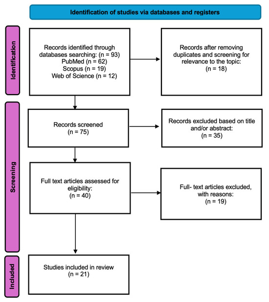
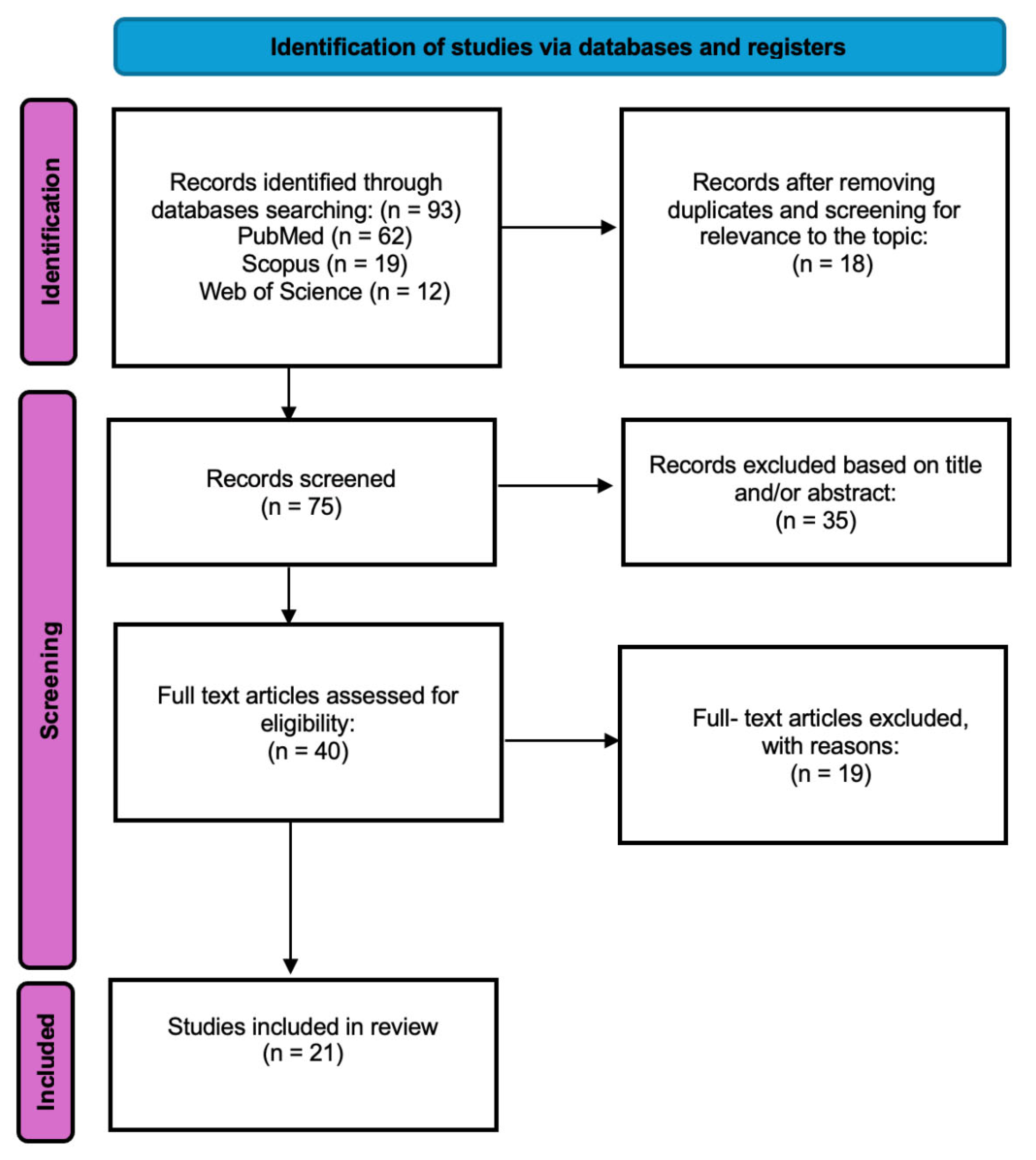
The study selection process is summarized in a PRISMA flow diagram (Figure 1).
Figure 1.
Prisma flowchart.
The initial search identified 93 articles. After excluding articles that do not match the criteria 75 studies were included for abstract review. Finally, 40 were selected for full text appraisal of which 21 met all the inclusion criteria and were included in this review. This systematic review included 21 articles including over 8627 patients. The complete dataset is summarized in Table 1 and Table 2 and Supplementary Table S1.
Due to substantial heterogeneity in keloid treatment modalities, study designs, and outcome measures, the available literature was considered too diverse to permit a formal meta-analysis.
Following data was abstracted from original studies:
- General study information: authors, publication year, country, institution.
- Patient characteristics: number of patients, sex, age.
- Recurrence rate.
- Lesion localisation.
3. Results
3.1. Physical Methods
3.1.1. Brachytherapy
In 4 studies describing a total of 3927 patients, brachytherapy was presented as an adjuvant method in the postoperative treatment of keloids. This modality was most frequently applied within 24 h following surgical excision. One study demonstrated a particularly low recurrence rate of 3.1% among 43 treated patients [9] whereas the remaining publications reported recurrence rates ranging from 9.7% to 15%, indicating generally favourable outcomes with this combined approach [10,11,12]. The timing of brachytherapy was consistent across studies, with all reporting initiation within 24 h postoperatively.
3.1.2. Radiotherapy
In 10 of the analysed studies including a total of 2941 patients, radiotherapy was used as an adjuvant treatment modality, most administered within 72 h following surgical excision. In two studies, radiotherapy was initiated on the fourth postoperative day [13,14]. Another report, a triple-therapy approach combining surgical excision and radiotherapy with an additional modality (e.g., hyperbaric oxygen therapy, pressure therapy, platelet-rich plasma, or 5fluorouracil) resulted in a recurrence rate of 11.2% [15]. A study focusing on auricular keloids treated with adjuvant radiotherapy reported a recurrence rate of 4.8%, while a separate investigation using a combination of surgery, platelet-rich plasma, and superficial photon radiation achieved a 95.5% non-recurrence rate [16]. Across the remaining studies, reported recurrence rates ranged from 14% to 29.3% [13,14,17,18,19,20,21,22]. Overall, the timing of radiotherapy varied across studies, although most protocols involved initiation within the first 24–72 h after surgery.
3.1.3. External-Beam Radiation Therapy (EBRT)
External-beam radiation therapy (EBRT) for the treatment of keloids was reported in three studies encompassing a total of 3130 patients. In one study, EBRT administered within 72 h after surgical excision resulted in recurrence rates of 16% for keloids located at nonauricular sites and 11% for auricular lesions [22]. In the remaining studies, reported recurrence rates ranged from 17% to 28.4%, with EBRT most applied within 24 h postoperatively [11,12]. The timing of EBRT was relatively consistent, with most studies reporting administration within 24–72 h postoperatively.
3.1.4. Compression Therapy
Compression therapy was evaluated by the authors in 2 studies with reported recurrence rates were 10.66% and 14%, suggesting that this non-invasive modality may provide satisfactory outcomes in reducing keloid recurrence. However, the effectiveness varied depending on treatment protocols and anatomical location [11,23]. The timing of compression therapy was not clearly defined and was inconsistently reported across studies.
3.2. Pharmacological Methods
Data from studies investigating the use of 5-fluorouracil (5-FU), triamcinolone (TAC), and Imiquimod 5% as adjuvant therapies following surgical treatment of keloids were analysed.
3.2.1. 5-Fluorouracil (5-FU)
The analysis included 3 studies, encompassing a total of 282 patients who received 5-FU as an adjuvant treatment administered immediately following surgical excision.
While no recurrences were reported in the included studies, one study [24] described a reduction in recurrence rates from 87% to 32% compared to surgical excision alone. In most studies, 5-FU was initiated immediately after surgery and continued as part of repeated injection protocols at defined intervals.
3.2.2. Triamcinolone (TAC)
The use of TAC injections as a standalone surgical adjuvant was reported in 7 studies, involving a total of 1026 patients. Treatment protocols varied, most commonly administered within 7 to 30 days after surgery [25,26]. Two studies employed repeated injections at intervals ranging from 14 to 90 days [20,24], while two others used intraoperative TAC injections [11,27]. Reported recurrence rates varied considerably, with the lowest rate of 6.6% observed in the study by Zhang et al. (2024) and the highest rate of 33% reported by Reid et al. (2025) [9,25]. Overall, the timing of TAC administration was highly variable, ranging from intraoperative use to delayed postoperative regimens extending up to several weeks.
3.2.3. Imiquimod 5%
The analysis was based on a single study that met the inclusion criteria [28]. The group consisted of 77 patients over the age of 12. The topical treatment was initiated either immediately after surgery or with a delay of up to seven days.
The meta-analysis revealed a relatively high, yet highly variable, recurrence rate of 39%. The timing of imiquimod initiation showed limited variability but was based on data from a single study.
3.2.4. Combination Therapies
Studies evaluating combination pharmacological therapies were also analysed. In one study Bijlard et al., a dual regimen combining 5-FU and triamcinolone acetonide (TAC) was administered in a cohort of 24 patients on days 0, 28, and 56 [24]. The results demonstrated superior outcomes with combination therapy compared with 5-FU monotherapy. In another study involving 24 patients [29], a treatment protocol combining corticosteroid injections with self-administered topical steroid ointment was evaluated. The protocol included a single injection at the time of suture removal, followed by injections every two weeks for a total of five sessions; each injection consisted of 1 mL triamcinolone and 1 mL procaine hydrochloride. This approach resulted in a reported recurrence rate of 14.3%. The timing of combination therapies was heterogeneous, reflecting differences in individual components and treatment protocols.
- Follow-Up
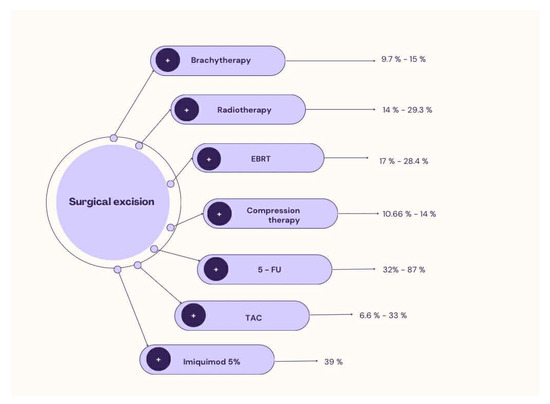
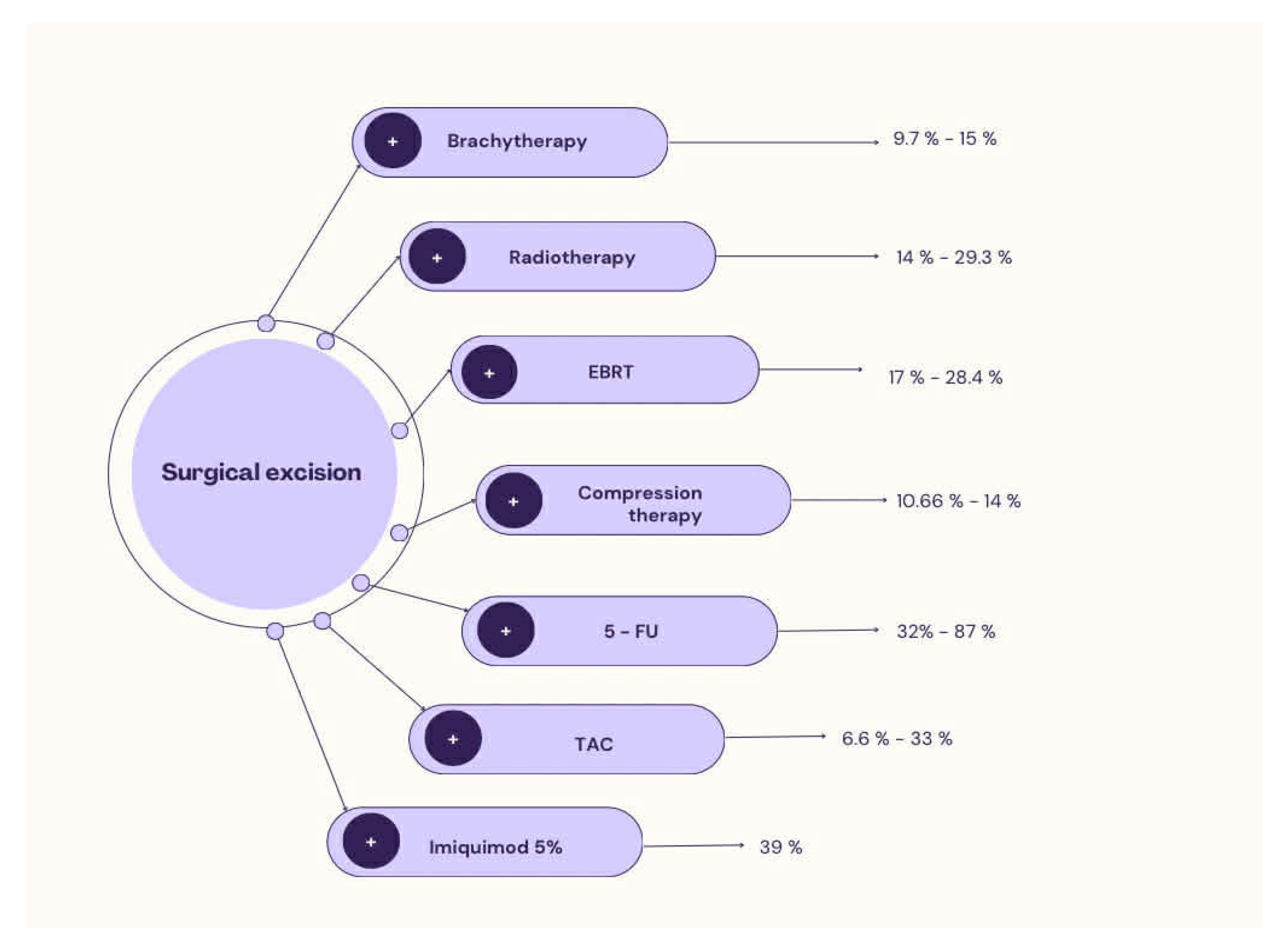
The outcome assessment time was reported in 17 out of 21 studies; it was not standardized, and its range varied from 3–6 months up to 16 years. In only 2 studies was the follow-up period shorter than 12 months. The available data demonstrate heterogeneity both in the duration of follow-up and in the methods of its reporting. All the articles included in our study are presented in Table 1 and Table 2. Recurrence rate of keloids after dual therapy flowchart is presented in Figure 2.
Table 1.
Overview of Studies Evaluating Adjunctive Methods in Surgery and Their Impact on Recurrence Rates.
Table 2.
Summary of Treatment Methods, Timing of Triamcinolone Injection, and Recurrence Rates.
Figure 2.
Recurrence rate of keloids after dual therapy flowchart.
3.3. Surgical Methods
Most studies did not specify the surgical techniques for keloid removal. One study used tangential/shaving excision. Three studies described the use of core excision as the primary surgical approach.
4. Discussion
The pathogenesis of keloid formation is primarily driven by excessive fibroblast proliferation, mediated by a complex interplay of pro-inflammatory and anti-inflammatory cytokines, chemokines, and growth factors, resulting in abnormal collagen synthesis and deposition [30]. Additional contributing factors include immunological, genetic, endocrine, and mechanical influences such as skin tension and trauma [31].
Despite extensive investigation, no definitive gold-standard treatment has been established that reliably eliminates keloids without recurrence [32]. Surgical treatment continues to be regarded as a promising modality, as it provides immediate relief from discomfort such as pain and pruritus; however, when used as monotherapy recurrence rates range from 45% to 100%, underscoring the necessity of adjuvant treatment strategies [1].
This systematic review synthesizes the evidence supporting the effectiveness of combining surgical excision with adjuvant physical therapy and pharmacology. Although differences in lesion severity, anatomical location, and treatment protocols complicate direct comparisons, the results consistently indicate that surgery is an effective primary intervention [33].
One significant limitation of the study is the lack of detailed description of the excision techniques used, leading to heterogeneous and inconsistent results and hindering the selection of the optimal treatment modality.
The follow-up period was reported in 17 studies and ranged from 3–6 months to 16 years. The results are too heterogeneous to allow for evaluation. This undoubtedly highlights the opportunity to conduct a prospective study that would enable a reliable assessment of the efficacy of a specific adjunctive treatment for keloid management.
Radiotherapy is considered a valuable therapeutic option for keloids that do not respond adequately to conventional treatments [34]. By inhibiting fibroblast proliferation, it disrupts the excessive wound-healing response and helps prevent recurrent lesion formation [33]. However, optimal timing of postoperative irradiation remains controversial [35]. In the present analysis, radiotherapy was most frequently initiated within the first 72 h after surgery, producing recurrence rates between 14% and 29.3%. Renu Sah et al. demonstrated that irradiation delivered on the first postoperative day ideally within two hours produced the most favourable outcomes, with recurrence rates of 5–7%. In contrast, delaying treatment beyond six hours, however, led to significantly poorer outcomes [36]. Conversely, Chin-Ling Hsieh and colleagues found no significant differences between irradiation performed within 24 h and later postoperative administration [37]. These findings collectively indicate that considerable uncertainty remains regarding the optimal timing of radiotherapy, and this topic continues to attract substantial research interest [35].
The two principal radiotherapy modalities employed in keloid management are brachytherapy and external beam radiotherapy (EBRT) [38]. Brachytherapy delivers gamma radiation directly to the targeted tissue while minimizing exposure to surrounding structures. This method is particularly useful for irregular anatomical regions that would otherwise necessitate multiple radiation fields [39]. Evidence summarized in our review indicates that postoperative brachytherapy is associated with comparatively lower recurrence rates (9.7–15%) than EBRT (14–29.3%) [40].
In contrast, EBRT typically requires higher radiation doses to achieve equivalent therapeutic outcomes. This modality remains well suited for treating more superficial lesions [41].
Compression therapy following surgical excision demonstrated recurrence rates of 10.66% and 14% in the studies included in our analysis. Its proposed mechanism involves inducing localized hypoxia, which promotes fibroblast apoptosis, while increased collagenase activity contributes to stabilization of the scar tissue [23]. This approach appears particularly effective for auricular keloids when combined with surgery, achieving recurrence rates as low as 6.7–10.6% [42].
Pharmacological therapies play a crucial role in the management of keloids. In this review, we summarize evidence regarding the use of triamcinolone acetonide (TAC), 5-fluorouracil (5FU), and 5% imiquimod.
TAC is widely regarded as the primary non-surgical treatment option for keloids [43]. Its therapeutic effect is believed to stem from the suppression of inflammatory signalling pathways as well as the inhibition of fibroblast activity and collagen synthesis [42]. Across the studies included in our review, TAC was administered intralesional according to various postoperative schedules, most commonly at intervals of 7–30 days, although some protocols extended the interval to 14–90 days or incorporated intraoperative administration. The lowest recurrence rate observed was 6.6%. In the study by Young-Jun Choi et al., combined therapy achieved a recurrence rate of 5% during one year of follow-up [44]. TAC is frequently combined with 5-FU, as this regimen has demonstrated superior efficacy and safety compared with monotherapy [45,46].
5-FU acts as an antimetabolite that inhibits fibroblast proliferation by disrupting RNA synthesis and reducing type I collagen gene expression [45]. In most of the studies assessed in our review, the use of 5-FU as an adjuvant treatment resulted in no reported recurrences, although the follow-up duration varied considerably. One study documented a reduction in recurrence rates from 87% to 32% when compared with surgery alone. Findings from Steven P. Davison et al. further demonstrated that combining surgery with TAC and 5-FU produced excellent outcomes a mean 92% reduction in lesion size-compared with 81% when pharmacologic therapy was used without surgery [47].
Imiquimod, a potent immune response modifier, is typically applied topically to the excision site for 6–8 weeks. A recent review reported a recurrence rate of 24.7% at six months of follow-up [42], whereas our analysis identified a recurrence rate of 14.3%.
Limitations
Unfortunately there are some limitations in our work. The studies included in our review did not categorise patients as individual units, but treated them as a group. Nor did they provide a breakdown by race, information on the comorbidities of the patients included, or whether they were taking long-term medication. Information on specific drug manufacturers was inconsistently reported across studies and was therefore not systematically analyzed.
5. Conclusions
Surgical excision combined with adjuvant therapy remains the most effective strategy for keloid management. Evidence from this systematic review indicates that multimodal approaches significantly reduce recurrence rates compared with surgery alone. Among physical modalities, postoperative brachytherapy appears to provide the lowest recurrence rates, while EBRT remains a viable option for selected superficial lesions. Pharmacological adjuvants, particularly triamcinolone acetonide combined with 5-fluorouracil, demonstrate superior efficacy compared with monotherapy. However, substantial heterogeneity in treatment protocols, timing, and lesion characteristics limits direct comparison across studies. Further high-quality, standardized clinical trials are required to establish optimal treatment algorithms and define evidence-based guidelines for keloid management.
Supplementary Materials
The following supporting information can be downloaded at: https://www.mdpi.com/article/10.3390/medicina62050916/s1, PRISMA 2020 Checklist [8]; Table S1: Summary of Included Studies.
Author Contributions
Conceptualization, M.W. and K.K.; methodology, K.K.; software, B.J.; validation, B.O., W.B. and H.S.; formal analysis, M.W.; investigation, M.W.; resources, A.S., I.Z., Z.D. and J.W.; data curation, B.O., W.B., H.S., A.S., I.Z., Z.D. and J.W.; writing—original draft preparation, K.K.; writing—review and editing, M.W.; visualization, M.W.; supervision, J.J.; project administration, K.K.; funding acquisition, M.W., J.J. All authors have read and agreed to the published version of the manuscript.
Funding
This research received no external funding.
Institutional Review Board Statement
Not applicable.
Informed Consent Statement
Not applicable.
Data Availability Statement
No new data were created or analyzed in this study.
Conflicts of Interest
The authors declare no conflicts of interest.
References
- Fu, S.; Duan, L.; Zhong, Y.; Zeng, Y. Comparison of surgical excision followed by adjuvant radiotherapy and laser combined with steroids for the treatment of keloids: A systematic review and meta-analysis. Int. Wound J. 2024, 21, e14449. [Google Scholar] [CrossRef] [Scilit]
- Zhao, J.; Zhai, X.; Xu, Z.; Zhou, S.; Gu, L.; Chen, L.; Zhou, B.; Hua, H. Novel needle-type electrocoagulation and combination pharmacotherapy: Basic and clinical studies on efficacy and safety in treating keloids. J. Cosmet. Dermatol. 2024, 23, 3693–3704. [Google Scholar] [CrossRef] [Scilit]
- Limandjaja, G.C.; Niessen, F.B.; Scheper, R.J.; Gibbs, S. Hypertrophic scars and keloids: Overview of the evidence and practical guide for differentiating between these abnormal scars. Exp. Dermatol. 2021, 30, 146–161. [Google Scholar] [CrossRef] [Scilit]
- Gauglitz, G.G.; Korting, H.C.; Pavicic, T.; Ruzicka, T.; Jeschke, M.G. Hypertrophic Scarring and Keloids: Pathomechanisms and Current and Emerging Treatment Strategies. Mol. Med. 2011, 17, 113–125. [Google Scholar] [CrossRef] [Scilit] [PubMed]
- Kim, H.J.; Kim, Y.H. Comprehensive Insights into Keloid Pathogenesis and Advanced Therapeutic Strategies. Int. J. Mol. Sci. 2024, 25, 8776. [Google Scholar] [CrossRef] [Scilit]
- Jagdeo, J.; Kerby, E.; Glass, D.A. Keloids. JAMA Dermatol. 2021, 157, 744. [Google Scholar] [CrossRef] [Scilit]
- Qi, W.; Xiao, X.; Tong, J.; Guo, N. Progress in the clinical treatment of keloids. Front. Med. 2023, 10, 1284109. [Google Scholar] [CrossRef] [Scilit] [PubMed]
- Page, M.J.; McKenzie, J.E.; Bossuyt, P.M.; Boutron, I.; Hoffmann, T.C.; Mulrow, C.D.; Shamseer, L.; Tetzlaff, J.M.; Akl, E.A.; Brennan, S.E.; et al. The PRISMA 2020 statement: An updated guideline for reporting systematic reviews. BMJ 2021, 372, n71. [Google Scholar] [CrossRef] [Scilit]
- Reid, D.; Malak, S.; Khadka, M.; Hanna, R.; Pharr, T.; Wyant, W.A.; Albers, S. Keloids and hypertrophic scars in individuals with darker Fitzpatrick skin types: A systematic review of treatment efficacy and quality of life outcomes. Arch. Dermatol. Res. 2025, 317, 795. [Google Scholar] [CrossRef] [Scilit]
- Kuribayashi, S.; Miyashita, T.; Ozawa, Y.; Iwano, M.; Ogawa, R.; Akaishi, S.; Dohi, T.; Hyakusoku, H.; Kumita, S. Post-keloidectomy irradiation using high-dose-rate superficial brachytherapy. J. Radiat. Res. 2011, 52, 365–368. [Google Scholar] [CrossRef] [Scilit] [PubMed]
- Zawadiuk, L.R.R.; Van Slyke, A.C.; Bone, J.; Redfern, B.; Carr, N.J.; Arneja, J.S. What Do We Know About Treating Recalcitrant Auricular Keloids? A Systematic Review and Meta-Analysis. Plast. Surg. 2022, 30, 49–58. [Google Scholar] [CrossRef] [Scilit] [PubMed]
- Van Leeuwen, M.C.E.; Stokmans, S.C.; Bulstra, A.E.J.; Meijer, O.W.M.; Heymans, M.W.; Ket, J.C.F.; Ritt, M.J.P.F.; van Leeuwen, P.A.M.; Niessen, F.B. Surgical excision with adjuvant irradiation for treatment of keloid scars: A systematic review. Plast. Reconstr. Surg.–Glob. Open 2015, 3. [Google Scholar] [CrossRef] [Scilit]
- Akita, S.; Akino, K.; Yakabe, A.; Imaizumi, T.; Tanaka, K.; Anraku, K.; Yano, H.; Hirano, A. Combined surgical excision and radiation therapy for keloid treatment. J. Craniofacial Surg. 2007, 18, 1164–1169. [Google Scholar] [CrossRef] [Scilit]
- Ogawa, R.; Miyashita, T.; Hyakusoku, H.; Akaishi, S.; Kuribayashi, S.; Tateno, A. Postoperative radiation protocol for keloids and hypertrophic scars: Statistical analysis of 370 sites followed for over 18 months. Ann. Plast. Surg. 2007, 59, 688–691. [Google Scholar] [CrossRef] [Scilit]
- Cardenas, D.; Cinclair, T.; Dogaroiu, A.; Hinson, C.; Sink, M.; Bruce, B.; Odobescu, A.; Sammer, D.; Zhang, A.Y. Wound Coverage, Adjuvant Treatments, and Surgical Outcomes for Major Keloid Scars: A Systematic Review and Meta-Analysis. Aesthetic Surg. J. Open Forum 2024, 7, ojae129. [Google Scholar] [CrossRef] [Scilit]
- Ogawa, R.; Akaishi, S.; Dohi, T.; Kuribayashi, S.; Miyashita, T.; Hyakusoku, H. Analysis of the Surgical Treatments of 63 Keloids on the Cartilaginous Part of the Auricle. Plast. Reconstr. Surg. 2015, 135, 868–875. [Google Scholar] [CrossRef] [Scilit]
- Mankowski, P.; Kanevsky, J.; Tomlinson, J.; Dyachenko, A.; Luc, M. Optimizing Radiotherapy for Keloids: A Meta-Analysis Systematic Review Comparing Recurrence Rates between Different Radiation Modalities. Ann. Plast. Surg. 2017, 78, 403–411. [Google Scholar] [CrossRef] [Scilit]
- Miles, O.J.; Zhou, J.; Paleri, S.; Fua, T.; Ramakrishnan, A. Chest keloids: Effect of surgical excision and adjuvant radiotherapy on recurrence, a systematic review and meta-analysis. ANZ J. Surg. 2021, 91, 1104–1109. [Google Scholar] [CrossRef] [Scilit] [PubMed]
- Ran, X.; Guo, X.; Liu, Y.; Zhu, S.; Li, S.; Chen, Z.; Han, T.; Jin, S.; Zhou, M.; Zang, M. Effectiveness of Core Excision Technique for Keloids: A Systematic Review. Plast. Reconstr. Surg. 2025, 155, 801e–808e. [Google Scholar] [CrossRef] [Scilit] [PubMed]
- Shin, J.Y.; Lee, J.W.; Roh, S.G.; Lee, N.H.; Yang, K.M. A Comparison of the Effectiveness of Triamcinolone and Radiation Therapy for Ear Keloids after Surgical Excision: A Systematic Review and Meta-Analysis. Plast. Reconstr. Surg. 2016, 137, 1718–1725. [Google Scholar] [CrossRef] [Scilit]
- Ellis, M.M.; Jones, L.R.; Siddiqui, F.; Sunkara, P.R.; Ozog, D.M. The efficacy of surgical excision plus adjuvant multimodal therapies in the treatment of keloids: A systematic review and metaanalysis. Dermatol. Surg. 2020, 46, 1054–1059. [Google Scholar] [CrossRef] [Scilit]
- Hwang, N.H.; Chang, J.H.; Lee, N.K.; Yang, K.S. Effect of the biologically effective dose of electron beam radiation therapy on recurrence rate after keloid excision: A meta-analysis. Radiother. Oncol. 2022, 173, 146–153. [Google Scholar] [CrossRef] [Scilit]
- Tahir, S.M.; Ihebom, D.; Simman, R. Compression Therapy for Keloid Scars: A Systematic Review and Meta-analysis. Plast. Reconstr. Surg. Glob. Open 2024, 12, e5864. [Google Scholar] [CrossRef] [Scilit] [PubMed]
- Bijlard, E.; Steltenpool, S.; Niessen, F.B. Intralesional 5-fluorouracil in keloid treatment: A systematic review. Acta Derm. Venereol. 2015, 95, 778–782. [Google Scholar] [CrossRef] [Scilit]
- Zhang, Y.; Wu, M.; Liu, D.; Panayi, A.C.; Xu, X.; Luo, L.; Feng, J.; Ou, Y.; Lin, T.; Cui, Y. Recurrence and Complications of Peri-operative Steroid Injection of Keloids: A Systematic Review and Meta-analysis. Aesthetic Plast. Surg. 2024, 48, 2927–2940. [Google Scholar] [CrossRef] [Scilit]
- Shin, J.Y.; Kim, J.S. Could 5-Fluorouracil or Triamcinolone Be an Effective Treatment Option for Keloid after Surgical Excision? A Meta-Analysis. J. Oral Maxillofac. Surg. 2016, 74, 1055–1060. [Google Scholar] [CrossRef] [Scilit]
- Al Aradi, I.K.; Alawadhi, S.A.; Alkhawaja, F.A. Earlobe keloids: A pilot study of the efficacy of keloidectomy with core fillet flap and adjuvant intralesional corticosteroids. Dermatol. Surg. 2013, 39, 1514–1519. [Google Scholar] [CrossRef] [Scilit]
- Klotz, T.; Munn, Z.; Aromataris, E.C.; Greenwood, J.E. Imiquimod to prevent keloid recurrence postexcision: A systematic review and meta-analysis. Wound Repair Regen. 2020, 28, 145–156. [Google Scholar] [CrossRef] [Scilit]
- Hayashi, T.; Furukawa, H.; Oyama, A.; Funayama, E.; Saito, A.; Murao, N.; Yamamoto, Y. A New Uniform Protocol of Combined Corticosteroid Injections and Ointment Application Reduces Recurrence Rates After Surgical Keloid/Hypertrophic Scar Excision. Dermatol. Surg. 2012, 38, 893–897. [Google Scholar] [CrossRef] [Scilit] [PubMed]
- Liu, M. Cytokines, chemokines and growth factors involved in keloids pathogenesis. An. Bras. Dermatol. 2025, 100, 300–307. [Google Scholar] [CrossRef] [Scilit] [PubMed]
- Xia, G.; Dohi, T.; Abdelhakim, M.; Tosa, M.; Ogawa, R. The effects of systemic diseases, genetic disorders and lifestyle on keloids. Int. Wound J. 2024, 21, e14865. [Google Scholar] [CrossRef] [Scilit] [PubMed]
- Ojeh, N.; Bharatha, A.; Gaur, U.; Forde, A.L. Keloids: Current and emerging therapies. Scars Burn. Heal. 2020, 6, 1–18. [Google Scholar] [CrossRef] [Scilit]
- Ekstein, S.F.; Wyles, S.P.; Moran, S.L.; Meves, A. Keloids: A review of therapeutic management. Int. J. Dermatol. 2021, 60, 661–671. [Google Scholar] [CrossRef] [Scilit]
- Walsh, L.A.; Wu, E.; Pontes, D.; Kwan, K.R.; Poondru, S.; Miller, C.H.; Kundu, R.V. Keloid treatments: An evidence-based systematic review of recent advances. Syst. Rev. 2023, 12, 42. [Google Scholar] [CrossRef] [Scilit] [PubMed]
- Dong, W.; Qiu, B.; Fan, F. Adjuvant Radiotherapy for Keloids. Aesthetic Plast. Surg. 2022, 46, 489–499. [Google Scholar] [CrossRef] [Scilit]
- Peng, Q.; Lu, Y.; Huang, R.; Chen, R. Should We Do Postoperative Radiotherapy After Keloid Excision as Soon as Possible? A Systematic Review and Meta-Analysis. Aesthetic Plast. Surg. 2025, 49, 4671–4682. [Google Scholar] [CrossRef] [Scilit]
- Hsieh, C.L.; Chi, K.Y.; Lin, W.Y.; Lee, L.T.J. Timing of Adjuvant Radiotherapy After Keloid Excision: A Systematic Review and Meta-Analysis. Dermatol. Surg. 2021, 47, 1438–1443. [Google Scholar] [CrossRef] [Scilit]
- Liu, E.K.; Cohen, R.F.; Chiu, E.S. Radiation therapy modalities for keloid management: A critical review. J. Plast. Reconstr. Aesthetic Surg. 2022, 75, 2455–2465. [Google Scholar] [CrossRef] [Scilit]
- Bryant, C.; Mendenhall, W.M. Radiation Therapy. In Juvenile Angiofibroma; Springer International Publishing: Cham, Switzerland, 2017; pp. 225–242. [Google Scholar] [CrossRef] [Scilit]
- Frech, F.S.; Hernandez, L.; Urbonas, R.; Zaken, G.A.; Dreyfuss, I.; Nouri, K. Hypertrophic Scars and Keloids: Advances in Treatment and Review of Established Therapies. Am. J. Clin. Dermatol. 2023, 24, 225–245. [Google Scholar] [CrossRef] [Scilit]
- Goutos, I.; Ogawa, R. Brachytherapy in the adjuvant management of keloid scars: Literature review. Scars Burn. Heal. 2017, 3, 1–14. [Google Scholar] [CrossRef] [Scilit] [PubMed]
- Thornton, N.J.; Garcia, B.A.; Hoyer, P.; Wilkerson, M.G. Keloid Scars: An Updated Review of Combination Therapies. Cureus 2021, 13, e12999. [Google Scholar] [CrossRef] [Scilit]
- Morelli Coppola, M.; Salzillo, R.; Segreto, F.; Persichetti, P. Triamcinolone acetonide intralesional injection for the treatment of keloid scars: Patient selection and perspectives. Clin. Cosmet. Investig. Dermatol. 2018, 11, 387–396. [Google Scholar] [CrossRef] [Scilit]
- Choi, Y.; Lee, Y.H.; Lee, H.J.; Lee, G.; Kim, W. Auricular keloid management in Asian skin: Clinical outcome of intralesional excision and postoperative triamcinolone acetonide intralesional injection. J. Cosmet. Dermatol. 2020, 19, 3041–3047. [Google Scholar] [CrossRef] [Scilit]
- Srivastava, S.; Patil, A.N.; Prakash, C.; Kumari, H. Comparison of Intralesional Triamcinolone Acetonide, 5-Fluorouracil, and Their Combination for the Treatment of Keloids. Adv. Wound Care 2017, 6, 393–400. [Google Scholar] [CrossRef] [Scilit] [PubMed]
- Wu, W.; Zhao, Y.; Chen, Y.; Zhong, A. Comparing the Efficacy of Multiple Drugs Injection for the Treatment of Hypertrophic Scars and Keloid: A Network Meta-Analysis. Aesthetic Plast. Surg. 2023, 47, 465–472. [Google Scholar] [CrossRef] [Scilit] [PubMed]
- Davison, S.P.; Dayan, J.H.; Clemens, M.W.; Sonni, S.; Wang, A.; Crane, A. Efficacy of Intralesional 5Fluorouracil and Triamcinolone in the Treatment of Keloids. Aesthetic Surg. J. 2009, 29, 40–46. [Google Scholar] [CrossRef] [Scilit] [PubMed]
Disclaimer/Publisher’s Note: The statements, opinions and data contained in all publications are solely those of the individual author(s) and contributor(s) and not of MDPI and/or the editor(s). MDPI and/or the editor(s) disclaim responsibility for any injury to people or property resulting from any ideas, methods, instructions or products referred to in the content. |
© 2026 by the authors. Published by MDPI on behalf of the Lithuanian University of Health Sciences. Licensee MDPI, Basel, Switzerland. This article is an open access article distributed under the terms and conditions of the Creative Commons Attribution (CC BY) license.