


Abstract
Background and Objectives: Anemia is a frequent complication of chronic kidney disease (CKD), primarily attributed to erythropoietin deficiency. Interstitial fibrosis (IF), which disrupts the renal interstitium where erythropoietin-producing cells reside, may contribute to anemia independent of glomerular filtration rate (GFR). However, data in primary glomerulonephritis (PGN) are limited and conflicting. Materials and Methods: In this nationwide multicenter registry analysis (TSN-GOLD), 2794 adults with biopsy-proven PGN were included. Interstitial fibrosis was graded semi quantitatively (0–3). Anemia was defined according to KDIGO/WHO criteria. Multivariable logistic regression models were constructed to evaluate the independent association between IF severity and anemia, adjusting for age, sex, eGFR, log-transformed proteinuria, hypertension, diabetes mellitus, and biopsy diagnosis. Interaction between IF and eGFR was assessed. A predefined subgroup analysis was performed in patients with preserved renal function (eGFR ≥ 60 mL/min/1.73 m2). Results: Anemia was present in 34.4% of patients. Although moderate-to-severe IF was more frequent among anemic patients (p < 0.001), IF severity was not independently associated with anemia in multivariable analysis (p-trend = 0.72). Female sex and lower eGFR were independently associated with anemia. A statistically significant IF×eGFR interaction was observed (p = 0.0029), indicating effect modification across renal function levels. The model demonstrated moderate discrimination (AUC = 0.705). In patients with preserved renal function, IF severity was not associated with anemia. Conclusions: In this large multicenter cohort of PGN patients, interstitial fibrosis severity was not independently associated with anemia after adjustment for renal function and clinical covariates. These findings suggest that the association between interstitial fibrosis and anemia in PGN appears largely mediated by renal functional status rather than fibrosis severity alone.
1. Introduction
Anemia is one of the most common complications of chronic kidney disease (CKD), second only to hypertension, and the stage of CKD is the strongest determinant of its prevalence. Previous studies have reported that 21–62% of non-dialysis-dependent CKD patients have anemia, with prevalence increasing progressively as kidney function declines [1,2,3]. In addition to CKD stage, female sex, black race, and advanced age (≥75 years) are independently associated with a higher prevalence of anemia [4,5]. Beyond disease stage, the underlying etiology of CKD also influences anemia risk. Anemia is more frequently observed in patients with diabetic nephropathy, whereas it appears to be less common in hypertensive nephropathy and autosomal dominant polycystic kidney disease. In a study including 2420 non-dialysis-dependent CKD patients, the prevalence of anemia among those with stage 3 CKD was 58.7% in diabetic nephropathy, compared with 36.6% in hypertensive nephropathy and 51.9% in glomerulonephritis [6].
The principal mechanisms underlying anemia in CKD include relative erythropoietin (EPO) deficiency, reduced responsiveness to EPO, and iron deficiency. Chronic inflammation and uremic toxins may further impair cellular responsiveness to EPO, contributing to functional EPO resistance. EPO is synthesized by peritubular interstitial cells located in the renal cortex, which have been identified as peritubular fibroblasts [7,8,9]. These fibroblasts are interstitial mesenchymal cells responsible for producing extracellular matrix and structurally supporting the epithelium. Experimental models of interstitial nephropathy have demonstrated that interstitial fibroblasts exhibit reduced capacity for EPO production following renal injury [10]. Therefore, structural alterations within the renal interstitium―particularly interstitial fibrosis―may directly compromise EPO synthesis.
Notably, in diabetic CKD, anemia tends to occur more frequently and at earlier stages compared with other etiologies [11,12]. Although the precise mechanisms remain incompletely understood, proposed contributors include EPO deficiency, iron deficiency, EPO resistance, inhibition of hypoxia-inducible factor signaling due to hyperglycemia, and tubulointerstitial injury mediated by advanced glycation end-products [13,14]. Inomata et al. [15] reported that interstitial fibrosis contributes to anemia in diabetic nephropathy. Similarly, Mise et al. [16] demonstrated, in biopsy-proven diabetic nephropathy, that higher interstitial fibrosis and tubular atrophy (IFTA) scores were more strongly associated with lower hemoglobin levels in multivariable models adjusted for clinical and histopathological variables. Furthermore, in a retrospective analysis of patients with biopsy-proven diabetic nephropathy, lower hemoglobin levels were associated with an increased risk of renal events and mortality in those with severe IFTA compared to those with mild IFTA [17].
In contrast, data regarding the relationship between tubulointerstitial injury and anemia in patients with primary glomerulonephritis (PGN) are limited. In a cohort of 462 patients with IgA nephropathy, female sex, hypoalbuminemia, reduced estimated glomerular filtration rate (eGFR), and severe renal tubulointerstitial lesions were independently associated with renal anemia; notably, tubulointerstitial involvement exceeding 50% was associated with a 2.57-fold increased risk of anemia [18]. In a single-center biopsy-based study conducted in our country, interstitial fibrosis (IF) severity correlated with lower hemoglobin levels in PGN; however, IF was not identified as an independent determinant in multivariable analyses [19]. Thus, existing evidence in PGN remains limited and partially conflicting, and the independent impact of interstitial fibrosis severity on anemia across different histopathological grades has not been fully elucidated.
In patients with primary glomerulonephritis, fibrosis of the renal interstitium may contribute to anemia development; however, whether this association is independent of renal function decline remains uncertain. Accordingly, in this multicenter registry analysis, we aimed to evaluate the association between interstitial fibrosis severity and anemia parameters in patients with primary glomerulonephritis, independent of renal function indices.
2. Materials and Methods
2.1. Study Population
For the purposes of this nationwide retrospective multicenter study, data were obtained from the registry of the Glomerular Diseases Working Group of the Turkish Society of Nephrology (TSN-GOLD). This study follows the Strengthening the Reporting of Observational Studies in Epidemiology (STROBE) reporting guideline. The TSN-GOLD registry and the studies derived from its data were approved by Istanbul University Istanbul Faculty of Medicine Ethical Committee (2011/1164), and complied with the Declaration of Helsinki and its later amendments. A total of 4399 patients from 47 centers recorded in the TSN-GOLD registry between May 2009 and May 2019 were initially evaluated. After exclusion of 524 patients without complete light microscopy and immunofluorescence findings, 3875 patients with biopsy-proven primary glomerulonephritis remained eligible. Patients with minimal change disease (n = 259), acute proliferative glomerulonephritis (n = 31), and crescentic glomerulonephritis (n = 244) were excluded due to distinct clinicopathological characteristics. An additional 547 patients were excluded because of missing interstitial fibrosis grading and/or hemoglobin data. Consequently, 2794 patients from 45 centers with complete clinical, laboratory, and histopathological data were included in the final analysis.
2.2. Clinical Data Collection
Demographic, clinical and laboratory characteristics of all patients were obtained and entered into the registry by an attending nephrologist at every center. Sex, age at biopsy, height (cm), weight (kg), and body mass index (BMI; calculated as weight [kg]/height [m]2) were recorded. Comorbid conditions, including hypertension and diabetes mellitus, as well as the presence of edema at the time of biopsy, were documented. Use of renin–angiotensin system inhibitors, including angiotensin-converting enzyme inhibitors and angiotensin receptor blockers, at the time of biopsy was recorded. Laboratory parameters at biopsy included blood urea nitrogen, serum creatinine, eGFR, uric acid, albumin, alanine aminotransferase, LDL cholesterol, HDL cholesterol, hemoglobin, hematocrit, and proteinuria. eGFRs were calculated by using the Chronic Kidney Disease Epidemiology Collaboration (CKD-EPI) 2009 formula [20]. Proteinuria was analyzed as the recorded daily proteinuria value (mg/day) at the time of biopsy. At examination of urinary sediment, microscopic hematuria was described as >5 erythrocytes per high power field and leukocyturia was described as >5 leukocytes per high power field. Anemia was defined according to KDIGO anemia guideline / World Health Organization criteria as a hemoglobin level <13 g/dL in men and <12 g/dL in women [21,22].
2.3. Histopathological Evaluation
A nephropathologist at every center evaluated individual kidney biopsies. Histopathological details were gathered from the individual biopsy data. IF and tubular atrophy (TA) were graded using a semiquantitative scale from 0 to 3: 0, normal; 1 (mild), <25% of interstitium; 2 (moderate), 25–50%; and 3 (severe), >50%. Histopathological variables included total number of glomeruli, globally sclerotic glomeruli, and segmentally sclerotic glomeruli.
2.4. Statistical Analysis
Continuous variables were evaluated for normality using visual inspection of histograms and the Shapiro–Wilk test. As most variables were not normally distributed, continuous data are presented as median (interquartile range, IQR; 25th–75th percentile). Between-group comparisons for continuous variables were performed using the Mann–Whitney U test. Categorical variables are presented as counts and percentages and were compared using the chi-square test or Fisher’s exact test, as appropriate.
Multivariable logistic regression analysis was performed to identify independent predictors of anemia. Covariates were selected a priori based on clinical relevance and prior literature and included age, sex, estimated glomerular filtration rate (eGFR), log-transformed proteinuria [ln(mg/day + 1)], hypertension, diabetes mellitus, biopsy diagnosis, and interstitial fibrosis (IF) severity (normal, mild, moderate, severe). Continuous predictors were modeled as linear terms on the log-odds scale, and the linearity assumption was assessed visually.
Missing data were minimal for most covariates (<5%), except for proteinuria (8.2%). Given the relatively low overall level of missingness and the large sample size, multivariable analyses were performed using complete-case data. Multicollinearity was assessed using variance inflation factors (VIF), with all VIF values < 2 indicating no evidence of significant multicollinearity. Model discrimination was evaluated using the area under the receiver operating characteristic curve (AUC).
To explore potential effect modification, an interaction term between IF severity and eGFR was introduced into the regression model. To reduce collinearity between the interaction components, eGFR was mean-centered prior to inclusion in the interaction model.
A predefined subgroup analysis was conducted in patients with preserved renal function (eGFR ≥ 60 mL/min/1.73 m2). Because eGFR was used to define the subgroup (range restriction), it was excluded from the subgroup regression model to avoid overadjustment and collinearity.
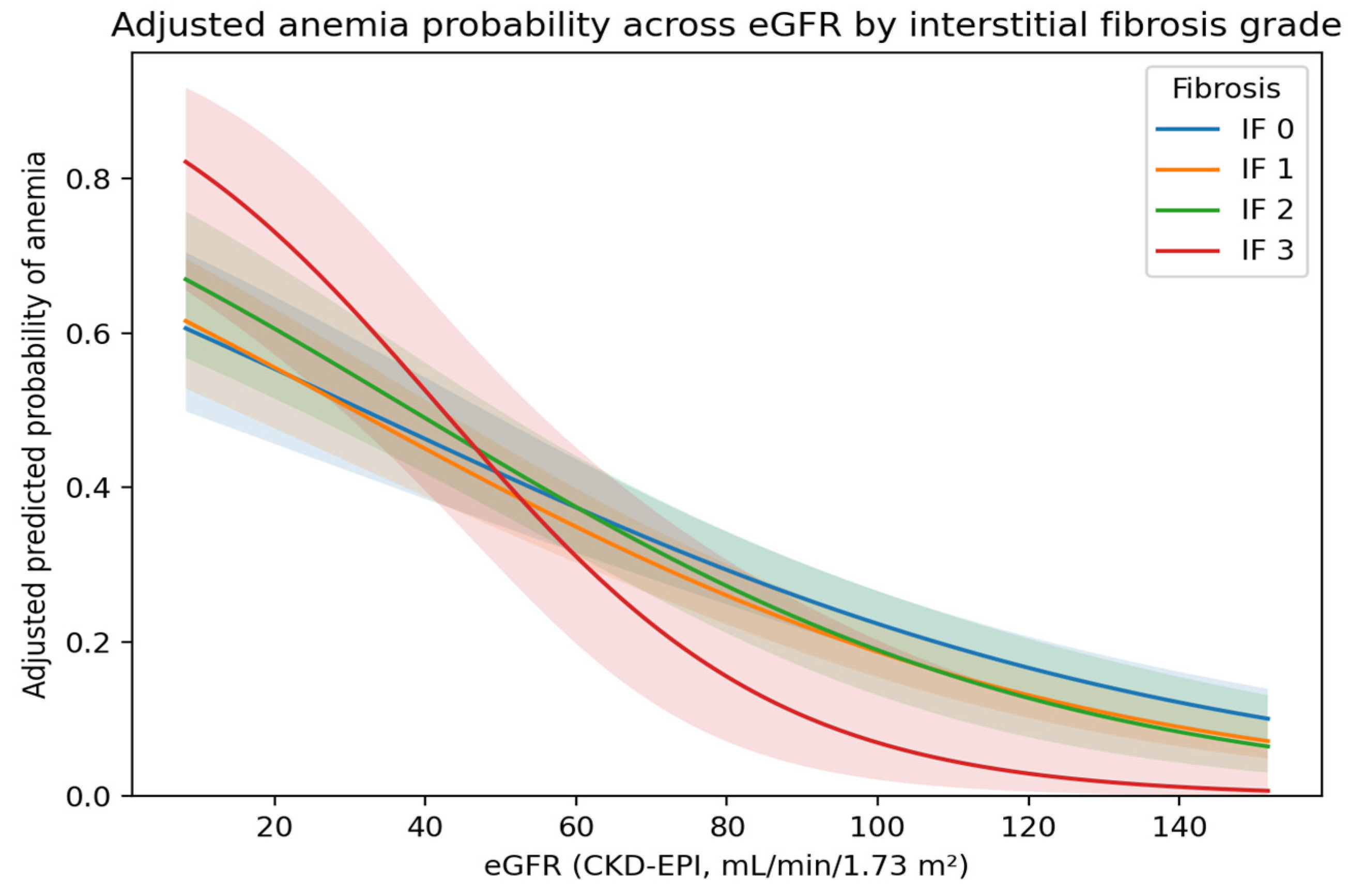
Adjusted predicted probabilities of anemia were estimated from the final interaction model and plotted across the observed eGFR range for IF grades 0–3 while holding other covariates constant at their mean (continuous variables) or reference (categorical variables) values.
All analyses were performed using SPSS for Windows (version 25.0; IBM Corp., Armonk, NY, USA) and Python (version 3.9; Python Software Foundation, Wilmington, DE, USA). All statistical tests were two-sided, and a p-value < 0.05 was considered statistically significant.
3. Results
3.1. Study Population
A total of 2794 patients with biopsy-proven primary glomerulonephritis were included. The median age was 40.0 years (IQR 29.9–51.5), and 56.9% were male. Median serum creatinine was 1.0 mg/dL (IQR 0.7–1.5), corresponding to a median eGFR of 85.6 mL/min/1.73 m2 (IQR 52.1–113.1). Median proteinuria was 3423 mg/day (IQR 1544–6400). Median hemoglobin was 13.1 g/dL (IQR 11.9–14.5).
3.2. Baseline Characteristics According to Anemia Status
Of the 2794 patients, 960 (34.4%) were classified as anemic and 1834 (65.6%) as non-anemic. Median hemoglobin levels were significantly lower in the anemic group compared with the non-anemic group (11.2 g/dL [IQR 10.3–11.9] vs. 14.0 g/dL [IQR 13.1–15.0], p < 0.001) and median hematocrit levels were significantly lower in the anemic group compared with the non-anemic group (33% [IQR 31–36] vs. 41% [IQR 39–44]), p < 0.001). Anemic patients were older and more frequently female. Hypertension and edema were more common among anemic patients. Serum creatinine was higher and eGFR was lower in the anemic group (median eGFR 60.5 vs. 94.8 mL/min/1.73 m2; p < 0.001). The prevalence of anemia differed across CKD stages, with proportions of 23.4%, 28.5%, 47.4%, 72.2%, and 86.8% in stages 1 through 5, respectively. (p < 0.001). Baseline characteristics are presented in Table 1.
Table 1.
Baseline clinical, laboratory, and histopathological characteristics of patients with primary glomerulonephritis, stratified by anemia status.
3.3. Relationship Between Interstitial Fibrosis and Anemia Status
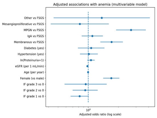
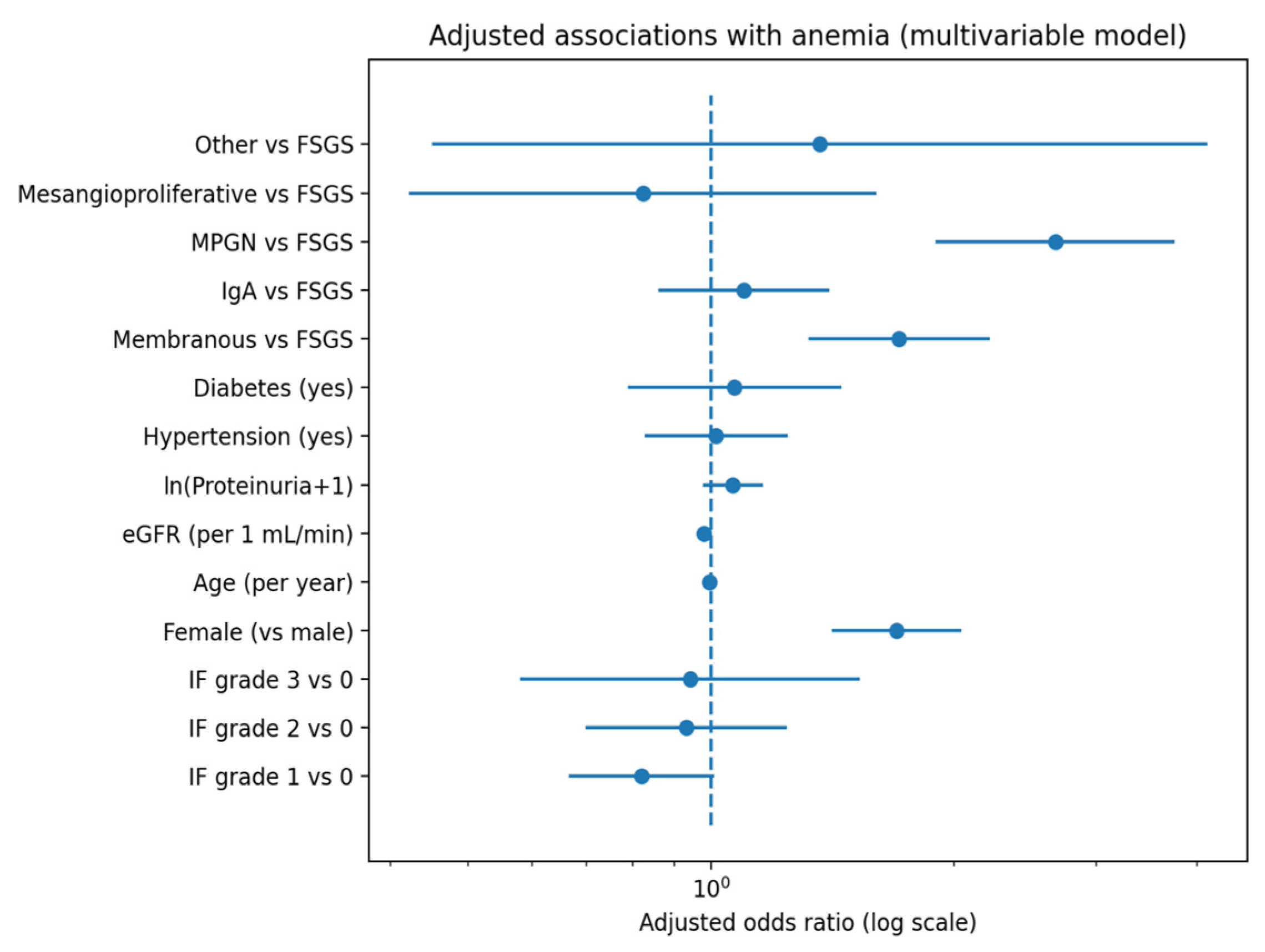
Regarding histopathology, moderate-to-severe IF and TA were more frequent among anemic patients (p < 0.001 for both distributions). In the complete-case multivariable model including 2486 patients (AUC = 0.705), interstitial fibrosis severity was not independently associated with anemia (Table 2). Forest plot of adjusted odds ratios for anemia from the multivariable logistic regression model are presented in Figure 1. Modeling IF severity as an ordinal variable likewise demonstrated no significant linear trend between increasing fibrosis grade and anemia risk (β (log-odds) = 0.018; OR per grade = 1.02 (95% CI 0.91–1.14), p-trend = 0.72).
Table 2.
Multivariable logistic regression analysis of factors associated with anemia.
Figure 1.
Forest plot of adjusted odds ratios for anemia from the multivariable logistic regression model. Adjusted odds ratios (ORs) with 95% confidence intervals for anemia from the main multivariable logistic regression model. The vertical dashed line indicates OR = 1. IF grades are shown relative to IF grade 0, and biopsy diagnosis categories are shown relative to FSGS (reference). ORs are displayed on a logarithmic scale.
Female sex (OR 1.66, 95% CI 1.38–2.00; p < 0.001), lower eGFR (OR 0.98 per 1 mL/min increase; p < 0.001) were independently associated with anemia.
In the alternative model including IF as a binary variable, IF remained non-significant (OR 0.94, 95% CI 0.77–1.14; p = 0.52; AUC = 0.704) (Table 3).
Table 3.
Multivariable logistic regression model including interstitial fibrosis as a binary variable.
3.4. Interaction and Subgroup Analyses
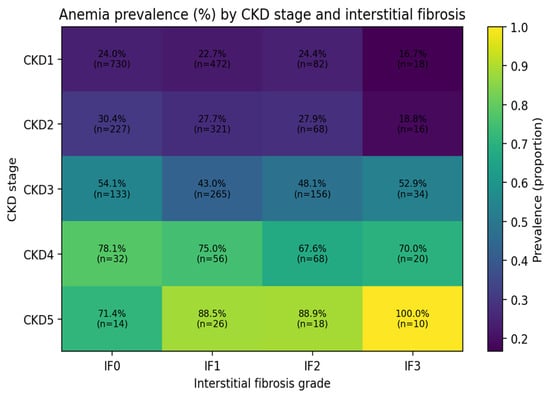
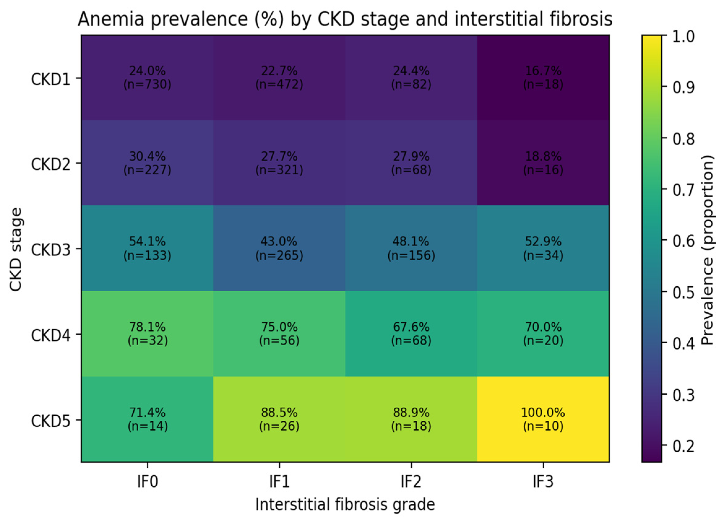
Lower eGFR was independently associated with anemia (OR 0.979 per 1 mL/min increase, p < 0.001) (Table 2). Expressed in clinically meaningful terms, each 10 mL/min/1.73 m2 decrease in eGFR corresponded to approximately 20% higher odds of anemia. Anemia prevalence across the cross-classification of CKD stage and IF grade are presented in Figure 2.
Figure 2.
Heatmap of anemia prevalence by CKD stage and interstitial fibrosis grade. Heatmap showing the observed anemia prevalence (color intensity) across the cross-classification of CKD stage (rows) and IF grade (columns). Each cell is annotated with prevalence (%) and the number of patients (n) contributing to that estimate. This figure illustrates effect heterogeneity of anemia prevalence across combined structural (IF) and functional (CKD stage) strata.
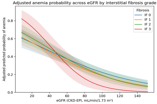
A statistically significant interaction between IF severity and eGFR was observed (β = −0.005; OR 0.995 per 1 mL/min increase; 95% CI 0.992–0.998; p = 0.0029). Adjusted predicted probabilities derived from the interaction model are presented in Figure 3.
Figure 3.
Adjusted predicted probability of anemia across eGFR by interstitial fibrosis grade (IF×eGFR interaction model). Model-based adjusted predicted probability of anemia across the eGFR spectrum (CKD-EPI, mL/min/1.73 m2), stratified by IF grades 0–3. Predictions were derived from a multivariable logistic regression model including an IF×eGFR interaction term; other covariates were held constant at their mean values (continuous) or reference categories (categorical). Shaded bands denote 95% confidence intervals.
Among 1722 patients (subgroup) with preserved renal function (eGFR ≥ 60 mL/min/1.73 m2) included in the adjusted model (AUC = 0.604), IF severity was not associated with anemia (Table 4).
Table 4.
Complete-case multivariable logistic regression analysis of interstitial fibrosis severity and anemia in patients with preserved renal function (eGFR ≥ 60 mL/min/1.73 m2).
4. Discussion
In this nationwide cohort of patients with biopsy-proven primary PGN, IF severity was more frequently observed among anemic patients in unadjusted analyses; however, IF severity was not independently associated with anemia after multivariable adjustment. These findings indicate that the crude association between IF and anemia in PGN is largely explained by coexisting clinical and functional determinants―particularly renal function―rather than by fibrosis grade per se. While this observation differs from biopsy-based studies in diabetic nephropathy, in which advanced tubulointerstitial lesions have been strongly linked to anemia and lower hemoglobin levels in adjusted models [15,16,17], it is consistent with prior evidence in PGN populations demonstrating that IF correlates with hemoglobin levels in univariate analyses but does not remain an independent predictor after adjustment for confounding factors [19]. Importantly, data examining the relationship between tubulointerstitial injury and anemia in PGN are limited and partially conflicting. Although a significant association between severe tubulointerstitial involvement and anemia has been demonstrated in patients with IgA nephropathy [18], similar associations have not been consistently confirmed across other PGN subtypes [19]. The present study extends the available evidence by evaluating this relationship in the largest multicenter PGN cohort to date, encompassing diverse histopathological diagnoses and a broad spectrum of renal function. The nationwide registry design and substantial sample size enhance the generalizability and robustness of our findings within PGN populations. Although experimental studies demonstrate that renal erythropoietin-producing interstitial cells undergo phenotypic transformation in fibrogenic microenvironments, leading to reduced EPO production, our findings suggest that in PGN this biological process may not translate into an independent clinical effect once overall renal function is accounted for [23]. This observation suggests that the apparent association between fibrosis and anemia may largely reflect its correlation with reduced renal function, rather than indicating an independent pathological mechanism; however, this interpretation should be considered associative rather than causal given the cross-sectional design. The nationwide multicenter design and large sample size enhance the external validity of the findings across diverse PGN populations.
Renal function remained the dominant determinant of anemia in our cohort, and the probability of anemia increased progressively as eGFR declined. This finding aligns with the well-established pathophysiological framework of chronic kidney disease-related anemia, in which reduced nephron mass and impaired erythropoietin production represent central mechanisms. Contemporary clinical guidance consistently emphasizes that anemia prevalence increases across CKD stages in parallel with declining GFR [22]. Large observational studies have reported that 21–62% of non-dialysis-dependent CKD patients exhibit anemia, with a stepwise rise in prevalence as kidney function deteriorates [1,2,3]. Notably, in a multicenter Chinese cohort of 2420 non-dialysis CKD patients―of whom approximately 60% had chronic glomerulonephritis―the prevalence of anemia increased from 22.4% in CKD stage 1 to 51.9% in stage 3 and 87.7% in stage 5 [6]. These data underscore that, irrespective of underlying etiology, CKD stage remains the principal determinant of anemia burden. Mechanistically, declining GFR reflects both quantitative nephron loss and functional impairment of renal erythropoietin-producing cells, leading to relative erythropoietin deficiency, impaired iron utilization, chronic inflammation, and altered hypoxia signaling pathways. Thus, even in diseases primarily characterized by glomerular pathology, the transition from structural injury to functional renal impairment appears to be the key mediator linking kidney disease to anemia.
In our PGN cohort, eGFR retained an independent association with anemia in multivariable models, whereas interstitial fibrosis did not. This pattern supports the interpretation that the apparent fibrosis–anemia relationship observed in unadjusted analyses is largely mediated through its association with reduced renal function rather than representing an autonomous pathological pathway. In other words, in PGN patients, functional renal reserve appears to be a stronger clinical determinant of anemia than histologic fibrosis grade alone. Importantly, we observed a statistically significant IF grade × eGFR interaction, visualized by model-based predicted probabilities. This finding indicates that the association between fibrosis grade and anemia varies across levels of renal function, consistent with effect modification in observed associations rather than a causal interaction. A plausible interpretation is that the clinical impact of structural interstitial injury on erythropoiesis becomes more discernible in the setting of reduced functional reserve, whereas at higher eGFR levels, compensatory mechanisms may predominate. Therefore, the observed interaction should be interpreted as a statistical phenomenon within the modeling framework, rather than as evidence of a direct biological interaction.
Female sex was independently associated with anemia in our multivariable models. This finding may partly reflect known sex-related differences in hemoglobin distribution and iron balance, as well as the sex-specific hemoglobin thresholds used to define anemia. Residual confounding by unmeasured iron indices and inflammatory markers is also possible, as these variables were not available in the registry. In this context, the persistent association of female sex with anemia supports the need for comprehensive anemia phenotyping (including iron indices and inflammation) when interpreting biopsy-based correlates of anemia.
We also observed higher odds of anemia among patients with membranoproliferative glomerulonephritis (MPGN) compared with FSGS, including within the preserved-eGFR subgroup model. MPGN comprises biologically heterogeneous entities and is often associated with systemic inflammatory or complement-mediated activity, which could contribute to anemia through inflammatory pathways and altered iron handling. Because CRP, ferritin/transferrin saturation, erythropoietin levels, and anemia treatments were not captured, we cannot determine whether the MPGN association reflects inflammation-related anemia, iron-restricted erythropoiesis, or other unmeasured mechanisms. Complement-mediated injury and chronic immune activation may further impair erythropoiesis through inflammatory iron sequestration pathways. Future studies incorporating standardized iron indices and inflammatory profiling are needed to clarify anemia phenotypes across PGN subtypes.
This registry analysis is limited by its cross-sectional design, lack of iron indices (ferritin, transferrin saturation), inflammatory markers, erythropoietin levels, and anemia treatments (iron/ESA), all of which influence hemoglobin and may confound associations. Because exposure (fibrosis severity) and outcome (hemoglobin/anemia) were measured at the same time point (biopsy), temporal direction cannot be established and reverse causality cannot be excluded. Single time-point laboratory measurements may not reflect longitudinal anemia status or treatment effects, and prospective follow-up analyses are needed. We did not account for potential center-level clustering effects, which may influence practice patterns and laboratory variability. Another limitation is the absence of albuminuria measurements. Although total proteinuria was used as a pragmatic and widely available measure across centers, albuminuria may have provided more specific information regarding glomerular injury and could have refined the interpretation of proteinuria-related associations. Histopathology was assessed locally at multiple centers without centralized review, introducing potential inter-observer variability that could attenuate true associations. Nonetheless, the nationwide scope and biopsy-confirmed diagnoses provide strong external validity for PGN populations and support the clinical interpretation that anemia risk is primarily driven by renal functional status, with fibrosis severity offering limited incremental information except through its interaction with eGFR.
5. Conclusions
In this large nationwide cohort of biopsy-proven primary glomerulonephritis, interstitial fibrosis severity was not independently associated with anemia after multivariable adjustment. Renal function was a major determinant of anemia, and the fibrosis–anemia relationship varied across eGFR levels as indicated by a significant IF×eGFR interaction. These findings suggest that, in PGN, anemia is primarily driven by functional renal impairment, while histological fibrosis alone provides limited incremental information.
Author Contributions
Conceptualization, E.C., K.T. and S.Ö.; Methodology, E.C., K.T., Z.Y., S.P. (Saime Paydaş) and S.Ö.; Software, E.C. and S.Ö.; Validation, E.C., S.Ö., A.Ş. and K.T.; Formal Analysis, E.C. and S.Ö.; Investigation, E.C., K.T., S.Ö., Y.Ö., S.G.B., A.Ş., N.S., M.Y., S.P. (Serhan Pişkinpaşa), Ö.F.A., T.S., G.Ş. (Garip Şahin), B.T., G.Ş. (Gülizar Şahin), İ.B., B.D., S.S. (Savaş Sipahi), A.Ö., G.S., S.U., F.B.G., S.K. (Sim Kutlay), E.P., İ.K., N.Y.S., Y.Y., M.G., C.Ç., M.T., Z.A., D.O., S.K. (Serdar Kahvecioğlu), M.Ü.K., D.T. (Dilek Torun), D.T. (Dilek Taymez), M.K. (Mehmet Küçük), S.D., L.K., S.S. (Siren Sezer), M.D., S.B., L.A., M.K. (Mehmet Koç), A.A., A.R.O., Z.Y. and S.P (Saime Paydaş).; Resources, all authors; Data Curation, all authors; Writing—Original Draft Preparation, E.C. and S.Ö.; Writing—Review and Editing, all authors; Visualization, E.C. and S.Ö.; Supervision, K.T., S.Ö. and S.P. (Saime Paydaş); Project Administration, E.C. and S.Ö.; Funding Acquisition, None. All authors have read and agreed to the published version of the manuscript.
Funding
The establishment and maintenance of the registry database were financially supported by the Turkish Society of Nephrology. Logistic support was provided by the authors’ affiliated departments. No funding was received for the publication of this article.
Institutional Review Board Statement
The study was conducted in accordance with the Declaration of Helsinki, and approved by the The Ethics Committee of Istanbul University, Istanbul Medical Faculty approved the study , 2011/1164 , 2011-07-08.
Informed Consent Statement
The Ethics Committee of Istanbul University, Istanbul Medical Faculty approved the study. Informed consent was waived due to the retrospective design.
Data Availability Statement
We created a database for primary glomerular diseases with the name of ‘Turkish Society of Nephrology Glomerular Diseases Working Group (TSNGOLD) in 04.04.2008. The data can be found at the following address: http://pgh.tsn.org.tr/login.php. Data are not publicly available due to legal and ethical reasons. Deidentified data are available upon reasonable request from the corresponding author.
Acknowledgments
The authors express special thanks to the Turkish Society of Nephrology for the organization of the study background, and to the nephropathologists in each center for their contributions to patient care and their kind help in providing these data. ChatGPT (OpenAI GPT-5.2) was used exclusively for text organization, linguistic refinement, figure legend standardization, and generate graphics.
Conflicts of Interest
The authors declare that they have no competing interests.
Abbreviations
The following abbreviations are used in this manuscript:
| ALT | Alanine Aminotransferase |
| AUC | Area Under the Receiver Operating Characteristic Curve |
| BMI | Body Mass Index |
| BUN | Blood Urea Nitrogen |
| CI | Confidence Interval |
| CKD | Chronic Kidney Disease |
| CKD-EPI | Chronic Kidney Disease Epidemiology Collaboration |
| EPO | Erythropoietin |
| FSGS | Focal Segmental Glomerulosclerosis |
| GFR | Glomerular Filtration Rate |
| HDL | High-Density Lipoprotein |
| IF | Interstitial Fibrosis |
| IFTA | Interstitial Fibrosis and Tubular Atrophy |
| IgA | Immunoglobulin A |
| IQR | Interquartile Range |
| KDIGO | Kidney Disease: Improving Global Outcomes |
| LDL | Low-Density Lipoprotein |
| MPGN | Membranoproliferative Glomerulonephritis |
| OR | Odds Ratio |
| PGN | Primary Glomerulonephritis |
| REP | Renal Erythropoietin-Producing Cells |
| TA | Tubular Atrophy |
| TSN-GOLD | Turkish Society of Nephrology Glomerular Diseases Working Group Registry |
| VIF | Variance Inflation Factor |
| WHO | World Health Organization |
References
- Wong, M.M.Y.; Tu, C.; Li, Y.; Perlman, R.L.; Pecoits-Filho, R.; Lopes, A.A.; Narita, I.; Reichel, H.; Port, F.K.; Sukul, N.; et al. Anemia and iron deficiency among chronic kidney disease Stages 3-5ND patients in the Chronic Kidney Disease Outcomes and Practice Patterns Study: Often unmeasured, variably treated. Clin. Kidney J. 2019, 13, 613–624. [Google Scholar] [CrossRef] [Scilit] [PubMed]
- Awan, A.A.; Walther, C.P.; Richardson, P.A.; Shah, M.; Winkelmayer, W.C.; Navaneethan, S.D. Prevalence, correlates and outcomes of absolute and functional iron deficiency anemia in nondialysis-dependent chronic kidney disease. Nephrol. Dial. Transplant. 2019, 36, 129–136. [Google Scholar] [CrossRef] [Scilit] [PubMed]
- Minutolo, R.; Locatelli, F.; Gallieni, M.; Bonofiglio, R.; Fuiano, G.; Oldrizzi, L.; Conte, G.; De Nicola, L.; Mangione, F.; Esposito, P.; et al. Anaemia management in non-dialysis chronic kidney disease (CKD) patients: A multicentre prospective study in renal clinics. Nephrol. Dial. Transplant. 2013, 28, 3035–3045. [Google Scholar] [CrossRef] [Scilit] [PubMed]
- Farrington, D.K.; Sang, Y.; Grams, M.E.; Ballew, S.H.; Dunning, S.; Stempniewicz, N.; Coresh, J. Anemia Prevalence, Type, and Associated Risks in a Cohort of 5.0 Million Insured Patients in the United States by Level of Kidney Function. Am. J. Kidney Dis. 2023, 81, 201–209.e1. [Google Scholar] [CrossRef] [Scilit]
- Kovesdy, C.P.; Davis, J.R.; Duling, I.; Little, D.J. Prevalence of anaemia in adults with chronic kidney disease in a representative sample of the United States population: Analysis of the 1999-2018 National Health and Nutrition Examination Survey. Clin. Kidney J. 2022, 16, 303–311. [Google Scholar] [CrossRef] [Scilit]
- Li, Y.; Shi, H.; Wang, W.M.; Peng, A.; Jiang, G.R.; Zhang, J.Y.; Ni, Z.H.; He, L.Q.; Niu, J.Y.; Wang, N.S.; et al. Prevalence, awareness, and treatment of anemia in Chinese patients with nondialysis chronic kidney disease: First multicenter, cross-sectional study. Medicine 2016, 95, e3872, Erratum in Medicine 2016, 95, e5074. https://doi.org/10.1097/01.md.0000490009.39850.74. [Google Scholar] [CrossRef] [Scilit]
- Schuster, S.J.; Koury, S.T.; Bohrer, M.; Salceda, S.; Caro, J. Cellular sites of extrarenal and renal erythropoietin production in anaemic rats. Br. J. Haematol. 1992, 81, 153–159. [Google Scholar] [CrossRef] [Scilit]
- Maxwell, P.H.; Osmond, M.K.; Pugh, C.W.; Heryet, A.; Nicholls, L.G.; Tan, C.C.; Doe, B.G.; Ferguson, D.J.; Johnson, M.H.; Ratcliffe, P.J. Identification of the renal erythropoietin-producing cells using transgenic mice. Kidney Int. 1993, 44, 1149–1162. [Google Scholar] [CrossRef] [Scilit]
- Suzuki, N.; Yamamoto, M. Roles of renal erythropoietin-producing (REP) cells in the maintenance of systemic oxygen homeostasis. Pflügers Arch.-Eur. J. Physiol. 2016, 468, 3–12. [Google Scholar] [CrossRef] [Scilit]
- Maxwell, P.H.; Ferguson, D.J.; Nicholls, L.G.; Johnson, M.H.; Ratcliffe, P.J. The interstitial response to renal injury: Fibroblast-like cells show phenotypic changes and have reduced potential for erythropoietin gene expression. Kidney Int. 1997, 52, 715–724. [Google Scholar] [CrossRef] [Scilit]
- El-Achkar, T.M.; Ohmit, S.E.; Mccullough, P.A.; Crook, E.D.; Brown, W.W.; Grimm, R.; Bakris, G.L.; Keane, W.F.; Flack, J.M. Kidney Early Evaluation Program. Higher prevalence of anemia with diabetes mellitus in moderate kidney insufficiency: The Kidney Early Evaluation Program. Kidney Int. 2005, 67, 1483–1488. [Google Scholar] [CrossRef] [Scilit]
- Ritz, E.; Haxsen, V. Diabetic nephropathy and anaemia. Eur. J. Clin. Investig. 2005, 35, 66–74. [Google Scholar] [CrossRef] [Scilit] [PubMed]
- Mehdi, U.; Toto, R.D. Anemia, diabetes, and chronic kidney disease. Diabetes Care 2009, 32, 1320–1326. [Google Scholar] [CrossRef] [Scilit] [PubMed]
- Xie, L.; Shao, X.; Yu, Y.; Gong, W.; Sun, F.; Wang, M.; Yang, Y.; Liu, W.; Huang, X.; Wu, X.; et al. Anemia is a risk factor for rapid eGFR decline in type 2 diabetes. Front. Endocrinol. 2023, 14, 1052227. [Google Scholar] [CrossRef] [Scilit] [PubMed]
- Inomata, S.; Itoh, M.; Imai, H.; Sato, T. Serum levels of erythropoietin as a novel marker reflecting the severity of diabetic nephropathy. Nephron 1997, 75, 426–430. [Google Scholar] [CrossRef] [Scilit] [PubMed]
- Mise, K.; Hoshino, J.; Ueno, T.; Imafuku, A.; Kawada, M.; Sumida, K.; Hiramatsu, R.; Hasegawa, E.; Yamanouchi, M.; Hayami, N.; et al. Impact of tubulointerstitial lesions on anaemia in patients with biopsy-proven diabetic nephropathy. Diabet. Med. 2015, 32, 546–555. [Google Scholar] [CrossRef] [Scilit]
- Shimizu, M.; Furuichi, K.; Kitajima, S.; Toyama, T.; Oshima, M.; Ogura, H.; Sato, K.; Nakagawa, S.; Yamamura, Y.; Miyagawa, T.; et al. Impact of the relationship between hemoglobin levels and renal interstitial fibrosis on long-term outcomes in type 2 diabetes with biopsy-proven diabetic nephropathy. BMC Nephrol. 2021, 22, 319. [Google Scholar] [CrossRef] [Scilit]
- Wang, Y.; Wei, R.B.; Su, T.Y.; Huang, M.J.; Li, P.; Chen, X.M. Clinical and pathological factors of renal anaemia in patients with IgA nephropathy in Chinese adults: A cross-sectional study. BMJ Open 2019, 9, e023479. [Google Scholar] [CrossRef] [Scilit]
- Urvasızoğlu, A.Ö; Hoca, E.; Cebeci, E.; Uzun, S.; Karadağ, S.; Öztürk, S. Determinants of anemia in patients with primary glomerulonephritis: A biopsy-based analysis. Acta Clin. Belg. 2025, 81, 70–79. [Google Scholar] [CrossRef] [Scilit]
- Levey, A.S.; Stevens, L.A.; Schmid, C.H.; Zhang, Y.L.; Castro, A.F., 3rd; Feldman, H.I.; Kusek, J.W.; Eggers, P.; Van Lente, F.; Greene, T.; et al. A new equation to estimate glomerular filtration rate. Ann. Intern. Med. 2009, 150, 604–612, Erratum in Ann. Intern. Med. 2011, 155, 408. [Google Scholar] [CrossRef] [Scilit]
- World Health Organization. Nutritional anemias: Report of a WHO scientific group. World Health Organ. Tech. Rep. Ser. 1968, 405, 5–37. [Google Scholar]
- Kidney Disease: Improving Global Outcomes (KDIGO) Anemia Work Group. KDIGO 2026 Clinical Practice Guideline for the Management of Anemia in Chronic Kidney Disease (CKD). Kidney Int. 2026, 109, S1–S99. [CrossRef] [Scilit]
- Souma, T.; Yamazaki, S.; Moriguchi, T.; Suzuki, N.; Hirano, I.; Pan, X.; Minegishi, N.; Abe, M.; Kiyomoto, H.; Ito, S.; et al. Plasticity of renal erythropoietin-producing cells governs fibrosis. J. Am. Soc. Nephrol. 2013, 24, 1599–1616. [Google Scholar] [CrossRef] [Scilit]

Disclaimer/Publisher’s Note: The statements, opinions and data contained in all publications are solely those of the individual author(s) and contributor(s) and not of MDPI and/or the editor(s). MDPI and/or the editor(s) disclaim responsibility for any injury to people or property resulting from any ideas, methods, instructions or products referred to in the content. |
© 2026 by the authors. Published by MDPI on behalf of the Lithuanian University of Health Sciences. Licensee MDPI, Basel, Switzerland. This article is an open access article distributed under the terms and conditions of the Creative Commons Attribution (CC BY) license.