


Abstract
Background and Objectives: Intense physical activity induces physiological adaptations affecting immune function, oxidative stress, erythrocyte stability, and hormonal balance. Brazilian Jiu-Jitsu (BJJ) uniquely combines anaerobic and aerobic demands, providing a unique model for studying exercise-induced responses. This study investigated hematological, oxidative, erythrocyte, hormonal, and biochemical responses to acute BJJ training. Materials and Methods: In this uncontrolled, exploratory pre–post pilot study, fifteen BJJ practitioners (12 men and 3 women; age range: 18–39 years) completed a standardized 60 min Brazilian Jiu-Jitsu training session comprising a progressive warm-up, technical and situational drills, and controlled sparring. Blood samples were collected before and 10 min after exercise. Complete blood counts were analyzed, while erythrocyte fragility, hemolysis, phosphatidylserine (PS) exposure, intracellular calcium (iCa2+), and reactive oxygen species (iROS) were assessed. Biochemical and hormonal markers were measured using automated analyzers. Results: White blood cells (estimated median change of +1.9 × 10³/μL, 95% CI 0.8 to 2.9, or +18.9%, 95% CI 9.4 to 45.9; q = 0.0006) and platelets (+34 × 103/μL, 95% CI 19 to 53, or +13.3%, 95% CI 7.9 to 22.6; q = 0.004) increased post-exercise. Mechanical fragility showed a nominal increase (unadjusted p = 0.0419), which did not remain significant after FDR correction, whereas no statistically significant changes were detected in oxidative lysis, osmotic fragility, or cryohemolysis. Free hemoglobin, iROS, PS, and iCa2+ showed no significant changes. Testosterone (+0.5 ng/mL, 95% CI 0.04 to 1.6, or +11.4%, 95% CI 4.3 to 45.5; q = 0.042) and creatine phosphokinase (CPK; +49 U/L, 95% CI 38 to 85, or +18.0%, 95% CI 13.7 to 26.8; q = 0.0004) increased. LDH, urea, and γ-GT increased, while triglycerides decreased. In the volume-adjusted sensitivity analysis, increases in WBC, platelet count, PCT, P-LCC and CPK remained significant, whereas changes in testosterone, LDH, triglycerides, γ-GT and urea did not. Exploratory correlations were observed between CPK and testosterone and, more tentatively, between mechanical fragility and LDH. Conclusions: Acute BJJ training was associated with leukocytosis, increased platelet indices, and increased CPK activity, while no statistically detectable changes in the measured oxidative or eryptotic markers were observed at 10 min post-exercise.
1. Introduction
High-intensity physical exercise induces complex physiological responses involving immune activation, oxidative stress, red blood cell (RBC) integrity, and endocrine regulation. Acute excessive exercise is known to cause transient leukocytosis, platelet activation, muscle damage, and metabolic derangement as a normal response to physical stress [1,2,3]. These responses are shaped by the intensity, duration, and mode of exercise, with intermittent high-intensity activities causing particularly marked hematological, biochemical and metabolic changes [4,5,6,7,8].
Brazilian Jiu-Jitsu (BJJ) is a modern ground-based martial art that developed in Brazil in the early 20th century, with origins in Japanese Kodokan judo and traditional jiu-jitsu [9]. The competitive and training nature of the sport places high demands on both aerobic and anaerobic energy systems, as matches are characterized by repeated bursts of strength and power combined with prolonged muscular fatigue [10]. BJJ is characterized by intermittent bouts of high-intensity isometric and dynamic muscular contractions, frequent transitions between dominant control positions, and phases that demand high technical accuracy. Previous studies have shown that both training sessions and competitive matches cause significant increases in heart rate, elevated blood lactate concentrations, and intense neuromuscular activation [9,11]. These findings highlight that the physical fitness required for successful performance in BJJ is multifaceted and highly specialized [10,12].
Intense physical activity has been shown to influence RBC stability and hemolysis through mechanical stress, oxidative damage, and membrane perturbations [13,14,15]. Exercise-induced hemolysis is a well-described phenomenon, particularly in high-impact and endurance sports, and is associated with increased plasma-free hemoglobin and elevated lactate dehydrogenase (LDH) levels [4]. Moreover, oxidative stress and intracellular calcium influx as key regulators of eryptosis, a programmed form of RBC death distinguished by phosphatidylserine (PS) externalization [16] are significant. However, data on RBC responses to combat sports such as BJJ remain limited.
In parallel, acute exercise elicits marked endocrine and metabolic responses. Increases in testosterone and creatine phosphokinase (CPK) are commonly observed following high-intensity training, reflecting anabolic signaling and muscle microtrauma [6,17]. Alterations in lipid metabolism, liver enzymes, and nitrogen balance further reflect metabolic stress [18,19,20,21]. Testosterone was included as an exploratory marker of the early endocrine response to BJJ exercise. We examined whether its concentration changed from baseline to 10 min post-exercise, without a pre-specified directional hypothesis, recognizing that sex, baseline concentrations, time of day, and exercise-related volume changes may influence its interpretation.
Despite the growing popularity of BJJ, limited research has examined its effects on red blood cell integrity, oxidative stress, hemolysis, and their relationship with hormonal and biochemical responses. Understanding these mechanisms is essential for optimizing training strategies and monitoring athletes’ health.
Erythrocyte stability encompasses several distinct properties. Fragility assays assess susceptibility to lysis under defined experimental stressors, while plasma-free hemoglobin serves as a marker of intravascular hemolysis, interpreted with consideration of sample handling. Fluorescence-based measurements provide complementary information on intracellular oxidative activity, calcium-associated signals, and phosphatidylserine exposure, supporting the assessment of erythrocyte stress and eryptosis-related changes. Hematological and biochemical measurements place these findings within the broader physiological response to exercise, although their changes are not necessarily attributable to erythrocyte injury alone.
This exploratory pilot study investigated whether a single Brazilian Jiu-Jitsu training session was associated with measurable changes in erythrocyte stability at 10 min post-exercise in BJJ practitioners aged 18–39 years with at least six months of BJJ training experience. Erythrocyte fragility and hemolysis measurements were complemented by hematological, biochemical, oxidative, and eryptotic markers to characterize the early response to exercise-related mechanical and metabolic stress. Sampling at 10 min post-exercise allowed assessment of early responses but could not capture delayed changes. No single primary outcome was pre-specified; therefore, all analyses were considered exploratory.
2. Materials and Methods
2.1. Standardized Training Protocol
Participants were recruited through convenience sampling from the available training classes of a local BJJ club/gym. Fifteen BJJ practitioners volunteered to participate. No prospective sample-size calculation or precision target was specified; recruitment was determined by participant availability and willingness to participate. The study was exploratory, with confidence intervals used to describe the precision of the estimated within-participant changes. Fifteen adults (12 men and 3 women), aged 18–39 years and with at least six months of regular Brazilian Jiu-Jitsu training experience, completed a 60 min BJJ training session. All participants completed both blood sampling time points, with no withdrawals after enrollment. All participants had a BMI within the normal adult range and reported no known medical conditions that would prevent exercise participation. One participant reported a well-controlled thyroid condition under medication; the remaining participants reported no medication use. Eight participants were smokers. None of the three female participants reported menstruating at the time of sampling or using hormonal contraceptives. The specific menstrual cycle phase was not recorded. Individuals with an acute illness or injury that could limit participation were excluded. The study was approved by the Research Ethics and Deontology Committee of the University of West Attica (approval reference no. 39648, dated 7 April 2026; protocol submission reference no. 32738, dated 18 March 2026) and was conducted in accordance with the Declaration of Helsinki. All participants provided written informed consent before enrollment in the study. The session comprised a 10 min progressive warm-up, 15 min of moderate-intensity technical drills, 10 min of situational drills performed at moderate-to-high intensity, and 20 min of controlled sparring, with brief transitions between phases. The session concluded with 5 min of stretching as a cool-down, bringing the total session duration to 60 min. Technical and situational drills incorporated grappling sequences, takedown entries, clinch work, positional control, ground transitions, guard passing, and escapes. The sparring phase consisted of five 3 min rounds, separated by 1 min recovery periods. Thus, the 20 min phase included 15 min of active sparring, 4 min of inter-round recovery, and a final 1 min recovery period to facilitate the transition to stretching. The standardized training protocol is summarized in Supplementary Table S7. Training partners were paired according to similar body mass and BJJ experience. The entire session was supervised by the same instructor, who provided consistent instructions and maintained the prescribed duration of each training phase. Participants were instructed to refrain from additional exercise on the testing day and maintain their habitual dietary intake. They were advised not to attend in a completely fasted state and to allow at least 4 h after a main meal or at least 90 min after a light snack. To minimize potential time-of-day-related variation in hematological and biochemical parameters, all training sessions were conducted between 19:00 and 22:00 [22,23]. Data collection was conducted on different days to accommodate laboratory processing and experimental measurements, with each participant’s paired blood samples collected on the same day, before and 10 min after their respective training session.
2.2. Sample Collection and Preparation
Peripheral venous blood samples were obtained at two predefined time points: before the training session (pre-exercise) and 10 min after its completion (post-exercise). Blood samples were collected in EDTA-containing tubes for complete blood count analysis, serum tubes for biochemical and testosterone analyses, and sodium citrate-containing tubes for the remaining experimental assays. Samples were transported to the laboratory in an insulated container with ice packs, and assays requiring fresh blood were performed within 3 h of collection. A complete blood count analysis was performed using the CONVERGYS® X3 hematology analyzer (Convergent Technologies, Cölbe, Germany). Instrument calibration and internal quality control were performed according to the manufacturers’ instructions and routine laboratory procedures. Hemolytic evaluation was conducted to assess exercise-associated changes in erythrocyte stability and hemolysis-related markers and included quantification of free plasma hemoglobin (Hb), mechanical and osmotic fragility indices and cryohemolysis testing. Intracellular red blood cell (RBC) stress responses were further examined by flow cytometric analysis of reactive oxygen species (ROS) production, intracellular calcium (iCa2+) levels, and PS externalization serving as indicators of oxidative stress and RBCs’ aging. A broad panel of biochemical and hormonal markers was evaluated pre- and post-exercise to evaluate metabolic, inflammatory and organ function responses. Serum tubes were centrifuged at 3000× g for 7 min. Serum was then separated, stored at −80 °C, and analyzed in a single batch after one freeze–thaw cycle.
2.3. Hemolytic Assays
Oxidative susceptibility of erythrocytes was assessed by incubation with phenylhydrazine (PHZ, final concentration 17 mM) at a final hematocrit of approximately 8%, at 37 °C for 45 min. Hemoglobin released into the supernatant was subsequently quantified spectrophotometrically using Harboe’s method, as previously described [24]. Plasma-free Hb was measured using the same spectrophotometric approach with Allen correction [25,26]. Briefly, plasma was diluted 1:10 in distilled water, mixed for 30 s, and incubated for 30 min. Absorbance was measured at 380, 415, and 450 nm using distilled water as the blank. Mechanical and osmotic fragility tests were performed to assess the resistance of erythrocytes to mechanical shear stress and hypotonic conditions, in accordance with established protocols [24,26]. Osmotic fragility was assessed using whole blood without prior standardization of hematocrit. Osmotic fragility was expressed as the NaCl concentration corresponding to 50% hemolysis, determined from the hemolysis–NaCl concentration curve. Hemolysis was expressed relative to the complete-lysis condition at 0% NaCl. For cryohemolysis, washed and packed erythrocytes (50 μL) were incubated in 2 mL of 0.7 M sucrose at 37 °C for 10 min, followed by 10 min on ice. After centrifugation at 3000 rpm for 10 min, supernatant hemoglobin was measured at 540 nm. Assays were performed in duplicate.
2.4. Intracellular RBCs’ Stress Responses
Intracellular oxidative activity and calcium-associated fluorescence were assessed by flow cytometry using CM-H2DCFDA and Fluo-4 AM, respectively, and expressed as mean fluorescence intensity (MFI). CM-H2DCFDA fluorescence was interpreted as a probe-dependent relative indicator of intracellular oxidative activity rather than a measurement of a specific ROS. Fluo-4 AM fluorescence was interpreted as a relative calcium-associated signal, recognizing its dependence on probe loading and intracellular esterase activity. Phosphatidylserine (PS) externalization was assessed by Annexin V staining. Erythrocyte staining and flow cytometric analysis were performed according to previously published protocols from our group [26,27,28]. Briefly, washed erythrocytes were incubated with CM-H2DCFDA (20 μM) for 60 min in PBS supplemented with 5 mM glucose and 0.1% BSA, or with Fluo-4 AM (1 μM) for 50 min in calcium-containing buffer (1.8 mM CaCl2) supplemented with 0.1% BSA, to assess intracellular ROS generation and Ca2+ levels, respectively. Both probe incubations were performed at 37 °C. Both probes were obtained from Invitrogen (Thermo Fisher Scientific, Waltham, MA, USA). PE-Annexin V kit combined with FITC-conjugated anti-CD235 antibody (BD Biosciences, San Jose, CA, USA) was used for the detection of PS exposure according to the manufacturer’s instructions. Briefly, whole blood diluted 1:10 in PBS was stained with Annexin V–PE and anti-CD235a–FITC for 20 min at room temperature in the dark in isotonic binding buffer containing 10 mM HEPES–NaOH (pH 7.4), 140 mM NaCl, 2.5 mM CaCl2, and 3% BSA. Flow cytometric acquisition was performed with a FACSCanto II Cytometer (BD Biosciences, San Jose, CA, USA) and data were analyzed with BD FACSDivaTM Software (version 7; BD Biosciences, San Jose, CA, USA). A minimum of 30,000 erythrocyte events were acquired per sample. Singlets were selected using FSC-A versus FSC-H gating to exclude doublets and aggregates. Unstained and fluorescence-minus-one (FMO) controls were included. PS exposure was reported as the percentage of Annexin V-positive cells within the CD235a-positive singlet population.
2.5. Biochemical Parameters
For the biochemical analysis of serum components (urea, creatinine, total cholesterol, triglycerides, high-density lipoprotein (HDL), sodium, aspartate aminotransferase [AST], alanine aminotransferase [ALT], gamma-glutamyl transferase [γ-GT], total bilirubin, creatine phosphokinase [CPK], lactate dehydrogenase [LDH]), and C-reactive protein (CRP), an automated clinical chemistry analyzer (Alinity c, Abbott Laboratories, Abbott Park, IL, USA) was used. Serum testosterone concentrations were determined using a chemiluminescent microparticle immunoassay (CMIA) on the Alinity i system (Abbott Laboratories, Abbott Park, IL, USA), according to the manufacturer’s instructions. Instrument calibration and internal quality control were performed according to the manufacturers’ instructions and routine laboratory procedures.
2.6. Statistical Analysis
The Statistical Package for Social Sciences (IBM SPSS Software; version 27.0 for Windows, IBM Corp., Armonk, NY, USA; administered by UNIWA) was used for statistical analysis. The results of each experiment are represented as median (interquartile range, IQR). Due to the small sample size and non-normal distribution of variables (Shapiro–Wilk test), the Wilcoxon signed-rank test was applied for paired comparisons. Two-sided exact p-values were computed, with zero differences excluded from the test; no imputation was performed, and the number of pairs analyzed for each outcome is given in Supplementary Table S1. Shapiro–Wilk tests of the paired differences indicated departure from normality (p < 0.05) for 15 of the 28 parameters; as normality cannot be reliably established with 15 pairs, the Wilcoxon test was used throughout. Spearman’s rank correlation coefficient was used to assess associations between variables. The four correlations reported were exploratory and not pre-specified, were selected for their biological relevance (Section 4.4.) and were the only correlations examined; 95% confidence intervals were obtained by bootstrap and Benjamini–Hochberg correction was applied across the four tests. Effect sizes (Wilcoxon r) with 95% bootstrap confidence intervals were calculated for all comparisons (Wilcoxon r = Z/√N), and the Benjamini–Hochberg FDR procedure was applied within each of five parameter families corresponding to Section 3.1, Section 3.2, Section 3.3, Section 3.4 and Section 3.5 (6, 5, 3, 6 and 8 parameters, respectively; unadjusted p and adjusted q values in Supplementary Table S1). The families were defined by physiological domain and were not pre-specified. In addition, Bayes factors (BF10) based on the JZS Bayesian paired t-test with a Cauchy prior (scale r = 0.707), computed in Python (version 3.13; SciPy version 1.18) by numerical integration (scipy.integrate.quad) of the marginal likelihood [29], were calculated for all comparisons to further characterize the strength of evidence, particularly for parameters showing borderline significance. Sensitivity to the prior scale (r = 0.5, 1 and 1.414) is given in Supplementary Table S1. Bayes factors complement, and are not equivalent to, the Wilcoxon-based p-values. BF10 was interpreted as anecdotal (1/3–3), moderate (3–10), strong (10–30), very strong (30–100) or extreme (>100) evidence [30]. Unadjusted p-values below 0.05 were considered nominally significant, whereas statistical significance after correction for multiple comparisons was determined using Benjamini–Hochberg-adjusted q-values below 0.05. As this was an exploratory pilot study, no a priori power calculation was performed and findings are considered hypothesis-generating. Men (n = 12) and women (n = 3) were compared descriptively (Supplementary Tables S2 and S3). Paired changes are reported as Hodges–Lehmann estimates of the median change with 95% bootstrap confidence intervals. Bootstrap intervals were percentile intervals (2000 resamples of the pairs for the Hodges–Lehmann estimates and 10,000 for the relative changes); relative changes are the median of the individual percentage changes. Between-sex comparisons used the Mann–Whitney U test, and the number of pairs, zero differences and ties for each outcome are given in Supplementary Table S1.
As an exploratory sensitivity analysis, post-exercise values were adjusted for estimated changes in blood or plasma volume. Plasma-volume change (ΔPV) was estimated from paired hemoglobin (Hb) and hematocrit (Hct) measurements using the Dill–Costill equation [31]. Absolute cell counts (WBC, PLT and P-LCC) and plateletcrit (PCT) were multiplied by . Serum or plasma concentrations including plasma-free Hb, were multiplied by plasma-volume adjustment factor, defined as:
Hct expressed as a percentage [31,32]. Other percentage and index variables, erythrocyte fragility measures, and intracellular oxidative and eryptosis-related markers were not adjusted. These estimates assume approximately constant circulating hemoglobin mass. Paired comparisons were repeated using the same statistical procedures and outcome families as in the primary analysis (Supplementary Table S6). Graphical representation of the data was performed using GraphPad Prism 8 (GraphPad Software, San Diego, CA, USA).
3. Results
3.1. Exercise Alters Immune and Platelet Profiles
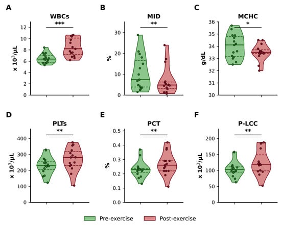
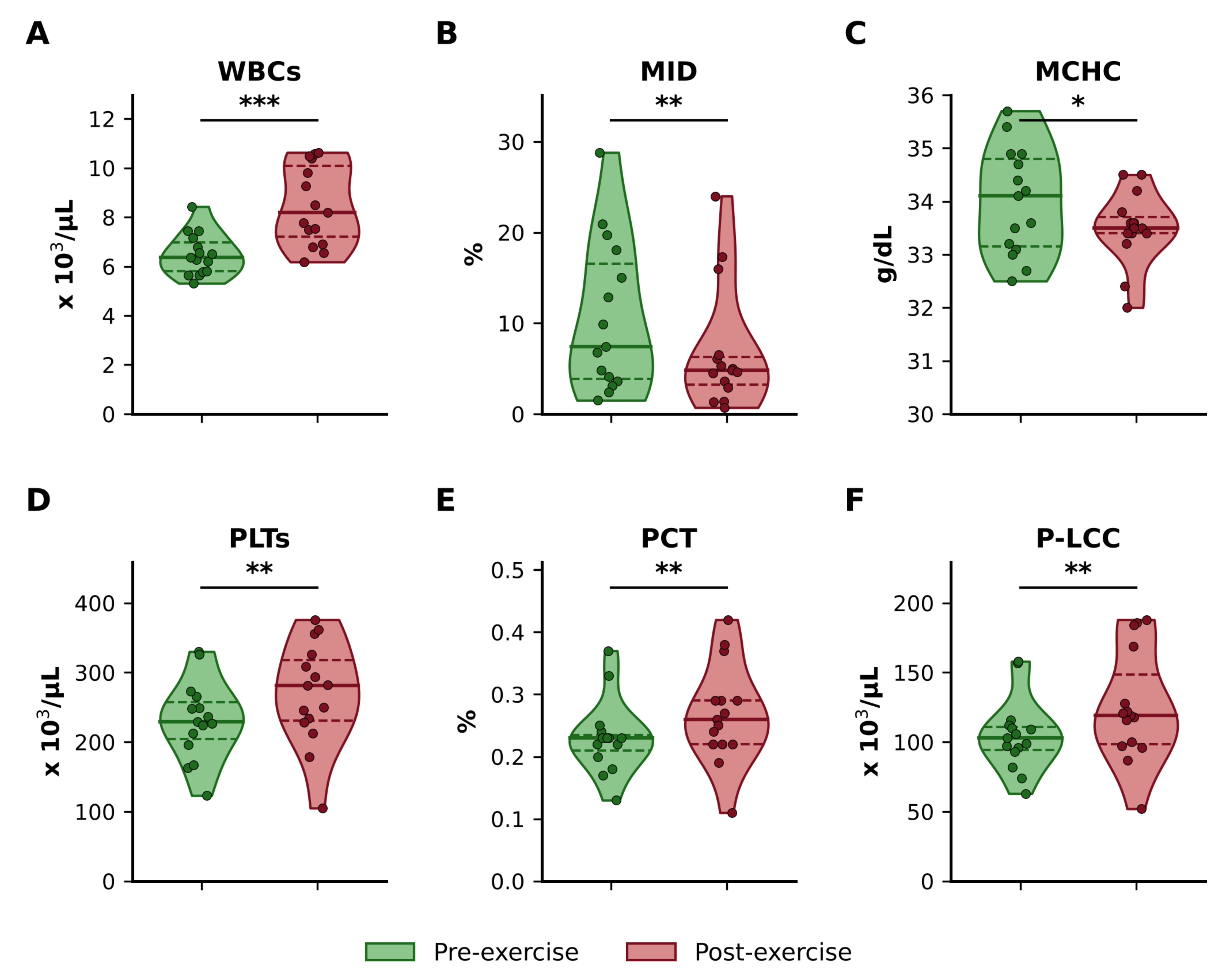
Acute exercise induced significant alterations in hematological parameters (Figure 1A–F). Total white blood cell (WBC) counts increased significantly following exercise compared with pre-exercise levels (p = 0.0001; Figure 1A). In contrast, the number of MID cells did not differ significantly post-exercise, although the percentage of MID cells showed a significant decrease (p = 0.002) (Figure 1B). A small but statistically significant reduction in mean corpuscular hemoglobin concentration (MCHC) was observed after exercise (p = 0.0339; Figure 1C). Platelet-related indices showed marked increases following exercise. Specifically, platelet counts (PLTs) were significantly elevated post-exercise (p = 0.0012; Figure 1D). Similarly, plateletcrit (PCT) increased significantly after exercise (p = 0.0049; Figure 1E), indicating an overall rise in circulating platelet mass. Furthermore, the number of large platelets (P-LCC) was significantly higher after exercise compared to pre-exercise values (p = 0.0056; Figure 1F), indicating an acute increase in circulating large platelets. Collectively, these findings demonstrate that acute exercise elicits immune and platelet responses, characterized by leukocytosis and increased platelet indices, alongside modest changes in erythrocyte parameters.
Figure 1.
Effects of acute exercise on hematological parameters. (A) White blood cell count (WBCs), (B) MID cell percentage (monocytes, eosinophils, and basophils), (C) mean corpuscular hemoglobin concentration (MCHC), (D) platelet count (PLTs), (E) plateletcrit (PCT), and (F) large platelet count (P-LCC) measured before (green) and after (red) exercise. Violin plots show the median (solid line) and interquartile range (dashed lines); dots represent individual BJJ practitioners. Statistical significance between pre- and post-exercise values was determined using the Wilcoxon signed-rank test. Asterisks indicate unadjusted significance levels: * p < 0.05, ** p < 0.01, *** p < 0.001. Displayed p-values are unadjusted; all six outcomes remained significant after Benjamini–Hochberg FDR correction (q < 0.05). Adjusted q-values are provided in Supplementary Table S1.
3.2. Exercise-Induced Changes in Erythrocyte Stability
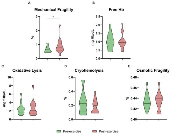
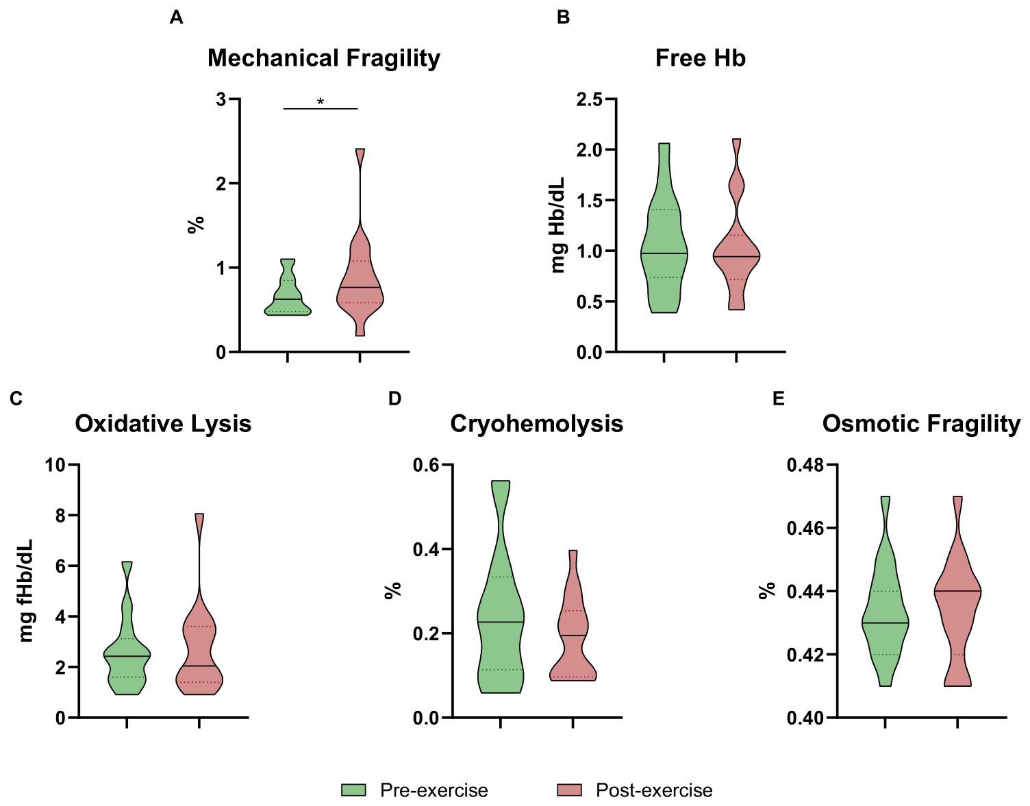
Erythrocyte fragility and hemolysis-related markers were assessed before and after exercise (Figure 2A–E). Mechanical fragility showed a nominal increase following exercise (unadjusted p = 0.0419; Figure 2A), although this finding did not remain significant after FDR correction (q = 0.21). Free hemoglobin did not change significantly after exercise (Figure 2B; estimated median change −0.04 mg/dL, 95% CI −0.19 to 0.15). Oxidative lysis did not differ significantly between pre- and post-exercise conditions (Figure 2C), with no detectable increase in oxidative susceptibility of erythrocytes. Similarly, cryohemolysis showed no statistically significant changes following exercise (Figure 2D), with no detectable change under cold stress. Osmotic fragility was also not significantly affected by exercise (Figure 2E), with no detectable change in resistance to hypotonic stress.
Figure 2.
Effects of acute exercise on erythrocyte fragility and hemolysis markers. Violin plots illustrate the distributions of paired measurements of (A) Mechanical fragility (%), (B) free hemoglobin (Free Hb; mg/dL), (C) oxidative lysis (mg Hb/dL), (D) cryohemolysis (%), and (E) osmotic fragility (%) measured before (green) and after (red) acute exercise. Solid and dashed horizontal lines indicate the median and interquartile range, respectively. Pre- and post-exercise values were compared using the Wilcoxon signed-rank test. Displayed p-values are unadjusted. The nominal increase in mechanical fragility (p = 0.0419) did not remain significant after Benjamini–Hochberg FDR correction across the five outcomes (q = 0.21). None of the five outcomes reached FDR-adjusted statistical significance. Adjusted q-values are provided in Supplementary Table S1. Asterisk (*) indicates p < 0.05 (unadjusted).
3.3. Effects of Acute Exercise on Erythrocyte Oxidative Stress and Eryptosis Markers
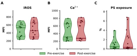
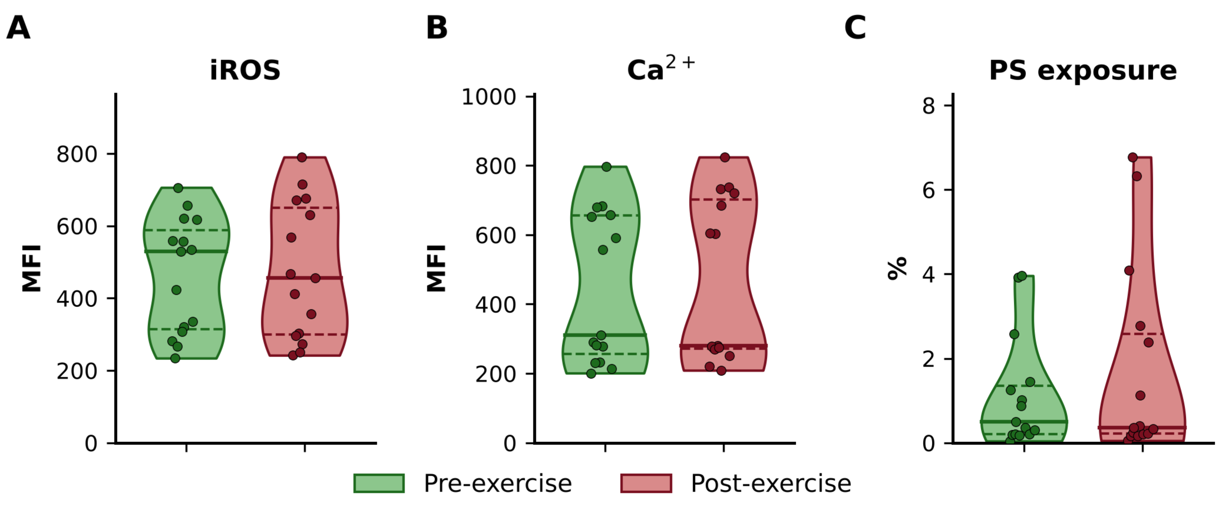
Acute exercise did not significantly alter intracellular reactive oxygen species (iROS) levels in erythrocytes (Figure 3A), as mean fluorescence intensity (MFI) values remained comparable between pre- and post-exercise conditions. Similarly, intracellular calcium (Ca2+) levels showed no significant change following exercise (Figure 3B), with no detectable change in calcium levels. Phosphatidylserine (PS) exposure on the erythrocyte membrane also did not differ significantly between conditions (Figure 3C), although a small increase and greater variability were observed post-exercise. No statistically significant changes were detected in the measured intracellular oxidative activity, calcium-associated fluorescence, or PS exposure at 10 min post-exercise. Individual paired changes are shown in Supplementary Figure S1.
Figure 3.
Effects of acute exercise on erythrocyte oxidative stress and eryptosis markers. (A) Intracellular reactive oxygen species (iROS), expressed as mean fluorescence intensity (MFI), (B) intracellular calcium (Ca2+; MFI), and (C) phosphatidylserine (PS) exposure (% positive cells) measured before (green) and after (red) acute exercise. Violin plots show the median (solid line) and interquartile range (dashed lines); dots represent individual BJJ practitioners. Paired comparisons were performed using the Wilcoxon signed-rank test. No statistically significant differences were detected before or after Benjamini–Hochberg FDR correction across the three outcomes. Unadjusted p-values and FDR-adjusted q-values are provided in Supplementary Table S1.
3.4. Alterations in Hormonal and Biochemical Markers
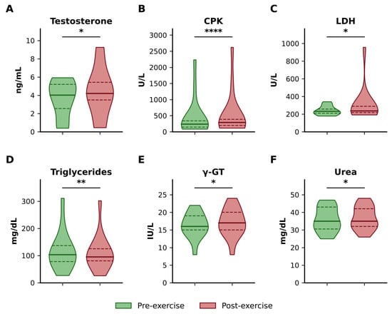
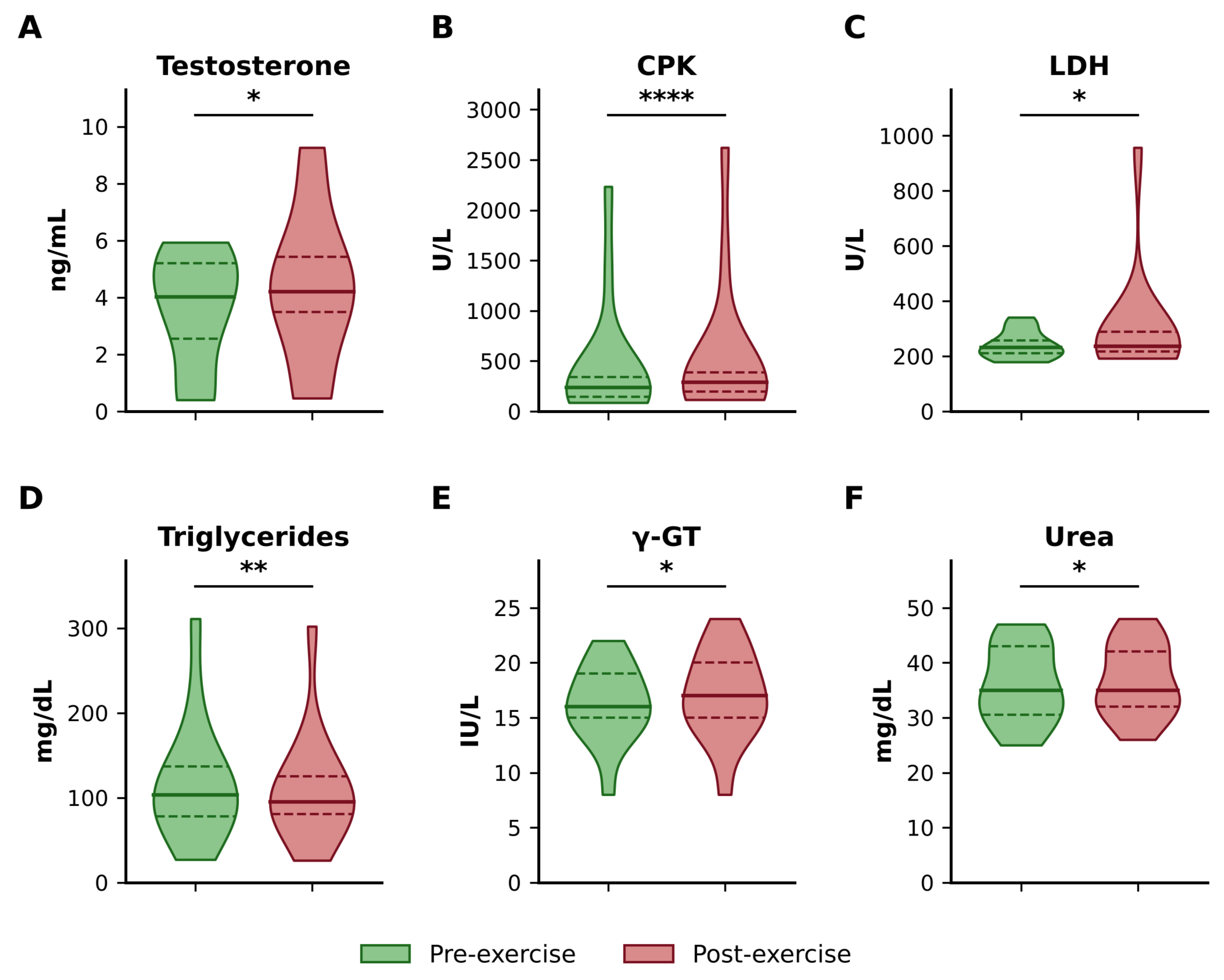
In the primary analysis, several hormonal and biochemical markers differed between pre- and post-exercise measurements (Figure 4A–F). Serum testosterone levels increased significantly following exercise (p = 0.0282; Figure 4A). Creatine phosphokinase (CPK) exhibited a marked post-exercise elevation (p < 0.0001; Figure 4B), indicating an early exercise-associated increase in circulating CPK activity. Lactate dehydrogenase (LDH) levels also increased significantly after exercise (p = 0.0205; Figure 4C). In contrast, triglyceride concentrations decreased significantly post-exercise (p = 0.0084; Figure 4D). Modest increases were observed in gamma-glutamyl transferase (γ-GT) (p = 0.0430; Figure 4E) and urea (p = 0.0488; Figure 4F). The mechanisms underlying these changes were not directly assessed. All six changes remained significant after FDR correction in the primary analysis. However, only the CPK increase retained FDR-adjusted significance in the plasma-volume-adjusted sensitivity analysis (Section 3.8).
Figure 4.
(A) Testosterone (ng/mL), (B) creatine phosphokinase (CPK; U/L), (C) lactate dehydrogenase (LDH; U/L), (D) triglycerides (mg/dL), (E) gamma-glutamyl transferase (γ-GT; U/L), and (F) urea (mg/dL) measured before (green) and after (red) acute exercise. Violin plots show the median (solid line) and interquartile range (dashed lines). Paired comparisons were performed using the Wilcoxon signed-rank test. Asterisks indicate unadjusted significance levels: * p < 0.05, ** p < 0.01, **** p < 0.0001. Displayed p-values are unadjusted. All six outcomes remained significant after Benjamini–Hochberg FDR correction in the primary analysis (q < 0.05; Supplementary Table S1). After plasma-volume adjustment and FDR correction, only CPK remained significant among these six outcomes (Supplementary Table S6).
3.5. Lipid Profile, Liver Enzymes, and Inflammatory Response
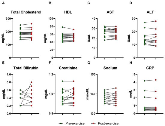
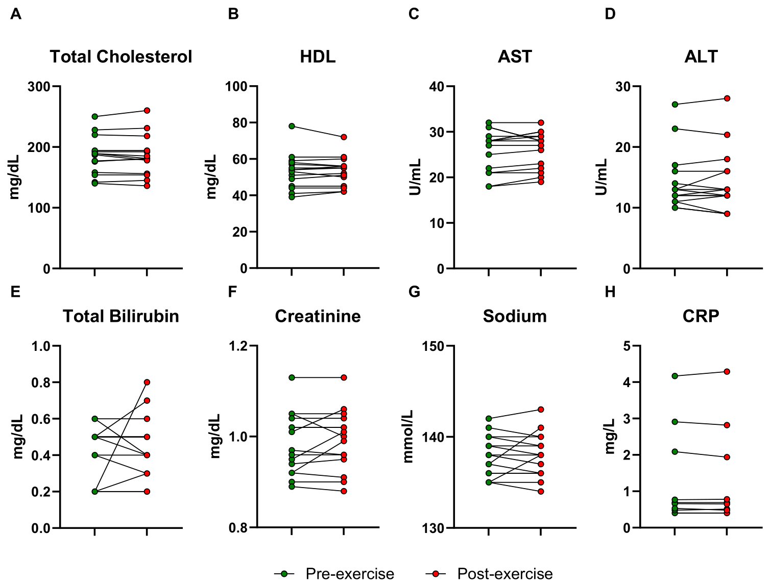
No statistically significant changes were detected in the eight parameters presented in Figure 5A–H. Total cholesterol (Figure 5A) and high-density lipoprotein (HDL; Figure 5B) levels showed no statistically significant changes post-exercise. Similarly, liver enzymes aspartate aminotransferase (AST; Figure 5C) and alanine aminotransferase (ALT; Figure 5D) showed no significant alterations, with no detectable change at 10 min post-exercise. Total bilirubin concentrations were stable before and after exercise (Figure 5E). Renal function, assessed by creatinine levels, also showed no statistically significant changes (Figure 5F). Sodium concentrations did not differ significantly between conditions (Figure 5G), with no detectable change at 10 min post-exercise. C-reactive protein (CRP), an inflammatory marker, showed no significant post-exercise change (Figure 5H), with no detectable change at 10 min post-exercise.
Figure 5.
Paired individual values of (A) total cholesterol, (B) high-density lipoprotein (HDL), (C) aspartate aminotransferase (AST), (D) alanine aminotransferase (ALT), (E) total bilirubin, (F) creatinine, (G) sodium, and (H) C-reactive protein (CRP) measured before (green) and after (red) acute exercise. The Y-axis of panel G (sodium) does not start at zero. Paired comparisons were performed using the Wilcoxon signed-rank test. No statistically significant differences were detected before or after Benjamini–Hochberg FDR correction across the eight outcomes. Unadjusted p-values and FDR-adjusted q-values are provided in Supplementary Table S1.
3.6. Correlations Between Hematological, Erythrocyte, and Biochemical Parameters
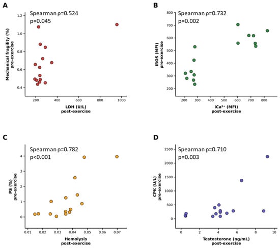
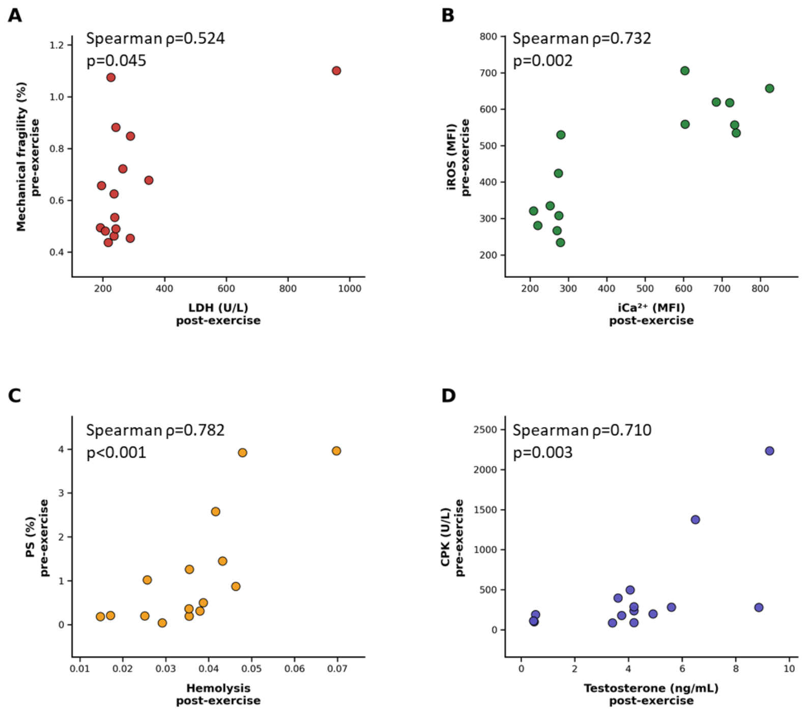
Exploratory correlation analyses identified associations between selected hematological, erythrocyte, and biochemical parameters (Figure 6A–D). Mechanical fragility measured before exercise showed a positive correlation with post-exercise lactate dehydrogenase (LDH) levels (ρ = 0.5237, p = 0.045, 95% CI −0.004 to 0.846; Figure 6A); this association may indicate that individuals with higher baseline erythrocyte fragility exhibited higher post-exercise LDH activity. A strong positive relationship was observed between pre-exercise intracellular reactive oxygen species (iROS) and post-exercise intracellular calcium (iCa2+) levels (ρ = 0.7321, p = 0.002, 95% CI 0.377 to 0.888; Figure 6B); this association may indicate a potential coordination between oxidative stress and calcium homeostasis mechanisms following exercise. Phosphatidylserine (PS) exposure prior to exercise correlated positively with post-exercise hemolysis (ρ = 0.7821, p < 0.001, 95% CI 0.429 to 0.945; Figure 6C); this association may indicate that baseline eryptotic signaling is related to increased susceptibility of erythrocytes during physical stress. Additionally, creatine phosphokinase (CPK) levels measured before exercise were positively associated with post-exercise testosterone concentrations (ρ = 0.7097, p = 0.003, 95% CI 0.306 to 0.910; Figure 6D), suggesting a link between muscle stress and endocrine responses. These exploratory correlations remained significant after Benjamini–Hochberg correction across the four tests (q = 0.002–0.045). In a leave-one-out sensitivity analysis, the associations of iROS with iCa2+, PS with hemolysis, and CPK with testosterone were stable, whereas the association between mechanical fragility and LDH was not and should be regarded as tentative (Supplementary Table S4). The bootstrap 95% CI of the mechanical fragility–LDH correlation includes zero, in keeping with its borderline p-value (0.045).
Figure 6.
Scatter plots illustrate associations between selected variables (Spearman’s rank correlation), with post-exercise values plotted on the X-axis and pre-exercise values plotted on the Y-axis. (A) Mechanical fragility (%) versus lactate dehydrogenase (LDH, U/L), (B) intracellular reactive oxygen species (iROS, MFI) versus intracellular calcium (iCa2+, MFI), (C) phosphatidylserine (PS, %) versus hemolysis, and (D) creatine phosphokinase (CPK, U/L) versus testosterone (ng/mL). Spearman’s ρ and p-values are shown for each panel (n = 15).
3.7. Effect Sizes, Correction for Multiple Comparisons and Bayes Factors
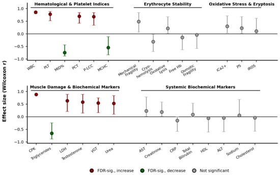
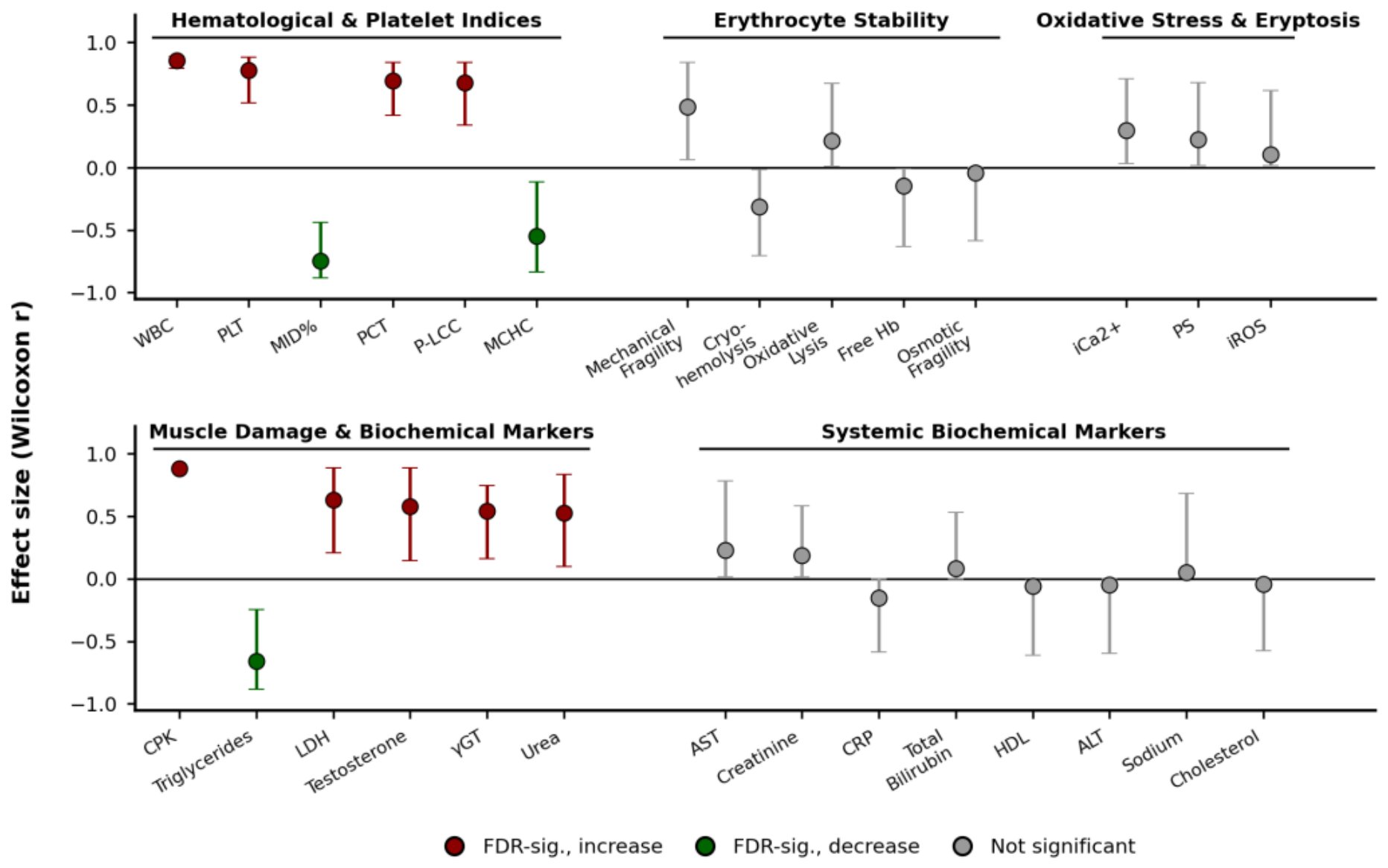
To assess the robustness of the observed pre- to post-exercise changes given the limited sample size, effect sizes (Wilcoxon matched-pairs r) and Bayes factors (BF10, Cauchy prior r = 0.707) were calculated for all measured parameters, and the Benjamini–Hochberg false discovery rate (FDR) procedure was applied within each domain (28 comparisons in five families; Supplementary Table S1). In the primary analysis, twelve parameters remained statistically significant after FDR correction: all six hematological/platelet parameters (WBC, PLT, MID cell percentage, PCT, P-LCC, MCHC) and CPK, together with testosterone, LDH, triglycerides, γ-GT, and urea (red/green markers in Figure 7). Effect sizes for these twelve parameters were uniformly moderate to large (r = 0.53–0.88), with CPK, WBC, and PLT showing the largest magnitudes (Figure 7).
Figure 7.
Effect sizes (Wilcoxon matched-pairs r, with 95% bootstrap confidence intervals) for pre- to post-exercise changes, grouped by domain as in Section 3.1, Section 3.2, Section 3.3, Section 3.4 and Section 3.5. Red markers indicate a significant increase and green markers a significant decrease post-exercise, surviving Benjamini–Hochberg FDR correction applied within each domain (q < 0.05). Gray markers indicate changes that did not reach significance after correction. Results refer to the primary analysis without volume adjustment. Volume-adjusted sensitivity results are provided in Supplementary Table S6.
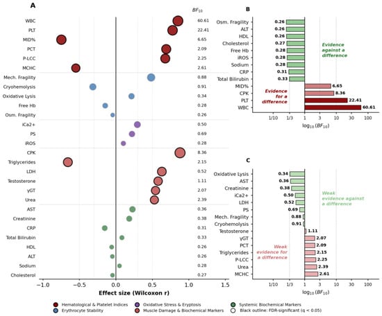
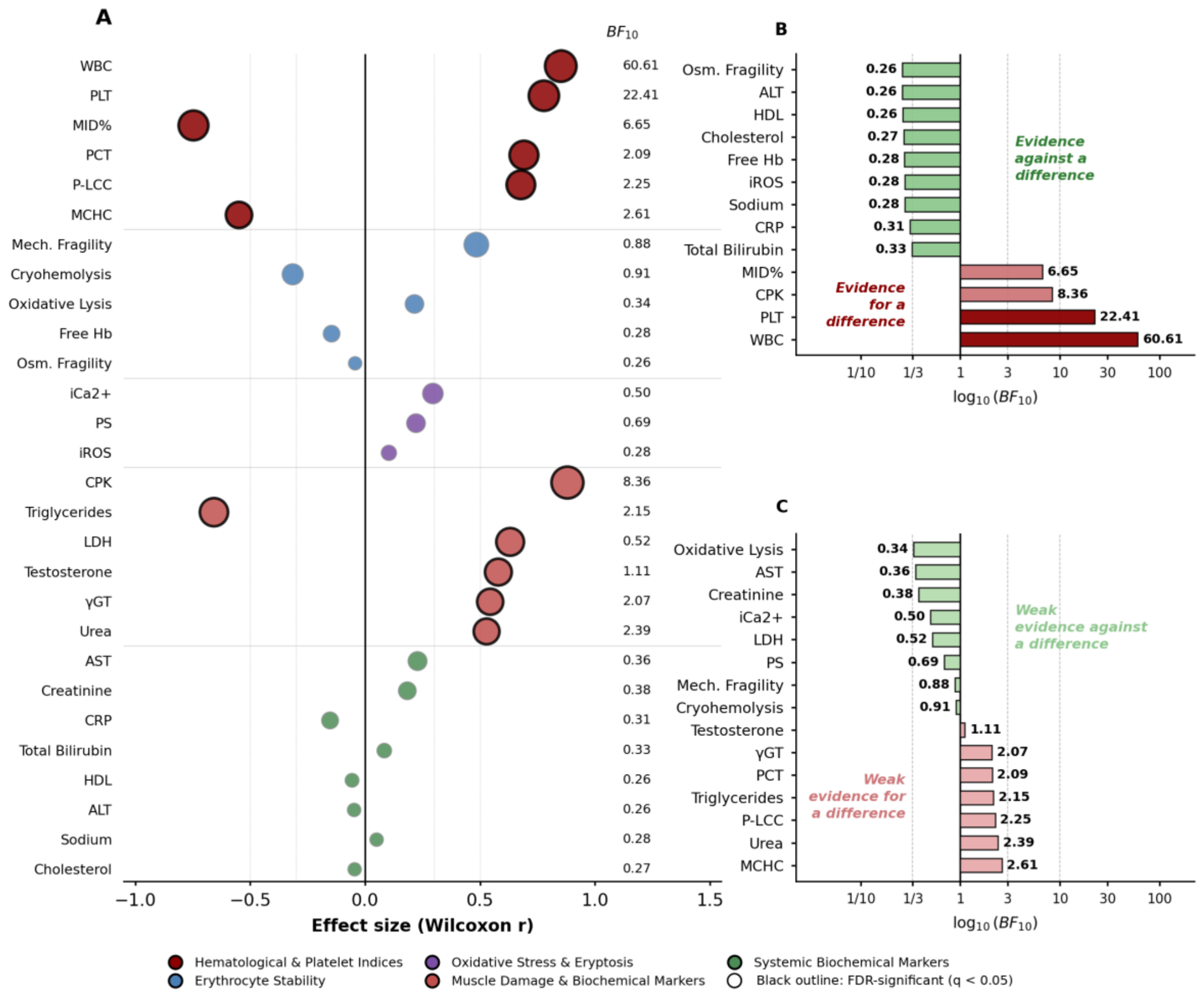
However, the strength of Bayesian evidence varied considerably across these twelve parameters (Figure 8). WBC showed very strong (BF10 = 60.6), PLT strong (BF10 = 22.4), and CPK and MID cell percentage moderate (BF10 = 8.4 and 6.7) Bayesian evidence for a difference. In contrast, MCHC, P-LCC, PCT, triglycerides, γ-GT, urea, and testosterone showed only anecdotal Bayesian support (BF10 = 1.1–2.6), and LDH showed essentially no informative evidence either way (BF10 = 0.52) despite reaching frequentist significance. Bayes factors depend on the prior scale (Supplementary Table S1). WBC, PLT and CPK are therefore the most robust findings, the others being tentative.
Figure 8.
Bayesian analysis of pre- to post-exercise changes. (A) Effect sizes (Wilcoxon r) by domain. (B,C) Bayes factors (BF10), split into (B) BF10 < 1/3 or > 3 (at least moderate evidence) and (C) 1/3 < BF10 < 3 (anecdotal or inconclusive evidence). Results refer to the primary analysis without volume adjustment. Bayes factors complement, but are not equivalent to, the Wilcoxon-based p-values or FDR-adjusted q-values.
3.8. Sensitivity Analysis: Correction for Plasma-Volume Change
Median Hb was 14.7 g/dL [IQR 14.3–15.9] before and 14.6 g/dL [13.8–15.7] after exercise, and median Hct was 44.3% [42.9–46.2] before and 45.1% [41.3–45.7] after exercise. The estimated median change in plasma volume was −2.9% [IQR −7.4 to +5.1], with large individual variability (Supplementary Table S6). After correction for ΔPV, the increases in WBC, PLT, PCT, P-LCC and CPK remained significant after FDR correction, whereas the changes in testosterone, LDH, triglycerides, γ-GT and urea did not (Supplementary Table S6). Bayes factors for WBC, PLT and CPK were lower after correction (16.1, 3.9 and 3.3, respectively). The correlations (Section 3.6) were computed on uncorrected values and may be affected by plasma-volume changes; the associations involving testosterone, LDH and CPK should therefore be interpreted with particular caution.
4. Discussion
In the present study, hematological parameters, erythrocyte stability, oxidative stress, and biochemical and hormonal markers were investigated in BJJ practitioners following an acute BJJ training session. Increases in WBC, platelet count, PCT, P-LCC, and CPK remained significant after FDR correction in the volume-adjusted sensitivity analysis. Changes in testosterone, LDH, triglycerides, γ-GT, and urea did not retain significance after plasma-volume adjustment and should be interpreted cautiously. Mechanical erythrocyte fragility showed a nominal increase that did not remain significant after FDR correction, while no statistically significant changes were detected in the measured oxidative activity or eryptosis-related markers at 10 min post-exercise.
4.1. Immune and Platelet Responses to BJJ Exercise
Total WBC counts increased significantly following exercise, aligning with reports of exercise-induced leukocytosis resulting from high-intensity or intermittent exercise in the literature [33,34,35,36]. Consistent with our findings, a recent case study in elite para-jiu-jitsu athletes reported post-fight increases in leukocytes and neutrophils, along with a similar upward trend in platelet count [37]. This occurs because catecholamines release leukocytes from the vascular endothelium and the spleen, while cortisol mobilizes cells from the bone marrow [5,38,39,40]. The percentage of MID cells decreased after exercise, whereas their absolute count did not change significantly. Although relative shifts toward neutrophil predominance have been reported following intense physical exercise [41,42], the change in MID percentage alone does not establish such a shift in the present study. MCHC was found to be reduced immediately after exercise, as has been reported after activities such as intensive judo exercise in adolescents [43], a half-marathon in recreational middle-aged women runners [44] or after four months of training for amateur cross-country skiers [45]. Platelet indices, including platelet count, PCT, and P-LCC, exhibited a statistically significant elevation following exercise, consistent with acute platelet mobilization. Comparable physiological responses have been documented in both combat and endurance sports, attributable to shear stress, sympathetic nervous system activation, and inflammatory processes [46,47,48,49]. The observed increase in large platelets is noteworthy because larger platelets have been associated with greater metabolic activity and prothrombotic potential [50]. However, increased large platelet counts do not establish platelet activation. Collectively, these findings suggest an acute hematological response to Brazilian Jiu-Jitsu training, characterized by leukocytosis and increased platelet indices. As platelet function and coagulation markers were not assessed, no conclusions regarding thrombotic risk can be drawn from the present findings.
4.2. RBC Stability and Hemolysis
Mechanical fragility showed a nominal increase after exercise that did not remain significant after FDR correction (q = 0.21) and should be regarded as tentative. Mechanical factors have been implicated in exercise-induced hemolysis [51]. However, their contribution to the nominal increase in mechanical fragility observed here remains uncertain, particularly because this finding did not survive FDR correction. Free hemoglobin levels, oxidative lysis, cryohemolysis, and osmotic fragility did not show statistically significant changes, which does not exclude small changes; because no equivalence test was performed and power was limited, preserved erythrocyte integrity cannot be concluded [51]. Similarly, the levels of intracellular reactive oxygen species (ROS), intracellular Ca2+, and phosphatidylserine externalization were not shown to differ significantly, although these non-significant findings cannot exclude smaller effects or responses occurring outside the sampling window [16,52,53]. It should be noted, however, that blood sampling was performed 10 min post-exercise, which may not capture delayed peaks in oxidative or inflammatory markers reported in the literature.
4.3. Biochemical and Hormonal Responses
Changes in testosterone, LDH, triglycerides, γ-GT, and urea did not retain statistical significance after plasma-volume adjustment. Their interpretation as exercise-associated endocrine or metabolic responses is therefore uncertain. Serum testosterone concentrations were higher after exercise (estimated median change +0.5 ng/mL, 95% CI 0.04 to 1.6), although this difference was sensitive to plasma-volume adjustment and does not establish activation of a specific endocrine pathway [54]. Analogous effects were reported after high-intensity resistance and combat sport training, driven by activation of the hypothalamic–pituitary–gonadal axis and sympathetic stimulation [55,56]. The CPK increase remained significant after plasma-volume adjustment and FDR correction and may be compatible with the muscular demands of BJJ, although it does not establish muscle injury [57,58]. The LDH finding was sensitive to volume adjustment (Supplementary Table S6). Similarly, Lopes et al. reported a significant post-fight increase in LDH, whereas sodium showed no statistically significant changes. Notably, concomitant increases in hematocrit, erythrocyte count, and hemoglobin were also observed, highlighting a possible contribution of exercise-induced hemoconcentration to acute changes in concentration-based biomarkers [37].
In the primary analysis, triglycerides were lower after exercise (estimated median change −6.5 mg/dL, 95% CI −11 to −2.5), while small increases were observed in γ-GT and urea. None of these changes retained statistical significance after plasma-volume adjustment (Supplementary Table S6). Exercise-related changes in lipid metabolism have been discussed previously [59,60].
Importantly, no significant changes in cholesterol, liver transaminases, creatinine, sodium, and CRP were present. Together, the absence of significant changes in CRP, AST, ALT, creatinine, and sodium indicates no detectable change in these variables within the 10 min post-exercise sampling window. However, this interpretation should be considered preliminary because delayed inflammatory, oxidative, hepatic, or renal responses can occur after strenuous exercise and require longer follow-up [20,61].
4.4. Correlations Between Hematological and Biochemical Markers
The exploratory correlations suggested links between red blood cell parameters and metabolic markers. The positive relationship between baseline mechanical fragility and post-exercise lactate dehydrogenase (LDH) levels may suggest that individuals with lower erythrocyte stability tend to exhibit higher LDH activity after intense exercise. This exploratory association may indicate that baseline erythrocyte mechanical properties are related to the post-exercise biochemical response; however, given the small sample size, this finding should be interpreted cautiously and requires confirmation in larger cohorts [51,62]. In addition, the observed associations between pre-exercise intracellular reactive oxygen species (iROS) and post-exercise intracellular Ca2+, as well as between PS exposure and hemolysis, should be regarded as exploratory. Although oxidative stress, altered Ca2+ homeostasis, and PS externalization are recognized features of eryptosis [63,64,65], the present correlations do not establish activation of a specific mechanistic pathway. Similarly, the positive association between baseline CPK and post-exercise testosterone represents a hypothesis-generating observation that may reflect interindividual variation in muscular and endocrine responses to exercise [6].
5. Conclusions
In this uncontrolled exploratory pilot study, increases in WBC, platelet count, PCT, P-LCC, and CPK measured 10 min after exercise remained significant after FDR correction in the volume-adjusted sensitivity analysis. Changes in testosterone, LDH, triglycerides, γ-GT, and urea did not retain significance after plasma-volume adjustment. No statistically detectable changes in the measured oxidative or eryptosis-related markers were observed at 10 min post-exercise. These findings describe the early response to a single BJJ session but do not establish preserved erythrocyte integrity or an efficient adaptive response. Non-significant results should not be interpreted as evidence of equivalence or the absence of a physiological effect. The small sample, absence of a control group, potential plasma-volume effects, and single post-exercise sampling point limit interpretation. Larger controlled studies with serial sampling are needed to characterize these responses and their potential practical relevance.
This study has some limitations. The small, self-selected sample from a single club/gym and the absence of a control group limit the generalizability and causal interpretation of the findings. No a priori power calculation was performed, so findings are hypothesis-generating and non-significant results do not demonstrate equivalence. Only three women were included, precluding formal sex-stratified analysis; in an exploratory comparison (Supplementary Tables S2 and S3), differences between men and women were found only for parameters with known sex dependence (testosterone, HDL-cholesterol, total bilirubin) and, post-exercise, for CPK and P-LCC, with no difference detected in the remaining parameters (power was very limited). With 15 pairs, the paired comparisons had 80% power only to detect large standardized effects (dz ≈ 0.8); smaller changes could not be reliably detected. In an exploratory sensitivity analysis restricted to men (n = 12), the direction of change was the same for all twelve key findings and nine remained nominally significant (Supplementary Table S5). Because 28 outcomes and four correlations were examined in a sample of 15 BJJ practitioners, the risk of false-positive findings is not negligible. FDR correction was applied within five families of outcomes, the correlations were exploratory and not pre-specified, and all findings should be regarded as hypothesis-generating; the mechanical fragility–LDH association was unstable in a leave-one-out analysis (Supplementary Table S4). Additionally, blood sampling at a single post-exercise time point (10 min) may not capture transient earlier or delayed physiological responses. Pre-exercise standardization was limited to instructions to avoid additional exercise on the testing day and maintain habitual dietary intake. Strenuous exercise during the preceding 24–48 h, fluid intake, caffeine and alcohol consumption, sleep, supplement use, and recovery status were not systematically standardized or recorded. Although minimum intervals between food intake and exercise were recommended, the exact composition and quantity of food consumed were not standardized or recorded. Hydration status was not directly assessed. These factors may have influenced baseline measurements and the observed pre–post changes, particularly in CPK and metabolic markers. Plasma-volume adjustment was based on indirect Hb/Hct estimates and is subject to measurement error and the assumption of constant circulating hemoglobin mass. Sampling at 10 min post-exercise was intended to characterize the early circulating response to BJJ training. This single time point cannot capture delayed biomarker peaks or establish the extent of muscle damage. Future studies should include sampling at 1, 3, 24, and 48 h post-exercise to characterize the response over time. Future studies with larger cohorts, multi-time-point assessments, prospective measurement of plasma volume and hydration status, and consideration of factors such as sex differences and metabolic variability are warranted.
Supplementary Materials
The following supporting information can be downloaded at https://www.mdpi.com/article/10.3390/medicina62112246/s1. Table S1: Pre- versus post-exercise comparisons for all 28 parameters: descriptive statistics, paired change, effect size, p, q and Bayes factor; Table S2: Pre-exercise values in men and women; Table S3: Post-exercise values in men and women; Table S4: Exploratory Spearman correlations: confidence intervals, multiplicity correction and leave-one-out sensitivity analysis; Table S5: Sensitivity analysis restricted to men; Table S6: Hemoglobin, hematocrit, estimated plasma-volume change, and sensitivity analysis accounting for estimated blood- and plasma-volume changes; Table S7: Standardized 60 min Brazilian Jiu-Jitsu training protocol; Figure S1: Individual paired changes in intracellular Ca2+, intracellular ROS and phosphatidylserine exposure.
Author Contributions
Conceptualization, S.P.F. and A.G.K.; methodology, S.P.F., S.V., G.D. and A.G.K.; software, E.G.P. (Effie G. Papageorgiou); validation, G.D. and A.G.K.; formal analysis, S.P.F., S.L., A.T.A. and V.L.T.; investigation, S.P.F., S.A., M.-A.K., E.G.P. (Efthymia G. Pavlou), E.M., K.D.D. and G.K.; resources, M.K., E.G.N. and A.G.K.; data curation, S.P.F., S.A., M.-A.K., E.G.P. (Efthymia G. Pavlou), E.M., E.S., A.G.T. and A.T.A.; writing—original draft preparation, S.P.F.; writing—review and editing, S.P.F., S.A., M.-A.K., E.G.P. (Efthymia G. Pavlou), S.L., E.M., E.S., A.T.A., V.L.T., K.D.D., A.G.T., G.K., E.G.P. (Effie G. Papageorgiou), M.K., E.G.N., K.S., S.V., D.C., G.D. and A.G.K.; visualization, S.P.F. and S.L.; supervision, A.G.K.; project administration, A.G.K.; funding acquisition, A.G.K. All authors have read and agreed to the published version of the manuscript.
Funding
This research received no external funding.
Institutional Review Board Statement
The study was conducted in accordance with the Declaration of Helsinki and approved by the Research Ethics and Deontology Committee of the University of West Attica (approval reference no. 39648, dated 7 April 2026; protocol submission reference no. 32738, dated 18 March 2026).
Informed Consent Statement
Informed consent was obtained from all subjects involved in the study.
Data Availability Statement
The data presented in this study are available from the corresponding author upon reasonable request. The data are not publicly available because they form part of an ongoing research project.
Acknowledgments
The authors would like to thank the BJJ practitioners who participated in the study. We are very grateful to the post-graduate programs “Biomedical methods and Technology in Diagnosis” and “Education Sciences through Innovative Technologies and Biomedical Approaches” of the University of West Attica for supporting this study. ChatGPT (OpenAI) was used to improve English grammar and clarity and assist in preparing the graphical abstract. All AI-assisted content was reviewed and edited by the authors, who take full responsibility for the final manuscript and graphical abstract.
Conflicts of Interest
The authors declare no conflicts of interest.
Abbreviations
The following abbreviations are used in this manuscript:
| ALP | Alkaline Phosphatase |
| ALT | Alanine Aminotransferase |
| AST | Aspartate Aminotransferase |
| BJJ | Brazilian Jiu-Jitsu |
| CMIA | Chemiluminescent Microparticle Immunoassay |
| CPK | Creatine Phosphokinase |
| CRP | C-Reactive Protein |
| Free Hb | Free Hemoglobin |
| Hb | Hemoglobin |
| HDL | High-Density Lipoprotein |
| iCa2+ | Intracellular Calcium |
| iROS | Intracellular Reactive Oxygen Species |
| LDH | Lactate Dehydrogenase |
| MCHC | Mean Corpuscular Hemoglobin Concentration |
| MFI | Mean Fluorescence Intensity |
| PCT | Plateletcrit |
| PHZ | Phenylhydrazine |
| PLT(s) | Platelet(s) |
| PS | Phosphatidylserine |
| P-LCC | Platelet Large Cell Count |
| RBC(s) | Red Blood Cell(s) |
| ROS | Reactive Oxygen Species |
| WBC(s) | White Blood Cell(s) |
| γ-GT | Gamma-Glutamyl Transferase |
References
- Nieman, D.C.; Pedersen, B.K. Exercise and immune function: Recent developments. Sports Med. 1999, 27, 73–80. [Google Scholar] [CrossRef] [Scilit] [PubMed]
- Banfi, G.; Colombini, A.; Lombardi, G.; Lubkowska, A. Metabolic markers in sports medicine. Adv. Clin. Chem. 2012, 56, 1–54. [Google Scholar] [CrossRef] [Scilit] [PubMed]
- Skouras, A.Z.; Antonakis-Karamintzas, D.; Tsantes, A.G.; Triantafyllou, A.; Papagiannis, G.; Tsolakis, C.; Koulouvaris, P. The Acute and Chronic Effects of Resistance and Aerobic Exercise in Hemostatic Balance: A Brief Review. Sports 2023, 11, 74. [Google Scholar] [CrossRef] [Scilit] [PubMed]
- Peeling, P.; Dawson, B.; Goodman, C.; Landers, G.; Wiegerinck, E.T.; Swinkels, D.W.; Trinder, D. Effects of exercise on hepcidin response and iron metabolism during recovery. Int. J. Sport Nutr. Exerc. Metab. 2009, 19, 583–597. [Google Scholar] [CrossRef] [Scilit] [PubMed]
- Gleeson, M. Immune function in sport and exercise. J. Appl. Physiol. 2007, 103, 693–699. [Google Scholar] [CrossRef] [Scilit] [PubMed]
- Kraemer, W.J.; Ratamess, N.A. Hormonal responses and adaptations to resistance exercise and training. Sports Med. 2005, 35, 339–361. [Google Scholar] [CrossRef] [Scilit] [PubMed]
- Minuzzi, L.G.; Carvalho, H.M.; Brunelli, D.T.; Rosado, F.; Cavaglieri, C.R.; Gonçalves, C.E.; Gaspar, J.M.; Rama, L.M.; Teixeira, A.M. Acute Hematological and Inflammatory Responses to High-intensity Exercise Tests: Impact of Duration and Mode of Exercise. Int. J. Sports Med. 2017, 38, 551–559. [Google Scholar] [CrossRef] [Scilit] [PubMed]
- Nemkov, T.; Cendali, F.; Stefanoni, D.; Martinez, J.L.; Hansen, K.C.; San-Millán, I.; D’Alessandro, A. Metabolic Signatures of Performance in Elite World Tour Professional Male Cyclists. Sports Med. 2023, 53, 1651–1665. [Google Scholar] [CrossRef] [Scilit] [PubMed]
- Andreato, L.V.; Lara, F.J.D.; Andrade, A.; Branco, B.H.M. Physical and Physiological Profiles of Brazilian Jiu-Jitsu Athletes: A Systematic Review. Sports Med. Open 2017, 3, 9. [Google Scholar] [CrossRef] [Scilit] [PubMed]
- Andreato, L.; Moraes, S.; Esteves, J.V.; Pereira, R.R.A.; Gomes, T.; Andreato, T.V.; Franchini, E. Physiological responses and rate of perceived exertion in brazilian jiu-jitsu athletes. Kinesiology 2012, 44, 173–181. [Google Scholar]
- Da Silva, B.; Marocolo, M.; Monteiro, G.; Junior, L.; Simim, M.; Mendes, E.; Mota, G. Blood Lactate Response After Brazilian Jiu-Jitsu Simulated Matches. J. Exerc. Physiol. Online 2013, 16, 63. [Google Scholar]
- Rufino, H.V.O.; Franchini, E.; Forte, L.D.M.; da Silva, T.B.O.; Meireles, C.L.S.; Soares, Y.M. Physiological and Perceptual Responses of a Guard Passing Test and a Simulated Brazilian Jiu-Jitsu Combat: A Pilot Study. J. Strength Cond. Res. 2024, 38, e574–e578. [Google Scholar] [CrossRef] [Scilit] [PubMed]
- Telford, R.D.; Sly, G.J.; Hahn, A.G.; Cunningham, R.B.; Bryant, C.; Smith, J.A. Footstrike is the major cause of hemolysis during running. J. Appl. Physiol. 2003, 94, 38–42. [Google Scholar] [CrossRef] [Scilit] [PubMed]
- Sentürk, U.K.; Gündüz, F.; Kuru, O.; Koçer, G.; Ozkaya, Y.G.; Yesilkaya, A.; Bor-Küçükatay, M.; Uyüklü, M.; Yalçin, O.; Baskurt, O.K. Exercise-induced oxidative stress leads hemolysis in sedentary but not trained humans. J. Appl. Physiol. 2005, 99, 1434–1441. [Google Scholar] [CrossRef] [Scilit] [PubMed]
- Nemkov, T.; Skinner, S.C.; Nader, E.; Stefanoni, D.; Robert, M.; Cendali, F.; Stauffer, E.; Cibiel, A.; Boisson, C.; Connes, P.; et al. Acute Cycling Exercise Induces Changes in Red Blood Cell Deformability and Membrane Lipid Remodeling. Int. J. Mol. Sci. 2021, 22, 896. [Google Scholar] [CrossRef] [Scilit] [PubMed]
- Lang, F.; Qadri, S.M. Mechanisms and significance of eryptosis, the suicidal death of erythrocytes. Blood Purif. 2012, 33, 125–130. [Google Scholar] [CrossRef] [Scilit] [PubMed]
- Brancaccio, P.; Maffulli, N.; Limongelli, F.M. Creatine kinase monitoring in sport medicine. Br. Med. Bull. 2007, 81–82, 209–230. [Google Scholar] [CrossRef] [Scilit] [PubMed]
- Tipton, K.D.; Wolfe, R.R. Exercise-induced changes in protein metabolism. Acta Physiol. Scand. 1998, 162, 377–387. [Google Scholar] [CrossRef] [Scilit] [PubMed]
- Durstine, J.L.; Haskell, W.L. Effects of exercise training on plasma lipids and lipoproteins. Exerc. Sport Sci. Rev. 1994, 22, 477–521. [Google Scholar] [CrossRef] [Scilit]
- Suzuki, K.; Tominaga, T.; Ruhee, R.T.; Ma, S. Characterization and Modulation of Systemic Inflammatory Response to Exhaustive Exercise in Relation to Oxidative Stress. Antioxidants 2020, 9, 401. [Google Scholar] [CrossRef] [Scilit] [PubMed]
- Meyer-Lindemann, U.; Moggio, A.; Dutsch, A.; Kessler, T.; Sager, H.B. The Impact of Exercise on Immunity, Metabolism, and Atherosclerosis. Int. J. Mol. Sci. 2023, 24, 3394. [Google Scholar] [CrossRef] [Scilit] [PubMed]
- Ammar, A.; Chtourou, H.; Souissi, N. Effect of Time-of-Day on Biochemical Markers in Response to Physical Exercise. J. Strength Cond. Res. 2017, 31, 272–282. [Google Scholar] [CrossRef] [Scilit] [PubMed]
- Alberty, R.; PupiŠ, M.; VachalÍk, V.; BÁtovskÝ, M. Diurnal variation in red blood cell variables in athletes after single and repeated bouts of exercise. J. Sports Med. Phys. Fit. 2021, 61, 269–279. [Google Scholar] [CrossRef] [Scilit] [PubMed]
- Tzounakas, V.L.; Anastasiadi, A.T.; Stefanoni, D.; Cendali, F.; Bertolone, L.; Gamboni, F.; Dzieciatkowska, M.; Rousakis, P.; Vergaki, A.; Soulakis, V.; et al. Beta thalassemia minor is a beneficial determinant of red blood cell storage lesion. Haematologica 2022, 107, 112–125. [Google Scholar] [CrossRef] [Scilit] [PubMed]
- Harboe, M. A method for determination of hemoglobin in plasma by near-ultraviolet spectrophotometry. Scand. J. Clin. Lab. Investig. 1959, 11, 66–70. [Google Scholar] [CrossRef] [Scilit] [PubMed]
- Fortis, S.P.; Batrinou, A.; Georgatzakou, H.T.; Tsamesidis, I.; Alvanidis, G.; Papageorgiou, E.G.; Stamoulis, K.; Gkiliopoulos, D.; Pouroutzidou, G.K.; Theocharidou, A.; et al. Effect of silica-based mesoporous nanomaterials on human blood cells. Chem.-Biol. Interact. 2024, 387, 110784. [Google Scholar] [CrossRef] [Scilit] [PubMed]
- Bouchla, A.; Kriebardis, A.G.; Georgatzakou, H.T.; Fortis, S.P.; Thomopoulos, T.P.; Lekkakou, L.; Markakis, K.; Gkotzias, D.; Panagiotou, A.; Papageorgiou, E.G.; et al. Red Blood Cell Abnormalities as the Mirror of SARS-CoV-2 Disease Severity: A Pilot Study. Front. Physiol. 2021, 12, 825055. [Google Scholar] [CrossRef] [Scilit] [PubMed]
- Pavlou, E.G.; Georgatzakou, H.T.; Fortis, S.P.; Tsante, K.A.; Tsantes, A.G.; Nomikou, E.G.; Kapota, A.I.; Petras, D.I.; Venetikou, M.S.; Papageorgiou, E.G.; et al. Coagulation Abnormalities in Renal Pathology of Chronic Kidney Disease: The Interplay between Blood Cells and Soluble Factors. Biomolecules 2021, 11, 1309. [Google Scholar] [CrossRef] [Scilit] [PubMed]
- Rouder, J.N.; Speckman, P.L.; Sun, D.; Morey, R.D.; Iverson, G. Bayesian t tests for accepting and rejecting the null hypothesis. Psychon. Bull. Rev. 2009, 16, 225–237. [Google Scholar] [CrossRef] [Scilit] [PubMed]
- Lee, M.; Wagenmakers, E.-J. Bayesian Cognitive Modeling: A Practical Course; Cambridge University Press: Cambridge, UK, 2013; p. 265. [Google Scholar] [CrossRef] [Scilit]
- Dill, D.B.; Costill, D.L. Calculation of percentage changes in volumes of blood, plasma, and red cells in dehydration. J. Appl. Physiol. 1974, 37, 247–248. [Google Scholar] [CrossRef] [Scilit] [PubMed]
- Matomäki, P.; Kainulainen, H.; Kyröläinen, H. Corrected whole blood biomarkers—The equation of Dill and Costill revisited. Physiol. Rep. 2018, 6, e13749. [Google Scholar] [CrossRef] [Scilit] [PubMed]
- Pedersen, B.K.; Hoffman-Goetz, L. Exercise and the immune system: Regulation, integration, and adaptation. Physiol. Rev. 2000, 80, 1055–1081. [Google Scholar] [CrossRef] [Scilit] [PubMed]
- Walsh, N.P.; Gleeson, M.; Shephard, R.J.; Gleeson, M.; Woods, J.A.; Bishop, N.C.; Fleshner, M.; Green, C.; Pedersen, B.K.; Hoffman-Goetz, L.; et al. Position statement Part one: Immune function and exercise. Exerc. Immunol. Rev. 2011, 17, 6–63. [Google Scholar] [PubMed]
- Ottone, V.d.O.; De Paula, F.; Brozinga, P.F.A.; Matos, M.A.d.; Duarte, T.C.; Costa, K.B.; Garcia, B.C.C.; Silva, T.J.; Magalhães, F.D.C.; Coimbra, C.C.; et al. Modulation of Leukocyte Subsets Mobilization in Response to Exercise by Water Immersion Recovery. Front. Physiol. 2022, 13, 867362. [Google Scholar] [CrossRef] [Scilit] [PubMed]
- Neves, P.; Tenório, T.; Lins, T.A.; Muniz, M.T.C.; Pithon-Curi, T.C.; Botero, J.P.; Do Prado, W.L. Acute effects of high- and low-intensity exercise bouts on leukocyte counts. J. Exerc. Sci. Fit. 2015, 13, 24–28. [Google Scholar] [CrossRef] [Scilit] [PubMed]
- Lopes, J.; Magalhães Neto, A.M.; Gonçalves, L.C.; Andrade, C.M.B. Case Report: Effects of a simulated jiu-jitsu fight on hematological, biochemical, and electrolyte responses in athletes with transfemoral amputation: A multiple-case study. Front. Sports Act. Living 2026, 8, 1893195. [Google Scholar] [CrossRef] [Scilit] [PubMed]
- Ince, L.M.; Weber, J.; Scheiermann, C. Control of Leukocyte Trafficking by Stress-Associated Hormones. Front. Immunol. 2018, 9, 3143. [Google Scholar] [CrossRef] [Scilit] [PubMed]
- Adams, G.R.; Zaldivar, F.P.; Nance, D.M.; Kodesh, E.; Radom-Aizik, S.; Cooper, D.M. Exercise and leukocyte interchange among central circulation, lung, spleen, and muscle. Brain Behav. Immun. 2011, 25, 658–666. [Google Scholar] [CrossRef] [Scilit] [PubMed]
- Muir, A.L.; Cruz, M.; Martin, B.A.; Thommasen, H.; Belzberg, A.; Hogg, J.C. Leukocyte kinetics in the human lung: Role of exercise and catecholamines. J. Appl. Physiol. Respir. Environ. Exerc. Physiol. 1984, 57, 711–719. [Google Scholar] [CrossRef] [Scilit] [PubMed]
- Peake, J.M. Exercise-induced alterations in neutrophil degranulation and respiratory burst activity: Possible mechanisms of action. Exerc. Immunol. Rev. 2002, 8, 49–100. [Google Scholar] [PubMed]
- Peake, J.M.; Neubauer, O.; Walsh, N.P.; Simpson, R.J. Recovery of the immune system after exercise. J. Appl. Physiol. 2017, 122, 1077–1087. [Google Scholar] [CrossRef] [Scilit] [PubMed]
- Hazratian, M.-R.; TaheriChadorneshin, H.; Rashidi, A.J. The Effect of One Bout of Intensive Judo Exercise on Select Hematological and Immunological Parameters in Adolescent Elite Judo Athletes. Asian J. Sports Med. 2020, 11, e101364. [Google Scholar] [CrossRef] [Scilit]
- Bonet, J.B.; Javierre, C.; Guimarães, J.T.; Martins, S.; Rizo-Roca, D.; Beleza, J.; Viscor, G.; Pagès, T.; Magalhães, J.; Torrella, J.R. Benefits on Hematological and Biochemical Parameters of a High-Intensity Interval Training Program for a Half-Marathon in Recreational Middle-Aged Women Runners. Int. J. Environ. Res. Public Health 2022, 19, 498. [Google Scholar] [CrossRef] [Scilit] [PubMed]
- Grzebisz-Zatońska, N. The Effect of Four-Month Training on Biochemical Variables in Amateur Cross-Country Skiers. J. Clin. Med. 2024, 13, 6026. [Google Scholar] [CrossRef] [Scilit] [PubMed]
- Heber, S.; Volf, I. Effects of Physical (In)activity on Platelet Function. BioMed Res. Int. 2015, 2015, 165078. [Google Scholar] [CrossRef] [Scilit] [PubMed]
- Hanke, A.A.; Staib, A.; Görlinger, K.; Perrey, M.; Dirkmann, D.; Kienbaum, P. Whole blood coagulation and platelet activation in the athlete: A comparison of marathon, triathlon and long distance cycling. Eur. J. Med. Res. 2010, 15, 59. [Google Scholar] [CrossRef] [Scilit] [PubMed]
- Schmidt, K.G.; Rasmussen, J.W. Exercise-induced changes in the in vivo distribution of 111In-labelled platelets. Scand. J. Haematol. 1984, 32, 159–166. [Google Scholar] [CrossRef] [Scilit] [PubMed]
- Skouras, A.Z.; Tsantes, A.G.; Koulouvaris, P. Platelet Function: Acute versus Chronic Responses to Physical Exercise: A Review. Semin. Thromb. Hemost. 2025, 52, 777–789. [Google Scholar] [CrossRef] [Scilit] [PubMed]
- Ahmadizad, S.; El-Sayed, M.S. The Effects of Graded Resistance Exercise on Platelet Aggregation and Activation. Med. Sci. Sports Exerc. 2003, 35, 1026–1032. [Google Scholar] [CrossRef] [Scilit] [PubMed]
- Lippi, G.; Sanchis-Gomar, F.J. Epidemiological, biological and clinical update on exercise-induced hemolysis. Ann. Transl. Med. 2019, 7, 270. [Google Scholar] [CrossRef] [Scilit] [PubMed]
- Lang, F.; Abed, M.; Lang, E.; Föller, M. Oxidative stress and suicidal erythrocyte death. Antioxid. Redox Signal. 2014, 21, 138–153. [Google Scholar] [CrossRef] [Scilit] [PubMed]
- Vollaard, N.B.; Shearman, J.P.; Cooper, C.E. Exercise-induced oxidative stress:Myths, realities and physiological relevance. Sports Med. 2005, 35, 1045–1062. [Google Scholar] [CrossRef] [Scilit] [PubMed]
- Tu, Q.; Li, G.; Wang, S. Effect of acute exercise on the dynamics of testosterone levels: A systematic review of randomized controlled trials. PeerJ 2026, 14, e20615. [Google Scholar] [CrossRef] [Scilit] [PubMed]
- Wheeler, G.; Cumming, D.; Burnham, R.; Maclean, I.; Sloley, B.D.; Bhambhani, Y.; Steadward, R.D. Testosterone, cortisol and catecholamine responses to exercise stress and autonomic dysreflexia in elite quadriplegic athletes. Paraplegia 1994, 32, 292–299. [Google Scholar] [CrossRef] [Scilit] [PubMed]
- Loebel, C.C.; Kraemer, W.J. Testosterone and Resistance Exercise in Men. J. Strength Cond. Res. 1998, 12, 57–63. [Google Scholar] [CrossRef] [Scilit]
- Fernandes, G.T.; Lopes, J.S.S.; Souza, G.T.S.; Silva, A.d.; Magalhães Neto, A.M.d.; Andrade, C.M.B. Assessment of the muscle damage markers following simulated para jiu-jitsu combat in elite amputee athletes. Int. J. Health Manag. Rev. 2025, 11, e403. [Google Scholar] [CrossRef] [Scilit]
- Carvalho, G.; Barcellos, L.; Martins, C.; Andrade, V.; Sobarzo, D.; Aedo-Muñoz, E.; Brito, C.; Miarka, B. Analysis of acute responses from cellular injury biomarkers and oxidative stress in Brazilian jiu-jitsu combat. J. Phys. Educ. Sport 2022, 22, 1327–1332. [Google Scholar] [CrossRef] [Scilit]
- Bülbül, A.; Özsoy, Ş.; Ocaklı, S.; Olcucu, B. The Effect of Heavy Exercise on Plasma Lipid Levels in Elite Volleyball Male Athletes. Mediterr. J. Sport Sci. 2022, 5, 604–612. [Google Scholar] [CrossRef] [Scilit]
- Romijn, J.A.; Coyle, E.F.; Sidossis, L.S.; Gastaldelli, A.; Horowitz, J.F.; Endert, E.; Wolfe, R.R. Regulation of endogenous fat and carbohydrate metabolism in relation to exercise intensity and duration. Am. J. Physiol.-Endocrinol. Metab. 1993, 265, E380–E391. [Google Scholar] [CrossRef] [Scilit] [PubMed]
- Gleeson, M.; Bishop, N.C.; Stensel, D.J.; Lindley, M.R.; Mastana, S.S.; Nimmo, M.A. The anti-inflammatory effects of exercise: Mechanisms and implications for the prevention and treatment of disease. Nat. Rev. Immunol. 2011, 11, 607–615. [Google Scholar] [CrossRef] [Scilit] [PubMed]
- Grau, M.; Bruns, J.; Stücher, M.; John, L.; Munk, M.; Siebers, M.; Siebers, C.; Bloch, W.; Bizjak, D.A. Running on the edge: Hematological responses to a non-stop ultramarathon with a focus on nitric oxide–mediated red blood cell deformability. Front. Physiol. 2025, 16, 1715513. [Google Scholar] [CrossRef] [Scilit] [PubMed]
- Lang, F.; Lang, K.S.; Lang, P.A.; Huber, S.M.; Wieder, T. Mechanisms and significance of eryptosis. Antioxid. Redox Signal. 2006, 8, 1183–1192. [Google Scholar] [CrossRef] [Scilit] [PubMed]
- Tkachenko, A.; Havranek, O.J. Redox Status of Erythrocytes as an Important Factor in Eryptosis and Erythronecroptosis. Folia Biol. 2023, 69, 116–126. [Google Scholar] [CrossRef] [Scilit] [PubMed]
- Boas, F.E.; Forman, L.; Beutler, E. Phosphatidylserine exposure and red cell viability in red cell aging and in hemolytic anemia. Proc. Natl. Acad. Sci. USA 1998, 95, 3077–3081. [Google Scholar] [CrossRef] [Scilit] [PubMed]
Disclaimer/Publisher’s Note: The statements, opinions and data contained in all publications are solely those of the individual author(s) and contributor(s) and not of MDPI and/or the editor(s). MDPI and/or the editor(s) disclaim responsibility for any injury to people or property resulting from any ideas, methods, instructions or products referred to in the content. |
© 2026 by the authors. Published by MDPI on behalf of the Lithuanian University of Health Sciences. Licensee MDPI, Basel, Switzerland. This article is an open access article distributed under the terms and conditions of the Creative Commons Attribution (CC BY) license.