


Abstract
Background and Objectives: Renal ischemia–reperfusion (I/R) injury can cause remote liver injury. This study compared levosimendan and N-acetylcysteine (NAC) against renal I/R-induced kidney dysfunction and remote liver injury in pediatric rats. Materials and Methods: Male Sprague–Dawley rats (postnatal day 25; n = 104) were assigned to Control, Sham, levosimendan (LV), NAC, I/R, I/R+NAC, and I/R+LV groups. Renal I/R was induced by 30 min of right renal pedicle clamping and 24 h of reperfusion; levosimendan (24 µg/kg) or NAC (150 mg/kg) was given intraperitoneally 30 min before ischemia. Serum urea, creatinine, ALT, and AST, renal and hepatic histopathology, and hepatic heat shock and apoptosis-related proteins (Western blotting) were evaluated. Results: Compared with Sham, renal I/R caused tubular injury and significantly increased serum urea, creatinine, ALT, AST, and the hepatic histopathological injury score. Both treatments significantly reduced the serum markers and the hepatic injury score; urea and creatinine did not differ significantly from Sham, whereas transaminases and hepatic injury scores remained elevated. In exploratory Western blot analyses, hepatic Hsp70 was lower in the I/R+LV group than in the I/R group; Hsp90, pro-caspase-3, and cleaved caspase-3 did not differ significantly, and the lower Bax/Bcl-2 ratio with levosimendan was significant only after log transformation. No serum or histopathological parameter differed significantly between the two treatments. Conclusions: NAC and levosimendan both attenuated renal I/R-induced kidney dysfunction and remote liver injury in juvenile rats. Further studies are needed to determine whether this hepatoprotection is direct or secondary to reduced renal injury.
1. Introduction
Ischemia–reperfusion (I/R) injury arises when blood flow is restored to previously ischemic tissue: the burst of reactive oxygen species (ROS), leukocyte activation, and pro-inflammatory cytokine release during reperfusion paradoxically aggravates the injury caused by ischemia. Renal I/R is common in clinical practice, occurring in circulatory failure states such as sepsis and hypotension and during procedures such as kidney transplantation and partial nephrectomy, and is a major determinant of morbidity and mortality in critically ill patients [1]. In critically ill children, acute kidney injury is common and independently associated with increased mortality and longer hospital stay [2], yet pediatric renal I/R remains relatively understudied.
Renal I/R may also injure remote organs, including the lungs, heart, and brain [3] and the liver [4,5,6]. In the liver, renal I/R has been shown to induce leukocyte-mediated oxidative stress [4], to increase AST, ALT, and malondialdehyde levels while decreasing superoxide dismutase activity [5], and to increase the Bax/Bcl-2 ratio and caspase-3 expression [6]. Heat shock proteins (Hsp70, Hsp90) are molecular chaperones that form part of the adaptive response to I/R, and HSP70 has been implicated in limiting inflammatory and oxidative damage in hepatic I/R [7].
N-acetylcysteine (NAC) limits oxidative injury both as a glutathione precursor and as a direct free-radical scavenger [8]; it protects against renal I/R-induced oxidative injury [9] and, after intestinal I/R, lowered serum transaminase and intercellular adhesion molecule-1 levels and improved renal histology, suggesting a remote-organ effect [10]. Levosimendan, a calcium sensitizer that also opens ATP-sensitive K+ channels, reduces hepatic I/R injury through a mechanism resembling pharmacological preconditioning [11] and by decreasing apoptotic markers and activating Akt/eNOS signaling [12], and it protects against renal I/R injury in pigs [13]. In a direct hepatic I/R model in adult rats, each agent alone, and particularly their combination, reduced histopathological injury and serum transaminases [14].
It remains unknown whether these agents protect the liver when hepatic injury arises secondarily, as a remote-organ consequence of renal I/R, and how they affect the hepatic heat shock and apoptosis-related protein response in juvenile animals, whose response to I/R may differ from that of adults. The present study was not designed to identify a new mechanism of action; rather, we examined whether the established protective effects of levosimendan and NAC extend to this remote-organ, pediatric setting by assessing serum urea, creatinine, ALT, and AST, hepatic heat shock proteins (Hsp70, Hsp90) and apoptosis-related proteins (Bax, Bcl-2, pro-caspase-3, cleaved caspase-3), and renal and hepatic histopathology in a pediatric rat model.
2. Materials and Methods
2.1. Ethics Statement and Experimental Design
This experimental study was conducted at the BADABEM (Bağcılar Experimental Research and Skills Development Center) Laboratory following approval from the Health Sciences University thesis committee and the Local Animal Experiments Ethics Committee of Istanbul Bağcılar Training and Research Hospital (Meeting No. 152, dated 25 March 2025; project no. 2025/02, decision no. 2025/18). All procedures complied with the European Convention for the Protection of Vertebrate Animals Used for Experimental and Other Scientific Purposes (ETS 123). Funding was provided by the budget of Bağcılar Training and Research Hospital, Republic of Turkey Ministry of Health.
2.2. Experimental Groups and Drug Administration
A total of 104 male Sprague–Dawley rats (BADABEM, Turkey), postnatal day 25, weighing 50–80 g, were included in the study to model the remote hepatic injury induced by pediatric renal I/R. Group sizes (n = 16 per group, except n = 8 in the Control group) were chosen on the basis of an internal pilot study (n = 2 per group in all seven groups; the pilot animals were included in the final analysis) and of group sizes used in comparable studies [14]; no formal power calculation was performed. The dose of levosimendan (24 µg/kg) was selected on the basis of a previous rat I/R study [15], and that of NAC (150 mg/kg) on the basis of a previous rat renal I/R study [9]; because pharmacokinetic data for juvenile rats are not available, the same weight-based doses were used without age-specific adjustment. Levosimendan (L5545, Sigma-Aldrich, St. Louis, MO, USA) was dissolved in a small volume of dimethyl sulfoxide (DMSO) and diluted with 0.9% saline to a final levosimendan concentration of 4.8 µg/mL, and administered at a volume of 5 mL/kg (e.g., 0.25 mL for a 50-g rat). NAC (Asist®, 300 mg/3 mL ampoule) was diluted with 0.9% saline to a final concentration of 30 mg/mL and administered at the same volume (5 mL/kg). Animals were housed under standard conditions (23–25 °C, 50% relative humidity, 12-h light cycle with lights on at 07:00) with ad libitum access to food and water. Animals meeting the inclusion criteria (postnatal day 25, body weight 50–80 g) were taken at random from their cages and allocated to the seven groups until the predetermined size of each group was reached; no computer-generated randomization sequence was used; the investigators who performed the biochemical, Western blot, and statistical analyses were blinded to group allocation:
- Group 1—Control (n = 8): 3% sevoflurane (Sevoflurane-Baxter; Baxter Healthcare Corporation, Deerfield, IL, USA) anesthesia for at least 30 min (equal to the ischemia period) without surgery or study drug (vehicle and paracetamol were given as in the other groups); animals were sacrificed via cardiac puncture 24 h later, with blood, kidney, and liver tissue collected as part of the terminal tissue-harvesting procedure.
- Group 2—Sham (n = 16): Dorsolateral flank incision and exposure of the right renal pedicle under general anesthesia, identical to the I/R procedure (including 30 min of anesthesia and wound closure) except that the pedicle was not clamped; tissue harvested 24 h later.
- Group 3—Levosimendan (LV, n = 16): Single-dose intraperitoneal levosimendan (24 µg/kg) without I/R induction; tissue harvested at 24 h.
- Group 4—N-acetylcysteine (NAC, n = 16): A single intraperitoneal dose of NAC (150 mg/kg), without I/R induction; tissue harvested at 24 h.
- Group 5—Renal I/R (I/R, n = 16): 30 min of renal ischemia followed by 24 h of reperfusion.
- Group 6—Renal I/R and N-acetylcysteine (I/R+NAC, n = 16): NAC (150 mg/kg, i.p.) was administered as a single dose 30 min before ischemia, followed by 30 min of ischemia and 24 h of reperfusion.
- Group 7—Renal I/R and Levosimendan (I/R+LV, n = 16): Levosimendan (24 µg/kg, i.p.) administered as a single dose 30 min before ischemia, followed by 30 min of ischemia and 24 h of reperfusion.
All groups, including the Control, LV, and NAC groups, received sevoflurane anesthesia for at least 30 min, equal to the duration of ischemia. Animals that did not receive a study drug (Control, Sham, and I/R groups) received 0.9% saline as vehicle (i.p.) in the same volume as the drug solutions (5 mL/kg, i.e., 0.25–0.40 mL per animal); all injections were given 30 min before ischemia or at the corresponding time point. The LV and NAC groups, which received the study drugs without surgery, were used to assess the effects of the drugs alone.
2.3. Anesthesia and Surgical Technique
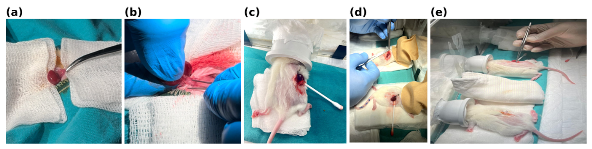
Anesthesia was induced and maintained with 3% sevoflurane (Sevoflurane-Baxter) delivered via a Datex-Ohmeda S-5 Avance machine (General Electric Company, GE Healthcare, Madison, WI, USA) in a temperature-controlled plexiglass chamber. The retroperitoneal space was accessed through a ~2 cm dorsolateral flank incision, the right kidney was mobilized, and the renal pedicle (renal artery and vein) was occluded en bloc with an atraumatic microvascular clamp (Fine Science Tools, Heidelberg, Germany) for 30 min. Ischemia was confirmed by initial blanching of the kidney followed by dark discoloration, reflecting venous congestion, and by loss of arterial pulsation; reperfusion was initiated by clamp release, confirmed by the return of renal color and pulsation, and maintained for 24 h. No renal vessel was ligated and no kidney was removed; the kidney was returned to the retroperitoneum, and the flank incision was closed with sutures (Figure 1). Intraoperative normothermia was maintained with a heating lamp. Animals were not fasted, no perioperative fluids were administered, and food and water were freely available after recovery from anesthesia. For analgesia, a single dose of paracetamol (Atabay Kimya Sanayi ve Ticaret A.Ş., Istanbul, Türkiye, 100 mg/kg, i.p.) was given at the end of the procedure to all groups, including the non-operated Control, LV, and NAC groups.
Figure 1.
Intraoperative photographs of the renal ischemia procedure, shown in procedural order. (a) Exposure of the renal pedicle with forceps. (b) Application of the atraumatic microvascular clamp to the renal pedicle. (c) The clamped right kidney, exteriorized through the dorsolateral flank incision and gently retracted with a cotton-tipped applicator, showing dark discoloration indicating ischemia. (d) Lower animal: Microvascular clamp on the renal pedicle, with dark discoloration of the kidney indicating ischemia; upper animal: suture closure of the flank incision after clamp removal. (e) Upper animal: Under sevoflurane anesthesia delivered via a nose cone, the kidney after clamp removal, with reperfusion initiated; lower animal: closed flank incision.
At sacrifice, blood was collected by cardiac puncture for serum biochemistry before PBS perfusion, and kidney and liver tissues were then harvested. Kidney tissue and a portion of liver tissue were fixed for histopathology, and the remaining liver tissue was snap-frozen in liquid nitrogen and stored at −80 °C for Western blotting. Liver tissue was collected from all animals; however, some samples did not yield evaluable results because of tissue degradation and fixation artifacts (histopathology) or insufficient material (Western blotting), and the number of animals evaluated is given for each analysis.
2.4. Serum Biochemical Analysis
Blood samples were allowed to clot at room temperature and centrifuged at 3000 rpm for 10 min. Serum urea, creatinine, alanine aminotransferase (ALT), and aspartate aminotransferase (AST) were measured with a Mindray BS-2000 automated clinical chemistry analyzer using the manufacturer’s kits (Mindray, Shenzhen, China). Urea and creatinine were used as indicators of renal function, and ALT and AST as indicators of hepatocellular injury rather than of hepatic functional capacity; because AST is also released from skeletal muscle, myocardium, kidney, and erythrocytes, it was interpreted together with the more liver-specific ALT.
2.5. Renal and Hepatic Histopathology
The right kidneys (the clamped kidney in the I/R groups) were fixed in 4% formalin and embedded in paraffin. Longitudinal sections 4 µm thick were cut from the formalin-fixed, paraffin-embedded blocks and stained with hematoxylin and eosin (H&E). The H&E-stained slides were evaluated by a pathologist blinded to group allocation under a light microscope (Nikon Eclipse 80i; Nikon Corporation, Tokyo, Japan). Tubular injury was assessed on the basis of tubular dilatation, loss of brush border, necrosis, hyaline casts, lymphocytic infiltration, increased mesangial cellularity, and dilatation of Bowman’s space. The tubular injury score was determined semi-quantitatively according to the percentage of damaged tubules: 0 = no injury, 1 = 1–25%, 2 = 26–50%, 3 = 51–75%, and 4 = 76–100%. Two sections were evaluated for each kidney, and the tubular injury score of each animal was calculated as the mean of the two section scores. Renal histopathology was performed only in the Control, Sham, and I/R groups to confirm the I/R model. Because sections of adequate quality for evaluation could not be obtained from all animals, evaluable sections were available from eight animals per group, and all of them were assessed.
For hepatic histopathology, a portion of liver tissue was fixed in 4% formalin and processed in the same way (paraffin embedding, 4 µm sections, and H&E staining), and the slides were examined by the same pathologist, blinded to group allocation. Sinusoidal congestion, hepatocellular (ballooning) degeneration, necrosis, inflammatory cell infiltration, and portal tract changes were each graded semi-quantitatively from 0 to 4 according to a modified Suzuki scoring system [16] (congestion, degeneration, inflammatory infiltration, and portal tract changes: 0, none; 1, minimal; 2, mild; 3, moderate; 4, severe; necrosis: 0, none; 1, single-cell necrosis; 2, <30%; 3, 30–60%; 4, >60%), and a total hepatic injury score (0–20) was calculated for each animal as the sum of the five parameter scores. Because tissue degradation and fixation artifacts in the remaining liver preparations precluded reliable evaluation, hepatic histopathology was assessed in the animals with evaluable sections: Control (n = 8), Sham (n = 10), LV (n = 12), NAC (n = 11), I/R (n = 12), I/R+NAC (n = 14), and I/R+LV (n = 14).
2.6. Western Blot Analysis
To evaluate cellular stress and apoptosis in the liver, the expression levels of heat shock proteins (Hsp70, Hsp90) and apoptosis-related proteins (Bax, Bcl-2, pro-caspase-3, cleaved caspase-3) were assessed by Western blotting.
Liver tissues were pulverized in liquid nitrogen and homogenized in cold lysis buffer (50 mM Tris-HCl pH 7.6, 150 mM NaCl, 1% NP-40, 1% Triton X-100, 10% glycerol, 1 mM PMSF, protease inhibitor cocktail); proteins were purified using the methanol-chloroform precipitation method of Wessel and Flügge (1984) [17]. Protein concentrations were determined using the Pierce™ BCA assay kit (Thermo Fisher Scientific, Waltham, MA, USA) based on absorbance at 562 nm.
Protein samples (50 µg per lane) were separated by SDS-PAGE and transferred onto PVDF membranes. Membranes were incubated overnight at 4 °C with the primary antibodies listed in Table 1, followed by incubation with the appropriate HRP-conjugated secondary antibody. Bands were visualized using a ChemiDoc MP system (Bio-Rad Laboratories, Hercules, CA, USA) and quantified with Image Lab software (v6.0.1; Bio-Rad Laboratories, Hercules, CA, USA). Hsp70, Hsp90, pro-caspase-3, and cleaved caspase-3 band intensities were normalized to α-tubulin, whereas Bax and Bcl-2 band intensities were normalized to vinculin. The corresponding loading controls were analyzed on the same membranes as their respective target proteins. For each sample, the densitometric intensity of each target protein band was first normalized to that of its corresponding loading control (α-tubulin or vinculin). The resulting value was then expressed relative to the loading-control-normalized value of the Control sample on the same membrane, which was assigned a value of 1 (100%). Different Control animals were used as reference samples on separate Western blot membranes. Thus, relative protein levels were expressed as fold changes compared with the membrane-specific Control reference. Western blot analysis was performed on liver samples from n = 8–16 animals per group (Control n = 8; Sham n = 15; LV n = 16; NAC n = 16; I/R n = 14; I/R+NAC n = 14; I/R+LV n = 14); of the 104 animals, Western blot analysis could not be performed in seven: three that died during the 24-h reperfusion/observation period and four (two each in the I/R and I/R+NAC groups) from which sufficient liver material could not be obtained. The uncropped Western blot membrane images are provided in Supplementary Figures S1–S4, organized according to the target proteins: Hsp70 (Figure S1), Hsp90 (Figure S2), pro-caspase-3 and cleaved caspase-3 (Figure S3), and Bax and Bcl-2 (Figure S4). The membranes are presented with their corresponding sample identifiers and loading-control images. On some of the additional membranes shown in Supplementary Figures S1–S4, GAPDH or β-actin was used as the loading control (Table 1). Molecular-weight markers are visible on some membranes only, because membranes were cut according to the molecular weights of the target proteins and some were stripped and reprobed.
Table 1.
Antibodies used for Western blot analysis.
2.7. Statistical Analysis
Serum biochemical and Western blot data are presented as mean ± standard deviation (SD), and renal and hepatic histopathological scores, as ordinal data, as median (interquartile range). Serum biochemistry was analyzed in all surviving animals. Normality was assessed with the Shapiro–Wilk test and homogeneity of variances with the Brown–Forsythe test. Because variances were heterogeneous, serum biochemical parameters were analyzed using Welch’s one-way ANOVA followed by the Games–Howell test (exact multiplicity-adjusted p-values are given in Supplementary Table S1). Western blot data were reanalyzed using Welch’s one-way ANOVA, with Brown–Forsythe ANOVA performed as an additional variance-robust omnibus test. Three selected pairwise comparisons (I/R versus Sham, I/R versus I/R+LV, and I/R versus I/R+NAC) were evaluated using the Games–Howell multiple-comparisons test, and multiplicity-adjusted p-values were reported. Because relative protein-expression data may exhibit right-skewed distributions, log10-transformed values were also examined in a sensitivity analysis where all observations were positive. The effect of I/R was assessed against the Sham group for serum biochemistry, histopathology, and Western blot data, so that the effects of anesthesia and surgery were separated from those of ischemia–reperfusion. For Western blotting, treatment-associated differences were assessed by comparing I/R with I/R+LV and I/R+NAC. For serum biochemistry, the treated I/R groups were also compared with each other, and the LV, NAC, and Sham groups with the Control group. For the comparison between the I/R+NAC and I/R+LV groups, mean differences with 95% confidence intervals are reported (Supplementary Table S2); because a non-significant difference does not demonstrate equivalence, no claim of equivalent efficacy is made. Renal tubular injury scores were compared with the Kruskal–Wallis test followed by Dunn’s test with Bonferroni correction. For hepatic scores, each group was compared with the Sham group (Kruskal–Wallis test across the seven groups, followed by Dunn’s test with Bonferroni correction for six comparisons), and the three I/R groups (I/R, I/R+NAC, and I/R+LV) were compared with each other (Kruskal–Wallis test followed by Dunn’s test with Bonferroni correction). A p-value < 0.05 was considered statistically significant. Serum biochemical and Western blot data were analyzed with GraphPad Prism (v11.0, GraphPad Software, San Diego, CA, USA), and histopathological scores with Python (v3.13; Python Software Foundation, Wilmington, DE, USA) using the SciPy library (v1.18).
3. Results
Three animals died during the 24-h reperfusion/observation period: one animal in the Sham group, whose death was attributed to anesthesia, and two animals in the I/R+LV group from an undetermined cause. No blood samples could be obtained from these animals, which were excluded from all analyses; the dead animals were not replaced, so the final group sizes were n = 15 in the Sham group and n = 14 in the I/R+LV group. No animal died in the other groups.
3.1. Protein Expression (Western Blot) Findings
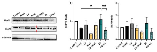
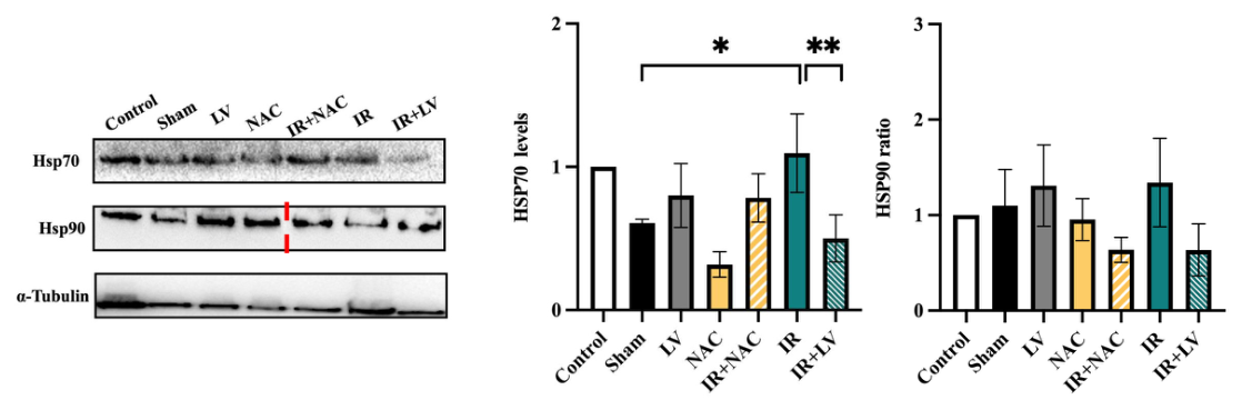
In the analysis of the original normalized data, hepatic Hsp70 expression (Figure 2) did not differ significantly between I/R and Sham (Games–Howell adjusted p = 0.0864), whereas Hsp70 expression was significantly lower in I/R+LV than in I/R (adjusted p = 0.0351). The difference between I/R and I/R+NAC was not significant (adjusted p = 0.3888). In the log10-transformed sensitivity analysis, the I/R versus Sham (adjusted p = 0.0210) and I/R versus I/R+LV (adjusted p = 0.0088) comparisons were significant. For Hsp90, none of the three selected comparisons reached statistical significance in either analysis.
Figure 2.
Representative Western blot images and densitometric analysis of Hsp70 and Hsp90 in liver tissues from the Control, Sham, LV, NAC, I/R, I/R+NAC, and I/R+LV groups (n = 8–16 per group). α-Tubulin was used as the loading control for Hsp70 and Hsp90. Protein band intensities were first normalized to α-tubulin and subsequently expressed relative to the normalized intensity of the Control sample included on the same membrane, which was assigned a value of 1 (100%). Different Control animals were used as membrane-specific reference samples across separate Western blot membranes. Data are presented as mean ± SD of the original normalized protein-expression values (fold change). Statistical significance indicated in the graphs was assessed using the Games–Howell multiple-comparisons test on log10-transformed data. For Hsp70, significant differences were observed between I/R and Sham (* adjusted p = 0.0210) and between I/R and I/R+LV (** adjusted p = 0.0088). No significant differences were detected for Hsp90 in the three selected comparisons. Complete Games–Howell post hoc results for both original normalized and log10-transformed data are provided in Supplementary Table S6. Uncropped Western blot membrane images corresponding to Hsp70 and Hsp90 are provided in Supplementary Figures S1 and S2, respectively. (Non-adjacent lanes from the same membrane are separated by visible splice lines in the representative Hsp90 panel. The red vertical dashed line in the representative Hsp90 panel indicates a splice junction separating non-adjacent lanes from the same membrane. The corresponding uncropped membrane images are provided in Supplementary Figure S2).
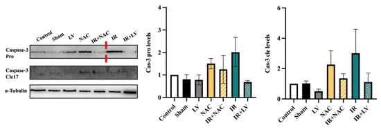
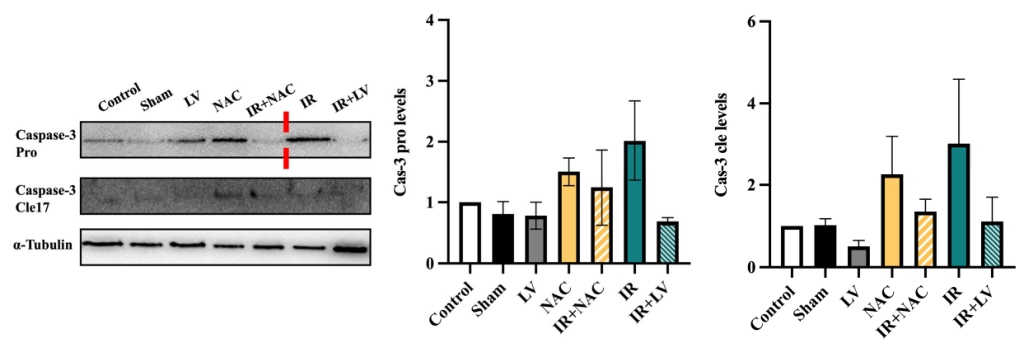
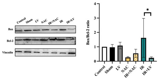
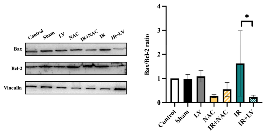
Neither pro-caspase-3 nor cleaved caspase-3 expression (Figure 3) differed significantly between I/R and Sham, I/R and I/R+LV, or I/R and I/R+NAC in the analyses of the original normalized or log10-transformed data (all Games–Howell adjusted p > 0.05). For the Bax/Bcl-2 ratio (Figure 4), none of the three selected comparisons was statistically significant using the original normalized values (I/R versus Sham, p = 0.8337; I/R versus I/R+LV, p = 0.2742; I/R versus I/R+NAC, p = 0.4814). However, the log10-transformed sensitivity analysis identified a significantly lower Bax/Bcl-2 ratio in I/R+LV than in I/R (adjusted p = 0.0154), while the other two comparisons remained non-significant. Because the membrane-specific control reference was assigned a value of 1, comparisons involving this reference group have inherent limitations. The analyses therefore focused on comparisons with Sham and I/R and used variance-robust statistical procedures. Nevertheless, the relatively small and variable numbers of evaluable observations, together with the sensitivity of some findings to log10 transformation, warrant cautious interpretation.
Figure 3.
Representative Western blot images and densitometric analysis of pro-caspase-3 and cleaved caspase-3 protein levels in liver tissues from the Control, Sham, LV, NAC, I/R, I/R+NAC, and I/R+LV groups (n = 8–16 per group). α-Tubulin was used as the loading control for the corresponding membrane. Pro-caspase-3 and cleaved caspase-3 band intensities were first normalized to α-tubulin and subsequently expressed relative to the normalized intensity of the Control sample included on the same membrane, which was assigned a value of 1 (100%). Different Control animals were used as membrane-specific reference samples across separate Western blot membranes. Data are presented as mean ± SD of the original normalized protein-expression values (fold change). Statistical comparisons were performed using the Games–Howell multiple-comparisons test on log10-transformed data. No significant differences were detected between I/R and Sham, I/R and I/R+LV, or I/R and I/R+NAC for either pro-caspase-3 or cleaved caspase-3 (all adjusted p > 0.05). (Non-adjacent lanes from the same membrane are separated by visible splice lines in the representative blot panels. The red vertical dashed line in the representative pro-caspase-3 panel indicates a splice junction separating non-adjacent lanes from the same membrane. The corresponding uncropped Western blot membrane images are provided in Supplementary Figure S3).
Figure 4.
Representative Western blot images and densitometric analysis of the Bax/Bcl-2 ratio in liver tissues from the Control, Sham, LV, NAC, I/R, I/R+NAC, and I/R+LV groups (n = 8–16 per group). Vinculin was used as the loading control for Bax and Bcl-2. Bax and Bcl-2 band intensities were individually normalized to their corresponding vinculin band intensities. The Bax/Bcl-2 ratio was calculated from the normalized Bax and Bcl-2 values and subsequently expressed relative to the Bax/Bcl-2 ratio of the Control sample included on the same membrane, which was assigned a value of 1 (100%). Different Control animals were used as membrane-specific reference samples across separate Western blot membranes. Data are presented as mean ± SD of the relative Bax/Bcl-2 ratio (fold change). Statistical significance was assessed using the Games–Howell multiple-comparisons test on log10-transformed data. The I/R versus I/R+LV comparison was significant (* adjusted p = 0.0154), whereas the I/R versus Sham (adjusted p = 0.9586) and I/R versus I/R+NAC (adjusted p = 0.2225) comparisons were not significant. The corresponding analysis of the original normalized values did not identify a significant difference between I/R and I/R+LV (adjusted p = 0.2742); complete results are provided in Supplementary Table S6. (The corresponding uncropped Western blot membrane images are provided in Supplementary Figure S4).
Overall, the most consistent Western blot finding was the lower hepatic Hsp70 expression in I/R+LV relative to I/R, which was statistically significant in both the original normalized and log10-transformed analyses. An I/R-associated increase in Hsp70 relative to Sham and a reduction in the Bax/Bcl-2 ratio following levosimendan were detected only in the log10-transformed analyses. No significant differences were detected in the selected comparisons for Hsp90, pro-caspase-3, or cleaved caspase-3. These molecular findings should therefore be interpreted as exploratory and considered alongside the biochemical and histopathological results.
3.2. Serum Biochemical Findings
Serum urea and creatinine were significantly higher in the I/R group than in the Sham group (urea: 55.8 ± 7.4 vs. 36.9 ± 13.5 mg/dL, p = 0.002; creatinine: 0.89 ± 0.22 vs. 0.59 ± 0.19 mg/dL, p = 0.005; Table 2). NAC and levosimendan pretreatment reduced both parameters relative to I/R (all p < 0.001), and values in both treated groups did not differ significantly from Sham (urea: p = 0.33 and p = 0.36; creatinine: p > 0.99 for both).
Table 2.
Serum biochemical parameters across experimental groups (mean ± SD).
Serum ALT and AST were markedly higher in the I/R group than in the Sham group (ALT: 107.9 ± 32.4 vs. 19.0 ± 3.1 U/L; AST: 325.5 ± 50.3 vs. 35.7 ± 6.2 U/L; both p < 0.0001). NAC and levosimendan reduced ALT (p = 0.007 and p = 0.005, respectively) and AST (p < 0.0001 for both) relative to I/R, but both parameters remained higher than in the Sham group (all p < 0.0001). No parameter differed between the I/R+NAC and I/R+LV groups (all p > 0.99); the mean differences (I/R+LV minus I/R+NAC) were −0.2 mg/dL (95% CI −3.3 to 3.0) for urea, 0.00 mg/dL (−0.13 to 0.13) for creatinine, −1.4 U/L (−19.3 to 16.5) for ALT, and −3.9 U/L (−51.9 to 44.2) for AST (Supplementary Table S2); these intervals do not exclude clinically relevant differences between the two agents.
No biochemical parameter differed significantly between the LV or NAC group and the Control group (all p > 0.40) or between the Sham and Control groups (all p > 0.60).
Data are presented as mean ± SD. Groups were compared using Welch’s ANOVA (p < 0.001 for all four parameters) followed by the Games–Howell test; exact p-values for all pairwise comparisons are given in Supplementary Table S1.
3.3. Renal Histopathological Findings
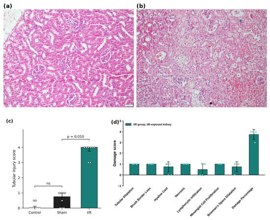
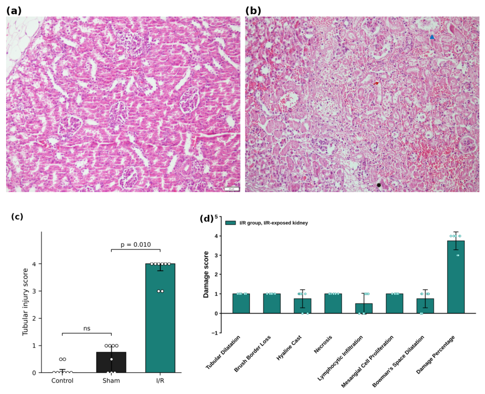
Renal histopathological evaluation was performed in the Control, Sham, and I/R groups (n = 8 per group) to confirm the I/R model. Kidneys from the Control and Sham groups showed normal or near-normal tubular and glomerular morphology (Figure 5a), whereas the clamped kidneys of the I/R group showed tubular dilatation, loss of brush border, tubular necrosis, hyaline casts, inflammatory cell infiltration, and interstitial hemorrhage (Figure 5b). On lesion-based assessment of the I/R group, tubular dilatation, brush border loss, tubular necrosis, and increased mesangial cellularity were present in all eight animals, hyaline casts and Bowman’s space dilatation in six, and lymphocytic infiltration in four; the tubular injury score was 4 in six animals and 3 in two (median 4, IQR 3.75–4; Figure 5d; Supplementary Table S4). The tubular injury score was significantly higher in the I/R group than in the Sham group (median 4 [IQR 3.75–4] vs. 0.75 [0–1]; p = 0.010), whereas the Control and Sham groups did not differ significantly (Control median 0 [0–0.125]; p = 0.72; Figure 5c).
Figure 5.
Renal histopathology. Representative hematoxylin and eosin (H&E)-stained sections of the right kidney from (a) the Control group, showing normal tubular and glomerular architecture, and (b) the I/R group, showing tubular dilatation, loss of brush border, tubular necrosis, intratubular casts, and interstitial hemorrhage. Red arrow: tubular epithelial necrosis, loss of brush border, and luminal accumulation of sloughed cellular debris; blue arrowhead: eosinophilic proteinaceous (hyaline/granular) casts within the tubular lumen; black asterisk: microvascular fibrin thrombi and focal necrotic debris accompanied by interstitial hemorrhage. Scale bar in (a) = 20 µm. (c) Tubular injury scores in the Control, Sham, and I/R groups (n = 8 per group; 0 = no injury; 4 = injury in 76–100% of tubules). Each value is the mean of the scores of two sections per animal. Bars show the median and error bars the interquartile range; dots represent individual animals. Kruskal–Wallis test followed by Dunn’s test with Bonferroni correction; ns, not significant. (d) Lesion-based scores in the I/R group (n = 8): each histopathological finding was scored as present (1) or absent (0), and the damage percentage was graded 0–4. Data are presented as mean ± SD with individual data points.
3.4. Hepatic Histopathological Findings
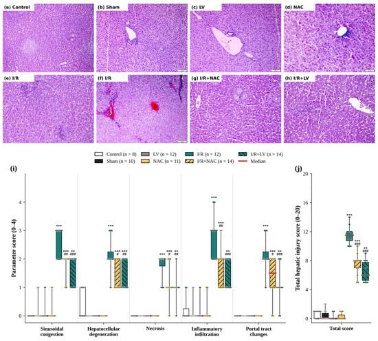
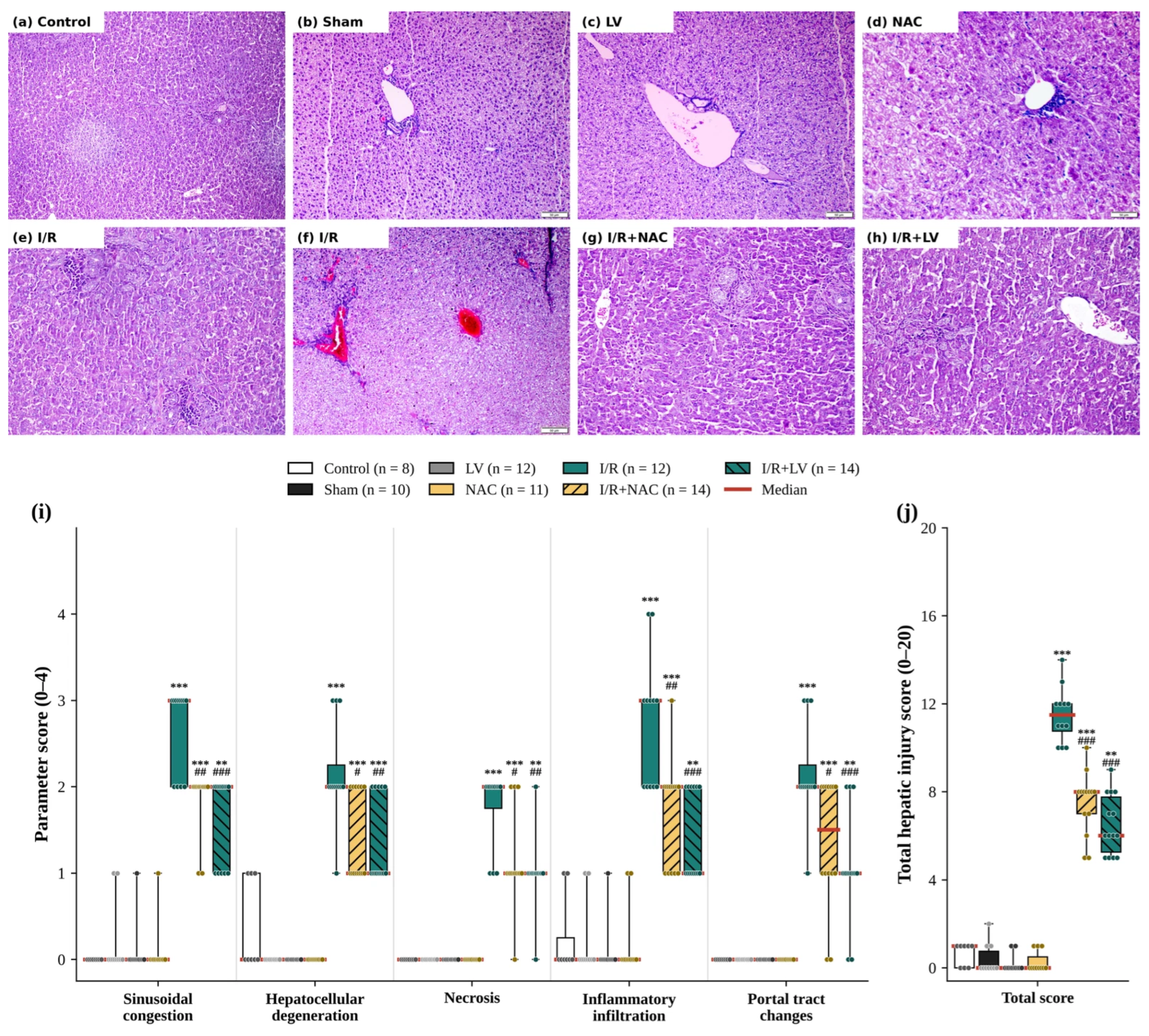
Hepatic histopathology was evaluated in the Control (n = 8), Sham (n = 10), LV (n = 12), NAC (n = 11), I/R (n = 12), I/R+NAC (n = 14), and I/R+LV (n = 14) groups. Livers from the Control, Sham, LV, and NAC groups showed preserved lobular architecture with regular hepatocyte cords and intact portal tracts (Figure 6a–d); only minimal changes (total score 0–2) were observed in these four groups. The I/R group showed sinusoidal congestion, periportal hepatocellular ballooning degeneration, focal necrosis, and periportal inflammatory cell infiltration (Figure 6e,f). These changes were less pronounced in both treated groups: the I/R+NAC group showed attenuated congestion and fewer degenerative changes (Figure 6g), and the I/R+LV group showed largely preserved hepatic architecture with mild sinusoidal dilatation (Figure 6h). On semi-quantitative scoring (Figure 6i,j; Supplementary Table S5), the total hepatic injury score was significantly higher in the I/R group than in the Sham group (median 11.5, IQR 10.75–12 vs. 0, IQR 0–0.75; p < 0.001), whereas the Control, LV, and NAC groups did not differ significantly from Sham (p > 0.99). Among the I/R groups, the total score was significantly lower in the I/R+NAC (8, IQR 7–8; p < 0.001) and I/R+LV (6, IQR 5.25–7.75; p < 0.001) groups than in the I/R group, with no significant difference between the two treated groups (p = 0.61); both treated groups remained significantly higher than the Sham group (p < 0.001 and p = 0.005, respectively). Both treatments also significantly reduced each of the five individual parameters relative to I/R (I/R+NAC, p = 0.002–0.032; I/R+LV, p < 0.001–0.004), and the two treated groups did not differ in any parameter (p ≥ 0.75).
Figure 6.
Representative hematoxylin and eosin (H&E)-stained liver sections. (a) Control: Normal lobular architecture with regular hepatocyte cords and intact portal tracts (×100). (b) Sham, (c) LV, and (d) NAC: Preserved lobular architecture and portal tracts (scale bars = 50 µm). (e,f) I/R: (e) periportal inflammatory cell infiltration and hepatocellular degeneration (×200); (f) marked congestion of the central and portal veins (scale bar = 50 µm). (g) I/R+NAC: Attenuated congestion and fewer degenerative changes (×200). (h) I/R+LV: Largely preserved hepatic architecture with mild sinusoidal dilatation (×200). (i) Individual parameter scores and (j) total hepatic injury score (0–20) in the Control (n = 8), Sham (n = 10), LV (n = 12), NAC (n = 11), I/R (n = 12), I/R+NAC (n = 14), and I/R+LV (n = 14) groups. Boxes show the interquartile range, red lines the median, and whiskers the minimum and maximum; dots represent individual animals. ** p < 0.01, *** p < 0.001 vs. Sham (Kruskal–Wallis test across all groups followed by Dunn’s test with Bonferroni correction); # p < 0.05, ## p < 0.01, ### p < 0.001 vs. I/R (Kruskal–Wallis test among the three I/R groups followed by Dunn’s test with Bonferroni correction).
4. Discussion
Renal ischemia–reperfusion (I/R) injury is common in clinical practice, arising from circulatory failure or from the vascular occlusion required during renal surgery and transplantation [1]. Through the systemic release of reactive oxygen species (ROS), pro-inflammatory cytokines, and damage-associated molecular patterns, it can injure distant organs, including the lungs, heart, and brain [3] and the liver [4,5,6]. This organ crosstalk is increasingly recognized as a determinant of morbidity in acute kidney injury (AKI), and the liver is one of its most studied targets [18,19]; renohepatic crosstalk has been reviewed in detail [20,21] and is also relevant in children, in whom AKI is common and associated with mortality [2] and organ crosstalk complicates the management of critical illness and congenital disease [22]. In the present juvenile rat model, renal I/R increased serum transaminases and the hepatic histopathological injury score relative to Sham, and pretreatment with NAC or levosimendan attenuated these changes, without a significant difference between the two agents.
Hepatic Hsp70 expression was significantly higher in I/R than in Sham in the log10-transformed analysis, although this difference was not significant in the original normalized data; no significant differences were detected for Hsp90 in the selected comparisons. Heat shock proteins are molecular chaperones induced as part of an adaptive response to oxidative and proteotoxic stress [23], and HSP70 has been implicated in limiting hepatic inflammatory and oxidative damage in direct hepatic I/R [7]. The lower Hsp70 expression in the I/R+LV group may therefore reflect either a reduced stress burden or suppression of an adaptive response; because these possibilities cannot be distinguished, it should not be interpreted as evidence of reduced injury.
By contrast, Onody et al. found that levosimendan did not alter hepatic HSP72 expression after direct hepatic I/R induced by hepatoduodenal ligament occlusion [11], whereas Hsp70 was lower in the I/R+LV group in the present study. This difference may reflect the injury paradigm: in the present model, the liver was exposed to circulating inflammatory mediators and ROS rather than to local ischemia, and the heat shock response and its modulation by levosimendan may differ between these settings.
In the variance-robust analyses, neither pro-caspase-3 nor cleaved caspase-3 differed significantly between I/R and Sham or between I/R and either treatment group, whereas the Bax/Bcl-2 ratio was significantly lower in I/R+LV than in I/R only after log10 transformation. Thus, the present data do not provide evidence of activation of the intrinsic apoptotic pathway, which proceeds through permeabilization of the outer mitochondrial membrane by pro-apoptotic BCL-2 family members and culminates in caspase-3 activation [24] and has been documented after kidney I/R [6]; demonstrating such activation would require evidence such as caspase-3 activity, cytochrome c release, or TUNEL staining. The two treatments were not compared directly in the Western blot analyses, and their mechanisms, which were not tested here, may differ: the anti-apoptotic action of levosimendan in the liver has been attributed to mitochondrial K-ATP channel opening and Akt/eNOS-dependent survival signaling [12], whereas NAC acts mainly upstream by scavenging ROS [8,9] and has shown remote-organ protection after intestinal I/R [10]. Because levosimendan is also a vasodilator and blood pressure was not measured, part of its effect may reflect improved perfusion.
Both agents are already used in children. Levosimendan is used in pediatric cardiac surgery, where prophylactic use reduced the incidence of low cardiac output syndrome without a significant effect on mortality [25], and NAC is the standard antidote for acetaminophen-induced hepatotoxicity [26]. This clinical familiarity supports further study of both agents for the prevention of remote liver injury in children undergoing procedures that place the kidney at ischemic risk.
In adult rats with direct hepatic I/R, levosimendan and NAC, alone and particularly in combination, reduced histopathological injury, transaminases, malondialdehyde, and myeloperoxidase, although serum TNF-α was not significantly affected [14]. In the present study, the liver was not itself ischemic, and our findings suggest that the protective effects of both agents extend to remote hepatic injury in juvenile animals. However, because both agents also lowered serum urea and creatinine, the design cannot determine whether the hepatic effects are direct or secondary to reduced renal injury. Unlike that study, we did not include a combination group. The direct comparison between the agents was not significant for any serum or histopathological endpoint, and whether either agent is superior can only be determined in an adequately powered comparative study.
The serum biochemical and histopathological findings support both the renal injury model and the presence of remote hepatic injury. Because the contralateral kidney was left intact, a rise in urea and creatinine was not necessarily expected; it may reflect systemic effects of renal I/R on the contralateral kidney or the limited compensatory hyperfiltration of immature kidneys. Tubular injury was confirmed in the clamped kidney (Figure 5), but because the contralateral kidney was not examined and glomerular filtration was not measured, these explanations cannot be distinguished. The rise in ALT and AST, together with the increased hepatic histopathological injury score, is consistent with remote hepatocellular injury. ALT and AST reflect hepatocellular injury rather than hepatic function, and the AST/ALT ratio of approximately 3 in the I/R group suggests extrahepatic contributions to AST, such as muscle injury from the flank incision, the injured kidney, or hemolysis during cardiac puncture. The transaminase elevation (approximately 6- to 9-fold over Sham) also exceeded that reported in adult rat models, in which AST increased approximately 1.7- to 1.9-fold and ALT up to 3.5-fold [5,27] and 30 min of renal ischemia did not increase transaminases [27]. This difference may reflect the comparatively immature antioxidant defenses of the pediatric liver and the greater vulnerability of juvenile animals to perioperative fluid shifts, accentuated by the absence of fluid resuscitation; because no adult group was included, these explanations remain hypotheses. Both agents reduced all four biochemical parameters and the hepatic injury score relative to I/R; urea and creatinine did not differ from Sham, whereas transaminases and hepatic injury scores remained elevated, indicating partial protection. The absence of biochemical or histological differences between the drug-alone groups and the Control or Sham group suggests that neither agent was associated with toxicity at the doses used, although measurements at a single time point are insufficient to establish safety.
Limitations
This study has several limitations. First, because of tissue degradation and fixation artifacts, liver histopathology could be evaluated in only 8–14 animals per group, and no apoptosis-specific staining (e.g., TUNEL) was performed; renal histopathology was performed only in the Control, Sham, and I/R groups to confirm the model. Second, oxidative stress, inflammatory mediators, caspase activity, mitochondrial function, and the signaling targets of levosimendan were not measured, so the mechanisms discussed remain hypothetical, and the design cannot distinguish a direct hepatoprotective effect from one secondary to reduced renal injury. Third, systemic hemodynamics and renal perfusion were not monitored; given its vasodilatory properties, the effects of levosimendan may partly reflect improved perfusion. Fourth, samples were collected at a single time point (24 h), only male rats were studied, ischemia was unilateral with the contralateral kidney left intact, and each agent was given as a single dose adapted from adult studies without dose–response evaluation. Fifth, the LV-alone and NAC-alone groups did not undergo sham surgery; paracetamol, given to all groups for analgesia, may have interacted with NAC through hepatic glutathione; levosimendan was dissolved in a minimal volume of DMSO, and no DMSO-vehicle I/R group was included; and no perioperative fluids were administered. Sixth, Western blot protein expression values were normalized to membrane-specific Control reference samples assigned a value of 1, which limits the representation of Control-group variability and the interpretation of comparisons involving this reference group. Furthermore, unequal variances among experimental groups, particularly for the Bax/Bcl-2 ratio, may affect the robustness of the statistical findings. Although variance-robust analyses and log10-transformed sensitivity analyses were performed, the relatively small and unequal numbers of evaluable observations and the sensitivity of some findings to data transformation warrant cautious interpretation of the Western blot results. Seventh, no formal power calculation or computer-generated randomization sequence was used, and the two deaths of undetermined cause in the I/R+LV group, in which levosimendan-related hypotension cannot be excluded, may have biased the results in favor of this group. Finally, the transaminase elevation was larger than that typically reported in adult models, an age-dependent difference that was not directly tested. These limitations should be addressed in future studies.
5. Conclusions
Unilateral renal I/R in juvenile rats caused tubular injury, increased serum urea and creatinine, and induced remote liver injury, reflected by elevated ALT and AST and an increased hepatic histopathological injury score. Pretreatment with NAC or levosimendan attenuated these changes: urea and creatinine did not differ from Sham, whereas transaminases and hepatic injury scores were only partially reduced. The two agents did not differ significantly, although the study was not designed to test equivalence, and neither agent alone affected serum biochemistry or liver histology. The Western blot findings were largely limited to lower hepatic Hsp70 expression with levosimendan, should be interpreted cautiously, and do not independently establish a hepatoprotective mechanism or superiority of either treatment. Both agents may therefore protect against renal I/R-associated remote liver injury in the pediatric setting; mechanistic, hemodynamic, and dose–response studies are needed to determine whether this effect is direct or secondary to reduced renal injury and to clarify its mechanisms.
Supplementary Materials
The following supporting information can be downloaded at https://www.mdpi.com/article/10.3390/medicina62112192/s1: Table S1: Games-Howell pairwise comparisons of serum biochemical parameters (multiplicity-adjusted p-values); Table S2: Mean differences between the I/R+LV and I/R+NAC groups with 95% confidence intervals; Table S3: Individual animal serum biochemical data; Table S4: Lesion-based renal histopathological findings in the I/R group, with the corresponding methods and results; Table S5: Individual animal hepatic histopathological scores; Table S6: Games–Howell multiple-comparisons analysis of Western blot protein-expression data in liver tissues; Figure S1: Uncropped Western blot images of Hsp70; Figure S2: Uncropped Western blot images of Hsp90; Figure S3: Uncropped Western blot images of pro-caspase-3 and cleaved caspase-3; Figure S4: Uncropped Western blot images of Bax and Bcl-2.
Author Contributions
Conceptualization, S.K.S., S.D. and T.B.Ö.; Methodology, S.K.S., S.D., E.Ö.U. and T.B.Ö.; Software, B.Ç.; Validation, S.D., E.Ö.U. and S.K.S.; Formal Analysis, B.Ç., Ö.B. and T.Ş.; Investigation, S.K.S., S.D., R.O.K., L.Ş., C.G., E.B.D., B.Ç., Ö.B., T.Ş. and T.B.Ö.; Resources, E.Ö.U. and C.G.; Data Curation, E.B.D.; Writing—Original Draft Preparation, S.K.S.; Writing—Review & Editing, S.D., E.Ö.U. and T.B.Ö.; Visualization, B.Ç.; Supervision, E.Ö.U. and S.D.; Project Administration, S.K.S.; Funding Acquisition, S.K.S. All authors have read and agreed to the published version of the manuscript.
Funding
This research was funded by the budget of Bağcılar Training and Research Hospital, Ministry of Health of the Republic of Türkiye (grant number 2026/15).
Institutional Review Board Statement
The animal study protocol was approved by the Local Animal Experiments Ethics Committee of Istanbul Bağcılar Training and Research Hospital (Meeting No. 152, project no. 2025/02, decision no. 2025/18, approved 25 March 2025). All procedures complied with the European Convention for the Protection of Vertebrate Animals Used for Experimental and Other Scientific Purposes (ETS 123).
Informed Consent Statement
Not applicable.
Data Availability Statement
Individual animal data for all serum biochemical measurements are provided in Supplementary Table S3; individual histopathological scores are shown in Figure 5c and Figure 6i,j and Supplementary Table S5; densitometric data are available from the corresponding author upon reasonable request.
Conflicts of Interest
The authors declare no conflicts of interest.
References
- Chen, Y.; Li, Z.; Zhang, H.; Chen, H.; Hao, J.; Liu, H.; Li, X. Mitochondrial Metabolism and Targeted Treatment Strategies in Ischemic-Induced Acute Kidney Injury. Cell Death Discov. 2024, 10, 69. [Google Scholar] [CrossRef] [Scilit] [PubMed]
- Kaddourah, A.; Basu, R.K.; Bagshaw, S.M.; Goldstein, S.L.; AWARE Investigators. Epidemiology of Acute Kidney Injury in Critically Ill Children and Young Adults. N. Engl. J. Med. 2017, 376, 11–20. [Google Scholar] [CrossRef] [Scilit] [PubMed]
- Saberi, S.; Najafipour, H.; Rajizadeh, M.A.; Etminan, A.; Jafari, E.; Iranpour, M. NaHS Protects Brain, Heart, and Lungs as Remote Organs from Renal Ischemia/Reperfusion-Induced Oxidative Stress in Male and Female Rats. BMC Nephrol. 2024, 25, 373. [Google Scholar] [CrossRef] [Scilit] [PubMed]
- Khastar, H.; Kadkhodaee, M.; Sadeghipour, H.R.; Seifi, B.; Hadjati, J.; Najafi, A.; Soleimani, M. Liver Oxidative Stress after Renal Ischemia-Reperfusion Injury is Leukocyte Dependent in Inbred Mice. Iran. J. Basic Med. Sci. 2011, 14, 534–539. [Google Scholar] [PubMed]
- Seifi, B.; Kadkhodaee, M.; Najafi, A.; Mahmoudi, A. Protection of Liver as a Remote Organ after Renal Ischemia-Reperfusion Injury by Renal Ischemic Postconditioning. Int. J. Nephrol. 2014, 2014, 120391. [Google Scholar] [CrossRef] [Scilit] [PubMed]
- Bagheri, Y.; Aghajani, S.; Hosseinzadeh, M.; Hoshmandan, F.; Abdollahpour, A.; Zununi Vahed, S. Protective Effects of Gamma Oryzanol on Distant Organs after Kidney Ischemia-Reperfusion in Rats: A Focus on Liver Protection. Hum. Exp. Toxicol. 2021, 40, 1022–1030. [Google Scholar] [CrossRef] [Scilit] [PubMed]
- Wu, H.H.; Huang, C.C.; Chang, C.P.; Lin, M.T.; Niu, K.C.; Tian, Y.F. Heat Shock Protein 70 (HSP70) Reduces Hepatic Inflammatory and Oxidative Damage in a Rat Model of Liver Ischemia/Reperfusion Injury with Hyperbaric Oxygen Preconditioning. Med. Sci. Monit. 2018, 24, 8096–8104. [Google Scholar] [CrossRef] [Scilit] [PubMed]
- Zhang, F.; Lau, S.S.; Monks, T.J. The Cytoprotective Effect of N-Acetyl-L-Cysteine against ROS-Induced Cytotoxicity Is Independent of Its Ability to Enhance Glutathione Synthesis. Toxicol. Sci. 2011, 120, 87–97. [Google Scholar] [CrossRef] [Scilit] [PubMed]
- Sehirli, A.O.; Sener, G.; Satiroglu, H.; Ayanoğlu-Dülger, G. Protective Effect of N-Acetylcysteine on Renal Ischemia/Reperfusion Injury in the Rat. J. Nephrol. 2003, 16, 75–80. [Google Scholar] [CrossRef] [Scilit]
- Alexandropoulos, D.; Bazigos, G.V.; Doulamis, I.P.; Tzani, A.; Konstantopoulos, P.; Tragotsalou, N.; Kondi-Pafiti, A.; Kotsis, T.; Arkadopoulos, N.; Smyrniotis, V.; et al. Protective Effects of N-Acetylcysteine and Atorvastatin against Renal and Hepatic Injury in a Rat Model of Intestinal Ischemia-Reperfusion. Biomed. Pharmacother. 2017, 89, 673–680. [Google Scholar] [CrossRef] [Scilit]
- Onody, P.; Stangl, R.; Fulop, A.; Rosero, O.; Garbaisz, D.; Turoczi, Z.; Lotz, G.; Rakonczay, Z., Jr.; Balla, Z.; Hegedus, V.; et al. Levosimendan: A Cardiovascular Drug to Prevent Liver Ischemia-Reperfusion Injury? PLoS ONE 2013, 8, e73758. [Google Scholar] [CrossRef] [Scilit] [PubMed]
- Grossini, E.; Pollesello, P.; Bellofatto, K.; Sigaudo, L.; Farruggio, S.; Origlia, V.; Mombello, C.; Mary, D.A.S.G.; Valente, G.; Vacca, G. Protective Effects Elicited by Levosimendan against Liver Ischemia/Reperfusion Injury in Anesthetized Rats. Liver Transpl. 2014, 20, 361–375. [Google Scholar] [CrossRef] [Scilit] [PubMed]
- Grossini, E.; Molinari, C.; Pollesello, P.; Bellomo, G.; Valente, G.; Mary, D.; Vacca, G.; Caimmi, P. Levosimendan Protection against Kidney Ischemia/Reperfusion Injuries in Anesthetized Pigs. J. Pharmacol. Exp. Ther. 2012, 342, 376–388. [Google Scholar] [CrossRef] [Scilit] [PubMed]
- Suakıtıcı, S.; Güven, B.B.; Tanoğlu, A.; Özkan, S. A Combination of Levosimendan and N-Acetylcysteine Shows Significant Favorable Efficacy on Experimental Liver Ischemia/Reperfusion Injury. Ulus. Travma Acil Cerrahi Derg. 2021, 27, 381–388. [Google Scholar] [CrossRef] [Scilit] [PubMed]
- Sezen, S.C.; Kucuk, A.; Özer, A.; Kılıç, Y.; Mardin, B.; Alkan, M.; Erkent, F.D.; Arslan, M.; Ünal, Y.; Oktar, G.L.; et al. Assessment of the Effects of Levosimendan and Thymoquinone on Lung Injury after Myocardial Ischemia Reperfusion in Rats. Drug Des. Devel. Ther. 2018, 12, 1347–1352. [Google Scholar] [CrossRef] [Scilit] [PubMed]
- Suzuki, S.; Toledo-Pereyra, L.H.; Rodriguez, F.J.; Cejalvo, D. Neutrophil Infiltration as an Important Factor in Liver Ischemia and Reperfusion Injury. Modulating Effects of FK506 and Cyclosporine. Transplantation 1993, 55, 1265–1272. [Google Scholar] [PubMed]
- Wessel, D.; Flügge, U.I. A Method for the Quantitative Recovery of Protein in Dilute Solution in the Presence of Detergents and Lipids. Anal. Biochem. 1984, 138, 141–143. [Google Scholar] [CrossRef] [Scilit] [PubMed]
- Doi, K.; Rabb, H. Impact of Acute Kidney Injury on Distant Organ Function: Recent Findings and Potential Therapeutic Targets. Kidney Int. 2016, 89, 555–564. [Google Scholar] [CrossRef] [Scilit] [PubMed]
- Lee, S.A.; Cozzi, M.; Bush, E.L.; Rabb, H. Distant Organ Dysfunction in Acute Kidney Injury: A Review. Am. J. Kidney Dis. 2018, 72, 846–856. [Google Scholar] [CrossRef] [Scilit] [PubMed]
- Lane, K.; Dixon, J.J.; MacPhee, I.A.M.; Philips, B.J. Renohepatic Crosstalk: Does Acute Kidney Injury Cause Liver Dysfunction? Nephrol. Dial. Transplant. 2013, 28, 1634–1647. [Google Scholar] [CrossRef] [Scilit] [PubMed]
- Khoshdel Rad, N.; Heydari, Z.; Tamimi, A.H.; Zahmatkesh, E.; Shpichka, A.; Barekat, M.; Timashev, P.; Hossein-khannazer, N.; Hassan, M.; Vosough, M. Review on Kidney-Liver Crosstalk: Pathophysiology of Their Disorders. Cell J. (Yakhteh) 2024, 26, 98–111. [Google Scholar] [CrossRef] [Scilit] [PubMed]
- Piatto, D.; De Biasio, D.; Cesaro, F.G.; Forcina, G.; Frattolillo, V.; Colucci, A.; Lamberti, F.; Marzuillo, P.; Miraglia del Giudice, E.; Di Sessa, A. Liver-Kidney Crosstalk in Major Pediatric Diseases: Unraveling the Complexities and Clinical Challenges. J. Clin. Med. 2025, 14, 3911. [Google Scholar] [CrossRef] [Scilit] [PubMed]
- Pearl, L.H. Review: The HSP90 Molecular Chaperone—An Enigmatic ATPase. Biopolymers 2016, 105, 594–607. [Google Scholar] [CrossRef] [Scilit] [PubMed]
- Czabotar, P.E.; Garcia-Saez, A.J. Mechanisms of BCL-2 Family Proteins in Mitochondrial Apoptosis. Nat. Rev. Mol. Cell Biol. 2023, 24, 732–748. [Google Scholar] [CrossRef] [Scilit] [PubMed]
- Wang, H.; Luo, Q.; Li, Y.; Zhang, L.; Wu, X.; Yan, F. Effect of Prophylactic Levosimendan on All-Cause Mortality in Pediatric Patients Undergoing Cardiac Surgery-An Updated Systematic Review and Meta-Analysis. Front. Pediatr. 2020, 8, 456. [Google Scholar] [CrossRef] [Scilit] [PubMed]
- Yesil, Y.; Ozdemir, A.A. Evaluation of the Children with Acute Acetaminophen Overdose and Intravenous N-Acetylcysteine Treatment. Pak. J. Med. Sci. 2018, 34, 590–594. [Google Scholar] [CrossRef] [Scilit] [PubMed]
- Kadkhodaee, M.; Golab, F.; Zahmatkesh, M.; Ghaznavi, R.; Hedayati, M.; Arab, H.A.; Ostad, S.N.; Soleimani, M. Effects of Different Periods of Renal Ischemia on Liver as a Remote Organ. World J. Gastroenterol. 2009, 15, 1113–1118. [Google Scholar] [CrossRef] [Scilit] [PubMed]

Disclaimer/Publisher’s Note: The statements, opinions and data contained in all publications are solely those of the individual author(s) and contributor(s) and not of MDPI and/or the editor(s). MDPI and/or the editor(s) disclaim responsibility for any injury to people or property resulting from any ideas, methods, instructions or products referred to in the content. |
© 2026 by the authors. Published by MDPI on behalf of the Lithuanian University of Health Sciences. Licensee MDPI, Basel, Switzerland. This article is an open access article distributed under the terms and conditions of the Creative Commons Attribution (CC BY) license.