


Abstract
Background and Objectives: Sentinel lymph node biopsy (SLNB) is the standard procedure for regional nodal staging in patients with clinically localized cutaneous melanoma. However, most patients undergoing SLNB have negative sentinel lymph nodes, highlighting the need for predictive tools to optimize selection. The Memorial Sloan Kettering Cancer Center (MSKCC) nomogram estimates the probability of sentinel lymph node metastasis using clinicopathological variables. This study evaluated the discrimination, calibration, and net benefit of the MSKCC nomogram in a Romanian cohort of patients with cutaneous melanoma. Materials and Methods: We conducted a retrospective, single-center cohort study including 117 patients with primary cutaneous melanoma who underwent SLNB between 2020 and 2025. The predicted probability of sentinel lymph-node (SLN) metastasis was calculated using the official MSKCC nomogram, and histopathological SLN status served as the reference standard. Discrimination was evaluated using receiver operating characteristic analysis, and threshold-based classification was assessed at predicted risks of 5%, 10%, and the exploratory Receiver Operating Characteristic (ROC) curve-derived threshold of 19%. Calibration was assessed using the observed-to-expected ratio and the calibration intercept and slope, and net benefit was evaluated by decision curve analysis. Results: Patients with positive sentinel lymph nodes had higher MSKCC-predicted risks than patients with negative nodes (median 20.0% vs. 10.0%; p = 0.0004). The MSKCC score was associated with SLN positivity (OR 1.065 per 1-percentage-point increase; 95% CI 1.026–1.106; p = 0.0011). The model showed moderate discrimination (AUC = 0.714; 95% CI 0.614–0.814). At the 5% threshold, 24 patients (20.5%) were classified below the threshold, and no SLN-positive case was observed in this subgroup; however, the subgroup was small. Higher thresholds classified more patients below the biopsy threshold but reduced sensitivity. The nomogram underestimated the observed risk (mean predicted 15.6% vs. observed 26.5%; observed-to-expected ratio 1.70, 95% CI 1.15–2.41; calibration slope 0.89). Compared with SLNB for all patients, nomogram-based selection yielded a net reduction of 20.5 biopsies per 100 patients at the 5% threshold but no net benefit at the 10% threshold. Conclusions: In this cohort, the MSKCC nomogram showed moderate discrimination for SLN positivity but underestimated the absolute risk of SLN metastasis. It may be useful as an additional tool in selected or borderline cases, but it should be applied with caution and should not replace established clinicopathological criteria and guideline-based SLNB recommendations. Validation in larger multicenter cohorts, including recalibration, is required before any risk threshold can be recommended.
1. Introduction
Melanoma is the most aggressive form of skin cancer. In 2020, an estimated 325,000 new cases and 57,000 deaths from cutaneous melanoma occurred worldwide, and both numbers are projected to increase substantially by 2040, making it a significant public health concern [1]. Although melanoma most commonly arises in the skin, it can also develop in other melanocyte-containing tissues, such as the uveal tract of the eye and mucosal surfaces [2].
Established risk factors for cutaneous malignant melanoma include ultraviolet (UV) radiation exposure, particularly from sunlight; Fitzpatrick skin phototypes I and II; the presence of dysplastic nevi; indoor tanning; increasing age and a personal or family history of melanoma [3].
The diagnosis of cutaneous melanoma relies on clinical examination, supported by the ABCD (a clinical and self-screening guide used to identify potential signs of melanoma) rule, the EFG (a clinical mnemonic) criteria for nodular melanoma, and the “ugly duckling” sign. Dermoscopy significantly increases diagnostic accuracy, while digital monitoring techniques may improve early detection in high-risk individuals. The definitive diagnosis is established by complete excisional biopsy with narrow margins and histopathological assessment according to the AJCC 8th edition TNM staging system [4]. The principal histopathological prognostic factors in primary melanoma are Breslow thickness, ulceration, and mitotic rate. Among these, Breslow thickness is the most important predictor of outcome, while ulceration is associated with a worse prognosis. Although mitotic rate retains prognostic value, it is no longer incorporated into the AJCC 8th edition staging system for thin melanomas [5].
The Sentinel Lymph Node (SLN) is defined as the primary node within a direct drainage pathway from the tumor to the regional basin; its identification relies on the principle that lymphatic flow follows a predictable anatomical route, directing malignant cells toward specific “gatekeeper” nodes [6]. According to the latest European Society for Medical Oncology (ESMO) guidelines, SLNB is recommended for patients with primary melanomas > 1.0 mm in Breslow thickness (AJCC pT2a or higher) and should be considered for selected patients with pT1b melanomas (0.8–1.0 mm or <0.8 mm with ulceration), while it is generally not recommended for pT1a lesions in the absence of additional high-risk features [4]. SLNB provides pathological staging information that may inform prognosis, adjuvant systemic therapy eligibility, and surveillance planning while limiting the morbidity associated with more extensive nodal surgery [7,8].
Despite the established prognostic value of SLNB, the procedure is associated with surgical risks and a high rate of negative results. Approximately 80–85% of patients do not harbor nodal metastases, although this percentage is highly contingent upon the Breslow thickness of the primary tumor [9,10]. This underscores the clinical need for non-invasive, predictive tools that can accurately estimate the risk of SLN positivity.
The Memorial Sloan Kettering Cancer Center (MSKCC) melanoma nomogram was developed to address this challenge by integrating specific clinicopathological variables such as patient age, tumor thickness, Clark level, ulceration, and anatomical site into a unified predictive model. While its efficacy has been demonstrated in large-scale international cohorts [11], its correlation with real-world histopathological outcomes in diverse clinical settings remains a subject of investigation.
The present study aimed to externally evaluate the MSKCC nomogram in a Romanian cohort of patients undergoing SLNB for cutaneous melanoma. The primary objective was to assess discrimination between SLN-positive and SLN-negative patients. The secondary objectives were to examine associations between individual clinicopathological variables and SLN status and to describe classification performance at clinically relevant risk thresholds.
2. Materials and Methods
2.1. Study Design and Setting
This retrospective, observational, single-center cohort study was conducted in the Department of Plastic, Aesthetic and Reconstructive Surgery at Elias University Emergency Hospital, Bucharest, Romania. The study population comprised consecutive patients who underwent sentinel lymph node biopsy for primary cutaneous melanoma between 2020 and 2025.
2.2. Eligibility Criteria
All patients were clinically node-negative at the time SLNB was indicated, with no known distant metastases before the procedure. Regional nodal metastases identified through SLNB were considered the study outcome and did not constitute an exclusion criterion. Any distant metastases recorded in the dataset were diagnosed after SLNB.
Patients were eligible if they underwent SLNB for primary cutaneous melanoma during the study period and had the variables required by the MSKCC nomogram available: age, Breslow thickness, Clark level, ulceration status, and primary tumor location. A definitive histopathological SLN result was also required. Patients with synchronous or metachronous non-melanoma malignancies were not excluded. Patients with documented metastatic melanoma at presentation were retained if SLNB had been performed and all required data were available.
Patients were excluded when one or more required nomogram predictors or the final SLN histopathological status was unavailable. The analysis therefore used complete cases for all variables included in the nomogram and the primary outcome.
2.3. Data Sources and Variables
Demographic, clinical, and pathological data were retrospectively extracted from electronic medical records and pathology reports. Variables used in the present analysis were age, sex, histological subtype (nodular, superficial spreading, acral, desmoplastic, or nevoid), primary tumor location, Breslow thickness, Clark level, ulceration status, sampled lymphatic basin, final SLN status, and presence and location of metastatic disease at diagnosis. Data was anonymized before statistical processing.
2.4. Sentinel Lymph Node Procedure and Pathological Outcome
The sampled lymphatic basin was recorded as axillary, inguinal, cervical, or supraclavicular. The reference outcome was the final histopathological interpretation of the sentinel lymph node and was dichotomized as SLN-positive when any metastatic melanoma cells were identified in the SLN, irrespective of the size of the deposit and of whether they were detected on hematoxylin and eosin (H&E) or immunohistochemical sections, in accordance with the AJCC 8th edition [12], and as SLN-negative otherwise. Therefore, no quantitative lower threshold of tumor burden was applied to define positivity.
SLNs were identified using preoperative lymphoscintigraphy with a 99mTc-labelled radiocolloid and an intraoperative gamma probe, with or without blue dye. In the pathology department, SLNs were bisected along their long axes, entirely embedded, and examined on serial H&E sections, with later immunohistochemistry for S100, HMB-45, and/or Melan-A. Quantitative measures of SLN tumor burden, such as the maximum diameter of the largest metastatic deposit, were not consistently reported and were not analyzed.
2.5. MSKCC Nomogram Calculation
For each patient, the predicted probability of sentinel lymph node metastasis was obtained from the official Memorial Sloan Kettering Cancer Center online nomogram. The required inputs were age, Breslow thickness, Clark level, ulceration status, and anatomical site of the primary melanoma. The calculated probability was recorded as a percentage and analyzed as a continuous predictor and at prespecified 5% and 10% thresholds; the 19% threshold was derived exploratorily from the study receiver operating characteristic(ROC) curve. The 5% and 10% thresholds were prespecified because they delimit the risk categories used in the NCCN guidelines, which do not recommend SLNB when the risk of a positive SLN is below 5% and recommend discussing and considering SLNB when the risk is 5–10% [13]. The same thresholds were evaluated in the previous validation studies discussed in Section 4, and the nomogram itself was used without modification, with the inputs and coefficients of the official online calculator. The 19% threshold, selected by the Youden index, was included only to illustrate the point of maximal balance between sensitivity and specificity in our cohort and was not intended as a clinical decision threshold.
Primary tumor locations were mapped to the calculator categories as follows: trunk lesions to “Trunk”; upper- and lower-limb lesions to “Extremity”; and facial, scalp, and neck lesions to “Head or Neck.”
2.6. Outcomes
The primary outcome was the discriminatory performance of the MSKCC-predicted probability of histopathologically confirmed SLN positivity. Discrimination was quantified using the area under the receiver operating characteristic curve. Threshold-based performance was described using the number of patients classified below each threshold, the number of SLN-positive cases below the threshold, sensitivity, specificity, positive predictive value, and negative predictive value.
Secondary analyses tested the associations of age, Breslow thickness, and ulceration with SLN status and compared Clark level, sex, histological subtype, and anatomical location between SLN-positive and SLN-negative patients. Calibration of the predicted probabilities and the net benefit of nomogram-based selection for SLNB were evaluated as additional measures of model performance, and discrimination and calibration were explored separately in nodular and non-nodular melanomas.
2.7. Statistical Analysis
Continuous variables were summarized using the median and interquartile range, except age, which was reported as median and range. Categorical variables were summarized as counts and percentages. Percentages were calculated using the relevant non-missing denominator.
Continuous and ordinal variables (age, Breslow thickness, Clark level, and MSKCC predicted risk) were compared between SLN-positive and SLN-negative groups using the Mann–Whitney U test, because Breslow thickness and predicted risk were not normally distributed (Shapiro–Wilk p < 0.001 in at least one group) and Clark level is an ordinal variable; for age, which was approximately normally distributed, the same test was used for consistency, and an independent-samples t-test gave a similar result. Associations between categorical variables, such as ulceration, sex, histological subtype, and tumor location, were evaluated using Fisher’s exact test (Fisher–Freeman–Halton extension for variables with more than two categories), which does not rely on large-sample approximations and remains valid when expected cell counts are small, as in the stratified analyses; odds ratios and 95% confidence intervals were reported where appropriate. The relationship between Breslow thickness and binary SLN status was additionally summarized using Spearman’s rank correlation coefficient.
Discrimination of the MSKCC nomogram was assessed using receiver operating characteristic curve analysis and summarized by the area under the curve. The exploratory ROC-derived threshold was selected using the Youden index. Univariate logistic regression was used to quantify the association between the predicted MSKCC probability, expressed per 1-percentage-point increase, and SLN positivity. Classification measures were calculated at thresholds of less than 5%, less than 10%, and less than 19%. The 95% confidence interval (CI) for the Area Under the Curve (AUC) was calculated using the nonparametric method of DeLong et al. [14].
Calibration was assessed according to the framework described by Van Calster et al. [15]. Calibration-in-the-large was evaluated by comparing the mean predicted probability with the observed SLN positivity rate and expressed as the observed-to-expected (O/E) ratio, with a 95% CI based on the exact Poisson distribution of the observed number of events. The calibration intercept was estimated by logistic regression of SLN status with the logit of the predicted probability as an offset and the calibration slope as the regression coefficient of the logit of the predicted probability; the ideal values are 0 and 1, respectively. The Brier score was also calculated. In addition, the observed SLN positivity rate, with exact Clopper–Pearson 95% CIs, was compared with the mean predicted risk across strata defined by the 5%, 10%, and 19% thresholds, and a logistic calibration curve was plotted.
Clinical usefulness was assessed by decision curve analysis [16]. For each threshold probability (pt), the net benefit of selecting patients for SLNB according to the nomogram was calculated as TP/n − FP/n × pt/(1 − pt), where TP and FP are the numbers of SLN-positive and SLN-negative patients with a predicted risk at or above the threshold, and n is the total number of patients. The nomogram was compared with the default strategies of performing SLNB in all patients and in none. Net benefit was plotted for thresholds between 1% and 40% and reported in detail at the 5%, 10%, and 19% thresholds, together with the net reduction in SLNBs per 100 patients, calculated as (net benefit of the nomogram − net benefit of SLNB for all)/(pt/(1 − pt))× 100. The 95% CIs of the net reduction were obtained by bootstrap resampling (2000 samples).
Because nodular melanoma was over-represented in the cohort, discrimination and calibration-in-the-large were also explored separately in nodular and non-nodular melanomas. These subgroup analyses were exploratory.
In additional exploratory analyses, SLN positivity was compared across sex, age groups (<50, 50–64, and ≥65 years), histological subtypes (nodular, superficial spreading, and other), tumor locations (trunk, extremity, and head and neck, corresponding to the nomogram categories), and ulceration status, in the whole cohort and separately in women and men; within each category, SLN positivity was also compared between women and men. These analyses were not adjusted for multiple comparisons and were considered hypothesis-generating.
All tests were two-sided, and p < 0.05 was considered statistically significant.
Statistical analyses were performed using IBM SPSS v.25 Statistics (IBM Corp., Armonk, NY, USA). The DeLong confidence interval, calibration measures, decision curve analysis, and bootstrap resampling were computed in Python (version 3.13) using the NumPy 2.1.0, SciPy 1.18.1, and Matplotlib 3.11.2 libraries.
3. Results
3.1. Cohort Characteristics
A total of 117 patients with primary cutaneous melanoma who underwent sentinel lymph node biopsy (SLNB) were included in the analysis. The median age was 58 years (range 20–93), and 54.7% of patients were female. The most frequent histological subtype was nodular melanoma (56.4%), followed by superficial spreading melanoma (35.9%). The trunk (37.6%) and lower limb (32.5%) were the most common tumor locations. Ulceration was present in 30.8% of cases. Sentinel lymph node invasion was identified in 31 patients (26.5%), while 86 patients (73.5%) had a negative SLN (Table 1).
Table 1.
Patient and tumor characteristics.
3.2. Sentinel Lymph Node Positivity, Patient Age, and Tumor Characteristics
3.2.1. Age
The median age was 59 years (range 29–85) among SLN-positive patients and 57.5 years (range 20–93) among SLN-negative patients, with no significant differences between the groups (Mann–Whitney U = 1406.0; p = 0.654) (Table 2).
Table 2.
Comparison of patient and tumor characteristics between patients with and without SLN invasion.
3.2.2. Ulceration
SLN positivity was observed in 14 of 36 patients with ulcerated melanoma (38.9%) and in 17 of 81 patients without ulceration (21.0%). Correspondingly, ulceration was present in 14 of 31 SLN-positive patients (45.2%) and 22 of 86 SLN-negative patients (25.6%). The odds of SLN positivity were 2.4-fold higher for ulcerated melanomas (OR = 2.396), although the association did not reach statistical significance (Fisher’s exact p = 0.068).
3.2.3. Breslow Thickness
Patients with positive sentinel lymph nodes had greater Breslow thickness than patients with negative sentinel lymph nodes (median 3.00 mm ([IQR 1.85–4.90] vs. 1.50 mm (IQR 0.90–2.80); Mann–Whitney U = 1936.5; p = 0.0002). Spearman analysis showed a moderate positive association between Breslow thickness and SLN positivity (rho = 0.346; p = 0.0001).
3.2.4. Clark Level
Clark level was higher in SLN-positive than in SLN-negative patients (Mann–Whitney U = 1730.5; p = 0.006). Although the median level was IV in both groups, Clark level V was present in 6 of 31 SLN-positive patients (19.4%) and in 3 of 86 SLN-negative patients (3.5%).
3.2.5. MSKCC Risk Score and SLN Invasion
Patients with a positive sentinel lymph node had significantly higher Sentinel Lymph Node Metastasis Risk Scores than patients with a negative sentinel lymph node. The median score was 20.0% (IQR 12.5–29.5) in the SLN-positive group, which can be compared with 10.0% (IQR 4.0–18.5) in the SLN-negative group (Mann–Whitney U = 1903.0, p = 0.0004).
3.2.6. Logistic Regression
Univariate logistic regression showed that the MSKCC predicted probability was associated with SLN positivity (beta = 0.063; OR = 1.065 per 1-percentage-point increase; 95% CI 1.026–1.106; p = 0.0011). Thus, each 1-percentage-point increase in predicted risk corresponded to an estimated 6.5% increase in the odds of SLN metastasis.
3.2.7. ROC Curve Analysis
ROC curve analysis showed moderate discrimination of the MSKCC predicted probability for SLN metastasis, with an AUC of 0.714 (95% CI 0.614–0.814). The exploratory cut-off selected by the Youden index was 19%, corresponding to a sensitivity of 58.1% and a specificity of 74.4%. Because this threshold was derived and evaluated in the same cohort, it should not be interpreted as a clinically optimal decision threshold.
3.3. Threshold-Based Classification at 5%, 10%, and 19%
At a predicted risk threshold of less than 5%, 24 of 117 patients (20.5%) were classified below the threshold (Table 3). None had a histopathological positive SLN, corresponding to an observed NPV of 100% and a sensitivity of 100%. Because only 24 patients were classified below this threshold, these point estimates should be interpreted with their uncertainty: the exact 95% CIs were 85.8–100.0% for NPV and 88.8–100.0% for sensitivity.
Table 3.
Threshold-based classification performance of the MSKCC nomogram for sentinel lymph node positivity.
At a predicted risk threshold of less than 10%, 45 patients (38.5%) were classified below the threshold. Of these, 39 were SLN-negative, and 6 were SLN-positive. Sensitivity was 80.6%, specificity was 45.3%, PPV was 34.7%, and NPV was 86.7%.
At the exploratory ROC-derived threshold of less than 19%, 77 patients (65.8%) were classified below the threshold and 40 (34.2%) at or above it. Among patients below the threshold, 64 were SLN-negative and 13 were SLN-positive. Sensitivity was 58.1%, specificity was 74.4%, PPV was 45.0%, and NPV was 83.1%. The 13 missed SLN-positive cases represented 41.9% of all patients with a positive SLN.
3.4. Calibration
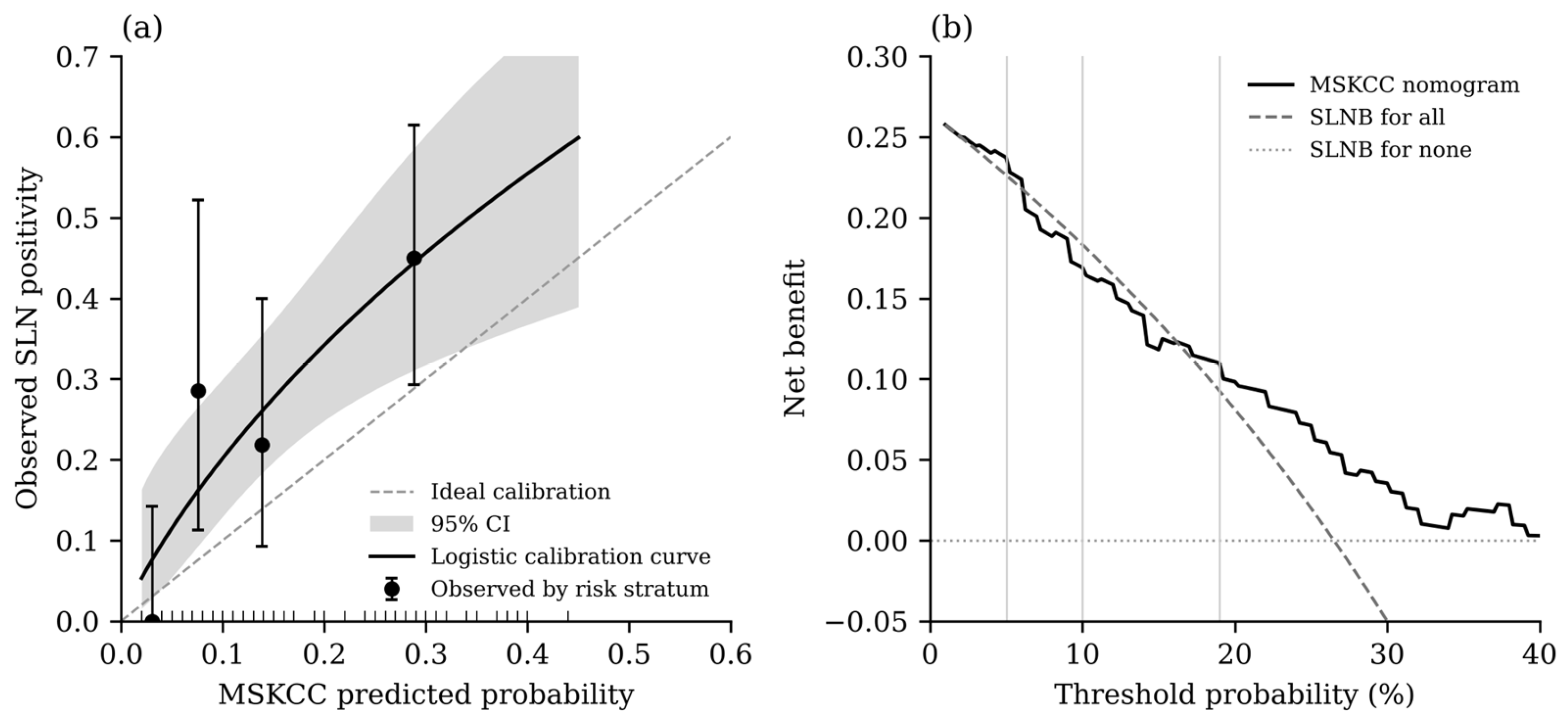
The mean predicted risk was 15.6%, whereas the observed SLN positivity rate was 26.5% (31 observed vs. 18.3 expected events), corresponding to an O/E ratio of 1.70 (95% CI 1.15–2.41). The calibration intercept was 0.76 (95% CI 0.31–1.20; p < 0.001), indicating systematic underestimation of risk, whereas the calibration slope was 0.89 (95% CI 0.37–1.40) and did not differ significantly from 1. The Brier score was 0.188, compared with 0.195 for a non-informative model assigning the observed prevalence to all patients. Across the predicted-risk strata (Table 4, Figure 1a), no SLN metastasis was observed below 5% (0/24; mean predicted risk 3.0%). In the 5–<10%, 10–<19%, and ≥19% strata, the observed SLN positivity rates were 28.6%, 21.9%, and 45.0%, which can be compared with mean predicted risks of 7.5%, 13.8%, and 28.8%, respectively.
Table 4.
Observed sentinel lymph node positivity across MSKCC predicted-risk strata.
Figure 1.
Calibration and decision curve analysis of the MSKCC nomogram (n = 117). (a) Calibration plot: points show the observed SLN positivity (with exact 95% CIs) against the mean predicted probability in the four predicted-risk strata (<5%, 5–<10%, 10–<19%, and ≥19%); the solid line is the logistic calibration curve with its 95% CI (shaded area), the dashed line represents ideal calibration, and tick marks on the x-axis show individual predicted probabilities. (b) Decision curve showing the net benefit of nomogram-based selection for SLNB compared with performing SLNB in all patients or in none; vertical lines mark the 5%, 10%, and 19% thresholds.
3.5. Net Benefit (Decision Curve Analysis)
The decision curve is shown in Figure 1b, and the net benefit at the prespecified thresholds is summarized in Table 5. Nomogram-based selection provided a higher net benefit than SLNB for all patients at thresholds of 3–6% and from 17% upwards, but a lower net benefit at thresholds between 7% and 16%. At the 5% threshold, the nomogram yielded a net reduction of 20.5 SLNBs per 100 patients without missed SLN metastases. At the 10% threshold, the net benefit of the nomogram was lower than that of SLNB for all patients (net reduction −12.8 per 100 patients), because the SLN positivity rate among patients classified below this threshold (13.3%; 6/45) exceeded the threshold itself. At the exploratory 19% threshold, the net reduction was 7.3 SLNBs per 100 patients. The bootstrap 95% CIs at the 10% and 19% thresholds included zero, indicating considerable uncertainty.
Table 5.
Net benefit of MSKCC nomogram-based selection for SLNB compared with performing SLNB in all patients.
3.6. Exploratory Subgroup Analyses
In nodular melanomas (n = 66; 22 SLN-positive, 33.3%), the AUC was 0.636 (95% CI 0.500–0.771) and the O/E ratio was 1.75 (95% CI 1.10–2.65; mean predicted risk 19.0%). In non-nodular melanomas (n = 51; 9 SLN-positive, 17.6%), the AUC was 0.735 (95% CI 0.542–0.929) and the O/E ratio was 1.58 (95% CI 0.72–3.00; mean predicted risk 11.2%).
SLN positivity according to sex, age group, histological subtype, tumor location, and ulceration, in the whole cohort and separately in women and men, is shown in Table 6. In the whole cohort, SLN positivity was 34.0% in men and 20.3% in women (p = 0.140) and did not differ significantly across age groups (p = 0.101), histological subtypes (p = 0.180), or tumor locations (p = 0.231); the association with ulceration was not significant (p = 0.068). In women, SLN positivity differed by tumor location (p < 0.001)—8.3% for extremity, 22.7% for trunk, and 83.3% for head and neck melanomas (5 of 6 patients)—whereas no such difference was observed in men (p = 0.379). Consequently, SLN positivity was higher in men than in women for extremity melanomas (42.1% vs. 8.3%; p = 0.005) and lower for head and neck melanomas (16.7% vs. 83.3%; p = 0.013). Ulcerated melanomas had numerically higher SLN positivity in both women (28.6% vs. 16.3%; p = 0.324) and men (53.3% vs. 26.3%; p = 0.106). The remaining sex-specific comparisons were not significant.
Table 6.
Sentinel lymph node positivity according to sex, age group, histological subtype, tumor location, and ulceration (exploratory analysis).
4. Discussion
In this external Romanian cohort of patients with primary cutaneous melanoma, the MSKCC nomogram showed moderate discrimination between patients with and without sentinel lymph node metastasis. Patients with positive SLNs had higher predicted risks than patients with negative SLNs, and the predicted probability was associated with SLN positivity in univariate logistic regression. Threshold-based analyses demonstrated the expected trade-off: higher thresholds classified more patients below the biopsy threshold but reduced sensitivity and increased the number of SLN-positive cases that would have been missed. However, the nomogram systematically underestimated the absolute risk of SLN metastasis in our cohort (O/E ratio 1.70), and its net benefit compared with SLNB for all patients depended on the threshold considered.
Sentinel lymph node status is recognized as the single most important prognostic factor in patients with clinically localized melanoma and represents the cornerstone of regional nodal staging [17]. Building upon this concept, the eighth edition of the AJCC melanoma staging system identifies Breslow thickness and ulceration as the principal characteristics of the primary tumor for T-category classification and for selecting patients who should undergo sentinel lymph node biopsy [18]. Consistent with these established principles, patients with positive sentinel lymph nodes in our cohort had significantly greater Breslow thickness and a higher Clark level than those with negative nodes, while ulceration was associated with a 2.4-fold increase in the odds of nodal metastasis, although statistical significance was not reached. The estimate was directionally consistent with previous evidence but remained imprecise and did not reach statistical significance in this cohort. When taken as a whole, these results support the biological foundation of the MSKCC nomogram, which uses these known clinico-pathological variables to calculate the probability of sentinel lymph node metastasis.
The present study demonstrated that the Sentinel Lymph Node Metastasis Risk Score has a moderate discriminatory ability for predicting sentinel lymph node metastasis, with an AUC comparable to those reported in previous external validation studies. Drebin et al. reported an AUC of 0.720 in a cohort of 2464 patients, while Olofsson Bagge et al. observed an AUC of 0.708 in a population-based validation including 10,089 patients, both closely matching the AUC of 0.714 obtained in our cohort. Despite differences in sample size and patient populations, these findings consistently support the reproducibility of the discriminative performance of the MSKCC nomogram across independent cohorts [19,20]. Our estimate is also consistent with the pooled C-statistic of 0.73 (95% CI 0.69–0.78) reported for MSKCC models in a recent meta-analysis [21] and with the AUC of 0.729 reported by Mesgarzadeh et al. in a high-UV-index region [22], although the 95% CI of our AUC (0.614–0.814) was wide, reflecting the small number of events. The main findings regarding the present cohort are compared with those of the original MSKCC study and previous external validations in Table 7.
Table 7.
Main findings of the present cohort compared with the original MSKCC study and previous external validations of the MSKCC nomogram.
Discrimination alone, however, does not indicate whether the predicted probabilities correspond to the observed risk. In our cohort, the calibration slope was close to 1, suggesting that the relative ranking of patients was preserved, but the positive calibration intercept and an O/E ratio of 1.70 indicate that the nomogram systematically underestimated the absolute probability of SLN metastasis. A similar underprediction was reported by Drebin et al. [19], whereas the nomogram was well calibrated in the Swedish population-based cohort and in the cohort of Mesgarzadeh et al. [20,22]. Underestimation is clinically relevant when fixed thresholds are applied, because patients classified below a threshold may have an actual risk above it.
Previous studies have also explored the clinical implications of applying predefined risk thresholds for selecting patients for sentinel lymph node biopsy. Drebin et al. reported that the MSKCC nomogram would have reduced the number of biopsies performed by 13.6% and 45.4% at the 5% and 10% risk thresholds, respectively; however, after accounting for missed metastases in decision curve analysis, neither the MSKCC nor the Melanoma Institute Australia (MIA) model provided a net reduction in avoidable SLNBs at thresholds of 10% or lower [19]. In the population-based cohort studied by Olofsson Bagge et al., neither model added a net benefit at the 5% threshold, whereas a net benefit was observed at thresholds of 10% or higher [20]. In our cohort, the pattern differed: nomogram-based selection was favorable at the 5% threshold, where no SLN-positive cases were observed among 24 patients, but not at the 10% threshold, where the SLN positivity rate among patients classified as low-risk (13.3%) exceeded the threshold. This is consistent with the underestimation of risk observed in our cohort, and the favorable result at 5% rests on a small subgroup. More generally, increasing the decision threshold resulted in a greater number of avoided biopsies at the expense of reduced sensitivity and a higher number of missed nodal metastases, emphasizing the trade-off between minimizing unnecessary interventions and maintaining adequate oncological safety [19,20].
Similar observations were reported by Tassavor et al., who evaluated the performance of the MSKCC nomogram alongside the integrated i31-GEP model. Although the proportion of patients classified as low risk differed between the two cohorts, the frequency of sentinel lymph node metastasis among patients categorized as low risk remained comparable at the 10% threshold. Interestingly, no patient classified as low risk using the 5% threshold in our cohort had histopathologically confirmed sentinel lymph node metastasis, whereas Tassavor et al. reported an SLN positivity rate of 10% within this subgroup. These differences are likely attributable to variations in patient characteristics, case selection, and sample size and further highlight the importance of external validation before widespread implementation of predictive models in different clinical settings [23].
In addition to evaluating the predefined 5% and 10% risk thresholds commonly reported in the literature, our study identified an exploratory ROC-derived cut-off of 19% using receiver operating characteristic analysis and the Youden index. This threshold maximized the Youden index and provided the mathematical balance between sensitivity (58.1%) and specificity (74.4%) in our cohort, reflecting the highest overall discriminatory performance of the MSKCC nomogram. Application of this threshold would have classified 65.8% of patients as low-risk, classifying 77 patients below the biopsy threshold; however, 13 patients with histopathologically confirmed nodal metastases would have been misclassified as low risk. These findings illustrate that, although increasing the decision threshold improves specificity and reduces the number of potentially unnecessary biopsies, it inevitably decreases sensitivity and increases the risk of false-negative classifications. Therefore, the ROC-derived threshold identified in this cohort may not necessarily represent the most appropriate clinical decision threshold. Instead, selection of the cut-off should balance diagnostic performance with the clinical consequences of missing sentinel lymph node metastases. In decision-analytic terms, a 19% threshold implies that a clinician would accept performing no more than approximately five SLNBs to detect one nodal metastasis, compared with 20 and 10 SLNBs at the 5% and 10% thresholds, respectively. Such a threshold reflects a much greater willingness to forgo the detection of nodal metastases, and the net benefit observed at 19% should therefore be interpreted with caution.
Although the MSKCC nomogram demonstrated moderate discriminatory performance in our cohort, there remains potential for further improvement. A study evaluating the incorporation of microphthalmia-associated transcription factor (MITF) expression into the MSKCC prediction model reported an AUC of 0.650 for the original nomogram in their cohort; after the model was refitted to the local data, the AUC was 0.742, and the addition of MITF further increased it to 0.825 (p < 0.0001) [24]. These findings suggest that combining clinicopathological prediction models with molecular biomarkers may further enhance the accuracy of sentinel lymph node metastasis prediction. Future studies should therefore investigate whether integrating additional biological markers with established clinicopathological models, such as the MSKCC nomogram, can improve risk stratification and optimize patient selection for sentinel lymph node biopsy.
The case mix of our cohort should also be considered when interpreting these results. Nodular melanoma accounted for more than half of the patients (56.4%), whereas superficial spreading melanoma is generally the most common histological subtype, as reported, for example, in Norway [25]. The SLN positivity rate (26.5%) was also higher than in the large validation cohorts studied by Drebin et al. (23.0%) and Olofsson Bagge et al. (17.9%) [19,20]. This distribution may reflect the referral of higher-risk patients to a tertiary plastic surgery center and limits the generalizability of our findings. Histological subtype is not an input variable of the MSKCC nomogram. In the exploratory subgroup analysis, discrimination was lower in nodular than in non-nodular melanomas (AUC 0.636 vs. 0.735), and risk was underestimated in both subgroups (O/E ratio 1.75 and 1.58, respectively). These subgroup results are based on few events and should be considered hypothesis-generating; they suggest that the predominance of nodular melanomas may have contributed to, but does not fully explain, the underestimation observed in the overall cohort.
The exploratory subgroup analyses did not show significant differences in SLN positivity by sex, age group, histological subtype, or tumor location in the whole cohort. In women, SLN positivity varied markedly by tumor location, with five of six head and neck melanomas being SLN-positive, whereas no such pattern was observed in men. These findings are based on very small subgroups and on multiple comparisons without adjustment, so they may reflect chance; they should be regarded as hypothesis-generating and examined in larger cohorts. Sex is not an input variable of the MSKCC nomogram.
Several factors may explain the differences between our results and those of the original MSKCC study and of the large US and Swedish validation cohorts [11,19,20]. First, the case mix differed: SLN positivity was 26.5% in our cohort, which can be compared with 23.0% at the MSKCC and 17.9% in Sweden [19,20], and nodular melanomas predominated. In Central and Eastern Europe, melanoma incidence is less than half that of Western Europe, whereas mortality is similar, a disparity possibly related to missed opportunities for early diagnosis and incomplete reporting [26]; patients undergoing SLNB in Romania may therefore present with more advanced primary tumors. Second, the indications for SLNB, the referral pathways, and the surgical technique vary between centers and countries and determine which patients enter a validation cohort. Third, the pathological examination of SLNs is not fully standardized. According to the AJCC 8th edition, any metastatic melanoma cells in an SLN define a positive node, whether detected on H&E or immunohistochemistry, so there is no quantitative lower threshold for positivity [12]. The probability of detecting small deposits, however, depends on the number of sections examined and on the use of immunohistochemistry: with the extensive European Organisation for. Research and Treatment of Cancer (EORTC) protocol, the SLN positivity rate was 28.7% in a cohort with a median Breslow thickness of 2.1 mm, and submicrometastases smaller than 0.1 mm were identified in 18% of cases [27]. More extensive SLN examination increases the observed positivity rate and could therefore contribute to an apparent underestimation of risk by a model developed with different pathological procedures. These factors would be expected to affect calibration more than discrimination, which is consistent with our finding of an AUC similar to that reported in other countries together with a higher-than-predicted observed risk. Finally, differences in genetic background, phototype, and ultraviolet exposure between populations could influence tumor biology; these factors are not included in the nomogram and could not be assessed in our study.
Therefore, in addition to further validation of the MSKCC nomogram in Romanian and other populations, international comparability would benefit from standardization in two areas. First, decision thresholds should be prespecified and uniform, such as the 5% and 10% risk categories used in the NCCN guidelines [13]. Second, the pathological evaluation and reporting of SLNs should be standardized, for example according to the updated EORTC protocol, which defines the sectioning procedure and immunohistochemistry and recommends reporting the number of deposits, their intranodal location, and the maximum dimension of the largest deposit [28]. Reporting these elements in validation studies would allow differences in model performance to be attributed more reliably to population characteristics rather than to procedural variation.
The present study has several strengths. It evaluated a consecutive cohort treated at a tertiary referral center and used the final histopathological SLN result as the reference outcome. It also provides data from a Romanian clinical setting, extending the geographical evidence regarding the discriminatory performance of the MSKCC nomogram. In addition to discrimination, calibration and net benefit were assessed [15,16]. Finally, the analysis examined the predefined 5% and 10% thresholds and an exploratory ROC-derived threshold of 19%, illustrating the trade-off between the number of patients classified below a threshold and the number of missed SLN-positive cases.
Several limitations should be acknowledged. First, the retrospective, single-center design may introduce selection and information bias and limits generalizability. Second, the cohort was relatively small, with 31 SLN-positive events, resulting in imprecise estimates for subgroup and threshold-based analyses. The inclusion period started in 2020 because complete electronic records with all nomogram variables were available only from this year onward; extension of the cohort and multicenter collaboration are planned. Third, patients with documented metastatic disease at presentation were retained, which may differ from the intended clinically node-negative population of the MSKCC nomogram; a sensitivity analysis excluding these patients is warranted. Fourth, the cohort had a high proportion of nodular melanomas and an SLN positivity rate of 26.5%, suggesting a relatively high-risk case mix that may not be representative of other settings. In addition, the SLN examination protocol and tumor burden could not be compared with those of the development cohort. Finally, with 31 events, the calibration and net benefit estimates were imprecise, the 19% threshold was derived from the same data, and the model was not recalibrated to the local population; therefore, our findings do not allow a preferred clinical threshold to be established.
5. Conclusions
In this Romanian cohort, the MSKCC nomogram showed moderate discrimination for histopathologically confirmed SLN positivity but tended to underestimate the absolute risk of SLN metastasis. The model may be useful as an additional tool in selected or borderline cases, e.g., during preoperative counselling, but it should be applied with caution and should not replace established clinicopathological criteria and current guideline recommendations.
Threshold selection materially changed the balance between the number of patients classified below the biopsy threshold and the number of missed SLN-positive cases. Nomogram-based selection provided a net benefit over SLNB for all patients at the 5% threshold but not at the 10% threshold. The absence of observed SLN metastases below 5% was encouraging but was based on only 24 patients and therefore remains uncertain.
Larger multicenter studies including recalibration of the model to the local population, sensitivity analyses restricted to the intended nomogram population, and prospective validation are needed before a preferred clinical threshold can be proposed. Uniform risk thresholds and standardized pathological evaluation of SLNs would further improve the comparability of results across countries.
Author Contributions
Conceptualization, S.-M.R.; methodology, R.-D.S.; writing—original draft preparation, A.-C.E.; writing—review and editing, A.-L.P., P.-A.C. and K.-A.J.; supervision, S.-M.R.; formal analysis, R.-D.S. All authors have read and agreed to the published version of the manuscript.
Funding
This research received no external funding.
Institutional Review Board Statement
This study was conducted in accordance with the Declaration of Helsinki and approved by the Ethics Committee of Elias University Emergency Hospital, Bucharest, Romania (code: 5080/8 September 2026).
Informed Consent Statement
Informed consent for participation was obtained from all subjects involved in the study.
Data Availability Statement
The original contributions presented in this study are included in the article. Further inquiries can be directed to the corresponding author.
Acknowledgments
Publication of this paper was supported by the University of Medicine and Pharmacy Carol Davila through the Institutional Program “Publish not Perish”.
Conflicts of Interest
The authors declare no conflicts of interest.
Abbreviations
The following abbreviations are used in this manuscript:
| AJCC | American Joint Committee on Cancer |
| AUC | area under the receiver operating characteristic curve |
| CI | confidence interval |
| EORTC | European Organisation for Research and Treatment of Cancer |
| H&E | hematoxylin and eosin |
| IQR | interquartile range |
| MIA | Melanoma Institute Australia |
| MITF | microphthalmia-associated transcription factor |
| MSKCC | Memorial Sloan Kettering Cancer Center |
| NCCN | National Comprehensive Cancer Network |
| NPV | negative predictive value |
| O/E | observed-to-expected ratio |
| OR | odds ratio |
| PPV | positive predictive value |
| ROC | receiver operating characteristic |
| SLN | sentinel lymph node |
| SLNB | sentinel lymph node biopsy |
References
- Arnold, M.; Singh, D.; Laversanne, M.; Vignat, J.; Vaccarella, S.; Meheus, F.; Cust, A.E.; de Vries, E.; Whiteman, D.C.; Bray, F. Global Burden of Cutaneous Melanoma in 2020 and Projections to 2040. JAMA Dermatol. 2022, 158, 495–503. [Google Scholar] [CrossRef] [Scilit]
- Elder, D.E.; Bastian, B.C.; Cree, I.A.; Massi, D.; Scolyer, R.A. The 2018 World Health Organization Classification of Cutaneous, Mucosal, and Uveal Melanoma: Detailed Analysis of 9 Distinct Subtypes Defined by Their Evolutionary Pathway. Arch. Pathol. Lab. Med. 2020, 144, 500–522. [Google Scholar] [CrossRef] [Scilit]
- Lauters, R.; Brown, A.D.; Harrington, K.A. Melanoma: Diagnosis and Treatment. Am. Fam. Physician 2024, 110, 367–377. [Google Scholar]
- Amaral, T.; Ottaviano, M.; Arance, A.; Blank, C.; Chiarion-Sileni, V.; Donia, M.; Dummer, R.; Garbe, C.; Gershenwald, J.; Gogas, H.; et al. Cutaneous melanoma: ESMO Clinical Practice Guideline for diagnosis, treatment and follow-up. Ann. Oncol. 2025, 36, 10–30. [Google Scholar] [CrossRef] [Scilit]
- Garbe, C.; Amaral, T.; Peris, K.; Hauschild, A.; Arenberger, P.; Basset-Seguin, N.; Bastholt, L.; Bataille, V.; Brochez, L.; del Marmol, V.; et al. European consensus-based interdisciplinary guideline for melanoma. Part 1: Diagnostics—Update 2024. Eur. J. Cancer 2025, 215, 115152. [Google Scholar] [CrossRef] [Scilit]
- Brănişteanu, D.E.; Cozmin, M.; Porumb-Andrese, E.; Brănişteanu, D.; Toader, M.P.; Iosep, D.; Sinigur, D.; Brănişteanu, C.I.; Brănişteanu, G.; Porumb, V.; et al. Sentinel Lymph Node Biopsy in Cutaneous Melanoma, a Clinical Point of View. Medicina 2022, 58, 1589. [Google Scholar] [CrossRef] [Scilit]
- Ross, M.I.; Gershenwald, J.E. Sentinel lymph node biopsy for melanoma: A critical update for dermatologists after two decades of experience. Clin. Dermatol. 2013, 31, 298–310. [Google Scholar] [CrossRef] [Scilit]
- Wong, S.L.; Faries, M.B.; Kennedy, E.B.; Agarwala, S.S.; Akhurst, T.J.; Ariyan, C.; Balch, C.M.; Berman, B.S.; Cochran, A.; Delman, K.A.; et al. Sentinel Lymph Node Biopsy and Management of Regional Lymph Nodes in Melanoma: American Society of Clinical Oncology and Society of Surgical Oncology Clinical Practice Guideline Update. J. Clin. Oncol. 2018, 36, 399–413. [Google Scholar] [CrossRef] [Scilit]
- Morton, D.L.; Thompson, J.F.; Cochran, A.J.; Mozzillo, N.; Nieweg, O.E.; Roses, D.F.; Hoekstra, H.J.; Karakousis, C.P.; Puleo, C.A.; Coventry, B.J.; et al. Final trial report of sentinel-node biopsy versus nodal observation in melanoma. N. Engl. J. Med. 2014, 370, 599–609. [Google Scholar] [CrossRef] [Scilit]
- Bartlett, E.K.; Peters, M.G.; Blair, A.; Etherington, M.S.; Elder, D.E.; Xu, X.G.; Guerry, D.; Ming, M.E.; Fraker, D.L.; Czerniecki, B.J.; et al. Identification of Patients with Intermediate Thickness Melanoma at Low Risk for Sentinel Lymph Node Positivity. Ann. Surg. Oncol. 2016, 23, 250–256. [Google Scholar] [CrossRef] [Scilit]
- Wong, S.L.; Kattan, M.W.; McMasters, K.M.; Coit, D.G. A nomogram that predicts the presence of sentinel node metastasis in melanoma with better discrimination than the American Joint Committee on Cancer staging system. Ann. Surg. Oncol. 2005, 12, 282–288. [Google Scholar] [CrossRef] [Scilit]
- Amin, M.B.; Edge, S.B.; Greene, F.L.; Byrd, D.R.; Brookland, R.K.; Washington, M.K.; Gershenwals, J.F.; Compton, C.C.; Hess, K.R.; Sullivan, D.C.; et al. (Eds.) AJCC Cancer Staging Manual, 8th ed.; Springer: New York, NY, USA, 2017. [Google Scholar]
- National Comprehensive Cancer Network. NCCN Clinical Practice Guidelines in Oncology: Melanoma: Cutaneous, Version 3. 2023. Available online: https://www.nccn.org/guidelines (accessed on 19 October 2026).
- DeLong, E.R.; DeLong, D.M.; Clarke-Pearson, D.L. Comparing the Areas under Two or More Correlated Receiver Operating Characteristic Curves: A Nonparametric Approach. Biometrics 1988, 44, 837–845. [Google Scholar] [CrossRef] [Scilit]
- Van Calster, B.; McLernon, D.J.; van Smeden, M.; Wynants, L.; Steyerberg, E.W. Calibration: The Achilles Heel of Predictive Analytics. BMC Med. 2019, 17, 230. [Google Scholar] [CrossRef] [Scilit]
- Vickers, A.J.; Elkin, E.B. Decision Curve Analysis: A Novel Method for Evaluating Prediction Models. Med. Decis. Mak. 2006, 26, 565–574. [Google Scholar] [CrossRef] [Scilit]
- Morton, D.L.; Cochran, A.J.; Thompson, J.F.; Elashoff, R.; Essner, R.; Glass, E.C.; Mozzillo, N.; Nieweg, O.E.; Roses, D.F.; Hoekstra, H.J.; et al. Sentinel node biopsy for early-stage melanoma: Accuracy and morbidity in MSLT-I, an international multicenter trial. Ann. Surg. 2005, 242, 302–313. [Google Scholar] [CrossRef] [Scilit]
- Keung, E.Z.; Gershenwald, J.E. The eighth edition American Joint Committee on Cancer (AJCC) melanoma staging system: Implications for melanoma treatment and care. Expert Rev. Anticancer Ther. 2018, 18, 775–784. [Google Scholar] [CrossRef] [Scilit]
- Drebin, H.M.; Hosein, S.; Kurtansky, N.R.; Nadelmann, E.; Moy, A.P.; Ariyan, C.E.; Bello, D.M.; Brady, M.S.M.; Coit, D.G.M.; Marchetti, M.A.; et al. Clinical Utility of Melanoma Sentinel Lymph Node Biopsy Nomograms. J. Am. Coll. Surg. 2024, 238, 23–31. [Google Scholar] [CrossRef] [Scilit]
- Olofsson Bagge, R.; Mikiver, R.; Marchetti, M.A.; Lo, S.N.; van Akkooi, A.C.J.; Coit, D.G.; Ingvar, C.; Isaksson, K.; Scolyer, R.A.; Thompson, J.F.; et al. Population-Based Validation of the MIA and MSKCC Tools for Predicting Sentinel Lymph Node Status. JAMA Surg. 2024, 159, 260–268. [Google Scholar] [CrossRef] [Scilit]
- Ma, B.; Gandhi, M.; Czyz, S.; Jia, J.; Rankin, B.; Osman, S.; Jonsson, E.L.; Robertson, L.; Parsons, L.; Temple-Oberle, C. Risk Prediction Models for Sentinel Node Positivity in Melanoma: A Systematic Review and Meta-Analysis. JAMA Dermatol. 2025, 161, 523–532. [Google Scholar] [CrossRef] [Scilit]
- Mesgarzadeh, S.; Amamoo, R.S.; Ameneni, G.; Gong, A.H.; Ayoade, O.O.; Stratton, D.B.; Latour, E.; Yu, W.; Curiel-Lewandrowski, C.; Abraham, I.; et al. Validation of Risk Prediction Models for Sentinel Lymph Node Metastasis in Melanoma in a High UV Index Region. JAAD Int. 2025, 21, 61–69. [Google Scholar] [CrossRef] [Scilit]
- Tassavor, M.; Martin, B.J.; Glazer, A.M. The Integrated i31-GEP Test Outperforms the MSKCC Nomogram at Predicting SLN Status in Melanoma Patients. Anticancer Res. 2023, 43, 4511–4516. [Google Scholar] [CrossRef] [Scilit]
- Naffouje, S.A.; Naffouje, R.; Chen, J.; Salti, G.I. Validation and Enhancement of the Clinicopathological Melanoma Nomogram via Incorporation of a Molecular Marker in the Primary Tumor. Anticancer Res. 2016, 36, 6603–6610. [Google Scholar] [CrossRef] [Scilit]
- Robsahm, T.E.; Helsing, P.; Svendsen, H.L.; Veierød, M.B. Clinical Suspicion Sensitivity of Nodular and Superficial Spreading Melanoma. Acta Derm. Venereol. 2021, 101, 786. [Google Scholar] [CrossRef] [Scilit]
- Forsea, A.M.; del Marmol, V.; de Vries, E.; Bailey, E.E.; Geller, A.C. Melanoma Incidence and Mortality in Europe: New Estimates, Persistent Disparities. Br. J. Dermatol. 2012, 167, 1124–1130. [Google Scholar] [CrossRef] [Scilit]
- van der Ploeg, A.P.T.; van Akkooi, A.C.J.; Schmitz, P.I.M.; Koljenovic, S.; Verhoef, C.; Eggermont, A.M.M. EORTC Melanoma Group Sentinel Node Protocol Identifies High Rate of Submicrometastases According to Rotterdam Criteria. Eur. J. Cancer 2010, 46, 2414–2421. [Google Scholar] [CrossRef] [Scilit]
- Cook, M.G.; Massi, D.; Szumera-Ciećkiewicz, A.; Van den Oord, J.; Blokx, W.; van Kempen, L.C.; Balamurugan, T.; Bosisio, F.; Koljenović, S.; Portelli, F.; et al. An Updated European Organisation for Research and Treatment of Cancer (EORTC) Protocol for Pathological Evaluation of Sentinel Lymph Nodes for Melanoma. Eur. J. Cancer 2019, 114, 1–7. [Google Scholar] [CrossRef] [Scilit]

Disclaimer/Publisher’s Note: The statements, opinions and data contained in all publications are solely those of the individual author(s) and contributor(s) and not of MDPI and/or the editor(s). MDPI and/or the editor(s) disclaim responsibility for any injury to people or property resulting from any ideas, methods, instructions or products referred to in the content. |
© 2026 by the authors. Published by MDPI on behalf of the Lithuanian University of Health Sciences. Licensee MDPI, Basel, Switzerland. This article is an open access article distributed under the terms and conditions of the Creative Commons Attribution (CC BY) license.