


Abstract
Background and Objectives: Secondary bloodstream infection in patients hospitalized with coronavirus disease 2019 (COVID-19) can lengthen admission and increase antimicrobial exposure, yet pathogen-specific evidence from the Omicron period is scarce. We examined how often Klebsiella pneumoniae was recovered from blood among adults admitted with SARS-CoV-2 (severe acute respiratory syndrome coronavirus 2) infection during the Omicron BA.5 wave, and which clinical features and outcomes accompanied it. Materials and Methods: This retrospective single-center cohort comprised 395 adults admitted in Timișoara, Romania, from 12 July to 25 September 2022. Patients with K. pneumoniae in blood culture were compared with all remaining patients, whether their cultures showed no growth or another pathogen. Groups were compared with the Mann–Whitney U and Fisher’s exact tests. The association between K. pneumoniae and a hospital stay of at least 14 days was estimated with Firth penalized logistic regression, adjusted for age, sex, and admission oxygen saturation. Results: Blood cultures grew K. pneumoniae in 16 patients (4.1%), another pathogen in 73 (18.5%), and nothing in 306 (77.5%). Median length of stay was 17.5 days (IQR (interquartile range) 9.8–25.2) with K. pneumoniae and 9.0 days (IQR 7.0–16.0) in the rest of the cohort (p = 0.006); a stay of at least 14 days occurred in 56.3% versus 28.0% (unadjusted OR 3.31, p = 0.022). The association persisted after adjustment (Firth adjusted OR 3.18, 95% CI 1.14–8.87; p = 0.027; maximum-likelihood estimate 3.25, 95% CI 1.16–9.12; p = 0.025). In-hospital death occurred in 1 of 16 exposed patients (6.3%) and in 61 of 379 others (16.1%; Fisher’s exact p = 0.485); with a single exposed death, the age- and sex-adjusted Firth estimate was imprecise (adjusted OR 0.48, 95% CI 0.04–2.21). Conclusions: K. pneumoniae bloodstream isolation was infrequent in this Omicron BA.5 cohort but was accompanied by a markedly longer hospital stay. Causality and an effect on mortality cannot be inferred: culture dates, place of acquisition, device exposure, and isolate susceptibility were not available, the untimed exposure is prone to immortal-time bias, and no non-COVID-19 comparator was studied. Prospective surveillance combining time-stamped microbiology with resistance phenotyping is needed.
1. Introduction
Although antibiotics were prescribed widely early in the pandemic, bacterial co-infection is uncommon when patients with COVID-19 (coronavirus disease 2019) first present to hospital, whereas secondary infections become more frequent as illness turns critical, organ support is introduced, and time in hospital lengthens [1,2,3]. Bloodstream infection carries particular weight among them. It may signal a severe focal infection, an infected vascular catheter, translocation from the gut, or a healthcare-acquired event, and it usually prompts broad-spectrum therapy. Pooled data from hospitalized COVID-19 cohorts put its frequency at about 7%, with considerably higher rates in intensive care [4], and large observational series associate it with prolonged admission, need for organ support, and higher crude mortality [5,6,7,8,9,10].
The organisms responsible for bloodstream infection in COVID-19 vary with clinical setting and pandemic period. Gram-positive bacteria predominated in most cohorts included in the meta-analysis cited above [4]. In an Italian intensive care unit (ICU) cohort from the first pandemic wave, 67.4% of patients developed at least one bloodstream infection and Enterococcus spp. caused 55.8% of episodes [11]; in the same setting, the enterococcal share fell from 57.4% in the first wave to 32.7% in the fourth, while Gram-positive bacteria still caused 74.6% of all episodes [12]. Among 731 patients hospitalized in Milan during the first wave, 71.7% of 106 bloodstream isolates were Gram-positive, mostly coagulase-negative staphylococci, whereas Gram-negative bacteria accounted for 21.7%, led by Acinetobacter baumannii and Escherichia coli [13]. K. pneumoniae is therefore one of several competing causes of COVID-19-associated bloodstream infection, alongside enterococci, staphylococci, E. coli, and non-fermenting Gram-negative bacilli, and its relative contribution is likely to depend on local ecology and case mix.
Risk factors for secondary bloodstream infection in COVID-19 have been characterized mainly in early-wave and critically ill cohorts. In the Milan cohort, severe lymphopenia (≤0.7 × 10⁹/L), a lower ratio of arterial oxygen partial pressure to fraction of inspired oxygen (PaO2/FiO2) at admission, and ICU admission within the first 48 h independently predicted secondary infection, most episodes of which were bloodstream infections [13]. In a French multicenter study of 4010 ICU patients, 19.5% developed bloodstream infection, 92% of which were ICU-acquired; male sex, a higher Simplified Acute Physiology Score II (SAPS II), a longer interval between hospital and ICU admission, and intubation were independent risk factors, and bloodstream infection was associated with higher 90-day mortality [14]. A recent meta-analysis of ICU studies identified tracheal intubation, mechanical ventilation, central venous catheterization, extracorporeal membrane oxygenation, diabetes, sex, methylprednisolone exposure (alone or combined with tocilizumab), higher SAPS II, and longer ICU stay as risk factors [15], and COVID-19 itself has been associated with an increased risk of ICU-acquired bloodstream infection [9]. Several of these factors, including invasive devices, ventilatory support, and the timing of immunomodulatory therapy, were not captured in the present dataset, which constrains adjustment (Section 4.5).
Klebsiella pneumoniae merits separate consideration. It frequently causes healthcare-associated pneumonia, urinary tract infection, and bacteremia, and it acquires extended-spectrum beta-lactamase and carbapenemase genes with ease. The pandemic combined several pressures that favor the spread of multidrug-resistant Gram-negative bacteria: overstretched infection-control resources, intense antibiotic and corticosteroid use, long admissions, and rapidly expanded intensive care capacity [16,17,18,19,20]. Outbreak investigations and cohort studies reported colonization or infection with carbapenem-resistant K. pneumoniae in patients with severe COVID-19, mostly in ICUs [21,22,23,24]. Most of this evidence, however, comes from early waves and from selected critically ill patients.
Patients admitted in later waves differ from early-pandemic cohorts in vaccination coverage, prior infection, viral virulence, and treatment practice [25,26], and organism-specific data from Central and Eastern Europe during the Omicron period remain scarce. Many studies also pool respiratory, urinary, and blood isolates, or do not separate colonization from infection, which makes their clinical significance difficult to judge. Limiting the exposure to K. pneumoniae recovered from blood increases specificity, although on its own it does not reveal the source, timing, or clinical relevance of each episode.
We therefore aimed to characterize adults hospitalized with SARS-CoV-2 infection during the Omicron BA.5 wave in whom K. pneumoniae was confirmed in blood culture, and to compare their clinical features and outcomes with those of the remaining patients. We hypothesized that confirmed isolation would accompany a longer hospital stay. Because few deaths were anticipated among exposed patients, mortality was treated as a secondary and explicitly exploratory outcome.
2. Materials and Methods
2.1. Study Design and Setting
This retrospective cohort study was carried out at a single tertiary center, the Clinical Hospital of Infectious Diseases and Pneumophthisiology “Dr. Victor Babeș”, Timișoara, Romania. It included consecutive adults admitted with laboratory-confirmed SARS-CoV-2 infection from 12 July to 25 September 2022; in the hospital database this interval is recorded as the sixth local wave, which coincided with circulation of Omicron BA.5. Reporting follows the Strengthening the Reporting of Observational Studies in Epidemiology (STROBE) statement [27]. The same admission cohort was previously analyzed for a different exposure and outcome, namely carbapenem-resistant Acinetobacter baumannii isolation and in-hospital mortality [28]; the present study addresses K. pneumoniae bloodstream isolation and length of stay.
2.2. Participants
Adults in the hospital cohort were eligible if SARS-CoV-2 infection was confirmed and a blood-culture result was available. All 395 records in the study database met these criteria and were retained for the primary analysis. Analytic outputs, tables, and figures contain no patient-level identifiers.
2.3. Exposure Definition and Comparator Groups
The primary exposure, microbiologically confirmed K. pneumoniae bloodstream isolation, required that the recorded blood-culture result name K. pneumoniae. All other patients formed the main comparator, whether their cultures showed no growth or another pathogen; for descriptive purposes, these two subgroups were also reported separately.
The study database also held a derived K. pneumoniae indicator that assigned 37 additional culture-negative patients to the K. pneumoniae category on the basis of a clinical-laboratory score (infection source, inflammatory biomarkers, intensive care admission, antibiotic exposure, age, comorbidity, duration of oxygen therapy, and length of stay). We did not regard these patients as confirmed cases and did not include them in the primary exposure. Because the score contains length of stay and other analyzed variables, using it as the exposure would have produced incorporation bias and circular inference.
2.4. Variables and Outcomes
Baseline characteristics comprised age, sex, area of residence, COVID-19 vaccination, body mass index, smoking, and type 2 diabetes. At admission, we recorded peripheral oxygen saturation (SpO2), leukocyte count, C-reactive protein, procalcitonin, interleukin-6, creatinine, and D-dimer. Treatment data covered oxygen therapy, systemic corticosteroids, and antibacterial agents. Length of hospital stay was the primary outcome; to obtain a clinically interpretable binary endpoint, a stay of at least 14 days was defined a priori as prolonged hospitalization. In-hospital death, the secondary outcome, was taken from the recorded discharge status, because a separate numeric mortality field was not consistently coded as binary.
2.5. Microbiology
Blood-culture results were used as entered in the clinical record, and a result naming K. pneumoniae was taken as confirmed bloodstream isolation. The dataset did not record the date of culture collection, the number of positive sets, time to positivity, adjudicated source, central-line status, colonization history, the species identification platform, minimum inhibitory concentrations, resistance genes, or isolate-level susceptibility. The analyses therefore cannot separate community-onset from hospital-onset infection, contamination from clinically adjudicated infection for organisms other than Klebsiella, or susceptible from multidrug-resistant K. pneumoniae. Generic antimicrobial-class labels in the database were not treated as isolate-specific antibiograms.
2.6. Statistical Analysis
Because continuous variables were skewed or contained influential values, they are presented as medians with interquartile ranges (IQRs); categorical variables are presented as counts and percentages. Patients with and without confirmed K. pneumoniae were compared using the two-sided Mann–Whitney U test for continuous data and Fisher’s exact test for categorical data. The 17 baseline and clinical comparisons were exploratory, so Holm-adjusted p values [29] were calculated for this family of tests, from which length of stay, the primary outcome, was excluded. Unadjusted p values below 0.05 that did not remain significant after correction were regarded as hypothesis-generating only. Exact p values are given and should be read alongside effect sizes and confidence intervals.
The association between confirmed K. pneumoniae and hospitalization of at least 14 days was estimated with a parsimonious multivariable logistic model. Its covariates (age per 10 years, sex, and admission SpO2 per 5 percentage points) were chosen for clinical relevance and completeness before any model was fitted and before group differences were inspected; with only 16 exposed patients, data-driven selection and additional parameters were deliberately avoided. Current smoking, which differed nominally between groups (Table 1), was not added to the primary model because only one exposed patient was a current smoker; with a single exposed observation in one stratum, adjustment for smoking is highly vulnerable to sparse-data bias and could introduce rather than remove distortion. Its influence was instead examined in a post hoc sensitivity model that added current smoking to the primary covariates (390 patients with recorded smoking status). Maximum-likelihood estimation tends to overstate effects when an exposure is rare (16 exposed patients, 9 with the outcome); every logistic model was therefore fitted with Firth’s penalized likelihood [30,31], and 95% confidence intervals (CIs) and p values were obtained from the profile penalized likelihood. Firth estimates are presented as the principal adjusted odds ratios (aORs), with the original maximum-likelihood estimates shown for comparison. Since only one patient with K. pneumoniae died, a maximum-likelihood mortality model would have been unstable; an exploratory Firth model adjusted for age and sex was fitted instead, and its estimate is shown for completeness only, without inferential use.
Table 1.
Baseline and clinical characteristics according to confirmed K. pneumoniae bloodstream isolation.
Missing data were not imputed; each analysis used the available observations, and denominators can be read from the tables. Confidence intervals for proportions were computed with the Wilson score method, and statistical significance was set at a two-sided p value below 0.05. Data handling, statistics, and graphics were done in Python (version 3.11.4; Python Software Foundation), using pandas (version 2.0.3), NumPy (version 1.24.3), SciPy (version 1.11.1), Matplotlib (version 3.7.2), and Seaborn (version 0.12.2). The Firth models used a penalized Newton–Raphson algorithm implemented in Python, with profile penalized-likelihood confidence intervals.
2.7. Ethics
The study complied with the Declaration of Helsinki. It was approved by the Ethics Committee of the Clinical Hospital of Infectious Diseases and Pneumophthisiology “Dr. Victor Babeș”, Timișoara, Romania (approval no. 4711/20 May 2026), for the retrospective analysis and publication of anonymized clinical data collected in routine care from 1 March 2020 to 31 December 2024. All patients (or, where applicable, their legal representatives) signed the hospital’s standard written informed consent form at admission, which includes consent to the use of anonymized clinical data for research purposes; consent was therefore obtained during hospitalization from every patient, including those who subsequently died. Ethics Committee approval for this specific retrospective analysis was requested after data collection had been completed and before manuscript submission, which explains why the approval date post-dates the study period; the approval explicitly specifies the retrospective design of the study. The Ethics Committee waived the requirement for additional study-specific informed consent, as the analysis involved no patient contact or additional procedures. Only de-identified data were analyzed, and data processing met institutional and data-protection requirements.
3. Results
3.1. Cohort and Blood Culture Findings
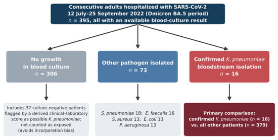
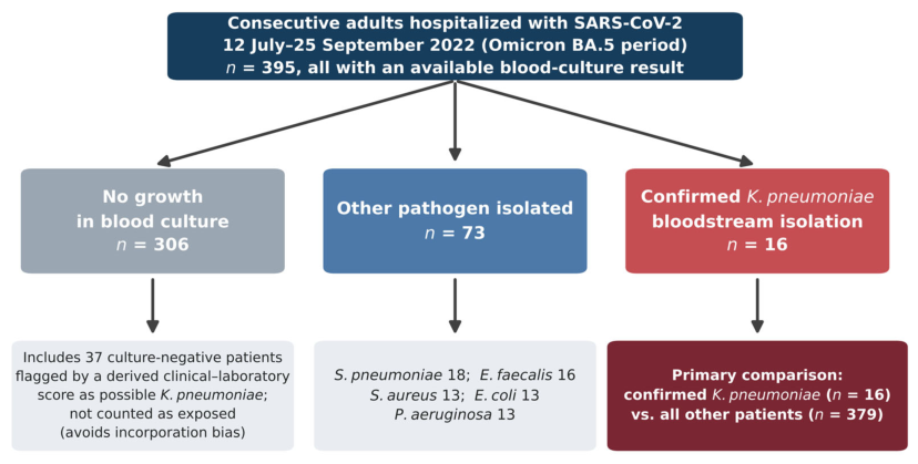
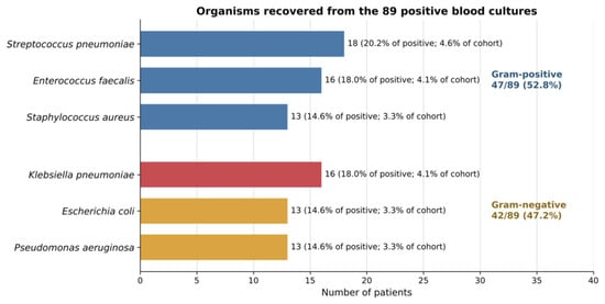
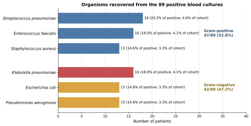
The cohort included 395 adults hospitalized with SARS-CoV-2 infection. Blood cultures showed no growth in 306 patients (77.5%), another identified pathogen in 73 (18.5%), and K. pneumoniae in 16 (4.1%). K. pneumoniae accounted for 16 of 89 positive blood-culture results (18.0%). The other recorded organisms were Streptococcus pneumoniae (n = 18), Enterococcus faecalis (n = 16), Staphylococcus aureus (n = 13), Escherichia coli (n = 13), and Pseudomonas aeruginosa (n = 13). Figure 1 presents the cohort classification and the primary exposure definition.
Figure 1.
Cohort classification and primary exposure definition. The primary exposure required microbiological confirmation of Klebsiella pneumoniae in blood culture. Algorithm-supported, culture-negative classifications were not treated as confirmed infections. The 37 culture-negative patients flagged by the derived score remained in the no-growth group; organisms in the other-pathogen group are listed.
3.2. Baseline and Clinical Characteristics
Median age was similar in patients with and without confirmed K. pneumoniae (72.5 versus 72.0 years; p = 0.744), and 50.0% of confirmed cases were female, as shown in Table 1. Admission SpO2 and the measured inflammatory biomarkers did not differ significantly between groups. Current smoking was recorded in 1 of 16 patients (6.3%) with confirmed K. pneumoniae and in 128 of 374 patients (34.2%) without (Fisher’s exact p = 0.026; Firth unadjusted OR 0.19, 95% CI 0.02–0.76). This difference rests on a single exposed smoker and did not remain significant after Holm correction for the 17 baseline comparisons (adjusted p = 0.416); it is therefore most consistent with a chance finding and is not interpreted as a protective association (Section 4.2). All 16 patients with confirmed K. pneumoniae received antibacterial therapy, compared with 269 of 379 patients (71.0%) without confirmed K. pneumoniae (Fisher’s exact p = 0.008; Holm-adjusted p = 0.136). Because antimicrobial initiation may precede or follow blood-culture collection and treatment details were not time-stamped, this association is descriptive rather than causal.
3.3. Length of Stay
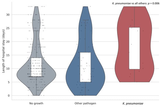
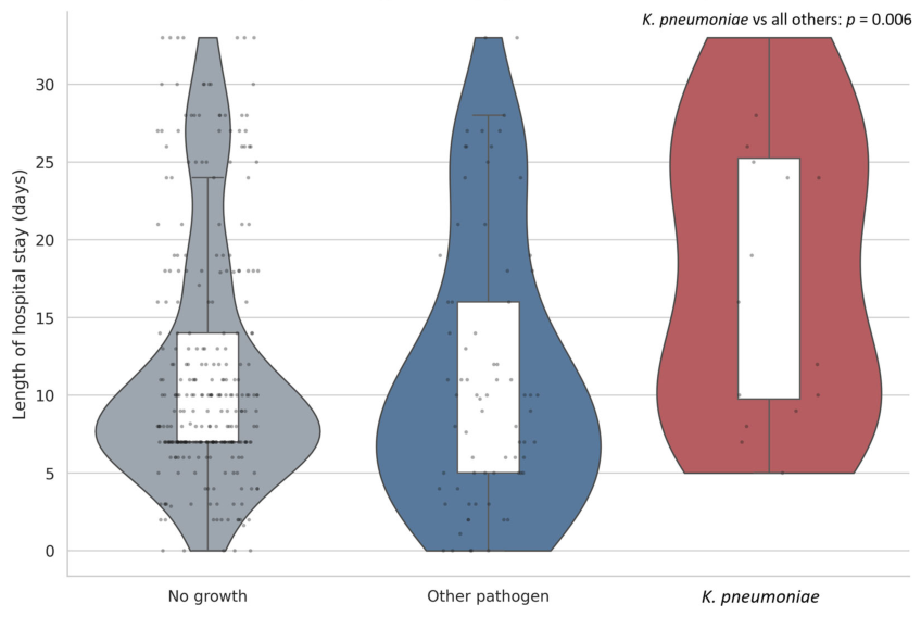
Hospitalization was longer among patients with confirmed K. pneumoniae: median 17.5 days (IQR 9.8–25.2) versus 9.0 days (IQR 7.0–16.0) among all other patients (p = 0.006). A stay of at least 14 days occurred in 9 of 16 patients (56.3%; 95% CI 33.2–76.9%) and 106 of 379 patients (28.0%; 95% CI 23.7–32.7%), corresponding to an unadjusted OR of 3.31 (Firth-corrected OR 3.25, 95% CI 1.22–9.00; Fisher’s exact p = 0.022). When the comparison was restricted to patients with another identified bloodstream pathogen, median length of stay remained longer in the K. pneumoniae group (17.5 versus 10.0 days; p = 0.012), whereas the difference in the binary prolonged-stay endpoint did not reach conventional statistical significance (56.3% versus 31.5%; Fisher’s exact p = 0.085; Table S2). The distribution of length of stay by blood-culture group is shown in Figure 2.
Figure 2.
Hospital length of stay by blood-culture group. Violin shapes show distributions, white boxes show the interquartile range, and points represent individual observations. The displayed p value compares confirmed K. pneumoniae with all other patients using the two-sided Mann–Whitney U test.
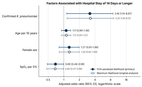
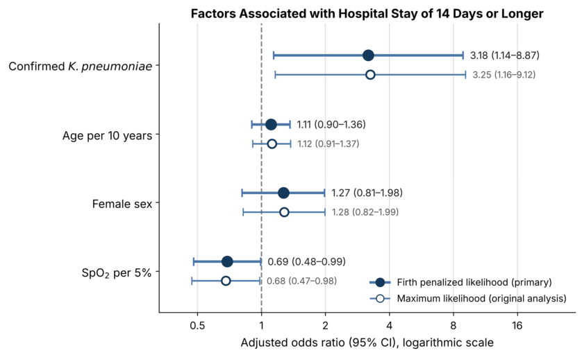
In the prespecified parsimonious multivariable model, which included 395 patients and 115 prolonged-stay events, confirmed K. pneumoniae was associated with higher adjusted odds of hospitalization for at least 14 days after adjustment for age, sex, and admission SpO2 (Firth aOR 3.18, 95% CI 1.14–8.87; p = 0.027); the maximum-likelihood estimate from the original analysis was aOR 3.25 (95% CI 1.16–9.12; p = 0.025). Higher admission SpO2 was inversely associated with prolonged hospitalization (Firth aOR per 5 percentage points 0.69, 95% CI 0.48–0.99; p = 0.041). Full model estimates are reported in Table 2 and displayed in Figure 3. The wide confidence interval around the K. pneumoniae estimate reflects the small number of exposed patients and should temper precision claims. In the post hoc sensitivity model additionally adjusted for current smoking (390 patients), the Firth aOR for confirmed K. pneumoniae was 3.12 (95% CI 1.11–8.75; p = 0.030; Table 2).
Table 2.
Multivariable logistic models for hospitalization of at least 14 days: Firth penalized-likelihood estimates (primary), maximum-likelihood estimates (original analysis), and a sensitivity model additionally adjusted for current smoking.
Figure 3.
Multivariable logistic regression for hospitalization of at least 14 days. Filled points represent Firth penalized-likelihood adjusted odds ratios (primary estimates), open points the corresponding maximum-likelihood estimates, and bars 95% confidence intervals. The model included confirmed K. pneumoniae bloodstream isolation, age, sex, and admission SpO2. CI, confidence interval; SpO2, peripheral oxygen saturation.
3.4. In-Hospital Mortality
In-hospital death occurred in 1 of 16 patients with confirmed K. pneumoniae (6.3%; 95% CI 1.1–28.3%) and 61 of 379 patients without confirmed K. pneumoniae (16.1%; 95% CI 12.7–20.1%). The unadjusted odds ratio was 0.35, but the confidence around the exposed-group proportion was wide and Fisher’s exact p was 0.485 (Table 3 and Table S1). These data do not demonstrate lower mortality; they indicate that the cohort was underpowered for organism-specific mortality inference. The Firth-corrected unadjusted OR was 0.50 (95% CI 0.05–2.07). In the exploratory Firth model adjusted for age and sex, the aOR was 0.48 (95% CI 0.04–2.21); with a single exposed death, this estimate is reported for completeness only.
Table 3.
Clinical outcomes according to blood-culture group.
4. Discussion
4.1. Principal Findings
Three findings emerge from this single-center cohort of the BA.5 period. First, K. pneumoniae was recovered from blood in 4.1% of hospitalized patients and accounted for 18.0% of positive cultures. Second, patients with confirmed isolation stayed considerably longer, by a median of 8.5 days, and had about three times the adjusted odds of a stay of at least 14 days. Third, no excess mortality was observed in the exposed group, but with a single death among 16 patients the estimate is too imprecise for either reassuring or comparative conclusions about mortality.
The finding is therefore best read not as lower mortality with K. pneumoniae, but as a marker of a subgroup that used more hospital resources. The association held after limited adjustment for age, sex, and admission oxygen saturation, yet it remains an association. Without time-stamped cultures, a longer stay may simply provide more opportunity to acquire or detect K. pneumoniae, whereas the infection may itself extend treatment and postpone discharge; the two mechanisms are not mutually exclusive.
4.2. Comparison with Previous Studies
Any positive blood culture was recorded in 22.5% of patients, more than pooled estimates for unselected hospitalized COVID-19 cohorts but within the range reported for severe and intensive care populations [4,5,6,7,8,9]. Comparison is limited because the database does not show whether all patients were sampled in the same way, whether cultures were taken at admission or later, or whether repeated episodes were merged. A 4.1% prevalence of confirmed K. pneumoniae is plausible for a tertiary infectious-diseases hospital, but it should not be extrapolated as a population incidence.
In an Italian COVID-19 hospital, Arcari and colleagues documented transmission of and infection with K. pneumoniae, illustrating how pandemic care pathways interact with resistant Gram-negative bacteria [21]. Clusters of carbapenem-resistant K. pneumoniae were likewise reported among ICU patients with COVID-19, against a background of infection-control strain, broad-spectrum antibiotic use, invasive devices, and prolonged critical care [22,23,24]. Because resistance phenotype could not be determined, our dataset should not be grouped automatically with carbapenem-resistant cohorts. Species identity does not by itself indicate multidrug resistance, and generic class-level labels cannot replace susceptibility testing of individual isolates.
The organism distribution also differed from early-wave intensive care series. In our cohort, Gram-negative bacteria (K. pneumoniae, E. coli, and P. aeruginosa) accounted for 42 of 89 positive cultures (47.2%) and E. faecalis for 16 (18.0%) (Figure 4), whereas Italian ICU cohorts reported Gram-positive predominance, with enterococci causing between one-third and more than one-half of episodes [11,12], and a hospital-wide first-wave cohort found mainly coagulase-negative staphylococci [13]. No coagulase-negative staphylococci were recorded in our dataset, possibly because probable contaminants were not entered as positive results; this could not be verified. Differences in case mix (a hospital-wide, later-wave cohort rather than ICU-only populations), culture practices, and data recording should therefore be considered when comparing pathogen distributions.
Figure 4.
Organisms recovered from the 89 positive blood cultures, grouped by Gram stain. Blue bars indicate Gram-positive organisms and orange bars indicate Gram-negative organisms; the red bar highlights Klebsiella pneumoniae, the focus of this study, which is included in the Gram-negative group. Labels give the number of patients, the percentage of positive cultures, and the percentage of the full cohort (N = 395). The 306 no-growth results are shown in Figure 1.
The lower recorded prevalence of current smoking among patients with confirmed K. pneumoniae (6.3% versus 34.2%) should not be interpreted as a protective association. It depended on a single exposed smoker, did not survive correction for multiple comparisons, lacks a plausible biological rationale in the observed direction, and may reflect chance or incomplete documentation of smoking history in more severely ill patients. Adding smoking to the adjusted model yielded a Firth aOR of 3.12 for confirmed K. pneumoniae, compared with 3.18 in the primary model, indicating that the association with prolonged stay was not materially affected by this imbalance.
Longer hospitalization and more intensive support have also accompanied secondary bloodstream infection in multicenter studies [5,6,7,8,32,33]. Bhatt and colleagues found prolonged admissions in patients with severe COVID-19 complicated by bloodstream infection [6], and ICU cohorts showed that the risk rose with the duration of exposure [7,8,9]. Together, these data support a two-way relationship: severe or protracted illness provides the time and procedures that allow bloodstream infection to occur, and the infection in turn prolongs treatment, monitoring, and recovery.
4.3. Clinical and Stewardship Implications
These results argue for targeted microbiological work-up rather than blanket escalation of antibacterial therapy. Every patient with confirmed K. pneumoniae received antibiotics, but antibacterial treatment was also frequent in patients without it. Studies from earlier in the pandemic consistently found that empirical antibiotic use far exceeded the low prevalence of bacterial co-infection [1,2,3,34], a gap that can accelerate antimicrobial resistance and make subsequent healthcare-associated infections harder to treat.
When a hospitalized patient worsens or fails to improve, reasonable stewardship includes drawing blood cultures before escalating antimicrobials, examining urinary and pulmonary sources, reviewing vascular devices, and de-escalating promptly once microbiology results are available. If K. pneumoniae is isolated, therapy should follow the isolate’s antibiogram, the source and severity of infection, renal function, previous colonization, and local resistance patterns. Our data do not justify any particular regimen and allow no inference about extended-spectrum beta-lactamase production or carbapenem resistance.
The association with length of stay is also relevant to hospital operations. A Gram-negative bloodstream isolate may require repeat cultures, source control, intravenous therapy, infection-control precautions, and a later transfer or discharge. Prospective surveillance should therefore record, alongside the organism, the timing and source of infection, device exposure, appropriateness of antimicrobial therapy, and patient-days at risk. Such data would permit incidence-density analyses and clarify whether infection leads to a longer stay or results from it.
4.4. Methodological Strengths
The study covers a clearly defined late-pandemic period and analyzes the entire cohort rather than a convenience sample. It distinguishes microbiologically confirmed K. pneumoniae from culture-negative cases assigned by an algorithm, which favors exposure specificity and avoids circular analyses of variables built into that score. The model was kept deliberately small and was estimated with Firth’s penalized likelihood to reduce small-sample bias, the mortality analysis was not overfitted, and absolute risks are reported with their uncertainty. Finally, the three-group description separates K. pneumoniae from other positive cultures and from cultures without growth.
4.5. Limitations
These limitations are substantial and determine how far the results can be generalized. First, a retrospective design at a single center is exposed to selection bias, residual confounding, and local culture practices. Second, with only 16 confirmed K. pneumoniae cases, confidence intervals are wide, and neither reliable organism-specific mortality modeling nor extensive covariate adjustment was possible. Firth’s penalized likelihood reduces small-sample bias in the exposure estimate but does not remove residual confounding or the imprecision inherent to 16 exposed patients. Third, culture timing was not available, so we could not separate community-onset from hospital-onset infection, treat K. pneumoniae as a time-dependent exposure, express episodes per 1000 patient-days, or address immortal-time bias. Because K. pneumoniae isolation was analyzed as a fixed exposure although it may have occurred at any point during admission, exposed patients had to remain hospitalized, and alive, long enough for the culture to be drawn and become positive. This immortal-time structure would be expected to inflate the association with prolonged stay and to bias mortality toward an apparently lower risk in the exposed group, and it may partly explain the low mortality observed among exposed patients.
Fourth, the database did not record the indication for culture, the number of sets, repeated cultures, time to positivity, adjudicated source, invasive devices, mechanical ventilation, central-line days, previous colonization, recent hospitalization, timing of antibiotics, or appropriateness of empirical therapy. Fifth, no isolate-level susceptibility results, minimum inhibitory concentrations, resistance mechanisms, or molecular typing were available, and the uniform class-level fields in the database were not analyzed as antibiograms. Consequently, findings from the extensive literature on carbapenem-resistant K. pneumoniae in COVID-19 cannot be extrapolated to this cohort. Sixth, coding was inconsistent in some clinical fields; outcomes were taken from the most internally consistent fields, and unreliably coded variables were excluded rather than corrected through undocumented assumptions. In addition, many baseline comparisons were made, and nominally significant differences that did not survive Holm correction, such as current smoking, are probably due to chance.
Seventh, without a cohort of patients hospitalized without COVID-19, we cannot isolate an effect specific to SARS-CoV-2 or tell whether the prevalence and the association with length of stay differ in patients without COVID-19. Eighth, no viral sequencing was available, so attribution to the BA.5 period rests on the recorded wave dates rather than on patient-level sequencing. Finally, the association between K. pneumoniae and prolonged stay may arise from reverse causation, unmeasured severity, unequal intensity of culture sampling, or healthcare exposure, and it should not be interpreted as a causal effect size.
4.6. Research Priorities
Future prospective, multicenter studies should define bloodstream infection by time-stamped cultures together with clinical criteria; record when cultures were drawn relative to admission and to antimicrobial therapy; document infection source, devices, organ support, and previous colonization; and pair every isolate with a complete susceptibility profile. Incidence should be reported per patient-days at risk, using competing-risk or time-dependent methods where appropriate. Whole-genome sequencing could separate sporadic cases from transmission clusters, and antimicrobial process measures could assess whether empirical and definitive therapy were adequate and timely. Larger samples would also allow adjusted analyses of mortality, recurrence, readmission, and specific resistance phenotypes.
5. Conclusions
In this cohort of 395 adults hospitalized with SARS-CoV-2 infection during the Omicron BA.5 wave, blood cultures confirmed K. pneumoniae in 16 patients (4.1%). Confirmed isolation was associated with a longer hospital stay and, after limited adjustment, with higher odds of a stay of at least 14 days. Increased mortality was not demonstrated, although the exposed group was too small for a firm conclusion. Because culture timing was unavailable, immortal-time bias may partly explain the association with length of stay, and without a non-COVID-19 comparator the association cannot be attributed specifically to SARS-CoV-2. The findings point to a clinically relevant signal of resource use and support prospective, time-resolved microbiological surveillance that integrates infection source, device exposure, appropriateness of antimicrobial therapy, and isolate-level resistance data.
Supplementary Materials
The following supporting information can be downloaded at https://www.mdpi.com/article/10.3390/medicina62112158/s1: Supplementary Methods S1: operational definitions and prespecified analytic decisions; Table S1: distribution of blood-culture findings and clinical outcomes; Table S2: sensitivity comparison with other pathogens; supplementary reporting checklist (STROBE, cohort studies).
Author Contributions
Conceptualization, C.G.B. and F.S.; methodology, C.G.B. and M.D.; software, C.G.B.; validation, C.G.B., F.S. and M.D.; formal analysis, C.G.B.; investigation, C.G.B., F.S. and M.D.; resources, F.S.; data curation, C.G.B. and M.D.; writing—original draft preparation, C.G.B.; writing—review and editing, C.G.B., F.S. and M.D.; visualization, C.G.B.; supervision, F.S.; project administration, C.G.B. and F.S. All authors have read and agreed to the published version of the manuscript.
Funding
This research received no external funding. The article processing charge was covered by the “Victor Babeș” University of Medicine and Pharmacy Timișoara, Romania, which had no role in the design of the study, the collection, analysis, or interpretation of data, the writing of the manuscript, or the decision to publish.
Institutional Review Board Statement
The study complied with the Declaration of Helsinki and was approved by the Ethics Committee of the Clinical Hospital of Infectious Diseases and Pneumophthisiology “Dr. Victor Babeș”, Timișoara, Romania (approval no. 4711/20 May 2026), for the retrospective analysis and publication of anonymized clinical data collected in routine care from 1 March 2020 to 31 December 2024. The approval, which specifies the retrospective design of the study, was granted after data collection had been completed.
Informed Consent Statement
Written informed consent, including consent to the use of anonymized clinical data for research, was obtained from all patients (or their legal representatives) at hospital admission as part of the hospital’s standard admission procedure. The Ethics Committee (approval no. 4711/20 May 2026) waived the requirement for additional study-specific consent for this retrospective analysis of de-identified data.
Data Availability Statement
The de-identified clinical and laboratory data underlying this study can be obtained from the corresponding author on reasonable request, subject to institutional approval, data-sharing policies, and privacy regulations.
Acknowledgments
We thank the “Victor Babeș” University of Medicine and Pharmacy, Timișoara, Romania, for covering the publication costs of this article. The authors reviewed and edited all outputs and take full responsibility for the content of the publication.
Conflicts of Interest
The authors declare no conflicts of interest.
Abbreviations
The following abbreviations are used in this manuscript:
| aOR | adjusted odds ratio |
| CI | confidence interval |
| COVID-19 | coronavirus disease 2019 |
| ICU | intensive care unit |
| IQR | interquartile range |
| OR | odds ratio |
| SARS-CoV-2 | severe acute respiratory syndrome coronavirus 2 |
| SpO2 | peripheral oxygen saturation |
| STROBE | Strengthening the Reporting of Observational Studies in Epidemiology |
References
- Langford, B.J.; So, M.; Raybardhan, S.; Leung, V.; Westwood, D.; MacFadden, D.R.; Soucy, J.-P.R.; Daneman, N. Bacterial co-infection and secondary infection in patients with COVID-19: A living rapid review and meta-analysis. Clin. Microbiol. Infect. 2020, 26, 1622–1629. [Google Scholar] [CrossRef] [Scilit] [PubMed]
- Russell, C.D.; Fairfield, C.J.; Drake, T.M.; Turtle, L.; Seaton, R.A.; Wootton, D.G.; Sigfrid, L.; Harrison, E.M.; Docherty, A.B.; de Silva, T.I.; et al. Co-infections, secondary infections, and antimicrobial use in patients hospitalised with COVID-19 during the first pandemic wave from the ISARIC WHO CCP-UK study: A multicentre, prospective cohort study. Lancet Microbe 2021, 2, e354–e365. [Google Scholar] [CrossRef] [Scilit] [PubMed]
- Alshaikh, F.S.; Godman, B.; Sindi, O.N.; Seaton, R.A.; Kurdi, A. Prevalence of bacterial coinfection and patterns of antibiotics prescribing in patients with COVID-19: A systematic review and meta-analysis. PLoS ONE 2022, 17, e0272375. [Google Scholar] [CrossRef] [Scilit] [PubMed]
- Ippolito, M.; Simone, B.; Filisina, C.; Catalanotto, F.R.; Catalisano, G.; Marino, C.; Misseri, G.; Giarratano, A.; Cortegiani, A. Bloodstream infections in hospitalized patients with COVID-19: A systematic review and meta-analysis. Microorganisms 2021, 9, 2016. [Google Scholar] [CrossRef] [Scilit] [PubMed]
- Giacobbe, D.R.; Battaglini, D.; Ball, L.; Brunetti, I.; Bruzzone, B.; Codda, G.; Crea, F.; De Maria, A.; Dentone, C.; Di Biagio, A.; et al. Bloodstream infections in critically ill patients with COVID-19. Eur. J. Clin. Investig. 2020, 50, e13319. [Google Scholar] [CrossRef] [Scilit] [PubMed]
- Bhatt, P.J.; Shiau, S.; Brunetti, L.; Xie, Y.; Solanki, K.; Khalid, S.; Mohayya, S.; Au, P.H.; Phan, K.; Uprety, P.; et al. Risk factors and outcomes of hospitalized patients with severe COVID-19 and secondary bloodstream infections: A multicenter, case-control study. Clin. Infect. Dis. 2021, 72, e995–e1003. [Google Scholar] [CrossRef] [Scilit] [PubMed]
- Palanisamy, N.; Vihari, N.; Meena, D.S.; Kumar, D.; Midha, N.; Tak, V.; Sharma, A.; Bohra, G.K.; Kothari, N.; Dutt, N.; et al. Clinical profile of bloodstream infections in COVID-19 patients: A retrospective cohort study. BMC Infect. Dis. 2021, 21, 933. [Google Scholar] [CrossRef] [Scilit] [PubMed]
- Grasselli, G.; Scaravilli, V.; Mangioni, D.; Scudeller, L.; Alagna, L.; Bartoletti, M.; Bombino, M.; Cereda, D.; Colombo, A.; Coluccello, A.; et al. Hospital-acquired infections in critically ill patients with COVID-19. Chest 2021, 160, 454–465. [Google Scholar] [CrossRef] [Scilit] [PubMed]
- Buetti, N.; Ruckly, S.; de Montmollin, E.; Reignier, J.; Terzi, N.; Cohen, Y.; Siami, S.; Dupuis, C.; Timsit, J.-F. COVID-19 increased the risk of ICU-acquired bloodstream infections: A case-cohort study from the multicentric OUTCOMEREA network. Intensive Care Med. 2021, 47, 180–187. [Google Scholar] [CrossRef] [Scilit] [PubMed]
- Mateescu, D.-M.; Ilie, A.-C.; Cotet, I.; Guse, C.; Muresan, C.-O.; Pah, A.-M.; Badalica-Petrescu, M.; Iurciuc, S.; Craciun, M.-L.; Avram, A.; et al. Global Burden of Bloodstream Infections in COVID-19: Prevalence, Antimicrobial Resistance, and Mortality Risk. Viruses 2025, 17, 1353. [Google Scholar] [CrossRef] [Scilit] [PubMed]
- Bonazzetti, C.; Morena, V.; Giacomelli, A.; Oreni, L.; Casalini, G.; Galimberti, L.R.; Bolis, M.; Rimoldi, M.; Ballone, E.; Colombo, R.; et al. Unexpectedly high frequency of enterococcal bloodstream infections in coronavirus disease 2019 patients admitted to an Italian ICU: An observational study. Crit. Care Med. 2021, 49, e31–e40. [Google Scholar] [CrossRef] [Scilit] [PubMed]
- Pozza, G.; Casalini, G.; Ciubotariu, C.L.; Giacomelli, A.; Galimberti, M.; Zacheo, M.; Rabbione, A.; Pieruzzi, M.; Oreni, L.; Galimberti, L.; et al. Bloodstream infections in intensive care unit during four consecutive SARS-CoV-2 pandemic waves. Antibiotics 2023, 12, 1448. [Google Scholar] [CrossRef] [Scilit] [PubMed]
- Ripa, M.; Galli, L.; Poli, A.; Oltolini, C.; Spagnuolo, V.; Mastrangelo, A.; Muccini, C.; Monti, G.; De Luca, G.; Landoni, G.; et al. Secondary infections in patients hospitalized with COVID-19: Incidence and predictive factors. Clin. Microbiol. Infect. 2021, 27, 451–457. [Google Scholar] [CrossRef] [Scilit] [PubMed]
- Massart, N.; Maxime, V.; Fillatre, P.; Razazi, K.; Ferré, A.; Moine, P.; Legay, F.; Voiriot, G.; Amara, M.; Santi, F.; et al. Characteristics and prognosis of bloodstream infection in patients with COVID-19 admitted in the ICU: An ancillary study of the COVID-ICU study. Ann. Intensive Care 2021, 11, 183. [Google Scholar] [CrossRef] [Scilit] [PubMed]
- Wang, J.; Jiang, T. Risk factors for bloodstream infection in COVID-19 patients in intensive care units: A systematic review and meta-analysis. BMC Infect. Dis. 2025, 25, 13. [Google Scholar] [CrossRef] [Scilit] [PubMed]
- Kariyawasam, R.M.; Julien, D.A.; Jelinski, D.C.; Larose, S.L.; Rennert-May, E.; Conly, J.M.; Dingle, T.C.; Chen, J.Z.; Tyrrell, G.J.; Ronksley, P.E.; et al. Antimicrobial resistance in COVID-19 patients: A systematic review and meta-analysis. Antimicrob. Resist. Infect. Control 2022, 11, 45. [Google Scholar] [CrossRef] [Scilit] [PubMed]
- Baker, M.A.; Sands, K.E.; Huang, S.S.; Kleinman, K.; Septimus, E.J.; Varma, N.; Blanchard, J.; Poland, R.E.; Coady, M.H.; Yokoe, D.S.; et al. The impact of coronavirus disease 2019 (COVID-19) on healthcare-associated infections. Clin. Infect. Dis. 2022, 74, 1748–1754. [Google Scholar] [CrossRef] [Scilit] [PubMed]
- Langford, B.J.; So, M.; Simeonova, M.; Leung, V.; Lo, J.; Kan, T.; Raybardhan, S.; Sapin, M.E.; Mponponsuo, K.; Farrell, A.; et al. Antimicrobial resistance in patients with COVID-19: A systematic review and meta-analysis. Lancet Microbe 2023, 4, e179–e191. [Google Scholar] [CrossRef] [Scilit] [PubMed]
- Monaco, M.; Floridia, M.; Giuliano, M.; Palmieri, L.; Noce, C.L.; Pantosti, A.; Palamara, A.T.; Brusaferro, S.; Onder, G.; Italian National Institute of Health COVID-19 Mortality Group. Hospital-acquired bloodstream infections in patients deceased with COVID-19 in Italy (2020–2021). Front. Med. 2022, 9, 1041668. [Google Scholar] [CrossRef] [Scilit] [PubMed]
- Mateescu, D.-M.; Toma, A.-O.; Gavrilescu, D.-M.; Ilie, A.-C.; Florescu, E.; Rosca, O.; Oancea, C.; Iurciuc, S.; Muresan, C.-O.; Enache, A. Patterns of Antibiotic Use in Hospitalized COVID-19 Patients and Association with Secondary Infections and Outcomes. Antibiotics 2026, 15, 240. [Google Scholar] [CrossRef] [Scilit] [PubMed]
- Arcari, G.; Raponi, G.; Sacco, F.; Bibbolino, G.; Di Lella, F.M.; Alessandri, F.; Coletti, M.; Trancassini, M.; Deales, A.; Pugliese, F.; et al. Klebsiella pneumoniae infections in COVID-19 patients: A 2-month retrospective analysis in an Italian hospital. Int. J. Antimicrob. Agents 2021, 57, 106245. [Google Scholar] [CrossRef] [Scilit] [PubMed]
- Montrucchio, G.; Corcione, S.; Sales, G.; Curtoni, A.; De Rosa, F.G.; Brazzi, L. Carbapenem-resistant Klebsiella pneumoniae in ICU-admitted COVID-19 patients: Keep an eye on the ball. J. Glob. Antimicrob. Resist. 2020, 23, 398–400. [Google Scholar] [CrossRef] [Scilit] [PubMed]
- Dumitru, I.M.; Dumitrascu, M.; Vlad, N.D.; Cernat, R.C.; Ilie-Serban, C.; Hangan, A.; Slujitoru, R.E.; Gherghina, A.; Mitroi-Maxim, C.; Curtali, L.; et al. Carbapenem-resistant Klebsiella pneumoniae associated with COVID-19. Antibiotics 2021, 10, 561. [Google Scholar] [CrossRef] [Scilit] [PubMed]
- Falcone, M.; Tiseo, G.; Galfo, V.; Giordano, C.; Leonildi, A.; Marciano, E.; De Simone, P.; Biancofiore, G.; Boggi, U.; Barnini, S.; et al. Bloodstream infections in patients with rectal colonization by Klebsiella pneumoniae producing different type of carbapenemases (CHIMERA study). Clin. Microbiol. Infect. 2022, 28, 298.e1–298.e7. [Google Scholar] [CrossRef] [Scilit] [PubMed]
- Nyberg, T.; Ferguson, N.M.; Nash, S.G.; Webster, H.H.; Flaxman, S.; Andrews, N.; Hinsley, W.; Bernal, J.L.; Kall, M.; Bhatt, S.; et al. Comparative analysis of the risks of hospitalisation and death associated with SARS-CoV-2 omicron (B.1.1.529) and delta (B.1.617.2) variants in England: A cohort study. Lancet 2022, 399, 1303–1312. [Google Scholar] [CrossRef] [Scilit] [PubMed]
- Tegally, H.; Moir, M.; Everatt, J.; Giovanetti, M.; Scheepers, C.; Wilkinson, E.; Subramoney, K.; Makatini, Z.; Moyo, S.; Amoako, D.G.; et al. Emergence of SARS-CoV-2 Omicron lineages BA.4 and BA.5 in South Africa. Nat. Med. 2022, 28, 1785–1790. [Google Scholar] [CrossRef] [Scilit] [PubMed]
- von Elm, E.; Altman, D.G.; Egger, M.; Pocock, S.J.; Gotzsche, P.C.; Vandenbroucke, J.P.; STROBE Initiative. The Strengthening the Reporting of Observational Studies in Epidemiology statement: Guidelines for reporting observational studies. Lancet 2007, 370, 1453–1457. [Google Scholar] [CrossRef] [Scilit] [PubMed]
- Bujor, C.G.; Dinuti, M.; Sfrijan, F. Carbapenem-Resistant Acinetobacter baumannii Isolation and In-Hospital Mortality in Adults Hospitalized with COVID-19 in Romania: A Retrospective Single-Center Cohort Study. J. Clin. Med. 2026, 15, 8093. [Google Scholar] [CrossRef] [Scilit] [PubMed]
- Holm, S. A simple sequentially rejective multiple test procedure. Scand. J. Stat. 1979, 6, 65–70. [Google Scholar]
- Firth, D. Bias reduction of maximum likelihood estimates. Biometrika 1993, 80, 27–38. [Google Scholar] [CrossRef]
- Heinze, G.; Schemper, M. A solution to the problem of separation in logistic regression. Stat. Med. 2002, 21, 2409–2419. [Google Scholar] [CrossRef] [Scilit] [PubMed]
- Protonotariou, E.; Mantzana, P.; Meletis, G.; Tychala, A.; Kassomenaki, A.; Vasilaki, O.; Kagkalou, G.; Gkeka, I.; Archonti, M.; Kati, S.; et al. Microbiological characteristics of bacteremias among COVID-19 hospitalized patients in a tertiary referral hospital in Northern Greece during the second epidemic wave. FEMS Microbes 2021, 2, xtab021. [Google Scholar] [CrossRef] [Scilit] [PubMed]
- Costa, R.L.D.; Lamas, C.C.; Simvoulidis, L.F.N.; Espanha, C.A.; Moreira, L.P.M.; Bonancim, R.A.B.; Weber, J.V.; Ramos, M.R.F.; Silva, E.C.F.; Oliveira, L.P.; et al. Secondary infections in a cohort of patients with COVID-19 admitted to an intensive care unit: Impact of Gram-negative bacterial resistance. Rev. Inst. Med. Trop. Sao Paulo 2022, 64, e6. [Google Scholar] [CrossRef] [Scilit] [PubMed]
- Afzal, A.; Gutierrez, V.P.; Gomez, E.; Mon, A.M.; Sarmiento, C.M.; Khalid, A.; Echevarria, K.; Gratrix, A.; Rizzo, V. Bloodstream infections in hospitalized patients before and during the COVID-19 surge in a community hospital in the South Bronx. Int. J. Infect. Dis. 2022, 116, 43–46. [Google Scholar] [CrossRef] [Scilit] [PubMed]

Disclaimer/Publisher’s Note: The statements, opinions and data contained in all publications are solely those of the individual author(s) and contributor(s) and not of MDPI and/or the editor(s). MDPI and/or the editor(s) disclaim responsibility for any injury to people or property resulting from any ideas, methods, instructions or products referred to in the content. |
© 2026 by the authors. Published by MDPI on behalf of the Lithuanian University of Health Sciences. Licensee MDPI, Basel, Switzerland. This article is an open access article distributed under the terms and conditions of the Creative Commons Attribution (CC BY) license.