


Abstract
Background and Objectives: Mortality following percutaneous endoscopic gastrostomy (PEG) may reflect illness severity, nutritional impairment, systemic inflammation, and patient selection. The prognostic value of routinely available indices, including the Prognostic Nutritional Index (PNI) and C-reactive protein-to-albumin ratio (CAR), remains uncertain in critically ill patients undergoing PEG. Materials and Methods: This retrospective cohort analyzed 93 adult intensive care patients who underwent PEG, with one index PEG retained per patient. Pre-PEG feeding and swallowing context, functional dependence, clinical-procedure consent documentation, and the availability of decision-relevant fields were described from retrospective hospital records. The primary outcome was date-defined 90-day all-cause mortality, calculated from the calendar-day difference between recorded PEG and death dates, with PEG placement defined as day 0; 30-day mortality was secondary. Separate multivariable logistic models included age, sex, APACHE II, and either PNI or log2-transformed CAR. Results: Date-derived death occurred within 30 days in 25/93 patients (26.9%; 95% CI, 18.9–36.7). Among 92 patients classifiable at 90 days from the available date fields, 45 deaths occurred by day 90 (48.9%; 95% CI, 38.9–59.0), and 47 patients had no recorded death through day 90; one patient with a last-known-alive date at day 81 and no recorded death was not classifiable for the 90-day endpoint. APACHE II was associated with 90-day death in both models (PNI model OR per 5 points, 1.95; 95% CI, 1.17–3.26; p = 0.011; CAR model OR, 1.96; 95% CI, 1.17–3.27; p = 0.011), whereas PNI and CAR were not statistically significantly associated. In the CAR model, age was also associated with 90-day death (OR per 10 years, 1.36; 95% CI, 1.02–1.81; p = 0.039). Primary PEG indications were predominantly acute or chronic neurological dysphagia (73.1%) and prolonged mechanical ventilation or ICU stay (18.3%), with dysphagia due to dementia recorded as the primary indication in only one patient (1.1%, alive at 90 days); all 93 patients had documented swallowing assessments and signed procedure consent after a median of 23 days of nasogastric/nasojejunal feeding. Among the 45 deaths by day 90, recorded causes were sepsis, septic shock, or multiorgan dysfunction in 32 (71.1%), acute respiratory failure in 6 (13.3%), cardiovascular/cardiorenal causes in 4 (8.9%), and cerebrovascular causes in 3 (6.7%), with no recorded deaths directly attributed to the PEG procedure (0/93) and four acute or subacute post-PEG events descriptively noted. Conclusions: In this retrospective intensive care cohort, higher APACHE II scores were associated with date-defined 90-day all-cause mortality. PNI and CAR were not statistically significantly associated with 90-day mortality in separate adjusted models. These findings describe all-cause outcomes following PEG and do not establish mortality attributable to PEG or the appropriateness of individual PEG decisions.
1. Introduction
Percutaneous endoscopic gastrostomy (PEG) provides durable gastric access for enteral nutrition, hydration, and medication administration in patients who cannot meet their requirements orally despite having a functional gastrointestinal tract. Neurological dysphagia, head and neck or upper gastrointestinal malignancies, and prolonged critical illness are common settings in which PEG is considered. Current European guidance supports temporary transnasal feeding when enteral support is expected to be short-term and consideration of percutaneous access when longer support is anticipated [1,2]. In critically ill patients, however, technical feasibility alone does not establish likely benefit; the decision also requires evaluation of prognosis, reversibility of the underlying condition, treatment goals, and patient preferences.
Early all-cause mortality following PEG remains clinically important and varies markedly across populations. A national English cohort of 87,862 patients reported an overall 30-day mortality of 8.9%, with improvement over time [3]. A Swedish multicentre cohort reported mortality of 15% at 30 days and 28% at 90 days [4], whereas other hospital and intensive care series have documented wide variations in early mortality and procedural adverse events depending on underlying neurological, malignant, or critical-illness indications [5,6,7,8]. These figures should not be interpreted as direct comparisons of procedural performance because PEG indications, underlying disease burden, critical-care exposure, patient-selection practices, and outcome ascertainment differ. Mortality is therefore best described as all-cause mortality following PEG rather than mortality caused by the procedure.
Nutritional and inflammatory variables have been investigated as pragmatic risk markers in PEG candidates. In prospective and retrospective cohorts, hypoalbuminemia combined with elevated C-reactive protein (CRP) has been associated with early post-PEG mortality and morbidity [7,8,9,10]. The Prognostic Nutritional Index (PNI), originally derived from serum albumin and total lymphocyte count [11], has also been evaluated; PNI below 37 was associated with early mortality in one cohort [12]. Similarly, higher CRP-to-albumin ratio (CAR) independently predicted complications and 30-day mortality in another study [13]. Findings have not been uniform, however, and a recent 90-day cohort found that nutritional indices did not consistently retain independent associations after adjustment [5]. Study-specific cutoffs should therefore not be assumed to be universal.
The Acute Physiology and Chronic Health Evaluation II (APACHE II) score offers a complementary assessment of global illness severity by integrating acute physiological abnormalities, age, and chronic health status [14]. Studies of surgical and medical ICU cohorts have reported associations between illness severity or organ dysfunction and mortality after PEG [6,15]. In a recent cohort of 487 intensive care patients receiving PEG, APACHE II above 20 and PNI below 33 were independently associated with mortality [15]. Evidence directly comparing physiological severity with nutritional-inflammatory indices for a fixed 90-day outcome is heterogeneous, including recent intensive care cohorts [5,15]. The primary aim of this small exploratory study was to describe date-derived 90-day all-cause mortality following PEG in intensive care patients and examine its associations with APACHE II, PNI, and CAR. Secondary objectives were to describe 30-day mortality and recorded post-PEG events and to explore the discriminatory performance of routinely available severity and nutritional-inflammatory indices.
2. Materials and Methods
2.1. Study Design and Reporting
This retrospective cohort study evaluated fixed-horizon 30-day and 90-day all-cause mortality after PEG among adult intensive care patients treated at Muş State Hospital. Data were retrospectively abstracted from hospital electronic records, including intensive care, endoscopy, consultation, discharge, and available in-hospital follow-up documentation. ICU admission dates ranged from 15 January 2021 to 30 September 2025, and index-PEG dates ranged from 26 February 2021 to 17 October 2025. Reporting was structured according to the STROBE recommendations for observational cohort studies [16].
2.2. Participants
The analytic cohort comprised 93 adult intensive care patients who underwent PEG. One index PEG per patient was retained. All patients had complete core baseline covariates and a recorded index-PEG date.
2.3. Available Pre-PEG Clinical Context
Available pre-PEG contextual data were retrospectively abstracted from hospital documentation into the study dataset. They included the primary PEG indication and specialist consultation; enteral feeding route and duration; the specific rationale for discontinuing nasogastric (NG) or nasojejunal (NJ) feeding; swallowing-assessment timing, modality, and findings, including fiberoptic endoscopic evaluation of swallowing (FEES) or bedside clinical assessment; consciousness, Glasgow Coma Scale (GCS), and sedation notes; functional status and care dependence; background comorbidity labels; and signed clinical-procedure written informed consent, including its date and the signer’s relationship to the patient. Baseline clinical and laboratory characteristics are summarized in Table 1; detailed pre-PEG categories are presented in Table 2. These variables were used descriptively and were not added to the existing regression models.
Primary PEG indications were distinguished from background comorbidity labels. The underlying comorbidity entries were “dementia” and “malignancy,” without documented dementia stage or oncological terminal-stage classification in those fields. These fields were therefore treated as unstaged comorbidity labels rather than confirmed advanced dementia or terminal malignancy. The separate recorded label “irreversible organ failure” encompassed chronic renal failure and/or heart failure without a standardized terminal-prognosis assessment. Severe dysphagia due to dementia was recorded as a primary PEG indication in one patient.
Multidisciplinary ICU, neurology, and surgical consultation records, swallowing assessments, and signed clinical-procedure written informed consent were documented in all 93 patients. The hospital electronic health record system (HBYS), however, did not provide separate structured electronic fields for quantitative life-expectancy estimates or formal palliative-care narratives (0/93 structured fields available for analysis). This describes the structure of the available electronic data, not the frequency with which clinicians considered prognosis or discussed care. Formal capacity assessment, legal representative authority, neurological prognosis or rehabilitation expectations, individualized benefit–burden deliberation, quantified expected duration of feeding, patient preferences, care goals, and treatment-limitation decisions were not available as standardized analytic variables. We did not infer these constructs from GCS, dependence, comorbidity labels, consultation presence, or consent signatures.
2.4. Data Sources and Variables
The de-identified analytic dataset included age, sex, airway-support status, days from ICU admission to index PEG, PEG indication, comorbidity labels, SOFA, APACHE II, albumin, CRP, absolute lymphocyte count (ALC), recorded PEG, death, last-known-alive dates, and post-PEG event and tube-related notes. Albumin, CRP, and ALC were measured 24.4–32.4 h before PEG (median, 28.5 h; IQR, 26.3–30.3 h). APACHE II was recorded on the calendar day before PEG, but time of day was unavailable; SOFA was available as a summary score without a time stamp. No post-PEG laboratory measurements were used in the analyses.
PNI was calculated as 10 × serum albumin (g/dL) + 5 × ALC (109/L). CAR was calculated as CRP (mg/L) divided by serum albumin (g/dL); reported numerical values retain the original (mg/L)/(g/dL) scale. Conversion to mg/g would require division of these numerical values by 10; no rescaling was applied. Because stored CAR values were not fully consistent with the corresponding recorded CRP and albumin values, all CAR analyses used values recalculated directly from recorded CRP and albumin. PNI < 40 was used only for descriptive characterization and was not entered into regression models.
2.5. Outcomes
The primary outcome was all-cause mortality by 90 days after index PEG, with the PEG date defined as day 0. Outcomes were classified using calendar-day differences between recorded PEG and death dates. A patient was classifiable at 90 days if death occurred by day 90, if the last-known-alive date was at least 90 days after PEG, or if a later recorded death date established survival through day 90. Transfer or discharge alone was not treated as evidence of survival through day 90. One patient had a last-known-alive date at day 81 without a recorded death and was not classifiable for the 90-day endpoint. The secondary outcome was 30-day all-cause mortality, derived using the same date-based definition. Outcome dates were retrospectively abstracted from hospital records; no linkage to an external vital-status registry was available. These were therefore date-derived outcome classifications based on documented hospital-record dates rather than independently verified external vital-status determinations.
Post-PEG events were abstracted from event fields in the study worksheet. Four acute or subacute events were recorded. A retrospective source note supplied simple relationship labels for these events, but no prespecified attribution or adjudication framework was available; the labels are presented descriptively and do not establish causation. The worksheet also contained 15 heterogeneous tube-related free-text entries. Their timing, indication, and procedure type were not systematically recorded; therefore, they were not classified as revision/replacement events or included in the acute-event total.
Recorded causes of death were additionally described from the cause-of-death field. The recorded labels were grouped as sepsis, septic shock, and multiorgan dysfunction; acute respiratory failure; cardiovascular and cardiorenal causes; or cerebrovascular causes, retaining every specific label within its parent category. Each death contributed once to a specific cause category and once to its parent subtotal. Distributions were tabulated for deaths within 30 days (n = 25), deaths on days 31–90 (n = 20), all date-defined deaths by day 90 (n = 45), and all recorded deaths (n = 46), including one death on day 106. Percentages used the number of deaths in the corresponding interval as the denominator. Procedure-related mortality was reported separately as the number of deaths explicitly attributed in the available records to PEG, including peritonitis, perforation, or major bleeding directly attributable to the procedure, divided by all 93 recipients. These classifications were descriptive; no independent causal adjudication, competing-risk analysis, or change to the fixed-horizon mortality models was undertaken.
2.6. Data Quality Checks
Before analysis, data for the 93-patient cohort were checked for completeness, range validity, formula consistency, and internal date reconciliation. Age, sex, APACHE II, SOFA, albumin, CRP, ALC, PNI, recalculated CAR, and the recorded PEG, death, and last-known-alive date fields were available for all patients; a blank death-date field denoted no recorded death rather than a missing baseline variable. The retrospective pre-PEG contextual fields were also populated for all patients. The final 30-day and 90-day classifications were calculated from exact recorded date fields; one patient with a last-known-alive date at day 81 and no recorded death was not classifiable for the 90-day endpoint. No imputation was performed.
2.7. Sample Size
No a priori sample-size calculation was performed because the analysis used the 93-patient cohort. With 45 date-defined 90-day deaths, each multivariable model was limited to four prespecified predictors: age, sex, APACHE II, and either PNI or CAR.
2.8. Statistical Analysis
Continuous variables are summarized as median and interquartile range (IQR), and categorical variables as count and percentage. Baseline characteristics were compared between patients with no recorded death through day 90 and patients with date-defined death by day 90, among the 92 patients classifiable at 90 days from available date fields, using the Mann–Whitney U test for continuous variables and Fisher’s exact test for categorical variables. Proportions are accompanied by Wilson 95% confidence intervals.
Date-defined 90-day all-cause death was modeled among the 92 patients classifiable at 90 days from available date fields using separate multivariable logistic regressions. Model 1 included age, sex, APACHE II, and PNI; Model 2 included age, sex, APACHE II, and log2-transformed recalculated CAR. Continuous predictors were scaled as age per 10 years, APACHE II per 5 points, PNI per 5 points, and CAR per doubling. PNI and CAR were modeled separately because both include albumin.
Adjusted odds ratios are reported with Wald 95% confidence intervals and two-sided p-values. Apparent discrimination was summarized with the area under the receiver operating characteristic curve (AUC), and overall prediction error with the Brier score. No resampling-based internal validation, external validation, or data-derived clinical cutoff analysis was performed. No multivariable model was fitted for post-PEG events because only four acute or subacute events were recorded.
All analyses were performed using Python version 3.11 (Python Software Foundation, Wilmington, DE, USA). Data processing used pandas version 2.2 and NumPy version 1.26. Mann–Whitney U tests and two-sided Fisher’s exact tests were performed using scipy.stats (SciPy version 1.12). Wilson 95% confidence intervals and multivariable logistic regression models (Logit) were implemented using statsmodels version 0.14. The area under the receiver operating characteristic curve (AUC) and Brier scores were calculated using scikit-learn version 1.4.
2.9. Ethics
An ethics committee decision covering the retrospective 90-day mortality analysis was issued by the Muş Alparslan University Scientific Research and Publication Ethics Board (Decision 43; meeting decision no. 87; 11 February 2026; electronic document no. 234682). Institutional site permission was granted by the Muş Provincial Health Directorate on 27 February 2026 (reference E-35465298-619-306452106).
3. Results
3.1. Cohort Description and Data Completeness
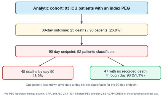
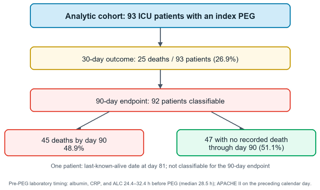
The analytic cohort comprised 93 adult intensive care patients undergoing PEG (Figure 1). Core baseline severity scores and laboratory variables, as well as PEG, death, and last-known-alive date fields, were available for all patients. Figure 1 summarizes the cohort, timing of pre-PEG laboratory measurements, and date-derived outcomes.
Figure 1.
Analytic cohort (n = 93), timing of recorded pre-PEG laboratory measures, and date-derived 30-day and 90-day outcomes. Blue indicates the analytic cohort, yellow the 30-day outcome, red the 90-day endpoint and deaths, and green patients with no recorded death through day 90.
3.2. Baseline Characteristics
Among the 93 patients, the median age was 76.0 years (IQR, 63.0–83.0), and 41 patients (44.1%) were male. Thirty-five patients (37.6%) were receiving advanced airway support at the time of PEG. The median interval from ICU admission to index PEG was 24.0 days (IQR, 11.0–45.0). Median SOFA was 4.0 (IQR, 3.0–6.0) and median APACHE II was 15.0 (IQR, 12.0–17.0). Median albumin was 2.65 g/dL (IQR, 2.30–3.00), recalculated CAR was 18.33 (IQR, 8.96–35.71), and PNI was 33.70 (IQR, 29.60–39.90). Baseline comparisons in the 92 patients classifiable at 90 days from available date fields are shown in Table 1. Compared with patients with no recorded death through day 90, patients with date-defined death by day 90 were older, had higher APACHE II scores, lower albumin, lower PNI, and a higher prevalence of PNI < 40.
Table 1.
Baseline clinical and laboratory characteristics among patients classifiable at 90 days from available date fields (n = 92).
The expanded pre-PEG context is shown in Table 2. Primary indications were post-stroke dysphagia in 34 patients (36.6%); hypoxic-ischemic brain injury and traumatic brain injury in 10 each (10.8% each); dysphagia after prolonged mechanical ventilation in 9 (9.7%); dysphagia due to prolonged ICU stay and head and neck malignancy-related dysphagia in 8 each (8.6% each); ALS in 6 (6.5%); Parkinson’s disease-related dysphagia in 5 (5.4%); other neurodegenerative disease-related dysphagia in 2 (2.2%); and severe dysphagia due to dementia in 1 (1.1%). The corresponding 30-day death counts were 14, 1, 1, 2, 2, 2, 2, 1, 0, and 0. The 90-day deaths/classifiable patients were 19/34, 5/10, 6/10, 4/9, 4/8, 3/7, 2/6, 2/5, 0/2, and 0/1, respectively. The patient whose primary indication was dementia-related dysphagia survived beyond 90 days.
Table 2.
Available pre-PEG clinical-context data (n = 93). Panel A. Primary PEG indications and date-defined mortality. Panel B. Feeding, swallowing, neurological, functional, and consent details. Panel C. Background comorbidity labels and electronic-record structure.
Background labels were dementia in 33/93 patients (35.5%), malignancy in 6/93 (6.5%), and “irreversible organ failure” in 3/93 (3.2%). These were distinct from the primary indication field. Dementia and malignancy were unstaged recorded comorbidity labels rather than confirmed advanced or terminal conditions; the organ-failure label also did not establish a terminal prognosis. Their frequencies must not be interpreted as the numbers undergoing PEG primarily for advanced dementia, terminal cancer, or end-stage organ failure.
Before PEG, 80/93 patients (86.0%) received NG feeding and 13/93 (14.0%) received NJ feeding for a median of 23 days (IQR, 10–44 days). Persistent severe dysphagia requiring prolonged access or inability to achieve safe or adequate oral intake accounted for transition in 72/93 (77.4%): anticipated prolonged enteral nutrition in 31 (33.3%), inadequate intake due to severe dysphagia in 21 (22.6%), and inability to achieve safe oral intake in 20 (21.5%). NG/NJ intolerance or mechanical failure accounted for 21/93 (22.6%): recurrent dislodgement in 7 (7.5%), intolerance in 6 (6.5%), inability to maintain transnasal feeding in 6 (6.5%), and recurrent occlusion in 2 (2.2%).
Swallowing assessments were documented in all 93 patients a median of 3 days before PEG. FEES was performed in 52 (55.9%), documenting thin-liquid aspiration in 11 (11.8%), pharyngeal residue with aspiration in 11 (11.8%), secretion pooling with aspiration in 10 (10.8%), silent aspiration in 10 (10.8%), and overt aspiration during swallowing in 10 (10.8%). Bedside clinical assessment was performed in 41 (44.1%), documenting marked oropharyngeal dysphagia with unsafe oral intake in 11 (11.8%), coughing and choking during swallowing in 10 (10.8%), impaired swallowing coordination in 10 (10.8%), and severe clinical dysphagia with inadequate intake in 10 (10.8%). Thus, all 93 patients had a documented pre-procedural assessment confirming severe swallowing impairment or aspiration risk prior to PEG placement.
Recorded pre-PEG neurological categories were confusion/disorientation or following simple/partial commands (GCS 13–15) in 27/93 (29.0%); somnolence with eye-opening or verbal response (GCS 10–12 or E3–4 V-NT M5) in 26/93 (28.0%); decreased consciousness responsive to verbal/painful stimuli (GCS 12 or E2 V-NT M4) in 21/93 (22.6%); and stupor or response only to painful stimuli (GCS 7–8 or E2 V-NT M5) in 13/93 (14.0%). Six patients (6.5%) were intubated under continuous intravenous sedation/analgesia with remifentanil, fentanyl, and/or midazolam, precluding GCS evaluation. These recorded categories were not used to infer capacity or long-term neurological prognosis.
Total care dependence was recorded in 73/93 patients (78.5%): 38 (40.9%) were bedridden without active mobilization or required positional assistance; 26 (28.0%) were bedridden with passive mobilization, limited limb movement, or total assistance with activities of daily living; and 9 (9.7%) had severe functional loss requiring full mobilization support. Severe care dependence was recorded in 20/93 (21.5%), comprising 10 (10.8%) requiring two-person assistance for sitting or transfer and 10 (10.8%) with severe mobility limitation.
Multidisciplinary ICU, neurology, and surgical consultations and signed clinical-procedure written informed consent were documented in 93/93 patients (100%). Consent was signed a median of 2 days before PEG (range, 1–3 days), by a spouse in 35 (37.6%), a son in 26 (28.0%), a daughter in 26 (28.0%), and the patient in 6 (6.5%). Separate structured HBYS fields for quantitative life expectancy or formal palliative-care narratives were unavailable (0/93); this was not an absence of the documented consultations, swallowing assessments, or signed consent forms. The contents needed to judge formal capacity, representative authority, detailed goals-of-care deliberation, and individual benefit were not available as standardized analytic variables.
3.3. Mortality Outcomes and Recorded Post-PEG Events
Date-derived death occurred within 30 days in 25/93 patients (26.9%; 95% CI, 18.9–36.7). Among 92 patients classifiable at 90 days from available date fields, 45 deaths occurred by day 90 (48.9%; 95% CI, 38.9–59.0) and 47 patients had no recorded death through day 90 (51.1%; 95% CI, 41.0–61.1) (Table 3). One patient with a last-known-alive date at day 81 and no recorded death was not classifiable for the 90-day endpoint. Four acute or subacute post-PEG events were recorded, three within 30 days. Other heterogeneous tube-related notes were retained separately and were not counted as acute/subacute events.
Table 3.
Mortality, recorded causes of death, and post-PEG events.
Across all available follow-up, 46 deaths were recorded: 25 within 30 days, 20 on days 31–90, and one on day 106. The day-106 death does not increase the 90-day death count; it establishes survival through day 90 for that patient. The causes-of-death panel of Table 3 separates these intervals and retains all recorded cause labels. Among the 45 deaths by day 90, 32 (71.1%) were recorded as sepsis, septic shock, or multiorgan dysfunction; 6 (13.3%) as acute respiratory failure; 4 (8.9%) as cardiovascular or cardiorenal causes; and 3 (6.7%) as cerebrovascular causes. Corresponding distributions among deaths within 30 days were 18/25 (72.0%), 4/25 (16.0%), 2/25 (8.0%), and 1/25 (4.0%), and on days 31–90 were 14/20 (70.0%), 2/20 (10.0%), 2/20 (10.0%), and 2/20 (10.0%).
The additional day-106 death was recorded as sepsis and multiorgan failure, giving 33/46 (71.7%) sepsis-related, 6/46 (13.0%) respiratory, 4/46 (8.7%) cardiovascular/cardiorenal, and 3/46 (6.5%) cerebrovascular deaths across all recorded follow-up. No death was attributed in the available records to PEG-related peritonitis, perforation, or major bleeding, and no other directly PEG-attributed death was recorded: 0/93 (0.0%). The four acute or subacute post-PEG events remain reported separately in Table 4.
Table 4.
Recorded details and retrospective source-note relationship labels for four acute or subacute post-PEG events.
Date-derived mortality estimates use recorded PEG and death dates, with PEG placement defined as day 0. The 90-day analysis includes only patients classifiable at 90 days from available date fields. Confidence intervals are Wilson intervals.
Values are n (%), with percentages calculated within each death column, not among all PEG recipients. The ≤30-day and days 31–90 columns are disjoint; together they comprise all 45 date-defined deaths by day 90. The all-recorded-deaths column additionally includes one death on post-PEG day 106, recorded as sepsis and multiorgan failure. That patient remains classified as alive through day 90. Parent rows are subtotals and must not be summed with their component rows. Recorded deaths directly attributed to PEG (including peritonitis, perforation, or major bleeding directly attributable to PEG): 0/93 (0.0%). ARDS, acute respiratory distress syndrome; COPD, chronic obstructive pulmonary disease; MODS, multiorgan dysfunction syndrome.
3.4. Multivariable Analyses
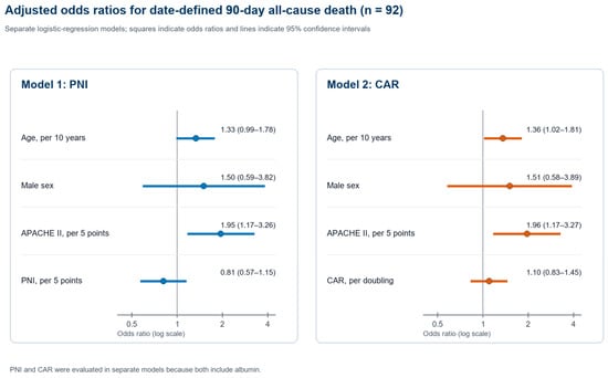
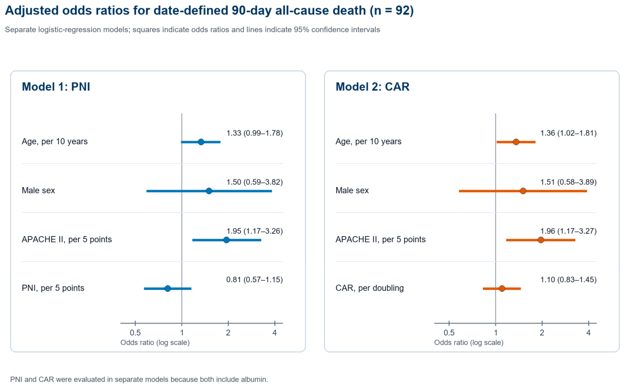
Table 5 presents the two adjusted 90-day logistic models among the 92 patients classifiable at 90 days from available date fields. In both models, higher APACHE II was associated with date-defined 90-day all-cause death. PNI was not statistically significantly associated in Model 1, and recalculated CAR was not statistically significantly associated in Model 2. Age was statistically significantly associated in the CAR model but not in the PNI model. The apparent AUC was 0.745 for the PNI model and 0.740 for the CAR model; Brier scores were 0.206 and 0.207, respectively. Table 6 reports variable availability and outcome classification handling. Adjusted odds ratios with 95% confidence intervals are shown in Figure 2.
Table 5.
Adjusted logistic regression models for date-derived 90-day all-cause death among patients classifiable at 90 days from available date fields (n = 92; events = 45).
Table 6.
Availability of variables, pre-PEG contextual fields, and date-derived outcome classifications in the 93-patient cohort.
Figure 2.
Adjusted odds ratios with 95% confidence intervals from two separate models for date-defined 90-day all-cause death among patients classifiable at 90 days from available date fields. PNI and CAR were evaluated in separate models. The x-axis is logarithmic; the vertical reference line indicates an odds ratio of 1.
4. Discussion
In this cohort of 93 intensive care patients undergoing PEG, date-defined all-cause mortality was 25/93 (26.9%) within 30 days and 45/92 (48.9%) among patients classifiable at 90 days. Higher APACHE II scores were associated with 90-day death in both adjusted models, whereas PNI and CAR were not statistically significantly associated after adjustment. Incorporating the detailed pre-PEG clinical context and recorded causes of death clarifies the clinical indications, pre-procedural swallowing assessments, and terminal critical-illness syndromes observed in this intensive care population.
A crucial distinction is between the primary condition prompting durable enteral access and a background comorbidity diagnosis. Dementia was recorded as a background comorbidity label in 33 patients, without a FAST or CDR stage in that field, and the six malignancy comorbidity labels likewise did not specify terminal cancer. Examples within the malignancy comorbidity field included operated brain tumors and other malignancies that cannot be assigned a terminal stage from the diagnostic label alone. Similarly, the three “irreversible organ failure” entries referred to chronic renal failure and/or heart failure without a standardized terminal-prognosis assessment. Severe dysphagia attributed to dementia was the primary PEG indication in only one patient (1.1%), who survived beyond 90 days, whereas the remaining 98.9% of procedures were performed for post-stroke dysphagia, acute brain injury, prolonged mechanical ventilation or ICU stay, head and neck malignancy, or neuromuscular disorders.
A documented clinical rationale for transitioning to durable enteral access was present in every patient: 72/93 (77.4%) had persistent severe dysphagia or unsafe/inadequate oral intake, and 21/93 (22.6%) had transnasal tube intolerance or mechanical failure. NG/NJ feeding had preceded PEG for a median of 23 days (IQR, 10–44 days), and all 93 patients underwent pre-procedural FEES (55.9%) or bedside clinical swallowing assessment (44.1%) confirming aspiration or unsafe oral intake. These findings demonstrate that PEG placement was prompted by documented enteral access and swallowing indications following prolonged transnasal feeding rather than indiscriminate referral based on comorbidity labels.
ESGE recommends temporary transnasal feeding when enteral support is expected for less than 4 weeks and consideration of percutaneous access when longer support is anticipated, depending on the clinical setting [1]. ESPEN’s home-enteral-nutrition guideline similarly describes nasal feeding for short-term use, up to 4–6 weeks, and percutaneous access when longer support is required [2]. Our cohort’s preceding transnasal feeding duration (median 23 days) and documented transition rationales align with this clinical framework for considering durable enteral access in patients with persistent dysphagia. At the same time, in critically ill patients, elapsed feeding duration and technical feasibility must always be weighed alongside overall clinical stability, prognosis, and treatment goals [1,2].
Our 30-day mortality is within the range reported in some recent ICU cohorts, although these populations are not interchangeable. In the 487-patient study by Sermet et al., the late-PEG group (>14 days; n = 341) had reported APACHE II and PNI values of 22.7 and 32.8, respectively, and 30-day mortality of 31.7%, compared with 15.1% in the early group [15]. Our median ICU-to-PEG interval was 24.0 days; our overall median APACHE II was 15.0, and the median PNI in the 90-day death group was 32.50. These differences in severity, subgroup definition, and summary measures preclude a matched comparison. Oruç et al. reported 125/364 in-hospital deaths (34.3%), with 56/125 deaths (44.8%) occurring within 14 days, and a median admission-to-PEG interval of 25 days [17]. Their endpoint was in-hospital mortality, not fixed 90-day mortality.
Other ICU series have reported lower early but high overall follow-up mortality, underscoring substantial heterogeneity across critical-care settings. Koç et al. reported 6.4% 30-day mortality, 45% overall mortality during follow-up, and 0% procedure-related mortality among 31 neurosurgical ICU patients [18]. Zippi et al. reported 0% procedure-related mortality in 36 selected ICU patients undergoing bedside PEG [19]. In Pih et al.’s surgical ICU cohort of 27 patients, median SOFA and APACHE II scores were 4 and 16, the median interval from surgery to PEG was 30 days, and 30-day mortality was 14.8%, with no procedure-related deaths in the ICU group [6]. Furthermore, in the multicentre study by Stenberg et al., 90-day mortality reached 41/80 (51%) among patients with a malignancy indication and 16/31 (52%) among those with baseline CRP ≥100 mg/L [4]. Collectively, these series indicate that while procedural mortality in the ICU is consistently low, 30- and 90-day all-cause mortality varies widely according to baseline physiological severity, systemic inflammation, and underlying critical illness.
The recorded causes of death further clarify the clinical syndromes underlying post-PEG mortality in our cohort: sepsis, septic shock, and multiorgan dysfunction accounted for 32/45 (71.1%) deaths by day 90, acute respiratory failure for 6/45 (13.3%), cardiovascular or cardiorenal causes for 4/45 (8.9%), and cerebrovascular causes for 3/45 (6.7%), with no deaths directly attributed to the PEG procedure (0/93). This distribution closely parallels prior hospital and ICU cohorts. Pih et al. reported no procedure-related deaths in their surgical ICU cohort, where all early deaths resulted from aggravated pneumonia, sepsis, cardiac arrest, myocardial infarction, renal failure, or intracranial hemorrhage [6], and similarly noted in their 401-patient cohort that early post-PEG deaths were predominantly driven by pneumonia, uncontrolled infection, and organ failure [7]. Stenberg et al. likewise identified malignancy, aspiration or COPD-related pneumonia, stroke, cardiac events, and sepsis as the principal causes of 30- and 90-day mortality [4]. The predominance of critical-illness syndromes in our dataset underscores that fixed-horizon mortality following ICU PEG primarily reflects the trajectory of the underlying critical illness and ICU-acquired infections rather than direct procedural lethality.
The prognostic models should inform risk discussion, not determine access to PEG in isolation. Our PNI and CAR models had apparent AUCs of 0.745 and 0.740 and Brier scores of 0.206 and 0.207, respectively. In Oruç et al., an in-hospital mortality model had an AUC of 0.761 but sensitivity of only 41.9% at a probability threshold of 0.5, despite specificity of 86.8% [17]. This illustrates that moderate discrimination does not permit reliable individual prediction or establish a threshold for withholding PEG. The endpoints and models differ, so the AUCs are not evidence of comparative superiority. Conversely, uncertainty in prediction does not remove the obligation to consider poor prognosis. Clinical trajectory, reversibility, patient values, alternatives, and treatment goals must accompany any risk estimate.
The lack of statistically significant adjusted PNI and CAR associations should not be interpreted as evidence that nutritional status or inflammation is clinically unimportant. Both indices reflect processes that may overlap with global illness severity, and estimates in a small cohort are imprecise. Prior studies have reported associations between albumin, CRP, PNI, or CAR and early outcomes [7,8,9,10,11,12,13], with variation in populations, endpoints, and adjustment. Our results support cautious use of these measures as components of assessment, rather than universal cutoffs or stand-alone selection rules.
The finding of 0/93 available structured fields requires a precise interpretation. ICU, neurology, and surgical consultations, swallowing assessments, and signed written procedure consent were documented in all 93 patients. What was unavailable was a separate structured HBYS field for quantitative life expectancy or a formal palliative-care narrative. Absence of such fields is not evidence that prognosis, alternatives, or care goals were never considered. At the same time, documentation of consultation or consent cannot establish the content or quality of a benefit–burden discussion, formal capacity assessment, or goals-concordant decision. These unmeasured dimensions remain limitations and should be recorded prospectively in future cohorts.
This single-center retrospective cohort is also limited by its size, possible selection and documentation biases, lack of a non-PEG comparator, and lack of standardized functional recovery, quality-of-life, or patient-centered benefit outcomes. Mortality was based on recorded calendar dates without external vital-status linkage; one patient with a last-known-alive date at day 81 remained unclassifiable at 90 days. All 46 recorded deaths were retained descriptively, while the day-106 death remained outside the 45-death 90-day endpoint. The additional cause-of-death labels were not independently adjudicated, and background dementia/cancer labels were not staged. No resampling-based internal validation or external validation was performed; apparent model performance may be optimistic. Accordingly, these data describe indications, care documentation, and prognostic associations after PEG, but cannot establish that each PEG decision was appropriate or that PEG improved survival.
5. Conclusions
In this retrospective intensive care cohort, higher APACHE II scores were associated with date-defined 90-day all-cause mortality, whereas PNI and CAR were not statistically significantly associated in separate adjusted models. Detailed records document primary dysphagia-related indications, preceding NG/NJ feeding, swallowing assessment, multidisciplinary consultation, and signed written consent. Background dementia and malignancy labels were unstaged and must not be described as confirmed advanced dementia or terminal cancer. Most recorded deaths by day 90 involved sepsis, septic shock, or multiorgan dysfunction, and no directly PEG-attributed death was recorded. These findings support contextualized risk discussion; they do not establish individual procedural appropriateness, goals-concordance, or a survival benefit from PEG.
Author Contributions
Conceptualization, M.Y. (Metin Yıldız) and B.A.; Methodology, M.Y. (Metin Yıldız) and B.A.; Formal analysis, M.Y. (Metin Yıldız) and B.A.; Data curation, M.Y. (Metin Yıldız) and B.A.; Writing—original draft preparation, M.Y. (Metin Yıldız) and B.A.; Visualization, M.Y. (Metin Yıldız) and B.A.; Investigation, M.Y. (Metin Yıldız), B.A., A.B., M.Ş., F.A.U., M.Y. (Muhammet Yıldırım) and R.P.; Validation, M.Y. (Metin Yıldız), B.A., A.B. and M.Ş.; Writing—review and editing, M.Y. (Metin Yıldız), B.A., A.B., M.Ş., F.A.U., M.Y. (Muhammet Yıldırım) and R.P.; Manuscript formatting and technical editing, F.A.U., M.Y. (Muhammet Yıldırım) and R.P.; Supervision, A.B. and M.Ş. All authors have read and agreed to the published version of the manuscript.
Funding
This research received no external funding.
Institutional Review Board Statement
An ethics committee decision covering the retrospective 90-day mortality analysis was issued by the Muş Alparslan University Scientific Research and Publication Ethics Board (Decision 43; meeting decision no. 87; 11 February 2026; electronic document no. 234682). Institutional site permission was granted by the Muş Provincial Health Directorate (27 February 2026; reference E-35465298-619-306452106).
Informed Consent Statement
Signed written informed consent for the clinical PEG procedure was documented in all 93 patients, including the date and the signer’s relationship to the patient. These clinical-procedure records are distinct from consent for retrospective research and do not, by themselves, establish formal decision-making capacity, legal representative authority, or goals-concordance. The retrospective research was conducted under the ethics approval and institutional permission reported above.
Data Availability Statement
The data are not publicly available due to ethical and privacy restrictions related to patient-level hospital data. De-identified data may be available from the corresponding author upon reasonable request and subject to institutional approval.
Conflicts of Interest
The authors declare no conflicts of interest.
References
- Arvanitakis, M.; Gkolfakis, P.; Despott, E.J.; Ballarin, A.; Beyna, T.; Boeykens, K.; Elbe, P.; Gisbertz, I.; Hoyois, A.; Mosteanu, O.; et al. Endoscopic management of enteral tubes in adult patients—Part 1: Definitions and indications. European Society of Gastrointestinal Endoscopy (ESGE) Guideline. Endoscopy 2021, 53, 81–92. [Google Scholar] [CrossRef] [Scilit] [PubMed]
- Bischoff, S.C.; Austin, P.; Boeykens, K.; Chourdakis, M.; Cuerda, C.; Jonkers-Schuitema, C.; Lichota, M.; Nyulasi, I.; Schneider, S.M.; Stanga, Z.; et al. ESPEN practical guideline: Home enteral nutrition. Clin. Nutr. 2022, 41, 468–488. [Google Scholar] [CrossRef] [Scilit] [PubMed]
- Kamran, U.; Lee, P.C.; Coupland, B.; Abbasi, A.; Steed, H.; Ispoglou, S.; Varyani, F.; Trudgill, N. Improving 30-day mortality after PEG tube placement in England from 2007 to 2019: A retrospective national cohort analysis of 87,862 patients. Gastrointest. Endosc. 2022, 96, 943–953.e11. [Google Scholar] [CrossRef] [Scilit] [PubMed]
- Stenberg, K.; Eriksson, A.; Odensten, C.; Darehed, D. Mortality and complications after percutaneous endoscopic gastrostomy: A retrospective multicentre study. BMC Gastroenterol. 2022, 22, 361. [Google Scholar] [CrossRef] [Scilit] [PubMed]
- Mutlu Bilgic, N.; Kahveci, G.; Aykut, H.; Ozer, Y.; Ozsenel, E.B.; Basat, S. Risk factors for complications and 90-day mortality after percutaneous endoscopic gastrostomy: The role of nutritional and inflammatory markers. Medicina 2025, 61, 1916. [Google Scholar] [CrossRef] [Scilit] [PubMed]
- Pih, G.Y.; Na, H.K.; Hong, S.K.; Ahn, J.Y.; Lee, J.H.; Jung, K.W.; Kim, D.H.; Choi, K.D.; Song, H.J.; Lee, G.H.; et al. Clinical outcomes of percutaneous endoscopic gastrostomy in the surgical intensive care unit. Clin. Endosc. 2020, 53, 705–712. [Google Scholar] [CrossRef] [Scilit] [PubMed]
- Pih, G.Y.; Na, H.K.; Ahn, J.Y.; Jung, K.W.; Kim, D.H.; Lee, J.H.; Choi, K.D.; Song, H.J.; Lee, G.H.; Jung, H.-Y. Risk factors for complications and mortality of percutaneous endoscopic gastrostomy insertion. BMC Gastroenterol. 2018, 18, 101. [Google Scholar] [CrossRef] [Scilit] [PubMed]
- Seyhan, S.; Tosun Tasar, P.; Karasahin, O.; Albayrak, B.; Sevinç, C.; Şahin, S. Complications and factors associated with mortality in patients undergoing percutaneous endoscopic gastrostomy. Clin. Sci. Nutr. 2024, 6, 97–106. [Google Scholar] [CrossRef] [Scilit]
- Blomberg, J.; Lagergren, P.; Martin, L.; Mattsson, F.; Lagergren, J. Albumin and C-reactive protein levels predict short-term mortality after percutaneous endoscopic gastrostomy in a prospective cohort study. Gastrointest. Endosc. 2011, 73, 29–36. [Google Scholar] [CrossRef] [Scilit] [PubMed]
- Karasahin, O.; Tasar, P.T.; Timur, O.; Binici, D.N.; Yilmaz, T.K.; Aslan, A.; Sahin, S. High C-reactive protein and low albumin levels predict high 30-day mortality in patients undergoing percutaneous endoscopic gastrotomy. Gastroenterol. Res. 2017, 10, 172–176. [Google Scholar] [CrossRef] [Scilit] [PubMed]
- Onodera, T.; Goseki, N.; Kosaki, G. Prognostic nutritional index in gastrointestinal surgery of malnourished cancer patients. Nihon Geka Gakkai Zasshi 1984, 85, 1001–1005. [Google Scholar] [PubMed]
- Adachi, Y.; Akino, K.; Nojima, M.; Himori, R.; Kikuchi, T.; Mita, H.; Nakamura, M.; Tsukuda, H.; Yamano, H.-O.; Sasaki, Y.; et al. Prognostic nutritional index and early mortality with percutaneous endoscopic gastrostomy. QJM 2018, 111, 635–641. [Google Scholar] [CrossRef] [Scilit] [PubMed]
- Duzenli, T.; Ketenci, M.; Akyol, T.; Koseoglu, H.; Tanoglu, A.; Kaplan, M.; Yazgan, Y. Predictive factors of complications and 30-day mortality in patients undergoing percutaneous endoscopic gastrostomy: The utility of C-reactive protein to albumin ratio. Acta Gastroenterol. Belg. 2021, 84, 283–288. [Google Scholar] [CrossRef] [Scilit] [PubMed]
- Knaus, W.A.; Draper, E.A.; Wagner, D.P.; Zimmerman, J.E. APACHE II: A severity of disease classification system. Crit. Care Med. 1985, 13, 818–829. [Google Scholar] [CrossRef] [Scilit]
- Sermet, M.; Kocoglu, H.; Houssaini, D.; Ekinci, O.; Alimoglu, O. Impact of percutaneous endoscopic gastrostomy timing on mortality in intensive care unit patients: The modifying role of the prognostic nutritional index. Clin. Nutr. ESPEN 2026, 73, 103309. [Google Scholar] [CrossRef] [Scilit] [PubMed]
- von Elm, E.; Altman, D.G.; Egger, M.; Pocock, S.J.; Gøtzsche, P.C.; Vandenbroucke, J.P. The Strengthening the Reporting of Observational Studies in Epidemiology (STROBE) statement: Guidelines for reporting observational studies. Ann. Intern. Med. 2007, 147, 573–577. [Google Scholar] [CrossRef] [Scilit] [PubMed]
- Oruç, M.; Şahingöz, E.; Canpolat, E.; Akın, T.; Terzioğlu, S.G.; Çetinkaya, E. Pre-procedural predictors of in-hospital mortality in ICU patients undergoing endoscopic PEG placement. Turk. J. Surg. 2026, 42, 298–305. [Google Scholar] [CrossRef] [Scilit] [PubMed]
- Koç, D.; Gerçek, A.; Gençosmanoğlu, R.; Tözün, N. Percutaneous endoscopic gastrostomy in the neurosurgical intensive care unit: Complications and outcome. JPEN J. Parenter. Enter. Nutr. 2007, 31, 517–520. [Google Scholar] [CrossRef] [Scilit] [PubMed]
- Zippi, M.; Fiorani, S.; De Felici, I.; Febbraro, I.; Mattei, E.; Traversa, G.; Barbaro, F.; Scafetti, S.; Occhigrossi, G. Percutaneous endoscopic gastrostomy (PEG) in critically ill patients performed at bed in Intensive Care Unit: Report of our experience. Clin. Ter. 2009, 160, 359–362. Available online: https://pubmed.ncbi.nlm.nih.gov/19997680/ (accessed on 12 September 2026). [PubMed]

Disclaimer/Publisher’s Note: The statements, opinions and data contained in all publications are solely those of the individual author(s) and contributor(s) and not of MDPI and/or the editor(s). MDPI and/or the editor(s) disclaim responsibility for any injury to people or property resulting from any ideas, methods, instructions or products referred to in the content. |
© 2026 by the authors. Published by MDPI on behalf of the Lithuanian University of Health Sciences. Licensee MDPI, Basel, Switzerland. This article is an open access article distributed under the terms and conditions of the Creative Commons Attribution (CC BY) license.