


Abstract
Background and Objectives: Initial high-sensitivity cardiac troponin I (hs-cTnI) measurements can be difficult to interpret in patients with renal dysfunction because elevated concentrations may occur without acute myocardial infarction (AMI). This systematic review compared reported optimal hs-cTnI cutoffs and their diagnostic performance across assay platforms and patient subgroups. Materials and Methods: PubMed/MEDLINE, Embase, the Cochrane Library, and Web of Science were searched from inception to 21 August 2026. Studies evaluating initial hs-cTnI measurements in adults with renal dysfunction and reporting diagnostic cutoffs with corresponding sensitivity and specificity for AMI were eligible. Risk of bias was assessed using QUADAS-2. Findings were synthesized narratively because of clinical and methodological heterogeneity; no meta-analysis was performed. Results: Five studies were included. In studies analyzing patients with an estimated glomerular filtration rate below 60 mL/min/1.73 m2 as a single group, optimal cutoffs ranged from 25.9 to 54 ng/L, with sensitivities of 76–88.4% and specificities of 83–90%. One study reported substantially higher cutoffs of 500–2610 ng/L across selected subgroups defined by CKD stage, age, and sex, with sensitivities of 80.4–91.4% and specificities of 92.1–100%. In patients receiving dialysis, cutoffs were 75 ng/L for hemodialysis and 144 ng/L for peritoneal dialysis, yielding sensitivities of 93.3% and 100% and specificities of 60.8% and 83.1%, respectively. All five studies were rated as having a high risk of bias in the index test domain. Conclusions: Reported initial hs-cTnI cutoffs varied considerably across assays and patient populations. The available evidence does not establish a single cutoff applicable to all patients with renal dysfunction. These thresholds require external validation and should be interpreted alongside clinical findings and serial troponin measurements.
1. Introduction
Chronic kidney disease (CKD) is closely linked to cardiovascular disease, with the risks of cardiovascular events and death increasing progressively as kidney function declines. Patients with CKD also have a substantially greater risk of acute myocardial infarction (AMI) and cardiovascular mortality than those without CKD [1,2,3]. This association has also been observed in patients without overt renal dysfunction. Among patients undergoing coronary angiography with an estimated glomerular filtration rate (eGFR) ≥ 60 mL/min/1.73 m2, lower eGFR within the normal or mildly reduced range was associated with higher long-term all-cause mortality [4]. The early diagnosis of AMI in patients with CKD is often challenging. These patients are less likely to present with typical chest pain and may instead report nonspecific symptoms such as dyspnea, fatigue, nausea, or generalized weakness. Moreover, left ventricular hypertrophy and baseline electrocardiographic abnormalities are common in CKD, and repolarization changes related to structural heart disease or electrolyte disturbances may obscure new ischemic findings. Consequently, clinical symptoms and a single electrocardiogram may be insufficient to reliably distinguish AMI from other acute conditions in this population [5,6].
High-sensitivity cardiac troponin (hs-cTn) assays can detect circulating troponin at substantially lower concentrations than conventional assays and identify small changes over short time intervals, thereby enabling earlier recognition of myocardial injury [7,8]. However, hs-cTn concentrations are frequently elevated in patients with CKD even in the absence of acute myocardial ischemia. This persistent elevation is thought to reflect the combined effects of structural heart disease and ongoing subclinical myocardial injury, including left ventricular hypertrophy, systemic inflammation, anemia, and volume overload, as well as the metabolic and hemodynamic disturbances associated with renal dysfunction [9]. Although high-sensitivity cardiac troponin I (hs-cTnI) appears to be less affected by declining renal function than high-sensitivity cardiac troponin T (hs-cTnT), important analytical differences remain among hs-cTnI assays. Measurement ranges and 99th-percentile upper reference limits vary across manufacturers, making results difficult to compare directly. The available studies also differ substantially in CKD severity, dialysis status, AMI prevalence, and the reference standards used to establish the final diagnosis. Consequently, no single hs-cTnI cutoff has yet been established for use across all patients with CKD and all available assay platforms [10].
This systematic review examined the diagnostic performance of initial hs-cTnI measurements for AMI in patients with renal dysfunction. We compared the receiver operating characteristic (ROC)-derived optimal cutoffs and their corresponding sensitivities and specificities across different degrees of renal dysfunction, dialysis status, and assay platforms. Given the clinical and methodological heterogeneity among the included studies, meta-analysis was not performed, and the findings were synthesized narratively with an emphasis on their clinical interpretation.
2. Materials and Methods
2.1. Study Design
We conducted a systematic review and narrative synthesis to examine how well initial high-sensitivity cardiac troponin I (hs-cTnI) measurements identify AMI in patients with renal dysfunction. The review was conducted and reported in accordance with the Preferred Reporting Items for Systematic Reviews and Meta-Analyses (PRISMA) 2020 statement [11]. The completed PRISMA 2020 checklist is provided in Supplementary Checklist S1.
Before beginning the literature search, we defined the review objectives, eligibility criteria, search strategy, data extraction process, and approach to evidence synthesis. Meta-analysis was considered inappropriate because of substantial differences across studies in patient characteristics, severity of renal dysfunction, dialysis status, assay platforms, reference standards, and reported subgroups. The results were therefore summarized using a narrative approach.
2.2. Search Strategy and Information Sources
We searched PubMed/MEDLINE, Embase, the Cochrane Library, and Web of Science from their inception to 21 August 2026. The search strategy combined terms related to high-sensitivity cardiac troponin I, acute myocardial infarction, chronic kidney disease, renal dysfunction, and dialysis. We also reviewed the reference lists of the included articles and relevant reviews to identify additional eligible studies. The complete database-specific search strategies, including the applied filters and search dates, are provided in Supplementary Table S1.
2.3. Study Selection and Eligibility Criteria
We included studies of adults with renal dysfunction who underwent evaluation for suspected AMI or acute coronary syndrome. Both patients with CKD not receiving dialysis and patients with kidney failure receiving hemodialysis or peritoneal dialysis were eligible. Studies were required to measure hs-cTnI at the initial clinical assessment using an identified assay platform and to provide an assay-specific or subgroup-specific diagnostic threshold with the corresponding sensitivity and specificity. The diagnosis of AMI had to be based on an established clinical definition or independent clinical adjudication.
Studies were excluded if they assessed hs-cTnT alone, used only conventional troponin assays, did not present separate results for patients with renal dysfunction, or lacked sufficient data on diagnostic thresholds or test performance. Review articles, editorials, case reports, conference abstracts, publications without original data, and duplicate reports were also excluded. Following removal of duplicate records, one reviewer (S.K. Oh) screened titles and abstracts and assessed potentially eligible articles in full text against the eligibility criteria.
2.4. Methodological Quality and Risk-of-Bias Assessment
The methodological quality of the included studies was assessed using the Quality Assessment of Diagnostic Accuracy Studies-2 (QUADAS-2) tool [12]. Risk of bias was evaluated across four domains: patient selection, index test, reference standard, and flow and timing. Applicability concerns were also examined for the first three domains. Following the QUADAS-2 guidance and signaling questions, each domain was rated as having a low, high, or unclear risk of bias or concern regarding applicability.
Two reviewers (S.K. Oh and S.U. Cho) independently assessed the risk of bias in all included studies. Differences in judgment were resolved through discussion and consensus. The assessments are presented in a study-level summary table and a risk-of-bias figure.
2.5. Data Extraction and Narrative Synthesis
Data extraction was performed by a single reviewer (S.K. Oh). For each study, we extracted the patient population, degree of renal dysfunction, dialysis status and modality, hs-cTnI assay platform, and ROC-derived optimal cutoff, together with the corresponding sensitivity and specificity. Age- and sex-specific results were recorded separately when available. Results for multiple assays or patient subgroups were tabulated individually but were not treated as independent studies.
Cutoffs were standardized to ng/L by multiplying values reported in ng/mL by 1000. This conversion ensured consistency in reporting units but did not imply analytical equivalence across assays. Thresholds and diagnostic performance were therefore interpreted in relation to the assay platform and study population.
The findings were organized by assay platform, renal function category, and dialysis status and synthesized narratively. Differences in patient selection, AMI adjudication procedures, and study-derived thresholds, together with the limited number of comparable independent studies, led us to forgo both overall and subgroup-specific meta-analyses. Even studies using the same assay differed in their populations and subgroup definitions. We therefore presented cutoffs, sensitivities, and specificities in tables and compared them descriptively, without formal statistical testing of between-study differences.
3. Results
3.1. Literature Search and Study Selection
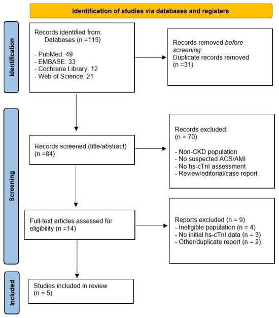
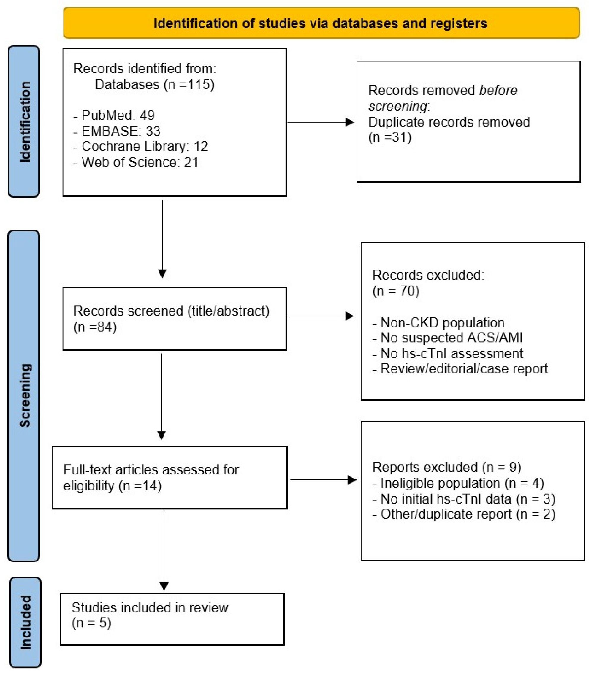
The database searches identified 115 records: 49 from PubMed, 33 from Embase, 12 from the Cochrane Library, and 21 from Web of Science. After removing 31 duplicates, 84 records underwent title and abstract screening. Of these, 70 were excluded because they did not involve patients with renal dysfunction, did not address suspected acute coronary syndrome or AMI, did not evaluate hs-cTnI, or were reviews, editorials, or case reports.
The remaining 14 articles were assessed in full text. Nine records were excluded after full-text assessment. Four did not meet the eligibility criteria because they did not report separate results for patients with renal dysfunction, did not enroll patients with suspected AMI, or included only patients with confirmed AMI without a non-AMI comparison group. Three were excluded because they reported only serial changes, peak concentrations, or subsequent measurements rather than initial values; provided insufficient data to extract or calculate sensitivity and specificity at the reported thresholds; or evaluated only prespecified thresholds without reporting a ROC-derived optimal cutoff for initial hs-cTnI. The remaining two were duplicate records that had been missed during initial deduplication and were identified and removed during full-text assessment. Five studies met the inclusion criteria and were included in the systematic review and narrative synthesis [12,13,14,15,16]. The study selection process is shown in Figure 1, and the main characteristics of the included studies are summarized in Table 1.
Figure 1.
PRISMA flow diagram of the study selection process.
Table 1.
Characteristics of the Included Studies.
3.2. Risk-of-Bias Assessment Results
The QUADAS-2 assessments of the five included studies are presented in Table 2. Risk of bias varied across studies and domains, with every study rated as high risk in at least one domain.
Table 2.
Risk of Bias Assessment of the Included Studies Using QUADAS-2.
For patient selection, Twerenbold et al. [13] were judged to have a low risk of bias, whereas Ren et al. [15] and Lim et al. [16] were rated as high risk. The assessments for Kraus et al. [14] and Li et al. [17] were unclear. All five studies were classified as high risk in the index test domain. In each study, the thresholds extracted for this review were derived from the study data, and their sensitivity and specificity were evaluated in the same dataset. This may have led to overestimation of diagnostic performance and was the main reason for the high-risk judgments. In the reference standard domain, Twerenbold et al. [13] , Kraus et al. [14], and Lim et al. [16] were rated as low risk, Ren et al. [15] as high risk, and Li et al. [17] as unclear. For flow and timing, Twerenbold et al. [13] were judged to have a low risk of bias and Lim et al. [16] a high risk, while the remaining three studies received unclear ratings.
The index test domain was the main source of concern, although high or unclear ratings were also observed in patient selection and flow and timing. These methodological limitations were considered when interpreting the reported cutoffs and diagnostic performance.
3.3. Overview of the Included Studies
The five included studies were published between 2015 and 2025, and their main characteristics are summarized in Table 1 [13,14,15,16,17]. Twerenbold et al. examined patients with renal dysfunction within an international multicenter prospective cohort, whereas Kraus et al. combined a prospective multicenter cohort with retrospective clinical registry data [13,14]. The studies by Ren, Lim, and Li et al. were conducted retrospectively at single centers [15,16,17].
The study populations ranged from patients with renal dysfunction not receiving dialysis for renal impairment to those receiving maintenance dialysis. Twerenbold et al. enrolled 2813 patients with suspected AMI, of whom 447 had an eGFR below 60 mL/min/1.73 m2 [13]. The hs-cTnI cohort in Kraus et al. comprised 1494 patients with suspected NSTE-AMI, including 280 with CKD [14]. Ren et al. included 761 patients with an eGFR of 15–59 mL/min/1.73 m2 within a cohort of 3295 patients and reported subgroup analyses by age and sex [15]. Lim et al. studied 1144 patients receiving maintenance dialysis, including 995 receiving hemodialysis and 149 receiving peritoneal dialysis [16]. Li et al. compared 237 patients with CKD and 261 with normal renal function among 498 patients evaluated for suspected NSTE-ACS. Of the patients with CKD, 71 had an eGFR below 30 mL/min/1.73 m2 [17].
The hs-cTnI assays also differed across studies. Twerenbold et al. compared several troponin assays, including hs-cTnI assays from Abbott, Siemens, and Beckman Coulter [13]. Kraus and Ren et al. used the Abbott ARCHITECT STAT assay, Lim et al. used the Siemens Centaur XPT assay, and Li et al. used the Ortho VITROS 5600 platform [14,15,16,17]. Reference diagnoses drew on clinical findings, electrocardiography, serial troponin measurements, cardiac imaging, and coronary angiography, although the diagnostic criteria and adjudication procedures varied. The MI types evaluated also differed: Twerenbold et al. included both type 1 and type 2 AMI, whereas Lim et al. explicitly applied diagnostic criteria for type 1 MI [13,16].
The studies addressed several related questions: assay-specific diagnostic thresholds, algorithms incorporating initial and serial troponin measurements, thresholds within renal function, age, and sex subgroups, differences between dialysis modalities, and the performance of ROC-derived cutoffs compared with the conventional 99th-percentile upper reference limit [13,14,15,16,17].
3.4. Initial Hs-cTnI Cutoffs and Diagnostic Performance
The optimal cutoffs for initial hs-cTnI measurements and their corresponding sensitivities and specificities are summarized in Table 3. Both the reported thresholds and diagnostic performance varied across assay platforms and patient populations.
Table 3.
Study-Specific Initial hs-cTnI Cutoffs and Diagnostic Performance.
In the study by Twerenbold et al., the optimal cutoffs for patients with an eGFR below 60 mL/min/1.73 m2 were 29.4 ng/L for the Abbott assay, 32.0 ng/L for the Siemens assay, and 25.9 ng/L for the Beckman assay. The corresponding sensitivities were 76%, 82%, and 81%, respectively, with specificities of 85%, 83%, and 83% [13].
Kraus et al. reported an optimal cutoff of 54 ng/L for the Abbott hs-cTnI assay in patients with CKD, yielding a sensitivity of 82% and a specificity of 90% [14].
Ren et al. reported different thresholds according to CKD stage, age, and sex. Among the CKD stage 3 subgroups presented in Table 3, the cutoffs were 720 ng/L for men younger than 60 years, 500 ng/L for men aged 60 years or older, and 590 ng/L for women aged 60 years or older. Sensitivities were 81.4%, 89.5%, and 91.4%, respectively, while specificity was 100% in all three subgroups. Among patients with CKD stage 4 aged 60 years or older, the cutoffs were 2610 ng/L for men and 1900 ng/L for women. The corresponding sensitivities were 80.4% and 89.3%, and specificities were 92.1% and 100%, respectively [15]. Cutoffs originally reported in ng/mL were converted to ng/L for consistency with the other studies.
In the dialysis cohort studied by Lim et al., the optimal cutoff for the Siemens hs-cTnI assay differed by dialysis modality. For patients receiving hemodialysis, a cutoff of 75 ng/L yielded a sensitivity of 93.3% and a specificity of 60.8%. For those receiving peritoneal dialysis, the cutoff was 144 ng/L, with a sensitivity of 100% and a specificity of 83.1% [16].
Li et al. reported an optimal cutoff of 53.4 ng/L for the Ortho VITROS hs-cTnI assay in patients with CKD and an eGFR below 60 mL/min/1.73 m2. At this threshold, sensitivity was 88.4%, and specificity was 88.1% [17].
4. Discussion
Diagnosing AMI in patients with CKD requires careful interpretation of both clinical findings and troponin measurements. Atypical presentations and pre-existing electrocardiographic abnormalities may obscure evidence of acute ischemia, while persistently elevated hs-cTn concentrations can limit the diagnostic value of a single measurement [5,6,9]. Analytical differences among hs-cTnI assays further complicate the interpretation of these results [10]. In this review, the reported cutoffs varied considerably across studies. This variation should be interpreted in light of differences in assay platforms and patient characteristics, rather than taken as evidence for a threshold applicable to all patients with renal dysfunction.
Against this background, we synthesized five studies evaluating the diagnostic performance of initial hs-cTnI measurements for AMI in patients with renal dysfunction. ROC-derived optimal cutoffs varied considerably across assay platforms and patient populations, as did the corresponding sensitivities and specificities. In studies that analyzed patients with an eGFR below 60 mL/min/1.73 m2 as a single group, cutoffs ranged from 25.9 to 54 ng/L [13,14,17]. Higher thresholds were reported by Ren et al., who examined subgroups defined by CKD stage, age, and sex [15]. Lim et al. also reported different cutoffs for patients receiving hemodialysis and peritoneal dialysis [16]. These differences highlight the importance of considering both the assay used and the characteristics of the patient population when interpreting an initial hs-cTnI result. The available evidence does not support a single diagnostic cutoff applicable to all patients with impaired renal function.
In patients with CKD, elevated troponin concentrations may reflect chronic myocardial injury as well as acute myocardial ischemia. An isolated value above the assay-specific 99th-percentile upper reference limit established in a healthy reference population is therefore insufficient to diagnose AMI [18,19]. This limitation does not mean that troponin testing lacks clinical value, even in advanced CKD. Serial measurements can help distinguish acute from chronic myocardial injury, although a rise or fall alone does not establish an ischemic cause. Troponin results should therefore be interpreted together with symptoms, electrocardiographic findings, and, when indicated, cardiac imaging, rather than replaced by non-troponin parameters alone. The cutoffs reported in this review should be considered within this diagnostic framework and should not be used as stand-alone criteria for AMI.
The variation in cutoffs observed in this systematic review should first be considered in relation to differences in assay platforms. Twerenbold et al. reported different optimal thresholds for the Abbott, Siemens, and Beckman assays within the same cohort of patients with renal dysfunction [13]. However, thresholds also differed between studies using the same manufacturer’s assay: Twerenbold et al. and Kraus et al. reported Abbott hs-cTnI cutoffs of 29.4 and 54 ng/L, respectively [13,14]. Assay manufacturer alone therefore cannot explain the observed variation. Differences in clinical characteristics, the distribution of renal function, the interval between symptom onset and blood sampling, and diagnostic adjudication procedures may also have contributed. Conversely, similar numerical cutoffs across different assays should not be taken as evidence of analytical or clinical equivalence.
The high cutoffs reported by Ren et al. should be interpreted in light of patient selection and the methods used to derive the thresholds [15]. This single-center retrospective study excluded patients receiving renal replacement therapy and calculated cutoffs for subgroups defined by renal function, age, and sex. AMI was diagnosed in 83.7% of the overall cohort, but the corresponding proportions among men and women aged ≥ 60 years with CKD stage 4 were 57.3% and 50.0%, respectively. The high overall prevalence of AMI therefore does not, by itself, explain the elevated thresholds in these subgroups. Patient selection and differences in clinical characteristics may have contributed, although the reported data cannot distinguish their effects from those of renal dysfunction. The cutoffs remained high after conversion to ng/L, indicating that the discrepancy was not simply due to different reporting units. Although ROC analysis was performed, the specific criterion for selecting the optimal cutoff was not stated, and some small subgroups were not included in the ROC analyses. No assessment in a separate validation cohort was reported, so reproducibility in independent populations remains unestablished. Deriving thresholds and evaluating their performance in the same dataset may produce optimistic estimates. These cutoffs should therefore not be attributed solely to CKD severity, and caution is needed before applying them to broader CKD populations.
The findings in patients receiving dialysis are based on a single retrospective study, which limits their generalizability. Lim et al. reported a cutoff of 75 ng/L for patients receiving hemodialysis, with 93.3% sensitivity and 60.8% specificity [16]. This limited specificity indicates that positive results remained common among patients without AMI. Among patients receiving peritoneal dialysis, a threshold of 144 ng/L yielded 100% sensitivity and 83.1% specificity, but only seven patients had MI. The absence of false-negative results in this small sample does not establish that the threshold can safely exclude AMI in other populations. The study used hs-cTnI measured at emergency department presentation but did not report a standardized interval between dialysis and blood sampling. Patient characteristics, sampling time relative to dialysis, and dialysis-related conditions may influence hs-cTnI concentrations, although their contribution to the observed differences was not directly established. The variation in cutoffs therefore cannot be attributed to dialysis modality alone. Both thresholds require validation in independent cohorts with clearly documented patient characteristics, sampling times, and dialysis conditions.
Initial hs-cTnI measurements can inform early clinical assessment, but the ROC-derived cutoffs identified in this review are not interchangeable with validated rule-in or rule-out thresholds. A rule-in or high-risk designation within a diagnostic pathway signals the need for prompt further evaluation rather than confirming AMI on its own [20]. This distinction is especially relevant in CKD, where chronic myocardial injury may elevate baseline troponin concentrations. Low specificity can lead to false-positive results in patients without AMI. Raising the cutoff may improve specificity, but at the cost of reduced sensitivity and more missed cases of AMI. A threshold with insufficient sensitivity is therefore unsuitable for excluding the diagnosis. Before CKD-specific cutoffs are adopted in practice, they need validation in independent cohorts and evidence that they add diagnostic value to established serial testing algorithms. The findings of this review alone do not justify using the reported thresholds as clinical rule-in or rule-out criteria.
The findings should also be interpreted in light of the risk of bias in the included studies. All five studies were rated as having a high risk of bias in the index test domain of QUADAS-2. Selecting an optimal cutoff and evaluating its performance in the same dataset may yield more favorable estimates than would be obtained in an independent cohort. Some studies also had high or unclear risk of bias in patient selection, the reference standard, or flow and timing. Reported diagnostic performance should therefore be considered alongside study design and patient characteristics; numerical differences alone do not establish the superiority of a particular assay or cutoff.
Previous studies have explored both diagnostic thresholds and the clinical utility of troponin testing in patients with renal dysfunction. Kampmann et al. reviewed 15 studies and included six in a meta-analysis, deriving cutoffs of approximately 42 ng/L for troponin I and 48 ng/L for troponin T [21]. Gunsolus et al. assessed initial and serial hs-cTnI measurements using sex-specific 99th-percentile thresholds. Specificity declined from 93 to 95% in patients with normal renal function to 57–61% in those with an eGFR below 30 mL/min/1.73 m2 and 40–41% in patients receiving dialysis [22]. These findings illustrate how the performance of a fixed threshold can vary with renal function and dialysis status.
In a secondary analysis of High-STEACS, Gallacher et al. found that hs-cTnI concentrations exceeded the 99th percentile in 46% of patients with renal dysfunction, compared with 16% of those without. Although introducing hs-cTnI testing increased the detection of myocardial injury and infarction, disparities in treatment and outcomes between the two groups persisted [23]. Greater detection of troponin elevation therefore does not necessarily translate into better clinical outcomes for patients with renal dysfunction.
This review has several limitations. First, only five studies were included, and they differed in patient selection, renal function categories, dialysis status, assay platforms, and AMI adjudication procedures. The MI types evaluated also varied across studies, so the reported thresholds should not be interpreted as uniformly specific to type 1 MI. Some evaluated patients with an eGFR < 60 mL/min/1.73 m2 as a single group, whereas others examined subgroups defined by CKD stage, age, or sex, or populations receiving dialysis. Sensitivity and specificity were also reported at different study-derived thresholds, making the clinical meaning of pooled estimates uncertain. Subgroup-specific pooling was limited by the scarcity of comparable independent studies: those using Abbott assays still differed in population and subgroup definitions, while dialysis findings came from only one study. We therefore compared the results descriptively without pooling estimates or formally testing between-study differences. This approach limited our ability to quantify the variation or determine its underlying causes. Second, although some studies reported results for multiple assays or subgroups, these findings came from the same study and should not be interpreted as replication across independent cohorts. Independent validation of the reported thresholds was also lacking. Together with the limited evidence base, this restricts our ability to identify a reliable hs-cTnI cutoff applicable across different patient populations and assay platforms. Third, our focus on ROC-derived cutoffs for initial hs-cTnI measurements limited the assessment of diagnostic approaches based on serial troponin changes or established 99th-percentile thresholds. Fourth, study selection and data extraction were performed by a single reviewer, which may have increased the risk of errors in determining study eligibility and extracting data. Finally, the small number of studies and their heterogeneity prevented a reliable assessment of publication bias. The possibility that unpublished findings or selective reporting influenced the available evidence cannot be excluded.
Future prospective multicenter studies should recruit sufficient numbers of patients across CKD stages and dialysis groups to evaluate both initial hs-cTnI concentrations and absolute and relative changes during serial testing. Analyses should be stratified by renal function and assay platform, with standardized sampling intervals and AMI adjudication criteria. Proposed thresholds and diagnostic algorithms should be validated in independent cohorts and interpreted alongside clinical evidence of ischemia to assess their ability to distinguish chronic myocardial injury from AMI. Direct comparisons with approaches based on established 99th-percentile upper reference limits and serial troponin algorithms are needed to determine whether renal function-specific strategies reduce false-positive results without increasing missed AMI. Their clinical value should also be assessed in terms of time to diagnosis, unnecessary investigations and admissions, and patient safety.
5. Conclusions
In this systematic review, reported cutoffs and diagnostic performance for initial hs-cTnI measurements in patients with renal dysfunction varied across assay platforms and study populations. Given the small number of studies, their heterogeneity, and the risk of bias, the available evidence does not support a single cutoff for all patients with renal dysfunction. Initial hs-cTnI results should be interpreted alongside symptoms, electrocardiographic findings, and serial troponin measurements, taking the assay and patient characteristics into account. Prospective multicenter validation in independent cohorts is needed before these thresholds can be adopted in clinical practice.
Supplementary Materials
The following supporting information can be downloaded at: https://www.mdpi.com/article/10.3390/medicina62112089/s1; Checklist S1: PRISMA 2020 checklist [11]. Table S1: Detailed Search Strategies for Each Database.
Author Contributions
Conceptualization, S.K.O.; methodology, S.K.O.; software, S.K.O.; validation, S.K.O. and S.U.C.; formal analysis, S.K.O.; investigation, S.K.O.; resources, S.K.O.; data curation, S.K.O.; writing—original draft preparation, S.K.O.; writing—review and editing, S.K.O.; visualization, S.K.O.; supervision, S.K.O.; project administration, S.K.O. All authors have read and agreed to the published version of the manuscript.
Funding
This research received no external funding.
Institutional Review Board Statement
Ethical approval was not required because this systematic review used only aggregate data from previously published studies and involved no direct participant recruitment or access to identifiable patient information.
Data Availability Statement
The data supporting the findings of this review are available in the included publications. Extracted data are presented in this article and its Supplementary Materials.
Acknowledgments
ChatGPT (GPT-6 Astra; OpenAI, San Francisco, CA, USA) was used to assist with drafting, translation, and language editing of the manuscript and responses to reviewers during manuscript preparation and revision.
Conflicts of Interest
The authors declare no conflicts of interest.
Abbreviations
The following abbreviations are used in this manuscript:
| AMI | Acute myocardial infarction |
| CKD | Chronic kidney disease |
| eGFR | Estimated glomerular filtration rate |
| hs-cTnI | High-sensitivity cardiac troponin I |
| hs-cTnT | High-sensitivity cardiac troponin T |
| NSTE-ACS | Non-ST-segment elevation acute coronary syndrome |
| NSTE-AMI | Non-ST-segment elevation acute myocardial infarction |
| ROC | Receiver operating characteristic |
| PRISMA | Preferred Reporting Items for Systematic Reviews and Meta-Analyses |
| QUADAS-2 | Quality Assessment of Diagnostic Accuracy Studies 2 |
References
- Sarnak, M.J.; Levey, A.S.; Schoolwerth, A.C.; Coresh, J.; Culleton, B.; Hamm, L.L.; Mccullough, P.A.; Kasiske, B.L.; Kelepouris, E.; Klag, M.J.; et al. Kidney disease as a risk factor for development of cardiovascular disease: A statement from the American Heart Association Councils on Kidney in Cardiovascular Disease, High Blood Pressure Research, Clinical Cardiology, and Epidemiology and Prevention. Circulation 2003, 108, 2154–2169. [Google Scholar] [PubMed]
- Go, A.S.; Chertow, G.M.; Fan, D.; McCulloch, C.E.; Hsu, C.Y. Chronic kidney disease and the risks of death, cardiovascular events, and hospitalization. N. Engl. J. Med. 2004, 351, 1296–1305. [Google Scholar] [CrossRef] [Scilit] [PubMed]
- Meisinger, C.; Döring, A.; Löwel, H.; KORA Study Group. Chronic kidney disease and risk of incident myocardial infarction and all-cause and cardiovascular disease mortality in middle-aged men and women from the general population. Eur. Heart J. 2006, 27, 1245–1250. [Google Scholar] [CrossRef] [Scilit] [PubMed]
- Zornitzki, L.; Sadon, S.; Wenkert, A.; Steinvil, A.; Konigstein, M.; Ben-Shoshan, J.; Bassat, O.K.-B.; Sirota, S.; Halkin, A.; Bazan, S.; et al. The impact of normal range estimated glomerular filtration rate on mortality in selected patients undergoing coronary angiography—A long-term follow-up. Coron. Artery Dis. 2021, 32, 302–308. [Google Scholar] [CrossRef] [Scilit] [PubMed]
- Sosnov, J.; Lessard, D.; Goldberg, R.J.; Yarzebski, J.; Gore, J.M. Differential symptoms of acute myocardial infarction in patients with kidney disease: A community-wide perspective. Am. J. Kidney Dis. 2006, 47, 378–384. [Google Scholar] [CrossRef] [Scilit] [PubMed]
- Shroff, G.R.; Frederick, P.D.; Herzog, C.A. Renal failure and acute myocardial infarction: Clinical characteristics in patients with advanced chronic kidney disease, on dialysis, and without chronic kidney disease. Am. Heart J. 2012, 163, 399–406. [Google Scholar] [CrossRef] [Scilit] [PubMed]
- Reichlin, T.; Hochholzer, W.; Bassetti, S.; Steuer, S.; Stelzig, C.; Hartwiger, S.; Biedert, S.; Schaub, N.; Buerge, C.; Potocki, M.; et al. Early diagnosis of myocardial infarction with sensitive cardiac troponin assays. N. Engl. J. Med. 2009, 361, 858–867. [Google Scholar] [CrossRef] [Scilit] [PubMed]
- Reichlin, T.; Schindler, C.; Drexler, B.; Twerenbold, R.; Reiter, M.; Zellweger, C.; Moehring, B.; Ziller, R.; Hoeller, R.; Gimenez, M.R.; et al. One-hour rule-out and rule-in of acute myocardial infarction using high-sensitivity cardiac troponin T. Arch. Intern. Med. 2012, 172, 1211–1218. [Google Scholar] [CrossRef] [Scilit] [PubMed]
- Chuang, A.M.Y.; Nguyen, M.T.; Kung, W.M.; Lehman, S.; Chew, D.P. High-sensitivity troponin in chronic kidney disease: Considerations in myocardial infarction and beyond. Rev. Cardiovasc. Med. 2020, 21, 191–203. [Google Scholar] [CrossRef] [Scilit] [PubMed]
- Raber, I.; McCarthy, C.P.; Januzzi, J.L., Jr. A test in context: Interpretation of high-sensitivity cardiac troponin assays in different clinical settings. J. Am. Coll. Cardiol. 2021, 77, 1357–1367. [Google Scholar] [CrossRef] [Scilit] [PubMed]
- Page, M.J.; McKenzie, J.E.; Bossuyt, P.M.; Boutron, I.; Hoffmann, T.C.; Mulrow, C.D.; Shamseer, L.; Tetzlaff, J.M.; Akl, E.A.; Brennan, S.E.; et al. The PRISMA 2020 statement: An updated guideline for reporting systematic reviews. BMJ 2021, 372, n71. [Google Scholar] [CrossRef] [Scilit] [PubMed]
- Whiting, P.; Rutjes, A.W.; Reitsma, J.B.; Bossuyt, P.M.; Kleijnen, J. The development of QUADAS: A tool for the quality assessment of studies of diagnostic accuracy included in systematic reviews. BMC Med. Res. Methodol. 2003, 3, 25. [Google Scholar] [CrossRef] [Scilit] [PubMed]
- Twerenbold, R.; Wildi, K.; Jaeger, C.; Rubini Gimenez, M.; Reiter, M.; Reichlin, T.; Walukiewicz, A.; Gugala, M.; Krivoshei, L.; Marti, N.; et al. Optimal cutoff levels of more sensitive cardiac troponin assays for the early diagnosis of myocardial infarction in patients with renal dysfunction. Circulation 2015, 131, 2041–2050. [Google Scholar] [CrossRef] [Scilit] [PubMed]
- Kraus, D.; von Jeinsen, B.; Tzikas, S.; Palapies, L.; Zeller, T.; Bickel, C.; Fette, G.; Lackner, K.J.; Drechsler, C.; Neumann, J.T.; et al. Cardiac troponins for the diagnosis of acute myocardial infarction in chronic kidney disease. J. Am. Heart Assoc. 2018, 7, e008032. [Google Scholar] [CrossRef] [Scilit] [PubMed]
- Ren, D.; Huang, T.; Liu, X.; Xu, G. High-sensitive cardiac troponin for the diagnosis of acute myocardial infarction in different chronic kidney disease stages. BMC Cardiovasc. Disord. 2021, 21, 100. [Google Scholar] [CrossRef] [Scilit] [PubMed]
- Lim, E.; Lee, M.J. Optimal cut-off value of high-sensitivity troponin I in diagnosing myocardial infarction in patients with end-stage renal disease. Medicine 2020, 99, e18580. [Google Scholar] [CrossRef] [Scilit] [PubMed]
- Li, Z.; Tao, J.; Wang, H.; Zhang, Q.; Liu, Q.; Wang, Q.; Feng, W.; Tan, Y.; Yang, B.; Xu, Z.; et al. High-sensitivity cardiac troponin I in the diagnosis of acute myocardial infarction: A study in patients with renal insufficiency. Am. Heart J. Plus 2025, 59, 100631. [Google Scholar] [CrossRef] [Scilit] [PubMed]
- Miller-Hodges, E.; Anand, A.; Shah, A.S.V.; Chapman, A.R.; Gallacher, P.; Lee, K.K.; Farrah, T.E.; Halbesma, N.; Blackmur, J.P.; Newby, D.E.; et al. High-sensitivity cardiac troponin and the risk stratification of patients with renal impairment presenting with suspected acute coronary syndrome. Circulation 2018, 137, 425–435. [Google Scholar] [CrossRef] [Scilit] [PubMed]
- Knott, J.D.; Ola, O.; De Michieli, L.; Akula, A.; Mehta, R.A.; Dworak, M.; Crockford, E.; Lobo, R.; Slusser, J.; Rastas, N.; et al. Diagnosis of acute myocardial infarction in patients with renal failure using high-sensitivity cardiac troponin T. Eur. Heart J. Acute Cardiovasc. Care 2024, 13, 546–558. [Google Scholar] [CrossRef] [Scilit] [PubMed]
- Mills, N.L.; Newby, L.K.; Zaman, S.; Almahmeed, W.A.; Bucciarelli-Ducci, C.; Colvin, M.M.; Dangas, G.D.; A DeVon, H.A.; Dharmakumar, R.; A Gaziano, T.A.; et al. Fifth universal definition of myocardial infarction (2026). Eur. Heart J. 2026, 47, 5911–5958. [Google Scholar] [CrossRef] [Scilit] [PubMed]
- Kampmann, J.; Heaf, J.; Backer Mogensen, C.; Pedersen, A.K.; Granhøj, J.; Mickley, H.; Brandt, F. Troponin cut-offs for acute myocardial infarction in patients with impaired renal function—A systematic review and meta-analysis. Diagnostics 2022, 12, 276. [Google Scholar] [CrossRef] [Scilit] [PubMed]
- Gunsolus, I.; Sandoval, Y.; Smith, S.W.; Sexter, A.; Schulz, K.; Herzog, C.A.; Apple, F.S. Renal dysfunction influences the diagnostic and prognostic performance of high-sensitivity cardiac troponin I. J. Am. Soc. Nephrol. 2018, 29, 636–643. [Google Scholar] [CrossRef] [Scilit] [PubMed]
- Gallacher, P.J.; Miller-Hodges, E.; Shah, A.S.V.; Farrah, T.E.; Halbesma, N.; Blackmur, J.P.; Chapman, A.R.; Adamson, P.D.; Anand, A.; Strachan, F.E.; et al. High-sensitivity cardiac troponin and the diagnosis of myocardial infarction in patients with kidney impairment. Kidney Int. 2022, 102, 149–159. [Google Scholar] [CrossRef] [Scilit] [PubMed]

Disclaimer/Publisher’s Note: The statements, opinions and data contained in all publications are solely those of the individual author(s) and contributor(s) and not of MDPI and/or the editor(s). MDPI and/or the editor(s) disclaim responsibility for any injury to people or property resulting from any ideas, methods, instructions or products referred to in the content. |
© 2026 by the authors. Published by MDPI on behalf of the Lithuanian University of Health Sciences. Licensee MDPI, Basel, Switzerland. This article is an open access article distributed under the terms and conditions of the Creative Commons Attribution (CC BY) license.