


Abstract
Background: Clinical and survival data on pulmonary arterial hypertension (PAH) from Central Asia remain limited. This study aimed to characterize the clinical profile, survival outcomes, and prognostic value of baseline COMPERA 2.0 risk stratification in patients with PAH in Kazakhstan. Methods: This retrospective observational study included 206 adult patients with right heart catheterization-confirmed PAH hospitalized at the largest pulmonary hypertension referral center in Kazakhstan between 2020 and 2024. Patients were categorized into idiopathic PAH (IPAH), congenital heart disease-associated PAH (CHD-PAH), and other PAH subgroups. Kaplan–Meier survival was assessed from the index hospitalization, and Cox regression used patients with complete data for the variables included in each model. The multivariable model included 182 patients (39 deaths). Baseline risk stratification was assessed retrospectively using the COMPERA 2.0 four-strata model. Results: Etiological groups comprised 121 (58.7%) with IPAH, 73 (35.4%) with CHD-PAH, and 12 (5.8%) with other forms of PAH. The mean age was 39.5 ± 13.7 years, and 80.6% were female. At baseline, 62.6% were classified as WHO functional class III–IV. Overall survival did not differ significantly according to PAH etiology (log-rank p = 0.407). Baseline COMPERA 2.0 four-strata risk stratification differentiated survival (log-rank p < 0.001). In multivariable Cox regression analysis, baseline COMPERA 2.0 four-strata risk category remained associated with mortality (overall p = 0.006), with an adjusted HR of 4.67 (95% CI 1.22–17.86) for high-risk versus low-risk patients. Higher tricuspid annular plane systolic excursion (TAPSE) was independently associated with lower mortality hazard (HR 0.89 per mm increase, 95% CI 0.8–0.99; p = 0.026). Conclusions: In this hospitalized, single-center cohort from Kazakhstan, patients were predominantly young women with advanced functional impairment at baseline. Baseline COMPERA 2.0 four-strata risk assessment and lower TAPSE were associated with mortality after multivariable adjustment. These findings support multidimensional risk assessment in patients with PAH and highlight the need for prospective regional PAH registries.
1. Introduction
Pulmonary arterial hypertension (PAH), classified as Group 1 pulmonary hypertension (PH), is a rare, life-threatening condition. It is characterized by pulmonary vascular remodeling, leading to increased pulmonary vascular resistance, right ventricular failure, and premature death [1]. PAH comprises a heterogeneous group of conditions, including idiopathic PAH, heritable forms associated with genetic mutations (most commonly BMPR2), and drug- and toxin-induced PAH. Furthermore, PAH is associated with conditions such as connective tissue disorders, congenital heart disease, portal hypertension, HIV infection, and schistosomiasis [2]. These subgroups share a common pathophysiology, but differ in precipitating risk factors, clinical characteristics, and therapeutic considerations [2,3]. Despite advances in classification and treatment, PAH remains associated with substantial morbidity and mortality [4].
Globally, PAH remains a rare disease, with an estimated age-standardized prevalence of 2.28 cases per 100,000 population in 2021, according to the Global Burden of Disease study [5]. Although age-standardized mortality rates have declined over time, the absolute global burden of PAH continues to increase. Regional disparities are substantial, with low socio-demographic index regions demonstrating higher disease burden and worse outcomes. Central Asia experienced one of the greatest increases in PAH-related mortality and disability-adjusted life years between 1990 and 2021 [5]. However, clinical characteristics and survival outcomes among patients with PAH in Central Asia, including Kazakhstan, remain insufficiently characterized [6]. Additionally, previous Asian cohorts have demonstrated differences in PAH etiology compared with Western registries, including a higher proportion of congenital heart disease-associated and connective tissue disease-associated PAH, highlighting the importance of region-specific data [7].
Current management of PAH relies heavily on multiparametric risk assessment, which guides treatment decisions and predicts clinical outcomes [2,8]. The ESC/ERS guidelines recommend regular risk stratification with comprehensive assessment at diagnosis and a simplified four-strata model during follow-up [2]. COMPERA 2.0 incorporates World Health Organization functional class (WHO-FC), six-minute walk distance, and natriuretic peptide levels [9]. Although these risk assessment models have been extensively validated in European registries and have demonstrated prognostic value in an Asian cohort [10], their applicability in underrepresented populations, including those from Central Asia, remains insufficiently studied [10]. Furthermore, survival data and prognostic factors among patients with PAH in this region are scarce. Therefore, the aim of this study was to characterize the clinical profile, treatment patterns, survival outcomes, and predictors of mortality among patients with right heart catheterization-confirmed PAH in Kazakhstan and to evaluate the prognostic performance of the COMPERA 2.0 risk stratification model in a real-world Central Asian cohort.
2. Methods
2.1. Study Design and Population
This retrospective observational study was conducted at the primary tertiary referral center for pulmonary hypertension in Kazakhstan. Medical records of adult patients (≥18 years) admitted with a diagnosis of pulmonary arterial hypertension (PAH) between 1 January 2020 and 31 December 2024 were retrospectively reviewed. Right heart catheterization (RHC) is performed only in an inpatient setting in Kazakhstan; therefore, hospitalized patients were used to identify the study population. The diagnostic evaluation included contrast-enhanced computed tomography, echocardiography with a bubble study when indicated, autoimmune work-up, and assessment to exclude chronic thromboembolic pulmonary hypertension and other causes of pulmonary hypertension. These investigations were reviewed to confirm the diagnosis of PAH and classify its underlying etiology. The diagnosis of PAH was established according to the 2022 ESC/ERS guideline criteria, defined by a mean pulmonary arterial pressure (mPAP) >20 mmHg, pulmonary arterial wedge pressure (PAWP) ≤15 mmHg, and pulmonary vascular resistance (PVR) >2 Wood units, confirmed by RHC [2]. RHC confirmed PAH either before or during the index hospitalization. Patients were categorized into three etiological groups: idiopathic PAH (IPAH), congenital heart disease-associated PAH (CHD-PAH), and other forms of PAH.
The study was conducted in two analytical stages. First, baseline analyses evaluated demographic characteristics, comorbidities, functional status, laboratory parameters, echocardiographic findings, available invasive hemodynamic measurements, and treatment patterns at the first recorded hospitalization within the study period. Functional status was assessed using the World Health Organization (WHO) functional class and the six-minute walk distance (6MWD). Echocardiographic parameters were measured according to guideline-recommended protocols. Invasive hemodynamic parameters were recorded when RHC was performed during the index hospitalization. Patients with a clinical diagnosis of Eisenmenger syndrome without RHC confirmation were excluded. Patients with Eisenmenger syndrome and RHC-confirmed PAH were eligible for inclusion. Baseline risk stratification was performed retrospectively using the COMPERA 2.0 four-strata risk assessment model, incorporating World Health Organization (WHO) functional class, six-minute walk distance (6MWD), and N-terminal pro-B-type natriuretic peptide (NT-proBNP) levels [9]. All three components were available for all 206 patients, and no imputation was required for risk-score calculation.
Second, survival analyses were performed in 206 patients with available follow-up data. Survival status was obtained through linkage with the official national death registry. Overall survival was calculated from the date of the index hospitalization until death, last documented follow-up, or the end of the observation period, whichever occurred first. Two patients underwent lung transplantation during follow-up and were censored at the date of transplantation.
2.2. Statistical Analysis
Statistical analysis was performed using SPSS version 27 (IBM Corp., Armonk, NY, USA). Continuous variables are presented as mean ± standard deviation (SD) for normally distributed data or median (interquartile range [IQR]) for non-normally distributed data. Categorical variables are presented as counts and percentages. Normality of distribution was assessed using the Shapiro–Wilk test.
Comparisons among the three etiological groups were performed using one-way analysis of variance (ANOVA) for normally distributed continuous variables, the Kruskal–Wallis test for non-normally distributed variables, and the χ2 test or Fisher’s exact test for categorical variables, as appropriate.
Overall survival was assessed using the Kaplan–Meier method and compared among etiological groups and baseline COMPERA 2.0 four-strata risk categories using the log-rank test. Univariable and multivariable Cox proportional hazards regression analyses were performed to identify predictors of mortality. Hazard ratios (HRs) are presented with 95% confidence intervals (CIs). Kaplan–Meier survival analyses included 206 patients. Cox regression analyses used complete cases for the variables included in each model, without imputation of missing values. The primary multivariable Cox regression model included age, COMPERA 2.0 risk category, and TAPSE, with 182 patients and 39 deaths. The proportional hazards assumption was assessed using time-dependent interactions with the logarithm of follow-up time. Potential nonlinear associations of age and TAPSE with mortality were evaluated by including quadratic terms. Median follow-up was estimated using the reverse Kaplan–Meier method. Sensitivity analyses included adjustment for incident versus prevalent status and a separate analysis restricted to incident patients.
3. Results
3.1. PAH Adult Population Baseline Description
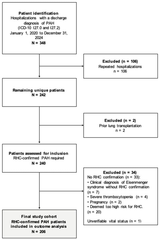
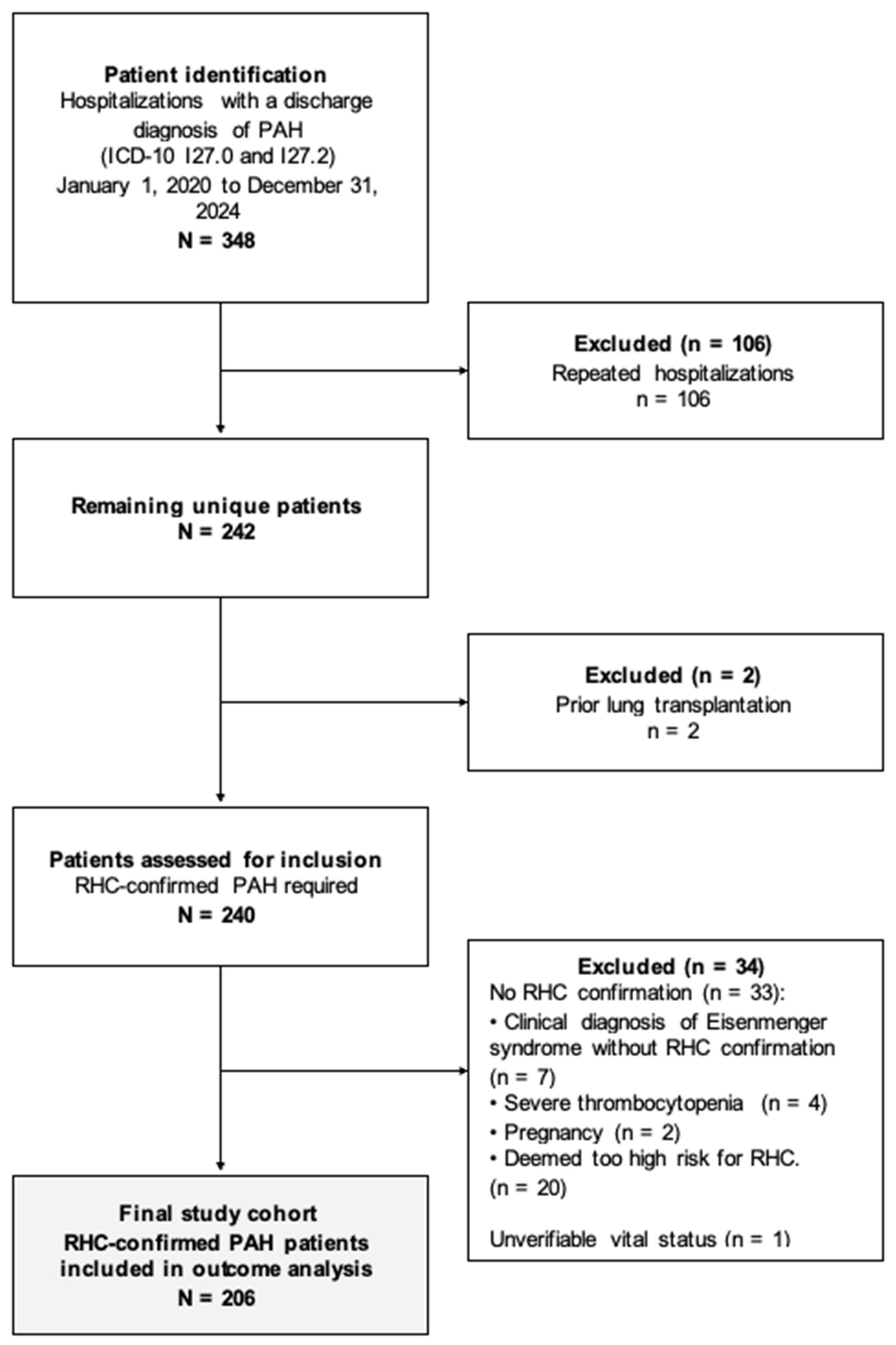
Between 2020 and 2024, 348 hospitalizations with a diagnosis of PAH were identified (Figure 1). After exclusion of repeated hospitalizations, 242 unique patients remained. Two patients with prior lung transplantation were excluded, leaving 240 eligible patients. Subsequently, 33 patients were excluded because of a clinical diagnosis of Eisenmenger syndrome without RHC confirmation (n = 7), severe thrombocytopenia (n = 4), pregnancy (n = 2), or contraindications to RHC (n = 20). One additional patient was excluded because of unverified vital status (n = 1). The final study cohort consisted of 206 patients with right heart catheterization-confirmed PAH who were included in the baseline and Kaplan–Meier survival analyses. Cox proportional hazards analyses included patients with complete data for the variables entered into each model. Other PAH etiologies included connective tissue disease-associated PAH (n = 6), porto-pulmonary hypertension (n = 5), and HIV-associated PAH (n = 1). The distribution of underlying congenital heart defects, including surgical correction status and the presence of Eisenmenger syndrome, is presented in Supplementary Table S3.
Figure 1.
Patient selection and study cohort formation.
Baseline demographic and clinical characteristics are summarized in Table 1. Baseline was defined as the first hospitalization during the study inclusion period. As this was a retrospective real-world cohort, a proportion of patients had an established diagnosis of PAH and were already receiving PAH-specific therapy before baseline assessment. The mean age of the cohort was 39.48 ± 13.73 years, and 80.6% were female. Age did not differ significantly among the etiological groups (p = 0.159), whereas female sex was more common in patients with IPAH than in those with CHD-PAH (86.8% vs. 68.5%, p = 0.005).
Table 1.
Demographic and clinical data (N = 206).
Most patients presented with advanced disease, with 129 (62.6%) classified as WHO functional class III–IV. Patients with IPAH more frequently presented in WHO functional class III–IV than those with CHD-PAH (69.4% vs. 52.1%), although the overall distribution among etiological groups did not differ significantly (p = 0.051). Hospitalization duration differed significantly among the groups (p = 0.032). Oxygen saturation (p = 0.011) and body mass index (p < 0.001) differed significantly among the etiological groups, with the lowest values observed in patients with congenital heart disease-associated PAH. Obesity (25.6%; overall p = 0.006) and syncope (p = 0.011) were more common among patients with IPAH. Laboratory evaluation demonstrated significant differences in hemoglobin concentration (p < 0.001) and NT-proBNP levels (p = 0.002) among the etiological groups. Patients with IPAH had the highest NT-proBNP levels, whereas patients with CHD-PAH had the highest hemoglobin concentrations. Ferritin concentration (p = 0.347) and uric acid (p = 0.123) did not differ significantly among the groups.
Baseline characteristics according to ferritin-defined low iron stores are presented in Supplementary Table S2. Patients with ferritin levels <30 µg/L were younger (p = 0.002), more likely to be female (p = 0.001), and had lower hemoglobin levels (p < 0.001). WHO functional class III–IV and mortality did not differ significantly between groups. Baseline COMPERA 2.0 four-strata risk stratification classified 43 patients (20.9%) as low risk, 58 (28.2%) as intermediate-low risk, 84 (40.8%) as intermediate-high risk, and 21 (10.2%) as high risk (Table 1). The largest proportion was classified as intermediate-high risk at baseline. Patients with IPAH were more frequently classified as intermediate-high risk than patients with CHD- PAH. The overall distribution of baseline COMPERA 2.0 four-strata risk categories differed significantly among the etiological groups (p = 0.026).
3.2. Echocardiographic and Hemodynamic Characteristics
Baseline echocardiographic and hemodynamic parameters are summarized in Table 2. Left ventricular dimensions differed significantly among the etiological groups. Patients with CHD-PAH had larger left ventricular end-diastolic and end-systolic volume indices than patients with IPAH (LVEDVi, p = 0.015; LVESVi, p = 0.022). Left ventricular ejection fraction remained preserved and did not differ significantly among the groups (p = 0.477).
Table 2.
Echocardiographic and hemodynamic parameters.
Among markers of right ventricular structure and function, right atrial volume index differed significantly among the etiological groups (p = 0.039), whereas right ventricular basal diameter, tricuspid annular plane systolic excursion (TAPSE), right ventricular systolic pressure, and right ventricular–pulmonary artery coupling ratio were comparable. The distribution of tricuspid regurgitation severity also differed significantly (p = 0.021), with severe or torrential tricuspid regurgitation observed more frequently in patients with IPAH than in those with CHD- PAH.
RHC demonstrated elevated pulmonary artery pressures across all etiological groups. Mean pulmonary artery pressure (p = 0.038) and pulmonary vascular resistance (p = 0.023) differed significantly among the groups, whereas pulmonary artery wedge pressure remained within the precapillary range and did not differ significantly (p = 0.300). Mixed venous oxygen saturation was significantly higher in patients with CHD-PAH than in the other etiological groups (p < 0.001), likely reflecting the presence of intracardiac shunting.
3.3. PAH-Targeted Therapy
Baseline PAH-targeted therapy patterns are summarized in Table 3. Overall, 103 patients (50.0%) were receiving triple combination therapy, 75 (36.4%) dual combination therapy, and 28 (13.6%) monotherapy or no PAH-targeted therapy. Treatment strategy differed significantly among the etiological groups (p = 0.017), with triple combination therapy used most frequently in patients with IPAH (58.7%).
Table 3.
PAH-targeted therapy (N = 206).
Endothelin receptor antagonists were the most frequently prescribed PAH-targeted medications (86.4%), followed by prostacyclin-pathway agents (56.3%), phosphodiesterase-5 inhibitors (53.4%), and soluble guanylate cyclase stimulators (40.8%). Treatment patterns differed significantly among the etiological groups. Patients with IPAH had higher proportions receiving endothelin receptor antagonists, prostacyclin-pathway agents, and soluble guanylate cyclase stimulators, whereas phosphodiesterase-5 inhibitors were prescribed more frequently in patients with CHD-PAH (all p < 0.01). Prostacyclin-pathway agents used during the study were iloprost and selexipag, both administered by non-parenteral routes.
Anticoagulation was prescribed more frequently in patients with IPAH, whereas antiplatelet therapy was more common in patients with CHD-PAH (both p < 0.001). Iron supplementation did not differ significantly among the etiological groups (p = 0.084), whereas diuretic use was comparable (p = 0.451).
3.4. Survival Analysis
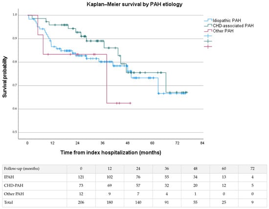
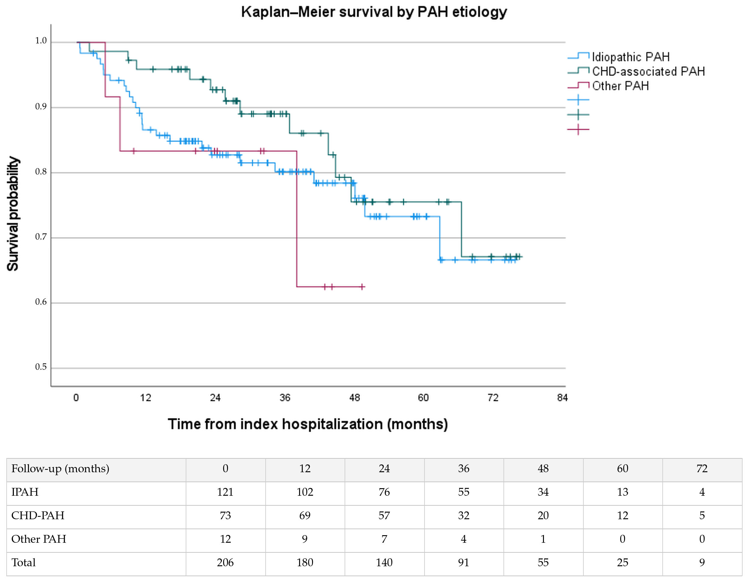
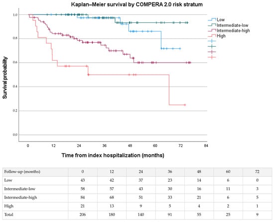
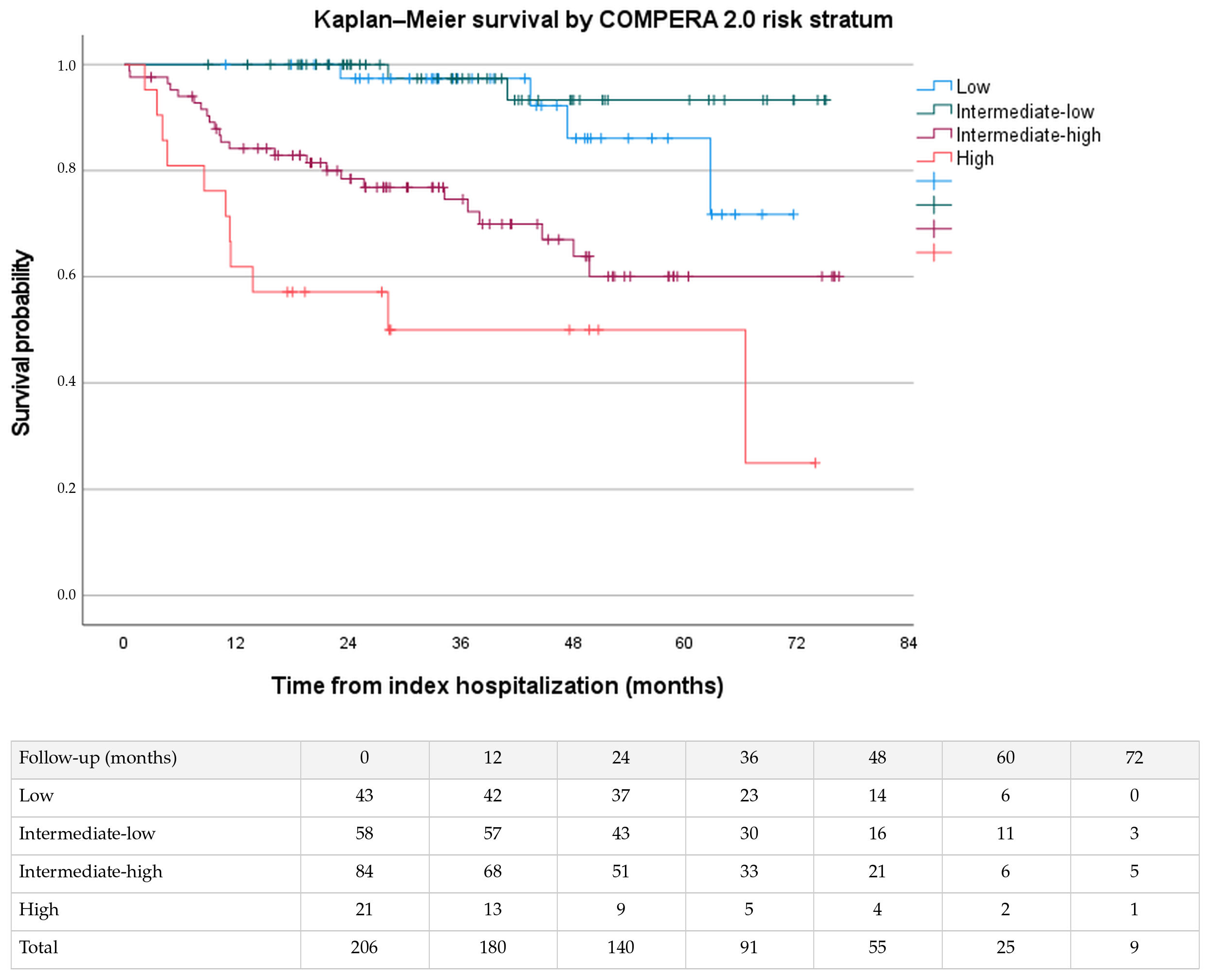
Survival analyses were performed in all 206 patients. During follow-up, 41 patients (19.9%) died. Median follow-up, estimated using the reverse Kaplan–Meier method, was 38.3 months (95% CI 33.9–42.7). Estimated overall survival at 1, 3 and 5 years was 89.7% (95% CI 85.6–93.8%), 83.4% (95% CI 77.9–88.9%), and 73.6% (95% CI 65.6–81.6%), respectively. Kaplan–Meier survival curves according to PAH etiology are shown in Figure 2. Overall survival did not differ significantly among patients with IPAH, CHD-PAH, and other forms of PAH (log-rank p = 0.407). Patients with CHD-PAH demonstrated the most favorable survival, whereas patients with other forms of PAH had the poorest survival; however, these differences were not statistically significant. Survival according to baseline COMPERA 2.0 risk category is shown in Figure 3. Baseline COMPERA 2.0 four-strata risk stratification differentiated survival, with progressively poorer outcomes observed across increasing risk categories (log-rank p < 0.001). Median survival was reached only among patients classified as high risk at baseline.
Figure 2.
Kaplan–Meier survival according to PAH etiology. Survival was calculated from the index hospitalization to death or last follow-up. Censoring is indicated by tick marks. Survival distributions did not differ significantly among etiologic groups (log-rank p = 0.407). Numbers at risk are below the plot.
Figure 3.
Kaplan–Meier survival curves stratified by baseline COMPERA 2.0 four-strata risk category. Survival was calculated from the index hospitalization to death or last follow-up. Censoring is indicated by marks. Differences in survival between risk groups were evaluated using the log-rank test (p < 0.001). Numbers at risk are shown below the plot.
Univariable and multivariable Cox proportional hazards regression analyses are presented in Supplementary Table S1. In univariable analysis, WHO functional class III–IV, COMPERA 2.0 four-strata risk category, body mass index, NT-proBNP, 6 min walk distance, TAPSE, right ventricular systolic pressure, right ventricular–pulmonary artery coupling and right ventricular basal diameter were associated with mortality. The multivariable model included 182 patients with complete covariate data (39 deaths). In the multivariable model, the high baseline COMPERA 2.0 risk category (HR 4.67, 95% CI 1.22–17.86; p = 0.024) and reduced TAPSE (HR 0.89 per 1 mm increase, 95% CI 0.80–0.99; p = 0.026) remained independently associated with mortality. The intermediate-high-risk COMPERA category also demonstrated an increased risk of death, although this association did not reach statistical significance (HR 2.88, 95% CI 0.96–8.68; p = 0.060).
4. Discussion
This study presents real-world data from the largest pulmonary hypertension referral center in Kazakhstan and represents one of the most comprehensive adult PAH cohorts reported from Central Asia. Several important findings emerged from this analysis. First, patients were predominantly young women and frequently presented with advanced functional impairment, highlighting the substantial burden of disease at referral. Second, combination PAH-targeted therapy was widely implemented in routine clinical practice, with approximately half of the patients receiving triple combination therapy at baseline. Third, baseline COMPERA 2.0 risk stratification differentiated long-term survival and remained independently associated with mortality, supporting its applicability in this Central Asian cohort. Finally, lower TAPSE, reflecting reduced right ventricular systolic function, remained independently associated with mortality in multivariable analysis.
Recent reports from national referral programs have described the clinical characteristics and survival outcomes of patients with chronic thromboembolic pulmonary hypertension and heart transplantation recipients, highlighting both the availability of specialized cardiovascular care and the continued need for robust local outcome data [11,12]. The present study expands this emerging body of evidence by providing comprehensive data on the clinical characteristics, treatment patterns, survival outcomes, and prognostic value of baseline COMPERA 2.0 risk stratification in patients with PAH in Kazakhstan.
In our cohort, patients were predominantly female (80.6%), with a mean age of 39.5 ± 13.7 years, and nearly two-thirds presented with WHO functional class III/IV, indicating advanced functional impairment at referral. The relatively young age and marked female predominance observed in our cohort are consistent with reports from selected Asian and developing-country registries [7,13]. Similar demographic characteristics have recently been described in a Kazakhstan cohort of patients with IPAH, in which the median age at diagnosis was approximately 40 years and women accounted for 86.8% of cases [14]. Comparable findings have also been reported in studies from China, Korea, Saudi Arabia, and Taiwan, where the mean age at diagnosis ranged from approximately 36 to 41 years and the proportion of female patients ranged from 63% to 81% [15,16,17,18]. In contrast, European registries, including SPAHR and COMPERA, have reported substantially older populations, with median ages ranging from 60 to 71 years [19,20].
The etiological distribution in our cohort also demonstrated several important regional characteristics. IPAH was the predominant subtype, accounting for 58.7% of cases, whereas CHD-PAH represented 35.4% of the cohort. This distribution differs somewhat from several Asian registries, particularly those from China and Southeast Asia, where CHD-PAH often equals or exceeds idiopathic PAH in proportion [10,15,16]. In contrast, Western registries generally report idiopathic PAH as the predominant etiology, accounting for approximately 55% of cases, whereas connective tissue disease-associated and CHD-PAH account for approximately 21% and 12% of cases, respectively [7]. The relatively high proportion of CHD-PAH observed in our study may reflect regional differences in the diagnosis and management of congenital heart disease (CHD), including the timing of defect repair. A national electronic healthcare database study documented CHD diagnosis extending into adulthood in Kazakhstan [21]. Conversely, connective tissue disease-associated PAH was uncommon in our cohort, comprising only six patients. This finding may reflect underdiagnosis, limited implementation of systematic screening among patients with connective tissue diseases, or referral patterns favoring congenital and cardiac forms of pulmonary hypertension to specialized centers [7,13].
Patients in our cohort presented with relatively advanced disease severity, with 62.6% classified as WHO functional class III–IV at baseline, a mean 6 min walk distance of approximately 300 m, and elevated NT-proBNP levels. Similar patterns of advanced functional limitation have been reported in cohorts from Saudi Arabia and Taiwan, where 72.9% and 80% of patients presented with WHO functional class III–IV disease [17,18]. Advanced functional impairment remains common in contemporary Western cohorts: functional class III-IV was reported in 85.3% of patients at baseline in the COMPERA 2.0 study [9] and 56.7% in a recent analysis of incident PAH patients from the US Pulmonary Hypertension Association Registry [22]. Longer diagnostic delay has been associated with poorer five-year survival in the Australian and New Zealand pulmonary hypertension registry [23]. The high proportion of patients with advanced functional limitation at baseline may reflect delayed recognition of PAH, referral to a national tertiary center after disease progression, and the limited availability of specialized pulmonary hypertension services in the region. These potential factors could not be directly assessed in the present study.
Echocardiographic and hemodynamic findings demonstrated marked right ventricular remodeling and severe pulmonary vascular disease. Mean right ventricular basal diameter was 4.85 ± 0.82 cm. Median right ventricular systolic pressure was 78 mmHg, median mean pulmonary artery pressure was 49 mmHg, and median pulmonary vascular resistance was 8.38 Wood units, consistent with advanced pulmonary arterial hypertension at presentation and comparable with findings reported in Asian and Western PAH registries [7,13,14,15,16,17,18]. Patients with CHD-PAH had numerically larger left ventricular volumes than patients with idiopathic PAH and higher recorded mixed venous oxygen saturation than the other etiological groups, potentially reflecting the underlying hemodynamic consequences of congenital cardiac shunts. In contrast, pulmonary vascular resistance and mean pulmonary artery pressure were highest among patients with IPAH. A major strength of the present study is that all included patients had right heart catheterization-confirmed PAH, although invasive hemodynamic measurements from the index hospitalization were available only in a subset of patients.
Treatment patterns in our cohort reflected contemporary PAH management, with half of the patients (50.0%) receiving triple combination therapy at baseline. This proportion is higher than that reported in several earlier Asian and Western registries, where monotherapy and dual combination therapy predominated [7,13,14,16]. However, comparisons should account for differences in enrolment periods and the inclusion of both incident and prevalent patients in our cohort. Endothelin receptor antagonists were the most frequently prescribed PAH-targeted medications (86.4%), followed by prostacyclin-pathway agents (56.3%), phosphodiesterase-5 inhibitors (53.4%), and soluble guanylate cyclase stimulators (40.8%). Nevertheless, access to prostacyclin-pathway therapy remained limited, with inhaled iloprost and oral selexipag representing the principal available agents during the study period, reflecting ongoing regional differences in treatment availability [2,7].
Despite substantial advances in targeted therapies, pulmonary arterial hypertension remains associated with considerable long-term mortality worldwide [4]. In our cohort, the largest proportion of patients (40.8%) was classified as intermediate-high risk according to the baseline COMPERA 2.0 four-strata model, reflecting the advanced clinical profile of the study population. Baseline COMPERA 2.0 four-strata risk stratification effectively differentiated long-term survival, with progressively poorer outcomes observed across increasing risk categories. These findings are consistent with previous studies demonstrating the prognostic value of COMPERA-based risk assessment and support the routine use of guideline-recommended multidimensional risk stratification in patients with PAH [2,9]. In contrast, although patients with CHD-PAH demonstrated numerically better survival than those with IPAH and other forms of PAH, these differences did not reach statistical significance (log-rank p = 0.407).
Previous registry studies have reported variable findings regarding survival differences between CHD-PAH and IPAH. In the COMPERA-CHD registry, 5-year survival was significantly higher among patients with CHD-PAH than among those with IPAH (76% vs. 54%) [24], whereas analyses from the REVEAL registry demonstrated comparable survival between CHD-PAH and idiopathic/heritable PAH despite important differences in hemodynamic characteristics [25]. More recently, the HOPE registry reported a 5-year survival of 81.9% among patients with CHD-PAH [26]. The numerically more favorable survival observed in our CHD-PAH cohort is therefore broadly consistent with these reports, although differences in cohort composition and follow-up limit direct comparisons. The absence of a statistically significant survival difference in our study may partly reflect limited statistical power, particularly given the small, heterogeneous other PAH subgroup; however, a true difference between etiological groups could not be established.
COMPERA 2.0 four-strata risk model has demonstrated prognostic value across multiple PAH populations and has become an integral component of contemporary risk assessment [2,9]. In the present study, the baseline COMPERA 2.0 four-strata risk model stratified survival and remained associated with mortality after multivariable adjustment. These findings provide additional evidence for its applicability in a hospitalized Central Asian referral cohort. Lower TAPSE also remained independently associated with mortality in the multivariable analysis, highlighting the prognostic importance of right ventricular systolic function in determining outcomes in PAH. Progressive right ventricular dysfunction plays a central role in clinical deterioration and death in PAH, and TAPSE has been associated with prognosis in previous cohorts [27]. Our findings further support the routine incorporation of echocardiographic assessment of right ventricular function into the comprehensive risk evaluation of patients with PAH.
5. Limitations
Several limitations should be acknowledged. First, this was a retrospective single-center study conducted at the largest pulmonary hypertension referral center in Kazakhstan, which may limit the generalizability of the findings to the broader regional population. Patients managed only as outpatients may not be represented. Second, the study included both incident and prevalent patients, and a proportion of patients were already receiving PAH-targeted therapy before baseline assessment, potentially introducing survivor bias and limiting direct comparison with incident PAH registries. Third, although all included patients had right heart catheterization-confirmed PAH, missing data and variations in routine clinical practice limited the availability of certain echocardiographic, laboratory, and invasive hemodynamic variables for all patients. Fourth, the relatively small size of the “other PAH” subgroup reduced the statistical power of subgroup comparisons and may have contributed to the absence of statistically significant survival differences between etiological groups. The limited number of deaths and wide confidence intervals for some adjusted estimates mean that these findings should be interpreted cautiously. Fifth, because this was an observational study, causal relationships between baseline characteristics and clinical outcomes cannot be established.
6. Conclusions
In this hospitalized, single-center cohort from Kazakhstan, patients with pulmonary arterial hypertension frequently had advanced functional impairment at baseline despite their relatively young age. Baseline COMPERA 2.0 four-strata risk stratification differentiated survival, while lower TAPSE was independently associated with mortality, highlighting the prognostic importance of right ventricular systolic function. These findings support the routine use of multidimensional risk assessment in patients with pulmonary arterial hypertension and underscore the need for prospective multicenter PAH registries in Kazakhstan and across Central Asia.
Supplementary Materials
The following supporting information can be downloaded at: https://www.mdpi.com/article/10.3390/medicina62112080/s1, Supplementary Table S1. Univariable and multivariable Cox proportional hazards regression analyses for predictors of mortality in patients with pulmonary arterial hypertension. Supplementary Table S2. Baseline characteristics according to ferritin-defined low iron stores. Supplementary Table S3. Characteristics of congenital heart disease in patients with CHD-associated pulmonary arterial hypertension (n = 73).
Author Contributions
Conceptualization, A.A., M.M. (Murat Mukarov) and Y.S.; Methodology, A.A., N.K. and Y.S.; Data curation, A.A., M.M. (Meruyert Muradaliyeva), G.D. and A.Z.; Formal analysis, A.A. and Y.S.; Investigation, M.M. (Murat Mukarov), N.K., M.M. (Meruyert Muradaliyeva), G.D. and A.Z.; Writing—original draft preparation, A.A. and N.K.; Writing—review and editing, all authors; Supervision, M.M. (Murat Mukarov) and Y.S.; Project administration, A.A., N.K., M.M. (Meruyert Muradaliyeva) and G.D. All authors have read and agreed to the published version of the manuscript.
Funding
This research received no external funding.
Institutional Review Board Statement
The study was conducted in accordance with the Declaration of Helsinki and approved by the Local Bioethics Committee of the Corporate Fund “University Medical Center”, Astana, Kazakhstan (Protocol No. 3, application No. 04-024; approved on 28 April 2026).
Informed Consent Statement
Patient consent was waived by the Local Bioethics Committee due to the retrospective observational design of the study and the use of de-identified, routinely collected clinical data.
Data Availability Statement
The datasets used and/or analyzed during the current study are available from the corresponding author on reasonable request.
Conflicts of Interest
The authors declare no conflicts of interest.
References
- Montani, D.; Günther, S.; Dorfmüller, P.; Perros, F.; Girerd, B.; Garcia, G.; Jaïs, X.; Savale, L.; Artaud-Macari, E.; Price, L.C.; et al. Pulmonary arterial hypertension. Orphanet J. Rare Dis. 2013, 8, 97. [Google Scholar] [CrossRef] [Scilit] [PubMed]
- Humbert, M.; Kovacs, G.; Hoeper, M.M.; Badagliacca, R.; Berger, R.M.F.; Brida, M.; Carlsen, J.; Coats, A.J.S.; Escribano-Subias, P.; Ferrari, P.; et al. ESC/ERS Scientific Document Group 2022 ESC/ERS Guidelines for the diagnosis treatment of pulmonary hypertension. Eur. Heart J. 2022, 43, 3618–3731. [Google Scholar] [CrossRef] [Scilit] [PubMed]
- Guignabert, C.; Aman, J.; Bonnet, S.; Dorfmüller, P.; Olschewski, A.J.; Pullamsetti, S.; Rabinovitch, M.; Schermuly, R.T.; Humbert, M.; Stenmark, K.R. Pathology and pathobiology of pulmonary hypertension: Current insights and future directions. Eur. Respir. J. 2024, 64, 2401095. [Google Scholar] [CrossRef] [Scilit] [PubMed]
- Chang, K.Y.; Duval, S.; Badesch, D.B.; Bull, T.M.; Chakinala, M.M.; De Marco, T.; Frantz, R.P.; Hemnes, A.; Mathai, S.C.; Rosenzweig, E.B.; et al. Mortality in pulmonary arterial hypertension in the modern era: Early insights from the Pulmonary Hypertension Association Registry. J. Am. Heart Assoc. 2022, 11, e024969. [Google Scholar] [CrossRef] [Scilit] [PubMed]
- Guan, A.; Dai, Z.; Lin, J.; Chen, Q.; Jiang, C. Global disease burden of pulmonary arterial hypertension: Patterns and inequality in 1990–2021, with projections to 2035. BMC Public Health 2026, 26, 4. [Google Scholar] [CrossRef] [Scilit] [PubMed]
- Huang, S.; Qiu, J.; Wang, A.; Ma, Y.; Wang, P.; Ding, D.; Qiu, L.; Li, S.; Liu, M.; Zhang, J.; et al. Burden of pulmonary arterial hypertension in Asia from 1990 to 2021: Findings from Global Burden of Disease Study 2021. Chin. Med. J. 2025, 138, 1324–1333. [Google Scholar] [CrossRef] [Scilit] [PubMed]
- Anderson, J.J.; Lau, E.M. Pulmonary hypertension definition, classification, and epidemiology in Asia. JACC Asia 2022, 2, 538–546. [Google Scholar] [CrossRef] [Scilit] [PubMed]
- Dardi, F.; Boucly, A.; Benza, R.; Frantz, R.; Mercurio, V.; Olschewski, H.; Rådegran, G.; Rubin, L.J.; Hoeper, M.M. Risk stratification and treatment goals in pulmonary arterial hypertension. Eur. Respir. J. 2024, 64, 2401323. [Google Scholar] [CrossRef] [Scilit] [PubMed]
- Hoeper, M.M.; Pausch, C.; Olsson, K.M.; Huscher, D.; Pittrow, D.; Grünig, E.; Staehler, G.; Vizza, C.D.; Gall, H.; Distler, O.; et al. COMPERA 2.0: A refined four-stratum risk assessment model for pulmonary arterial hypertension. Eur. Respir. J. 2022, 60, 2102311. [Google Scholar] [CrossRef] [Scilit] [PubMed]
- Jiang, H.; Tan, J.L.; Ruan, W.; Hon, J.S.; Ismail, A.; Lim, C.L.; Perumal, S.; Koh, M.; Sewa, D.W.; Phua, G.C.; et al. Performance of risk-stratification scores for patients with pulmonary arterial hypertension in a multi-ethnic Asian population. Pulm. Circ. 2025, 15, e70032. [Google Scholar] [CrossRef] [Scilit] [PubMed]
- Semenova, Y.; Shaisultanova, S.; Beyembetova, A.; Asanova, A.; Sailybayeva, A.; Novikova, S.; Myrzakhmetova, G.; Pya, Y. Examining a 12-year experience within Kazakhstan’s national heart transplantation program. Sci. Rep. 2024, 14, 10291. [Google Scholar] [CrossRef] [Scilit] [PubMed]
- Abbay, A.; Askanbekova, A.; Semenova, Y.; Kuzhakhmetova, A.; Duysenbay, G.; Mukarov, M.; Lesbekov, T. Clinical characteristics, management strategies, and survival outcomes of patients with chronic thromboembolic pulmonary hypertension in Central Asia: Experience from the sole pulmonary endarterectomy center. Front. Cardiovasc. Med. 2026, 13, 1786958. [Google Scholar] [CrossRef] [Scilit] [PubMed]
- Lau, E.M.T.; Giannoulatou, E.; Celermajer, D.S.; Humbert, M. Epidemiology and treatment of pulmonary arterial hypertension. Nat. Rev. Cardiol. 2017, 14, 603–614. [Google Scholar] [CrossRef] [Scilit] [PubMed]
- Taizhanova, D.; Nurpissova, T.; Abildinova, G.; Martynyuk, T.; Kulmyrzayeva, N.; Zholdybayeva, E. Hemodynamic and genetic associations with the risk of idiopathic pulmonary arterial hypertension development in an ethnic cohort of Kazakhs. Diagnostics 2024, 14, 2687. [Google Scholar] [CrossRef] [Scilit] [PubMed]
- Zhang, C.; Ding, H.; Li, J.; Ramey, D.R.; Zhu, B.; Zhou, Z.; Yang, M.; Lautsch, D. Epidemiology, disease burden of pulmonary arterial hypertension in China: A systematic literature review. Respir. Med. 2025, 240, 108007. [Google Scholar] [CrossRef] [Scilit] [PubMed]
- Cha, J.H.; Jang, S.Y.; Song, J.; Kang, I.S.; Huh, J.; Park, T.K.; Yang, J.H.; Park, S.W.; Kim, H.; Kim, D.K.; et al. A single center experience of pulmonary arterial hypertension management in Korea: A 25-year comparative analysis following the introduction of targeted therapy. Korean Circ. J. 2024, 54, 636–650. [Google Scholar] [CrossRef] [Scilit] [PubMed]
- Idrees, M.M.; Al-Najashi, K.; Khan, A.; Al-Dammas, S.; Al-Awwad, H.; Batubara, E.; Al Otai, A.; Abdulhameed, J.; Fayed, A.; Kashour, T. SAPH Registry Taskforce. Pulmonary arterial hypertension in Saudi Arabia: Patients’ clinical and physiological characteristics and hemodynamic parameters. A single center experience. Ann. Thorac. Med. 2014, 9, 209–215. [Google Scholar] [CrossRef] [Scilit] [PubMed]
- Wang, L.Y.; Lee, K.T.; Lin, C.P.; Hsu, L.A.; Wang, C.L.; Hsu, T.S.; Ho, W.J. Long-term survival of patients with pulmonary arterial hypertension at a single center in Taiwan. Acta Cardiol. Sin. 2017, 33, 498–509. [Google Scholar] [CrossRef] [Scilit] [PubMed]
- Hoeper, M.M.; Huscher, D.; Ghofrani, H.A.; Delcroix, M.; Distler, O.; Schweiger, C.; Grunig, E.; Staehler, G.; Rosenkranz, S.; Halank, M.; et al. Elderly patients diagnosed with idiopathic pulmonary arterial hypertension: Results from the COMPERA registry. Int. J. Cardiol. 2013, 168, 871–880. [Google Scholar] [CrossRef] [Scilit] [PubMed]
- Kylhammar, D.; Hjalmarsson, C.; Hesselstrand, R.; Jansson, K.; Kavianipour, M.; Kjellström, B.; Nisell, M.; Söderberg, S.; Rådegran, G. Predicting mortality during long-term follow-up in pulmonary arterial hypertension. ERJ Open Res. 2021, 7, 00837–2020. [Google Scholar] [CrossRef] [Scilit] [PubMed]
- Syssoyev, D.; Seitkamzin, A.; Lim, N.; Mussina, K.; Poddighe, D.; Gaipov, A.; Galiyeva, D. Epidemiology of congenital heart disease in Kazakhstan: Data from the Unified National Electronic Healthcare System 2014–2021. J. Clin. Med. Kazakhstan 2024, 21, 49–55. [Google Scholar] [CrossRef] [Scilit] [PubMed]
- Khan, S.L.; Paoli, C.J.; Kime, N.; Williams, K.; Rendon, G.G.; Tonelli, A.R.; Cajigas, H.; Mathai, S.C.; Sahay, S. Treatment patterns in patients with incident pulmonary hypertension: Real-world data from the Pulmonary Hypertension Association Registry. Chest 2026, 169, 1077–1090. [Google Scholar] [CrossRef] [Scilit] [PubMed]
- Khou, V.; Anderson, J.J.; Strange, G.; Corrigan, C.; Collins, N.; Celermajer, D.S.; Dwyer, N.; Feenstra, J.; Horrigan, M.; Keating, D.; et al. Diagnostic delay in pulmonary arterial hypertension: Insights from the Australian and New Zealand pulmonary hypertension registry. Respirology 2020, 25, 863–871. [Google Scholar] [CrossRef] [Scilit] [PubMed]
- Kaemmerer, H.; Gorenflo, M.; Huscher, D.; Pittrow, D.; Apitz, C.; Baumgartner, H.; Berger, F.; Bruch, L.; Brunnemer, E.; Budts, W.; et al. Pulmonary hypertension in adults with congenital heart disease: Real-world data from the international COMPERA-CHD registry. J. Clin. Med. 2020, 9, 1456. [Google Scholar] [CrossRef] [Scilit] [PubMed]
- Barst, R.J.; Ivy, D.D.; Foreman, A.J.; McGoon, M.D.; Rosenzweig, E.B. Four- and seven-year outcomes of patients with congenital heart disease-associated pulmonary arterial hypertension (from the REVEAL Registry). Am. J. Cardiol. 2014, 113, 147–155. [Google Scholar] [CrossRef] [Scilit] [PubMed]
- Patsiou, V.; Arvanitaki, A.; Farmakis, I.T.; Anthi, A.; Demerouti, E.; Apostolopoulou, S.; Feloukidis, C.; Gourgiotis, P.; Papadopoulos, G.E.; Chrysochoidis-Trantas, T.; et al. Survival prospects of pulmonary arterial hypertension associated with congenital heart disease: Insights from the HOPE registry. Int. J. Cardiol. 2025, 421, 132894. [Google Scholar] [CrossRef] [Scilit] [PubMed]
- Vonk Noordegraaf, A.; Galiè, N. The role of the right ventricle in pulmonary arterial hypertension. Eur. Respir. Rev. 2011, 20, 243–253. [Google Scholar] [CrossRef] [Scilit] [PubMed]

Disclaimer/Publisher’s Note: The statements, opinions and data contained in all publications are solely those of the individual author(s) and contributor(s) and not of MDPI and/or the editor(s). MDPI and/or the editor(s) disclaim responsibility for any injury to people or property resulting from any ideas, methods, instructions or products referred to in the content. |
© 2026 by the authors. Published by MDPI on behalf of the Lithuanian University of Health Sciences. Licensee MDPI, Basel, Switzerland. This article is an open access article distributed under the terms and conditions of the Creative Commons Attribution (CC BY) license.