


Abstract
Periodontitis is a chronic, dysbiosis-driven inflammatory disease, that, unlike gingivitis, results in irreversible, tooth-supporting soft tissue and bone loss. Clarifying the molecular pathways that sustain the host inflammatory response is therefore central to improving disease management. Lipopolysaccharide (LPS), a major pathogen-associated molecular pattern of Gram-negative bacteria, is one important—though not the sole—microbial stimulus that drives inflammation in the polymicrobial, dysbiotic periodontal environment. This narrative review summarizes how LPS-initiated signaling promotes production of the pro-inflammatory cytokines TNF-α and IL-1, how these cytokines couple inflammation to osteoclast-mediated bone loss, and whether their pharmacological blockade holds therapeutic promise. A literature search combining the terms “LPS,” “periodontitis,” “TNF-α,” and “IL-1” was conducted in PubMed/MEDLINE, Scopus, and Google Scholar for English-language articles published up to 2026, with priority given to primary studies over secondary citations. Preclinical models consistently show that TNF-α and IL-1 blockade reduces periodontal inflammation and bone loss, but the effect is time-dependent: short-term blockade can aid healing whereas prolonged blockade may impair it, and robust periodontitis-specific clinical trials are lacking. We conclude that targeted modulation of TNF-α and IL-1—particularly through locally delivered, appropriately timed agents used as an adjunct to conventional mechanical therapy—is a biologically rational strategy.
Keywords:
periodontitis; inflammation; TNF-α; IL-1β; lipopolysaccharide; LPS; molecular mechanisms; therapeutics 1. Introduction
Periodontitis is a chronic inflammatory disease and develops in response to a dysbiotic subgingival biofilm, leading to progressive destruction of the periodontal soft tissue and alveolar bone and, if untreated, to tooth loss. It is highly prevalent, affecting nearly half of adults worldwide, with roughly 10% experiencing severe forms [1,2]. Beyond the oral cavity, periodontitis is increasingly recognized as a contributor to systemic inflammatory burden, and epidemiological and mechanistic studies have linked it to cardiovascular disease [3,4,5,6], Alzheimer’s disease (AD) [7], Parkinson’s disease [8,9], type 2 diabetes mellitus (in a bidirectional relationship) [10], and rheumatoid arthritis (RA) [11]. TNF-α and IL-1 are implicated both locally and as mediators of this systemic crosstalk, which is one reason that dampening their activity could produce benefits extending beyond the periodontium [12,13,14,15].
The etiology of periodontitis is microbial, but the tissue destruction is driven by the host inflammatory response to microbial challenge [16]. The dysbiotic subgingival biofilm is characterized by a high abundance of Gram-negative bacteria. Among them, Porphyromonas gingivalis (P. gingivalis) acts as a keystone pathogen [17,18], orchestrating dysbiosis and altering host-microbial homeostasis to promote disease progression [19]. P. gingivalis has been detected in approximately 85.75% of subgingival plaque samples from periodontitis patients, and its abundance correlates with disease severity [20]. P. gingivalis and other Gram-negative bacteria possess LPS and various virulence factors, including gingipains, fimbriae, nucleic acids, and lipoproteins, which function as pathogen-associated molecular patterns (PAMPs). These PAMPs modulate host immune responses and thereby contribute to the pathogenesis of periodontitis [21]. LPS acts as a prominent inflammatory stimulus within this complex microbial community. This review focuses on LPS-initiated signaling because it is among the best-characterized routes to TNF-α and IL-1 induction, while keeping this broader context in view.
Conventional management of periodontitis rests on mechanical disruption of the biofilm (scaling and root planning, surgical access where needed) and, in selected cases, adjunctive antimicrobials. These approaches are effective for many patients but have well-recognized limitations: residual deep pockets, disease recurrence, a subset of “non-responding” patients, and—critically—an incomplete effect on the host inflammatory response that drives tissue breakdown. Persistent inflammation, ongoing bone destruction, and heterogeneous responses to standard therapy together motivate interest in host-modulation strategies that target the inflammatory mediators themselves [22,23,24,25,26,27].
Building on the clinical success of cytokine blockade in RA, psoriasis, and inflammatory bowel disease, we examine whether neutralizing TNF-α or IL-1 could similarly benefit the periodontium. The central hypothesis motivating this review is that targeted modulation of TNF-α and IL-1 signaling may serve as an adjunctive strategy—complementing, not replacing, mechanical biofilm control—to reduce periodontal inflammation and preserve the tooth-supporting tissues. We first summarize LPS structure and its receptor-level recognition; then trace the downstream signaling that generates TNF-α and IL-1 and couples them to bone loss; and finally appraise the preclinical and (limited) clinical evidence for anti-TNF-α and anti-IL-1 therapy, giving particular weight to the questions of delivery route, treatment timing, safety, and evidence quality that will determine any future translational path.
2. Materials and Methods
A literature search was conducted in PubMed/MEDLINE, Scopus, and Google Scholar for English-language articles published from 1980s up to July 2026. using combinations of keywords: “periodontitis”, “inflammation”, “lipopolysaccharide”, “LPS”, “TNF-α”, “IL-1β”, “IL-1”, “TNF/IL-1 inhibitor/biologic therapy”. Reference lists of relevant articles were hand-searched for additional primary sources. Records were screened manually in three sequential steps: (1) studies had to be relevant to LPS, TNF-α, or IL-1 in the context of periodontitis; (2) priority was given to original research over review articles that merely cited primary results; and (3) referenced findings were checked against the primary source. Because a narrative review does not apply a formal risk-of-bias assessment, we instead sought to signpost the level of evidence throughout the text—distinguishing in vitro studies, animal models, human observational studies, and controlled clinical trials—so that preclinical findings are not given the same interpretive weight as clinical evidence. The figures included in this article were created by the authors using “PowerPoint” with some basic shapes derived from “Servier Medical Art, licensed under Creative Commons Attribution 4.0 International (CC BY 4.0)”.
3. LPS—TNF-α/IL-1 Signaling in Periodontitis
3.1. LPS Structure and Its Immune-Stimulatory Function
3.1.1. LPS Structure
LPS is an integral component of Gram-negative bacteria outer membrane [28] and is composed of three main elements: (1) lipid A; (2) core oligosaccharide; and (3) the O antigen (Figure 1). The structure and composition of these elements vary across and within bacterial species [29], and this variation produces diverse host immune responses. For instance, the lipid A of Escherichia coli (E. coli) is highly immunogenic, whereas other lipid A structures are far less so; producing weakly immunogenic lipid A is one strategy used by certain bacteria to evade immune detection and persist without triggering strong activation [30].
Figure 1.
Schematic of the Gram-negative outer membrane and LPS structure.
3.1.2. Recognition of LPS by Toll-like Receptors
LPS triggers immune responses chiefly through its lipid A moiety, which is recognized by the host’s Toll-like receptors (TLRs) [31]. TLRs are pattern-recognition-receptors (PRRs) that detect conserved microbial structures; human express ten TLRs, whereas mice express thirteen. TLR activation has been implicated in inflammatory disease across many organs, including the oral cavity, where it contributes to periodontal inflammation and alveolar bone loss [32]. TLR4 primarily detects LPS from Gram-negative bacteria whereas TLR2 recognizes lipoteichoic acid (LTA) from Gram-positive bacteria as well as certain bacterial lipoproteins [33]; both TLR2 and TLR4 predominate in periodontal tissues [34,35,36,37].
A crucial and disease-relevant point is that the LPS of P. gingivalis is atypical. Unlike the relatively uniform LPS of enteric bacteria, P. gingivalis produces a heterogeneous mixture of lipid A species: tetra-acylated forms act as weak TLR4 agonists or even TLR4 antagonists, whereas penta-acylated forms activate TLR4-dependent signaling [38]. This structural flexibility is a core strategy by which P. gingivalis manipulates host immunity, and it underscores why LPS-driven signaling in periodontitis cannot be treated as a single, uniform input. In human gingival fibroblasts and macrophages, different P. gingivalis lipid A structures differentially alter NF-κB activation and the production of TNF-α, IL-1β, IL-6, and IL-8 [38].
3.1.3. Downstream Signaling from LPS-TLR Engagement
Signals from LPS–TLR engagement are transmitted intracellularly through adaptors and kinases including MyD88, interleukin-1,4 receptor-associated kinases (IRAK1,4), TNF receptor-associated factor 6 (TRAF6), and NF-κβ, leading to production of proinflammatory cytokines [39]. TLR4 signaling is notable in that it bifurcates into two branches (Figure 2). The MyD88-dependent branch, recruited at the plasma membrane via the sorting adaptor TIRAP, activates NF-κB and activator protein-1 (AP-1) to drive early transcription of TNF-α and IL-1 [39]. The TRIF-dependent (MyD88-independent) branch, engaged after TLR4 internalization via the adaptor TRAM, activates interferon regulatory factor 3 (IRF3) to induce type I interferons (IFNs) and also contributes to a later phase of NF-κB activation [40]. Distinguishing these branches matters biologically because IL-1 and TNF-α promote osteoclast differentiation and activity [41], whereas type I IFNs generally inhibit osteoclast formation [42]; the net osteoclastogenic output of TLR4 signaling thus reflects the balance between its MyD88- and TRIF-driven arms. In periodontal tissues, TLR2 signaling (also MyD88-dependent) provides an additional route to NF-κB activation, and P. gingivalis can exploit a TLR2–PI3K axis to escape immune clearance while still promoting bone resorption [43].
Figure 2.
Binding of LPS to TLR4 (with TLR2) activates the MyD88-dependent pathway, and, after receptor internalization, the TRIF-dependent (MyD88-independent) pathway; MyD88-dependent signaling drives NF-κB/AP-1 and production of IL-1 and TNF-α, whereas the TRIF branch activates IRF3/7 and type I IFNs. LPS: lipopolysaccharide; LBP: LPS binding protein; TLR: toll-like receptor; CD14: cluster of differentiation 14; MD-2: myeloid differentiation factor 2; LTA: lipoteichoic acid; LPN: lipoprotein; TIRAP: TIR domain-containing adaptor protein; MyD88: myeloid differentiation primary response 88; IRAK1/4: interleukin-1 receptor-associated kinase 1/4; TRAF: TNF receptor-associated factor; TAB: TAK-binding protein; TAK1: TGF-β-activated kinase 1; IKK: inhibitor of κB kinase; NF-κB: nuclear factor kappa-light-chain-enhancer of activated B cells; MAPK: mitogen-activated protein kinase; AP1: activator protein-1; TNF-α: tumor necrosis factor-alpha; IL-1: interleukin-1; TRAM: TRIF-related adaptor molecule; TRIF: TIR-domain-containing adaptor-inducing beta interferon; TBK1: TANK-binding kinase 1; IRF: interferon regulatory factor; IFNs: interferons.
3.1.4. Post-Transcriptional Regulation of LPS-Induced Cytokine Production
Although LPS-triggered activation of TLRs and transcription factors such as NF-κB is essential for initiating cytokine gene expression, transcription alone does not fully explain the persistently high levels of TNF-α, IL-1β and IL-6 seen in chronic periodontal lesions; post-transcriptional control of mRNA stability also contributes [44]. A well-characterized mechanism is the p38 mitogen-activated protein kinase (MAPK) pathway and its downstream effector MAPK-activated protein kinase 2 (MK2), which stabilizes mRNAs encoding proinflammatory cytokines [44,45]. Recognizing this post-transcriptional layer helps explain both the chronicity of periodontal inflammation and the variable responses observed with cytokine-targeted therapies [44].
3.2. LPS and the Host Immune Response
3.2.1. The Periodontal Immune-Cell Landscape
Analyses of gene-expression data (Gene Expression Omnibus) indicate that plasma cells (differentiated B cells), naïve B cells, and neutrophils are enriched in periodontitis lesions, whereas memory B cells, CD4+ memory T cells, and resting dendritic cells predominate in healthy periodontal tissue [46]. During inflammation, immune cells release reactive oxygen species (ROS) and proinflammatory cytokines—including TNF-α, IL-1, and prostaglandin E2 (PGE2)—that together promote osteoclast differentiation and bone resorption [47,48]. P. gingivalis fragments and virulence factors activate neutrophils and macrophages, driving cytokine release and RANKL-dependent osteoclastogenesis that ultimately causes alveolar bone loss [43].
3.2.2. LPS and Monocytes/Macrophages
Most myeloid-lineage cells express TLR4. LPS binding to TLR4 requires accessory molecules including LPS-binding protein (LBP), cluster of differentiation 14 (CD14), and MD-2, and specifically involves the acyl chains and phosphate groups of lipid A. In organisms such as E. coli, hexa-acylated and diphosphorylated lipid A is a potent TLR4 agonist; Conversely, under-acylation or dephosphorylation markedly reduces proinflammatory activity and is a key mechanism for down-regulating inflammation and promoting immune resolution [49]. This structural dependence again illustrates that “LPS” is not a single functional entity.
3.2.3. LPS and Neutrophils
Human neutrophils express TLR1, 2, 4, 5, 6, 7, 8, 9, and 10 but not TLR3 [50]. Unlike monocytes, LPS-activated neutrophils do not express type I IFN or IFN-dependent genes and thus do not mobilize the MyD88-independent/TRIF-dependent pathway [51]. In health, neutrophils continuously migrate through the junctional epithelium into the gingival sulcus to maintain microbial homeostasis. In periodontitis, heightened neutrophil responsiveness and exaggerated neutrophil extracellular trap (NET) formation can damage the gingival barrier, allowing penetration of bacteria and PAMPs and stimulating Th17 cells; resulting in further neutrophil recruitment and tissue damage [52,53,54].
3.2.4. TNF-α and IL-1 Within the Broader Inflammatory Network
Although this review focuses on TNF-α and IL-1, these cytokines act within an integrated inflammatory network rather than in isolation. LPS-initiated signaling and the TNF-α/IL-1 axis intersect with IL-6, the IL-17/IL-23–Th17 pathway, IFN-γ, chemokines, matrix metalloproteinases (MMPs), the RANKL/OPG axis, and the NLRP3/AIM2 inflammasomes to shape the net outcome of connective-tissue degradation and bone loss. TNF-α and IL-1 frequently function as upstream amplifiers within this network, for example by inducing IL-6 and PGE2 and by up-regulating RANKL, which is part of the rationale for targeting them.
3.3. TNF-a and IL-1 Mediate LPS-Induced Inflammation and Bone Loss
LPS has been implicated in the pathogenesis of periodontitis through induction of proinflammatory cytokines such as IL-1 and TNF-α [55] and consequent bone resorption [56].
3.3.1. LPS Exposure, Cytokine Production, and Periodontal Destruction
In vivo (animal): Periodontal-pocket injection of LPS caused alveolar bone loss in rats [57]. Wistar rats developed periodontitis and alveolar bone loss accompanied by elevated NF-κB and RANKL after palatal injection of LPS [58]. These two animal studies demonstrated that local LPS exposure induced periodontal inflammation and tissue loss, providing evidence that bacterial LPS contributes to the pathogenesis of periodontitis. Systemic LPS administration combined with ligature placement in Sprague-Dawley rats led to periodontitis and alveolar bone loss associated with increased TNF-α, IL-6, NLRP3, cleaved caspase-1, and RANKL [59].
In vitro: Mouse RAW264.7 macrophages treated with LPS displayed M1 polarization with increased TNF-α, IL-1β, IL-6, NLRP3, and cleaved caspase-1, and ROS [60]. Human THP1 macrophages showed M1 macrophage polarization with increased TNF-α, IL-1β, and IL-6 [61]. Human periodontal ligament stem cells (hPDLSCs) exposed to LPS exhibited increased TNF-α, IL-1β, IL-6, COX2 and iNOS with reduced osteogenesis [62]. Human periodontal ligament cells (hPDLCs) showed increased TNF-α, IL-6, iNOS, COX2 and ROS with decreased alkaline phosphatase (ALP) activity [63]. Collectively, these in vitro studies provided evidence that LPS exposure triggers an inflammatory cascade within various cells of the periodontium.
Altogether, these in vivo and in vitro studies indicate that LPS exposure elicits robust inflammatory responses and contributes to periodontal tissue destruction (Table 1 and Table 2). Because the two tables mix species, LPS sources, and model systems, they are best read as a catalogue of consistent directional effects rather than as directly comparable quantitative results.
Table 1.
Cytokine changes and subsequent effects after in-vivo LPS exposure.
Table 2.
Cytokine changes and subsequent effects after in-vitro LPS exposure.
3.3.2. Why This Review Focuses on TNF-α and IL-1
We concentrate on TNF-α and IL-1 for several reasons. First, TNF-α and its receptor TNFR1 are expressed in junctional epithelium, and TNF-α induces RANKL expression via TNFR1 and protein kinase A pathway in gingival epithelial cells [71]. TNF-α is elevated in the gingival crevicular fluid (GCF) [72] and serum [73] of patients with chronic periodontitis. Second, IL-1 is produced in the periodontium and elevated at disease sites [74], with increased IL-1β frequently detected in the saliva and GCF of patients [75,76]; applied alone to rat gingiva, IL-1 enhanced inflammation and bone resorption [77]. Third, TNF-α and IL-1 are rapidly induced by LPS, and other mediators such as IL-6 and PGE2 are downstream of them [78,79,80,81,82,83]. Fourth, TNF-α and IL-1 directly or indirectly regulate osteoclastogenesis and bone resorption, linking inflammation to inflammatory bone loss [84]. Fifth, TNF-α and IL-1 blockers are clinically available and effective in other inflammatory diseases such as RA. Sixth, in a Macaca fascicularis primate model of periodontitis, IL-1/TNF antagonists reduced osteoclast number and alveolar bone loss, and soluble IL-1/TNF antagonists reduced connective-tissue attachment loss by roughly half [85,86,87]. Other mediators (IL-6, IL-17 and arachidonic acid metabolites) also contribute to periodontal destruction; IL-6, for example, is directly induced by LPS [88,89] and further modulated by feedback within its own family [90,91]. These are acknowledged as part of the network (Section 3.2.4) but are outside this review’s primary focus.
3.3.3. TNF-α Biology and Periodontitis
TNF-α is a 17-kDa cytokine encoded on chromosome 6 near the HLA-B locus of the major histocompatibility complex (MHC), suggesting a link between TNF-α expression and MHC-associated inflammatory or autoimmune disorders [92]. It exists in soluble and membrane-bound forms [93] and signals through two receptors, TNFR1 and TNFR2, which share extracellular homology but differ in their intracellular regions. Monocytes and macrophages are the primary sources. The magnitude of TNF-α production varies among individuals, influenced by polymorphisms within the TNF locus [94], CD14 expression [95], and the presence of other cytokines. Functionally, TNF-α is a pleiotropic mediator, affecting antimicrobial defense, cell growth, differentiation, and immune modulation [96].
Elevated TNF-α has been consistently detected in the serum, gingival tissue, and GCF of patients with periodontitis [72]. Promoter polymorphisms (notably at −308 G/A and −863 C/A) have been associated with increased susceptibility [97]. Beyond its role as a proinflammatory mediator, TNF-α contributes to bone metabolism by upregulating RANKL in gingival epithelial cells, T cells, and osteoblasts [71,98], and it induces apoptosis in gingival fibroblasts and epithelial cells, aggravating tissue destruction [99]; P. gingivalis-induced fibroblast apoptosis was greatly reduced in TNFR−/− mice [100].
3.3.4. IL-1 Biology and Periodontitis
The IL-1 family relevant here comprises two agonists, IL-1α and IL-1β, and one natural antagonist, the IL-1 receptor antagonist (IL-1Ra). Both agonists signal through IL-1R1 [101].
IL-1β production is governed by two distinct signals: Signal 1 (priming): LPS–TLR engagement activates NF-κB, inducing transcription of IL1B and synthesis of the inactive precursor pro-IL-1β. This step also up-regulates NLRP3. Signal 2 (activation): Conversion of pro-IL-1β to the mature, secreted, biologically active cytokine requires assembly of an inflammasome (e.g., NLRP3 or AIM2) and activation of caspase-1, which cleaves both pro-IL-1β and gasdermin D; the latter forms membrane pores that mediate IL-1β release and pyroptotic cell death. Alternative, caspase-1-independent proteases can also process IL-1β in some contexts [40,102,103,104].
IL-1α is constitutively present in many cell types and released upon cell death and is bioactive in its precursor form without requiring caspase-1 processing [105,106,107].
The human IL1B gene lies on chromosome 2 and contains regulatory polymorphisms; Carriers of the IL1B 3953 T allele secrete approximately twice as much IL-1β as those with the CC genotype [108], and IL-1 gene polymorphisms have been associated with the susceptibility to, and progression of, periodontitis [109,110,111] and peri-implantitis [112].
Functionally, IL-1 shifts bone remodeling toward resorption: IL-1β administration increased osteoclast number and bone resorption while suppressing bone formation in rats [113,114,115,116], inhibited osteoblastic collagen and non-collagenous protein synthesis and ALP expression in vitro [115], and increased matrix metalloproteinase (MMP) expression, contributing to matrix degradation [117,118]. IL-1α exhibits similar effects as IL-1β does in osteoblasts [119]. IL-1β also induces secondary mediators (IL-6, IL-11, M-CSF) that further amplify the pro-inflammatory milieu and potentiate its osteoclastogenic effects [120]. Consistent with a central role, IL-1α/β double-knockout mice display greater bone mineral density and fewer osteoclasts than wild-type mice [114], elevated IL-1/IL-1Ra ratio in GCF tracks with disease severity [121]. IL-1Ra deficiency reduces the expression of osteogenic markers and formation of mineralized matrix by osteoblasts. IL-1Ra deficiency worsens infection-driven periodontal bone loss [122].
3.3.5. Osteoclastogenesis: Canonical RANKL-Dependent Versus TNF-/IL-1-Mediated Amplification
A precise account of how TNF-α and IL-1 drive bone loss requires distinguishing two mechanisms (Figure 3). Canonical osteoclastogenesis is RANKL-dependent: RANKL, presented by osteoblasts, stromal cells, and activated T/B cells, engages RANK on myeloid precursors to induce NFATc1—the master osteoclastogenic transcription factor—and downstream resorptive machinery (cathepsin K, TRAP). TNF-α and IL-1 mainly act to amplify this canonical pathway rather than to substitute for it: they up-regulate RANKL expression by stromal and immune cells, suppress the decoy receptor OPG (shifting the RANKL/OPG balance), enhance the survival and fusion of osteoclast precursors, and lower the RANKL threshold required for differentiation [123,124,125,126]. Under permissive levels of RANKL, TNF-α and IL-1 can further stimulate osteoclast formation and activity [126,127,128], and IL-1 and TNF-α have been reported to support RANKL-independent osteoclast differentiation in specific settings [41]. Framing TNF-α/IL-1 as amplifiers of, and cooperating signals with, RANKL-driven osteoclastogenesis—rather than as direct, standalone inducers of “RANK and NFATc1 production”—is both more accurate and more informative for therapy, because it predicts that cytokine blockade will blunt but not abolish osteoclastogenesis.
Figure 3.
TNF-α/TNFR1 and IL-1/IL-1R signaling amplify canonical RANKL–RANK–NFATc1 osteoclastogenesis—by increasing RANKL and reducing OPG, enhancing precursor survival/fusion, and lowering the RANKL threshold—leading to increased osteoclast formation and activity, whereas the TNF-α and TNFR2 signaling favors cell survival and proliferation. TNF-α: tumor necrosis factor-alpha; TNFR: TNF receptor; IL-1: interleukin-1; IL-1R: IL-1 receptor; MyD88: myeloid differentiation primary response 88; IRAK4: interleukin-1 receptor-associated kinase 4; TRAF: TNF receptor-associated factor; PI3K: phosphoinositide 3-kinase; Akt: protein kinase B; RIP1: receptor-interacting protein 1; CASP: caspase; TAK1: TGF-β-activated kinase 1; TAB: TAK-binding protein; MAPK: mitogen-activated protein kinase; IKK: inhibitor of κB kinase; AP1: activator protein-1; c/EBPβ: CCAAT/enhancer-binding protein beta; NF-κB : nuclear factor kappa-light-chain-enhancer of activated B cells; NFATc1: nuclear factor of activated T cells 1; RANK: receptor activator of nuclear factor kappa-B; RANKL: receptor activator of nuclear factor kappa-B ligand; OPG: osteoprotegerin; CTSK: cathepsin K; TRAP: tartrate-resistant acid phosphatase.
3.4. TNF-α and IL-1 Antagonism in the Management of Periodontitis
Anti-TNF-α and anti-IL-1 are established treatments for inflammatory and autoimmune diseases such as RA, ankylosing spondylitis, and psoriasis [129,130,131,132]. Because TNF-α and IL-1 contribute substantially to periodontal inflammation and alveolar bone loss, investigators have asked whether blocking TNF-α, IL-1, or both could mitigate periodontal destruction.
3.4.1. Systemic Biologic Therapy—And the Limits of Indirect Evidence
Anti-TNF-α. Infliximab, a TNF-α-neutralizing antibody, reduced gingival myeloperoxidase (MPO), IL-1β, TNF-α, MMP-1/-8, RANK, and RANKL and improved periodontal collagen network in a rat ligature model of periodontitis [133]. Etanercept ameliorated mandibular bone loss in a mouse model of systemic lupus erythematosus (FcγRIIb−/−) [134]—evidence of anti-resorptive effect, but in an autoimmune model that is not periodontitis, and therefore only indirectly relevant. In RA patients, infliximab treatment reduced periodontal attachment loss but increased gingivitis; the latter was not associated with more periodontal disease [135]. Anti-TNF-α therapy increased the effectiveness of non-surgical periodontal therapy in patients with RA and periodontitis [136]. A separate report cautioned that anti-TNF-α therapy might increase osteonecrosis risk in patients with RA and periodontitis [137].
Anti-IL-1. IL-1Ra (Anakinra) significantly reduced IL-1β-mediated leukocyte clustering and osteoclastogenesis in co-cultures of human periodontal ligament fibroblasts and peripheral blood mononuclear cells [138]; and in IL-1Ra-knockout macrophages, Aggregatibacter actinomycetemcomitans-LPS markedly increased IL-1α, IL-1β, TNF-α, and IL-6 with enhanced osteoclast formation [139]. Despite clear anti-inflammatory effects, Anakinra’s rapid renal clearance and short half-life limit its utility, motivating delivery strategies to extend its action [140,141,142,143].
Interpretive caution (Table 3). Several entries in Table 3 are not direct periodontitis- therapy models—for example, the FcγRIIb−/− lupus model [134], RA-patient cohorts [135], and psoriasis-comorbidity or diabetic-animal models [144]. These provide supportive, mechanistically consistent, indirect evidence and should not be weighed equally with direct periodontal-intervention studies.
Table 3.
The effects of anti-TNF-α and anti-IL-1β intervention on periodontal tissues.
3.4.2. Local and Targeted Delivery as a Distinct Translational Strategy
Systemic biologics carry systemic risk, whereas the periodontal lesion is anatomically accessible and locally confined, which makes local, sustained, site-specific delivery an attractive and mechanistically distinct approach. Reported strategies include IL-1Ra-releasing hydrogels, which reduced periodontal inflammation and alveolar bone absorption (and hyperglycemia) in diabetic rats [144], and other sustained-release systems designed to prolong local exposure while minimizing systemic drug levels [140,141,142,143]. Local delivery could, in principle, achieve therapeutic intratissue concentrations with a lower systemic immunosuppressive burden, and it aligns naturally with existing periodontal drug-delivery formats (gels, fibers, films). We treat local delivery as a separate translational track from systemic biologic therapy because the two differ fundamentally in pharmacokinetics, safety, regulatory path, and likely clinical indication [25,149,150,151,152].
3.4.3. The Treatment Window: Timing Determines Benefit Versus Harm
An important translational lesson from the periodontal literature is that the effect of TNF-α/IL-1 blockade is time-dependent and biphasic, because these cytokines have dual roles in inflammation and in normal tissue repair. In a Macaca mulatta model, locally administered soluble anti-TNF-α/anti-IL-1 blockade decreased inflammatory-cell infiltration and increased apoptosis of inflammatory cells, with increased epithelial attachment and cementum formation at day 14; by day 35, however, these effects reversed, with increased inflammatory infiltration and decreased epithelial attachment and cementum formation [146]. Thus, short-term blockade facilitated periodontal wound healing, whereas prolonged blockade had adverse effects. Converging observations support this: TNF-α blockade aided early-phase wound healing after P. gingivalis scalp inoculation in mice (etanercept reduced fibroblast apoptosis and caspase-3 activity early [153], yet TNF-α is also required for mesenchymal stem-cell and osteoclast recruitment during fracture healing and tissue repair, so its absence impairs bone-fracture healing [154,155]. Likewise, IL-1 promotes fibroblast and keratinocyte growth, collagen synthesis, and keratinocyte chemotaxis, and facilitates healing of challenging wounds by protecting against bacterial insult [156,157]. The practical implication is that treatment timing and duration—not merely the choice of target—will determine whether cytokine blockade helps or harms the periodontium. This “treatment-window” concept deserves to be a central theme of any future translational program.
3.4.4. Safety, Cost, and Patient-Selection Considerations
A balanced appraisal must weigh the risks of suppressing TNF-α or IL-1, not only the potential benefits. Systemic biologics are associated with immunosuppression and increased susceptibility to opportunistic and reactivation infections; anti-TNF-α therapy has been linked to osteonecrosis risk in the periodontally affected patients [137]; and the agents are costly, require careful patient selection and monitoring, and—crucially—lack robust randomized controlled trials conducted specifically in periodontitis. The absence of periodontitis-specific efficacy and safety trials means that current human evidence is largely indirect (from patients treated for RA or other conditions) and cannot yet establish a favorable risk–benefit balance for periodontal indications. These considerations argue for cautious, adjunctive, and ideally locally delivered use, restricted to well-characterized patient subgroups, rather than broad systemic application [158,159,160,161].
3.4.5. Biomarkers for Disease Activity and Therapeutic Monitoring
TNF-α and IL-1β are not only therapeutic targets but also candidate biomarkers. Both are measurable in GCF, saliva, and serum, and their levels track with disease presence and severity [72,73,75,76]. In a precision-dentistry framework, such biomarkers could help stratify patients (identifying “high-inflammatory” phenotypes most likely to benefit from host modulation), monitor treatment response, and time the initiation or withdrawal of therapy—directly supporting the treatment-window concept above. Standardized, validated assays and prospective correlation with clinical endpoints remain needed [76,162,163,164].
3.4.6. Positioning Cytokine Blockade Within the Host-Modulation Landscape
Anti-TNF-α/IL-1 strategies should be viewed alongside other host-modulation and adjunctive approaches under study, including specialized pro-resolving mediators (resolvins, lipoxins, protectins), NLRP3-inflammasome inhibitors, antioxidants and NRF2/KEAP1 pathway modulators [165,166], probiotics and oral-microbiome modulation, and regenerative strategies. Because periodontitis is multifactorial, combination approaches that pair microbial control with targeted host modulation may prove more effective than blocking any single mediator. Situating TNF-α/IL-1 blockade within this landscape guards against the oversimplified expectation that neutralizing two cytokines could control a network-driven disease on its own [24,167].
Overall, most preclinical evidence demonstrated that blocking TNF-α and IL-1β reduces inflammation and alveolar bone loss (Table 3). However, clinical evidence from periodontitis patients remains limited. The observational studies in patients receiving systemic anti-TNF-α/IL-1β therapy for inflammatory/autoimmune diseases provide indirect evidence for periodontal benefit (Table 3), but they are insufficient to establish long-term safety or efficacy when these agents are used specifically for periodontal treatment. In addition, the dual roles of TNF-α and IL-1β in inflammatory response and normal tissue repair/healing process warranted further studies on the treatment window and dosage of anti-TNF-α/IL-1β in periodontitis models.
3.5. Limitations of This Review
Several limitations should be acknowledged. First, much of the evidence summarized here derives from in vitro studies and animal models, whereas clinical studies of anti-TNF-α and anti-IL-1 therapy conducted specifically in periodontitis patients remain scarce; extrapolation of experimental findings to clinical practice should therefore be cautious. Second, the available studies are heterogeneous in experimental model, LPS source and species, diagnostic criteria, populations, therapeutic protocols, follow-up times, and outcomes, which limit direct comparison and generalization; the summary tables should be read with this in mind. Third, periodontitis is multifactorial: TNF-α and IL-1 are central but operate within a complex network of cytokines, chemokines, MMPs, intracellular pathways, and host–microbiome interactions, so targeting these two cytokines alone may be insufficient. Fourth, as a narrative review, this work did not apply a formal systematic- search protocol or risk-of-bias assessment, and some cited models (e.g., non-oral autoimmune models [134] differ from plaque-induced periodontitis and are included only as mechanistic or indirect context. Finally, the safety, cost, and implementation challenges of biologic therapy in dental practice are discussed here in a necessarily condensed form.
3.6. Future Perspectives
Future work should prioritize well-designed randomized controlled trials evaluating the efficacy and safety of anti-TNF-α and anti-IL-1 therapies as adjuncts to conventional periodontal treatment, with attention to the treatment-window concept (optimal timing, dose, and duration). Progress also depends on identifying and validating biomarkers—in GCF, saliva, and serum—that predict therapeutic response and disease progression, enabling patient stratification and biomarker-guided timing. Given the multifactorial nature of the disease, combination and multi-target strategies (cytokine networks, inflammasome activation, oxidative stress, host-immune modulation, and microbiome dysbiosis) warrant systematic exploration, as does local/controlled delivery to maximize local efficacy while minimizing systemic risk. Advances in precision dentistry—multi-omics, salivary diagnostics, and artificial-intelligence-assisted risk modeling—offer opportunities to individualize therapy. Finally, longitudinal studies integrating clinical, molecular, microbiological, and immunological data, together with evaluation of long- term safety and cost-effectiveness relative to conventional and emerging host-modulation strategies, will be essential to translate immunomodulatory therapy into routine care.
4. Conclusions
This review highlights how LPS-initiated signaling promotes TNF-α and IL-1 production and how these cytokines amplify RANKL-driven osteoclastogenesis to couple inflammation with alveolar bone loss in periodontitis. Several points frame the translational outlook. First, periodontitis is a multifactorial, dysbiosis-driven disease arising from the interplay of the subgingival biofilm, host immune response, and genetic and environmental factors; LPS is one important stimulus among many, and TNF-α and IL-1 act within a broader inflammatory network. Second, preclinical evidence consistently shows that TNF-α and IL-1 blockades reduce periodontal inflammation and bone loss, but the dual roles of these cytokines in inflammation and repair make treatment timing decisive—short-term blockade can aid healing while prolonged blockade can impair it. Third, cytokine blockade is best positioned as an adjunct to, not a replacement for, mechanical biofilm control, and given systemic-safety concerns, local/targeted delivery is a particularly promising route. Fourth, most current evidence is preclinical or indirect, and the number of high-quality clinical studies in periodontitis patients is still limited; conclusions should be calibrated accordingly. The principal open questions for translation are therefore the optimal delivery route, treatment timing and window, the role of concurrent microbial control, the safety of cytokine suppression, and biomarker-guided patient selection—resolving these is a prerequisite for meaningful clinical trials of anti-TNF-α/IL-1 therapy in periodontitis.
Author Contributions
M.V.S. contributed to conception, drafting and revising the manuscript; S.D. contributed to revising manuscript; Y.M. contributed to conception, drafting, and revising the manuscript. All authors have read and agreed to the published version of the manuscript.
Funding
The authors declared no funding sources to disclose.
Institutional Review Board Statement
Not applicable.
Informed Consent Statement
Not applicable.
Data Availability Statement
No new data were created or analyzed in this study. Data sharing is not applicable to this article.
Conflicts of Interest
The authors declare no conflict of interest.
References
- Tonetti, M.S.; Jepsen, S.; Jin, L.; Otomo-Corgel, J. Impact of the global burden of periodontal diseases on health, nutrition and wellbeing of mankind: A call for global action. J. Clin. Periodontol. 2017, 44, 456–462. [Google Scholar] [CrossRef] [Scilit]
- Kassebaum, N.J.; Bernabe, E.; Dahiya, M.; Bhandari, B.; Murray, C.J.; Marcenes, W. Global burden of severe periodontitis in 1990–2010: A systematic review and meta-regression. J. Dent. Res. 2014, 93, 1045–1053. [Google Scholar] [CrossRef] [Scilit]
- DeStefano, F.; Anda, R.F.; Kahn, H.S.; Williamson, D.F.; Russell, C.M. Dental disease and risk of coronary heart disease and mortality. BMJ 1993, 306, 688–691. [Google Scholar] [CrossRef] [Scilit]
- Friedewald, V.E.; Kornman, K.S.; Beck, J.D.; Genco, R.; Goldfine, A.; Libby, P.; Offenbacher, S.; Ridker, P.M.; Van Dyke, T.E.; Roberts, W.C. The American Journal of Cardiology and Journal of Periodontology editors’ consensus: Periodontitis and atherosclerotic cardiovascular disease. J. Periodontol. 2009, 80, 1021–1032. [Google Scholar] [CrossRef] [Scilit]
- Genco, R.; Offenbacher, S.; Beck, J. Periodontal disease and cardiovascular disease: Epidemiology and possible mechanisms. J. Am. Dent. Assoc. 2002, 133, 14S–22S. [Google Scholar] [CrossRef] [Scilit]
- Van Dyke, T.E.; Kholy, K.E.; Ishai, A.; Takx, R.A.P.; Mezue, K.; Abohashem, S.M.; Ali, A.; Yuan, N.; Hsue, P.; Osborne, M.T.; et al. Inflammation of the periodontium associates with risk of future cardiovascular events. J. Periodontol. 2021, 92, 348–358. [Google Scholar] [CrossRef] [Scilit]
- Kamer, A.R.; Craig, R.G.; Dasanayake, A.P.; Brys, M.; Glodzik-Sobanska, L.; de Leon, M.J. Inflammation and Alzheimer’s disease: Possible role of periodontal diseases. Alzheimers Dement. 2008, 4, 242–250. [Google Scholar] [CrossRef] [Scilit]
- Gusmao, N.A.; Mateus, A.P.M.; Oliveira, V.B.; Cota, L.O.M.; Lima, R.P.E.; Cortelli, J.R.; Cortelli, S.C.; Costa, F.O. Parkinson’s disease and Porphyromonas gingivalis levels: A case-control study. J. Appl. Oral Sci. 2026, 34, e20250780. [Google Scholar] [CrossRef] [Scilit]
- Kanimozhi; Aishwarya, K.; Yadav, S.; Chandy, A.A.; Muralikrishna, R.; Shinkre, R. Exploring the Link between Periodontal Disease and Systemic Conditions: Implications for Alzheimer’s, Parkinson’s, and Rheumatoid Arthritis. J. Pharm. Bioallied Sci. 2024, 16, S3775–S3777. [Google Scholar] [CrossRef] [Scilit]
- Graves, D.T.; Levine, M.A.; Aldosary, S.; Demmer, R.T. Understanding the Periodontitis-Diabetes Linkage: Mechanisms and Evidence. J. Dent. Res. 2026, 105, 21–30. [Google Scholar] [CrossRef] [Scilit]
- Bartold, P.M.; Marshall, R.I.; Haynes, D.R. Periodontitis and rheumatoid arthritis: A review. J. Periodontol. 2005, 76, 2066–2074. [Google Scholar] [CrossRef] [Scilit]
- Wang, R.P.; Huang, J.; Chan, K.W.Y.; Leung, W.K.; Goto, T.; Ho, Y.S.; Chang, R.C. IL-1β and TNF-α play an important role in modulating the risk of periodontitis and Alzheimer’s disease. J. Neuroinflamm. 2023, 20, 71. [Google Scholar] [CrossRef] [Scilit]
- Villar, A.; Paladini, S.; Cossatis, J. Periodontal Disease and Alzheimer’s: Insights from a Systematic Literature Network Analysis. J. Prev. Alzheimers Dis. 2024, 11, 1148–1165. [Google Scholar] [CrossRef] [Scilit]
- Martinez-Garcia, M.; Hernandez-Lemus, E. The Molecular Comorbidity Network of Periodontal Disease. Int. J. Mol. Sci. 2024, 25, 10161. [Google Scholar] [CrossRef] [Scilit]
- Bida, F.C.; Curca, F.R.; Lupusoru, R.V.; Virvescu, D.I.; Scurtu, M.; Rotundu, G.; Butnaru, O.M.; Tudorici, T.; Luchian, I.; Budala, D.G. The Systemic Link Between Oral Health and Cardiovascular Disease: Contemporary Evidence, Mechanisms, and Risk Factor Implications. Diseases 2025, 13, 354. [Google Scholar] [CrossRef] [Scilit]
- Ekstein, J.; Shapira, L.; Van Dyke, T.E. The pathogenesis of periodontal disease: A paradigm shift. Refuat Hapeh Vehashinayim (1993) 2010, 27, 35–39, 63. [Google Scholar]
- Socransky, S.S.; Haffajee, A.D.; Cugini, M.A.; Smith, C.; Kent, R.L., Jr. Microbial complexes in subgingival plaque. J. Clin. Periodontol. 1998, 25, 134–144. [Google Scholar] [CrossRef] [Scilit]
- Socransky, S.S.; Haffajee, A.D. Periodontal microbial ecology. Periodontol. 2000 2005, 38, 135–187. [Google Scholar] [CrossRef] [Scilit]
- Abusleme, L.; Dupuy, A.K.; Dutzan, N.; Silva, N.; Burleson, J.A.; Strausbaugh, L.D.; Gamonal, J.; Diaz, P.I. The subgingival microbiome in health and periodontitis and its relationship with community biomass and inflammation. ISME J. 2013, 7, 1016–1025. [Google Scholar] [CrossRef] [Scilit]
- How, K.Y.; Song, K.P.; Chan, K.G. Porphyromonas gingivalis: An Overview of Periodontopathic Pathogen below the Gum Line. Front. Microbiol. 2016, 7, 53. [Google Scholar] [CrossRef] [Scilit]
- Xu, W.; Zhou, W.; Wang, H.; Liang, S. Roles of Porphyromonas gingivalis and its virulence factors in periodontitis. Adv. Protein Chem. Struct. Biol. 2020, 120, 45–84. [Google Scholar] [CrossRef] [Scilit]
- Sugi, N.; Naruishi, K.; Kudo, C.; Hisaeda-Kako, A.; Kono, T.; Maeda, H.; Takashiba, S. Prognosis of periodontitis recurrence after intensive periodontal treatment using examination of serum IgG antibody titer against periodontal bacteria. J. Clin. Lab. Anal. 2011, 25, 25–32. [Google Scholar] [CrossRef] [Scilit]
- Shaddox, L.M.; Walker, C.B. Treating chronic periodontitis: Current status, challenges, and future directions. Clin. Cosmet. Investig. Dent. 2010, 2, 79–91. [Google Scholar] [CrossRef] [Scilit]
- Dewanjee, P.; Gogoi, A.; Srivastava, S.K.; Sikdar, C.; Rana, A. Host Modulation in Periodontology: Redefining Therapy Beyond Scaling and Root Planing. Cureus 2025, 17, e93880. [Google Scholar] [CrossRef] [Scilit]
- Valverde, A.; Naqvi, R.A.; Tokarski, M.; Ceredon, K.; Gluck, J.; Elshourbagy, S.; Martinez, G.; Brambilla, M.; Nares, S.; Schwartz, J.; et al. IL-17 Receptor inhibition with brodalumab limits experimental periodontitis. J. Transl. Med. 2026, 24, 1024. [Google Scholar] [CrossRef] [Scilit]
- Bicakcioglu, H.A.; Colak, G. Platelet-derived concentrates in the immunological management of periodontitis: From dysbiosis to personalized approaches. Odontology 2026, 114, 1149–1160. [Google Scholar] [CrossRef] [Scilit]
- Li, S.; Li, S.; Meng, L.; Gao, R.; Liu, H.; Li, M. Immunopathogenesis and immunotherapy of diabetes-associated periodontitis. Clin. Oral Investig. 2025, 29, 44. [Google Scholar] [CrossRef] [Scilit]
- Silhavy, T.J.; Kahne, D.; Walker, S. The bacterial cell envelope. Cold Spring Harb. Perspect. Biol. 2010, 2, a000414. [Google Scholar] [CrossRef] [Scilit]
- Klein, G.; Raina, S. Regulated Control of the Assembly and Diversity of LPS by Noncoding sRNAs. BioMed Res. Int. 2015, 2015, 153561. [Google Scholar] [CrossRef] [Scilit]
- Coats, S.R.; Pham, T.T.; Bainbridge, B.W.; Reife, R.A.; Darveau, R.P. MD-2 mediates the ability of tetra-acylated and penta-acylated lipopolysaccharides to antagonize Escherichia coli lipopolysaccharide at the TLR4 signaling complex. J. Immunol. 2005, 175, 4490–4498. [Google Scholar] [CrossRef] [Scilit]
- Dardelle, F.; Phelip, C.; Darabi, M.; Kondakova, T.; Warnet, X.; Combret, E.; Juranville, E.; Novikov, A.; Kerzerho, J.; Caroff, M. Diversity, Complexity, and Specificity of Bacterial Lipopolysaccharide (LPS) Structures Impacting Their Detection and Quantification. Int. J. Mol. Sci. 2024, 25, 3927. [Google Scholar] [CrossRef] [Scilit]
- Tominari, T.; Matsumoto, C.; Tanaka, Y.; Shimizu, K.; Takatoya, M.; Sugasaki, M.; Karouji, K.; Kasuga, U.; Miyaura, C.; Miyata, S.; et al. Roles of Toll-like Receptor Signaling in Inflammatory Bone Resorption. Biology 2024, 13, 692. [Google Scholar] [CrossRef] [Scilit]
- Aksel, E.G.; Akyuz, B. Effect of LPS and LTA stimulation on the expression of TLR-pathway genes in PBMCs of Akkaraman lambs in vivo. Trop. Anim. Health Prod. 2021, 53, 65. [Google Scholar] [CrossRef] [Scilit]
- Hatakeyama, J.; Tamai, R.; Sugiyama, A.; Akashi, S.; Sugawara, S.; Takada, H. Contrasting responses of human gingival and periodontal ligament fibroblasts to bacterial cell-surface components through the CD14/Toll-like receptor system. Oral Microbiol. Immunol. 2003, 18, 14–23. [Google Scholar] [CrossRef] [Scilit]
- AlQallaf, H.; Hamada, Y.; Blanchard, S.; Shin, D.; Gregory, R.; Srinivasan, M. Differential profiles of soluble and cellular toll like receptor (TLR)-2 and 4 in chronic periodontitis. PLoS ONE 2018, 13, e0200231. [Google Scholar] [CrossRef] [Scilit]
- Ilango, P.; Mahalingam, A.; Parthasarathy, H.; Katamreddy, V.; Subbareddy, V. Evaluation of TLR2 and 4 in Chronic Periodontitis. J. Clin. Diagn. Res. 2016, 10, ZC86–ZC89. [Google Scholar] [CrossRef] [Scilit]
- Jeepipalli, S.; Gurusamy, P.; Martins, A.R.L.; Colella, E.; Nadakuditi, S.R.; Desaraju, T.; Yada, A.; Onime, J.; William, J.T.; Bhattacharyya, I.; et al. Altered microRNA expression correlates with reduced TLR2/4-dependent periodontal inflammation and bone resorption induced by polymicrobial infection. Microbiol. Spectr. 2025, 13, e0016025. [Google Scholar] [CrossRef] [Scilit]
- Herath, T.D.; Wang, Y.; Seneviratne, C.J.; Lu, Q.; Darveau, R.P.; Wang, C.Y.; Jin, L. Porphyromonas gingivalis lipopolysaccharide lipid A heterogeneity differentially modulates the expression of IL-6 and IL-8 in human gingival fibroblasts. J. Clin. Periodontol. 2011, 38, 694–701. [Google Scholar] [CrossRef] [Scilit]
- Takeda, K.; Kaisho, T.; Akira, S. Toll-like receptors. Annu. Rev. Immunol. 2003, 21, 335–376. [Google Scholar] [CrossRef] [Scilit]
- Kim, H.J.; Kim, H.; Lee, J.H.; Hwangbo, C. Toll-like receptor 4 (TLR4): New insight immune and aging. Immun. Ageing 2023, 20, 67. [Google Scholar] [CrossRef] [Scilit]
- Liao, R.; Feng, Z.; Li, W.; Liu, R.; Xu, X.; Yao, S.; Tian, J. Interleukin-1 induces receptor activator of nuclear factor-κB ligand-independent osteoclast differentiation in RAW264.7 cells. Exp. Ther. Med. 2021, 21, 640. [Google Scholar] [CrossRef] [Scilit]
- Takayanagi, H.; Kim, S.; Taniguchi, T. Signaling crosstalk between RANKL and interferons in osteoclast differentiation. Arthritis Res. Ther. 2002, 4, S227–S232. [Google Scholar] [CrossRef] [Scilit]
- Makkawi, H.; Hoch, S.; Burns, E.; Hosur, K.; Hajishengallis, G.; Kirschning, C.J.; Nussbaum, G. Porphyromonas gingivalis Stimulates TLR2-PI3K Signaling to Escape Immune Clearance and Induce Bone Resorption Independently of MyD88. Front. Cell. Infect. Microbiol. 2017, 7, 359. [Google Scholar] [CrossRef] [Scilit]
- O’Neil, J.D.; Ammit, A.J.; Clark, A.R. MAPK p38 regulates inflammatory gene expression via tristetraprolin: Doing good by stealth. Int. J. Biochem. Cell Biol. 2018, 94, 6–9. [Google Scholar] [CrossRef] [Scilit]
- Zhao, W.; Liu, M.; D’Silva, N.J.; Kirkwood, K.L. Tristetraprolin regulates interleukin-6 expression through p38 MAPK-dependent affinity changes with mRNA 3′ untranslated region. J. Interferon Cytokine Res. 2011, 31, 629–637. [Google Scholar] [CrossRef] [Scilit]
- Li, W.; Zhang, Z.; Wang, Z.M. Differential immune cell infiltrations between healthy periodontal and chronic periodontitis tissues. BMC Oral Health 2020, 20, 293. [Google Scholar] [CrossRef] [Scilit]
- de Molon, R.S.; Vernal, R.; Oliveira, G.E.; Steffens, J.P.; Ervolino, E.; Theodoro, L.H.; van den Beucken, J.; Tetradis, S. Inflammatory bone loss and signaling pathways in periodontitis: Mechanistic insights and emerging therapeutic strategies. Bone Res. 2026, 14, 1. [Google Scholar] [CrossRef] [Scilit]
- Baima, G.; Arce, M.; Romandini, M.; Van Dyke, T. Inflammatory and Immunological Basis of Periodontal Diseases. J. Periodontal Res. 2025, early view. [Google Scholar] [CrossRef] [Scilit]
- Steimle, A.; Autenrieth, I.B.; Frick, J.S. Structure and function: Lipid A modifications in commensals and pathogens. Int. J. Med. Microbiol. 2016, 306, 290–301. [Google Scholar] [CrossRef] [Scilit]
- Hayashi, F.; Means, T.K.; Luster, A.D. Toll-like receptors stimulate human neutrophil function. Blood 2003, 102, 2660–2669. [Google Scholar] [CrossRef] [Scilit]
- Tamassia, N.; Le Moigne, V.; Calzetti, F.; Donini, M.; Gasperini, S.; Ear, T.; Cloutier, A.; Martinez, F.O.; Fabbri, M.; Locati, M.; et al. The MyD88-independent pathway is not mobilized in human neutrophils stimulated via TLR4. J. Immunol. 2007, 178, 7344–7356. [Google Scholar] [CrossRef] [Scilit]
- Vitkov, L.; Munoz, L.E.; Schoen, J.; Knopf, J.; Schauer, C.; Minnich, B.; Herrmann, M.; Hannig, M. Neutrophils Orchestrate the Periodontal Pocket. Front. Immunol. 2021, 12, 788766. [Google Scholar] [CrossRef] [Scilit]
- Scott, D.A.; Krauss, J. Neutrophils in periodontal inflammation. Front. Oral Biol. 2012, 15, 56–83. [Google Scholar] [CrossRef] [Scilit]
- Uriarte, S.M.; Hajishengallis, G. Neutrophils in the periodontium: Interactions with pathogens and roles in tissue homeostasis and inflammation. Immunol. Rev. 2023, 314, 93–110. [Google Scholar] [CrossRef] [Scilit]
- Takahashi, T.; Nishihara, T.; Ishihara, Y.; Amano, K.; Shibuya, N.; Moro, I.; Koga, T. Murine macrophage interleukin-1 release by capsularlike serotype-specific polysaccharide antigens of Actinobacillus actinomycetemcomitans. Infect. Immun. 1991, 59, 18–23. [Google Scholar] [CrossRef] [Scilit]
- Ishihara, Y.; Nishihara, T.; Maki, E.; Noguchi, T.; Koga, T. Role of interleukin-1 and prostaglandin in in vitro bone resorption induced by Actinobacillus actinomycetemcomitans lipopolysaccharide. J. Periodontal Res. 1991, 26, 155–160. [Google Scholar] [CrossRef] [Scilit]
- Qiao, X.; Tang, J.; Dou, L.; Yang, S.; Sun, Y.; Mao, H.; Yang, D. Dental Pulp Stem Cell-Derived Exosomes Regulate Anti-Inflammatory and Osteogenesis in Periodontal Ligament Stem Cells and Promote the Repair of Experimental Periodontitis in Rats. Int. J. Nanomed. 2023, 18, 4683–4703. [Google Scholar] [CrossRef] [Scilit]
- Souza, J.A.C.; Nogueira, A.V.B.; Souza, P.P.C.; Oliveira, G.; Medeiros, M.C.; Garlet, G.P.; Cirelli, J.A.; Rossa, C.J. Suppressor of cytokine signaling 1 expression during LPS-induced inflammation and bone loss in rats. Braz. Oral Res. 2017, 31, e75. [Google Scholar] [CrossRef] [Scilit]
- Ye, Q.; Lin, B.; Xu, P.; Zhang, F.; Wang, N.; Shou, D. Yunvjian decoction attenuates lipopolysaccharide-induced periodontitis by suppressing NFκB/NLRP3/IL-1β pathway. J. Ethnopharmacol. 2024, 319, 117279. [Google Scholar] [CrossRef] [Scilit]
- Yang, L.; Tao, W.; Xie, C.; Chen, Q.; Zhao, Y.; Zhang, L.; Xiao, X.; Wang, S.; Zheng, X. Interleukin-37 ameliorates periodontitis development by inhibiting NLRP3 inflammasome activation and modulating M1/M2 macrophage polarization. J. Periodontal Res. 2024, 59, 128–139. [Google Scholar] [CrossRef] [Scilit]
- La Rosa, M.; Spagnolo, A.; Gamonal, J.D.; Marin, M.J.; Figuero, E.; Sanz, M. In Vitro Infection of Human Macrophages with Porphyromonas gingivalis W83. Int. J. Mol. Sci. 2025, 26, 1054. [Google Scholar] [CrossRef] [Scilit]
- Yang, Y.; Chen, L. Omentin-1 attenuates lipopolysaccharide-induced inflammation and osteogenic differentiation in periodontal ligament stem cells and reduces M1 macrophages polarization through repressing endoplasmic reticulum stress. Prostaglandins Other Lipid Mediat. 2024, 174, 106882. [Google Scholar] [CrossRef] [Scilit]
- Li, Y.; Xu, C.; Mao, J.; Mao, L.; Li, W.; Liu, Z.; Shin, A.; Wu, J.; Hou, L.; Li, D.; et al. ZIF-8-based Nanoparticles for Inflammation Treatment and Oxidative Stress Reduction in Periodontitis. ACS Appl. Mater. Interfaces 2024, 16, 36077–36094. [Google Scholar] [CrossRef] [Scilit]
- Yamada, S.; Chea, C.; Furusho, H.; Oda, K.; Shiba, F.; Tanimoto, K.; Tate, S.I.; Miyauchi, M.; Takata, T. Effects of novel lactoferrin peptides on LPS-induced alveolar bone destruction in a rat model. Chem. Biol. Drug Des. 2024, 104, e14574. [Google Scholar] [CrossRef] [Scilit]
- Mao, H.Q.; Zhou, L.; Li, J.Q.; Wen, Y.H.; Chen, Z.; Zhang, L. STING inhibition alleviates bone resorption in apical periodontitis. Int. Endod. J. 2024, 57, 951–965. [Google Scholar] [CrossRef] [Scilit]
- Ye, Q.; Xu, H.; Liu, S.; Li, Z.; Zhou, J.; Ding, F.; Zhang, X.; Wang, Y.; Jin, Y.; Wang, Q. Apoptotic extracellular vesicles alleviate Pg-LPS induced inflammatory responses of macrophages via AMPK/SIRT1/NF-κB pathway and inhibit osteoclast formation. J. Periodontol. 2022, 93, 1738–1751. [Google Scholar] [CrossRef] [Scilit]
- Yang, K.; Yang, Y.; Long, T.; Wang, X.; Chen, Y.; He, C.; Li, L.; Yang, X.; Jiang, M.; Hu, Y.; et al. Hyperhomocysteinaemia aggravates periodontitis by suppressing the Nrf2/HO-1 signalling pathway. Redox Rep. 2025, 30, 2475691. [Google Scholar] [CrossRef] [Scilit]
- Kim, H.Y.; Song, M.K.; Lim, Y.; Jang, J.S.; An, S.J.; Kim, H.H.; Choi, B.K. Effects of extracellular vesicles derived from oral bacteria on osteoclast differentiation and activation. Sci. Rep. 2022, 12, 14239. [Google Scholar] [CrossRef] [Scilit]
- Zeng, X.; Li, T.; Yang, K.; Jiang, Y.; Chen, S.; Yang, S.; Zou, S.; Liu, J.; Duan, P. Natural compound phloretin restores periodontal immune homeostasis via HIF-1α-regulated PI3K/Akt and glycolysis in macrophages. Int. Immunopharmacol. 2024, 141, 112933. [Google Scholar] [CrossRef] [Scilit]
- Wu, L.; Luo, Z.; Chen, Y.; Yan, Z.; Fu, J.; Jiang, Y.; Xu, J.; Liu, Y. Butyrate Inhibits Dendritic Cell Activation and Alleviates Periodontitis. J. Dent. Res. 2023, 102, 1326–1336. [Google Scholar] [CrossRef] [Scilit]
- Fujihara, R.; Usui, M.; Yamamoto, G.; Nishii, K.; Tsukamoto, Y.; Okamatsu, Y.; Sato, T.; Asou, Y.; Nakashima, K.; Yamamoto, M. Tumor necrosis factor-α enhances RANKL expression in gingival epithelial cells via protein kinase A signaling. J. Periodontal Res. 2014, 49, 508–517. [Google Scholar] [CrossRef] [Scilit]
- Madureira, D.F.; Lucas De Abreu Lima, I.; Costa, G.C.; Lages, E.M.B.; Martins, C.C.; Aparecida Da Silva, T. Tumor Necrosis Factor-alpha in Gingival Crevicular Fluid as a Diagnostic Marker for Periodontal Diseases: A Systematic Review. J. Evid. Based Dent. Pract. 2018, 18, 315–331. [Google Scholar] [CrossRef] [Scilit]
- Gorska, R.; Gregorek, H.; Kowalski, J.; Laskus-Perendyk, A.; Syczewska, M.; Madalinski, K. Relationship between clinical parameters and cytokine profiles in inflamed gingival tissue and serum samples from patients with chronic periodontitis. J. Clin. Periodontol. 2003, 30, 1046–1052. [Google Scholar] [CrossRef] [Scilit]
- Jandinski, J.J.; Stashenko, P.; Feder, L.S.; Leung, C.C.; Peros, W.J.; Rynar, J.E.; Deasy, M.J. Localization of interleukin-1β in human periodontal tissue. J. Periodontol. 1991, 62, 36–43. [Google Scholar] [CrossRef] [Scilit]
- Rangbulla, V.; Nirola, A.; Gupta, M.; Batra, P.; Gupta, M. Salivary IgA, Interleukin-1β and MMP-8 as Salivary Biomarkers in Chronic Periodontitis Patients. Chin. J. Dent. Res. 2017, 20, 43–51. [Google Scholar] [CrossRef] [Scilit]
- Kinney, J.S.; Morelli, T.; Oh, M.; Braun, T.M.; Ramseier, C.A.; Sugai, J.V.; Giannobile, W.V. Crevicular fluid biomarkers and periodontal disease progression. J. Clin. Periodontol. 2014, 41, 113–120. [Google Scholar] [CrossRef] [Scilit]
- Koide, M.; Suda, S.; Saitoh, S.; Ofuji, Y.; Suzuki, T.; Yoshie, H.; Takai, M.; Ono, Y.; Taniguchi, Y.; Hara, K. In vivo administration of IL-1β accelerates silk ligature-induced alveolar bone resorption in rats. J. Oral Pathol. Med. 1995, 24, 420–434. [Google Scholar] [CrossRef] [Scilit]
- McGeough, M.D.; Pena, C.A.; Mueller, J.L.; Pociask, D.A.; Broderick, L.; Hoffman, H.M.; Brydges, S.D. Cutting edge: IL-6 is a marker of inflammation with no direct role in inflammasome-mediated mouse models. J. Immunol. 2012, 189, 2707–2711. [Google Scholar] [CrossRef] [Scilit]
- Bernheim, H.A. Is prostaglandin E2 involved in the pathogenesis of fever? Effects of interleukin-1 on the release of prostaglandins. Yale J. Biol. Med. 1986, 59, 151–158. [Google Scholar]
- Richards, D.; Rutherford, R.B. The effects of interleukin 1 on collagenolytic activity and prostaglandin-E secretion by human periodontal-ligament and gingival fibroblast. Arch. Oral Biol. 1988, 33, 237–243. [Google Scholar] [CrossRef] [Scilit]
- Al-Roub, A.; Al Madhoun, A.; Akhter, N.; Thomas, R.; Miranda, L.; Jacob, T.; Al-Ozairi, E.; Al-Mulla, F.; Sindhu, S.; Ahmad, R. IL-1β and TNFα Cooperativity in Regulating IL-6 Expression in Adipocytes Depends on CREB Binding and H3K14 Acetylation. Cells 2021, 10, 3228. [Google Scholar] [CrossRef] [Scilit]
- Confalone, E.; D’Alessio, G.; Furia, A. IL-6 Induction by TNFα and IL-1β in an Osteoblast-Like Cell Line. Int. J. Biomed. Sci. 2010, 6, 135–140. [Google Scholar] [CrossRef] [Scilit]
- McGee, D.W.; Bamberg, T.; Vitkus, S.J.; McGhee, J.R. A synergistic relationship between TNF-alpha, IL-1 beta, and TGF-beta 1 on IL-6 secretion by the IEC-6 intestinal epithelial cell line. Immunology 1995, 86, 6–11. [Google Scholar]
- Braun, T.; Schett, G. Pathways for bone loss in inflammatory disease. Curr. Osteoporos. Rep. 2012, 10, 101–108. [Google Scholar] [CrossRef] [Scilit]
- Assuma, R.; Oates, T.; Cochran, D.; Amar, S.; Graves, D.T. IL-1 and TNF antagonists inhibit the inflammatory response and bone loss in experimental periodontitis. J. Immunol. 1998, 160, 403–409. [Google Scholar] [CrossRef] [Scilit]
- Delima, A.J.; Oates, T.; Assuma, R.; Schwartz, Z.; Cochran, D.; Amar, S.; Graves, D.T. Soluble antagonists to interleukin-1 (IL-1) and tumor necrosis factor (TNF) inhibits loss of tissue attachment in experimental periodontitis. J. Clin. Periodontol. 2001, 28, 233–240. [Google Scholar] [CrossRef] [Scilit]
- Graves, D.T.; Delima, A.J.; Assuma, R.; Amar, S.; Oates, T.; Cochran, D. Interleukin-1 and tumor necrosis factor antagonists inhibit the progression of inflammatory cell infiltration toward alveolar bone in experimental periodontitis. J. Periodontol. 1998, 69, 1419–1425. [Google Scholar] [CrossRef] [Scilit]
- Jin, J.; Sundararaj, K.P.; Samuvel, D.J.; Zhang, X.; Li, Y.; Lu, Z.; Lopes-Virella, M.F.; Huang, Y. Different signaling mechanisms regulating IL-6 expression by LPS between gingival fibroblasts and mononuclear cells: Seeking the common target. Clin. Immunol. 2012, 143, 188–199. [Google Scholar] [CrossRef] [Scilit]
- Lohrer, P.; Gloddek, J.; Nagashima, A.C.; Korali, Z.; Hopfner, U.; Pereda, M.P.; Arzt, E.; Stalla, G.K.; Renner, U. Lipopolysaccharide directly stimulates the intrapituitary interleukin-6 production by folliculostellate cells via specific receptors and the p38α mitogen-activated protein kinase/nuclear factor-κB pathway. Endocrinology 2000, 141, 4457–4465. [Google Scholar] [CrossRef]
- Nguyen, H.N.; Noss, E.H.; Mizoguchi, F.; Huppertz, C.; Wei, K.S.; Watts, G.F.M.; Brenner, M.B. Autocrine Loop Involving IL-6 Family Member LIF, LIF Receptor, and STAT4 Drives Sustained Fibroblast Production of Inflammatory Mediators. Immunity 2017, 46, 220–232. [Google Scholar] [CrossRef] [Scilit]
- Rose-John, S. Interleukin-6 Family Cytokines. Cold Spring Harb. Perspect. Biol. 2018, 10, a028415. [Google Scholar] [CrossRef] [Scilit]
- Goeddel, D.V.; Aggarwal, B.B.; Gray, P.W.; Leung, D.W.; Nedwin, G.E.; Palladino, M.A.; Patton, J.S.; Pennica, D.; Shepard, H.M.; Sugarman, B.J.; et al. Tumor necrosis factors: Gene structure and biological activities. Cold Spring Harb. Symp. Quant. Biol. 1986, 51, 597–609. [Google Scholar] [CrossRef] [Scilit]
- Mohler, K.M.; Sleath, P.R.; Fitzner, J.N.; Cerretti, D.P.; Alderson, M.; Kerwar, S.S.; Torrance, D.S.; Otten-Evans, C.; Greenstreet, T.; Weerawarna, K.; et al. Protection against a lethal dose of endotoxin by an inhibitor of tumour necrosis factor processing. Nature 1994, 370, 218–220. [Google Scholar] [CrossRef] [Scilit]
- Louis, E.; Franchimont, D.; Piron, A.; Gevaert, Y.; Schaaf-Lafontaine, N.; Roland, S.; Mahieu, P.; Malaise, M.; De Groote, D.; Louis, R.; et al. Tumour necrosis factor (TNF) gene polymorphism influences TNF-α production in lipopolysaccharide (LPS)-stimulated whole blood cell culture in healthy humans. Clin. Exp. Immunol. 1998, 113, 401–406. [Google Scholar] [CrossRef] [Scilit]
- Treon, S.P.; Anand, B.; Ulevitch, R.; Broitman, S.A. CD14 mediated endogenous TNF-alpha release in HL60 AML cells: A potential model for CD14 mediated endogenous cytokine release in the treatment of AML. Leuk. Res. 1994, 18, 17–21. [Google Scholar] [CrossRef] [Scilit]
- Jang, D.I.; Lee, A.H.; Shin, H.Y.; Song, H.R.; Park, J.H.; Kang, T.B.; Lee, S.R.; Yang, S.H. The Role of Tumor Necrosis Factor Alpha (TNF-α) in Autoimmune Disease and Current TNF-α Inhibitors in Therapeutics. Int. J. Mol. Sci. 2021, 22, 2719. [Google Scholar] [CrossRef] [Scilit]
- Ding, C.; Ji, X.; Chen, X.; Xu, Y.; Zhong, L. TNF-α gene promoter polymorphisms contribute to periodontitis susceptibility: Evidence from 46 studies. J. Clin. Periodontol. 2014, 41, 748–759. [Google Scholar] [CrossRef] [Scilit]
- Kawai, T.; Matsuyama, T.; Hosokawa, Y.; Makihira, S.; Seki, M.; Karimbux, N.Y.; Goncalves, R.B.; Valverde, P.; Dibart, S.; Li, Y.P.; et al. B and T lymphocytes are the primary sources of RANKL in the bone resorptive lesion of periodontal disease. Am. J. Pathol. 2006, 169, 987–998. [Google Scholar] [CrossRef] [Scilit]
- Basso, F.G.; Pansani, T.N.; Turrioni, A.P.; Soares, D.G.; de Souza Costa, C.A.; Hebling, J. Tumor Necrosis Factor-α and Interleukin (IL)-1β, IL-6, and IL-8 Impair In Vitro Migration and Induce Apoptosis of Gingival Fibroblasts and Epithelial Cells, Delaying Wound Healing. J. Periodontol. 2016, 87, 990–996. [Google Scholar] [CrossRef] [Scilit]
- Graves, D.T.; Oskoui, M.; Volejnikova, S.; Naguib, G.; Cai, S.; Desta, T.; Kakouras, A.; Jiang, Y. Tumor necrosis factor modulates fibroblast apoptosis, PMN recruitment, and osteoclast formation in response to P. gingivalis infection. J. Dent. Res. 2001, 80, 1875–1879. [Google Scholar] [CrossRef] [Scilit]
- Vigers, G.P.; Anderson, L.J.; Caffes, P.; Brandhuber, B.J. Crystal structure of the type-I interleukin-1 receptor complexed with interleukin-1β. Nature 1997, 386, 190–194. [Google Scholar] [CrossRef] [Scilit]
- Luo, R.; Yao, Y.; Chen, Z.; Sun, X. An examination of the LPS-TLR4 immune response through the analysis of molecular structures and protein-protein interactions. Cell Commun. Signal. 2025, 23, 142. [Google Scholar] [CrossRef] [Scilit]
- Kelley, N.; Jeltema, D.; Duan, Y.; He, Y. The NLRP3 Inflammasome: An Overview of Mechanisms of Activation and Regulation. Int. J. Mol. Sci. 2019, 20, 3328. [Google Scholar] [CrossRef] [Scilit]
- Que, X.; Zheng, S.; Song, Q.; Pei, H.; Zhang, P. Fantastic voyage: The journey of NLRP3 inflammasome activation. Genes Dis. 2024, 11, 819–829. [Google Scholar] [CrossRef] [Scilit]
- Di Paolo, N.C.; Shayakhmetov, D.M. Interleukin 1α and the inflammatory process. Nat. Immunol. 2016, 17, 906–913. [Google Scholar] [CrossRef] [Scilit]
- Malik, A.; Kanneganti, T.D. Function and regulation of IL-1α in inflammatory diseases and cancer. Immunol. Rev. 2018, 281, 124–137. [Google Scholar] [CrossRef] [Scilit]
- Kim, D.H.; Lee, W.W. IL-1 Receptor Dynamics in Immune Cells: Orchestrating Immune Precision and Balance. Immune Netw. 2024, 24, e21. [Google Scholar] [CrossRef] [Scilit]
- Lau, S.; Bates, K.A.; Sohrabi, H.R.; Rodrigues, M.; Martins, G.; Dhaliwal, S.S.; Taddei, K.; Laws, S.M.; Martins, I.J.; Mastaglia, F.L.; et al. Functional effects of genetic polymorphism in inflammatory genes in subjective memory complainers. Neurobiol. Aging 2012, 33, 1054–1056. [Google Scholar] [CrossRef] [Scilit]
- Kornman, K.S.; Crane, A.; Wang, H.Y.; di Giovine, F.S.; Newman, M.G.; Pirk, F.W.; Wilson, T.G., Jr.; Higginbottom, F.L.; Duff, G.W. The interleukin-1 genotype as a severity factor in adult periodontal disease. J. Clin. Periodontol. 1997, 24, 72–77. [Google Scholar] [CrossRef] [Scilit]
- Thomson, W.M.; Edwards, S.J.; Dobson-Le, D.P.; Tompkins, G.R.; Poulton, R.; Knight, D.A.; Braithwaite, A.W. IL-1 genotype and adult periodontitis among young New Zealanders. J. Dent. Res. 2001, 80, 1700–1703. [Google Scholar] [CrossRef] [Scilit]
- McDevitt, M.J.; Wang, H.Y.; Knobelman, C.; Newman, M.G.; di Giovine, F.S.; Timms, J.; Duff, G.W.; Kornman, K.S. Interleukin-1 genetic association with periodontitis in clinical practice. J. Periodontol. 2000, 71, 156–163. [Google Scholar] [CrossRef] [Scilit]
- Shimpuku, H.; Nosaka, Y.; Kawamura, T.; Tachi, Y.; Shinohara, M.; Ohura, K. Genetic polymorphisms of the interleukin-1 gene and early marginal bone loss around endosseous dental implants. Clin. Oral Implants Res. 2003, 14, 423–429. [Google Scholar] [CrossRef] [Scilit]
- Nguyen, L.; Dewhirst, F.E.; Hauschka, P.V.; Stashenko, P. Interleukin-1 beta stimulates bone resorption and inhibits bone formation in vivo. Lymphokine Cytokine Res. 1991, 10, 15–21. [Google Scholar]
- Lee, Y.M.; Fujikado, N.; Manaka, H.; Yasuda, H.; Iwakura, Y. IL-1 plays an important role in the bone metabolism under physiological conditions. Int. Immunol. 2010, 22, 805–816. [Google Scholar] [CrossRef] [Scilit]
- Stashenko, P.; Dewhirst, F.E.; Rooney, M.L.; Desjardins, L.A.; Heeley, J.D. Interleukin-1β is a potent inhibitor of bone formation in vitro. J. Bone Miner. Res. 1987, 2, 559–565. [Google Scholar] [CrossRef] [Scilit]
- Ohmori, Y.; Hanazawa, S.; Amano, S.; Hirose, K.; Kumegawa, M.; Kitano, S. Effects of recombinant human interleukin 1α and interleukin 1β on cell growth and alkaline phosphatase of the mouse osteoblastic cell line MC3T3-E1. Biochim. Biophys. Acta 1988, 970, 22–30. [Google Scholar] [CrossRef] [Scilit]
- Salo, T.; Makela, M.; Kylmaniemi, M.; Autio-Harmainen, H.; Larjava, H. Expression of matrix metalloproteinase-2 and -9 during early human wound healing. Lab. Investig. 1994, 70, 176–182. [Google Scholar]
- Du, M.; Wang, Y.; Liu, Z.; Wang, L.; Cao, Z.; Zhang, C.; Hao, Y.; He, H. Effects of IL-1β on MMP-9 Expression in Cementoblast-Derived Cell Line and MMP-Mediated Degradation of Type I Collagen. Inflammation 2019, 42, 413–425. [Google Scholar] [CrossRef] [Scilit]
- Guo, C.; Yang, X.G.; Wang, F.; Ma, X.Y. IL-1α induces apoptosis and inhibits the osteoblast differentiation of MC3T3-E1 cells through the JNK and p38 MAPK pathways. Int. J. Mol. Med. 2016, 38, 319–327. [Google Scholar] [CrossRef] [Scilit]
- Sawada, S.; Chosa, N.; Ishisaki, A.; Naruishi, K. Enhancement of gingival inflammation induced by synergism of IL-1β and IL-6. Biomed. Res. 2013, 34, 31–40. [Google Scholar] [CrossRef] [Scilit]
- Ishihara, Y.; Nishihara, T.; Kuroyanagi, T.; Shirozu, N.; Yamagishi, E.; Ohguchi, M.; Koide, M.; Ueda, N.; Amano, K.; Noguchi, T. Gingival crevicular interleukin-1 and interleukin-1 receptor antagonist levels in periodontally healthy and diseased sites. J. Periodontal Res. 1997, 32, 524–529. [Google Scholar] [CrossRef] [Scilit]
- Izawa, A.; Ishihara, Y.; Mizutani, H.; Kobayashi, S.; Goto, H.; Okabe, E.; Takeda, H.; Ozawa, Y.; Kamiya, Y.; Sugita, Y.; et al. Inflammatory bone loss in experimental periodontitis induced by Aggregatibacter actinomycetemcomitans in interleukin-1 receptor antagonist knockout mice. Infect. Immun. 2014, 82, 1904–1913. [Google Scholar] [CrossRef] [Scilit]
- Hofbauer, L.C.; Lacey, D.L.; Dunstan, C.R.; Spelsberg, T.C.; Riggs, B.L.; Khosla, S. Interleukin-1β and tumor necrosis factor-α, but not interleukin-6, stimulate osteoprotegerin ligand gene expression in human osteoblastic cells. Bone 1999, 25, 255–259. [Google Scholar] [CrossRef] [Scilit]
- Lee, S.K.; Gardner, A.E.; Kalinowski, J.F.; Jastrzebski, S.L.; Lorenzo, J.A. RANKL-stimulated osteoclast-like cell formation in vitro is partially dependent on endogenous interleukin-1 production. Bone 2006, 38, 678–685. [Google Scholar] [CrossRef] [Scilit]
- Marahleh, A.; Kitaura, H.; Ohori, F.; Kishikawa, A.; Ogawa, S.; Shen, W.R.; Qi, J.; Noguchi, T.; Nara, Y.; Mizoguchi, I. TNF-α Directly Enhances Osteocyte RANKL Expression and Promotes Osteoclast Formation. Front. Immunol. 2019, 10, 2925. [Google Scholar] [CrossRef] [Scilit]
- Kitaura, H.; Marahleh, A.; Ohori, F.; Noguchi, T.; Nara, Y.; Pramusita, A.; Kinjo, R.; Ma, J.; Kanou, K.; Mizoguchi, I. Role of the Interaction of Tumor Necrosis Factor-α and Tumor Necrosis Factor Receptors 1 and 2 in Bone-Related Cells. Int. J. Mol. Sci. 2022, 23, 1481. [Google Scholar] [CrossRef] [Scilit]
- Lam, J.; Takeshita, S.; Barker, J.E.; Kanagawa, O.; Ross, F.P.; Teitelbaum, S.L. TNF-α induces osteoclastogenesis by direct stimulation of macrophages exposed to permissive levels of RANK ligand. J. Clin. Investig. 2000, 106, 1481–1488. [Google Scholar] [CrossRef] [Scilit] [PubMed]
- Luo, G.; Li, F.; Li, X.; Wang, Z.G.; Zhang, B. TNF-α and RANKL promote osteoclastogenesis by upregulating RANK via the NF-κB pathway. Mol. Med. Rep. 2018, 17, 6605–6611. [Google Scholar] [CrossRef] [Scilit]
- Croft, M.; Salek-Ardakani, S.; Ware, C.F. Targeting the TNF and TNFR superfamilies in autoimmune disease and cancer. Nat. Rev. Drug Discov. 2024, 23, 939–961. [Google Scholar] [CrossRef] [Scilit]
- Jan, Z.; El Assadi, F.; Velayutham, D.; Mifsud, B.; Jithesh, P.V. Pharmacogenomics of TNF inhibitors. Front. Immunol. 2025, 16, 1521794. [Google Scholar] [CrossRef] [Scilit]
- Urdaneta, M.; Jethwa, H.; Sultan, R.; Abraham, S. A review on golimumab in the treatment of psoriatic arthritis. Immunotherapy 2017, 9, 871–889. [Google Scholar] [CrossRef] [Scilit]
- LaMattina, K.C.; Goldstein, D.A. Adalimumab for the treatment of uveitis. Expert. Rev. Clin. Immunol. 2017, 13, 181–188. [Google Scholar] [CrossRef] [Scilit]
- Goncalves, D.C.; Evangelista, R.C.; da Silva, R.R.; Santos, M.J.; Silva, F.S., Jr.; Aragao, K.S.; Brito, G.A.; Lucena, H.B.; Leitao, R.C.; Oria, R.B. Infliximab attenuates inflammatory osteolysis in a model of periodontitis in Wistar rats. Exp. Biol. Med. 2014, 239, 442–453. [Google Scholar] [CrossRef] [Scilit]
- Sakunrangsit, N.; Metheepakornchai, P.; Kumpunya, S.; Greenblatt, M.B.; Leelahavanichkul, A.; Pisitkun, P.; Lotinun, S. Etanercept prevents TNF-α mediated mandibular bone loss in FcγRIIb−/− lupus model. PLoS ONE 2021, 16, e0250215. [Google Scholar] [CrossRef] [Scilit]
- Pers, J.O.; Saraux, A.; Pierre, R.; Youinou, P. Anti-TNF-α immunotherapy is associated with increased gingival inflammation without clinical attachment loss in subjects with rheumatoid arthritis. J. Periodontol. 2008, 79, 1645–1651. [Google Scholar] [CrossRef] [Scilit]
- Yemenoglu, H.; Beder, M.; Cure, O.; Kose, O.; Arpa, M.; Atak, M.; Bostan, S.A. The effect of anti-tumor necrosis factor-alpha and nonsurgical periodontal therapy in individuals with rheumatoid arthritis. BMC Oral Health 2026, 26, 1259. [Google Scholar] [CrossRef] [Scilit]
- Majdi Abunemer, R.; Saifuddin Shaheen, R.; Abudullah Alghamdi, R. Correlation of anti-TNF-a biological therapy with periodontal conditions and osteonecrosis in autoimmune patients: A systematic review. Saudi Dent. J. 2023, 35, 785–796. [Google Scholar] [CrossRef] [Scilit]
- Steemers, E.; Talbi, W.M.I.; Hogervorst, J.M.A.; Schoenmaker, T.; de Vries, T.J. IL-1 Receptor Antagonist Anakinra Inhibits the Effect of IL-1β-Mediated Osteoclast Formation by Periodontal Ligament Fibroblasts. Biology 2025, 14, 250. [Google Scholar] [CrossRef] [Scilit]
- Mizutani, H.; Ishihara, Y.; Izawa, A.; Fujihara, Y.; Kobayashi, S.; Gotou, H.; Okabe, E.; Takeda, H.; Ozawa, Y.; Kamiya, Y.; et al. Lipopolysaccharide of Aggregatibacter actinomycetemcomitans up-regulates inflammatory cytokines, prostaglandin E2 synthesis and osteoclast formation in interleukin-1 receptor antagonist-deficient mice. J. Periodontal Res. 2013, 48, 748–756. [Google Scholar] [CrossRef] [Scilit]
- Freeman, B.D.; Buchman, T.G. Interleukin-1 receptor antagonist as therapy for inflammatory disorders. Expert Opin. Biol. Ther. 2001, 1, 301–308. [Google Scholar] [CrossRef] [Scilit]
- Akash, M.S.; Shen, Q.; Rehman, K.; Chen, S. Interleukin-1 receptor antagonist: A new therapy for type 2 diabetes mellitus. J. Pharm. Sci. 2012, 101, 1647–1658. [Google Scholar] [CrossRef] [Scilit]
- Neven, B.; Marvillet, I.; Terrada, C.; Ferster, A.; Boddaert, N.; Couloignier, V.; Pinto, G.; Pagnier, A.; Bodemer, C.; Bodaghi, B.; et al. Long-term efficacy of the interleukin-1 receptor antagonist anakinra in ten patients with neonatal-onset multisystem inflammatory disease/chronic infantile neurologic, cutaneous, articular syndrome. Arthritis Rheum. 2010, 62, 258–267. [Google Scholar] [CrossRef] [Scilit]
- Akash, M.S.; Rehman, K.; Chen, S. IL-1Ra and its delivery strategies: Inserting the association in perspective. Pharm. Res. 2013, 30, 2951–2966. [Google Scholar] [CrossRef] [Scilit]
- Marruganti, C.; Gaeta, C.; Falciani, C.; Cinotti, E.; Rubegni, P.; Alovisi, M.; Scotti, N.; Baldi, A.; Bellan, C.; Defraia, C.; et al. The Synergetic Effect of Periodontal Therapy and TNF-α Inhibitor for the Treatment of Comorbid Periodontitis and Psoriasis. J. Clin. Periodontol. 2025, 52, 907–919. [Google Scholar] [CrossRef] [Scilit]
- Liu, Y.; Liu, C.; Wang, C.; Zhang, Q.; Qu, X.; Liang, C.; Si, C.; Wang, L. Treatment of Periodontal Inflammation in Diabetic Rats with IL-1ra Thermosensitive Hydrogel. Int. J. Mol. Sci. 2022, 23, 13939. [Google Scholar] [CrossRef] [Scilit]
- Zhang, X.; Kohli, M.; Zhou, Q.; Graves, D.T.; Amar, S. Short- and long-term effects of IL-1 and TNF antagonists on periodontal wound healing. J. Immunol. 2004, 173, 3514–3523. [Google Scholar] [CrossRef] [Scilit]
- Mayer, Y.; Balbir-Gurman, A.; Machtei, E.E. Anti-tumor necrosis factor-alpha therapy and periodontal parameters in patients with rheumatoid arthritis. J. Periodontol. 2009, 80, 1414–1420. [Google Scholar] [CrossRef] [Scilit]
- Kobayashi, T.; Yokoyama, T.; Ito, S.; Kobayashi, D.; Yamagata, A.; Okada, M.; Oofusa, K.; Narita, I.; Murasawa, A.; Nakazono, K.; et al. Periodontal and serum protein profiles in patients with rheumatoid arthritis treated with tumor necrosis factor inhibitor adalimumab. J. Periodontol. 2014, 85, 1480–1488. [Google Scholar] [CrossRef] [Scilit]
- Amato, M.; Santonocito, S.; Polizzi, A.; Tartaglia, G.M.; Ronsivalle, V.; Viglianisi, G.; Grippaudo, C.; Isola, G. Local Delivery and Controlled Release Drugs Systems: A New Approach for the Clinical Treatment of Periodontitis Therapy. Pharmaceutics 2023, 15, 1312. [Google Scholar] [CrossRef] [Scilit]
- Phanrungsuwan, A.; Huang, J.; Dharmaraj, N.; Cobos Perez, A.; Veiseh, O.; Young, S.; Lee, C.T. Host modulation therapy in periodontitis: From established therapies to emerging technologies. Front. Immunol. 2026, 17, 1762187. [Google Scholar] [CrossRef] [Scilit]
- Golub, L.M.; Lee, H.M.; Bacigalupo, J.; Gu, Y. Host modulation therapy in periodontitis, diagnosis and treatment—Status update. Front. Dent. Med. 2024, 5, 1423401. [Google Scholar] [CrossRef] [Scilit]
- Chilukuri, V.B.; Agnihotri, R.; Kamath, D.G. Chemically modified curcumin in periodontal therapy: A scoping review. Evid. Based Dent. 2026. [Google Scholar] [CrossRef] [Scilit]
- Liu, R.; Bal, H.S.; Desta, T.; Behl, Y.; Graves, D.T. Tumor necrosis factor-alpha mediates diabetes-enhanced apoptosis of matrix-producing cells and impairs diabetic healing. Am. J. Pathol. 2006, 168, 757–764. [Google Scholar] [CrossRef] [Scilit]
- Gerstenfeld, L.C.; Cho, T.J.; Kon, T.; Aizawa, T.; Tsay, A.; Fitch, J.; Barnes, G.L.; Graves, D.T.; Einhorn, T.A. Impaired fracture healing in the absence of TNF-α signaling: The role of TNF-α in endochondral cartilage resorption. J. Bone Miner. Res. 2003, 18, 1584–1592. [Google Scholar] [CrossRef] [Scilit]
- Glass, G.E.; Chan, J.K.; Freidin, A.; Feldmann, M.; Horwood, N.J.; Nanchahal, J. TNF-α promotes fracture repair by augmenting the recruitment and differentiation of muscle-derived stromal cells. Proc. Natl. Acad. Sci. USA 2011, 108, 1585–1590. [Google Scholar] [CrossRef] [Scilit]
- Sauder, D.N.; Kilian, P.L.; McLane, J.A.; Quick, T.W.; Jakubovic, H.; Davis, S.C.; Eaglstein, W.H.; Mertz, P.M. Interleukin-1 enhances epidermal wound healing. Lymphokine Res. 1990, 9, 465–473. [Google Scholar]
- Graves, D.T.; Nooh, N.; Gillen, T.; Davey, M.; Patel, S.; Cottrell, D.; Amar, S. IL-1 plays a critical role in oral, but not dermal, wound healing. J. Immunol. 2001, 167, 5316–5320. [Google Scholar] [CrossRef] [Scilit]
- Li, J.; Zhang, Z.; Wu, X.; Zhou, J.; Meng, D.; Zhu, P. Risk of Adverse Events After Anti-TNF Treatment for Inflammatory Rheumatological Disease. A Meta-Analysis. Front. Pharmacol. 2021, 12, 746396. [Google Scholar] [CrossRef] [Scilit]
- Xiao, R.; Tang, P.; Zhou, J.; Zheng, W.; Cao, Y.; Zhu, Y.; Xiao, W.; Tan, H.; Wen, T.; Abdirahman, A.; et al. Safety of TNF-α inhibitors therapy in patients with rheumatoid arthritis: An umbrella review. eClinicalMedicine 2025, 88, 103488. [Google Scholar] [CrossRef] [Scilit]
- Yu, L.; Luo, R.; Qin, G.; Zhang, Q.; Liang, W. Efficacy and safety of anti-interleukin-1 therapeutics in the treatment of knee osteoarthritis: A systematic review and meta-analysis of randomized controlled trials. J. Orthop. Surg. Res. 2023, 18, 100. [Google Scholar] [CrossRef] [Scilit]
- Dinarello, C.A.; Simon, A.; van der Meer, J.W. Treating inflammation by blocking interleukin-1 in a broad spectrum of diseases. Nat. Rev. Drug Discov. 2012, 11, 633–652. [Google Scholar] [CrossRef] [Scilit]
- Nazar Majeed, Z.; Philip, K.; Alabsi, A.M.; Pushparajan, S.; Swaminathan, D. Identification of Gingival Crevicular Fluid Sampling, Analytical Methods, and Oral Biomarkers for the Diagnosis and Monitoring of Periodontal Diseases: A Systematic Review. Dis. Markers 2016, 2016, 1804727. [Google Scholar] [CrossRef] [Scilit]
- Dhulipalla, R.; Vudathaneni, V.K.P.; Bodduru, R.; Nadella, S.B.; Kondapaneni, A.; Boyapati, R. Assessment of Salivary Biomarkers as Predictors of Periodontal Disease Severity in Smokers and Nonsmokers. J. Pharm. Bioallied Sci. 2025, 17, S1820–S1822. [Google Scholar] [CrossRef] [Scilit]
- Kinane, D.F.; Biyikoglu, B. Diagnostic Biomarkers of Periodontitis and Peri-Implantitis: The Past, the Present, the Future. J. Periodontal Res. 2026. [Google Scholar] [CrossRef] [Scilit]
- Tossetta, G.; Fantone, S.; Olivieri, F.; Mazzucchelli, R.; Togni, L.; Santarelli, A.; Marzioni, D.; Rippo, M.R. Effect of natural compounds on NRF2/KEAP1 signaling in periodontitis: A potential use to prevent age-related disorders. Mol. Biol. Rep. 2025, 52, 771. [Google Scholar] [CrossRef] [Scilit]
- Tossetta, G.; Fantone, S.; Togni, L.; Santarelli, A.; Olivieri, F.; Marzioni, D.; Rippo, M.R. Modulation of NRF2/KEAP1 Signaling by Phytotherapeutics in Periodontitis. Antioxidants 2024, 13, 1270. [Google Scholar] [CrossRef] [Scilit]
- de Molon, R.S.; Steffens, J.P.; de Avila, E.D.; Teughels, W.; Van Dyke, T.E. Adjunctive Therapies in Periodontitis: Current Concepts and the Future. J. Periodontal Res. 2026, 61, 138–164. [Google Scholar] [CrossRef] [Scilit]
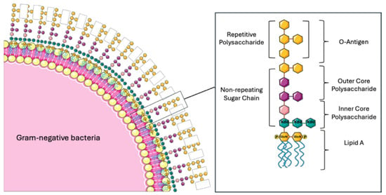
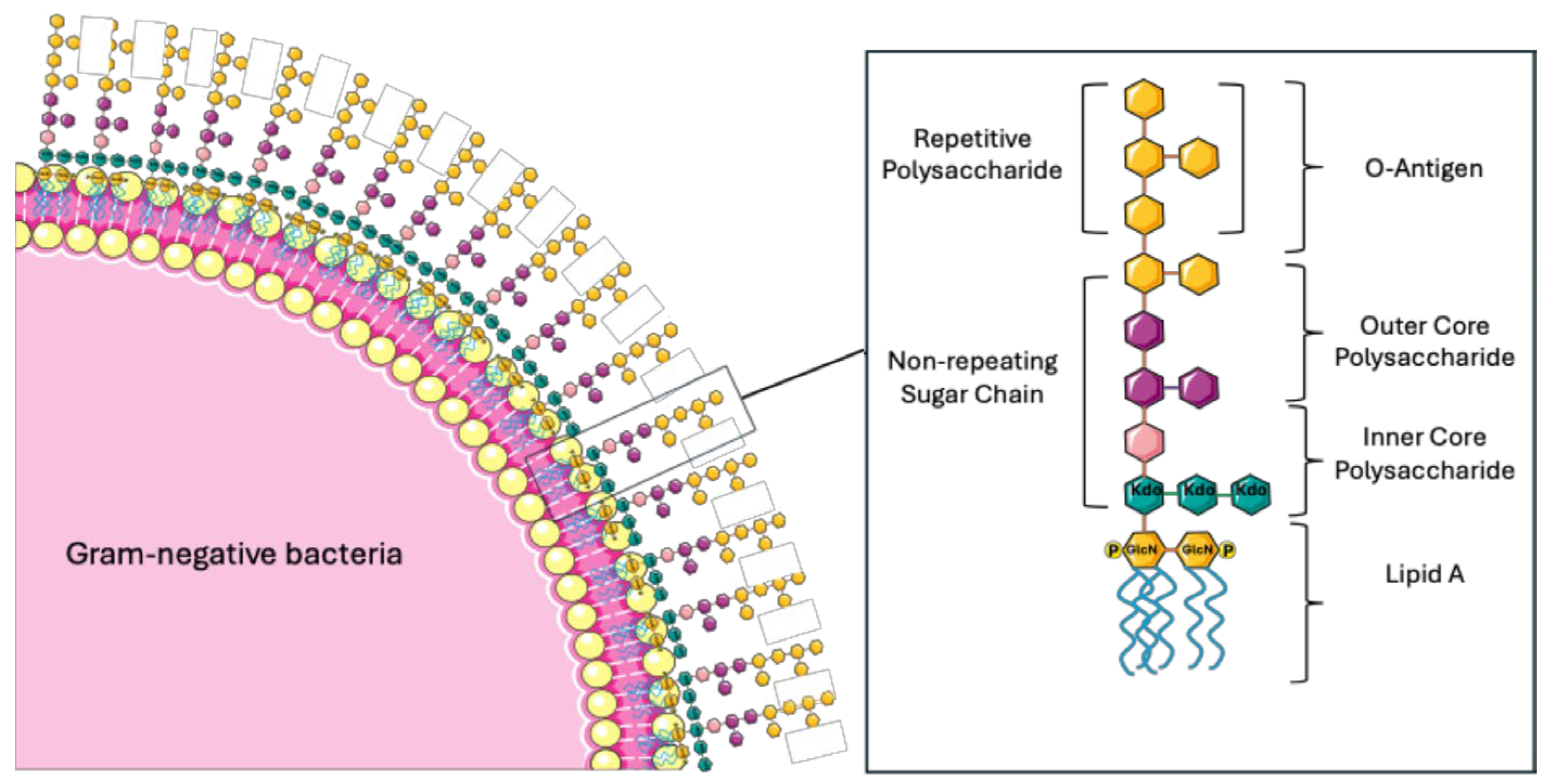
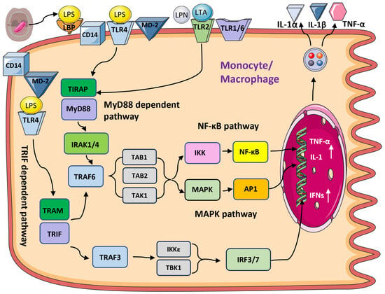
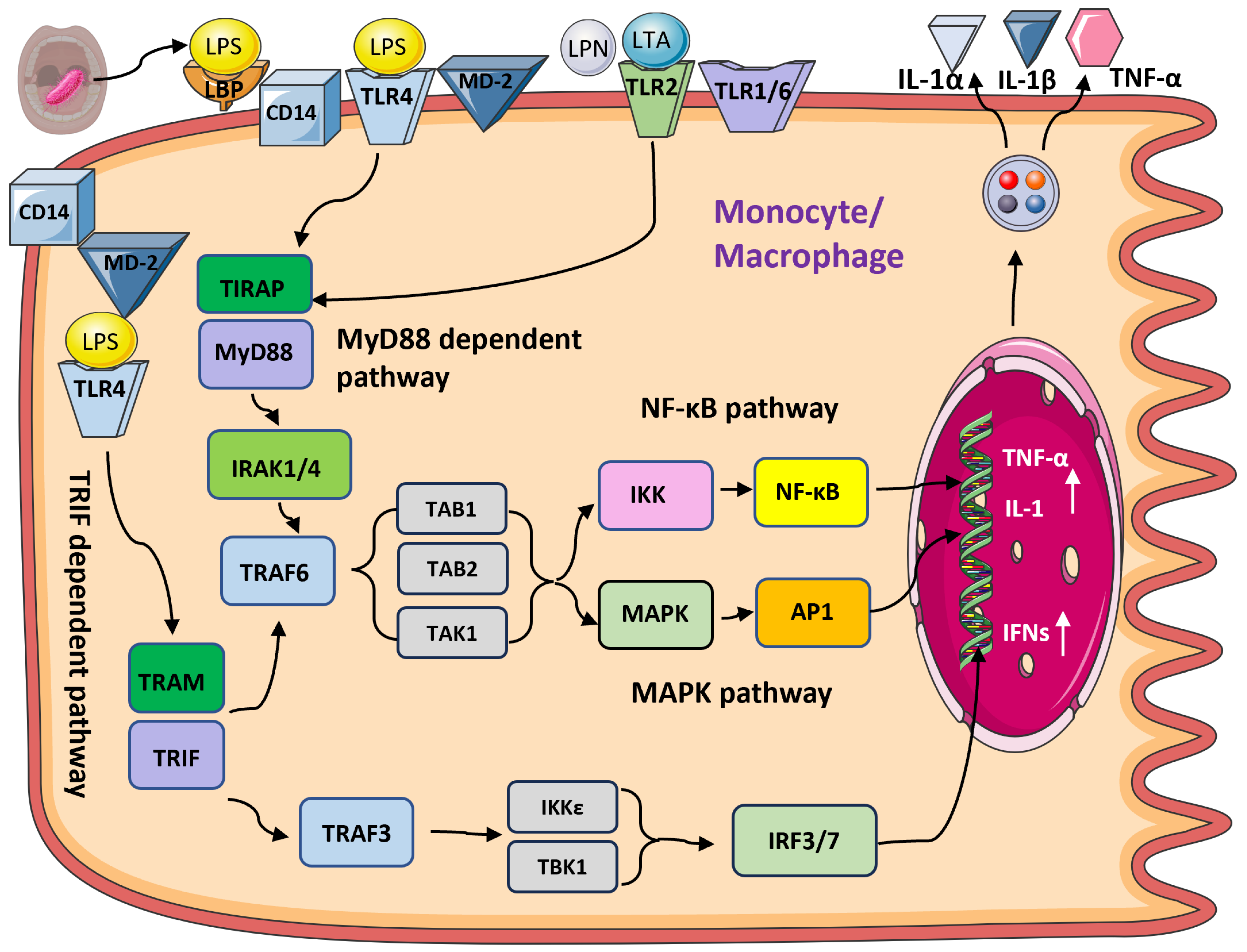
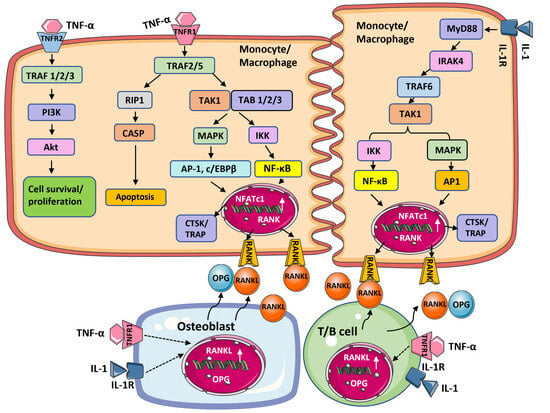
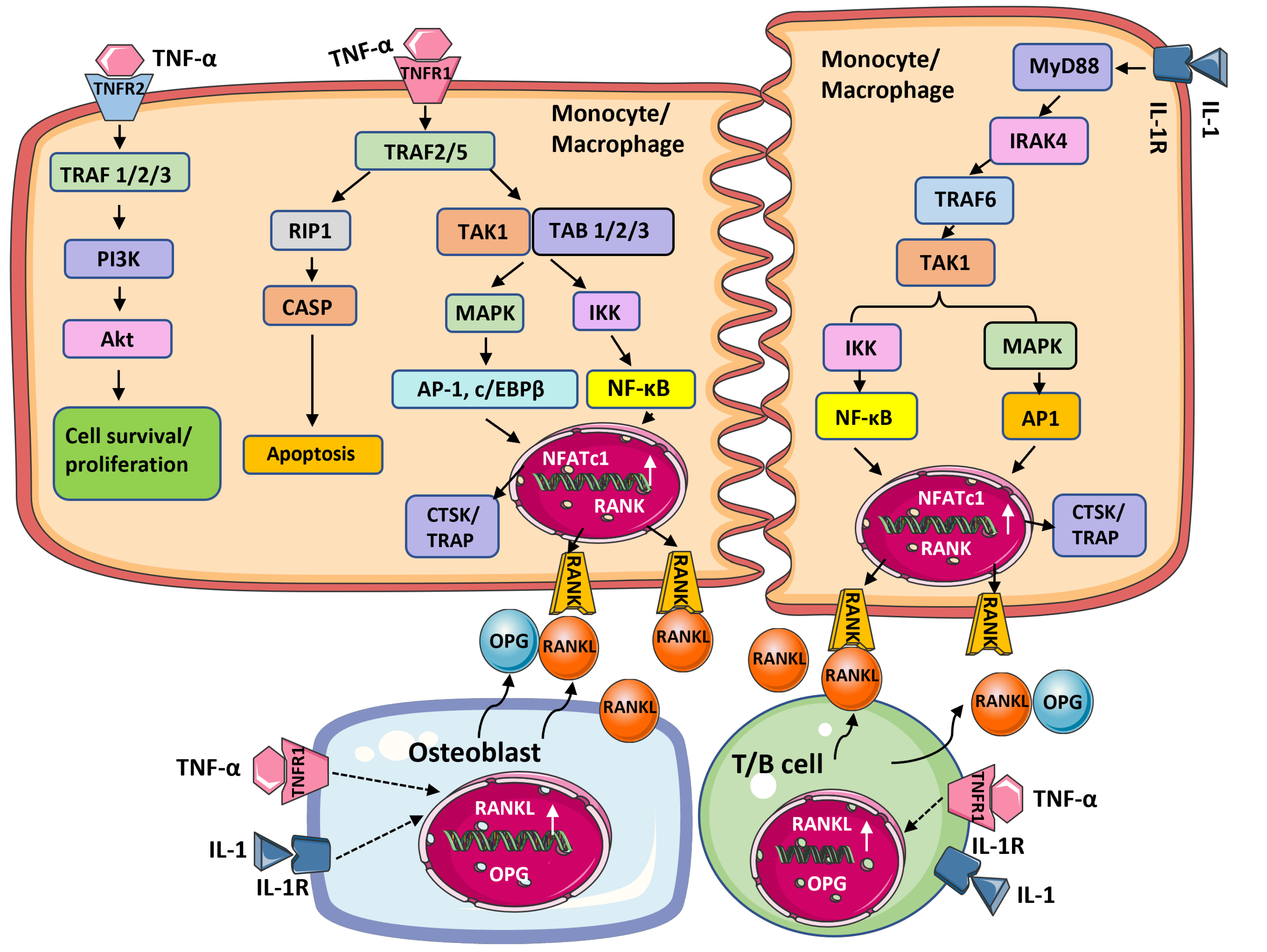

Disclaimer/Publisher’s Note: The statements, opinions and data contained in all publications are solely those of the individual author(s) and contributor(s) and not of MDPI and/or the editor(s). MDPI and/or the editor(s) disclaim responsibility for any injury to people or property resulting from any ideas, methods, instructions or products referred to in the content. |
© 2026 by the authors. Licensee MDPI, Basel, Switzerland. This article is an open access article distributed under the terms and conditions of the Creative Commons Attribution (CC BY) license.