


Abstract
Akebia trifoliata is an important Chinese traditional medicinal plant, and its abundant triterpenoids are the primary active medicinal components. However, the oxidosqualene cyclase (OSC) gene family responsible for constructing their triterpenoid skeletons has not yet been systematically characterized. In this study, nine AktOSC genes were identified genome-wide in A. trifoliata. Phylogenetic analysis revealed that most AktOSC members formed independent evolutionary subclades. Synteny and selective pressure analyses indicated that multiple duplication modes collectively contributed to the expansion of the AktOSC family, and all members were subjected to strong purifying selection. Sequence alignment showed that all members possess the characteristic motifs of the OSC family, whereas amino acid substitutions at critical active-site residues hinted at potential catalytic product diversity. Cis-acting element prediction revealed an abundance of environmental and phytohormone responsiveness elements within the AktOSC promoter regions. Expression profiling revealed that AktOSC genes exhibited distinct expression patterns across tissues, developmental stages, and disease-susceptibility groups. Finally, AktOSC8, which was specifically and highly expressed in the pericarp, was identified as a highest-priority candidate gene. Collectively, these findings provide essential foundational insights for further elucidating the mechanisms underlying triterpenoid biosynthesis and accumulation in A. trifoliata.
1. Introduction
Sterols and triterpenoids are important components of plant metabolites with essential roles in membrane stability, cell growth and development, signaling, and responses to biotic and abiotic stresses [1,2,3,4]. Beyond their physiological importance in plants, many triterpenoids also possess substantial pharmacological activities in humans, including anti-inflammatory, anticancer, antiviral, immunomodulatory, and cholesterol-lowering effects, making them valuable resources for the pharmaceutical, nutraceutical, and cosmetic industries [5,6,7].
Oxidosqualene cyclases (OSCs) are the key enzymes that catalyze the cyclization of 2,3-oxidosqualene into a wide variety of sterol and triterpene skeletons [8]. The product outcome of OSCs is closely associated with substrate folding: cyclization in the chair–boat–chair (C-B-C) conformation generally generates a protosteryl cation intermediate and leads to sterol-type tetracyclic products such as cycloartenol and lanosterol, whereas cyclization in the chair–chair–chair (C-C-C) conformation produces a dammarenyl cation intermediate, which typically gives rise to pentacyclic triterpene skeletons such as lupeol and amyrins [9]. The formation of these two conformations is dictated by the unique structural features of the OSC enzyme and the physicochemical properties of key amino acid residues within the catalytic domain [10]. Among these, the DCTAE, QW, and MWCYCR motifs represent the most extensively studied structural elements, and they are essential for catalytic activity and intermediate stabilization, with subtle variations within these regions influencing both substrate folding and the extent of the cyclization cascade [10,11,12]. Because minor amino acid substitutions within the active site can profoundly alter the product outcome [10,13,14], this plasticity has driven the extensive structural variation in triterpene skeletons and prompted the emergence of multiple OSC subfamilies, including cycloartenol synthases (CASs), lanosterol synthases (LASs), lupeol synthases (LUPs), dammarenediol synthases (DDSs), β-amyrin synthases (bASs), and multifunctional OSCs [4,15,16].
Akebia trifoliata (Thunb.) Koidz., a perennial woody vine of the family Lardizabalaceae [17], is a traditional medicinal plant whose stems and fruits are both used in traditional Chinese medicine (TCM) as “Mutong” and “Yuzhizi” [18,19], respectively. These two distinct medicinal parts are employed for different therapeutic indications: Mutong is exploited to treat neuralgia, menstrual pain, inflammatory disorders, and infections of the kidneys and urinary tract, whereas Yuzhizi is primarily prescribed for indigestion, inflammation, cardiovascular diseases, and as an adjuvant therapy for malignant tumors [18,19]. This divergence in clinical applications is fundamentally driven by variations in their secondary metabolite profiles. While Mutong accumulates a complex mixture of triterpene saponins, phenylethanoid glycosides, and lignans, Yuzhizi features a more pronounced enrichment of oleanane-type triterpenoids [19]. Recent phytochemical studies have revealed that the triterpenoid aglycones in A. trifoliata are predominantly composed of hederagenin, followed by arjunolic acid, dehydroarjunolic acid, oleanolic acid, and dehydrooleanolic acid [20,21]. The attached sugar moieties mainly include glucose, rhamnose, xylose, and arabinose. These triterpene saponins have been demonstrated to possess potent antitumor and anti-inflammatory activities, representing the primary source of medicinal value for both Mutong and Yuzhizi [20,22,23].
From a safety perspective, historical incidents of aristolochic acid-associated nephrotoxicity linked to medicinal materials marketed as Mutong were primarily attributed to the use of Aristolochia manshuriensis (“Guanmutong”), rather than Akebia-derived materials [18]. LC–UV/LC–MS analyses, including a subsequent multilaboratory validation, found aristolochic acid I to be below the reporting limit (<2.00 μg g−1) in the tested stem materials of A. trifoliata [24,25]. In a comparative rat study, Akebia quinata, another recognized botanical source of Mutong, caused no detectable renal injury at the tested dose, whereas A. manshuriensis induced marked nephrotoxicity attributable to its aristolochic acid content [26]. Collectively, these chemical and toxicological findings indicate that Akebia-derived medicinal materials carry a substantially lower risk of aristolochic acid-associated nephrotoxicity than A. manshuriensis.
Despite decades of extensive phytochemical and pharmacological characterization that has established the medicinal value of A. trifoliata, functional genomic research into this species remains in its infancy [27]. As a typical non-model medicinal plant, A. trifoliata lacks a stable and efficient genetic transformation system, which imposes a severe bottleneck on its molecular and genetic investigations and renders direct in vivo functional validation of metabolic genes highly challenging [28,29]. Under these methodological constraints, genome-wide bioinformatic analysis emerges as an indispensable research strategy rather than a mere computational tool. By utilizing comparative genomics, molecular evolutionary analysis, and expression profiling, a systematic genome-wide study can effectively circumvent current technical limitations to screen core candidate members within a gene family, thereby providing a critical theoretical foundation for future functional characterization and genetic engineering once transformation platforms mature.
In this study, we performed a genome-wide identification and comprehensive analysis of the OSC gene family in A. trifoliata. Specifically, we identified OSC family members from the reference genome, analyzed their gene and protein structures, conserved motifs, and chromosomal distributions, and inferred their evolutionary relationships and duplication history. We also examined cis-acting regulatory elements in the promoter regions and explored their expression patterns using available transcriptomic data, with the aim of prioritizing candidate AktOSC genes that may participate in triterpenoid biosynthesis. This work provides a foundation for future functional characterization of AktOSC genes and for elucidating the molecular mechanisms underlying triterpenoid metabolism in A. trifoliata.
2. Materials and Methods
2.1. Data Sources
The genome assembly and annotation files of A. trifoliata (accession no. GWHBISH00000000) were retrieved from the National Genomics Data Center (NGDC, https://ngdc.cncb.ac.cn/; accessed on 15 March 2024) [30]. Concurrently, the reference genome sequences and corresponding annotation files of Arabidopsis thaliana and Oryza sativa were downloaded from the Ensembl Plants database (https://plants.ensembl.org/; accessed on 19 March 2024 and 19 November 2024, respectively). The OSC protein sequences of A. thaliana (n = 13) [16] and O. sativa (n = 12) [31] were obtained from the TAIR database (https://www.arabidopsis.org/; accessed on 15 January 2026) and the Rice Genome Annotation Project database (https://rice.uga.edu/; accessed on 15 January 2026), respectively. Hidden Markov model (HMM) profiles of the squalene-hopene cyclase C-terminal domain (PF13243) and N-terminal domain (PF13249) were downloaded from the InterPro database (https://www.ebi.ac.uk/interpro/; accessed on 15 January 2026). Additionally, a total of 44 reviewed sequences belonging to the terpene cyclase/mutase family were collected from the UniProt database (https://www.uniprot.org/; accessed on 20 March 2026), spanning 14 species that range from the early-diverging green alga to the core eudicot.
Furthermore, two publicly available transcriptome datasets of A. trifoliata were utilized in this study. The fruit growth and development transcriptome data (NCBI BioProject accession no. PRJNA671772; https://www.ncbi.nlm.nih.gov/bioproject/671772; accessed on 30 March 2026) comprised 36 RNA-seq libraries generated from flesh, seed, and peel tissues at four developmental stages (immature, enlargement, coloring, and mature), with three biological replicates for each tissue–stage combination [27]. The disease-associated transcriptome data (NGDC BioProject accession no. PRJCA014987; https://ngdc.cncb.ac.cn/bioproject/browse/PRJCA014987; accessed on 30 March 2026) comprised nine peel RNA-seq libraries representing disease-resistant (RP), disease-susceptible (SP), and control (CP) samples, with each sample category collected during three sampling periods (May, June, and July) [32]. According to the BioProject metadata, these samples were collected under naturally occurring disease conditions without artificial pathogen inoculation. The RP and SP libraries were generated from the mixed pool of 24 lines exhibiting disease resistance and 50 lines exhibiting susceptibility to fungal disease, respectively, whereas the CP libraries were derived from fruit peels of the genome-sequenced cultivar ‘Shusen 1’.
2.2. Genome-Wide Identification of A. trifoliata OSC Gene Family
To systematically identify OSC gene family members within the A. trifoliata genome, a combination of hidden Markov model and homology-based BLAST searches was employed. Initially, the HMMER software (v3.3.2) was utilized to screen the genome assembly using the Pfam profiles PF13249 and PF13243, with an E-value threshold of 1 × 10⁻⁵ and default parameters for all other settings [33]. Concurrently, local BLASTP was performed using NCBI BLAST+ (v2.17.0+) against the A. trifoliata proteins using the known OSC protein sequences from A. thaliana (n = 13) and O. sativa (n = 12) as queries under the identical E-value cutoff [34].
The candidate proteins intersected from both the HMMER and BLASTP pipelines were subsequently submitted to the SMART database (https://smart.embl.de; accessed on 20 January 2026) to verify the presence of the OSC conserved domains [35]. Sequences that lacked a complete conserved domain were manually discarded. The remaining proteins were confirmed as high-confidence A. trifoliata OSC family members and designated as AktOSC proteins.
2.3. Physicochemical Property and Subcellular Localization Analysis
ProtParam on the ExPASy server (https://web.expasy.org/protparam/; accessed on 22 January 2026) was employed to predict the physicochemical properties of the AktOSC proteins [36]. Additionally, their subcellular localizations were analyzed using DeepLoc 2.1 (https://services.healthtech.dtu.dk/services/DeepLoc-2.1/; accessed on 25 January 2026) [37].
2.4. Protein Domain, Conserved Motif, and Gene Structure Characterization
The conserved functional domains of AktOSC proteins were analyzed using the NCBI Batch CD-Search tool (CDD, v3.21; https://www.ncbi.nlm.nih.gov/Structure/bwrpsb/bwrpsb.cgi; accessed on 30 January 2026) [38]. Conserved motifs within the proteins were predicted using the MEME Suite (MEME Suite, v5.5.9; https://meme-suite.org/meme/tools/meme; accessed on 30 January 2026) [39], with the maximum motif width set to 75 and the total number of motifs configured to 8, while maintaining default settings for all other parameters. Concurrently, the exon-intron structures of the AktOSC genes were extracted from the GFF3 file. Finally, the identified protein domains, conserved motifs, and gene structures were integrated and visualized using TBtools (v2.476) [40].
2.5. Phylogenetic Analysis
The AktOSC protein sequences, along with 44 reference OSC proteins retrieved from diverse plant species, were aligned using MAFFT (v7.525) [41]. The resulting multi-sequence alignment was automatically trimmed using trimAl (v1.5) to remove poorly aligned regions [42]. Subsequently, an ML phylogenetic tree was constructed using IQ-TREE (v2.3.6) [43]. The best-fit amino acid substitution model was automatically selected using the ModelFinder Plus algorithm, and branch support was evaluated with 1000 bootstrap replicates. The resulting tree file was visualized using the Interactive Tree Of Life platform (iTOL, v7; https://itol.embl.de/; accessed on 20 March 2026) [44].
2.6. Chromosomal Location, Gene Duplication, Collinearity and Ka/Ks Analysis
The physical chromosomal positions of the AktOSC genes were determined based on the A. trifoliata genome annotation file and visualized using TBtools. To investigate syntenic relationships, collinearity analyses of the OSC genes between A. trifoliata and two reference species (A. thaliana and O. sativa) were performed using the MCScanX [45] module integrated within TBtools, maintaining default parameters. Gene duplication events within the AktOSC genes were identified based on the outputs generated by the MCScanX module. Subsequently, the Ka/Ks ratio of the identified AktOSC genes was calculated by TBtools.
2.7. Analysis of Cis-Acting Elements
The putative cis-acting elements located in the promoter regions (2000 bp upstream of the start codon) of the AktOSC genes were computationally identified via the PlantCARE database (https://bioinformatics.psb.ugent.be/webtools/plantcare/html/; accessed on 26 March 2026) [46]. The predicted results were subsequently classified and visualized using TBtools.
2.8. Expression Analysis
The two public transcriptome datasets described in Section 2.1 were analyzed separately. RNA-seq reads were mapped to the A. trifoliata reference genome using HISAT2 (v2.1.0) [47]. Gene expression levels were quantified as fragments per kilobase of transcript per million mapped fragments (FPKM) using StringTie (v1.3.6) [48]. Subsequently, the FPKM values of the AktOSC genes were extracted and transformed as log2(FPKM + 1). Finally, expression heatmaps of the AktOSC genes were generated using TBtools.
3. Results
3.1. Identification of OSC Gene Family Members in A. trifoliata
A total of nine high-confidence AktOSC genes were systematically identified within the A. trifoliata genome (Table 1). Based on their genomic distribution, the nine genes were sequentially designated as AktOSC1 to AktOSC9. Chromosome 8 harbored the highest number of AktOSC genes, with three members forming a tandem cluster at the chromosome terminus (Figure S1). Although the genomic lengths of these nine AktOSC genes exhibited considerable variation, ranging from 10,570 to 24,638 bp, their coding sequence (CDS) lengths (2121–2310 bp) and the number of exons (17–20) were highly conserved.
Table 1.
Characteristics of the identified AktOSC genes and putative AktOSC proteins.
Furthermore, the physicochemical properties of the nine putative AktOSC proteins displayed a high degree of similarity, with amino acid lengths of 706–769 amino acids (AA), molecular weights of 80.31–88.32 kDa, theoretical isoelectric points (pI) of 5.86–6.51, aliphatic indices of 76.85–81.94, and grand average of hydropathy (GRAVY) values of −0.372 to −0.269. Notably, with the exception of AktOSC6, which was predicted to be stable with an instability index of 36.60, all other AktOSC proteins were classified as unstable, with their instability indices ranging from 43.66 to 50.13. Subcellular localization analysis showed that all AktOSC proteins were consistently localized to the endoplasmic reticulum (ER).
3.2. Gene Structure, Conserved Domain, and Motif Analysis
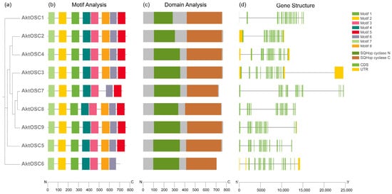
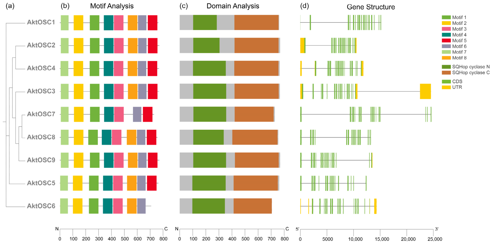
To reveal the structural characteristics of the AktOSC gene family, their exon–intron organizations and protein motif distributions were systematically characterized. MEME analysis identified eight conserved motifs within the AktOSC proteins, with Motif 5 and Motif 8 being specifically absent in AktOSC6 and AktOSC7 (Figure 1), respectively. Detailed sequence inspection verified that these predicted motifs encompassed the essential catalytic features required for oxidosqualene cyclization (Table 2). Motif 3 harbored the highly conserved DCTAE motif, which is directly associated with the OSC catalytic reaction. Furthermore, Motif 1 contained the MWCYCR signature sequence, which is a documented hallmark of pentacyclic triterpene synthases. Additionally, Motifs 2 and 6 each carried two copies of the conserved QX2–5GXW motif, which is important for the stability of OSC catalytic reaction. Concurrently, conserved domain analysis confirmed that the classic dual-domain architecture, comprising the squalene-hopene cyclase N-terminal and C-terminal domains, was intact across all identified AktOSC proteins (Figure 1).
Figure 1.
Structural and evolutionary analysis of the AktOSC gene family. (a) Phylogenetic relationship among the nine AktOSC proteins. (b) The distribution of protein conserved motifs. Different colored boxes indicate distinct motifs. (c) Conserved domain architecture. Green and brown boxes indicate SQHop_cyclase_C and SQHop_cyclase_N domains, respectively, and grey boxes indicate regions outside the annotated domains. (d) Exon-intron structures. Green boxes, yellow boxes, and black lines represent CDS, untranslated regions (UTR), and introns, respectively. Scale bars at the bottom indicate protein length (AA) for (b,c), and gene length (bp) for (d).
Table 2.
Information on the conserved motifs of AktOSC proteins.
Gene structure analysis indicated that AktOSC genes typically exhibited complex architectures, containing 17 to 20 exons (Figure 1). While the genomic lengths of the majority of AktOSC genes fell within a relatively uniform range of approximately 10 to 15 kb, AktOSC3 and AktOSC7 exhibited larger genomic regions of approximately 24 kb, a phenomenon primarily attributed to the substantial elongation of their intronic regions.
3.3. Phylogenetic Analysis of AktOSC Proteins
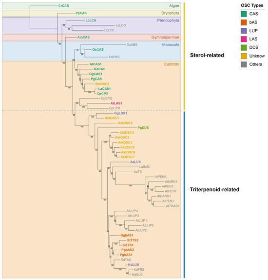
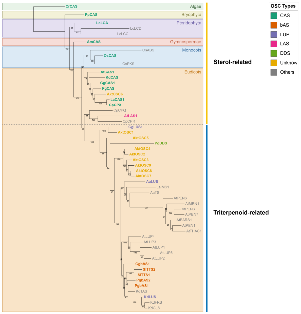
To investigate the evolutionary relationships and predict the functional contexts of the AktOSC proteins, a maximum likelihood (ML) phylogenetic tree was constructed using the amino acid sequences of the nine AktOSC proteins alongside 44 functionally characterized reference OSC proteins from diverse plant species (Figure 2, Table S1). The phylogenetic topology resolved into multiple clades and corresponding OSC functional categories, based on which the AktOSC proteins were classified. Specifically, AktOSC6 clustered robustly within the CAS clade with strong bootstrap support, suggesting high confidence in its orthology to known sterol-synthesizing enzymes. Meanwhile, AktOSC1 grouped within the LUP clade, exhibiting a close evolutionary relationship with reference LUPs from other species. In contrast, the remaining seven members (AktOSC2–AktOSC5 and AktOSC7–AktOSC9) partitioned into the broader triterpenoid synthase lineage but did not tightly cluster with any previously characterized reference OSCs. Notably, six of these members, with the exception of AktOSC5, formed an independent subclade, suggesting unique evolutionary trajectory of the OSC family within A. trifoliata.
Figure 2.
Phylogenetic relationships of nine AktOSC proteins and 44 reference OSC proteins from various plant species. The phylogenetic tree was constructed using the ML method based on amino acid sequences, with bootstrap support values from 1000 replicates indicated at the nodes. The CAS sequence from Chlamydomonas reinhardtii was utilized as the outgroup. Functional categories of OSCs are distinguished by distinct label colors, with gray representing multi-product/uncommon types and yellow denoting uncharacterized AktOSCs. Dashed lines demarcate the boundary between the sterol biosynthesis and triterpenoid biosynthesis clades. Detailed information for all analyzed OSCs is provided in Table S1.
3.4. Collinearity and Ka/Ks Analysis of OSC Family Members
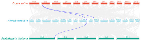
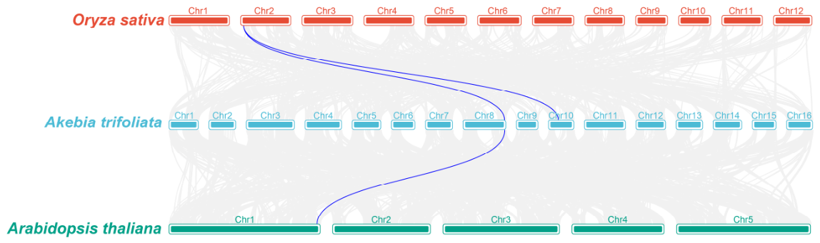
To elucidate the interspecific collinear relationship of the OSC genes, a comparative synteny analysis was performed between A. trifoliata and two representative plant genomes: the dicot A. thaliana and the monocot O. sativa (Figure 3). A total of three collinear OSC gene pairs were detected across these lineages. Specifically, two syntenic OSC gene pairs were identified between A. trifoliata and O. sativa, linking Chr2 of O. sativa to Chr8 and Chr10 of A. trifoliata. Additionally, one collinear gene pair was resolved between A. trifoliata (Chr8) and A. thaliana (Chr1). Notably, the OSC locus situated on Chr8 of A. trifoliata maintained highly conserved synteny with both monocot and dicot reference genomes, underscoring its ancestral origin and evolutionary stability across deeply divergent angiosperm lineages.
Figure 3.
Collinearity analyses of the OSC genes between A. trifoliata and two reference species (A. thaliana and O. sativa). Blue lines represent collinearity among different OSC genes.
To assess the selective pressure among AktOSC pairs, Ka/Ks analysis was conducted for the nine members, generating 36 gene pairs in total (Table S2). All pairwise Ka/Ks ratios ranged from 0.15 to 0.35, indicating that the evaluated AktOSC gene pairs have predominantly evolved under purifying selection.
3.5. Cis-Acting Elements Composition of AktOSC Promoters
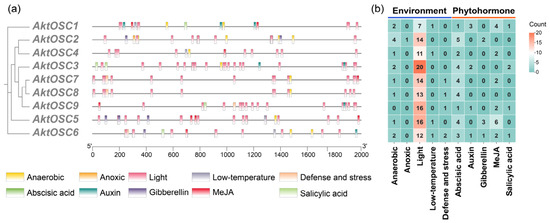
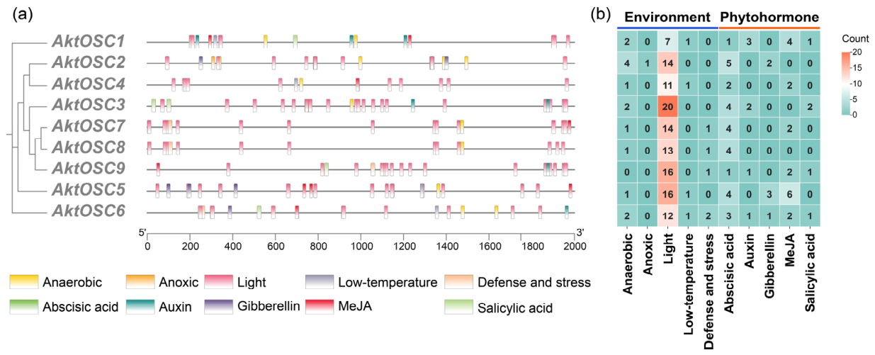
After removing basal promoter components (e.g., TATA-box and CAAT-box) and unannotated motifs from the raw predictions, a total of 211 functional cis-acting regulatory elements were identified within the 2000 bp upstream promoter regions of the nine AktOSC genes (Figure 4, Table S3). These elements were classified into two macro-categories encompassing 10 subcategories: environmental responsiveness (anaerobic-, anoxic-, light-, low-temperature-, and defense and stress-responsive elements) and phytohormone responsiveness (abscisic acid-, auxin-, gibberellin-, MeJA-, and salicylic acid-responsive elements). The abundance of cis-elements averaged 23.44 per gene, spanning from a minimum of 17 in AktOSC4 to a maximum of 31 in AktOSC5. Within the two macro-categories, light-responsive and abscisic acid-responsive elements represented the most abundant subcategories, accounting for 58.3% and 13.3% of the total pool, respectively. Notably, these two functional groups were universally distributed across the promoters of all nine AktOSC genes. Conversely, anoxic-responsive and salicylic acid-responsive elements were the least represented in their respective macro-categories, with only one and five elements detected, respectively.
Figure 4.
Prediction of cis-acting elements in the AktOSCs promoter. (a) The distribution of cis-acting elements in the 2000 bp upstream region of AktOSC genes. (b) The number of cis-acting elements of the two macro-categories in AktOSCs, indicated by different colors and numbers, respectively.
3.6. Expression Profiles of AktOSC Genes in Different Tissues and Stages
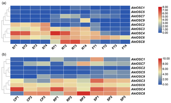
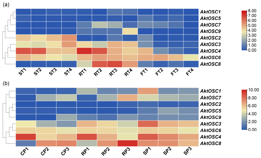
To elucidate the potential biological roles of AktOSC genes in A. trifoliata growth, development, and defense responses, their expression profiles were systematically evaluated using two transcriptomic datasets (Figure 5). Hierarchical clustering revealed pronounced spatial and temporal transcriptional divergence among the nine AktOSC genes during normal development. Specifically, AktOSC2 exhibited strict seed-specific expression; its transcript abundance progressively increased during seed maturation and peaked at the mature stage (ST4), whereas it remained barely detectable in peel and flesh tissues. AktOSC3 displayed moderate transcript accumulation restricted to the developmental sequences of both seeds and peels. Conversely, AktOSC8 expression was uniquely restricted to the peel, showing a sharp preferential enrichment during the fruit enlargement (RT2) and coloring (RT3) stages. AktOSC4 was heavily transcribed during early organogenesis, particularly in the immature and enlargement stage of seeds (ST1–ST2) and peels (RT1–RT2), before tapering off in later stages. In contrast to these highly tissue-specific patterns, AktOSC6 maintained a relatively stable, moderate expression baseline across all examined tissues and stages, including the flesh. The remaining four members (AktOSC1, AktOSC5, AktOSC7, and AktOSC9) were transcriptionally quiescent, maintaining very low or undetectable expression levels throughout normal development.
Figure 5.
Expression profiles of AktOSCs in different stages. The degree of change from blue to red indicates the intensity of the expression level. (a) Expression profiles of AktOSCs in tissue- and development-related transcriptomes. ST, FT, RT and 1, 2, 3, 4 represent three different tissues (seed, flesh, and peel tissues) and four different developmental stages (immature, enlargement, coloring, and mature stages) of A. trifoliata, respectively. (b) Expression profiles of AktOSCs in disease-associated transcriptomes. CP, RP, SP and 1, 2, 3 represent three peel sample categories (control, disease-resistant, and disease-susceptible peel group) and sampling periods (May, June, and July) of A. trifoliata, respectively.
Within the disease-associated transcriptome dataset, the AktOSC genes displayed distinct expression patterns across peel sample categories and sampling periods. Notably, AktOSC8 showed a pronounced increase in expression in the disease-resistant peel group collected in July (RP3) and maintained relatively high expression in the disease-susceptible peel group collected from May to July (SP1–SP3). Intriguingly, AktOSC1 and AktOSC7, which showed very low or undetectable expression in the fruit growth and development dataset, were detected at relatively high levels in some disease-associated peel samples. AktOSC1 reached its highest observed expression level in SP1 (May), whereas AktOSC7 showed relatively high expression in both RP3 and SP1. In contrast, AktOSC3 and AktOSC4 showed relatively high expression in the May samples of all three categories, followed by lower expression levels in June and July. Conversely, the remaining members (AktOSC2, AktOSC5, and AktOSC9) remained at very low or undetectable expression levels under all treatments.
4. Discussion
In plant triterpenoid biosynthesis, the cyclization of 2,3-oxidosqualene, catalyzed by OSC, is the initial committed step toward triterpenoid diversification, giving rise to more than 100 distinct triterpenoid skeletons [49]. Through heterologous expression in hosts such as yeast or tobacco, over 150 OSCs from more than 75 plant species have been functionally characterized to date [50]. Among these, bAS, CAS, and LUP constitute the most prevalent classes, accounting for 23.3%, 20.7%, and 14% of the total identified OSCs, respectively [50]. In the present study, nine AktOSC genes were identified in A. trifoliata. Phylogenetic analysis coupled with NCBI BlastP searches suggested that AktOSC1 and AktOSC6 may encode LUP and CAS (Figure 2, Table S4), respectively. In contrast, the remaining seven AktOSC proteins were positioned within the triterpene synthase-related branch, but they did not cluster closely with OSCs whose catalytic products have been experimentally characterized. Although their top BLASTP hits were annotated as bAS-like proteins, these sequence-based similarities are insufficient to establish their catalytic functions or product specificities. Given that minor modifications within the active site (such as a single amino acid substitution) can radically alter catalytic activity and yield diverse polycyclic triterpenoids [11], further biochemical verification is indispensable to conclusively determine the catalytic activities and product specificities of these AktOSC proteins.
4.1. Distinct Evolutionary Trajectories Shape the Functional Divergence of AktOSC Genes
Previous studies suggest that successive gene duplication and divergence events shaped the evolutionary trajectory of the plant OSC family, expanding OSC functions from ancestral roles in primary sterol biosynthesis in early-diverging plant lineages to increasingly diverse roles in specialized triterpenoid metabolism. Rather than resulting from a single ancestral radiation, this diversification appears to have involved repeated, lineage-specific innovations that generated distinct OSC repertoires across plant lineages [16,50]. Consistent with this evolutionary framework, most AktOSC members clustered within a distinct clade in the phylogenetic tree (Figure 2). Together with the identification of tandem, proximal, and dispersed duplicates (Table 1), this pattern suggests that multiple duplication modes contributed to the lineage-specific expansion and potential functional divergence of the AktOSC family. This finding agrees with the established consensus that numerous triterpene synthases evolved independently across diverse plant lineages [50]. Despite this lineage-specific expansion, the family-wide Ka/Ks analysis indicated that the AktOSC genes have experienced strong purifying selection (Table S2), suggesting that the duplicated members have remained under substantial evolutionary constraint, thereby preserving OSC functions important for sterol and triterpenoid biosynthesis in A. trifoliata.
Within this evolutionary framework, interspecific collinearity analysis was conducted among A. trifoliata (a basal eudicot), O. sativa (a monocot), and A. thaliana (a core eudicot) (Figure 3). Both AktOSC1 and AktOSC4 exhibited collinearity with the same OSC locus in O. sativa, indicating a possible one-to-many orthologous relationship. This pattern is consistent with their derivation from a common ancestral OSC followed by a lineage-specific duplication event in A. trifoliata. However, their subsequent evolutionary trajectories appear to have diverged. AktOSC1 retained collinear relationships with OSC loci in both O. sativa and A. thaliana, suggesting that its surrounding genomic region has remained relatively conserved across the examined angiosperm lineages. When considered together with its phylogenetic placement, this conservation is compatible with its tentative annotation as a LUP. By contrast, AktOSC4 retained detectable collinearity only with O. sativa, suggesting that its corresponding syntenic locus may have been lost or substantially reorganized along the lineage leading to A. thaliana through gene fractionation or localized genomic rearrangement. Similarly, the absence of detectable interspecific synteny for AktOSC6 does not contradict its tentative annotation as a CAS, which is supported primarily by its phylogenetic placement and conserved sequence characteristics. Instead, this absence may reflect extensive restructuring of its surrounding genomic region over evolutionary time, whereas the conservation of its coding sequence is consistent with its putative role in essential sterol biosynthesis.
4.2. Conserved Motifs and Amino Acid Variations May Contribute to AktOSC Catalytic Diversity
The function of an enzyme is closely associated with its functional domains, and key amino acids within the active center frequently determine its catalytic activity. Therefore, understanding the relationship between these amino acids and enzyme structure and function is highly important. The DCTAE and QW motifs are conserved regions in OSC proteins [12,51]. The DCTAE motif is involved in substrate binding, and the aspartate (D) residue within this motif is responsible for substrate protonation to initiate the cyclization cascade [52,53]. Mutation of this specific residue can lead to a complete loss of catalytic capacity, whereas mutations at other positions within the motif may result in a drastic reduction in enzyme activity or diversification of the product profiles [4,11]. In this study, seven of the nine identified AktOSC proteins retained the canonical DCTAE motif (Figure S2), whereas AktOSC5 and AktOSC9 contained the variant sequences DCTGE and DGTAE, respectively. The QW motif features a consensus sequence of [K/R][G/A]X2-3[F/Y/W][L/V]X3QX2-5GXW [12], which is often simplified as QX2-5GXW [54], and its primary function is to stabilize carbocation intermediates during the cyclization cascade [55]. All AktOSC members contained conserved QW repeats conforming to the QX2-5GXW consensus. Furthermore, bASs generally possess a conserved MWCYCR motif, which is thought to participate in stabilizing complex cationic intermediates [10,56]. LUPs commonly contain an MLCYCR sequence at this position, whereas CASs and LASs frequently contain the corresponding MWCHCR sequence [10]. Consistent with previous findings, AktOSC1 (putatively inferred as a LUP) and AktOSC6 (putatively inferred as a CAS) harbored the MLCYCR and MWCHCR motifs, respectively. Most other members predicted to be bAS featured the conserved MWCYCR motif, except for AktOSC3 (MWIYCR) and AktOSC7 (MWCHCR). In conclusion, minor variations within the active site can substantially influence enzyme activity and product profiles, representing one of the key mechanisms driving the diversification of plant triterpenoid metabolism. Nevertheless, the functional predictions for these AktOSC proteins require subsequent molecular and biochemical experiments to definitively confirm their enzymatic identities.
4.3. Promoter Cis-Elements and Developmental Expression Patterns Suggest Spatiotemporal Regulation of AktOSC Genes
Numerous studies have demonstrated that different members of the plant OSC family often exhibit distinct tissue-specific expression patterns, indicating that they fulfill diverse biological functions in growth and development, chemical defense, and environmental adaptation [4,57,58]. In the present study, the promoter regions of the AktOSC genes contained diverse predicted cis-acting elements associated with environmental and hormonal responses, providing sequence-based clues to their potential differential regulation during fruit development. Among these, light-responsive elements are widely enriched across all AktOSC promoters and represent the most abundant cis-element subcategories for each member (up to 20), implying that light signaling may be a key upstream environmental factor driving triterpenoid accumulation in A. trifoliata. For instance, AktOSC8, which was almost exclusively expressed in peel, contained 13 predicted light-responsive elements and exhibited relatively high transcript abundance during the immature and enlargement stages (RT1–RT2). This expression pattern broadly corresponds to the previously reported accumulation of triterpenoids in A. trifoliata peel during early fruit development [59], supporting the prioritization of AktOSC8 as a candidate potentially involved in peel triterpenoid biosynthesis. In contrast, AktOSC2, which was almost exclusively expressed in seeds and reached its highest transcript abundance at the mature stage (ST4), contained five predicted abscisic acid-responsive elements in its promoter. Given the established role of abscisic acid in seed maturation and dormancy, these observations suggest that AktOSC2 may be associated with triterpenoid biosynthesis during seed development. Additionally, AktOSC3 and AktOSC4, which were predominantly expressed in seeds and peels, exhibited opposing temporal expression trends. These patterns suggest possible temporal partitioning among AktOSC members during seed and peel development, although their precise metabolic roles remain to be determined.
4.4. AktOSC Genes Exhibit Distinct Expression Patterns in Disease-Associated Peel Samples
Within the disease-associated transcriptome dataset, the AktOSC genes also exhibited distinct expression patterns across peel samples of various categories and at different sampling periods, suggesting their potential involvement in the plant’s defense mechanisms. For example, AktOSC8 exhibited an increasing expression trend from May to July in the control and disease-resistant peel samples, whereas it showed a decreasing trend across the same sampling periods in the disease-susceptible peel samples, suggesting that its expression may be associated with variation in disease susceptibility among the sampled materials. Given the established defensive roles of some plant triterpenoids [60], AktOSC8 may represent a candidate for investigating the possible relationship between triterpenoid biosynthesis and disease-associated physiological responses. Interestingly, although AktOSC1 and AktOSC7 showed very low transcript abundance in the fruit developmental dataset, their transcripts were detected at relatively high levels in some disease-associated peel samples. These qualitative differences suggest possible context-dependent regulation.
4.5. Integrated Sequence and Expression Analyses Prioritize Candidate AktOSC Genes for Triterpenoid Biosynthesis
Integration of the conserved-motif, phylogenetic, and expression analyses allowed the AktOSC genes to be prioritized for subsequent functional characterization.
The DCTAE motif is directly involved in substrate binding and initiation of the 2,3-oxidosqualene cyclization reaction, making sequence conservation within this region particularly important for OSC catalytic function. AktOSC5 and AktOSC9 contained the noncanonical DCTGE and DGTAE sequences, respectively, and these substitutions may substantially affect their catalytic activities [11]. Together with their very low transcript abundance within both the fruit developmental and disease-associated datasets, these results substantially reduce the priority of AktOSC5 and AktOSC9 as candidates for the predominant triterpenoid biosynthetic pathways.
Variations in the MWCYCR-related region may influence OSC product specificity [10]. The phylogenetic positions and characteristic MLCYCR and MWCHCR sequences of AktOSC1 and AktOSC6 support their tentative annotations as LUP and CAS, respectively. Therefore, these proteins are less likely to encode the β-amyrin synthases primarily responsible for forming the oleanane-type scaffolds that predominate among the bioactive triterpenoids of A. trifoliata [18]. The relatively constitutive expression of AktOSC6 across the examined tissues and developmental stages is also consistent with its predicted role in primary sterol biosynthesis. By contrast, the catalytic functions of AktOSC3 and AktOSC7 remain uncertain because their variant MWIYCR and MWCHCR sequences alone are insufficient to determine product specificity. Moreover, although AktOSC1 and AktOSC7 showed very low transcript abundance in the fruit developmental dataset, their transcripts were detected at appreciable levels in some disease-associated peel libraries. These qualitative differences indicate that they may be associated with plant defense.
AktOSC2, AktOSC4, and AktOSC8 retained the canonical DCTAE and MWCYCR motifs and exhibited relatively high, tissue-associated expression. AktOSC2 was predominantly expressed in seeds, AktOSC8 was expressed in the peel, and AktOSC4 was expressed in both tissues. Together, these sequence and expression characteristics prioritize these three members as candidates potentially involved in triterpenoid biosynthesis. In particular, the developmental expression profile of AktOSC8 broadly corresponded to the previously reported accumulation pattern of triterpenoids in fruit peels [59], making it the highest-priority candidate. Elucidating the precise relationships between these candidate genes (particularly AktOSC8) and triterpenoid accumulation via experimental validation is our next objective.
5. Conclusions
In this study, nine AktOSC genes were identified from the reference genome of A. trifoliata, and their physicochemical and structural characteristics were analyzed. Phylogenetic analysis indicated that most AktOSC proteins formed independent evolutionary subclades, suggesting a unique evolutionary trajectory of this family in A. trifoliata. Synteny and selective pressure analyses revealed that multiple duplication modes, including tandem, proximal, and dispersed duplications, collectively contributed to the expansion of the AktOSC family, and all members were subjected to strong purifying selection. Combined analysis of promoter cis-acting elements and expression patterns demonstrated that the AktOSC genes exhibited distinct spatiotemporal transcriptional divergence during normal fruit growth and development. Among these genes, AktOSC8 exhibited relatively high, peel-enriched expression and contained numerous predicted light-responsive cis-elements, suggesting its potential association with light-related regulation and triterpenoid accumulation in the peel. In the disease-associated transcriptome dataset, the AktOSC genes displayed distinct expression patterns among peel samples from different disease-susceptibility groups, and AktOSC8 showed contrasting temporal trends between disease-resistant and disease-susceptible samples. These observations identify AktOSC8 as a candidate for further investigation of the possible relationship between triterpenoid biosynthesis and disease-associated physiological responses. In summary, this study characterized the basic features, evolutionary relationships, predicted regulatory elements, and spatiotemporal and disease-associated expression patterns of the AktOSC gene family in A. trifoliata, providing a theoretical foundation for further deciphering the regulatory mechanisms of triterpenoid biosynthesis and facilitating molecular breeding for disease resistance in A. trifoliata. Future studies could focus on utilizing molecular biology techniques to perform in vivo and in vitro functional characterization of key members (such as AktOSC8) and functional validation of cis-acting elements, thereby fully elucidating their molecular regulatory networks.
Supplementary Materials
The following supporting information can be downloaded at https://www.mdpi.com/article/10.3390/cimb48080795/s1.
Author Contributions
Conceptualization, P.L. and H.R.; methodology, J.L. and X.Y.; validation, Y.D.; formal analysis, H.L.; investigation, H.L.; resources, C.C.; data curation, H.L.; writing—original draft preparation, H.L.; writing—review and editing, P.L.; visualization, H.L.; supervision, H.R.; project administration, P.L. and C.C.; funding acquisition, P.L., C.C. and F.T. All authors have read and agreed to the published version of the manuscript.
Funding
This research was funded by the Central Guided Local Science and Technology Development Fund, grant number 2024ZYD0284.
Institutional Review Board Statement
Not applicable.
Informed Consent Statement
Not applicable.
Data Availability Statement
The original contributions presented in this study are included in the article/Supplementary Materials. Further inquiries can be directed to the corresponding author.
Conflicts of Interest
The authors declare no conflicts of interest.
Abbreviations
The following abbreviations are used in this manuscript:
| OSC | Oxidosqualene cyclase |
| C-B-C | Chair–boat–chair |
| C-C-C | Chair–chair–chair |
| CAS | Cycloartenol synthase |
| LAS | Lanosterol synthase |
| LUP | Lupeol synthase |
| DDS | Dammarenediol synthase |
| bAS | β-amyrin synthase |
| TCM | Traditional Chinese medicine |
| HMM | Hidden Markov model |
| FPKM | Fragments per kilobase of transcript per million mapped fragments |
| ML | Maximum likelihood |
References
- Dong, H.; Qi, X. Biosynthesis of Triterpenoids in Plants: Pathways, Regulation, and Biological Functions. Curr. Opin. Plant Biol. 2025, 85, 102701. [Google Scholar] [CrossRef] [Scilit] [PubMed]
- Du, Y.; Fu, X.; Chu, Y.; Wu, P.; Liu, Y.; Ma, L.; Tian, H.; Zhu, B. Biosynthesis and the Roles of Plant Sterols in Development and Stress Responses. Int. J. Mol. Sci. 2022, 23, 2332. [Google Scholar] [CrossRef] [Scilit] [PubMed]
- Schaller, H. The Role of Sterols in Plant Growth and Development. Prog. Lipid Res. 2003, 42, 163–175. [Google Scholar] [CrossRef] [Scilit] [PubMed]
- Thimmappa, R.; Geisler, K.; Louveau, T.; O’Maille, P.; Osbourn, A. Triterpene Biosynthesis in Plants. Annu. Rev. Plant Biol. 2014, 65, 225–257. [Google Scholar] [CrossRef] [Scilit] [PubMed]
- Jäger, S.; Trojan, H.; Kopp, T.; Laszczyk, M.N.; Scheffler, A. Pentacyclic Triterpene Distribution in Various Plants—Rich Sources for a New Group of Multi-Potent Plant Extracts. Molecules 2009, 14, 2016–2031. [Google Scholar] [CrossRef] [Scilit] [PubMed]
- Liby, K.T.; Yore, M.M.; Sporn, M.B. Triterpenoids and Rexinoids as Multifunctional Agents for the Prevention and Treatment of Cancer. Nat. Rev. Cancer 2007, 7, 357–369. [Google Scholar] [CrossRef] [Scilit] [PubMed]
- Liu, J. Oleanolic Acid and Ursolic Acid: Research Perspectives. J. Ethnopharmacol. 2005, 100, 92–94. [Google Scholar] [CrossRef] [Scilit] [PubMed]
- Haralampidis, K.; Bryan, G.; Qi, X.; Papadopoulou, K.; Bakht, S.; Melton, R.; Osbourn, A. A New Class of Oxidosqualene Cyclases Directs Synthesis of Antimicrobial Phytoprotectants in Monocots. Proc. Natl. Acad. Sci. USA 2001, 98, 13431–13436. [Google Scholar] [CrossRef] [Scilit] [PubMed]
- Xu, R.; Fazio, G.C.; Matsuda, S.P.T. On the Origins of Triterpenoid Skeletal Diversity. Phytochemistry 2004, 65, 261–291. [Google Scholar] [CrossRef] [Scilit] [PubMed]
- Kushiro, T.; Shibuya, M.; Masuda, K.; Ebizuka, Y. Mutational Studies on Triterpene Synthases: Engineering Lupeol Synthase into β-Amyrin Synthase. J. Am. Chem. Soc. 2000, 122, 6816–6824. [Google Scholar] [CrossRef] [Scilit]
- Abe, I. Enzymatic Synthesis of Cyclic Triterpenes. Nat. Prod. Rep. 2007, 24, 1311–1331. [Google Scholar] [CrossRef] [Scilit] [PubMed]
- Poralla, K.; Hewelt, A.; Prestwich, G.D.; Abe, I.; Reipen, I.; Sprenger, G. A Specific Amino Acid Repeat in Squalene and Oxidosqualene Cyclases. Trends Biochem. Sci. 1994, 19, 157–158. [Google Scholar] [CrossRef] [Scilit] [PubMed]
- Salmon, M.; Thimmappa, R.B.; Minto, R.E.; Melton, R.E.; Hughes, R.K.; O’Maille, P.E.; Hemmings, A.M.; Osbourn, A. A Conserved Amino Acid Residue Critical for Product and Substrate Specificity in Plant Triterpene Synthases. Proc. Natl. Acad. Sci. USA 2016, 113, E4407–E4414. [Google Scholar] [CrossRef] [Scilit] [PubMed]
- Zhang, F.; Wang, Y.; Yue, J.; Zhang, R.; Hu, Y.; Huang, R.; Ji, A.; Hess, B.A.; Liu, Z.; Duan, L.; et al. Discovering a Uniform Functional Trade-off of the CBC-Type 2,3-Oxidosqualene Cyclases and Deciphering Its Chemical Logic. Sci. Adv. 2023, 9, eadh1418. [Google Scholar] [CrossRef] [Scilit] [PubMed]
- Ghosh, S. Biosynthesis of Structurally Diverse Triterpenes in Plants: The Role of Oxidosqualene Cyclases. Proc. Indian Natl. Sci. Acad. 2016, 82, 1189–1210. [Google Scholar] [CrossRef] [Scilit]
- Xue, Z.; Duan, L.; Liu, D.; Guo, J.; Ge, S.; Dicks, J.; ÓMáille, P.; Osbourn, A.; Qi, X. Divergent Evolution of Oxidosqualene Cyclases in Plants. New Phytol. 2012, 193, 1022–1038. [Google Scholar] [CrossRef] [Scilit] [PubMed]
- Li, L.; Yao, X.; Zhong, C.; Chen, X.; Huang, H. Akebia: A Potential New Fruit Crop in China. HortScience 2010, 45, 4–10. [Google Scholar] [CrossRef] [Scilit]
- Li, X.; Xia, Y.; Li, G.; Zhan, Z.; Yao, R.; Li, M. Traditional Uses, Phytochemistry, Pharmacology, and Toxicology of Akebiae Caulis and Its Synonyms: A Review. J. Ethnopharmacol. 2021, 277, 114245. [Google Scholar] [CrossRef] [Scilit] [PubMed]
- Maciąg, D.; Dobrowolska, E.; Sharafan, M.; Ekiert, H.; Tomczyk, M.; Szopa, A. Akebia quinata and Akebia trifoliata—A Review of Phytochemical Composition, Ethnopharmacological Approaches and Biological Studies. J. Ethnopharmacol. 2021, 280, 114486. [Google Scholar] [CrossRef] [Scilit] [PubMed]
- Liu, X.; Wang, K.; Cai, G.; Li, H.; Guo, Y.; Gong, J. Comparative Chemical Diversity and Antioxidant Activities of Three Species of Akebia Herbal Medicines. Arab. J. Chem. 2023, 16, 104549. [Google Scholar] [CrossRef] [Scilit]
- Mimaki, Y.; Kuroda, M.; Yokosuka, A.; Harada, H.; Fukushima, M.; Sashida, Y. Triterpenes and Triterpene Saponins from the Stems of Akebia trifoliata. Chem. Pharm. Bull. 2003, 51, 960–965. [Google Scholar] [CrossRef] [Scilit] [PubMed]
- Gao, H.; Wang, Z. Triterpenoid Saponins and Phenylethanoid Glycosides from Stem of Akebia trifoliata var. australis. Phytochemistry 2006, 67, 2697–2705. [Google Scholar] [CrossRef] [Scilit] [PubMed]
- Wang, J.; Xu, Q.-L.; Zheng, M.-F.; Ren, H.; Lei, T.; Wu, P.; Zhou, Z.-Y.; Wei, X.-Y.; Tan, J.-W. Bioactive 30-Noroleanane Triterpenes from the Pericarps of Akebia trifoliata. Molecules 2014, 19, 4301–4312. [Google Scholar] [CrossRef] [Scilit] [PubMed]
- Trujillo, W.A.; Sorenson, W.R.; Luzerne, P.L.; Austad, J.W.; Sullivan, D. Determination of Aristolochic Acid in Botanicals and Dietary Supplements by Liquid Chromatography with Ultraviolet Detection and by Liquid Chromatography/Mass Spectrometry: Single Laboratory Validation Confirmation. J. AOAC Int. 2006, 89, 942–959. [Google Scholar] [CrossRef] [Scilit]
- Sorenson, W.R.; Sullivan, D. Determination of Aristolochic Acid I in Botanicals and Dietary Supplements Potentially Contaminated with Aristolochic Acid I Using LC-UV with Confirmation by LC/MS: Collaborative Study. J. AOAC Int. 2007, 90, 925–933. [Google Scholar] [PubMed]
- Liu, M.-C.; Maruyama, S.; Mizuno, M.; Morita, Y.; Hanaki, S.; Yuzawa, Y.; Matsuo, S. The Nephrotoxicity of Aristolochia manshuriensis in Rats Is Attributable to Its Aristolochic Acids. J. Clin. Exp. Nephrol. 2003, 7, 186–194. [Google Scholar] [CrossRef] [Scilit] [PubMed]
- Zhong, S.; Li, B.; Chen, W.; Wang, L.; Guan, J.; Wang, Q.; Yang, Z.; Yang, H.; Wang, X.; Yu, X.; et al. The Chromosome-Level Genome of Akebia trifoliata as an Important Resource to Study Plant Evolution and Environmental Adaptation in the Cretaceous. Plant J. 2022, 112, 1316–1330. [Google Scholar] [CrossRef] [Scilit] [PubMed]
- Bennur, P.L.; O’Brien, M.; Fernando, S.C.; Doblin, M.S. Improving Transformation and Regeneration Efficiency in Medicinal Plants: Insights from Other Recalcitrant Species. J. Exp. Bot. 2025, 76, 52–75. [Google Scholar] [CrossRef] [Scilit] [PubMed]
- Chen, W.; Shi, Y.; Lv, Z.; Chen, W. Genome Editing of Medicinal Plants: Advances, Challenges, and Prospects. J. Integr. Plant Biol. 2026, 68, 903–925. [Google Scholar] [CrossRef] [Scilit] [PubMed]
- Zhong, S.; Yang, H.; Guan, J.; Shen, J.; Ren, T.; Li, Z.; Tan, F.; Li, Q.; Luo, P. Characterization of the MADS-Box Gene Family in Akebia trifoliata and Their Evolutionary Events in Angiosperms. Genes 2022, 13, 1777. [Google Scholar] [CrossRef] [Scilit] [PubMed]
- Inagaki, Y.-S.; Etherington, G.; Geisler, K.; Field, B.; Dokarry, M.; Ikeda, K.; Mutsukado, Y.; Dicks, J.; Osbourn, A. Investigation of the Potential for Triterpene Synthesis in Rice through Genome Mining and Metabolic Engineering. New Phytol. 2011, 191, 432–448. [Google Scholar] [CrossRef] [Scilit] [PubMed]
- Yi, X.; Chen, W.; Guan, J.; Zhu, J.; Zhang, Q.; Yang, H.; Yang, H.; Zhong, S.; Chen, C.; Tan, F.; et al. Genome-Wide Analysis of the Polygalacturonase Gene Family Sheds Light on the Characteristics, Evolutionary History, and Putative Function of Akebia trifoliata. Int. J. Mol. Sci. 2023, 24, 16973. [Google Scholar] [CrossRef] [Scilit] [PubMed]
- Eddy, S.R. Accelerated Profile HMM Searches. PLoS Comput. Biol. 2011, 7, e1002195. [Google Scholar] [CrossRef] [Scilit] [PubMed]
- Camacho, C.; Coulouris, G.; Avagyan, V.; Ma, N.; Papadopoulos, J.; Bealer, K.; Madden, T.L. BLAST+: Architecture and Applications. BMC Bioinf. 2009, 10, 421. [Google Scholar] [CrossRef] [Scilit] [PubMed]
- Letunic, I.; Bork, P. SMART V10: Three Decades of the Protein Domain Annotation Resource. Nucleic Acids Res. 2026, 54, D499–D503. [Google Scholar] [CrossRef] [Scilit] [PubMed]
- Gasteiger, E.; Gattiker, A.; Hoogland, C.; Ivanyi, I.; Appel, R.D.; Bairoch, A. ExPASy: The Proteomics Server for in-Depth Protein Knowledge and Analysis. Nucleic Acids Res. 2003, 31, 3784–3788. [Google Scholar] [CrossRef] [Scilit] [PubMed]
- Ødum, M.T.; Teufel, F.; Thumuluri, V.; Almagro Armenteros, J.J.; Johansen, A.R.; Winther, O.; Nielsen, H. DeepLoc 2.1: Multi-Label Membrane Protein Type Prediction Using Protein Language Models. Nucleic Acids Res. 2024, 52, W215–W220. [Google Scholar] [CrossRef] [Scilit] [PubMed]
- Lu, S.; Wang, J.; Chitsaz, F.; Derbyshire, M.K.; Geer, R.C.; Gonzales, N.R.; Gwadz, M.; Hurwitz, D.I.; Marchler, G.H.; Song, J.S.; et al. CDD/SPARCLE: The Conserved Domain Database in 2020. Nucleic Acids Res. 2020, 48, D265–D268. [Google Scholar] [CrossRef] [Scilit] [PubMed]
- Bailey, T.L.; Johnson, J.; Grant, C.E.; Noble, W.S. The MEME Suite. Nucleic Acids Res. 2015, 43, W39–W49. [Google Scholar] [CrossRef] [Scilit] [PubMed]
- Chen, C.; Wu, Y.; Li, J.; Wang, X.; Zeng, Z.; Xu, J.; Liu, Y.; Feng, J.; Chen, H.; He, Y.; et al. TBtools-II: A “One for All, All for One” Bioinformatics Platform for Biological Big-Data Mining. Mol. Plant 2023, 16, 1733–1742. [Google Scholar] [CrossRef] [Scilit] [PubMed]
- Katoh, K.; Standley, D.M. MAFFT Multiple Sequence Alignment Software Version 7: Improvements in Performance and Usability. Mol. Biol. Evol. 2013, 30, 772–780. [Google Scholar] [CrossRef] [Scilit] [PubMed]
- Capella-Gutiérrez, S.; Silla-Martínez, J.M.; Gabaldón, T. trimAl: A Tool for Automated Alignment Trimming in Large-Scale Phylogenetic Analyses. Bioinformatics 2009, 25, 1972–1973. [Google Scholar] [CrossRef] [Scilit] [PubMed]
- Minh, B.Q.; Schmidt, H.A.; Chernomor, O.; Schrempf, D.; Woodhams, M.D.; von Haeseler, A.; Lanfear, R. IQ-TREE 2: New Models and Efficient Methods for Phylogenetic Inference in the Genomic Era. Mol. Biol. Evol. 2020, 37, 1530–1534, Erratum in Mol. Biol. Evol. 2020, 37, 2461. [Google Scholar] [CrossRef] [Scilit] [PubMed]
- Letunic, I.; Bork, P. Interactive Tree of Life (iTOL) v6: Recent Updates to the Phylogenetic Tree Display and Annotation Tool. Nucleic Acids Res. 2024, 52, W78–W82. [Google Scholar] [CrossRef] [Scilit] [PubMed]
- Wang, Y.; Tang, H.; DeBarry, J.D.; Tan, X.; Li, J.; Wang, X.; Lee, T.; Jin, H.; Marler, B.; Guo, H.; et al. MCScanX: A Toolkit for Detection and Evolutionary Analysis of Gene Synteny and Collinearity. Nucleic Acids Res. 2012, 40, e49. [Google Scholar] [CrossRef] [Scilit] [PubMed]
- Lescot, M.; Déhais, P.; Thijs, G.; Marchal, K.; Moreau, Y.; Van de Peer, Y.; Rouzé, P.; Rombauts, S. PlantCARE, a Database of Plant Cis-Acting Regulatory Elements and a Portal to Tools for in Silico Analysis of Promoter Sequences. Nucleic Acids Res. 2002, 30, 325–327. [Google Scholar] [CrossRef] [Scilit] [PubMed]
- Kim, D.; Paggi, J.M.; Park, C.; Bennett, C.; Salzberg, S.L. Graph-Based Genome Alignment and Genotyping with HISAT2 and HISAT-Genotype. Nat. Biotechnol. 2019, 37, 907–915. [Google Scholar] [CrossRef] [Scilit] [PubMed]
- Pertea, M.; Pertea, G.M.; Antonescu, C.M.; Chang, T.-C.; Mendell, J.T.; Salzberg, S.L. StringTie Enables Improved Reconstruction of a Transcriptome from RNA-Seq Reads. Nat. Biotechnol. 2015, 33, 290–295. [Google Scholar] [CrossRef] [Scilit] [PubMed]
- Shang, Y.; Huang, S. Multi-Omics Data-Driven Investigations of Metabolic Diversity of Plant Triterpenoids. Plant J. 2019, 97, 101–111. [Google Scholar] [CrossRef] [Scilit] [PubMed]
- Wang, J.; Guo, Y.; Yin, X.; Wang, X.; Qi, X.; Xue, Z. Diverse Triterpene Skeletons Are Derived from the Expansion and Divergent Evolution of 2,3-Oxidosqualene Cyclases in Plants. Crit. Rev. Biochem. Mol. Biol. 2022, 57, 113–132. [Google Scholar] [CrossRef] [Scilit] [PubMed]
- Abe, I.; Prestwich, G.D. Active Site Mapping of Affinity-Labeled Rat Oxidosqualene Cyclase. J. Biol. Chem. 1994, 269, 802–804. [Google Scholar] [CrossRef] [Scilit]
- Corey, E.J.; Cheng, H.; Baker, C.H.; Matsuda, S.P.T.; Li, D.; Song, X. Methodology for the Preparation of Pure Recombinant S. cerevisiae Lanosterol Synthase Using a Baculovirus Expression System. Evidence That Oxirane Cleavage and a-Ring Formation Are Concerted in the Biosynthesis of Lanosterol from 2,3-Oxidosqualene. J. Am. Chem. Soc. 1997, 119, 1277–1288. [Google Scholar] [CrossRef] [Scilit]
- Corey, E.J.; Cheng, H.; Baker, C.H.; Matsuda, S.P.T.; Li, D.; Song, X. Studies on the Substrate Binding Segments and Catalytic Action of Lanosterol Synthase. Affinity Labeling with Carbocations Derived from Mechanism-Based Analogs of 2,3-Oxidosqualene and Site-Directed Mutagenesis Probes. J. Am. Chem. Soc. 1997, 119, 1289–1296. [Google Scholar] [CrossRef] [Scilit]
- Zhang, Y.; Prach, L.M.; O’Brien, T.E.; DiMaio, F.; Prigozhin, D.M.; Corn, J.E.; Alber, T.; Siegel, J.B.; Tantillo, D.J. Crystal Structure and Mechanistic Molecular Modeling Studies of Mycobacterium Tuberculosis Diterpene Cyclase Rv3377c. Biochemistry 2020, 59, 4507–4515. [Google Scholar] [CrossRef] [Scilit] [PubMed]
- Wendt, K.U.; Poralla, K.; Schulz, G.E. Structure and Function of a Squalene Cyclase. Science 1997, 277, 1811–1815. [Google Scholar] [CrossRef] [Scilit] [PubMed]
- Zheng, Y.; Chen, N.; Ji, Z.; Ye, Q.; Huang, P.; Chen, X.; Cui, G.; Duan, L.; Zhang, F. Adjusting Catalytic Activity of β-Amyrin Synthase GgBAS by Utilizing the Plasticity Residues of an Active Site. J. Chem. Inf. Model. 2024, 64, 3933–3941. [Google Scholar] [CrossRef] [Scilit] [PubMed]
- Guo, C.; Xu, S.; Guo, X. Genome-Wide Analysis of Oxidosqualene Cyclase Genes in Artemisia Annua: Evolution, Expression, and Potential Roles in Triterpenoid Biosynthesis. Curr. Issues Mol. Biol. 2025, 47, 545. [Google Scholar] [CrossRef] [Scilit] [PubMed]
- Yang, C.; Halitschke, R.; O’Connor, S.E. OXIDOSQUALENE CYCLASE 1 and 2 Influence Triterpene Biosynthesis and Defense in Nicotiana attenuata. Plant Physiol. 2024, 194, 2580–2599. [Google Scholar] [CrossRef] [Scilit] [PubMed]
- Fang, W.; Qian, C.; Wu, R.; Yang, Y.; Ou, J. Transcriptomic Analyses of Akebiae Fructus and Identification of Genes Related to Triterpenoid Saponin Biosynthesis. Plant Mol. Biol. Rep. 2024, 43, 617–630. [Google Scholar] [CrossRef] [Scilit]
- Huang, X.; Zhang, W.; Liao, Y.; Ye, J.; Xu, F. Contemporary Understanding of Transcription Factor Regulation of Terpenoid Biosynthesis in Plants. Planta 2023, 259, 2. [Google Scholar] [CrossRef] [Scilit] [PubMed]

Disclaimer/Publisher’s Note: The statements, opinions and data contained in all publications are solely those of the individual author(s) and contributor(s) and not of MDPI and/or the editor(s). MDPI and/or the editor(s) disclaim responsibility for any injury to people or property resulting from any ideas, methods, instructions or products referred to in the content. |
© 2026 by the authors. Licensee MDPI, Basel, Switzerland. This article is an open access article distributed under the terms and conditions of the Creative Commons Attribution (CC BY) license.