


Abstract
Helicobacter pylori (H. pylori) infection has been investigated as a potential contributor to extra-gastric vascular injury, although its cardiovascular relevance remains uncertain and context-dependent. This state-of-the-art narrative review synthesizes clinical, translational, and experimental evidence linking H. pylori infection to endothelial dysfunction and arterial stiffness, two complementary phenotypes of early vascular aging. Evidence is strongest for endothelial dysfunction, particularly in the presence of active or cytotoxin-associated gene A (CagA)-positive infection, extracellular-vesicle-mediated signaling, oxidative stress, impaired endothelial repair, selective attenuation of endothelium-dependent vasodilation, and short-term improvement after eradication. Associations with arterial stiffness are less consistent and appear more evident in selected settings characterized by younger age, inflammatory or metabolic vulnerability, and severe gastric injury. Conversely, serology-based studies and studies using late structural vascular endpoints frequently report null or discordant findings. Overall, H. pylori should not be considered a universal cardiovascular risk factor, but it may amplify early vascular injury in susceptible subgroups. Prospective studies using active-infection testing, virulence profiling, gastric histology, and prespecified vascular endpoints are needed to determine whether eradication produces sustained vascular benefit.
1. Introduction
Helicobacter pylori (H. pylori) has long been regarded primarily as a gastroduodenal pathogen, but its effects may extend beyond the gastric mucosa in selected clinical contexts [1,2]. Its cardiovascular relevance, however, remains controversial. Gastric colonization by itself rarely leads to increased vascular risk. Systemic effects may arise when factors such as bacterial virulence, host susceptibility, metabolic stress, inflammation, and gastric injury interact to disrupt vascular homeostasis [3,4,5,6]. This interpretation is consistent with the contemporary view of vascular disease as a progressive process rather than the consequence of a single isolated exposure [7,8]. Atherosclerosis and vascular aging develop through prolonged preclinical phases in which endothelial dysfunction, oxidative stress, immune activation, arterial stiffening, and microvascular injury precede overt cardiovascular events [7,8,9,10]. Vascular aging refers to the progressive loss of endothelial homeostasis and arterial elasticity, accompanied by increased arterial stiffness, oxidative stress, inflammation, and structural remodeling [7,8,9,10]. When these alterations occur earlier or progress more rapidly than expected for chronological age, they are commonly described as early vascular aging [7,8,9,10]. In this framework, the relevant question is not whether H. pylori directly causes cardiovascular disease, but whether it can amplify early vascular injury in susceptible individuals [2,11,12].
Endothelial dysfunction represents the most plausible initial interface of H. pylori-related injury [5,13,14]. As an early, largely nitric oxide-dependent, and potentially reversible disturbance of vascular homeostasis, it is particularly suited to capture infection-related functional damage [7,8,9]. Accordingly, reduced flow-mediated dilation (FMD), impaired acetylcholine-mediated relaxation, preserved nitroglycerin responsiveness, and improvement after eradication define a pattern more consistent with selective endothelial injury than with nonspecific vascular impairment [5,13,14].
Arterial stiffness may represent a later stage of the same continuum. Unlike endothelial dysfunction, which captures a dynamic and potentially reversible abnormality, arterial stiffness reflects structural and functional arterial-wall remodeling, including extracellular-matrix reorganization, elastin fragmentation, collagen deposition, medial calcification, vascular-smooth-muscle-cell dysfunction, oxidative stress, and chronic inflammation [10,15,16]. For this reason, its association with H. pylori is expected to be less uniform and more dependent on age, blood pressure load, metabolic disease, inflammatory tone, and gastric-disease burden [11,17,18,19].
A useful conceptual framework is therefore one of selective susceptibility rather than universal cardiovascular causality [2,11,12]. H. pylori may become clinically relevant when infection is active, strain virulence is high, or the patient profile is already shaped by inflammatory, metabolic, or gastric pathology [12,19,20,21,22,23,24,25]. In this view, the stomach is not merely the anatomical site of infection, but a potential source of immune, metabolic, and extracellular-vesicle-mediated signals capable of influencing systemic vascular biology [5,6,10].
This review critically examines the evidence linking H. pylori infection with two complementary phenotypes: endothelial dysfunction and arterial stiffness. Rather than treating positive and negative studies as mutually incompatible, we interpret the literature according to the sensitivity of the vascular endpoint, and the clinical context in which the association was observed.
2. Methods
2.1. Review Design and Scope
This article was designed as a state-of-the-art narrative review aimed at critically synthesizing the clinical, translational, and experimental evidence linking H. pylori infection to early vascular injury, with a specific focus on endothelial dysfunction and arterial stiffness. The review was not conceived as a systematic review or meta-analysis; therefore, no formal protocol was registered, and no quantitative evidence synthesis or formal risk-of-bias assessment was performed. Nevertheless, a structured literature-search and study-selection approach was adopted to improve methodological transparency and reproducibility.
2.2. Literature Search Strategy
A targeted literature search was conducted in PubMed/MEDLINE, Scopus, Web of Science, and Google Scholar from database inception through May 2026. The search strategy combined terms related to H. pylori infection with terms related to endothelial function, arterial stiffness, vascular aging, bacterial virulence, and eradication therapy. Search terms included combinations of “Helicobacter pylori” or “H. pylori” with “endothelial dysfunction”, “flow-mediated dilation”, “FMD”, “arterial stiffness”, “pulse-wave velocity”, “PWV”, “brachial–ankle pulse-wave velocity”, “baPWV”, “cardio–ankle vascular index”, “CAVI”, “vascular aging”, “CagA”, “cytotoxin-associated gene A”, and “eradication therapy”. Reference lists of relevant original studies and reviews were also manually screened to identify additional eligible publications.
2.3. Eligibility Criteria
Eligible publications comprised original clinical studies, including cohort, case-control, interventional, and pre- and post-eradication studies, as well as experimental and translational investigations, pertinent reviews, and commentaries addressing H. pylori infection in relation to endothelial function, arterial stiffness, vascular aging, immune activation, oxidative stress, or infection-related vascular injury.
Priority was given to studies that directly evaluated H. pylori infection or gastric injury together with vascular-function endpoints, including FMD, pulse-wave velocity (PWV), brachial–ankle PWV (baPWV), estimated PWV, cardio–ankle vascular index (CAVI), vascular biomarkers, or vascular changes following eradication therapy. Studies were considered particularly informative when the infectious exposure was characterized using active-infection testing, bacterial virulence profiling, gastric histology, infection severity, or documented eradication response. Both studies reporting positive associations and studies reporting null or inconsistent findings were considered relevant to the review.
Publications were excluded when they did not address H. pylori infection or vascular outcomes relevant to the scope of the review, focused exclusively on gastrointestinal disease without systemic vascular or cardiometabolic endpoints, provided insufficient methodological or outcome information, were available only as conference abstracts without full-text data, or represented duplicate publications. When multiple reports appeared to include overlapping study populations, the most complete or methodologically informative publication was prioritized.
2.4. Study Selection and Data Extraction
Titles and abstracts identified through the literature search were screened for relevance to the review question. Potentially eligible articles were subsequently assessed in full text against the eligibility criteria described above. Studies were considered relevant when they evaluated H. pylori infection or gastric injury in relation to endothelial dysfunction, arterial stiffness, vascular aging, cardiometabolic vulnerability, or related mechanistic pathways, irrespective of whether the reported association was positive, negative, or inconclusive.
For each eligible study, relevant information was extracted regarding study design, population characteristics, method of H. pylori or gastric-injury assessment, vascular endpoint, main findings, adjustment for potential confounders, and interpretive relevance. Particular attention was paid to whether infection was defined by serology alone or by biologically more informative measures, including urea breath testing, biopsy, histology, cytotoxin-associated gene A (CagA) status, gastric inflammatory severity, atrophic gastritis, intestinal metaplasia, or documented vascular response following eradication.
2.5. Evidence Synthesis
Because of heterogeneity in study design, infection assessment, vascular endpoints, populations, follow-up duration, and adjustment strategies, findings were synthesized narratively rather than quantitatively. The evidence was organized according to vascular phenotype, distinguishing endothelial dysfunction from arterial stiffness, and according to the biological informativeness of the methods used to assess infection or gastric injury. Experimental and translational studies were considered separately from clinical studies and were used to evaluate mechanistic plausibility, including CagA-dependent signaling, extracellular-vesicle transfer, oxidative stress, endothelial-repair impairment, inflammatory endothelial programming, and sex-specific vascular susceptibility.
The synthesis considered the consistency of findings across study designs, the specificity of vascular endpoints, the definition of infectious exposure, reversibility following eradication, and the clinical contexts in which positive, negative, or inconsistent associations were observed. Given the narrative design and the predominantly observational nature of the clinical evidence, the conclusions were framed cautiously and should be regarded as hypothesis-generating rather than as definitive evidence of causality.
3. The Endothelium as the First Vascular Interface of Helicobacter Pylori
The endothelium provides the most biologically plausible vascular interface through which H. pylori may exert systemic effects, because endothelial homeostasis is highly responsive to inflammatory, oxidative, metabolic, and extracellular-vesicle-mediated signals [7,8,10]. In contrast to carotid intima-media thickness or coronary calcification, which reflect relatively late and cumulative structural remodeling, endothelial function captures an earlier, dynamic, and potentially reversible stage of vascular injury [5,9]. The most informative H. pylori studies go beyond measuring seropositivity or structural atherosclerotic changes. They focus on demonstrating selective impairment of endothelium-dependent vasodilation. This is especially meaningful when endothelium-independent smooth-muscle responsiveness is preserved, or when vascular function improves after eradication of the infection [5,13,14].
3.1. Functional Evidence: Selectivity, Reversibility, and Early Vascular Injury
The clinical evidence is most persuasive when two conditions are met: the vascular phenotype is functional rather than structural, and the infectious exposure reflects biological activity rather than remote serological contact [3,5]. This principle is illustrated by coronary physiology studies in which cumulative pathogen burden associates with coronary artery disease, with an odds ratio (OR) of approximately 1.3 per pathogen, but its more important implication is mechanistic rather than epidemiologic [13]. The vascular abnormality localizes predominantly to the endothelium, because pathogen burden tracks with acetylcholine-mediated endothelial dysfunction while responses to sodium nitroprusside and adenosine remain preserved [13]. Thus, the central signal is not simply that infection burden accompanies coronary disease, but that chronic infectious exposure identifies a selective disturbance of endothelial vasomotor regulation [13].
Active H. pylori infection provides a more direct test of this concept [5]. In young adults, infection is associated with reduced FMD, while experimental models reproduce the same biological pattern through impaired acetylcholine-mediated relaxation and preserved nitroglycerin-mediated responsiveness [5]. The improvement in endothelial function after eradication is pivotal, because it moves the observation beyond cross-sectional association and supports a potentially reversible infection-related vascular phenotype [5]. Pediatric data extend this sequence to an earlier stage of vascular vulnerability, showing substantially lower FMD in infected children, approximately 6% compared with 9.5–10% in comparator groups, together with improvement after eradication and parallel changes in inflammatory and lipid indices [14]. The correlation between FMD and gastric inflammatory severity further suggests that endothelial impairment may reflect biological burden rather than infection status alone [14]. However, the clinical evidence regarding vascular improvement after eradication remains preliminary and is based on two small, non-randomized pre/post studies. In one, baseline FMD was assessed in 18 infected participants and 13 controls, while only 10 treated patients underwent post-eradication reassessment [5]. In the other, 30 infected children were compared with 30 H. pylori-negative symptomatic children and 30 healthy controls, with vascular reassessment performed 3 months after a 2-week eradication regimen [14]. Although both studies reported improvement in endothelial function after successful eradication, the limited post-eradication sample sizes and short follow-up preclude conclusions regarding the durability of the vascular benefit. Moreover, the available data do not establish whether the observed improvement was independent of concurrent changes in systemic inflammation or lipid profile, and neither study assessed long-term arterial-stiffness progression or cardiovascular outcomes.
Negative studies materially limit the strength and generalizability of the endothelial hypothesis [3,4]. In healthy young men without cardiovascular risk factors, H. pylori seropositivity and cumulative infectious burden were not associated with brachial FMD, despite the assessment of both endothelium-dependent and endothelium-independent vascular responses [3]. This finding indicates that serological exposure alone does not identify a reproducible endothelial phenotype in low-risk populations. Differences in infection ascertainment, strain virulence, inflammatory activity, or host susceptibility may partly explain this null result; however, the possibility that H. pylori has no clinically meaningful vascular effect in such populations must also be acknowledged [3,5,26].
Microvascular observations add a plausible but less definitive metabolic extension of the endothelial hypothesis [27,28]. In slow coronary flow, H. pylori infection coexists with higher homocysteine, lower folate, and a higher TIMI frame count, suggesting a possible link among gastric infection, one-carbon metabolism, and coronary microvascular dysfunction [27]. This pathway should not be generalized, however, because cardiac syndrome X studies do not show a significant difference in homocysteine according to H. pylori status within the syndrome [28]. Homocysteine-related mechanisms may therefore represent a context-specific modifier rather than a universal mediator of H. pylori-associated endothelial dysfunction [27,28].
Overall, the functional evidence suggests a potentially relevant but conditional endothelial phenotype. Positive associations are more frequently observed when vascular assessment captures early and potentially reversible dysfunction and when infection is characterized by measures of biological activity rather than serology alone [3,5,13,14]. However, the limited number of studies, the heterogeneity of infection assessment, and the presence of null findings prevent firm conclusions regarding the consistency or generalizability of this association.
3.2. Mechanistic Convergence: CagA, Exosomes, Oxidative Stress, and Endothelial Repair
The evidence discussed in this section derives predominantly from experimental and translational models and should be interpreted as supporting biological plausibility rather than as establishing a causal vascular effect in humans. A central mechanistic issue is how a pathogen largely confined to the gastric mucosa can influence distant vascular beds. The most persuasive explanation is that H. pylori does not require direct vascular invasion to alter endothelial biology but may instead act through gastric-derived extracellular vesicles carrying bacterial virulence signals [5,6]. In this model, CagA-positive H. pylori induces gastric epithelial cells to release CagA-containing exosomes that are internalized by endothelial cells and impair migration, proliferation, and tube formation, thereby linking a mucosal infection to defective endothelial repair [5]. Serum exosomes from infected subjects and experimental animals reproduce endothelial dysfunction in vitro, whereas pharmacological inhibition of exosome release preserves endothelial function in infected mice, supporting extracellular-vesicle transfer as a biologically active mediator rather than an epiphenomenon [5].
Redox biology provides the second point of mechanistic convergence. CagA-positive, but not CagA-negative, infection increases aortic reactive oxygen species, impairs acetylcholine-mediated relaxation, and promotes early atherosclerotic changes in experimental models [6]. The prevention of these abnormalities by antioxidant treatment and by inhibition of exosome release places CagA-containing exosomes and oxidative stress upstream of endothelial dysfunction, and integrates microbial virulence, vesicular communication, redox imbalance, and vascular phenotype specificity into a single causal sequence [5,6].
This exosomal-redox pathway is complemented by inflammatory endothelial programming. In endothelial-cell systems and infected animals, H. pylori activates a GATA3–CHI3L1–p38 MAPK axis associated with impaired proliferation, migration, and tube formation [29]. The attenuation of this phenotype after GATA3 knockdown indicates that endothelial injury is not merely the passive consequence of oxidative stress, but also reflects regulated transcriptional and kinase-dependent inflammatory signaling [29].
Sex-specific experimental data further refine this model. In CagA-positive infection, male mice develop endothelial dysfunction and increased aortic reactive oxygen species, whereas female mice appear relatively protected [24]. Exosomes from infected male animals impair endothelial repair phenotypes in vitro, and antioxidant treatment prevents these effects, suggesting that sex may modify the vascular consequences of CagA-driven oxidative signaling [24]. This observation is clinically relevant because human stiffness studies also indicate stronger vascular associations in men, particularly when inflammatory activation is present [19,24].
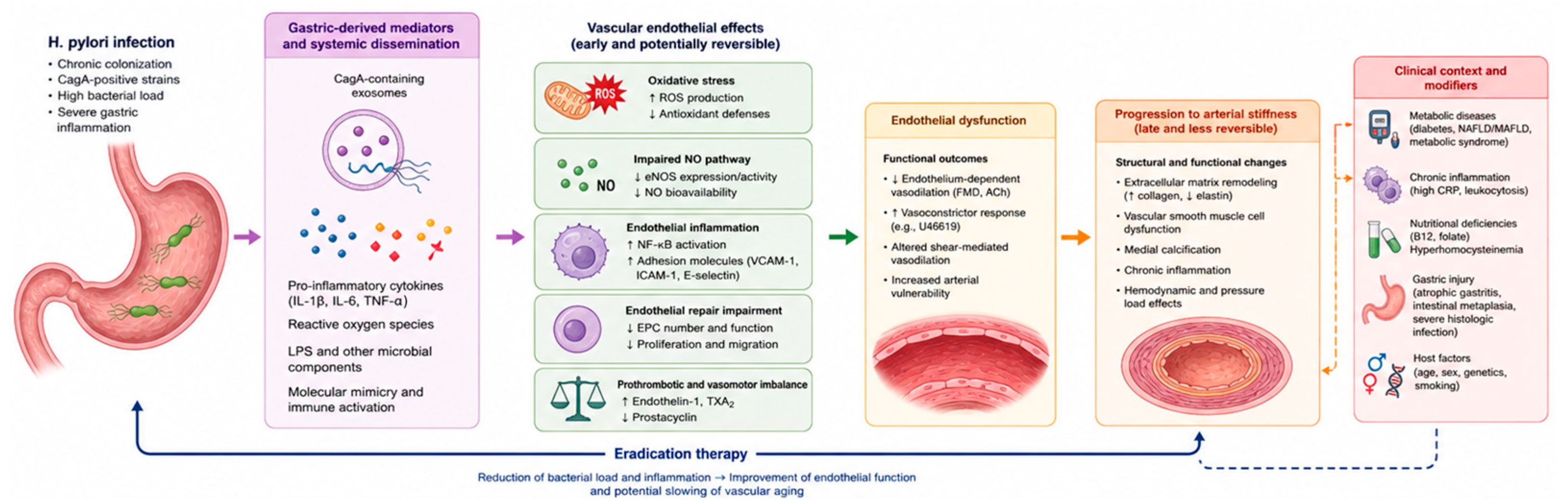
The mechanistic literature therefore supports a biologically integrated model in which virulent H. pylori strains generate gastric-derived vesicular signals that reach the endothelium, increase oxidative stress, impair endothelial repair, activate inflammatory pathways, and selectively compromise endothelium-dependent vasodilation [5,6,24,29]. This mechanistic convergence provides the strongest biological rationale for considering endothelial dysfunction as the primary vascular interface of H. pylori infection [5,13,14] (Figure 1).
Figure 1.
Pathophysiological mechanisms linking Helicobacter pylori infection to vascular dysfunction, including extracellular-vesicle signaling, oxidative stress, inflammatory endothelial programming, impaired endothelial repair, and arterial remodeling. Abbreviations: ACh, acetylcholine; CagA, cytotoxin-associated gene A; CRP, C-reactive protein; eNOS, endothelial nitric oxide synthase; EPC, endothelial progenitor cells; FMD, flow-mediated dilation; H. pylori, Helicobacter pylori; ICAM-1, intercellular adhesion molecule 1; IL-1β, interleukin 1 beta; IL-6, interleukin 6; LPS, lipopolysaccharide; MASLD, metabolic dysfunction-associated steatotic liver disease; NAFLD, nonalcoholic fatty liver disease; NF-κB, nuclear factor kappa B; NO, nitric oxide; ROS, reactive oxygen species; TNF-α, tumor necrosis factor alpha; TXA2, thromboxane A2; U46619, thromboxane A2 analog; VCAM-1, vascular cell adhesion molecule 1; ↑, increased; ↓, decreased.
3.3. Why the Endothelial Signal Is Stronger than the Structural Signal
Studies using structural vascular endpoints have not demonstrated a consistent association between H. pylori seropositivity and established atherosclerosis [4,17,30]. In the Multi-Ethnic Study of Atherosclerosis (MESA), neither H. pylori seropositivity nor cumulative pathogen burden was associated with carotid intima-media thickness or coronary artery calcification [4]. Other studies conducted in patients at increased cardiovascular risk have similarly shown limited or inconsistent relationships between H. pylori antibodies and structural vascular measures [17,30]. Taken together, these findings argue against H. pylori as a universal or major independent determinant of atherosclerotic burden.
Functional and structural vascular endpoints may nevertheless capture different stages of vascular injury. FMD and acetylcholine-mediated vasorelaxation assess dynamic and potentially reversible abnormalities, whereas carotid intima-media thickness and coronary artery calcification reflect cumulative arterial remodeling influenced by age, blood pressure, lipid and glycemic burden, renal function, smoking, diet, and socioeconomic factors [4,5,9,30]. This difference offers one possible explanation for the heterogeneous findings, but it does not establish that negative structural studies represent false-negative results or that endothelial dysfunction necessarily precedes structural vascular disease in individuals with H. pylori infection. Such a temporal model remains biologically plausible but requires prospective validation.
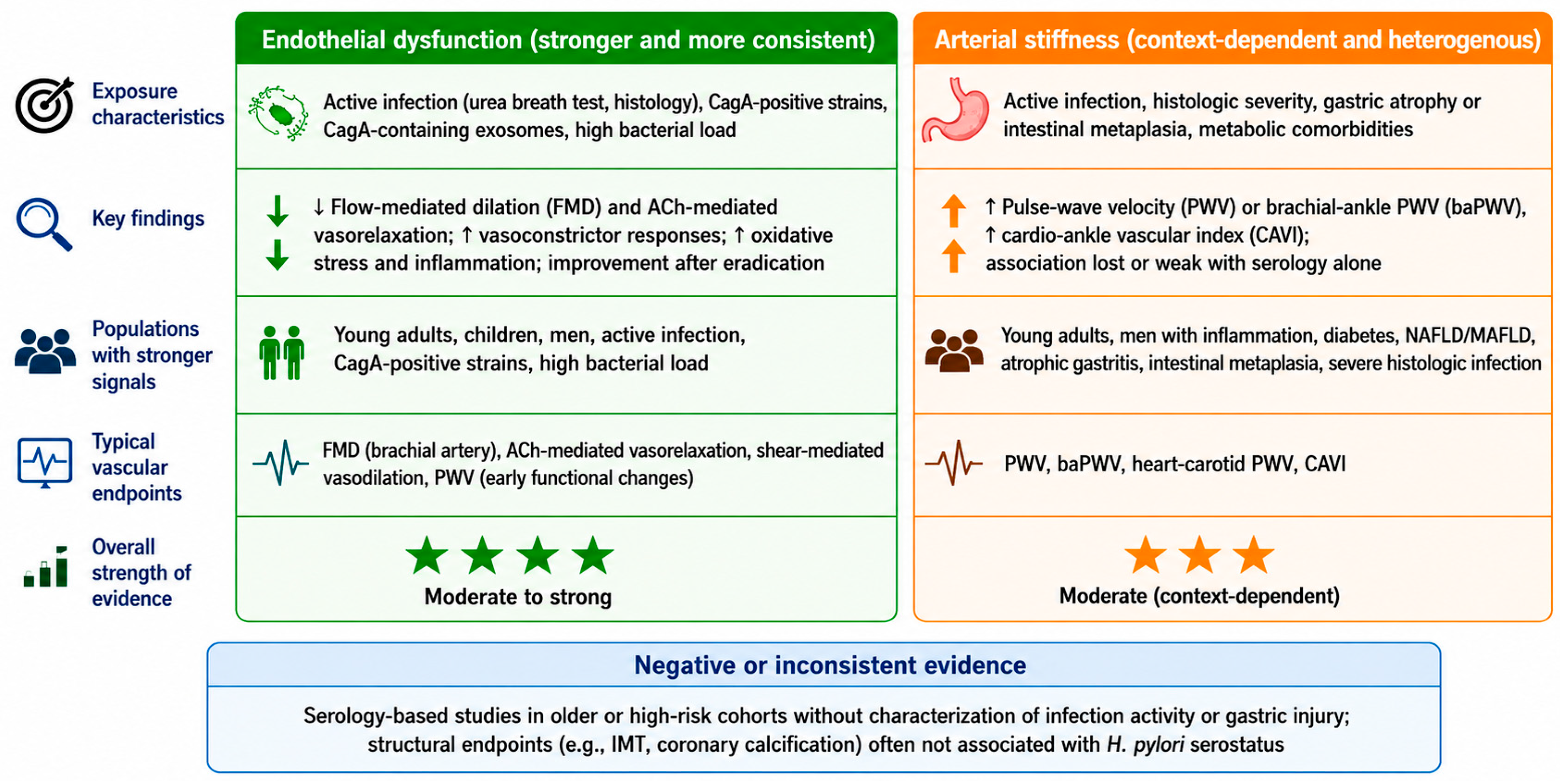
The positive evidence appears more coherent when active infection, gastric inflammatory severity, virulence characteristics, eradication response, and functional vascular endpoints are considered [3,4,5,6,14]. However, these observations remain context-dependent and should be interpreted as hypothesis-supporting rather than as definitive evidence of a causal vascular effect. The absence of consistent associations in serology-based and structural-imaging studies represents an important limitation of the proposed relationship rather than merely a consequence of less informative exposure or outcome assessment (Figure 2).
Figure 2.
Evidence landscape linking Helicobacter pylori infection to endothelial dysfunction and arterial stiffness. Abbreviations: ACh, acetylcholine; baPWV, brachial–ankle pulse-wave velocity; CagA, cytotoxin-associated gene A; CAVI, cardio-ankle vascular index; FMD, flow-mediated dilation; H. pylori, Helicobacter pylori; MAFLD, metabolic dysfunction-associated fatty liver disease; NAFLD, non-alcoholic fatty liver disease; PWV, pulse-wave velocity.
A rigorous interpretation of the endothelial literature therefore requires attention to both sides of the exposure–endpoint relationship. On the exposure side, active infection, virulence profiling, eradication response, and gastric inflammatory severity are more informative than antibody status alone [5,14,26]. On the endpoint side, FMD and acetylcholine-mediated vasorelaxation are better suited to detecting early endothelial injury than carotid intima-media thickness or coronary calcium [4,5,9]. The endothelial signal becomes most persuasive when these biologically meaningful exposure definitions are paired with sensitive functional vascular readouts (Table 1). The principal experimental and translational evidence supporting these mechanisms is summarized in Table 2.
Table 1.
Clinical evidence on H. pylori infection and endothelial or endothelial-adjacent vascular phenotypes.
Table 2.
Experimental and translational mechanisms linking H. pylori infection to endothelial injury.
4. Arterial Stiffness as the Vascular-Aging Readout of Chronic Infection
Arterial stiffness represents a more integrated and temporally accumulated vascular phenotype than endothelial dysfunction. Whereas endothelial dysfunction reflects an early and potentially reversible disturbance of vascular homeostasis, arterial stiffness embodies progressive remodeling of the arterial wall through extracellular-matrix reorganization, vascular-smooth-muscle-cell dysfunction, elastin fragmentation, collagen accumulation, medial calcification, inflammation, and altered central hemodynamics [10,15,16]. For this reason, H. pylori-related stiffness is expected to be harder to detect than endothelial impairment and more dependent on the duration of exposure, vascular aging, blood pressure load, metabolic disease, and gastric inflammatory burden [11,18,19,21].
4.1. Serology as an Imperfect Vascular Exposure
The arterial-stiffness literature does not support a simple or consistent relationship between H. pylori seropositivity and arterial remodeling [11,17]. In populations at increased cardiovascular risk, antibodies against H. pylori were not consistently associated with carotid intima-media thickness, carotid stenosis, or carotid elastic pressure modulus [17]. These null findings may partly reflect the inability of serology to distinguish remote exposure from active infection, but they also indicate that a broadly generalizable and independent vascular association has not been established [3,4,17].
Similarly, cumulative pathogen burden and individual pathogen serostatus have not shown consistent associations with carotid intima-media thickness or coronary artery calcification [4,30]. More informative associations have been reported in selected studies using active-infection testing, gastric histology, or measures of infection severity [21,22,25]. However, these findings derive from specific populations and require independent replication; they should not be interpreted as resolving or invalidating the negative evidence obtained in serology-based cohorts.
4.2. Age, Sex, and Inflammation as Biological Filters
Age, sex, and inflammatory tone appear to function as biological filters that determine whether an H. pylori-associated stiffness signal becomes detectable. In a Japanese screening cohort, seropositivity is accompanied by a modest inflammatory–metabolic profile, including lower high-density lipoprotein cholesterol [55.0 vs. 58.0 mg/dL; p < 0.0001] and higher leukocyte count [5907 vs. 5504 × 109/L; p < 0.0001] [18]. Although overall PWV does not differ by serostatus, heart–carotid PWV is higher in seropositive individuals younger than 39 years [632.2 ± 15.7 vs. 589.7 ± 10.6 cm/s; p = 0.027], whereas this difference is no longer evident with advancing age [18]. This age-dependent pattern suggests that infection-related arterial changes may be more discernible before chronological aging and pressure load become the dominant determinants of arterial stiffness [11,18].
Sex and inflammation further sharpen this phenotype. In men, H. pylori seropositivity is independently associated with high baPWV [OR, 1.27; 95% confidence interval (CI), 1.05–1.52], and the association becomes stronger when seropositivity coexists with C-reactive protein >0.045 mg/dL [OR, 1.50; 95% CI, 1.14–1.98] [19]. The signal is most pronounced in men aged ≤49 years, in whom combined seropositivity and elevated C-reactive protein confers a higher likelihood of increased stiffness [OR, 1.81; 95% CI, 1.16–2.80], while no independent association is evident in older men or in women [19]. This clinical pattern is consistent with experimental evidence showing male-selective endothelial oxidative injury after CagA-positive infection, suggesting that sex-related vascular susceptibility may modulate the arterial consequences of chronic gastric infection [24].
4.3. Biological Burden Beyond Antibody Status
The vascular signal becomes more informative when the exposure is defined by biological burden rather than by antibody status alone. In asymptomatic adults, H. pylori seropositivity is independently associated with high CAVI [adjusted OR, 1.36; 95% CI, 1.10–1.68; p = 0.005], suggesting that stiffness-related associations may be detectable when the vascular phenotype is assessed with an index less dependent on instantaneous blood pressure than conventional PWV [31]. Nevertheless, the most conceptually compelling evidence comes from histology-based assessment, where the severity of gastric infection, rather than seropositivity alone, defines the biological exposure [25].
In young adults with histologically confirmed infection, estimated PWV (ePWV) is higher in infected than uninfected participants [6.66 ± 0.60 vs. 6.33 ± 0.58 m/s; p < 0.001] [25]. More importantly, ePWV increases across Updated Sydney System severity categories, and severe infection is associated with a markedly higher likelihood of elevated ePWV [OR, 3.87; 95% CI, 3.25–4.60; p < 0.001] [25]. This graded relationship suggests that arterial stiffening may reflect the biological intensity of gastric infection more closely than the mere presence of circulating antibodies [25].
4.4. Metabolic Vulnerability as a Vascular Amplifier
Metabolic disease appears to make the vascular consequences of H. pylori more visible, not because infection becomes an isolated cause of arterial stiffness, but because it acts on an arterial wall already primed by insulin resistance, glycation, oxidative stress, endothelial dysfunction, inflammation, and pressure-related remodeling [15,16,20,23]. In type 2 diabetes, infected patients have higher PWV than uninfected patients [1877 ± 550 vs. 1585 ± 331 cm/s; p = 0.0005], and infection remains an independent determinant of PWV after adjustment for major hemodynamic covariates [β = 0.169; p = 0.0220] [20]. This association becomes more pronounced in a larger diabetic cohort using active-infection testing, in which infected subjects show substantially higher baPWV [2031.61 ± 525.48 vs. 1556.68 ± 227.54 cm/s; p < 0.01] and a higher prevalence of severe peripheral arterial stiffness [62.7% vs. 21.9%; p < 0.01] [23]. These findings position diabetes as a vascular amplification state in which chronic gastric infection may intensify, rather than independently initiate, arterial aging [20,23].
A similar interaction emerges in metabolic liver disease [12]. In asymptomatic individuals, nonalcoholic fatty liver disease combined with H. pylori infection confers higher odds of increased arterial stiffness [OR, 2.23; 95% CI, 1.63–3.06] than nonalcoholic fatty liver disease without infection [OR, 1.61; 95% CI, 1.15–2.26] [12]. Among individuals with metabolic dysfunction-associated fatty liver disease, H. pylori infection additively increases stiffness risk [OR, 2.13; 95% CI, 1.64–2.78], supporting convergence among gastric infection, hepatic–metabolic dysfunction, insulin resistance, systemic inflammation, and arterial-wall remodeling [12]. This interpretation is consistent with commentary proposing active H. pylori infection as a potential modifier of the relationship among metabolic syndrome, fatty liver disease, arterial stiffness, and cardio–cerebrovascular risk [32].
4.5. Gastric Injury as a Marker of Cumulative Vascular Exposure
Cumulative gastric injury may provide a more informative vascular exposure than infection status alone. In older adults, pepsinogen-defined atrophic gastritis is associated with higher adjusted baPWV [16.63 ± 3.50 vs. 15.59 ± 3.47 m/s; p = 0.010] and higher CAVI [8.59 ± 1.20 vs. 8.27 ± 1.19; p = 0.022], whereas H. pylori immunoglobulin G status alone shows only nonsignificant trends [21]. This distinction is biologically important because atrophic gastritis may mark prolonged gastric inflammation, cumulative microbial exposure, altered acid secretion, and nutritional disturbance even when serology becomes less informative [21].
Histologic gastric lesions reinforce the same concept. Patients with gastric intestinal metaplasia or atrophic gastritis have higher PWV than controls [10.07 ± 1.79 and 9.85 ± 1.84 vs. 8.99 ± 1.95 m/s], and PWV >10 m/s is substantially more frequent in both gastric-injury groups than in controls [49.5%, 47.8%, and 18.8%; p < 0.001] [22]. Low vitamin B12, gastric intestinal metaplasia, and atrophic gastritis independently predict elevated PWV, suggesting that malabsorption, micronutrient deficiency, homocysteine-related vascular stress, and chronic gastric inflammation may contribute to arterial stiffening [22].
This evidence reframes gastric pathology as a cumulative biological record rather than a purely local consequence of infection. In vascular terms, atrophic gastritis, intestinal metaplasia, vitamin B12 disturbance, and histologic infection severity may capture the duration and systemic consequences of the host–pathogen interaction more effectively than antibody status alone [21,22,25].
The principal clinical and contextual evidence linking H. pylori infection or gastric injury to arterial stiffness is summarized in Table 3.
Table 3.
Clinical and contextual evidence linking H. pylori infection or gastric injury to arterial stiffness.
4.6. Experimental Bridge and Interpretive Boundaries
Experimental evidence helps connect endothelial injury with later arterial remodeling, particularly when infection is studied in the presence of metabolic stress. In a guinea-pig model, persistent H. pylori infection combined with a high-fat diet increases systemic inflammation and reduces pulse-wave amplitude, a pattern consistent with impaired arterial elasticity [34]. The relevance of this model lies less in direct clinical extrapolation than in its biological convergence with human data, in which the vascular signal of infection becomes most apparent when metabolic vulnerability is present [12,34].
The boundaries of this model are equally important. Not all systemic consequences of H. pylori appear to be mediated by arterial stiffness, and infection-related risk may involve pathways that are not captured by PWV [33]. In a nested case–control study, H. pylori seropositivity was associated with cognitive decline in the final stepwise multivariable model [OR, 4.468; 95% CI, 1.535–13.00], whereas PWV was not retained as an independent predictor, suggesting that systemic immune or metabolic disruption may extend beyond measurable arterial stiffening [33]. More broadly, the clinical literature supports biological plausibility but does not establish the direction or causality of the reported associations. Most available studies are cross-sectional, case–control, or observational, and therefore cannot determine whether H. pylori infection preceded the development of endothelial dysfunction or arterial stiffness. Reverse causation and bidirectional relationships cannot be excluded, because individuals with cardiometabolic disease or established vascular impairment may differ in healthcare utilization, likelihood of undergoing gastrointestinal testing, dietary patterns, nutritional status, gastric pathology, and medication exposure, all of which may influence both infection ascertainment and vascular measurements [2,11,35].
Residual confounding also remains an important concern. Age, smoking, adiposity or body mass index, blood pressure, diabetes, renal function, lipid profile, diet, physical activity, socioeconomic conditions, and concomitant medications are associated with vascular function and may also correlate with the acquisition, persistence, diagnosis, or treatment of H. pylori infection. Although several studies adjusted for selected cardiovascular risk factors, the covariates considered and the methods used to measure them varied substantially, and some relevant factors were unavailable or incompletely captured. Reported adjusted associations should therefore not be interpreted as evidence that H. pylori independently causes vascular dysfunction.
A systematic review and meta-analysis of 24 observational studies reported higher CIMT and PWV and lower FMD in H. pylori-positive individuals; however, the FMD analysis included only two studies and between-study heterogeneity was very high, limiting the certainty and generalizability of the pooled estimates [36].
5. Integrated Pathophysiological Model
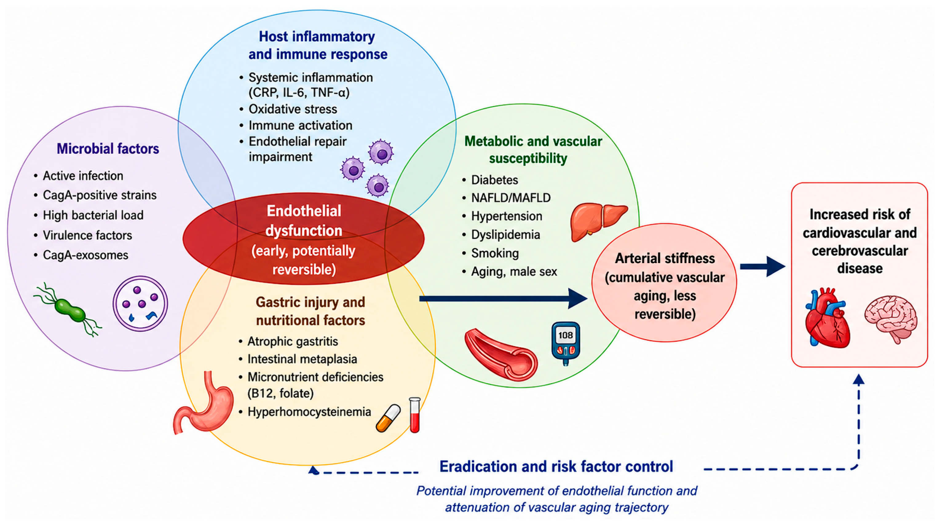
The available evidence supports a model in which H. pylori contributes to vascular dysfunction only when microbial, host, metabolic, and gastric-disease determinants converge [2,3,4,11]. Within this framework, the endothelium represents the earliest and most coherent vascular interface of infection, because active CagA-positive strains can generate gastric-derived exosomal signals, increase reactive oxygen species, impair nitric oxide-dependent vasodilation, disrupt endothelial repair, and activate inflammatory signaling pathways [5,6,29]. These mechanisms closely overlap with established pathways of endothelial dysfunction, including oxidative stress, impaired nitric oxide signaling, inflammatory activation, altered mechanotransduction, thrombogenicity, senescence, and defective vascular repair [7,8,9].
Arterial stiffness occupies a later position in this biological sequence. It may represent the stage at which repeated endothelial injury is incorporated into extracellular-matrix remodeling, vascular-smooth-muscle-cell dysfunction, altered central hemodynamics, and progressive arterial-wall aging [10,15,16]. Conditions characterized by insulin resistance, chronic inflammation, micronutrient disturbance, or advanced gastric pathology may facilitate this transition, making the vascular consequences of H. pylori more detectable in metabolically or histologically enriched populations [12,20,21,22,23,25].
This framework also helps explain the heterogeneity of clinical literature. Serology-based studies may underestimate relevant vascular exposure, whereas older cohorts may be dominated by age- and pressure-related arterial remodeling that obscures smaller infection-related effects [3,4,11,17,18]. By contrast, vascular signals are more likely to emerge in younger individuals, in men with inflammatory activation, in patients with cardiometabolic disease, and in cohorts characterized by active infection, gastric histology, or infection severity [5,12,14,19,23,24,25].
More broadly, H. pylori fits within an emerging immunovascular paradigm in which chronic mucosal infection may influence systemic vascular aging through persistent antigenic stimulation, immune remodeling, extracellular-vesicle signaling, oxidative stress, and post-infectious vascular injury [10,37,38]. Its relevance is therefore not that of an isolated cardiovascular pathogen, but that of a chronic biological stressor whose vascular effects become manifest when microbial activity and host susceptibility intersect [5,6,10] (Figure 3).
Figure 3.
Integrated model: Helicobacter pylori as a conditional amplifier of early vascular aging. Abbreviations: B12, vitamin B12; CagA, cytotoxin-associated gene A; CRP, C-reactive protein; IL-6, interleukin 6; MAFLD, metabolic dysfunction-associated fatty liver disease; NAFLD, nonalcoholic fatty liver disease; TNF-α, tumor necrosis factor alpha.
6. Clinical and Research Implications
The available evidence does not support H. pylori testing as a cardiovascular screening or risk-stratification strategy in unselected populations [3,4,17]. It also does not justify eradication therapy solely for the prevention or treatment of endothelial dysfunction or arterial stiffness. Accordingly, the present evidence should not modify established clinical indications for H. pylori testing and eradication. The potential vascular relevance of the infection should therefore be investigated primarily in research settings and in carefully phenotyped high-risk cohorts, using endpoints such as FMD, acetylcholine-mediated vasorelaxation, PWV, and CAVI [5,9,25,31].
The next generation of studies should therefore move beyond binary serology as a screening test and adopt a multidimensional exposure framework in which the detection of an active infection is needed. Active-infection testing, CagA and virulence profiling, gastric histologic grading, sex-stratified analyses, metabolic phenotyping, inflammatory biomarkers, and prespecified vascular endpoints should be incorporated into prospective and interventional designs [5,9,11,26]. Randomized eradication trials with prespecified endothelial and arterial-stiffness outcomes will be essential to determine whether H. pylori is only a marker of vascular vulnerability or a modifiable contributor to early vascular aging, and will be required before testing or eradication can be recommended for cardiovascular prevention [5,14].
7. Limitations
Publication bias and selective outcome reporting cannot be excluded. Some of the positive findings derive from relatively small, single-center observational, pre/post-eradication, or mechanistic studies, whereas null or inconclusive results may be less likely to be published or emphasized. In addition, despite the adoption of a structured literature-search strategy, the narrative design of this review entails a potential risk of selective study inclusion or interpretive emphasis. Because of the substantial heterogeneity in infection assessment, vascular endpoints, study populations, follow-up duration, and analytical methods, a formal assessment of publication bias was not feasible. Consequently, the apparent coherence of the positive evidence may overestimate the magnitude, reproducibility, or clinical relevance of the association between H. pylori infection and vascular dysfunction.
Most of the available clinical evidence is observational, predominantly cross-sectional or case–control, and therefore cannot establish temporality or causality. Comparability across studies is further limited by substantial heterogeneity in study populations, methods used to assess H. pylori exposure, vascular endpoints, adjustment strategies, and follow-up duration. In particular, serology, active-infection testing, bacterial virulence profiling, and gastric histology do not represent equivalent exposure measures, while FMD, PWV, baPWV, CAVI, ePWV, and structural vascular markers capture different vascular phenotypes. The limited number and sample size of eradication studies, together with the absence of long-term vascular outcomes, further restrict causal interpretation. Accordingly, the conclusions of this review should be regarded as hypothesis-generating rather than definitive.
The predominantly observational nature of the clinical evidence limits causal interpretation. Cross-sectional and case–control studies cannot establish whether H. pylori infection precedes endothelial or arterial abnormalities, and reverse causation or bidirectional associations remain possible. Patients with cardiometabolic disease may have different patterns of healthcare access, gastrointestinal investigation, previous antibiotic or acid-suppressive treatment, dietary behavior, nutritional status, and medication use, potentially influencing both the classification of infection and the measured vascular phenotype.
Residual confounding is also likely. Smoking, body mass index and adiposity, hypertension, diabetes, dyslipidemia, renal function, diet, physical activity, socioeconomic disadvantage, and concomitant therapies may be associated with both H. pylori exposure and vascular dysfunction. In particular, antihypertensive, lipid-lowering, glucose-lowering, and other cardiovascular therapies may directly affect endothelial function, blood pressure, inflammatory markers, or arterial-stiffness measurements, while previous antibiotics or proton-pump inhibitors may affect the detection of active infection. Adjustment for these variables was inconsistent across studies, and even extensive multivariable models cannot exclude unmeasured or imperfectly measured confounding. Consequently, associations reported as statistically independent should not be regarded as proof of a direct causal vascular effect of H. pylori.
8. Conclusions
H. pylori should be interpreted neither as an incidental serological finding nor as a universal cardiovascular pathogen. The most defensible interpretation is that chronic infection is associated with vascular dysfunction in selected biological contexts and may contribute to it when active infection or strain virulence coincides with a susceptible inflammatory, metabolic, or gastric-disease substrate. However, the predominantly observational evidence, uncertain temporal sequence, and potential for residual confounding preclude causal conclusions.
This model is most persuasive for endothelial dysfunction. Active infection, CagA-dependent exosome signaling, oxidative stress, selective impairment of endothelium-dependent relaxation, and improvement after eradication form a coherent mechanistic and translational sequence linking gastric infection to vascular homeostasis. Evidence for arterial stiffness is less uniform, but becomes more clinically meaningful in populations enriched for early vascular aging, inflammatory activation, cardiometabolic disease, or advanced gastric injury.
The broader implication is that H. pylori may contribute to early vascular aging as part of an immunometabolic network linking chronic mucosal infection, endothelial injury, extracellular-vesicle signaling, oxidative stress, and arterial-wall remodeling.
Author Contributions
Conceptualization, F.F., G.F. and A.F.G.C.; methodology, F.F., G.F., D.V. and A.F.G.C.; investigation, F.F., G.F., C.S., D.V. and A.F.G.C.; data curation, F.F., G.F., C.S., C.B., D.V. and A.F.G.C.; writing—original draft preparation, F.F., G.F., D.V. and A.F.G.C.; writing—review and editing, C.S. and C.B.; supervision, D.V. and A.F.G.C.; project administration, G.F.; funding acquisition, G.F. All authors have read and agreed to the published version of the manuscript.
Funding
This research was funded by Fondazione Carisbo, under the “Bando Ricerca scientifica e alta tecnologia 2025” programme, grant number 2025.0459, request no. 22583, awarded to Giulia Fiorini for the project “Correlation between H. pylori infection, inflammation, and vascular aging in patients at high cardiovascular risk”.
Institutional Review Board Statement
Not applicable.
Informed Consent Statement
Not applicable.
Data Availability Statement
No new data were created or analyzed in this study. Data sharing is not applicable to this article.
Acknowledgments
During the preparation of this manuscript, the author(s) used ChatGPT (OpenAI, GPT-5.5 Thinking) for technical support in the editing and refinement of graphical figures, including improvement of image resolution, legibility, and background standardization, without altering the scientific content of the figures. The author(s) reviewed and edited the output and take full responsibility for the content of this publication.
Conflicts of Interest
The authors declare no conflicts of interest.
Abbreviations
The following abbreviations are used in this manuscript:
| ABI | Ankle–brachial index |
| ACh | Acetylcholine |
| AG | Atrophic gastritis |
| A/G | Albumin-to-globulin ratio |
| baPWV | Brachial–ankle pulse-wave velocity |
| CagA | Cytotoxin-associated gene A |
| CAC | Coronary artery calcification |
| CAD | Coronary artery disease |
| CAVI | Cardio–ankle vascular index |
| C-CVD | Cardio–cerebrovascular disease |
| CHI3L1 | Chitinase 3-like 1 |
| CI | Confidence interval |
| CIMT | Carotid intima-media thickness |
| CMV | Cytomegalovirus |
| COVID-19 | Coronavirus disease 2019 |
| CRP | C-reactive protein |
| CV | Cardiovascular |
| EBV | Epstein–Barr virus |
| ePWV | Estimated pulse-wave velocity |
| FMD | Flow-mediated dilation |
| FRS | Framingham risk score |
| GATA3 | GATA-binding protein 3 |
| GIM | Gastric intestinal metaplasia |
| GW4869 | Exosome-release inhibitor |
| HAPWV | Heart–ankle pulse-wave velocity |
| HCPWV | Heart–carotid pulse-wave velocity |
| HDL | High-density lipoprotein |
| H. pylori | Helicobacter pylori |
| hs-CRP | High-sensitivity C-reactive protein |
| HUVECs | Human umbilical vein endothelial cells |
| ICAM-1 | Intercellular adhesion molecule-1 |
| ICVD | Ischemic cardiovascular disease |
| IgG | Immunoglobulin G |
| IS | Ischemic stroke |
| MAFLD | Metabolic dysfunction-associated fatty liver disease |
| MAPK | Mitogen-activated protein kinase |
| MESA | Multi-Ethnic Study of Atherosclerosis |
| MetS | Metabolic syndrome |
| MI | Myocardial infarction |
| NAFLD | Nonalcoholic fatty liver disease |
| OR | Odds ratio |
| PAI-1:Ag | Plasminogen activator inhibitor-1 antigen |
| PWV | Pulse-wave velocity |
| ROS | Reactive oxygen species |
| TIMI | Thrombolysis in Myocardial Infarction |
| TM | Thrombomodulin |
| tPA:Ag | Tissue plasminogen activator antigen |
| vWF | von Willebrand factor |
References
- Manolakis, A.; Kapsoritakis, A.N.; Potamianos, S.P. A review of the postulated mechanisms concerning the association of Helicobacter pylori with ischemic heart disease. Helicobacter 2007, 12, 287–297. [Google Scholar] [CrossRef] [Scilit] [PubMed]
- Xu, Z.; Li, J.; Wang, H.; Xu, G. Helicobacter pylori infection and atherosclerosis: Is there a causal relationship? Eur. J. Clin. Microbiol. Infect. Dis. 2017, 36, 2293–2301. [Google Scholar] [CrossRef] [Scilit] [PubMed]
- Khairy, P.; Rinfret, S.; Tardif, J.C.; Marchand, R.; Shapiro, S.; Brophy, J.; Dupuis, J. Absence of association between infectious agents and endothelial function in healthy young men. Circulation 2003, 107, 1966–1971. [Google Scholar] [CrossRef] [Scilit] [PubMed]
- Szklo, M.; Ding, J.; Tsai, M.Y.; Cushman, M.; Polak, J.F.; Lima, J.; Barr, R.G.; Sharrett, A.R. Individual pathogens, pathogen burden and markers of subclinical atherosclerosis: The Multi-Ethnic Study of Atherosclerosis. J. Cardiovasc. Med. 2009, 10, 747–751. [Google Scholar] [CrossRef] [Scilit] [PubMed]
- Xia, X.; Zhang, L.; Chi, J.; Li, H.; Liu, X.; Hu, T.; Li, R.; Guo, Y.; Zhang, X.; Wang, H.; et al. Helicobacter pylori infection impairs endothelial function through an exosome-mediated mechanism. J. Am. Heart Assoc. 2020, 9, e014120. [Google Scholar] [CrossRef] [Scilit] [PubMed]
- Xia, X.; Zhang, L.; Wu, H.; Chen, F.; Liu, X.; Xu, H.; Cui, Y.; Zhu, Q.; Wang, M.; Hao, H.; et al. CagA+ Helicobacter pylori, not CagA− Helicobacter pylori, infection impairs endothelial function through exosomes-mediated ROS formation. Front. Cardiovasc. Med. 2022, 9, 881372. [Google Scholar] [CrossRef] [Scilit] [PubMed]
- Godo, S.; Shimokawa, H. Endothelial functions. Arterioscler. Thromb. Vasc. Biol. 2017, 37, e108–e114. [Google Scholar] [CrossRef] [Scilit] [PubMed]
- Alexander, Y.; Osto, E.; Schmidt-Trucksäss, A.; Shechter, M.; Trifunovic, D.; Duncker, D.J.; Aboyans, V.; Bäck, M.; Badimon, L.; Cosentino, F.; et al. Endothelial function in cardiovascular medicine: A consensus paper of the European Society of Cardiology Working Groups on Atherosclerosis and Vascular Biology, Aorta and Peripheral Vascular Diseases, Coronary Pathophysiology and Microcirculation, and Thrombosis. Cardiovasc. Res. 2021, 117, 29–42. [Google Scholar] [CrossRef] [Scilit] [PubMed]
- Thijssen, D.H.J.; Bruno, R.M.; van Mil, A.C.C.M.; Holder, S.M.; Faita, F.; Greyling, A.; Zock, P.L.; Taddei, S.; Deanfield, J.E.; Luscher, T.; et al. Expert consensus and evidence-based recommendations for the assessment of flow-mediated dilation in humans. Eur. Heart J. 2019, 40, 2534–2547. [Google Scholar] [CrossRef] [Scilit] [PubMed]
- Herzog, M.J.; Müller, P.; Lechner, K.; Stiebler, M.; Arndt, P.; Kunz, M.; Ahrens, D.; Schmeißer, A.; Schreiber, S.; Braun-Dullaeus, R.C. Arterial stiffness and vascular aging: Mechanisms, prevention, and therapy. Signal Transduct. Target. Ther. 2025, 10, 282. [Google Scholar] [CrossRef] [Scilit] [PubMed]
- Chavez, L.; Mayrovitz, H.N. Assessing the impact of Helicobacter pylori infection and inflammatory bowel disease on pulse wave velocity and arterial stiffness. Cureus 2021, 13, e14944. [Google Scholar] [CrossRef] [Scilit] [PubMed]
- Choi, J.M.; Park, H.E.; Han, Y.M.; Lee, J.; Lee, H.; Chung, S.J.; Lim, S.H.; Yim, J.Y.; Chung, G.E. Non-alcoholic/metabolic-associated fatty liver disease and Helicobacter pylori additively increase the risk of arterial stiffness. Front. Med. 2022, 9, 844954. [Google Scholar] [CrossRef] [Scilit] [PubMed]
- Prasad, A.; Zhu, J.; Halcox, J.P.; Waclawiw, M.A.; Epstein, S.E.; Quyyumi, A.A. Predisposition to atherosclerosis by infections: Role of endothelial dysfunction. Circulation 2002, 106, 184–190. [Google Scholar] [CrossRef] [Scilit] [PubMed]
- Abdelhai, E.; El Amrousy, D.; Abou Taira, N.; Shareef, M.; Amin, S. Vascular endothelial function in pediatric patients with Helicobacter pylori infection and its response to Helicobacter pylori eradication. J. Pediatr. Gastroenterol. Nutr. 2026, 82, 988–995. [Google Scholar] [CrossRef] [Scilit] [PubMed]
- Vatner, S.F.; Zhang, J.; Vyzas, C.; Mishra, K.; Graham, R.M.; Vatner, D.E. Vascular stiffness in aging and disease. Front. Physiol. 2021, 12, 762437. [Google Scholar] [CrossRef] [Scilit] [PubMed]
- Maier, J.A.; Andrés, V.; Castiglioni, S.; Giudici, A.; Lau, E.S.; Nemcsik, J.; Seta, F.; Zaninotto, P.; Catalano, M.; Hamburg, N.M. Aging and vascular disease: A multidisciplinary overview. J. Clin. Med. 2023, 12, 5512. [Google Scholar] [CrossRef] [Scilit] [PubMed]
- Espinola-Klein, C.; Rupprecht, H.J.; Blankenberg, S.; Bickel, C.; Kopp, H.; Rippin, G.; Hafner, G.; Pfeifer, U.; Meyer, J. Are morphological or functional changes in the carotid artery wall associated with Chlamydia pneumoniae, Helicobacter pylori, cytomegalovirus, or herpes simplex virus infection? Stroke 2000, 31, 2127–2133. [Google Scholar] [CrossRef] [Scilit] [PubMed]
- Adachi, K.; Arima, N.; Takashima, T.; Miyaoka, Y.; Yuki, M.; Ono, M.; Komazawa, Y.; Kawamura, A.; Fujishiro, H.; Ishihara, S.; et al. Pulse-wave velocity and cardiovascular risk factors in subjects with Helicobacter pylori infection. J. Gastroenterol. Hepatol. 2003, 18, 771–777. [Google Scholar] [CrossRef] [Scilit] [PubMed]
- Saijo, Y.; Utsugi, M.; Yoshioka, E.; Horikawa, N.; Sato, T.; Gong, Y.; Kishi, R. Relationship of Helicobacter pylori infection to arterial stiffness in Japanese subjects. Hypertens. Res. 2005, 28, 283–292. [Google Scholar] [CrossRef] [Scilit] [PubMed]
- Ohnishi, M.; Fukui, M.; Ishikawa, T.; Ohnishi, N.; Ishigami, N.; Yoshioka, K.; Hasegawa, G.; Yoshikawa, T.; Nakamura, N. Helicobacter pylori infection and arterial stiffness in patients with type 2 diabetes mellitus. Metabolism 2008, 57, 1760–1764. [Google Scholar] [CrossRef] [Scilit] [PubMed]
- Torisu, T.; Takata, Y.; Ansai, T.; Matsumoto, T.; Sonoki, K.; Soh, I.; Awano, S.; Yoshida, A.; Hamasaki, T.; Kagiyama, S.; et al. Possible association of atrophic gastritis and arterial stiffness in healthy middle-aged Japanese. J. Atheroscler. Thromb. 2009, 16, 691–697. [Google Scholar] [CrossRef] [Scilit] [PubMed]
- Kutluana, U.; Kilciler, A.G. Is there a possible relationship between gastric intestinal metaplasia and systemic arterial stiffness? Rev. Esp. Enferm. Dig. 2019, 111, 500–506. [Google Scholar] [CrossRef] [Scilit] [PubMed]
- Yang, Y.F.; Li, Y.; Liu, J.H.; Wang, X.M.; Wu, B.H.; He, C.S.; Gu, J.W. Relation of Helicobacter pylori infection to peripheral arterial stiffness and 10-year cardiovascular risk in subjects with diabetes mellitus. Diab. Vasc. Dis. Res. 2020, 17, 1479164120953626. [Google Scholar] [CrossRef] [Scilit] [PubMed]
- Zhang, L.; Xia, X.; Wu, H.; Liu, X.; Zhu, Q.; Wang, M.; Hao, H.; Cui, Y.; Li, D.P.; Chen, S.Y.; et al. Helicobacter pylori infection selectively attenuates endothelial function in male mice via exosomes-mediated ROS production. Front. Cell. Infect. Microbiol. 2023, 13, 1142387. [Google Scholar] [CrossRef] [Scilit] [PubMed]
- Kim, H.L.; Kim, D.H.; Min, K.W.; Son, B.K.; Chung, J. Arterial stiffness and histologically confirmed Helicobacter pylori infection in young adults. Helicobacter 2025, 30, e70067. [Google Scholar] [CrossRef] [Scilit] [PubMed]
- Grabczewska, Z.; Nartowicz, E.; Kubica, J.; Rość, D. Endothelial function parameters in patients with unstable angina and infection with Helicobacter pylori and Chlamydia pneumoniae. Eur. J. Intern. Med. 2006, 17, 339–342. [Google Scholar] [CrossRef] [Scilit] [PubMed]
- Evrengul, H.; Tanriverdi, H.; Kuru, O.; Enli, Y.; Yuksel, D.; Kilic, A.; Kaftan, A.; Kirac, S.; Kilic, M. Elevated homocysteine levels in patients with slow coronary flow: Relationship with Helicobacter pylori infection. Helicobacter 2007, 12, 298–305. [Google Scholar] [CrossRef] [Scilit] [PubMed]
- Rasmi, Y.; Mehraban, K.; Sadreddini, M.; Zeynalzadeh, J.; Majidinia, M.; Seyyed-Mohammadzad, M.; Babazadeh, H. Lack of significant association between Helicobacter pylori infection and homocysteine levels in patients with cardiac syndrome X. Cardiol. J. 2012, 19, 466–469. [Google Scholar] [CrossRef] [Scilit] [PubMed]
- Chi, J.; Xia, X.; Zhang, L.; Liu, X.; Li, H.; Liu, P.; Wu, H.; Xu, C. Helicobacter pylori induces GATA3-dependent chitinase 3 like 1 (CHI3L1) upregulation and contributes to vascular endothelial injuries. Med. Sci. Monit. 2019, 25, 4837–4848. [Google Scholar] [CrossRef] [Scilit] [PubMed]
- Kurkowska-Jastrzebska, I.; Karlinski, M.A.; Błazejewska-Hyzorek, B.; Sarzynska-Dlugosz, I.; Filipiak, K.J.; Czlonkowska, A. Carotid intima media thickness and blood biomarkers of atherosclerosis in patients after stroke or myocardial infarction. Croat. Med. J. 2016, 57, 548–557. [Google Scholar] [CrossRef] [Scilit] [PubMed]
- Choi, J.M.; Lim, S.H.; Han, Y.M.; Lee, H.; Seo, J.Y.; Park, H.E.; Kwak, M.S.; Chung, G.E.; Choi, S.Y.; Kim, J.S. Association between Helicobacter pylori infection and arterial stiffness: Results from a large cross-sectional study. PLoS ONE 2019, 14, e0221643. [Google Scholar] [CrossRef] [Scilit] [PubMed]
- Kountouras, J.; Papaefthymiou, A.; Polyzos, S.A.; Liatsos, C.; Tzitiridou-Chatzopoulou, M.; Chatzopoulos, D.; Vardaka, E.; Gialambrinou, D.; Kotronis, G.; Doulberis, M. Potential impact of Helicobacter pylori and metabolic syndrome-related non-alcoholic fatty liver disease on cardio-cerebrovascular disease. Metabolism 2022, 135, 155276. [Google Scholar] [CrossRef] [Scilit] [PubMed]
- Koyama, T.; Kuriyama, N.; Ozaki, E.; Matsui, D.; Watanabe, I.; Miyatani, F.; Kondo, M.; Tamura, A.; Kasai, T.; Ohshima, Y.; et al. Serum albumin to globulin ratio is related to cognitive decline via reflection of homeostasis: A nested case-control study. BMC Neurol. 2016, 16, 253. [Google Scholar] [CrossRef] [Scilit] [PubMed]
- Krupa, A.; Gonciarz, W.; Rusek-Wala, P.; Rechciński, T.; Gajewski, A.; Samsel, Z.; Dziuba, A.; Śmiech, A.; Chmiela, M. Helicobacter pylori infection acts synergistically with a high-fat diet in the development of a proinflammatory and potentially proatherogenic endothelial cell environment in an experimental model. Int. J. Mol. Sci. 2021, 22, 3394. [Google Scholar] [CrossRef] [Scilit] [PubMed]
- Kountouras, J.; Boziki, M.; Kazakos, E.; Theotokis, P.; Kesidou, E.; Nella, M.; Bakirtzis, C.; Karafoulidou, E.; Vardaka, E.; Mouratidou, M.C.; et al. Impact of Helicobacter pylori and metabolic syndrome on mast cell activation-related pathophysiology and neurodegeneration. Neurochem. Int. 2024, 175, 105724. [Google Scholar] [CrossRef] [Scilit] [PubMed]
- Shi, H.; Li, Y.; Dong, C.; Si, G.; Xu, Y.; Peng, M.; Li, Y. Helicobacter pylori infection and the progression of atherosclerosis: A systematic review and meta-analysis. Helicobacter 2022, 27, e12865. [Google Scholar] [PubMed]
- Yu, H.T.; Youn, J.C.; Kim, J.H.; Seong, Y.J.; Park, S.H.; Kim, H.C.; Lee, W.W.; Park, S.; Shin, E.C. Arterial stiffness is associated with cytomegalovirus-specific senescent CD8+ T cells. J. Am. Heart Assoc. 2017, 6, e006535. [Google Scholar] [CrossRef] [Scilit] [PubMed]
- Bruno, R.M.; Badhwar, S.; Abid, L.; Agharazii, M.; Anastasio, F.; Bellien, J.; Burghuber, O.; Faconti, L.; Filipovsky, J.; Ghiadoni, L.; et al. Accelerated vascular ageing after COVID-19 infection: The CARTESIAN study. Eur. Heart J. 2025, 46, 3905–3918, Correction in Eur. Heart J. 2025, 46, 4984. https://doi.org/10.1093/eurheartj/ehaf709. [Google Scholar] [CrossRef] [Scilit] [PubMed]

Disclaimer/Publisher’s Note: The statements, opinions and data contained in all publications are solely those of the individual author(s) and contributor(s) and not of MDPI and/or the editor(s). MDPI and/or the editor(s) disclaim responsibility for any injury to people or property resulting from any ideas, methods, instructions or products referred to in the content. |
© 2026 by the authors. Licensee MDPI, Basel, Switzerland. This article is an open access article distributed under the terms and conditions of the Creative Commons Attribution (CC BY) license.