


Abstract
Glioblastoma (GBM) remains one of the most aggressive malignancies, characterized by profound heterogeneity and a dismal prognosis. While genomic and transcriptomic profiling have provided structural insights, they often fail to capture the dynamic interactions within the tumor microenvironment (TME). Secretome analysis—the study of proteins actively secreted by tumor cells—offers a functional readout of these interactions and a reservoir for potential biomarkers. In this review, we critically evaluate the role of MALDI-TOF Mass Spectrometry as a strategic tool for GBM secretome profiling. While Liquid Chromatography-Tandem Mass Spectrometry (LC-MS/MS) remains the gold standard for deep protein discovery, we argue that MALDI-TOF’s speed, cost-effectiveness, and high-throughput capabilities position it as an ideal platform for clinical screening and “spectral phenotyping.” We discuss the technical hurdles, such as ion suppression and the “leakome” (intracellular contamination), and highlight how integrating MALDI with Extracellular Vesicle (EV) enrichment and Artificial Intelligence (AI) can bridge the gap between in vitro discovery and clinical application.
Keywords:
glioblastoma; secretome; MALDI; MALDI-TOF; proteomics; secreted proteins; cell lines; extracellular vesicles 1. Introduction
Studies of the cancer secretome, including glioblastoma (GBM), are increasingly important. They allow us to understand communication between cancer cells and the tumor microenvironment at the molecular level. Secretome profiling involves identifying and quantifying proteins and other factors released by cells. In the context of GBM, mass spectrometry (MS) techniques, such as MALDI (Matrix-Assisted Laser Desorption/Ionization), enable the mapping of proteins from cell culture media. This mapping reveals differences in secretory profiles between lines with varying aggressiveness, invasive potential, or response to treatment [1,2,3]. For instance, label-free quantitative MS has revealed that iHDAC inhibitors can modulate tumor behavior by specifically remodeling the ‘angiogenic matrisome’—regulating factors like decorin and ADAM-family proteases (ADAM10, 12, 15)—rather than through direct VEGF inhibition [4]. This highlights a crucial mechanistic link: histone post-translational modifications (PTMs) regulate chromatin architecture to drive tumor progression, and targeting the enzymes responsible (e.g., deacetylases or methyltransferases) is a rapidly evolving therapeutic strategy [5]. Profiling the secretome serves as a functional readout of this epigenetic state. It allows researchers to monitor how epigenetic therapies remodel the tumor microenvironment. This approach enables the identification of candidate biomarkers. More importantly, it helps researchers understand how therapeutic changes influence cell secretion. These changes, in turn, affect microenvironment remodeling, angiogenesis, invasion, and immune response. In short, secretome profiling is a tool that combines basic research on cellular mechanisms with the search for biomarkers and therapeutic targets [6,7]. This approach allows for the identification of candidate biomarkers, but more importantly, it offers insights into the mechanisms of therapy resistance and relapse, which are the primary clinical challenges in GBM management. For instance, understanding the molecular shifts in Temozolomide (TMZ)-resistant cells is crucial for identifying new therapeutic vulnerabilities. Recent computational studies utilizing gene expression profiles have successfully identified BMS345541—a drug targeting the FOXG1 transcription factor—as a potential repurposed agent to reverse TMZ resistance [5]. Applying secretome profiling in this context could similarly reveal extracellular effectors of such resistance pathways, moving beyond mere description to potential functional intervention. While liquid chromatography-tandem mass spectrometry (LC-MS/MS) remains the gold standard for deep-dive protein discovery due to its high sensitivity, MALDI-TOF MS offers unique advantages for clinical secretome screening. Primarily, MALDI-TOF MS provides unmatched throughput and cost-efficiency, enabling the analysis of hundreds of samples in a fraction of the time required for LC-MS/MS. Furthermore, MALDI-TOF MS is particularly effective in the low-molecular-weight range (peptidomics), capturing bioactive peptide fragments (<10 kDa) that are often lost during the extensive fractionation or enzymatic digestion steps of shotgun proteomics. This allows for the generation of ‘spectral fingerprints’—rapid, diagnostic molecular signatures that can be used for real-time monitoring of tumor progression and therapy response in a clinical setting. In this review, we evaluate the potential of MALDI-TOF MS in GBM secretome profiling, discussing technical constraints, clinical translation challenges, and future perspectives.
2. Glioblastoma Pathophysiology and Research Models
Glioblastoma (GBM) is defined by its profound intra- and intertumoral heterogeneity, manifesting across genomic, transcriptomic, and epigenetic layers [8,9,10,11,12]. While clinical management relies on molecular subtyping—including the assessment of isocitrate dehydrogenase (IDH) mutations, which serve as a primary diagnostic and prognostic indicator, and O-6-methylguanine-DNA methyltransferase (MGMT) promoter methylation, a key predictor of sensitivity to alkylating chemotherapy—and transcriptional classification (proneural, mesenchymal, classical), these static markers often fail to capture the dynamic nature of tumor progression [10,11]. The primary clinical “pain point”—resistance to Temozolomide and inevitable relapse—is driven by a subset of cells that actively remodel their niche [8,11]. In this context, the secretome acts as a critical functional interface; tumor cells modulate the extracellular space to promote invasion, angiogenesis, and immune evasion [10,11]. Consequently, profiling secreted factors is a necessity to understand how GBM clones adapt to therapy and bypass the limitations of the blood–brain barrier [12,13]. Recent meta-analyses of whole-proteome datasets have identified a core set of dysregulated proteins in GBM, including 154 commonly upregulated and 116 downregulated factors compared to normal brain tissue. These shifts reflect critical alterations in mRNA splicing and immune system pathways, providing a systemic backdrop against which the secretome—as a functional readout of these changes—must be interpreted [14].
In vitro models serve as essential discovery platforms for MALDI-based secretomics, offering high reproducibility and simplified sample preparation required for high-throughput methodological optimization [15,16,17]. These controlled systems allow for the precise mapping of secretome shifts under specific stressors—such as epigenetic modulation or drug exposure—without the systemic noise inherent in in vivo environments [18,19]. However, the translational utility of traditional 2D cultures is limited by the absence of the glioblastoma microenvironment’s architectural complexity, oxygen gradients, and multi-cellular interactions (e.g., with immune and endothelial cells), which can lead to the loss of original tumor characteristics over long passages [8,11,18]. Consequently, while GBM cell lines are indispensable for the initial high-throughput screening of secretome signatures and the development of MALDI-TOF MS workflows, they must be viewed as a foundational step. Validating these in vitro “fingerprints” in more complex patient-derived 3D models (organoids) and longitudinal clinical samples is mandatory to ensure biological relevance and clinical applicability [11,18]. The transition from isolated cell line secretomes to “regional proteomes”—analyzing proteins directly from tumor margins—is the necessary next step. Cell lines serve as a discovery platform where MALDI signatures are first identified under simplified conditions before being validated against the “noisy” background of clinical samples.
3. The Secretome as a Diagnostic and Functional Hub
The cancer secretome serves as a dynamic signaling hub that governs the complex cross-talk between GBM cells and the tumor microenvironment (TME) [18,20,21]. Unlike whole-cell proteomics, secretome analysis provides a direct readout of the active mediators of niche remodeling—such as proteases, cytokines, and extracellular vesicles (EVs)—which are potentially detectable in clinical biofluids like plasma and cerebrospinal fluid (CSF) [7,11,18]. It is essential to distinguish soluble secretome components from EV-associated cargo, as they require distinct isolation workflows (e.g., ultrafiltration versus ultracentrifugation) [22,23,24]. Furthermore, while soluble proteins are often analyzed directly, EVs necessitate a specific lysis step to release encapsulated proteins for effective MALDI-TOF MS detection [25,26,27].
In GBM, these secreted factors are primary determinants of invasive growth. Specifically, the enrichment of metalloproteinases and adhesion factors in the extracellular space helps differentiate high-invasive from low-invasive cellular phenotypes [7]. Furthermore, secreted components and exosomal cargo act as paracrine effectors that reprogram surrounding microglia and immune cells, fostering an immunosuppressive environment that facilitates tumor progression and escape [18,28].
However, the identification of a secreted protein is merely a hypothesis-generating step. To move toward clinical utility, these findings must undergo rigorous functional validation, linking candidate markers to core oncogenic drivers, such as the PI3K/AKT signaling axis, or to established mechanisms of TMZ resistance [19,28]. In glioblastoma, oncogenic PI3K/AKT/mTOR signaling downstream of EGFR amplification or PTEN loss not only supports proliferation and therapy resistance but also enhances angiogenic output by increasing VEGF expression and secretion, thereby reshaping the secretome toward a pro-vascular phenotype [29,30,31,32]. In parallel, constitutively active NF-κB orchestrates a chronic inflammatory program in GBM cells, where TNF-α-driven NF-κB activation induces robust IL-6 production, sustains STAT3 signaling, and promotes expression of additional inflammatory mediators, collectively fostering an immunosuppressive, tumor-supportive microenvironment enriched in pro-inflammatory cytokines [33,34,35]. MORE. Moreover, malignant cells display elevated Ca2+ signaling dynamics, in which store depletion, store-operated Ca2+ entry and calpain activity drive extracellular vesicle (EV) biogenesis; blocking cytosolic Ca2+ with chelators such as BAPTA-AM effectively abolishes EV release, underscoring the reliance of vesiculation and docking on Ca2+-dependent pathways [35,36,37].
Together, these signaling axes converge to dictate the final composition of the GBM secretome—combining high levels of VEGF and MMP-associated invasive programs, NF-κB-dependent cytokines such as IL-6, and Ca2+-regulated EV cargo—to optimally support angiogenesis, invasion, immune evasion and therapeutic resistance.
4. Methodological Workflows in GBM Secretomics
Mass spectrometry (MS) is a fundamental tool in proteomics and secretome studies. It enables the identification and quantification of proteins released by cells. Various MS techniques differ in ionization methods, analyzers, and sample preparation. Consequently, the choice of method depends on the study objective and the required level of details [38].
4.1. Methodological Landscape: MALDI-MS and LC-MS in Secretomics
The selection of a mass spectrometry platform involves a trade-off between analytical depth and throughput. Direct MALDI-based workflows may involve spectral profiling (“fingerprinting”), peptide mass fingerprinting (PMF), or MALDI imaging mass spectrometry (MALDI-MSI), depending on the analytical objective. Spectral profiling focuses primarily on reproducible signal patterns and biomarker signatures, whereas PMF relies on peptide mass matching for protein identification. This provides a cost-effective method for screening many samples with minimal preparation [39,40]. However, MALDI is inherently limited by ion suppression in complex mixtures; high-abundance proteins (e.g., albumin fragments) often mask low-concentration signaling factors. While MALDI-TOF/TOF MS analysis enables sequence confirmation of selected precursor ions and rapid spectral characterization, direct MALDI-based workflows generally provide lower proteome coverage than LC-coupled tandem MS approaches due to the absence of prior chromatographic peptide separation [41]. In contrast to direct MALDI profiling approaches, LC-coupled tandem MS workflows provide substantially greater analytical depth by separating peptide mixtures prior to ionization. These workflows may utilize either electrospray ionization (LC-ESI-MS/MS) or MALDI-based detection (LC-MALDI-TOF/TOF). By reducing ion suppression and increasing peptide coverage, LC-coupled strategies enable the identification of low-abundance proteins and post-translational modifications (PTMs) [42]. Quantitative approaches such as SILAC, TMT, or iTRAQ further enhance comparative proteomic analyses [43,44,45].
A unique advantage of the MALDI platform is its extension into MALDI Mass Spectrometry Imaging (MSI). MSI bridges the gap between secretomics and histology by mapping the spatial distribution of molecular signals directly on tissue sections without the need for labels [2,46]. In glioblastoma research, this provides critical spatial context. Researchers can correlate protein localization with tumor boundaries and necrotic regions. This information is typically lost in bulk LC-MS/MS analyses [47,48].
4.2. Critical Comparison of MALDI and LC-MS Platforms
The choice between MALDI and LC-MS/MS is dictated by the specific requirements of the GBM study—whether the priority is deep discovery of signaling pathways or high-throughput screening of biomarker signatures. The Table 1 summarizes the technical trade-offs between these two dominant platforms.
Table 1.
Comparison of MALDI-TOF MS and LC-MS/MS in the Context of Glioblastoma Secretomics.
MALDI-TOF MS is not a replacement for LC-MS/MS in de novo biomarker discovery. LC-MS/MS remains the superior method for depth. Instead, the value of MALDI lies in its role as a clinical screening platform. Once discovery proteomics establishes a “hit list” of proteins, MALDI-TOF MS provides the speed and cost-efficiency to monitor these signatures. This allows for the analysis of large patient cohorts, which is often unfeasible for LC-MS/MS in routine clinical settings [54,58].
5. Technical Constraints and Strategic Solutions in MALDI-TOF MS Secretomics
MALDI-based approaches have been applied to glioblastoma tissues, cyst fluids, and model systems. These studies highlight technical constraints and offer strategies for optimization across sample preparation, acquisition, and bioinformatics [2,59]. Such insights are directly adaptable to GBM secretome-focused workflows.
5.1. Critical Parameters in Sample Preparation
Matrix and washing: Tissue and cell studies emphasize that washing must remove salts and contaminants without delocalizing analytes. Matrix coating must be homogeneous to preserve spatial and intensity information [15].
Secreted fluids: For GBM cyst fluid, optimized protocols (including weak cation exchange enrichment) were required to reproducibly detect low-mass secreted proteins around 6–7 kDa [60].
Low-volume preparation: For high-resolution MSI and single-cell work, mild fixation with isotonic washes balances morphology and molecular integrity. However, researchers must control for the leakage of cytosolic components to avoid artifacts [15,61].
5.2. Strategic Optimization for the GBM Secretome
Optimizing GBM secretome analysis involves three goals: (1) extracting low-abundance proteins like EV cargo, (2) linking them to spatial niches, and (3) monitoring therapy-induced changes (Table 2). The following strategies address these objectives.
Table 2.
Key optimization levers for GBM MALDI secretomics.
EV-focused proteomics: Bottom-up MS of GBM-EVs identifies thousands of proteins. These include invasion-linked markers and post-translational modifications that bulk assays often miss [61,62].
Source optimization: Conditioned media provide EVs highly enriched for tumor material. This improves the detection of low-abundance factors compared to blood samples [63].
Secretome fractionation: Separating EV and EV-depleted fractions reveals distinct proteomes. Both fractions contribute differently to immune and microenvironmental responses [64].
5.3. Signal Acquisition and the “Single Peptide” Identification Crisis
MALDI-TOF MS often yields a limited mass range and overlapping peaks, which complicates protein assignment [60]. To mitigate this, high-resolution MALDI-FTICR MSI can be paired with region-specific LC-MS/MS. This alignment reduces false positives by matching accurate masses with localized bottom-up identifications [2]. Furthermore, single-peptide identifications are prone to error. Specificity can be increased using stable isotope-based amino acid mass tagging. This technique allows for higher-confidence calls, particularly for low-abundance or membrane proteins [65].
5.4. Bioinformatics and Functional Interpretation of MALDI Data
GBM secretomics workflows can utilize existing bioinformatics pipelines to ensure data quality:
- Database search: Engines like Sequest should use stringent cutoffs, including an FDR <1% and a requirement for at least two unique peptides [66].
- Functional annotation: Analysis should focus on proteins linked to the angiogenic matrisome and GBM progression [67]. Using dedicated EV databases, such as Vesiclepedia [Available online: http://www.microvesicles.org/ (accessed on 01 June 2026)], helps contextualize tumor specificity [68].
- Clinical correlation: Linking MALDI patterns with patient outcomes demonstrates the feasibility of actionable biomarker panels [2,59].
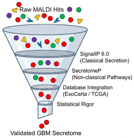
A multi-stage pipeline for GBM MALDI secretomics (Figure 1) should therefore: (1) distinguish classical from non-classical secretion signals [47]; (2) integrate results with prognostic datasets like TCGA [67,68,69,70]; and (3) apply strict statistical thresholds to define a refined secretome signature.
Figure 1.
Multi-stage bioinformatic pipeline for the high-confidence identification of the glioblastoma secretome. To ensure the biological relevance of MALDI-TOF MS profiles, raw protein hits undergo a rigorous filtration process to discriminate the authentic secretome from intracellular contaminants (leakome). Stage 1: Identification of classical secretion signals via SignalP 6.0. Stage 2: Prediction of non-classical (leaderless) secretion pathways using SecretomeP 2.0, critical for tumor stress response. Stage 3: Cross-referencing with extracellular vesicle (EV) and cancer repositories (e.g., ExoCarta, Vesiclepedia, TCGA) to contextualize protein origin and clinical relevance. Stage 4: Application of stringent statistical thresholds (FDR < 1% and minimum 2 unique peptides) to yield a validated set of clinically actionable GBM biomarkers. Note: The diagram represents the bioinformatic filtration pipeline following the initial sample preparation and EV isolation (created by the authors).
In summary, core MALDI constraints are mitigated through careful sample enrichment, high-resolution mass spectrometry, and FDR-controlled bioinformatics. When integrated with clinical data, these workflows transform fragile peptide hits into robust biomarker panels.
6. Synthesis of Published Research: From Deep Discovery to MALDI Targets
The synthesis of published research highlights a strategic pipeline: while deep discovery via LC-MS/MS has provided a comprehensive atlas of the GBM secretome, these findings now serve as the foundational ‘hit list’ for rapid MALDI-TOF MS clinical screening. The following sections evaluate these studies not as competing methodologies, but as complementary stages in biomarker development.
6.1. The “Core” GBM Secretome: High-Abundance MALDI Candidates
Despite varying methodologies, a subset of high-abundance proteins—primarily extracellular matrix (ECM) components—emerges in almost every study. Proteins such as Tenascin-C (TNC), Fibronectin (FN1), and SPARC are not only biologically significant as mediators of tumor stiffness and invasion but are also ideal targets for MALDI-TOF MS profiling due to their high concentration in conditioned media [71,72,73]. These proteins are not only central to GBM invasiveness but also yield high-intensity, reproducible peaks in MALDI spectra. Additionally, secreted factors like YKL-40 and Osteopontin, frequently dysregulated in aggressive GBM phenotypes, serve as robust ‘sentinel’ markers that can be monitored via MALDI-TOF MS to track the transition from low- to high-invasive states [74].
6.2. Phenotype-Specific Markers and Methodological Discrepancies
The reported discrepancies in protein counts between studies (ranging from 150 to nearly 1000) are primarily a reflection of the analytical platform used. For instance, proteins such as YKL-40, identified in invasive GBM phenotypes through MS/MS discovery workflows [6], could serve as candidate targets for future MALDI-based profiling strategies aimed at translational biomarker development. In contrast, patient-derived sphere models (GICs) secrete unique factors like Midkine (MDK) [75]. The fact that MDK is often missed in serum-cultured lines underscores the need for MALDI protocols that can operate in serum-free, chemically defined environments to capture these low-abundance stemness mediators. Table 3 shows a review of studies analyzing the secretome of the GBM lines.
Table 3.
Overview of studies analyzing the secretome of GBM lines.
6.3. Bridging the Gap: Why MALDI Matters for These Findings
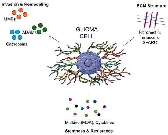
While deep LC-MS/MS by Gupta et al. [71] identified nearly 1000 proteins, many of these are low-abundance intracellular contaminants. For clinical translation, we do not need to detect every protein; we need to monitor the “secretory signature.” The predominant ECM and invasion proteins identified in these studies (Table 4) produce distinct, high-intensity ions in MALDI spectra. This confirms that the transition from LC-MS/MS “discovery” to MALDI-TOF MS “screening” is not only possible but biologically justified. Figure 2 shows proposed Key components of the Glioblastoma Secretome identified by MS proteomics.
Table 4.
Comparative overview of key protein identifications in GBM secretome studies.
Figure 2.
Proposed Key components of the Glioblastoma Secretome identified by MS proteomics. The secretome acts as a functional interface driven by core ECM proteins (e.g., Tenascins), invasion mediators (Proteases, ADAMs), and stem-cell specific factors (e.g., Midkine). These secreted molecules drive microenvironmental remodeling, therapy resistance, and tissue invasion (created by the authors).
6.4. Analytical Constraints of MALDI-TOF MS in Secretomics: From Quantitative Performance to Potential Failure Modes
In contrast to LC–ESI–MS/MS, MALDI-TOF(/TOF) typically offers moderate resolution (~10,000 or higher in reflector mode) with picomole–femtomole peptide sensitivity but suffers from limited quantitative robustness and lower analytical sensitivity in complex clinical samples, where it cannot reliably detect low analyte loads without enrichment [38,53,78]. Quantitative MS/MS comparisons using iTRAQ-labeled E. coli digests show that LC-MALDI-TOF/TOF and LC-ESI-QTOF yield very similar peptide ratios over 1:1–10:1 ranges, with mean ratio deviations of only 0.7–6.7%, but LC-MALDI uses fewer spectra and achieves slightly lower sequence coverage [53]. However, MALDI-based quantification is intrinsically hampered by poor shot-to-shot and spot-to-spot reproducibility due to heterogeneous matrix–analyte cocrystals and random laser sampling, requiring extensive averaging and internal standards to approximate the quantitative performance routinely achieved by LC–MS/MS [79,80]. In glioma secretome workflows, MALDI-TOF can fail when residual salts or ammonium buffers reduce peptide sensitivity and promote matrix cluster formation that dominates spectra below m/z 1200, unless carefully optimized desalting and washing (e.g., ammonium phosphate) are applied [78]. Highly abundant ECM proteins and adhesion molecules secreted by glioblastoma cells and EVs (e.g., proteins involved in cell–matrix adhesion, and migration) may dominate the proteomic composition of these samples, potentially limiting the detection of low-abundance chemokines [81,82]. This problem is exacerbated by “leakome” contamination in ex vivo or stressed cultures, where intracellular proteins released from damaged cells add intense background that obscures regulated, low-level secreted factors [83]. Conventional MALDI matrices generate a dense chemical background below ~700–800 m/z, making it difficult to distinguish low-molecular-weight cytokines from matrix clusters; in an ECM-rich glioma secretome this low-mass interference, combined with ion suppression by abundant matrisome proteins, provides a mechanistic explanation for false-negative MALDI-TOF results for small cytokines, even when LC–MS/MS can detect them [79,80,84].
7. Clinical Validation and the Translational “Handover”
The transition from an in vitro secretome “hit list” to a clinically useful biomarker requires a rigorous validation cascade. This process, termed the “Clinical Handover,” involves shifting from broad discovery platforms to targeted, high-sensitivity assays capable of detecting tumor-derived signals against the massive protein background of human biofluids.
7.1. From Discovery to Targeted Verification: SRM/PRM vs. Immunoassays
While immunological approaches such as ELISA or conventional IHC remain indispensable for single-target validation, they encounter substantial limitations when confronted with the extreme intratumoral heterogeneity of glioblastoma [85]. The shift away from simple immunological screening toward MALDI-based platforms is therefore not driven by inadequate antibody specificity—indeed, contemporary MALDI-IHC workflows explicitly exploit mass-tagged antibodies [86]. Instead, MALDI’s key advantage lies in its intrinsic multiplexing capacity. Whereas standard immunoassays are typically restricted to 1–5 analytes per run, a single MALDI acquisition can concurrently survey dozens of proteins, glycoforms and lipid species, generating a holistic “spectral fingerprint” that more faithfully captures the evolving molecular landscape of the tumor than any single-antibody assay [3,87].
7.2. The Analytical Challenge of Biofluid Matrices: CSF vs. Plasma
Translating secretome markers into clinical samples faces two major hurdles: the Blood–Brain Barrier (BBB) and the dynamic range of protein concentrations.
- Cerebrospinal Fluid (CSF): As the fluid in direct contact with the GBM microenvironment, CSF is enriched with the tumor secretome. However, the concentration of GBM-derived proteins is still several orders of magnitude lower than that of systemic albumin [88,89]. MALDI-based screening in CSF requires advanced pre-fractionation (e.g., immuno-depletion of albumin) to reveal the “hidden” tumor signature.
- Plasma/Serum: The BBB significantly restricts the efflux of large protein markers into the systemic circulation, often rendering secretome candidates undetectable in blood without ultra-sensitive enrichment of extracellular vesicles (EVs) [90,91].
From a translational perspective, detecting these markers in plasma remains a formidable challenge. The blood–brain barrier (BBB) acts as a highly selective filter, ensuring that brain-derived proteins are present in systemic circulation only at extremely low concentrations (picomolar to femtomolar range) [92,93]. Furthermore, the overwhelming abundance of plasma proteins—such as albumin and immunoglobulins—creates a significant ‘ionization noise’ in MALDI-TOF MS, often masking the subtle spectral signatures of glioblastoma-specific factors [94,95].
7.3. Standardizing the Pre-Analytical Pipeline
The “Clinical Handover” often fails due to a lack of pre-analytical rigor. Factors such as the time from sample collection to centrifugation, the number of freeze–thaw cycles, and the use of specific protease inhibitors drastically affect the stability of the secretome profile [96,97]. For MALDI-TOF MS to be viable in a clinical setting, a standardized SOP (Standard Operating Procedure) for biofluid handling must be implemented, ensuring that the “molecular fingerprint” recorded in the clinic matches the one discovered in the laboratory.
To address the in vivo relevance, MALDI-based workflows must move toward the analysis of regional proteomes from clinical specimens (e.g., tumor core vs. invasive margin). This approach, though analytically challenging due to tissue heterogeneity, provides a more accurate readout of the active mediators of niche remodeling. By correlating in vitro secretome profiles with spatial data from MALDI-Imaging (MSI) of patient tissues, researchers can bridge the gap between discovery and the biological reality of the GBM microenvironment [3].
8. Technical and Methodological Limitations: A Critical Assessment
The inherent complexity of the glioblastoma secretome—characterized by a vast dynamic range of protein concentrations and high levels of glycosylation—presents unique challenges for MALDI-TOF MS analysis. Unlike the relatively “clean” intracellular proteome, the secretome is prone to contamination from culture media additives and cellular debris, which can drastically compromise spectral quality [3].
8.1. The Dynamic Range and Ion Suppression Challenge
The primary limitation in MALDI secretomics is the masking effect of high-abundance proteins. A few dominant species (e.g., Albumin fragments or Tenascins) can monopolize the available matrix energy during laser desorption, effectively suppressing the ionization of low-abundance signaling peptides (cytokines, growth factors). This suppression is not merely a matter of instrument sensitivity but a fundamental physicochemical competition within the matrix crystals. To mitigate this, specific strategies for protein depletion and fractionated crystallization must be employed [98,99].
8.2. Troubleshooting and Optimization Strategies
To ensure reproducible and high-resolution spectral fingerprints, researchers must systematically address the most common failure modes in MALDI workflows. Table 5 provides a comprehensive guide to identifying and resolving these methodological hurdles.
Table 5.
Troubleshooting and Optimization Guide for MALDI-based GBM Secretome Profiling.
9. Future Research Frontiers: Toward Multimodal and Intelligent Diagnostics
Current evidence suggests a paradigm shift. MALDI-MSI is now being integrated with artificial intelligence and complementary imaging modalities to create multimodal diagnostic platforms. In this framework, AI facilitates the high-throughput translation of complex spectral data into distinct disease phenotypes. Additionally, the synergy of longitudinal datasets enables the reconstruction of a “molecular movie” of therapeutic response. Spatially registered MSI further permits high-resolution “secretomics” by correlating molecular signatures with specific cellular microenvironments.
9.1. AI-Driven Spectral Phenotyping
AI models are essential for processing complex MALDI data. For example, single-cell (Table 6) MALDI-MSI lipid profiles were recently combined with MALDI-IHC. This automated recognition model accurately distinguished GBM from neuronal cells using triglycerides and sphingomyelins as key features [3]. Similarly, “dry proteomics” (Table 6) employs unsupervised clustering on lipid and protein MALDI-MSI. This method defines GBM subgroups linked to prognosis and builds robust classification models [110]. Finally, fractal-based feature extraction from MALDI-TOF MS spectra of liquid biopsies has enabled the machine-learning diagnosis of GBM. This shows that even compressed pattern descriptors carry vital diagnostic information [111].
Table 6.
Different MALDI-based spectral phenotyping strategies in GBM.
9.2. The “Molecular Movie”: Longitudinal Monitoring of Therapy Response
Focusing on MALDI as a screening tool allows for high-frequency, longitudinal monitoring. This ‘molecular movie’ of the secretome allows for the detection of multi-target signatures that single-marker immunological tests would likely miss, providing a more robust framework for real-time therapeutic adjustments [113]. A multimodal platform recently combined MALDI-MSI drug mapping with phosphoproteomics in GBM models. This approach revealed heterogeneous drug distribution and adaptive signaling responses to the inhibitor adavosertib [114].
Furthermore, the use of REIMS during surgery, coupled with MALDI-MSI and LC-MS/MS, enabled survival-time classification based on lipid patterns [59]. In parallel, spatially resolved multi-omics pipelines in GBM models have been used to track metabolic therapy effects, such as arginine depletion. These studies highlight critical differences in response between the tumor core and its rim [115].
9.3. Spatial Secretomics: MALDI-MSI and the Peribascular Niche
While not always labeled “secretomics,” several MALDI-centric and multimodal studies interrogate spatial metabolic and proteomic niches in GBM: Ultra-high-resolution MALDI-FTICR MSI with microproteomics in a mouse glioma model delineated tumor-specific proteoforms and metabolic proteins, resolving heterogeneous regions that underlie local functional states [2]. Similarly, lipid and peptide MALDI-MSI in large GBM cohorts revealed spatially distinct microenvironments that correlate with patient survival [110,114].
Advanced MSI techniques, combined with isotope tracing, have also mapped metabolic fluxes in glioma models. This research exposed tumor-specific anabolic activity within the brain ecosystem [116]. Finally, spatial transcriptomics has identified perivascular and perinecrotic niches with distinct inflammatory programs [117,118]. Integrating MALDI-MSI with these datasets will enable true “spatial secretomics,” allowing for the detailed study of macrophage–endothelial–tumor interactions.
Beyond imaging, the integration of single-cell proteomics and microfluidic-based secretome analysis offers a path toward resolving the extreme cellular heterogeneity of GBM. While microfluidics allow for the high-sensitivity capture of secreted factors from minimal cell numbers, single-cell approaches ensure that the ‘average’ secretome signal does not mask rare, highly aggressive clones [119,120]. Furthermore, aligning these protein-level findings with spatial transcriptomics data will enable a complete multi-omic reconstruction of the tumor niche, providing a definitive map of how gene expression translates into the functional, secreted proteome [70,118].
10. General Discussion and Conclusions
Secretome profiling represents a critical functional layer in glioblastoma research, capturing the active interface between the tumor and its microenvironment—a layer often missed by static genomic or transcriptomic analyses. However, for MALDI-TOF MS to realize its potential as a diagnostic tool, the field must address the persistent challenges of ion suppression, dynamic range, and pre-analytical variability.
The synthesis of current research confirms that a “hybrid” approach—utilizing LC-MS/MS for deep discovery and MALDI-TOF MS for rapid, high-throughput validation—is the most robust strategy for biomarker development. While cell lines remain indispensable as discovery platforms, the integration of spatial imaging (MSI/IHC) and multi-omic data is necessary to bridge the gap between in vitro findings and clinical reality [121].
While this review focuses on glioblastoma, the potential of MALDI-TOF MS profiling extends to other malignancies. For instance, MALDI-based spectral fingerprinting and imaging are increasingly used to classify breast cancer subtypes, identify diagnostic markers in pancreatic cyst fluids, and map the metabolic heterogeneity of lung and prostate tumors [122,123,124,125,126]. The versatility of MALDI-TOF MS in capturing rapid “molecular signatures” across different solid tumors highlights its broad utility as a scalable clinical platform in oncology.
In conclusion, if methodological standards for sample preparation and bioinformatic filtering (e.g., SignalP, FDR control) are rigorously applied, MALDI-based secretomics will move from an exploratory research tool to a cornerstone of personalized GBM management. The transition from “identifying proteins” to “classifying molecular signatures” via AI-driven MALDI platforms offers a promising route to more precise, functionally relevant diagnostics and therapeutic monitoring [127,128].
Author Contributions
Conceptualization, D.A., K.D., R.M., I.R., K.R., A.K.-K., P.O., D.B.-A.; formal analysis, D.A., K.D., R.M., I.R., K.R., A.K.-K., P.O., D.B.-A.; resources, D.A., K.D., R.M., I.R., K.R., A.K.-K., P.O., D.B.-A.; data curation, D.A., K.D., R.M., I.R., K.R., A.K.-K., P.O., D.B.-A.; writing—original draft preparation, D.A., K.D., R.M., I.R., K.R., A.K.-K., P.O., D.B.-A.; writing—review and editing, D.A., K.D., R.M., I.R., K.R., A.K.-K., P.O., D.B.-A.; visualization, D.A., K.D., R.M., I.R., K.R., A.K.-K., P.O., D.B.-A.; supervision, D.A., A.K.-K. All authors have read and agreed to the published version of the manuscript.
Funding
This research received no external funding.
Data Availability Statement
No new data were created or analyzed in this study. Data sharing is not applicable to this article.
Conflicts of Interest
The authors declare no conflicts of interest.
References
- Kontrimaite, U.; Wong, K.F.C.; Martínez-Jarquín, S.; McCrorie, P.; Rahman, R.; Kim, D.H. Single-Cell Metabolic Profiling in a Glioblastoma Coculture Model Using AP-MALDI-Based Mass Spectrometry Imaging. Anal. Chem. 2026, 98, 11246–11260. [Google Scholar] [CrossRef] [Scilit] [PubMed]
- Dilillo, M.; Ait-Belkacem, R.; Esteve, C.; Pellegrini, D.; Nicolardi, S.; Costa, M.; Vannini, E.; de Graaf, E.L.; Caleo, M.; McDonnell, L.A. Ultra-High Mass Resolution MALDI Imaging Mass Spectrometry of Proteins and Metabolites in a Mouse Model of Glioblastoma. Sci. Rep. 2017, 7, 603. [Google Scholar] [CrossRef] [Scilit] [PubMed] [PubMed Central]
- Krestensen, K.K.; Hendriks, T.F.E.; Grgic, A.; Derweduwe, M.; De Smet, F.; Heeren, R.M.A.; Cuypers, E. Molecular Profiling of Glioblastoma Patient-Derived Single Cells Using Combined MALDI-MSI and MALDI-IHC. Anal. Chem. 2025, 97, 3846–3854. [Google Scholar] [CrossRef] [Scilit] [PubMed] [PubMed Central]
- Menezes, A.; Martins, Y.; Nogueira, F.C.S.; de Abreu Pereira, D.; Carneiro, K. Profiling the Secretome of Glioblastoma Cells Under Histone Deacetylase Inhibition Using Mass Spectrometry. Bio-Protocol 2025, 15, e5197. [Google Scholar] [CrossRef] [Scilit] [PubMed]
- Nayak, R.; Mallick, B. BMS345541 Is Predicted as a Repurposed Drug for the Treatment of TMZ-Resistant Glioblastoma Using Target Gene Expression and Virtual Drug Screening. Cancer Genet. 2024, 288–289, 20–31. [Google Scholar] [CrossRef] [Scilit] [PubMed]
- Formolo, C.A.; Williams, R.; Gordish-Dressman, H.; MacDonald, T.J.; Lee, N.H.; Hathout, Y. Secretome Signature of Invasive Glioblastoma Multiforme. J. Proteome Res. 2011, 10, 3149–3159. [Google Scholar] [CrossRef] [Scilit] [PubMed]
- Lee, W.S.; Woo, E.Y.; Kwon, J.; Park, M.J.; Lee, J.S.; Han, Y.H.; Bae, I.H. Bcl-w Enhances Mesenchymal Changes and Invasiveness of Glioblastoma Cells by Inducing Nuclear Accumulation of β-Catenin. PLoS ONE 2013, 8, e68030, Erratum in PLoS ONE 2013, 8, e68030.. [Google Scholar] [CrossRef] [Scilit] [PubMed] [PubMed Central]
- Carlotto, B.S.; Trevisan, P.; Provenzi, V.O.; Soares, F.P.; Rosa, R.F.M.; Varella-Garcia, M.; Zen, P.R.G. PDGFRA, KIT, and KDR Gene Amplification in Glioblastoma: Heterogeneity and Clinical Significance. NeuroMolecular Med. 2023, 25, 441–450. [Google Scholar] [CrossRef] [Scilit] [PubMed] [PubMed Central]
- Mathur, R.; Wang, Q.; Schupp, P.G.; Nikolic, A.; Hilz, S.; Hong, C.; Grishanina, N.R.; Kwok, D.; Stevers, N.O.; Jin, Q.; et al. Glioblastoma evolution and heterogeneity from a 3D whole-tumor perspective. Cell 2024, 187, 446–463.e16. [Google Scholar] [CrossRef] [Scilit] [PubMed] [PubMed Central]
- Yang, Y.; Jin, X.; Xie, Y.; Ning, C.; Ai, Y.; Wei, H.; Xu, X.; Ge, X.; Yi, T.; Huang, Q.; et al. The CEBPB+ glioblastoma subcluster specifically drives the formation of M2 tumor-associated macrophages to promote malignancy growth. Theranostics 2024, 14, 4107–4126. [Google Scholar] [CrossRef] [Scilit] [PubMed] [PubMed Central]
- Panek, P.; Jezela-Stanek, A. Genetic and molecular backgrounds of the development of glioblastoma. Postep. Biochem. 2024, 69, 254–263. [Google Scholar] [CrossRef] [Scilit]
- Jankowska, S.; Lewandowska, M.; Masztalewicz, M.; Sagan, L.; Nowacki, P.; Urasińska, E. Molecular Classification of Glioblastoma Based on Immunohistochemical Expression of EGFR, PDGFRA, NF1, IDH1, P53 and PTEN Proteins. Pol. J. Pathol. 2021, 72, 1–10. [Google Scholar] [CrossRef] [Scilit] [PubMed]
- Wu, W.; Klockow, J.L.; Zhang, M.; Lafortune, F.; Chang, E.; Jin, L.; Wu, Y.; Daldrup-Link, H.E. Glioblastoma multiforme (GBM): An overview of current therapies and mechanisms of resistance. Pharmacol. Res. 2021, 171, 105780. [Google Scholar] [CrossRef] [Scilit] [PubMed] [PubMed Central]
- Tribe, A.K.W.; McConnell, M.J.; Teesdale-Spittle, P.H. The Big Picture of Glioblastoma Malignancy: A Meta-Analysis of Glioblastoma Proteomics to Identify Altered Biological Pathways. ACS Omega 2021, 6, 24535–24544. [Google Scholar] [CrossRef] [Scilit] [PubMed]
- Bien, T.; Bessler, S.; Dreisewerd, K.; Soltwisch, J. Transmission-Mode MALDI Mass Spectrometry Imaging of Single Cells: Optimizing Sample Preparation Protocols. Anal. Chem. 2021, 93, 4513–4520. [Google Scholar] [CrossRef] [Scilit] [PubMed]
- Iakab, S.A.; Cordes, J.; Enzlein, T.; Keller, F.; Kastner, K.; Mulholland, T.; Fröhlich, B.C.; Gruber, L.; Cairns, J.L.; Schmidt, S.; et al. From Sample to Mixed Reality: A Translational 3D MALDI Imaging Platform for Advanced 3D Spatial Omics Analysis of 3D Cell Culture Disease Models. Adv. Sci. 2026, 13, e16098. [Google Scholar] [CrossRef] [Scilit]
- Haidas, D.; Bachler, S.; Köhler, M.; Blank, L.M.; Zenobi, R.; Dittrich, P.S. Microfluidic Platform for Multimodal Analysis of Enzyme Secretion in Nanoliter Droplet Arrays. Anal. Chem. 2019, 91, 2066–2073. [Google Scholar] [CrossRef] [Scilit] [PubMed]
- Baulenas-Farres, M.; Paco, S.; Marino, F.; Mohr, J.; Panisello, C.; Balaguer-Lluna, L.; Aschero, R.; Cuadrado-Vilanova, M.; Resa-Pares, C.; Rodriguez, E.; et al. Proteins of the Cancer Cell Secretome Induce the Protumoral Microenvironment of Diffuse Intrinsic Pontine Glioma. Neuro-Oncol. Adv. 2025, 7, vdaf132. [Google Scholar] [CrossRef] [Scilit] [PubMed]
- De Bem, T.H.D.; Tinning, H.; Vasconcelos, E.; Wang, D.; Forde, N. Endometrium On-a-Chip Reveals Insulin- and Glucose-induced Alterations in the Transcriptome and Proteomic Secretome. Endocrinology 2021, 162, bqab054. [Google Scholar] [CrossRef] [Scilit] [PubMed]
- Onzi, G.R.; Faccioni, J.L.; Pereira, L.C.; Thomé, M.P.; Bertoni, A.P.S.; Buss, J.H.; Fazolo, T.; Filippi-Chiela, E.; Wink, M.R.; Lenz, G. Adipose-Derived Stromal Cell Secretome Disrupts Autophagy in Glioblastoma. J. Mol. Med. 2019, 97, 1491–1506. [Google Scholar] [CrossRef] [Scilit] [PubMed]
- Pancholi, S.; Shah, R.; Bose, U.; Yadav, A.; Murukan, K.; Pillai, P. Hypoxic Secretome and Exosomes Derived from Human Glioblastoma Cells (U87MG) Promote Protumorigenic Phenotype of Microglia In Vitro. J. Cell Biochem. 2025, 126, e70002. [Google Scholar] [CrossRef] [Scilit] [PubMed]
- Aresta, A.M.; Vietro, N.D.; Zambonin, C. Analysis and Characterization of the Extracellular Vesicles Released in Non-Cancer Diseases Using Matrix-Assisted Laser Desorption Ionization/Mass Spectrometry. Int. J. Mol. Sci. 2024, 25, 4490. [Google Scholar] [CrossRef] [Scilit] [PubMed]
- Di Giuseppe, F.; Carluccio, M.; Zuccarini, M.; Giuliani, P.; Ricci-Vitiani, L.; Pallini, R.; De Sanctis, P.; Di Pietro, R.; Ciccarelli, R.; Angelucci, S. Proteomic Characterization of Two Extracellular Vesicle Subtypes Isolated from Human Glioblastoma Stem Cell Secretome by Sequential Centrifugal Ultrafiltration. Biomedicines 2021, 9, 146. [Google Scholar] [CrossRef] [Scilit] [PubMed]
- Sharma, N.; Angori, S.; Sandberg, A.; Mermelekas, G.; Lehtiö, J.; Wiklander, O.P.B.; Görgens, A.; Andaloussi, S.E.; Eriksson, H.; Pernemalm, M. Defining the Soluble and Extracellular Vesicle Protein Compartments of Plasma Using In-Depth Mass Spectrometry-Based Proteomics. J. Proteome Res. 2024, 23, 4114–4127. [Google Scholar] [CrossRef] [Scilit] [PubMed]
- Signore, M.; Alfonsi, R.; Federici, G.; Nanni, S.; Addario, A.; Bertuccini, L.; Aiello, A.; Di Pace, A.L.; Sperduti, I.; Muto, G.; et al. Diagnostic and prognostic potential of the proteomic profiling of serum-derived extracellular vesicles in prostate cancer. Cell Death Dis. 2021, 12, 636. [Google Scholar] [CrossRef] [Scilit] [PubMed] [PubMed Central]
- Subedi, P.; Schneider, M.; Atkinson, M.J.; Tapio, S. Isolation of Proteins from Extracellular Vesicles (EVs) for Mass Spectrometry-Based Proteomic Analyses. Methods Mol. Biol. 2021, 2261, 207–212. [Google Scholar] [CrossRef] [Scilit] [PubMed]
- Abramowicz, A.; Wojakowska, A.; Marczak, L.; Lysek-Gladysinska, M.; Smolarz, M.; Story, M.D.; Polanska, J.; Widlak, P.; Pietrowska, M. Ionizing radiation affects the composition of the proteome of extracellular vesicles released by head-and-neck cancer cells in vitro. J. Radiat. Res. 2019, 60, 289–297. [Google Scholar] [CrossRef] [Scilit] [PubMed] [PubMed Central]
- Liu, Y.; He, Q.; Zhu, L.; Zhong, M.; Zhuang, H.; Zhao, N.; Cai, Y.; Cheng, C.; Shao, J. TRIM29 Promotes Glioblastoma Progression via Ubiquitinating NEFL and Activating the PI3K/AKT Signaling Pathway. Cancer Genet. 2025, 296–297, 88–99. [Google Scholar] [CrossRef] [Scilit] [PubMed]
- Maity, A.; Cerniglia, G.J.; Karar, J. Radiosensitization by the Dual PI3K/mTOR Inhibitor NVP-BEZ235. Int. J. Radiat. Oncol. Biol. Phys. 2011, 81, S84–S85. [Google Scholar] [CrossRef] [Scilit]
- Nicolas, S.; Abdellatef, S.; Haddad, M.A.; Fakhoury, I.; El-Sibai, M. Hypoxia and EGF Stimulation Regulate VEGF Expression in Human Glioblastoma Multiforme (GBM) Cells by Differential Regulation of the PI3K/Rho-GTPase and MAPK Pathways. Cells 2019, 8, 1397. [Google Scholar] [CrossRef] [Scilit] [PubMed]
- Karar, J.; Maity, A. PI3K/AKT/mTOR Pathway in Angiogenesis. Front. Mol. Neurosci. 2011, 4, 51. [Google Scholar] [CrossRef] [Scilit] [PubMed]
- Liu, T.-J.; Koul, D.; LaFortune, T.; Tiao, N.; Shen, R.J.; Maira, S.-M.; Garcia-Echevrria, C.; Yung, W.K.A. NVP-BEZ235, a Novel Dual Phosphatidylinositol 3-Kinase/Mammalian Target of Rapamycin Inhibitor, Elicits Multifaceted Antitumor Activities in Human Gliomas. Mol. Cancer Ther. 2009, 8, 2204–2210. [Google Scholar] [CrossRef] [Scilit] [PubMed]
- Cui, X.; Zhao, J.; Li, G.; Yang, C.; Yang, S.; Zhan, Q.; Zhou, J.; Wang, Y.; Xiao, M.; Hong, B.; et al. Blockage of EGFR/AKT and mevalonate pathways synergize the antitumor effect of temozolomide by reprogramming energy metabolism in glioblastoma. Cancer Commun. 2023, 43, 1326–1353. [Google Scholar] [CrossRef] [Scilit] [PubMed] [PubMed Central]
- McFarland, B.C.; Hong, S.W.; Rajbhandari, R.; Twitty, G.B., Jr.; Gray, G.K.; Yu, H.; Benveniste, E.N.; Nozell, S.E. NF-κB-Induced IL-6 Ensures STAT3 Activation and Tumor Aggressiveness in Glioblastoma. PLoS ONE 2013, 8, e78728. [Google Scholar] [CrossRef] [Scilit] [PubMed]
- Ma, Z.; Sun, Y.; Yu, Y.; Xiao, W.; Xiao, Z.; Zhong, T.; Xiang, X.; Li, Z. Extracellular vesicles containing MFGE8 from colorectal cancer facilitate macrophage efferocytosis. Cell Commun. Signal. 2024, 22, 295. [Google Scholar] [CrossRef] [Scilit] [PubMed] [PubMed Central]
- Taylor, J.; Azimi, I.; Monteith, G.; Bebawy, M. Ca2+ Mediates Extracellular Vesicle Biogenesis through Alternate Pathways in Malignancy. J. Extracell. Vesicles 2020, 9, 1734326. [Google Scholar] [CrossRef] [Scilit] [PubMed]
- Al-Ewaidat, O.A.; Gogia, S.; Begiashvili, V.; Naffaa, M.M.; Al-Ewaidat, O.A.; Gogia, S.; Begiashvili, V.; Naffaa, M.M. The Multifaceted Role of Calcium Signaling Dynamics in Neural Cell Proliferation and Gliomagenesis. AIMS Biophys. 2024, 11, 296–328. [Google Scholar] [CrossRef] [Scilit]
- Gámez-Pozo, A.; Sánchez-Navarro, I.; Nistal, M.; Calvo, E.; Madero, R.; Díaz, E.; Camafeita, E.; de Castro, J.; López, J.A.; González-Barón, M.; et al. MALDI profiling of human lung cancer subtypes. PLoS ONE 2009, 4, e7731. [Google Scholar] [CrossRef] [Scilit] [PubMed] [PubMed Central]
- Clauser, K.R.; Baker, P.; Burlingame, A.L. Role of Accurate Mass Measurement (±10 Ppm) in Protein Identification Strategies Employing MS or MS/MS and Database Searching. Anal. Chem. 1999, 71, 2871–2882. [Google Scholar] [CrossRef] [Scilit] [PubMed]
- Ravi, V.M.; Will, P.; Kueckelhaus, J.; Sun, N.; Joseph, K.; Salié, H.; Vollmer, L.; Kuliesiute, U.; von Ehr, J.; Benotmane, J.K.; et al. Spatially resolved multi-omics deciphers bidirectional tumor-host interdependence in glioblastoma. Cancer Cell 2022, 40, 639–655.e13. [Google Scholar] [CrossRef] [Scilit] [PubMed]
- Neagu, A.N.; Jayathirtha, M.; Baxter, E.; Donnelly, M.; Petre, B.A.; Darie, C.C. Applications of Tandem Mass Spectrometry (MS/MS) in Protein Analysis for Biomedical Research. Molecules 2022, 27, 2411. [Google Scholar] [CrossRef] [Scilit] [PubMed]
- Seo, J.; Jeong, J.; Kim, Y.M.; Hwang, N.; Paek, E.; Lee, K.J. Strategy for comprehensive identification of post-translational modifications in cellular proteins, including low abundant modifications: Application to glyceraldehyde-3-phosphate dehydrogenase. J. Proteome Res. 2008, 7, 587–602. [Google Scholar] [CrossRef] [Scilit] [PubMed]
- Chen, X.; Wei, S.; Ji, Y.; Guo, X.; Yang, F. Quantitative Proteomics Using SILAC: Principles, Applications, and Developments. Proteomics 2015, 15, 3175–3192. [Google Scholar] [CrossRef] [Scilit] [PubMed]
- Megger, D.A.; Bracht, T.; Meyer, H.E.; Sitek, B. Label-Free Quantification in Clinical Proteomics. Biochim. Biophys. Acta (BBA)-Proteins Proteom. 2013, 1834, 1581–1590. [Google Scholar] [CrossRef] [Scilit] [PubMed]
- Parker, S.J.; Halligan, B.D.; Greene, A.S. Quantitative Analysis of SILAC Data Sets Using Spectral Counting. Proteomics 2010, 10, 1408–1415. [Google Scholar] [CrossRef] [Scilit] [PubMed]
- Li, D.; Qian, Y.; Yao, H.; Yu, W.; Ma, X. DeepS: Accelerating 3D Mass Spectrometry Imaging via a Deep Neural Network. Anal. Chem. 2023, 95, 10879–10886. [Google Scholar] [CrossRef] [Scilit] [PubMed]
- Théron, L.; Centeno, D.; Coudy-Gandilhon, C.; Pujos-Guillot, E.; Astruc, T.; Rémond, D.; Barthelemy, J.-C.; Roche, F.; Feasson, L.; Hébraud, M.; et al. A Proof of Concept to Bridge the Gap Between Mass Spectrometry Imaging, Protein Identification and Relative Quantitation: MSI~LC-MS/MS-LF. Proteomes 2016, 4, 32. [Google Scholar] [CrossRef] [Scilit] [PubMed]
- Bellido, M.L.; Radpour, R.; Lapaire, O.; De Bie, I.; Hösli, I.; Bitzer, J.; Hmadcha, A.; Zhong, X.Y.; Holzgreve, W. MALDI-TOF mass array analysis of RASSF1A and SERPINB5 methylation patterns in human placenta and plasma. Biol. Reprod. 2010, 82, 745–750. [Google Scholar] [CrossRef] [Scilit] [PubMed]
- Si, T.; Li, B.; Comi, T.J.; Wu, Y.; Hu, P.; Wu, Y.; Min, Y.; Mitchell, D.A.; Zhao, H.; Sweedler, J.V. Profiling of Microbial Colonies for High-Throughput Engineering of Multistep Enzymatic Reactions via Optically Guided Matrix-Assisted Laser Desorption/Ionization Mass Spectrometry. J. Am. Chem. Soc. 2017, 139, 12466–12473. [Google Scholar] [CrossRef] [Scilit] [PubMed] [PubMed Central]
- Haslam, C.; Hellicar, J.; Dunn, A.; Fuetterer, A.; Hardy, N.; Marshall, P.; Paape, R.; Pemberton, M.; Resemannand, A.; Leveridge, M. The Evolution of MALDI-TOF Mass Spectrometry Toward Ultra-High-Throughput Screening: 1536-Well Format and Beyond. J. Biomol. Screen. 2016, 21, 176–186. [Google Scholar] [CrossRef] [Scilit] [PubMed]
- Hendriks, T.F.E.; Eijkel, G.B.; Visvikis, T.; Balluff, B.; Heeren, R.M.A.; Cuypers, E. One Section, Two Worlds: Single-Cell Integration of MALDI-MSI and Spatial Transcriptomics on the Same Single Tissue Section. Sci. Rep. 2025, 15, 42660. [Google Scholar] [CrossRef] [Scilit] [PubMed]
- Maus, A.; Mignon, R.; Basile, F. Enhanced protein identification using graphite-modified MALDI plates for offline LC-MALDI-MS/MS bottom-up proteomics. Anal. Biochem. 2018, 545, 31–37. [Google Scholar] [CrossRef] [Scilit] [PubMed] [PubMed Central]
- Kuzyk, M.A.; Ohlund, L.B.; Elliott, M.H.; Smith, D.; Qian, H.; Delaney, A.; Hunter, C.L.; Borchers, C.H. A comparison of MS/MS-based, stable-isotope-labeled, quantitation performance on ESI-Quadrupole TOF and MALDI-TOF/TOF mass spectrometers. Proteomics 2009, 9, 3328–3340. [Google Scholar] [CrossRef] [Scilit] [PubMed]
- Hebert, A.S.; Prasad, S.; Belford, M.W.; Bailey, D.J.; McAlister, G.C.; Abbatiello, S.E.; Huguet, R.; Wouters, E.R.; Dunyach, J.J.; Brademan, D.R.; et al. Comprehensive Single-Shot Proteomics with FAIMS on a Hybrid Orbitrap Mass Spectrometer. Anal. Chem. 2018, 90, 9529–9537. [Google Scholar] [CrossRef] [Scilit] [PubMed] [PubMed Central]
- Zheng, R.; Matzinger, M.; Mayer, R.L.; Valenta, A.; Sun, X.; Mechtler, K. A High-Sensitivity Low-Nanoflow LC-MS Configuration for High-Throughput Sample-Limited Proteomics. Anal. Chem. 2023, 95, 18673–18678. [Google Scholar] [CrossRef] [Scilit] [PubMed] [PubMed Central]
- Lehtiö, J.; Arslan, T.; Siavelis, I.; Pan, Y.; Socciarelli, F.; Berkovska, O.; Umer, H.M.; Mermelekas, G.; Pirmoradian, M.; Jönsson, M.; et al. Proteogenomics of non-small cell lung cancer reveals molecular subtypes associated with specific therapeutic targets and immune evasion mechanisms. Nat. Cancer 2021, 2, 1224–1242. [Google Scholar] [CrossRef] [Scilit] [PubMed] [PubMed Central]
- Sanni, A.; Koraich, A.; Nwaiwu, J.; Delkhosh, A.S.; Mechref, Y. Direct Comparison of MRM-MS and PRM-MS Methods for Quantitative Ganglioside Analysis. J. Am. Soc. Mass Spectrom. 2026, 37, 1487–1499. [Google Scholar] [CrossRef] [Scilit] [PubMed]
- Klein, O.; Kanter, F.; Kulbe, H.; Jank, P.; Denkert, C.; Nebrich, G.; Schmitt, W.D.; Wu, Z.; Kunze, C.A.; Sehouli, J.; et al. MALDI-Imaging for Classification of Epithelial Ovarian Cancer Histotypes from a Tissue Microarray Using Machine Learning Methods. Proteom. Clin. Appl. 2019, 13, e1700181. [Google Scholar] [CrossRef] [Scilit] [PubMed]
- Hendriks, T.F.E.; Birmpili, A.; de Vleeschouwer, S.; Heeren, R.M.A.; Cuypers, E. Integrating Rapid Evaporative Ionization Mass Spectrometry Classification with Matrix-Assisted Laser Desorption Ionization Mass Spectrometry Imaging and Liquid Chromatography-Tandem Mass Spectrometry to Unveil Glioblastoma Overall Survival Prediction. ACS Chem. Neurosci. 2025, 16, 1021–1033. [Google Scholar] [CrossRef] [Scilit] [PubMed]
- Evangelou, P.; Groll, M.; Oppermann, H.; Gaunitz, F.; Eisenlöffel, C.; Müller, W.; Eschrich, K.; Schänzer, A.; Nestler, U. Assessment of ApoC1, LuzP6, C12orf75 and OCC-1 in Cystic Glioblastoma Using MALDI–TOF Mass Spectrometry, Immunohistochemistry and qRT-PCR. Med. Mol. Morphol. 2019, 52, 217–225. [Google Scholar] [CrossRef] [Scilit] [PubMed]
- Lee, Y.J.; Seo, C.W.; Lee, D.; Choi, D. Proteomics of Extracellular Vesicle in Glioblastoma. Brain Tumor Res. Treat. 2022, 10, 207–214. [Google Scholar] [CrossRef] [Scilit] [PubMed] [PubMed Central]
- Giuliani, P.; De Simone, C.; Febo, G.; Bellasame, A.; Tupone, N.; Di Virglio, V.; di Giuseppe, F.; Ciccarelli, R.; Di Iorio, P.; Angelucci, S. Proteomics Studies on Extracellular Vesicles Derived from Glioblastoma: Where Do We Stand? Int. J. Mol. Sci. 2024, 25, 9778. [Google Scholar] [CrossRef] [Scilit] [PubMed] [PubMed Central]
- Pink, R.C.; Beaman, E.M.; Samuel, P.; Brooks, S.A.; Carter, D.R.F. Utilising extracellular vesicles for early cancer diagnostics: Benefits, challenges and recommendations for the future. Br. J. Cancer. 2022, 126, 323–330. [Google Scholar] [CrossRef] [Scilit] [PubMed] [PubMed Central]
- Hutson, K.H.; Qin, G.; Cai, C.; Nestorova, G. Abstract 1531 Mass Spectrometry-Based Proteomic Analysis of Glioblastoma-Derived Extracellular Vesicles and Healthy Brain Cells for Biomarkers Identification. J. Biol. Chem. 2024, 300, 106637. [Google Scholar] [CrossRef] [Scilit]
- Pan, S.; Gu, S.; Bradbury, E.M.; Chen, X. Single Peptide-Based Protein Identification in Human Proteome Through MALDI-TOF MS Coupled with Amino Acids Coded Mass Tagging. Anal. Chem. 2003, 75, 1316–1324. [Google Scholar] [CrossRef] [Scilit] [PubMed]
- Moresi, F.; Rossetti, D.V.; Vincenzoni, F.; Simboli, G.A.; Rocca, G.L.; Olivi, A.; Urbani, A.; Sabatino, G.; Desiderio, C. Investigating Glioblastoma Multiforme Sub-Proteomes: A Computational Study of CUSA Fluid Proteomic Data. Int. J. Mol. Sci. 2022, 23, 2058. [Google Scholar] [CrossRef] [Scilit] [PubMed]
- Jangholi, E.; Tehran, H.A.; Ghasemi, A.; Hoseinian, M.; Firoozi, S.; Ghodsi, S.M.; Tamaddon, M.; Bereimipour, A.; Hadjighassem, M. Evaluation of Secretome Biomarkers in Glioblastoma Cancer Stem Cells: A Bioinformatics Analysis. Cancer Rep. 2024, 7, e2080. [Google Scholar] [CrossRef] [Scilit] [PubMed]
- Roux, Q.; Van Deun, J.; Dedeyne, S.; Hendrix, A. The EV-TRACK summary add-on: Integration of experimental information in databases to ensure comprehensive interpretation of biological knowledge on extracellular vesicles. J. Extracell. Vesicles 2020, 9, 1699367. [Google Scholar] [CrossRef] [Scilit] [PubMed] [PubMed Central]
- Hallal, S.M.; Tűzesi, Á.; Vijayan, A.; Shivalingam, B.; Buckland, M.E.; Gorolay, V.; Sim, H.; Satgunaseelan, L.; Vafaee, F.; Alexander, K.L. P05.21.A MACHINE LEARNING MODELS DETECT MULTIANALYTE BLOOD ‘FINGERPRINTS FOR ACCURATE GLIOBLASTOMA SURVEILLANCE. Neuro-Oncology 2025, 27, iii70. [Google Scholar] [CrossRef] [Scilit]
- Zhao, S.; Wang, Q.; Ni, K.; Zhang, P.; Liu, Y.; Xie, J.; Ji, W.; Cheng, C.; Zhou, Q. Combining Single-Cell Sequencing and Spatial Transcriptome Sequencing to Identify Exosome-Related Features of Glioblastoma and Constructing a Prognostic Model to Identify BARD1 as a Potential Therapeutic Target for GBM Patients. Front. Immunol. 2023, 14, 1263329. [Google Scholar] [CrossRef] [Scilit] [PubMed]
- Gupta, M.K.; Polisetty, R.V.; Ramamoorthy, K.; Tiwary, S.; Kaur, N.; Uppin, M.S.; Shiras, A.; Sirdeshmukh, R. Secretome Analysis of Glioblastoma Cell Line—HNGC-2. Mol. Biosyst. 2013, 9, 1390–1400. [Google Scholar] [CrossRef] [Scilit] [PubMed]
- Qi, J.; Esfahani, D.R.; Huang, T.; Ozark, P.; Bartom, E.; Hashizume, R.; Bonner, E.R.; An, S.; Horbinski, C.M.; James, C.D.; et al. Tenascin-C expression contributes to pediatric brainstem glioma tumor phenotype and represents a novel biomarker of disease. Acta Neuropathol. Commun. 2019, 7, 75. [Google Scholar] [CrossRef] [Scilit] [PubMed] [PubMed Central]
- de Sostoa, J.; Marinari, E.; Pedard, M.; Widmer, V.; Davanture, S.; Schaller, K.; Tissot, S.; De Palma, M.; Wolf, B.; Orend, G.; et al. Targeting the Extracellular Matrix with Tenascin-C-Specific CAR T Cells Extends Survival in Preclinical Models of Glioblastoma. J. Immunother. Cancer 2025, 13, e011382. [Google Scholar] [CrossRef] [Scilit] [PubMed]
- Lu, G.; Zhu, P.; Shao, R.; Zhu, J.-J. PATH-31. ELEVATION OF YKL-40, NORMALIZED TO CERULOPLASMIN, IN CEREBROSPINAL FLUID IS ASSOCIATED WITH GLIOBLASTOMA RECURRENCE. Neuro-Oncology 2017, 19, vi177. [Google Scholar] [CrossRef] [Scilit]
- Lachota, M.; Zielniok, K.; Zagożdżon, R. Midkine and pleiotrophin in glioma: From mechanistic insights to therapeutic potential. Neoplasia 2026, 76, 101304. [Google Scholar] [CrossRef] [Scilit] [PubMed]
- Polisetty, R.V.; Gupta, M.K.; Nair, S.C.; Ramamoorthy, K.; Tiwary, S.; Shiras, A.; Chandak, G.R.; Sirdeshmukh, R. Glioblastoma Cell Secretome: Analysis of Three Glioblastoma Cell Lines Reveal 148 Non-Redundant Proteins. J. Proteom. 2011, 74, 1918–1925. [Google Scholar] [CrossRef] [Scilit] [PubMed]
- Han, S.; Shin, H.; Lee, J.-K.; Liu, Z.; Rabadan, R.; Lee, J.; Shin, J.; Lee, C.; Yang, H.; Kim, D.; et al. Secretome Analysis of Patient-Derived GBM Tumor Spheres Identifies Midkine as a Potent Therapeutic Target. Exp. Mol. Med. 2019, 51, 1–11. [Google Scholar] [CrossRef] [Scilit] [PubMed]
- Wiangnon, K.; Cramer, R. Liquid MALDI MS Analysis of Complex Peptide and Proteome Samples. J. Proteome Res. 2016, 15, 2998–3008. [Google Scholar] [CrossRef] [Scilit] [PubMed]
- Petre, G.; Durand, H.; Pelletier, L.; Poulenard, M.; Nugue, G.; Ray, P.F.; Rendu, J.; Coutton, C.; Berger, F.; Bidart, M. Rapid Proteomic Profiling by MALDI-TOF Mass Spectrometry for Better Brain Tumor Classification. Proteom. Clin. Appl. 2020, 14, e1900116. [Google Scholar] [CrossRef] [Scilit] [PubMed]
- Treu, A.; Römpp, A. Matrix ions as internal standard for high mass accuracy matrix-assisted laser desorption/ionization mass spectrometry imaging. Rapid Commun. Mass Spectrom. 2021, 35, e9110. [Google Scholar] [CrossRef] [Scilit] [PubMed]
- Choi, D.; Montermini, L.; Kim, D.K.; Meehan, B.; Roth, F.P.; Rak, J. The Impact of Oncogenic EGFRvIII on the Proteome of Extracellular Vesicles Released from Glioblastoma Cells. Mol. Cell Proteom. 2018, 17, 1948–1964. [Google Scholar] [CrossRef] [Scilit] [PubMed] [PubMed Central]
- Marino, S.; Menna, G.; Di Bonaventura, R.; Lisi, L.; Mattogno, P.; Figà, F.; Bilgin, L.; D’Alessandris, Q.G.; Olivi, A.; Della Pepa, G.M. The Extracellular Matrix in Glioblastomas: A Glance at Its Structural Modifications in Shaping the Tumoral Microenvironment—A Systematic Review. Cancers 2023, 15, 1879. [Google Scholar] [CrossRef] [Scilit] [PubMed] [PubMed Central]
- Neri, I.; Marvi, M.V.; Righi, V.; Papa, V.; Boschetti, E.; Blando, S.; Franceschi, E.; Zoli, M.; Mazzatenta, D.; Neri, N.; et al. Ex vivo extraction of glioma secretome: A novel microenvironment-based protocol for metabolic and molecular profiling. Eur. J. Histochem. 2025, 69. [Google Scholar] [CrossRef] [Scilit]
- Oh, H.S.; Rutledge, J.; Nachun, D.; Pálovics, R.; Abiose, O.; Moran-Losada, P.; Channappa, D.; Urey, D.Y.; Kim, K.; Sung, Y.J.; et al. Organ aging signatures in the plasma proteome track health and disease. Nature 2023, 624, 164–172. [Google Scholar] [CrossRef] [Scilit] [PubMed] [PubMed Central]
- Hallal, S.M.; Tűzesi, Á.; Sida, L.A.; Xian, E.; Madani, D.; Muralidharan, K.; Shivalingam, B.; Buckland, M.E.; Satgunaseelan, L.; Alexander, K.L. Glioblastoma biomarkers in urinary extracellular vesicles reveal the potential for a ‘liquid gold’ biopsy. Br. J. Cancer 2024, 130, 836–851. [Google Scholar] [CrossRef] [Scilit] [PubMed] [PubMed Central]
- Anand, A.; Harwood, D.S.; Bishop, C.; Todd, K.; Ellis, R.; Ellis, R.; Poulsen, F.R.; Kristensen, B.W. P12.08.A Uncovering the Glioblastoma Tumor-Microenvironment by High-End Multiplexing with Imaging Mass Cytometry. Neuro-Oncology 2022, 24, ii78. [Google Scholar] [CrossRef] [Scilit]
- Lim, M.J.; Yagnik, G.; Henkel, C.; Frost, S.F.; Bien, T.; Rothschild, K.J. MALDI HiPLEX-IHC: Multiomic and Multimodal Imaging of Targeted Intact Proteins in Tissues. Front. Chem. 2023, 11, 1182404. [Google Scholar] [CrossRef] [Scilit] [PubMed]
- Kalinina, J.; Peng, J.; Ritchie, J.C.; Van Meir, E.G. Proteomics of gliomas: Initial biomarker discovery and evolution of technology. Neuro-Oncology 2011, 13, 926–942. [Google Scholar] [CrossRef] [Scilit] [PubMed] [PubMed Central]
- Khwaja, F.W.; Reed, M.S.; Olson, J.J.; Schmotzer, B.J.; Gillespie, G.Y.; Guha, A.; Groves, M.D.; Kesari, S.; Pohl, J.; Van Meir, E.G. Proteomic identification of biomarkers in the cerebrospinal fluid (CSF) of astrocytoma patients. J. Proteome Res. 2007, 6, 559–570. [Google Scholar] [CrossRef] [Scilit] [PubMed] [PubMed Central]
- Lerussi, G.; Villagrasa-Araya, V.; Moltó-Abad, M.; Del Toro, M.; Pintos-Morell, G.; Seras-Franzoso, J.; Abasolo, I. Extracellular Vesicles as Tools for Crossing the Blood-Brain Barrier to Treat Lysosomal Storage Diseases. Life 2025, 15, 70. [Google Scholar] [CrossRef] [Scilit] [PubMed]
- Dardet, J.P.; Serrano, N.; András, I.E.; Toborek, M. Overcoming Blood-Brain Barrier Resistance: Implications for Extracellular Vesicle-Mediated Drug Brain Delivery. Front. Drug Deliv. 2022, 2, 855017. [Google Scholar] [CrossRef] [Scilit]
- Dadas, A.; Washington, J.; Marchi, N.; Janigro, D. Improving the Clinical Management of Traumatic Brain Injury Through the Pharmacokinetic Modeling of Peripheral Blood Biomarkers. Fluids Barriers CNS 2016, 13, 21. [Google Scholar] [CrossRef] [Scilit]
- Murcko, R.; Marchi, N.; Bailey, D.; Janigro, D. Diagnostic Biomarker Kinetics: How Brain-Derived Biomarkers Distribute through the Human Body, and How This Affects Their Diagnostic Significance: The Case of S100B. Fluids Barriers CNS 2022, 19, 32. [Google Scholar] [CrossRef] [Scilit] [PubMed]
- Pietrowska, M.; Wlosowicz, A.; Gawin, M.; Widlak, P. MS-Based Proteomic Analysis of Serum and Plasma: Problem of High Abundant Components and Lights and Shadows of Albumin Removal. Adv. Exp. Med. Biol. 2019, 1073, 57–76. [Google Scholar] [CrossRef] [Scilit] [PubMed]
- Tu, C.; Rudnick, P.A.; Martinez, M.Y.; Cheek, K.L.; Stein, S.E.; Slebos, R.J.C.; Liebler, D.C. Depletion of Abundant Plasma Proteins and Limitations of Plasma Proteomics. J. Proteome Res. 2010, 9, 4982–4991. [Google Scholar] [CrossRef] [Scilit]
- Awasthi, S.; Spellman, D.S.; Hatcher, N.G. Proteomic Discovery and Validation of Novel Fluid Biomarkers for Improved Patient Selection and Prediction of Clinical Outcomes in Alzheimer’s Disease Patient Cohorts. Proteomes 2022, 10, 26. [Google Scholar] [CrossRef] [Scilit] [PubMed]
- Matafora, V.; Bachi, A. Secret3D Workflow for Secretome Analysis. STAR Protoc. 2020, 1, 100162. [Google Scholar] [CrossRef] [Scilit] [PubMed]
- Govorukhina, N.I.; Keizer-Gunnink, A.; van der Zee, A.G.; de Jong, S.; de Bruijn, H.W.; Bischoff, R. Sample preparation of human serum for the analysis of tumor markers. Comparison of different approaches for albumin and gamma-globulin depletion. J. Chromatogr. A 2003, 1009, 171–178. [Google Scholar] [CrossRef] [Scilit] [PubMed]
- Villanueva, J.; Philip, J.; Entenberg, D.; Chaparro, C.A.; Tanwar, M.K.; Holland, E.C.; Tempst, P. Serum peptide profiling by magnetic particle-assisted, automated sample processing and MALDI-TOF mass spectrometry. Anal. Chem. 2004, 76, 1560–1570. [Google Scholar] [CrossRef] [Scilit] [PubMed]
- Gobom, J.; Nordhoff, E.; Mirgorodskaya, E.; Ekman, R.; Roepstorff, P. Sample purification and preparation technique based on nano-scale reversed-phase columns for the sensitive analysis of complex peptide mixtures by matrix-assisted laser desorption/ionization mass spectrometry. J. Mass Spectrom. 1999, 34, 105–116. [Google Scholar] [CrossRef] [PubMed]
- Rappsilber, J.; Ishihama, Y.; Mann, M. Stop and go extraction tips for matrix-assisted laser desorption/ionization, nanoelectrospray, and LC/MS sample pretreatment in proteomics. Anal. Chem. 2003, 75, 663–670. [Google Scholar] [CrossRef] [Scilit] [PubMed]
- Echan, L.A.; Tang, H.Y.; Ali-Khan, N.; Lee, K.; Speicher, D.W. Depletion of multiple high-abundance proteins improves protein profiling capacities of human serum and plasma. Proteomics 2005, 5, 3292–3303. [Google Scholar] [CrossRef] [Scilit] [PubMed]
- Colantonio, D.A.; Dunkinson, C.; Bovenkamp, D.E.; Van Eyk, J.E. Effective removal of albumin from serum. Proteomics 2005, 5, 3831–3835. [Google Scholar] [CrossRef] [Scilit] [PubMed]
- Cohen, S.L.; Chait, B.T. Influence of matrix solution conditions on the MALDI-MS analysis of peptides and proteins. Anal. Chem. 1996, 68, 31–37. [Google Scholar] [CrossRef] [Scilit] [PubMed]
- Laugesen, S.; Roepstorff, P. Combination of two matrices results in improved performance of MALDI MS for peptide mass mapping and protein analysis. J. Am. Soc. Mass Spectrom. 2003, 14, 992–1002. [Google Scholar] [CrossRef] [Scilit] [PubMed]
- Tirumalai, R.S.; Chan, K.C.; Prieto, D.A.; Issaq, H.J.; Conrads, T.P.; Veenstra, T.D. Characterization of the low molecular weight human serum proteome. Mol. Cell Proteom. 2003, 2, 1096–1103. [Google Scholar] [CrossRef] [Scilit] [PubMed]
- Pang, M.; Jones, J.J.; Wang, T.Y.; Quan, B.; Kubat, N.J.; Qiu, Y.; Roukes, M.L.; Chou, T.F. Increasing Proteome Coverage Through a Reduction in Analyte Complexity in Single-Cell Equivalent Samples. J. Proteome Res. 2025, 24, 1528–1538. [Google Scholar] [CrossRef] [Scilit] [PubMed] [PubMed Central]
- Winter, M.; Ries, R.; Kleiner, C.; Bischoff, D.; Luippold, A.H.; Bretschneider, T.; Büttner, F.H. Automated MALDI Target Preparation Concept: Providing Ultra-High-Throughput Mass Spectrometry-Based Screening for Drug Discovery. SLAS Technol. Transl. Life Sci. Innov. 2019, 24, 209–221. [Google Scholar] [CrossRef] [Scilit] [PubMed]
- Toh-Boyo, G.M.; Wulff, S.S.; Basile, F. Comparison of sample preparation methods and evaluation of intra- and intersample reproducibility in bacteria MALDI-MS profiling. Anal. Chem. 2012, 84, 9971–9980. [Google Scholar] [CrossRef] [Scilit] [PubMed]
- Lagache, L.; Zirem, Y.; Rhun, É.L.; Fournier, I.; Salzet, M. Heterogeneity Assessment and Protein Pathway Prediction via Spatial Lipidomic and Proteomic Correlation: Advancing Dry Proteomics Concept for Human Glioblastoma Prognosis. bioRxiv 2024. [Google Scholar] [CrossRef] [Scilit]
- Lazari, L.C.; Azemi, G.; Russo, C.; Rosa-Fernandes, L.; Marie, S.K.N.; Ieva, A.D.; Palmisano, G. Fractal and Machine Learning Analyses of MALDI-TOF Mass Spectrometry Data in Glioblastoma. bioRxiv 2025. [Google Scholar] [CrossRef] [Scilit]
- Le Rhun, E.; Duhamel, M.; Drelich, L.; Zairi, F.; Reyns, N.; Wisztoski, M.; Maurage, C.A.; Escande, F.; Salzet, M.; Fournier, I. METB-10. EVALUATION OF NON-SUPERVISED MATRIX-ASSISTED LASER DESORPTION / IONIZATION MASS SPECTROMETRY IMAGING (MALDI) MASS SPECTROMETRY IMAGING (MSI) COMBINED WITH MICROPROTEOMICS FOR DETERMINATION OF GLIOBLASTOMA HETEROGENEITY. Neuro-Oncology 2017, 19, vi130. [Google Scholar] [CrossRef] [Scilit]
- Li, D.; Yi, J.; Han, G.; Qiao, L. MALDI-TOF Mass Spectrometry in Clinical Analysis and Research. ACS Meas. Sci. Au 2022, 2, 385–404. [Google Scholar] [CrossRef] [Scilit] [PubMed]
- Lopez, B.G.C.; Kohale, I.N.; Du, Z.; Korsunsky, I.; Abdelmoula, W.M.; Dai, Y.; Stopka, S.A.; Gaglia, G.; Randall, E.C.; Regan, M.S.; et al. Multimodal Platform for Assessing Drug Distribution and Response in Clinical Trials. Neuro-Oncology 2022, 24, 64–77. [Google Scholar] [CrossRef] [Scilit] [PubMed]
- Willis, B.; Sagi-Kiss, V.; Xiang, Y.; Ford, L.; Roberts, D.; Stavrakaki, S.M.; Takats, Z.; Syed, N.; Want, E.; Whitwell, H. TMET-36. A Spatially Resolved Multi-Omic Pipeline to Study Metabolic Therapy Response in Glioblastoma. Neuro-Oncology 2025, 27, v436. [Google Scholar] [CrossRef] [Scilit]
- Schwaiger-Haber, M.; Stancliffe, E.; Anbukumar, D.S.; Sells, B.; Yi, J.; Cho, K.; Adkins-Travis, K.; Chheda, M.G.; Shriver, L.P.; Patti, G.J. Using Mass Spectrometry Imaging to Map Fluxes Quantitatively in the Tumor Ecosystem. Nat. Commun. 2023, 14, 2876. [Google Scholar] [CrossRef] [Scilit] [PubMed]
- Onubogu, U.; Gatenbee, C.D.; Prabhakaran, S.; Wolfe, K.L.; Oakes, B.; Salatino, R.; Vaubel, R.; Szentirmai, O.; Anderson, A.R.A.; Janiszewska, M. Spatial Analysis of Recurrent Glioblastoma Reveals Perivascular Niche Organization. JCI Insight 2024, 9, e179853. [Google Scholar] [CrossRef] [Scilit] [PubMed]
- Ren, Y.; Huang, Z.; Zhou, L.; Xiao, P.; Song, J.; He, P.; Xie, C.; Zhou, R.; Li, M.; Dong, X.; et al. Spatial Transcriptomics Reveals Niche-Specific Enrichment and Vulnerabilities of Radial Glial Stem-like Cells in Malignant Gliomas. Nat. Commun. 2023, 14, 1028. [Google Scholar] [CrossRef] [Scilit] [PubMed]
- Matuła, K.; Rivello, F.; Huck, W.T.S. Single-Cell Analysis Using Droplet Microfluidics. Adv. Biosyst. 2020, 4, e1900188. [Google Scholar] [CrossRef] [Scilit] [PubMed]
- Lu, Y.; Chen, J.J.; Mu, L.; Xue, Q.; Wu, Y.; Wu, P.-H.; Li, J.; Vortmeyer, A.O.; Miller-Jensen, K.; Wirtz, D.; et al. High-Throughput Secretomic Analysis of Single Cells to Assess Functional Cellular Heterogeneity. Anal. Chem. 2013, 85, 2548–2556. [Google Scholar] [CrossRef] [Scilit] [PubMed]
- Popescu, I.D.; Codrici, E.; Albulescu, L.; Mihai, S.; Enciu, A.-M.; Albulescu, R.; Tanase, C.P. Potential Serum Biomarkers for Glioblastoma Diagnostic Assessed by Proteomic Approaches. Proteome Sci. 2014, 12, 47. [Google Scholar] [CrossRef] [Scilit] [PubMed]
- Aebisher, D.; Rudy, I.; Rogóż, K.; Bartusik-Aebisher, D. The MALDI Method to Analyze the Lipid Profile, Including Cholesterol, Triglycerides and Other Lipids. Curr. Issues Mol. Biol. 2026, 48, 59. [Google Scholar] [CrossRef] [Scilit] [PubMed]
- Sun, J.; Yu, G.; Yang, Y.; Qiao, L.; Xu, B.; Ding, C.; Liu, Y.; Yu, S. Evaluation of Prostate Cancer Based on MALDI-TOF MS Fingerprinting of Nanoparticle-Treated Serum Proteins/Peptides. Talanta 2020, 220, 121331. [Google Scholar] [CrossRef] [Scilit] [PubMed]
- Brégeon, F.; Brioude, G.; De Dominicis, F.; Atieh, T.; D’Journo, X.B.; Flaudrops, C.; Rolain, J.-M.; Raoult, D.; Thomas, P.A. MALDI-ToF Mass Spectrometry for the Rapid Diagnosis of Cancerous Lung Nodules. PLoS ONE 2014, 9, e97511. [Google Scholar] [CrossRef] [Scilit] [PubMed]
- Rauser, S.; Marquardt, C.; Balluff, B.; Deininger, S.O.; Albers, C.; Belau, E.; Hartmer, R.; Suckau, D.; Specht, K.; Ebert, M.P.; et al. Classification of HER2 receptor status in breast cancer tissues by MALDI imaging mass spectrometry. J. Proteome Res. 2010, 9, 1854–1863. [Google Scholar] [CrossRef] [Scilit] [PubMed]
- Thiruvengadam, N.; Park, W.G. Systematic Review of Pancreatic Cyst Fluid Biomarkers: The Path Forward. Clin. Transl. Gastroenterol. 2015, 6, e88. [Google Scholar] [CrossRef] [Scilit] [PubMed] [PubMed Central]
- Jayaram, S.; Gupta, M.K.; Polisetty, R.V.; Cho, W.C.S.; Sirdeshmukh, R. Towards Developing Biomarkers for Glioblastoma Multiforme: A Proteomics View. Expert Rev. Proteom. 2014, 11, 621–639. [Google Scholar] [CrossRef] [Scilit] [PubMed]
- de Castro, J.V.; Gomes, E.D.; Granja, S.; Anjo, S.I.; Baltazar, F.; Manadas, B.; Salgado, A.J.; Costa, B.M. Impact of Mesenchymal Stem Cells’ Secretome on Glioblastoma Pathophysiology. J. Transl. Med. 2017, 15, 200. [Google Scholar] [CrossRef] [Scilit] [PubMed]

Disclaimer/Publisher’s Note: The statements, opinions and data contained in all publications are solely those of the individual author(s) and contributor(s) and not of MDPI and/or the editor(s). MDPI and/or the editor(s) disclaim responsibility for any injury to people or property resulting from any ideas, methods, instructions or products referred to in the content. |
© 2026 by the authors. Licensee MDPI, Basel, Switzerland. This article is an open access article distributed under the terms and conditions of the Creative Commons Attribution (CC BY) license.