


Abstract
Circulating long non-coding RNAs (lncRNAs) have emerged as promising non-invasive biomarkers for colorectal cancer (CRC) detection; however, data in Vietnamese populations remain limited. In this study, a total of 218 participants (106 CRC, 80 adenomas, and 32 healthy controls) were included. Relative expression levels and diagnostic performance of three circulating lncRNAs—CCAT1, HOTAIR, and NEAT1—were quantified using RT-qPCR and analyzed by the 2−ΔΔCt method. Receiver operating characteristic (ROC) curve analysis was performed to assess the diagnostic accuracy of individual lncRNAs and their combinations. CCAT1, HOTAIR, and NEAT1 were significantly upregulated in CRC patients compared with adenoma patients and healthy controls (all p < 0.001). Expression levels were higher in advanced-stage (TNM III–IV) CRC than in early-stage disease. Among individual markers, HOTAIR demonstrated the highest diagnostic accuracy (AUC = 0.918), followed by CCAT1 (AUC = 0.908) and NEAT1 (AUC = 0.890). Combined biomarker models showed improved performance, with the CCAT1 + HOTAIR combination achieving the highest AUC (0.944). Overall, circulating CCAT1, HOTAIR, and NEAT1 demonstrated favorable diagnostic performance in a Vietnamese population and outperformed conventional markers (CEA and CA19-9). These findings support the potential utility of multi-lncRNA panels as non-invasive biomarkers for CRC detection, warranting further validation in larger, independent cohorts.
1. Introduction
Colorectal cancer is a major global health concern and one of the most lethal malignancies worldwide. According to GLOBOCAN 2022, CRC ranks as the third most commonly diagnosed cancer, with an estimated 1.93 million new cases, and stands as the second leading cause of cancer-related mortality, accounting for approximately 904,859 deaths annually [1]. Despite the increasing availability of treatment options, the prognosis of CRC largely depends on the stage at diagnosis. Early-stage detection dramatically improves survival rates, with five-year survival reaching over 90%, compared to less than 15% in metastatic cases [2]. However, due to the often silent nature of early CRC and limited screening coverage, many patients are diagnosed at advanced stages.
In Vietnam, CRC is among the five most common cancers. Based on GLOBOCAN 2022 data, there are an estimated 16,835 new cases and 8454 deaths each year. Importantly, a large proportion of patients are diagnosed at late stages, with 67.8% of cases detected at stages III–IV, underscoring significant shortcomings in current screening strategies within the Vietnamese healthcare system [3].
Currently, colonoscopy remains the gold standard for CRC screening and diagnosis. While highly specific and sensitive, this method is invasive, costly, and not widely accessible, especially in low- and middle-income countries like Vietnam [4]. Consequently, there is a pressing need for alternative, non-invasive, and reliable biomarkers that can facilitate early detection and improve patient outcomes [5]. In this context, there is a need to explore alternative approaches that are feasible within resource-limited settings while maintaining acceptable diagnostic performance.
In recent years, long non-coding RNAs have emerged as a novel class of regulatory RNAs with key roles in cancer biology [6]. LncRNAs are defined as RNA transcripts longer than 200 nucleotides that lack canonical open reading frames sufficient for encoding functional proteins; however, emerging evidence suggests that a small subset may encode short functional peptides, indicating that their protein-coding capacity is not entirely absent but remains highly limited and context-dependent [7]. These molecules are involved in diverse cellular processes including epigenetic regulation, chromatin remodeling, transcriptional control, and post-transcriptional modification [8,9]. Despite their biological importance, lncRNAs remain challenging to study due to their typically low expression levels, high tissue specificity, poor sequence conservation across species, and context-dependent regulatory functions [10]. In this study, these challenges were partially addressed by focusing on well-characterized lncRNAs with reproducible detection in peripheral blood and by applying sensitive quantitative real-time PCR methods to ensure reliable measurement.
Among the many lncRNAs implicated in CRC, CCAT1 (Colon Cancer-Associated Transcript 1), HOTAIR (HOX Transcript Antisense Intergenic RNA), and NEAT1 (Nuclear-Enriched Abundant Transcript 1) have been consistently reported to contribute to tumorigenesis, progression, and metastasis. CCAT1 is known to promote cell proliferation and invasion via MYC signaling and is overexpressed in CRC tissues and plasma [11]. HOTAIR is associated with poor prognosis and has been shown to regulate epithelial–mesenchymal transition (EMT), enhancing tumor aggressiveness [12]. NEAT1, involved in paraspeckle formation and gene regulation, has been linked to tumor progression, chemoresistance, and immune escape in CRC [13]. Notably, several studies have demonstrated that HOTAIR and NEAT1 exhibit high diagnostic accuracy for CRC when measured in peripheral blood, suggesting their utility in non-invasive screening [14,15].
In the Vietnamese context, several environmental and lifestyle factors may influence lncRNA expression and thereby affect their potential as biomarkers. These include a diet increasingly rich in red and processed meats and low in dietary fiber, a high prevalence of tobacco uses among males, widespread alcohol consumption, and exposure to foodborne contaminants such as nitrosamines and pesticide residues. In addition, chronic gastrointestinal infections and inflammation, along with alterations in gut microbiota associated with rapid urbanization, may further modulate gene expression patterns [16]. Mechanistically, these factors can contribute to epigenetic dysregulation, including aberrant DNA methylation, histone modification, and oxidative stress, which in turn may influence the expression of lncRNAs such as CCAT1, HOTAIR, and NEAT1.
While the roles of these lncRNAs in CRC have been extensively explored in other populations, evidence remains limited to specific geographic and ethnic cohorts, and cross-population generalizability has not been fully established. To date, there has been no published study investigating their expression or diagnostic value in a Vietnamese population, highlighting a critical gap in geographically representative data.
To address this gap, we conducted a study to evaluate the expression levels of CCAT1, HOTAIR, and NEAT1 in the peripheral blood of Vietnamese patients diagnosed with CRC, in comparison to patients with colorectal adenomas. Furthermore, we assessed the diagnostic performance of each lncRNA individually and in combination, to determine their potential clinical utility. By generating data from a clearly defined geographic population, this study aims to provide context-specific evidence and contribute to a more nuanced understanding of lncRNA-based biomarkers in CRC.
2. Materials and Methods
2.1. Study Population
This study was conducted at the 103 Military Hospital in collaboration with the Vietnam Academy of Science and Technology from October 2023 to October 2024. A total of 218 participants were enrolled and categorized into two main groups: CRC group consisting of 106 patients newly diagnosed with CRC, and a control group comprising 112 individuals, including 80 patients with histologically confirmed colorectal adenomas (adenomatous polyps) and 32 healthy individuals with no evidence of malignancy or colorectal disease.
Exclusion criteria included patients who had received prior treatment for CRC; those with secondary colorectal malignancies or other concurrent malignancies; patients with coexisting inflammatory bowel diseases such as ulcerative colitis or Crohn’s disease; individuals under 18 years of age; and those who did not provide informed consent.
Sample size was estimated based on ROC analysis using a hypothesis-testing approach for comparing the expected area under the curve (AUC) with the null hypothesis (AUC = 0.5). The calculation was performed using the following formula:
where AUC1 represents the expected AUC, AUC0 is the null hypothesis value (0.5), V1 and V0 denote the variance of the AUC in the diseased and non-diseased groups, respectively, and Zα and Zβ correspond to the desired significance level and statistical power.
Based on previous studies reporting AUC values ranging from 0.78 to 0.91, an expected AUC of 0.83 was assumed. With α = 0.05 and 80% power, the minimum required sample size was estimated to be 73 subjects per group. To improve model robustness and reduce potential overfitting, the sample size was increased to at least 100 subjects per group.
2.2. Study Design and Sample Selection
This was a descriptive cross-sectional study with a control group. Participants were selected by convenience sampling, based on the inclusion and exclusion criteria. Clinical and demographic data, including age, sex, presenting symptoms, tumor location (via colonoscopy), tumor size, and disease stage (based on abdominal CT scans and Tumor–Node–Metastasis (TNM) system), were collected using standardized forms.
Disease staging was classified according to the TNM system of the American Joint Committee on Cancer (AJCC Cancer Staging Manual, 8th edition, 2017).
Serum tumor markers, including carcinoembryonic antigen (CEA) and carbohydrate antigen 19-9 (CA19-9), were also collected as part of routine clinical evaluation. CEA levels were quantified using enzyme-linked immunosorbent assay (ELISA) and electrochemiluminescence immunoassay methods, as performed routinely in hospital laboratories, with a normal reference range of 0–5 ng/mL. CA19-9 levels were measured using electrochemiluminescence immunoassay, according to standard clinical protocols.
2.3. RNA Extraction and Quantification of lncRNA Expression
Expression levels of three lncRNAs (CCAT1, HOTAIR, and NEAT1) were assessed using qPCR in peripheral blood samples from a subset of 218 selected (106 CRC, 80 adenomas controls, and 32 healthy controls).
Primer sequences used in this study were verified against the latest NCBI GenBank database using BLAST (version 2.17.0) to ensure specificity and to avoid off-target amplification.
Sample preparation: A total of 2 mL of peripheral blood was collected in EDTA tubes. Samples were centrifuged at approximately 447× g for 10 min to reduce red blood cell content and obtain a leukocyte-enriched fraction, rather than isolating cell-free plasma. The processed blood fraction was retained for downstream RNA extraction and stored at −80 °C until further processing.
RNA isolation and cDNA synthesis: Total RNA was extracted from the processed whole blood fraction (including cellular components) using TRIzol reagent (Invitrogen, Carlsbad, CA, USA) according to the manufacturer’s protocol. RNA quantity and purity were assessed using a NanoDrop spectrophotometer (Thermo Fisher Scientific, Waltham, MA, USA). Only samples with an OD260/280 ratio between 1.8 and 2.0 were used. Complementary DNA (cDNA) was synthesized from 0.5 μg of total RNA using Maxime RT PreMix Kit (Intron Biotechnology, Seongnam, Republic of Korea).
Real-time PCR (qPCR): Gene expression levels of CCAT1, HOTAIR, NEAT1, and GAPDH (reference gene) were quantified using SYBR Green-based RT-qPCR (RealMOD™ Green W2 2X qPCR Mix, Intron Biotechnology, Seongnam, Republic of Korea). Each 11 µL reaction contained 5.5 µL of qPCR mix, 0.55 µL of each forward and reverse primer (Table 1), 1.1 µL of cDNA template, and 3.3 µL of DEPC-treated water. The amplification protocol included initial denaturation at 95 °C for 20 s, followed by 40 cycles of annealing (gene-specific temperature) for 40 s, and extension at 72 °C for 30 s.
Table 1.
Primer sequences used in this study.
2.4. Statistical Analysis
All data were processed and analyzed using R software (version 4.4.3, R Foundation for Statistical Computing, Vienna, Austria). Threshold cycle (Ct) values of target genes were obtained from qPCR assays, with GAPDH used as the endogenous reference gene for normalization. The ΔCt value was calculated as follows: ΔCt = Ct_target − Ct_GAPDH. The ΔΔCt value was then determined by subtracting the mean ΔCt of the healthy control group from the ΔCt of each study sample (ΔΔCt = ΔCt_sample − mean ΔCt_control). Relative gene expression levels were calculated using the 2−ΔΔCt method to compare expression differences among study groups [17]. These 2−ΔΔCt values represent relative fold changes in gene expression normalized to GAPDH, and calibrated against the healthy control group.
For quantitative analyses, including receiver operating characteristic curve analysis, determination of optimal cut-off values, calculation of sensitivity, specificity, positive predictive value, negative predictive value, and p-values, relative expression data expressed as 2−ΔCt were used [18]. The non-cancer control group included patients with adenoma and healthy individuals, allowing assessment of the diagnostic performance of the selected lncRNAs in distinguishing CRC from both precancerous and normal conditions.
All statistical analyses and data visualization were performed in the R environment using the dplyr, ggplot2, pROC, and writexl packages. All statistical tests were two-tailed, and a p-value < 0.05 was considered statistically significant.
Receiver operating characteristic (ROC) curve analysis was performed to evaluate the diagnostic performance of each biomarker. The optimal cut-off values were determined using Youden’s Index (J = sensitivity + specificity − 1), which identifies the point that maximizes the combined sensitivity and specificity.
3. Results
3.1. Participant Characteristics
The general characteristics of the study participants are summarized in Table 2. The mean age did not differ significantly among the CRC, adenoma, and healthy groups (Kruskal–Wallis test, p = 0.079). Similarly, no significant difference in gender distribution was observed across the study groups (Chi-square test, p = 0.165), although males predominated in the overall study population. These findings indicate that the study groups were comparable in terms of basic demographic characteristics, minimizing potential confounding effects of age and sex on subsequent analyses.
Table 2.
General characteristics of study participants.
3.2. Tumor Characteristics of Colorectal Cancer Patients
Tumor characteristics of CRC patients are summarized in Table 3. Tumors were most frequently located in the rectum (40.6%), followed by the left colon (38.7%) and right colon (20.7%). The majority of tumors involved ≥¾ of the bowel circumference (76.4%), whereas smaller tumors were less common. According to TNM staging, most patients were diagnosed at advanced stages, with stage III (35.8%) and stage IV (26.4%) accounting for more than half of the cases, while early-stage disease (stages I–II) represented 37.8% of the cohort.
Table 3.
Tumor characteristics in CRC patients (n = 106).
3.3. Expression Levels of CCAT1, HOTAIR, and NEAT1
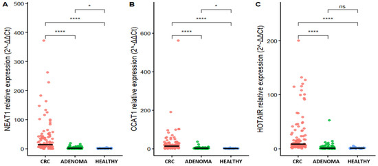
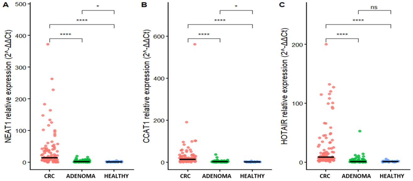
Relative expression levels were calculated using the 2−ΔΔCt method. Horizontal lines indicate median values. Statistical comparisons were performed using pairwise Wilcoxon rank-sum tests followed by post hoc pairwise comparisons. p-values are shown in the figure.
The median expression level of CCAT1 in CRC patients was 13.47 (IQR: 6.12–28.17), which was markedly higher than that in the adenoma group [2.30 (0.52–3.81)] and healthy controls [1.00 (0.41–2.37)]. Similarly, NEAT1 showed a median expression of 13.73 (5.75–40.66) in the CRC group, compared with 1.96 (0.47–4.04) in adenoma patients and 1.00 (0.29–1.77) in healthy individuals. For HOTAIR, CRC patients also exhibited elevated expression, with a median value of 8.52 (4.60–24.66), whereas expression levels in the adenoma [1.14 (0.45–2.26)] and healthy [1.01 (0.46–1.53)] groups were comparable.
As shown in Figure 1, the relative expression levels of CCAT1, NEAT1, and HOTAIR differed significantly among the CRC, adenoma, and healthy groups. CCAT1 and NEAT1 exhibited a clear gradient of increasing expression from healthy controls to adenoma patients and further to CRC patients, with the highest levels observed in the CRC group (all pairwise comparisons, p < 0.05). In contrast, HOTAIR expression was significantly elevated in CRC compared with both adenoma patients and healthy controls (p < 0.001), whereas no statistically significant difference was observed between the adenoma and healthy groups (p = 0.34).
Figure 1.
Relative expression levels of lncRNAs NEAT1 (A), CCAT1 (B), and HOTAIR (C) among colorectal cancer, adenoma, and healthy groups. Statistical comparisons were performed using the Wilcoxon rank-sum test. * p < 0.05; **** p < 0.0001, while “ns” indicates no statistically significant difference.
3.4. Association Between lncRNA Expression Levels and TNM Stage in Colorectal Cancer
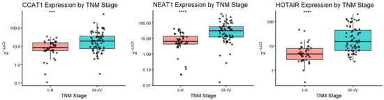
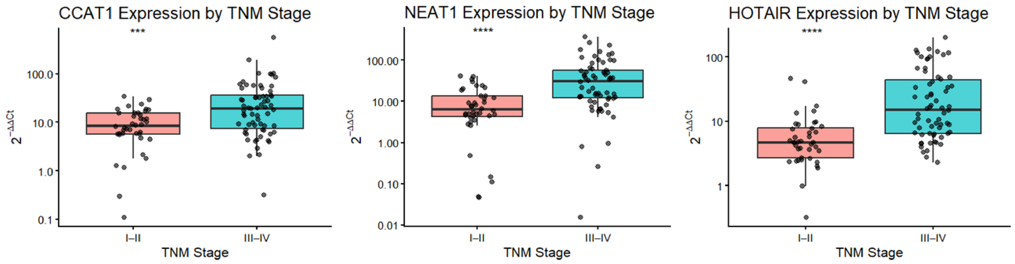
When stratified by tumor stage, the relative expression levels of CCAT1, HOTAIR, and NEAT1 were significantly higher in patients with advanced CRC (TNM stages III–IV) compared with those with early-stage disease (stages I–II) (Figure 2). Specifically, CCAT1 expression was significantly increased in stages III–IV relative to stages I–II (p < 0.001). Similarly, NEAT1 and HOTAIR showed markedly higher expression levels in advanced-stage CRC, with highly significant differences between the two stage groups (p < 0.0001 for both). These findings indicate that elevated expression of CCAT1, NEAT1, and HOTAIR is associated with more advanced tumor stage, suggesting a potential link between these lncRNAs and CRC progression.
Figure 2.
Relative expression levels of CCAT1, NEAT1, and HOTAIR according to TNM stage in colorectal cancer patients. Patients were grouped into early-stage (TNM I–II) and advanced-stage disease (TNM III–IV). Relative expression levels were calculated using the 2−ΔΔCt method. Data are presented as boxplots showing the median and interquartile range, with individual data points overlaid. Statistical comparisons were performed using the Wilcoxon rank-sum test. *** p < 0.001; **** p < 0.0001.
3.5. Diagnostic Performance of Individual and Combined lncRNAs
To further evaluate the diagnostic utility of CCAT1, HOTAIR, and NEAT1, ROC curve analysis was performed to assess the discriminative ability of each individual lncRNA and their combinations in distinguishing CRC patients from non-cancer controls (adenoma and healthy groups).
The diagnostic performance of CCAT1, NEAT1, HOTAIR, and their combined models for CRC is summarized in Table 4. Among individual biomarkers, HOTAIR achieved the highest diagnostic accuracy, with an area under the ROC curve (AUC) of 0.918 (95% CI: 0.823–0.922), a sensitivity of 87.7%, and a specificity of 84.8%. CCAT1 and NEAT1 also demonstrated good discriminative ability, with AUC values of 0.908 (95% CI: 0.841–0.935) and 0.890 (95% CI: 0.842–0.935), respectively (all p < 0.0001).
Table 4.
Diagnostic performance of lncRNAs and combinations.
Notably, combined biomarker models showed improved diagnostic performance compared with individual lncRNAs. The combination of CCAT1 and HOTAIR yielded the highest AUC (0.944), with a sensitivity of 94.3% and a specificity of 85.7%. Similarly, the HOTAIR + NEAT1 model achieved an AUC of 0.940, while the CCAT1 + NEAT1 combination showed the highest specificity (95.5%) among all models. These results indicate that combining lncRNAs enhances the diagnostic accuracy for CRC compared with single-marker approaches.
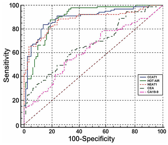
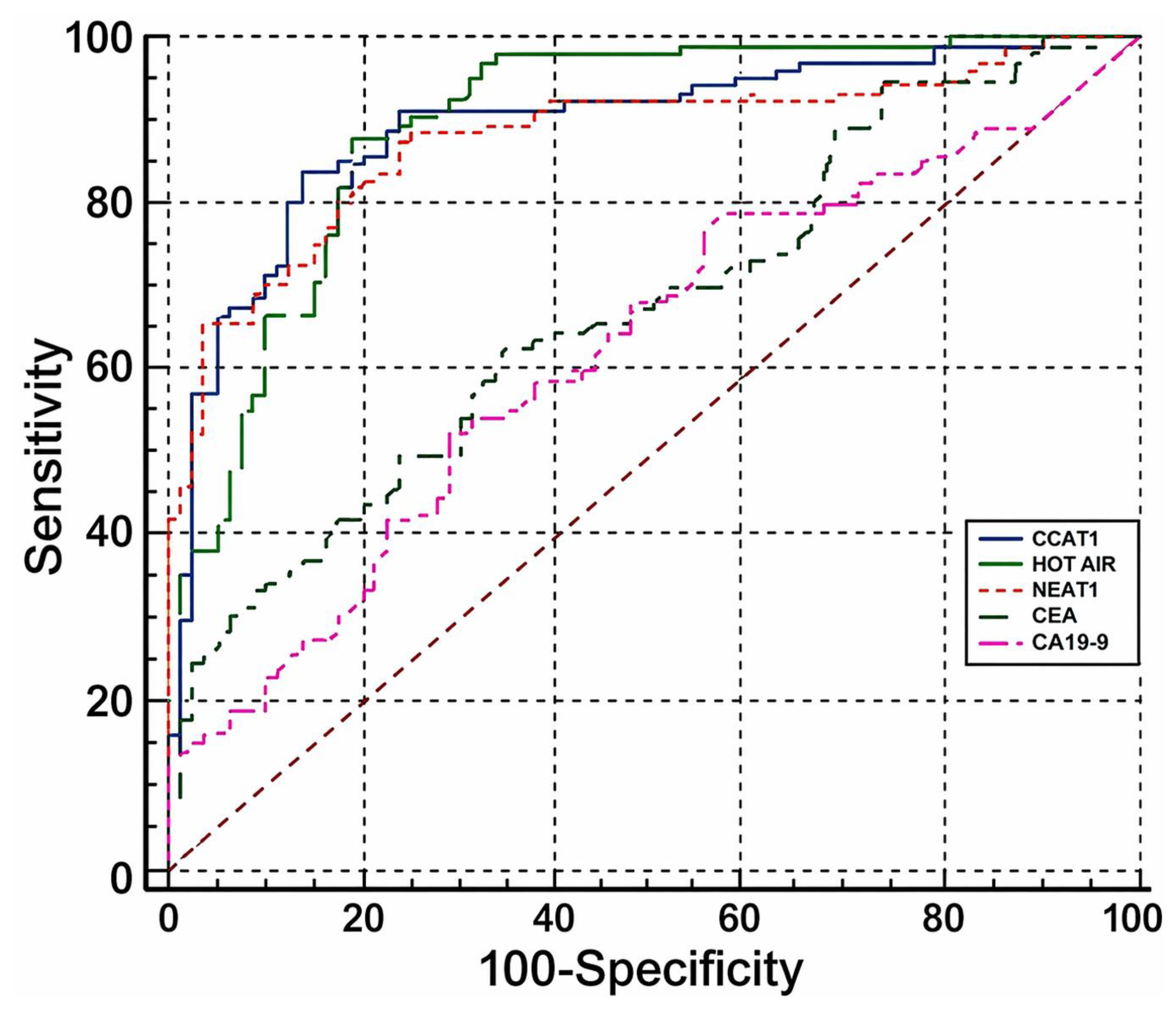
In addition, the diagnostic performance of the investigated lncRNAs was compared with conventional serum tumor markers, including CEA and CA19-9. The AUC values for CEA and CA19-9 were 0.675 (95% CI: 0.602–0.742) and 0.627 (95% CI: 0.553–0.697), respectively, which were markedly lower than those of all three lncRNAs. Statistical comparisons demonstrated that CCAT1, HOTAIR, and NEAT1 significantly outperformed both CEA and CA19-9 (all p < 0.001). These findings highlight the superior diagnostic value of circulating lncRNAs compared with conventional biomarkers (Figure 3).
Figure 3.
Receiver operating characteristic (ROC) curves of CCAT1, HOTAIR, NEAT1, CEA and CA19-9 for distinguishing CRC patients from non-cancer controls.
4. Discussion
In the present study, we investigated the expression patterns and diagnostic performance of three circulating lncRNAs—CCAT1, HOTAIR, and NEAT1—in patients with CRC compared with individuals with colorectal adenomas and healthy controls. Our results showed that all three lncRNAs were significantly upregulated in CRC patients and exhibited favorable diagnostic performance, both individually and in combination, suggesting their potential relevance as non-invasive biomarkers for CRC detection.
Among the investigated lncRNAs, HOTAIR exhibited markedly elevated expression levels in CRC patients, with a median value of 8.52, which was significantly higher than that observed in the adenoma group (median 1.14, p < 0.001). HOTAIR also demonstrated excellent diagnostic performance, with an AUC of 0.918; at the optimal cut-off value of 2.22, it achieved a sensitivity of 87.7% and a specificity of 84.8%. These findings are highly consistent with previous studies. Ismail et al. (2019) [12] reported that circulating HOTAIR expression was increased by approximately 7.55-fold in CRC patients compared with healthy controls and showed strong diagnostic accuracy. In addition, Chen et al. (2020) [14] confirmed that HOTAIR is highly expressed in CRC tissues and may play an important role in promoting tumor metastasis. Together, these data support the association between HOTAIR upregulation and CRC development.
NEAT1 has also been widely reported to be overexpressed in CRC. Wu et al. (2015) [13] demonstrated that circulating NEAT1 levels were significantly higher in CRC patients than in healthy individuals, and that two NEAT1 transcript variants (NEAT1-v1 and NEAT1-v2) showed good diagnostic accuracy, with AUC values of 0.732 and 0.845, respectively. Consistent with these findings, our study showed that NEAT1 expression was substantially higher in CRC patients (median 13.73) than in adenoma patients (median 1.96), suggesting a possible association between NEAT1 upregulation and disease progression. Furthermore, NEAT1 demonstrated good diagnostic performance in distinguishing CRC from adenoma and healthy groups, with an AUC of 0.890, sensitivity of 87.7%, and specificity of 81.2%. NEAT1 has been implicated in multiple oncogenic processes, including tumor proliferation, metastasis, chemoresistance, and regulation of the tumor immune microenvironment, which may partially explain its elevated expression in CRC.
Similarly, CCAT1 was significantly upregulated in CRC patients in our cohort, with the highest median expression level observed in the CRC group (13.47) compared with the adenoma group (2.30). CCAT1 also showed strong diagnostic performance, with an AUC of 0.908. These findings are in line with previous studies reporting elevated CCAT1 expression in CRC and its association with disease progression. Gu et al. (2020) [15] demonstrated that CCAT1 was highly expressed in CRC tissues and closely related to tumor progression. The observed stepwise increase in CCAT1 expression from healthy controls to adenoma patients and further to CRC patients suggests a possible involvement of CCAT1 in the adenoma–carcinoma sequence.
Importantly, when compared with conventional serum tumor markers such as CEA and CA19-9, all three lncRNAs demonstrated superior diagnostic performance. The AUC values of CEA (0.675) and CA19-9 (0.627) were markedly lower than those observed for CCAT1, HOTAIR, and NEAT1. These findings are in line with previous studies indicating that circulating lncRNAs may provide higher sensitivity and specificity than traditional protein biomarkers in CRC detection [19].
The improved diagnostic performance observed with combined lncRNA models is consistent with previous reports demonstrating synergistic effects between CCAT1 and HOTAIR [11]. However, rather than representing a conceptual breakthrough, our findings primarily serve as an external validation of previously reported biomarker combinations in a Vietnamese population. This highlights the importance of population-specific validation, as genetic background, environmental exposures, and lifestyle factors may influence biomarker expression and diagnostic performance.
From a practical perspective, the implementation of RT-qPCR-based multi-marker panels in routine clinical practice in Vietnam may face several challenges. These include limited laboratory infrastructure in lower-level healthcare settings, variability in pre-analytical sample handling, lack of standardized protocols for circulating RNA analysis, and cost-related constraints associated with multiplex testing. Similar barriers have been reported in other low- and middle-income countries, where access to advanced molecular diagnostics remains limited [5]. Therefore, further efforts are required to standardize methodologies and evaluate the feasibility and cost-effectiveness of these assays in real-world clinical settings.
Although we observed clear differences in lncRNA expression between CRC and non-cancer groups, and potential associations with disease stage, this study did not include longitudinal follow-up or survival analysis. Therefore, the prognostic value of these biomarkers, including their ability to predict overall survival or disease progression, could not be assessed and remains to be clarified in future studies.
Overall, our findings suggest that circulating CCAT1, HOTAIR, and NEAT1 are promising diagnostic biomarkers for CRC, particularly when used in combination.
This study has several limitations. First, the sample size was relatively modest and derived from a single center, which may limit the generalizability of the findings. Second, the cross-sectional design precludes assessment of temporal changes and causal relationships. Third, external validation in independent cohorts was not performed. Finally, the use of peripheral blood-derived RNA rather than strictly cell-free RNA may introduce variability when comparing results with plasma-based studies.
Future studies with larger, multi-center cohorts, standardized analytical protocols, and longitudinal follow-up are warranted to validate these findings, assess prognostic value, and further explore the clinical applicability of circulating lncRNA-based biomarker panels for CRC detection.
5. Conclusions
In conclusion, CCAT1, HOTAIR, and NEAT1 were significantly upregulated in CRC and demonstrated good diagnostic performance, particularly when combined. These lncRNAs outperformed conventional markers (CEA and CA19-9), supporting their potential as non-invasive biomarkers in a Vietnamese population. However, further validation in larger cohorts is required before clinical application.
Author Contributions
Conceptualization, T.T.B.V. and H.Q.D.; methodology, T.T.B.V., H.Q.D., K.N.N. and D.T.N.; software, D.T.N.; validation, T.T.B.V., K.N.N. and D.T.N.; formal analysis, T.T.B.V., H.Q.D., K.N.N. and D.T.N.; investigation, H.Q.D., K.N.N., C.P. and K.H.P.; resources, H.Q.D., K.N.N., C.P. and K.H.P.; data curation, T.T.B.V., H.Q.D., K.N.N. and D.T.N.; writing—original draft preparation, T.T.B.V. and D.T.N.; writing—review and editing, T.T.B.V.; visualization, T.T.B.V. and H.Q.D.; supervision, T.T.B.V. and H.Q.D.; project administration, H.Q.D. and K.N.N.; funding acquisition, K.N.N. All authors have read and agreed to the published version of the manuscript.
Funding
This research received no external funding.
Institutional Review Board Statement
The study was conducted in accordance with the Declaration of Helsinki and approved by the Human Research Ethics Committee of the 103 Military Hospital (Approval No. 341/CNChT-HĐĐĐ, dated 15 November 2023).
Informed Consent Statement
Informed consent for participation was obtained from all subjects involved in the study.
Data Availability Statement
The original contributions presented in this study are included in the article. Further inquiries can be directed to the corresponding author.
Conflicts of Interest
The authors declare no conflicts of interest.
Abbreviations
The following abbreviations are used in this manuscript:
| CRC | Colorectal cancer |
| lncRNAs | long non-coding RNAs |
| ROC | Receiver operating characteristic |
| CCAT1 | Colon Cancer-Associated Transcript 1 |
| HOTAIR | HOX Transcript Antisense Intergenic RNA |
| NEAT1 | Nuclear-Enriched Abundant Transcript 1 |
| cDNA | Complementary DNA |
| Ct | Threshold cycle |
| TNM | Tumor Stage |
References
- Bray, F.; Laversanne, M.; Sung, H.; Ferlay, J.; Siegel, R.L.; Soerjomataram, I.; Jemal, A. Global cancer statistics 2022: GLOBOCAN estimates of incidence and mortality worldwide for 36 cancers in 185 countries. CA Cancer J. Clin. 2024, 74, 229–263. [Google Scholar] [CrossRef] [Scilit] [PubMed]
- Siegel, R.L.; Miller, K.D.; Wagle, N.S.; Jemal, A. Cancer statistics, 2023. CA Cancer J. Clin. 2023, 73, 17–48. [Google Scholar] [CrossRef] [Scilit] [PubMed]
- International Agency for Research on Cancer. Viet Nam Fact Sheet 2022. In GLOBOCAN 2022; International Agency for Research on Cancer (IARC): Lyon, France, 2024. [Google Scholar]
- Ni, Y.; Xie, G.; Jia, W. Metabonomics of human colorectal cancer: New approaches for early diagnosis and biomarker discovery. J. Proteome Res. 2014, 13, 3857–3870. [Google Scholar] [CrossRef] [Scilit] [PubMed]
- Antwi, S.; Agbedinu, K.; Aduse-Poku, L.; Amponsah, F.; Larrious-Lartey, H.; Mensah, S.; Nsaful, J.; Akakpo, P.; Nyarko, V.; Nortey, M. Barriers to cancer diagnosis and treatment among women in low-and low-middle-income countries. Cancer Res. 2025, 85, 7113. [Google Scholar] [CrossRef] [Scilit]
- Lin, Y.; Zhao, W.; Lv, Z.; Xie, H.; Li, Y.; Zhang, Z. The functions and mechanisms of long non-coding RNA in colorectal cancer. Front. Oncol. 2024, 14, 1419972. [Google Scholar] [CrossRef] [Scilit] [PubMed]
- Patraquim, P.; Magny, E.G.; Pueyo, J.I.; Platero, A.I.; Couso, J.P. Translation and natural selection of micropeptides from long non-canonical RNAs. Nat. Commun. 2022, 13, 6515. [Google Scholar] [CrossRef] [Scilit] [PubMed]
- Bhan, A.; Soleimani, M.; Mandal, S.S. Long noncoding RNA and cancer: A new paradigm. Cancer Res. 2017, 77, 3965–3981. [Google Scholar] [CrossRef] [Scilit] [PubMed]
- Quinn, J.J.; Chang, H.Y. Unique features of long non-coding RNA biogenesis and function. Nat. Rev. Genet. 2016, 17, 47–62. [Google Scholar] [CrossRef] [Scilit] [PubMed]
- Adjeroh, D.A.; Zhou, X.; Paschoal, A.R.; Dimitrova, N.; Derevyanchuk, E.G.; Shkurat, T.P.; Loeb, J.A.; Martinez, I.; Lipovich, L. Challenges in lncRNA biology: Views and opinions. Non-Coding RNA 2024, 10, 43. [Google Scholar] [CrossRef] [Scilit] [PubMed]
- Zhao, W.; Song, M.; Zhang, J.; Kuerban, M.; Wang, H. Combined identification of long non-coding RNA CCAT1 and HOTAIR in serum as an effective screening for colorectal carcinoma. Int. J. Clin. Exp. Pathol. 2015, 8, 14131. [Google Scholar] [PubMed]
- Ismail, D.M.; Shaker, O.G.; Kandeil, M.A.; Hussein, R.M. Gene expression of the circulating long noncoding RNA H19 and HOTAIR in Egyptian colorectal cancer patients. Genet. Test. Mol. Biomark. 2019, 23, 671–680. [Google Scholar] [CrossRef] [Scilit] [PubMed]
- Wu, Y.; Yang, L.; Zhao, J.; Li, C.; Nie, J.; Liu, F.; Zhuo, C.; Zheng, Y.; Li, B.; Wang, Z. Nuclear-enriched abundant transcript 1 as a diagnostic and prognostic biomarker in colorectal cancer. Mol. Cancer 2015, 14, 191. [Google Scholar] [CrossRef] [Scilit] [PubMed]
- Chen, S.; Zhang, C.; Feng, M. Prognostic value of lncRNA HOTAIR in colorectal cancer: A meta-analysis. Open Med. 2020, 15, 76–83. [Google Scholar] [CrossRef] [Scilit] [PubMed]
- Gu, C.; Zou, S.; He, C.; Zhou, J.; Qu, R.; Wang, Q.; Qi, J.; Zhou, M.; Yan, S.; Ye, Z. Long non-coding RNA CCAT1 promotes colorectal cancer cell migration, invasiveness and viability by upregulating VEGF via negative modulation of microRNA-218. Exp. Ther. Med. 2020, 19, 2543–2550. [Google Scholar] [CrossRef] [Scilit] [PubMed]
- MIMS Vietnam. Ung Thư Đại Trực Tràng: Tổng Quan Về Bệnh. 2025. Available online: https://www.mims.com/vietnam/disease/colorectal-cancer/disease-background (accessed on 19 April 2026).
- Livak, K.J.; Schmittgen, T.D. Analysis of relative gene expression data using real-time quantitative PCR and the 2−ΔΔCT method. Methods 2001, 25, 402–408. [Google Scholar] [CrossRef] [Scilit] [PubMed]
- Graham, L.D.; Pedersen, S.K.; Brown, G.S.; Ho, T.; Kassir, Z.; Moynihan, A.T.; Vizgoft, E.K.; Dunne, R.; Pimlott, L.; Young, G.P. Colorectal neoplasia differentially expressed (CRNDE), a novel gene with elevated expression in colorectal adenomas and adenocarcinomas. Genes Cancer 2011, 2, 829–840. [Google Scholar] [CrossRef] [Scilit] [PubMed]
- Hou, S.; Jing, J.; Wang, Y.; Du, L.; Tian, B.; Xu, X.; Sun, T.; Shi, Y. Evaluation of Clinical Diagnostic and Prognostic Value of Preoperative Serum Carcinoembryonic Antigen, CA19-9, and CA24-2 for Colorectal Cancer. Altern. Ther. Health Med. 2023, 29, 192–197. [Google Scholar] [PubMed]

Disclaimer/Publisher’s Note: The statements, opinions and data contained in all publications are solely those of the individual author(s) and contributor(s) and not of MDPI and/or the editor(s). MDPI and/or the editor(s) disclaim responsibility for any injury to people or property resulting from any ideas, methods, instructions or products referred to in the content. |
© 2026 by the authors. Licensee MDPI, Basel, Switzerland. This article is an open access article distributed under the terms and conditions of the Creative Commons Attribution (CC BY) license.