


Abstract
Jellyfish, as representatives of the phylum Cnidaria, possess venoms characterized by structurally diverse and functionally complex toxins, rendering them a central focus in cnidarian toxin research. This article presents a systematic review of the physicochemical properties of jellyfish toxins, examines their mechanisms of action from a molecular biology perspective, investigates the patterns of toxin transformation in organisms, elucidates the structure–activity relationships between structure and toxicity, introduces advancements in research on novel jellyfish toxins, and offers an outlook on future developments in this field. By integrating modern proteomic techniques, such as liquid chromatography-tandem mass spectrometry, this review provides comprehensive theoretical support for the foundational research and application development of jellyfish toxins, as well as a scientific basis for practical applications, including antivenom serum development and novel marine drug design.
1. Introduction
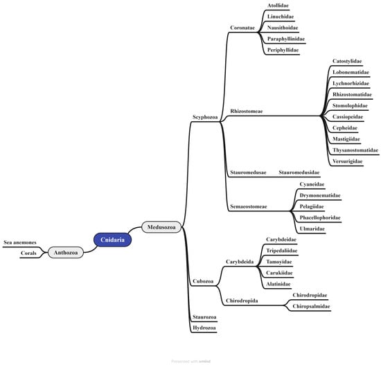
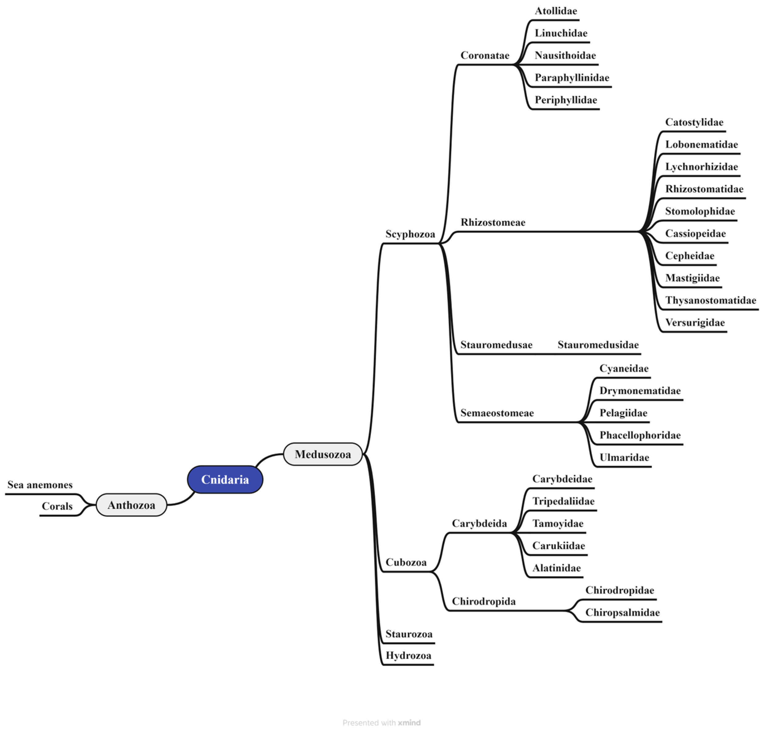
Jellyfish are members of the phylum Cnidaria, which includes over 10,000 known species. This phylum is divided into two primary lineages: Anthozoa (sea anemones and corals) and Medusozoa (jellyfish and hydras) (Figure 1). The subphylum Medusozoa is further classified into four classes: Scyphozoa (real jellyfish), Cubozoa (box jellyfish), Staurozoa (stalked jellyfish), and Hydrozoa (hydras) [1,2,3]. According to the World Register of Marine Species (http://www.marinespecies.org), there are currently 187 and 46 confirmed species of Scyphozoa and Cubozoa, respectively. The class Scyphozoa encompasses four orders: Coronatae (crown jellyfish), Rhizostomeae (root-mouth jellyfish), Stauromedusae, and Semaeostomeae (sea nettles) [4]. In contrast, the class Cubozoa comprises only two orders: Carybdeida and Chirodropida [5]. Santhanam. et al. has provided a more detailed taxonomic breakdown at the family level for the orders within Scyphozoa [6,7]: the order Coronatae contains 5 families (Atollidae, Linuchidae, Nausithoidae, Paraphyllinidae, Periphyllidae); Rhizostomeae includes 10 families (Catostylidae, Lobonematidae, Lychnorhizidae, Rhizostomatidae, Stomolophidae, Cassiopeidae, Cepheidae, Mastigiidae, Thysanostomatidae, Versurigidae); Stauromedusae is represented by a single family (Stauromedusidae); and Semaeostomeae consists of 5 families (Cyaneidae, Drymonematidae, Pelagiidae, Phacellophoridae, and Ulmaridae).
Figure 1.
Species classification of the Cnidaria, from phylum to order. The phylum Cnidaria is divided into two subphyla: Anthozoa and Medusozoa. The subphylum Medusozoa is further classified into four classes, which correspond to “jellyfish” [1,2,3,4,5,6,7,8,9].
Similarly, the two principal orders within the Cubozoa class can be taxonomically separated at the family level. The Carybdeida order consists of five families: Carybdeidae, Tripedaliidae, Tamoyidae, Carukiidae, and Alatinidae. The Chirodropida order is composed of two families: Chirodropidae and Chiropsalmidae [8]. As carnivorous animals, all cnidarians possess a defining feature—highly specialized stinging cells termed cnidocytes, which are densely distributed in the tentacular epithelium [3,9] (Figure 1). Cnidoblast are the precursor cells of cnidocytes. The cnidocyst vesicle is formed from post-Golgi vesicles in the cytoplasm of nematoblasts [4]. A mature cnidocyst is a complex intracellular structure consisting of a double-walled invaginated capsule, opercular lid, and intricately coiled and frequently barbed filament [10]. Each cnidocyte precursor synthesizes a single cnidocyst, containing a toxin-loaded filament that typically measures 200–800 μm in length. Cnidocysts discharge explosively in response to mechanical or chemical stimuli. The operculum opens, and the inverted filament rapidly everts, propelling the barbed tubule at a high speed into the target. This process results in the simultaneous injection of thousands of toxin-laden tubules into the prey [11,12]. In the early stages of envenomation, jellyfish stings can induce both immediate and delayed hypersensitivity reactions, and the venom components of some jellyfish species exhibit significant immunomodulatory activity. Conversely, jellyfish-derived collagen extracts and peptides can strongly promote the release of pro-inflammatory cytokines, especially tumor necrosis factor-α (TNF-α) and interferon-γ (IFN-γ). On the other hand, protein toxins and carbohydrate-rich components (e.g., glycoproteins and polysaccharides) in nematocysts can act as antigens and trigger clinical symptoms through cellular and/or humoral immune pathways [3]. The combined neurotoxic, cytotoxic, and cardiotoxic effects of the venom can lead to systemic pathophysiology and, in severe cases, death within a few minutes [9] (Figure 2).
Jellyfish envenomation has multiple toxicological consequences due to the complex composition of their venom, which contains a wide range of bioactive chemicals, such as proteins, peptides, and low-molecular-weight substances. These constituents exhibit a wide spectrum of biological activities, including hemolytic, cytotoxic, neurotoxic, and cardiovascular toxic effects [13,14,15,16] (Table 1). Advances in current separation and purification techniques, as well as proteomic technologies, have improved the identification and characterization of these venom components. Omic and biotechnological approaches have been further integrated to form a research field known as modern venomology. Mass spectrometry imaging is a highly promising technique in modern venomics. It combines classical histology with modern mass spectrometry-based omics and enables the generation of molecular spatial distribution maps based on molecular weight. The application of methods such as LC-MS/MS has allowed the precise identification of an increasing number of toxin proteins and peptides. Consequently, the chemical nature and functional attributes of an increasing number of jellyfish toxin components have been gradually elucidated [17,18,19,20,21,22,23,24].
Contemporary research on jellyfish toxins has advanced from initial phenomenological descriptions to molecular-level investigations, including chemical structure elucidation, mechanistic studies, and structure–activity relationship (SAR) analyses. However, owing to the remarkable diversity and complexity of venom constituents, the structure-function relationships of the majority of these toxins remain poorly defined. Consequently, the development and application of novel toxin analogs are still in the early stages. Therefore, a systematic review consolidating the current knowledge on the chemical properties, mechanisms of action, and SARs of jellyfish toxins is of considerable academic importance. Such synthesis is critical for advancing foundational research and facilitating translational applications in the field. Building upon recent research findings, this review focuses on elucidating the physicochemical properties, mechanisms of action, in vivo biotransformation pathways, and structure–toxicity relationships of jellyfish toxins. Furthermore, it introduces recent progress in the study of novel toxin analogs, with the goal of serving as a comprehensive reference and framework for future investigations.
Figure 2.
The stinging mechanism of Jellyfish and effects. (I) General morphology of tentacles and cnidocysts in Cubozoans. nemeb: nematocyst bands; lc: lancet. Reprinted from Refs. [25,26], (II) tentacular cnidocyst of several jellyfish species (from left to right: Rhizostoma pulmo, Aurelia sp., Cassiopea sp., and Rhizostoma luteum, 50 μm). Reprinted from Ref. [27], (III) Chirodropidae jellyfish sting wound. Reprinted from Ref. [28]. All images in the figure are taken from the cited references.
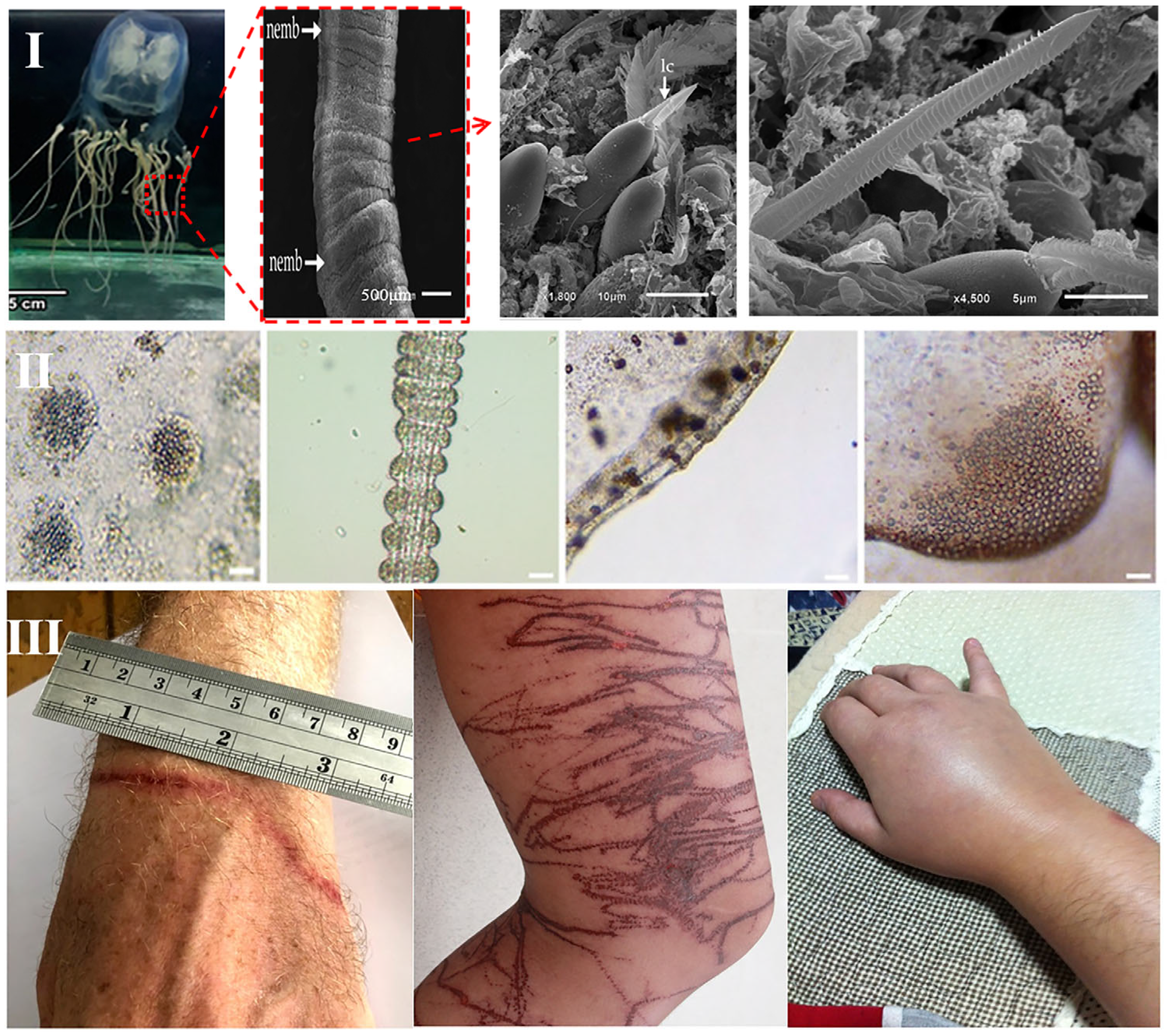
Table 1.
Distribution of several poisonous jellyfish and signs of “stings”.
2. Physicochemical Characterization of Jellyfish Toxins
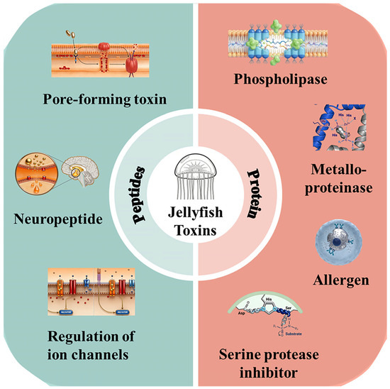
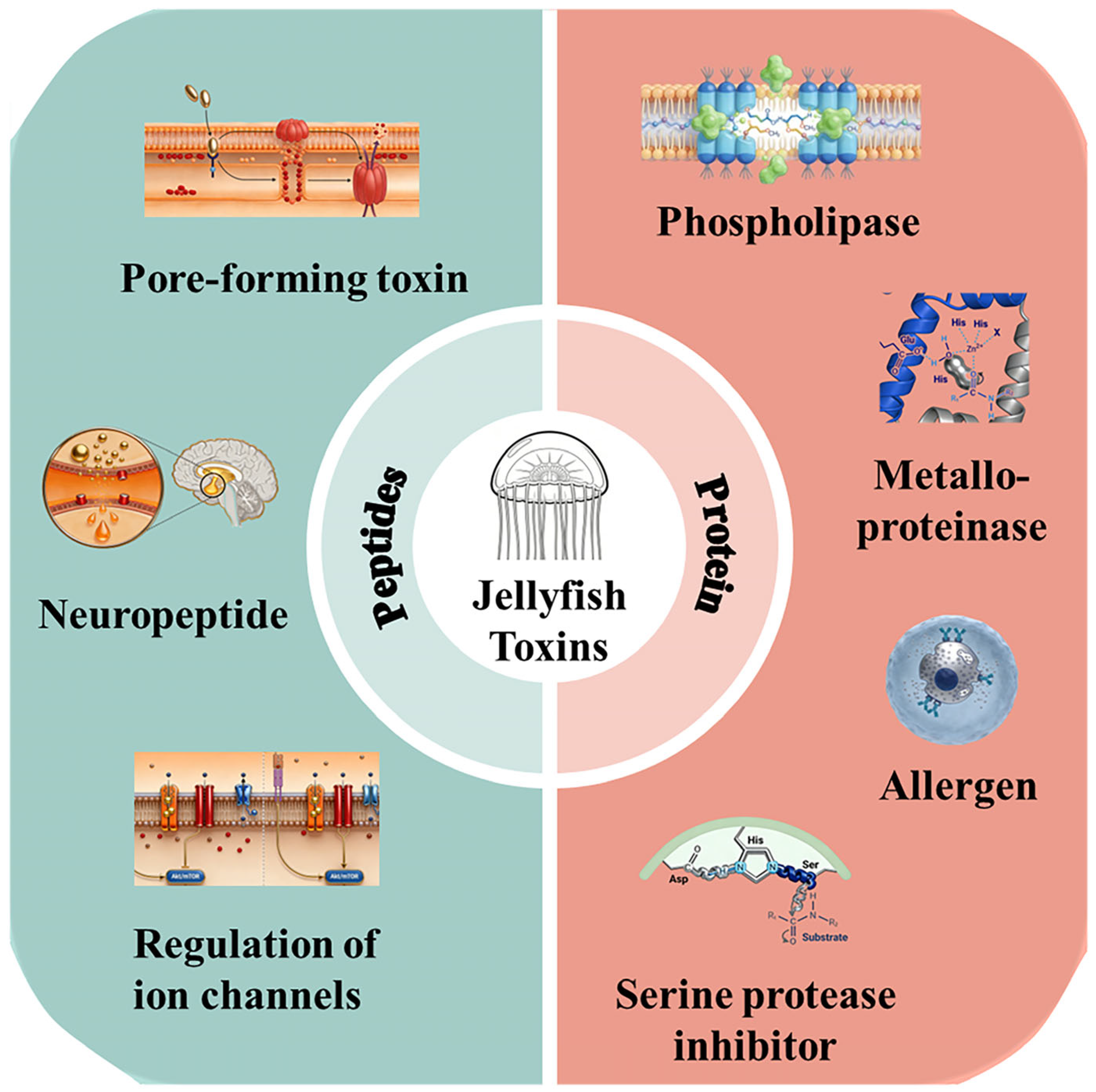
The physicochemical properties of jellyfish venom vary greatly between species and toxin classes. Toxic ingredients can be further divided into two categories based on their chemical composition: protein and peptide toxins (Figure 3). The unique physicochemical profile of each toxin class, which includes features such as molecular weight, stability, and solubility, determines its biological activity and mechanism of action.
Figure 3.
Classification of jellyfish toxins. Protein toxins are classified into four categories according to their mechanism of action: phospholipase, metalloproteinases, allergen, serine protease inhibitors; peptide toxins are classified into three categories according to their mechanism of action: pore-forming toxins, neuropeptide toxins, ion channel toxins.
2.1. Protein Toxins
Proteinaceous toxins constitute the predominant and functionally central components of jellyfish venom. This category encompasses a diverse array of molecules, including phospholipase, metalloproteinases, allergens and serine protease inhibitors. The key physicochemical parameters of these proteins, such as molecular weight, isoelectric point, and structural stability, are intrinsically linked to their specific biological activities and toxicological functions [56].
The molecular weights of jellyfish protein toxins vary widely, typically ranging from 10 to 200 kDa. For instance, metalloproteinases identified in the venom of Nemopilema nomurai exhibit molecular weights between 30 and 60 kDa [57], whereas members of the CfTX toxin family, key toxic components in Chironex fleckeri, have molecular weights of 40 to 50 kDa [58]. Similarly, protease toxins derived from Rhopilema esculentum venom have molecular weights ranging from 25 to 75 kDa, with distinct molecular weights often correlating with specific substrate selectivity and catalytic potency [23]. Molecular size critically influences toxin pharmacokinetics; lower molecular weight proteins may diffuse more readily across biological membranes, whereas larger toxins frequently rely on specific receptor binding on the cell surface to exert their effects.
The isoelectric point (pI) represents another fundamental physicochemical factor for protein toxins. Most jellyfish protein toxins have acidic or neutral pI values. For example, the pore-forming toxin TX2 from Chrysaora fuscescens has a pI of approximately 5.2 [59], indicating that it is an acidic protein, whereas a serine protease inhibitor from Chrysaora quinquecirrha exhibits a near-neutral pI of approximately 7.1 [60]. The pI determines the net charge of a toxin under specific physiological pH conditions, thereby modulating its structural integrity, solubility, and functional interactions. Following envenomation, the local acidic microenvironment at the sting site may potentiate the activities of certain toxins. Therapeutic alkaline intervention, on the other hand, has the ability to neutralize toxicity [61], but this method still requires further investigation.
Jellyfish protein toxins are notoriously unstable and highly sensitive to thermal stress, pH extremes, and proteolytic degradation. Most toxins undergo significant or complete inactivation when exposed to temperatures above 60 °C for 10 to 15 min. Extreme pH conditions (pH < 3 or >10) can cause conformational denaturation, resulting in an irreversible loss of biological activity [62]. Furthermore, naturals proteases (e.g., trypsin and pepsin) can hydrolyze and degrade these toxin proteins, constituting a key physiological mechanism for the spontaneous alleviation of symptoms following various envenomations. Notably, certain potent toxins exhibit exceptional structural resilience. For instance, CfTX-1 from Chironex fleckeri demonstrates considerable structural stability under physiological conditions, which may account for the severe and long-lasting toxic reactions it induces [58].
2.2. Peptide Toxins
Peptide toxins represent another crucial class of bioactive constituents found in jellyfish venom. These polypeptides are typically composed of 10 to 50 amino acid residues, and possess a low molecular mass ranging from approximately 1 to 5 kDa. Despite their compact tertiary structures, they exhibit prominent and specific biological activities. This class is exemplified by the pp3 and pp11 neuropeptides obtained from the venom of Lychnorhiza malayensis, which have molecular masses of about 1.2 kDa and 1.5 kDa, respectively [22].
Peptide toxins have different amino acid compositions, often with a high abundance of cysteine residues. These cysteine residues generate many intramolecular disulfide bonds, which are critical for maintaining the conformational integrity and stability of the toxins. For instance, neuropeptide toxins from box jellyfish commonly contain three to four disulfide bridges in their structures. These covalent crosslinks provide structural stability and directly modulate the affinity and specificity of the toxin for its target binding sites [26]. Furthermore, certain peptide toxins undergo specific post-translational modifications, such as hydroxylation or methylation. These modifications can affect the physicochemical properties and bioactivities of the toxins. For example, the hydroxylated peptide toxins in Aurelia coerulea (likely caused by the increased expression of tryptophan) may significantly enhance their hydrophobicity, thereby improving their binding efficiency to cell membranes. This may be the source of toxicity in the mucus on the body surface of this jellyfish species [11].
Compared to larger protein toxins, peptide toxins often exhibit greater intrinsic stability under ambient conditions than larger protein toxins. Many enzymes retain significant activity over several days at room temperature and demonstrate considerable tolerance across a broad pH range. For example, the pp3 neuropeptide from Lychnorhiza malayensis, maintains over 80% of its activity across a pH range of 2 to 9, a resilience attributed to its compact, disulfide-rich scaffold [22]. Paradoxically, despite this in vitro stability, peptide toxins generally have a short in vivo half-life, typically ranging from several minutes to tens of minutes [63].
3. Mechanism of Action of Jellyfish Toxin
The mechanisms of action that enable jellyfish envenomation are multifaceted, and driven by a diverse repertoire of bioactive constituents. These diverse toxin components exert harmful effects by selectively targeting specific molecular locations in the organism. This targeted interference disrupts essential physiological processes, including cellular signaling, metabolic homeostasis, and membrane integrity, ultimately resulting in the observed toxicological consequences. The major mechanisms can be categorized into four principal classes based on their primary molecular targets and engagement modalities.
3.1. Pore-Forming Action
Pore-forming toxins (PFTs) represent one of the most common and functionally consequential components of jellyfish venom (Table 2). Their primary mechanism involves the self-assembly of transmembrane pores or channels within the lipid bilayer of the target cell membrane. This breakdown of membrane integrity disrupts ion gradients and osmotic balance, leading to the uncontrolled efflux of cytosolic contents and influx of extracellular ions, ultimately inducing cytolysis, apoptosis, or necrotic cell death [64]. Canonical examples include CfTX-A/B toxins from Chironex fleckeri and TX-1 toxin from Chrysaora fuscescens [58,59].
PFTs typically operate through a sequential, multi-stage process. First, water-soluble toxin monomers bind to the target membrane, either by directly interacting with lipid components (e.g., cholesterol) or through specific protein receptors. Subsequently, the monomers undergo conformational rearrangement, revealing hydrophobic regions that integrate into the lipid bilayer. Finally, monomers injected into the membrane oligomerize to form a stable ring-like complex that forms a functional transmembrane pore [65]. To demonstrate this mechanism, the CfTX-A toxin initially binds to membrane cholesterol. The N-terminal amphipathic helix is then inserted into the bilayer, causing approximately six monomers to assemble into a β-barrel pore with an estimated diameter of 2 nm. This pore permits the rapid efflux of K+ and influx of Na+ and Ca2+, resulting in cell enlargement, depolarization, and eventual rupture [58].
The cytotoxicity of PFTs is not indiscriminate; rather, it is frequently characterized by strong cellular selectivity, which is determined by the distribution of target molecules. For instance, some PFTs show a high affinity for specific glycosphingolipids found on erythrocyte membranes, thereby displaying potent hemolytic activity. Others preferentially bind to receptors enriched in vascular endothelial cells, disrupting endothelial junctions and increasing vascular permeability, resulting in clinically significant edema and hemorrhage [66].
Table 2.
Medusozoan toxins with pore-forming toxin activity.
3.2. Enzymatic Activity
Enzymatic toxins constitute a critical class of bioactive agents found in jellyfish venom (Table 3), which exert toxicological effects by catalyzing the hydrolysis or modification of specific host substrates, thereby disrupting essential physiological pathways. Prominent enzyme families identified in these venoms include metalloproteinases and phospholipases A2 (PLA2).
Metalloproteinases are among the most prevalent enzymatic components in the human body. Multiple isoforms have been characterized in the venom of jellyfish, such as Nemopilema nomurai [23,67]. These zinc-dependent endopeptidases primarily target extracellular matrix (ECM) proteins, including collagen and fibronectin. Their proteolytic activity compromises tissue integrity, leading to localized damage, hemorrhage, and necrosis [69]. Certain metalloproteinases can activate pro-inflammatory cytokines, such as TNF-α and IL-6, thereby accelerating the inflammatory cascade [70]. A disintegrin and metalloproteinase (ADAM), identified in Nemopilema nomurai venom, degrades vascular basement membranes, increasing capillary permeability and contributing to pathological outcomes such as pulmonary edema [23].
PLA2 is another widely distributed enzyme in the venom of species such as Chironex fleckeri [20]. Its catalytic activity releases arachidonic acid and lysophosphatidylcholine from the membrane phospholipids. Arachidonic acid serves as a precursor for potent inflammatory mediators (e.g., prostaglandins and leukotrienes), whereas lysophosphatidylcholine acts as a direct cytolysin that destabilizes cell membranes and induces cell death [17].
In addition, hyaluronidase is another common venom enzyme detected in various animal venoms. Although this enzyme is non-toxic by itself, it is known as a “spreading factor” because it hydrolyzes connective tissue and promotes the diffusion of other venom components. Lee et al. detected hyaluronidase activity in the venom of Nemopilema nomurai, with corresponding molecular weights of 55 kDa and 95 kDa. These enzymes can directly act on and degrade extracellular matrix components, which may further lead to necrotic skin damage [69].
Table 3.
Medusozoan toxins with enzymatic activity.
3.3. Regulation of Ion Channels
Certain jellyfish toxins exert their effects by selectively modulating ion channel function. Toxins that specifically bind to potassium (K+), sodium (Na+), or calcium (Ca2+) channels disturb cellular ion homeostasis and electrophysiological signaling, which underlies a range of neurotoxic and cardiotoxic outcomes [72].
The most well-studied of these are the K+ channel inhibitors. For instance, the kappa-stichotoxin-Shd1a/b from Stichodactyla haddoni is a potent voltage-gated K+ channel blocker. Inhibition of K+ efflux causes membrane depolarization and disrupts neuronal repolarization and muscle contraction, contributing to cardiovascular failure [66]. Similarly, the secretory precursor Mp-332-1 from Lychnorhiza malayensis, which contains an ShK toxin-like domain, acts as a K+ channel blocker and induces prey immobilization [22].
Toxins that target Na+ channels typically modulate channel gating, thereby altering action potential generation. Certain jellyfish neurotoxins bind to site 3 of the voltage-gated Na+ channels, delaying rapid inactivation. This leads to prolonged channel opening, sustained membrane depolarization, and abnormal neuronal firing, which manifest clinically as intense pain, paresthesia, and muscle spasm [4].
Toxins that alter Ca2+ signaling, although less commonly documented, are also of significance. A calmodulin-like protein found in Nemopilema nomurai venom binds to Ca2+ with remarkable affinity. It can cause cellular toxicity by disrupting calcium-dependent processes, such as apoptosis and autophagy [66].
3.4. Immunotoxicity
Jellyfish toxins can induce immunotoxic effects through a various of interactions with the host immune system (Table 4). These interactions range from eliciting hypersensitivity reactions to directly modulating the immune cell function. A major immunopathological effect is the induction of an allergic response. Certain venom components operate as powerful allergens, causing clinical symptoms ranging from localized urticaria and pruritus to systemic anaphylaxis [52,73]. For instance, specific allergens in the venom of Chrysaora fuscescens can cross-link IgE antibodies bound to the surface of mast cells and basophils. This triggers degranulation and the release of preformed mediators like histamine, initiating a type I hypersensitivity reaction [59].
In addition to allergenicity, certain toxins directly impair immune cell survival and function. Toxins from certain jellyfish species have been shown to induce apoptosis in lymphocytes, reducing adaptive immune responses and potentially increasing host susceptibility to secondary infections [74]. Other venom components, on the other hand, promote pro-inflammatory properties by overactivating immune pathways. They can cause immune cells to produce excessive levels of pro-inflammatory cytokines, resulting in an unbalanced inflammatory cascade that contributes to tissue damage. For example, cytotoxic toxins from Craspedacusta sowerbyi trigger an amplified inflammatory response, exacerbating local tissue injury at the sting site [75].
Li et al. extracted and purified a novel polysaccharide, JSP-11, from Rhopilema esculentum, with a molecular weight of 1.25 × 10⁶ Da [76]. It is composed of mannose, galactose, and glucuronic acid, and exhibits significant immunomodulatory effects. JSP-11 markedly enhanced RAW 264.7 macrophage activity and promoted the release of NO, TNF-α, and IL-1β through the NF-κB, MAPKs, and PI3K/Akt signaling pathways.
Table 4.
Jellyfish toxins with regulation of ion channel activity and immunotoxicity.
3.5. Enzyme Inhibitory Activity
In addition to toxins with enzymatic activity, jellyfish venom also contains toxins with enzyme inhibitory activity, which affect bodily functions by inhibiting the activity of certain enzymes in stung patients. Prakash et al. hydrolyzed the venom of Nemopilema nomurai with papain and isolated two novel angiotensin-converting enzyme (ACE)-inhibitory peptides: IVGRPLANG (896.48 Da) and IGDEPRHQYL (1227.65 Da) [77]. Serine protease inhibitors have been identified in various jellyfish species, including three types: Serpin (serpin superfamily) [78], Kazal-type inhibitors [79], and Kunitz-type inhibitors [23]. These factors inhibit blood clot formation.
4. Biotransformation of Jellyfish Toxins In Vivo
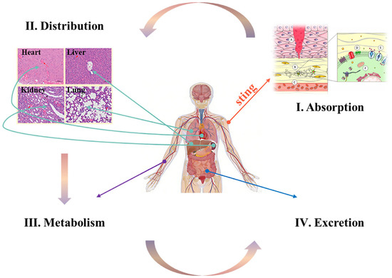
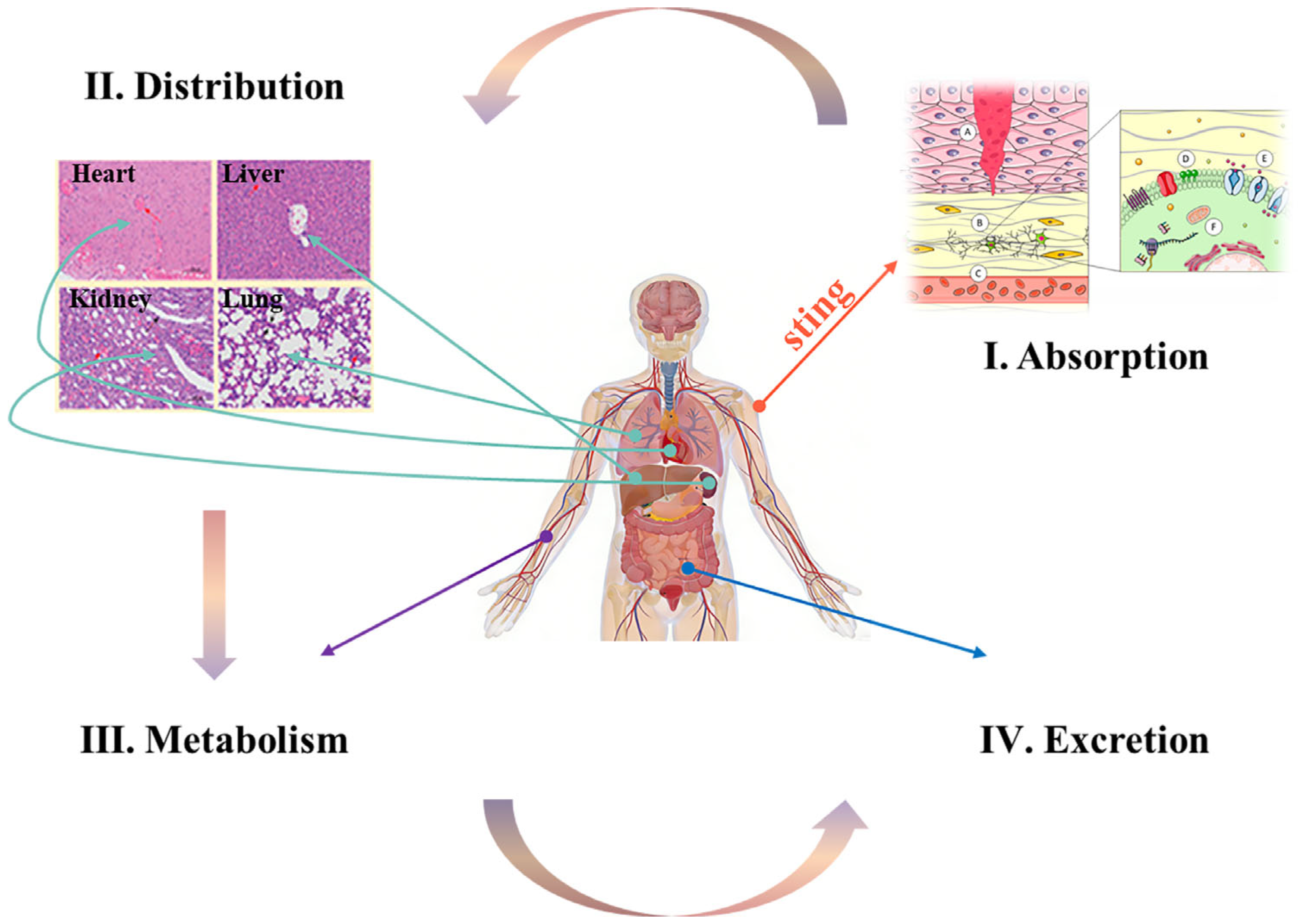
Upon entering a biological system, jellyfish toxins undergo the fundamental pharmacokinetic processes of absorption, distribution, metabolism, and excretion (ADME) (Figure 4). This journey involves a series of enzymatic bioconversion, processes that significantly impact both the manifestation of toxicity and the in vivo clearance kinetics of these bioactive molecules.
Figure 4.
The transformation process of different jellyfish toxins in the human body. The jellyfish toxins undergo the fundamental pharmacokinetic processes of absorption (A–F, Reprinted from ref. [80]), distribution (Reprinted with permission from ref. [68]. Copyright 2020 American Chemical Society.), metabolism, and excretion (ADME).
4.1. Absorption
Jellyfish toxins primarily enter organisms via transdermal administration after stings. Upon nematocyst discharge, the released venom comes into direct contact with and compromises the epidermal barrier, facilitating its absorption into the systemic circulation through multiple pathways [62]. The absorption kinetics of proteinaceous and polypeptide toxins are largely contingent on the degree of damage to epidermal integrity. Envenomation typically damages the epidermal barrier, allowing toxins direct and rapid access to the dermal capillary network [81]. Conversely, certain low-molecular-weight and lipophilic toxins can passively diffuse across the intact stratum corneum [82]. The rate of transdermal absorption is strongly associated with the key physicochemical properties of the toxins. Specifically, compounds with lower molecular masses and higher lipophilicity may exhibit faster absorption kinetics.
4.2. Distribution
Jellyfish toxins enter into the systemic circulation and are distributed throughout the body via hemodynamic perfusion. The extent and pattern of their biodistribution are governed by three key determinants: the physicochemical properties of the toxin, its plasma protein-binding capacity, and its inherent tissue-targeting affinity [83]. The physicochemical profile of a toxin dictates its ability to cross the biological barriers. Low-molecular-weight, highly lipophilic toxins can easily cross specific barriers, such as the blood-brain and placental barriers, gaining access to the central nervous system and to fetal tissues. In contrast, high-molecular-weight, hydrophilic toxins are mostly confined to the vascular compartment and readily perfused organs like the liver and kidneys, with limited extravasation into other tissues [84].
Plasma protein binding serves as a critical pharmacokinetic modulator. The majority of proteinaceous and polypeptide toxins exhibit a high binding affinity for plasma proteins, primarily albumin and globulins. This binding traps the toxin in an inactive, macromolecular complex within the bloodstream, limiting its volume of distribution and impeding its passage across biological membranes [85].
Ultimately, specific tissue-targeting affinity underpins the organ-specific tropism and toxicodynamics of many venom components. Certain toxins possess molecular motifs that confer a high affinity for receptors or lipids that are abundant in particular tissues, leading to selective accumulation. For instance, the CfTX-1 toxin from Chironex fleckeri displays a pronounced affinity for cardiac tissue, binding to specific targets on cardiomyocytes and disrupting cardiac function [86]. PLA2 from various jellyfish species shows a high affinity for skeletal muscle, leading to localized myotoxicity [87].
4.3. Metabolism
Current studies have demonstrated that the major functional components of jellyfish toxins are mostly proteins or polypeptide toxins. The major metabolic mechanism of protein and polypeptide toxins is enzymatic hydrolysis. Endogenous peptidases (e.g., aminopeptidases and carboxypeptidases) degrade these toxins into amino acids or small peptide fragments, thereby eliminating their bioactivity [88]. The pp3 neuropeptide from Lychnorhiza malayensis is rapidly hydrolyzed to free amino acids by aminopeptidases, exhibiting a short in vivo half-life of approximately 15 min [22].
A critical consideration in toxin metabolism is the pronounced species variability. Differences in enzyme composition and activity between species lead to changes in the metabolic rates and metabolite profiles of the same toxin [89]. For example, a metalloproteinase from Nemopilema nomurai showed a plasma half-life of 45 min in mice but 90 min in rats, accompanied by distinct metabolite profiles in each species [90]. This diversity presents significant challenges in extrapolating toxicity data and developing broadly effective antitoxin therapies.
4.4. Excretion
Jellyfish toxins and their metabolites are predominantly eliminated from the body via renal excretion in urine, with biliary-fecal excretion serving as a secondary pathway. Minor amounts may also be eliminated through respiration and perspiration [62]. The physicochemical properties of the chemicals involved determine the rate and efficiency of excretion. Metabolites with high water solubility and low-molecular-weight typically undergo rapid renal clearance, whereas those with high lipophilicity and larger molecular size are excreted more slowly and frequently, require alternative pathways [82].
Renal excretion constitutes the primary elimination mechanism. Toxins and metabolites are first filtered through the glomerular capillaries. They may then be actively secreted into or passively reabsorbed by the renal tubules. Small-molecule metabolites, such as those derived from alkaloid toxins, are easily filtered and rapidly excreted in the urine [91]. In contrast, metabolites such as amino acids from degraded protein toxins can be extensively reabsorbed by the tubular epithelium for reuse, with only the remaining portion being discharged in the urine [92].
Biliary excretion plays a crucial role in the elimination of large hydrophobic compounds. These substances are actively transported from hepatocytes into the bile canaliculi, secreted into the intestine via the bile duct, and eventually expelled in feces [93]. A representative example is the high-molecular-weight CfTX-1 toxin from Chironex fleckeri, for which fecal excretion accounts for more than 60% of the overall clearance, highlighting the significance of the hepatobiliary pathway [86].
5. Structure–Toxicity Relationship
The toxicity of jellyfish venom components is governed by a well-defined structure–activity relationship (SAR). The structural hierarchy of a toxin determines its potency, target specificity, and molecular mechanisms of action. This hierarchy includes the primary structure (amino acid sequence or chemical backbone), secondary structure (e.g., α-helices and β-sheets), tertiary three-dimensional fold, and quaternary oligomeric assembly (Figure 5). A comprehensive elucidation of these structure–toxicity relationships is crucial for scientific progress, as it provides critical insights into the molecular basis of envenomation and informs the rational design of targeted antitoxin therapies.
Figure 5.
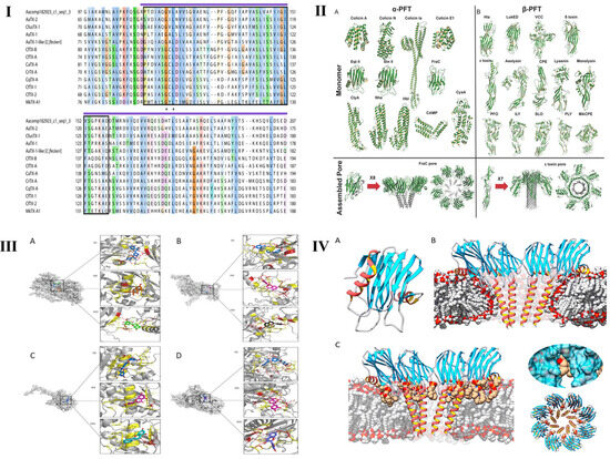
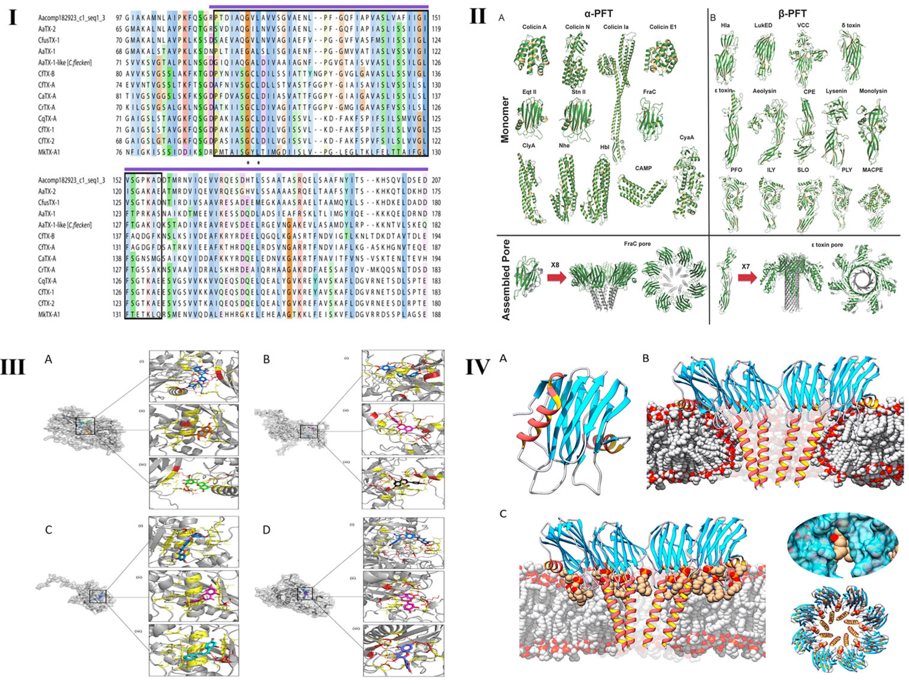
The structure–toxicity relationship of jellyfish toxins. (I) Partial multiple protein sequence alignment of CfusTX-1 and related jellyfish toxins, highlighting the regions of highest sequence similarity. Sequences were aligned using MUSCLE v5.3 and visualized using JalView v2.11.5.1. Amino acid residue shading is based on the Clustal protein colour scheme, with color intensity increasing as residue conservation increases from 25% to 100%. Identical residues are indicated by asterisks. Dashes represent the gaps introduced for better alignment. A predicted transmembrane spanning region (TSR1), common among jellyfish toxins, is indicated by a black outline. A purple line above the alignment corresponds to the predicted δ-endotoxin N-terminal-like domain. Reprinted from Ref. [59]. (II) The relationship between the secondary structure and toxicity of peptide toxins. (A): α-PFTs, upon binding to the membrane, α-helices undergo a conformational change to insert into the membrane and form membrane pore; (B): β-PFTs, monomer β-PFT first assembles in a pre-stem loop, and inserts into the membrane to form a partial β-barrel, and then combines with the other protomers to form a complete β-barrel. Reprinted from Ref. [94]. (III) The relationship between the tertiary structure and toxicity of peptide toxins. This is molecular docking studies of flavonoids against NnV-Mlp types and validated using AutoDock Vina v1.2.7 and PyMOL v3.1. (A), (B), (C) and (D) are the binding poses of flavonoids against Type 1/2/3/4 NnV-Mlp receptor, respectively. Reprinted from Ref. [57]. (IV) The relationship between the quaternary structure and toxicity of peptide toxins. (A): actinoporin monomers share a common fold: a stranded β-sandwich flanked by two short α-helixes. With regard to pore structure, two models have been proposed; (B): a tetrameric structure in which the lipid membrane adopts a toroidal shape around the pore walls; (C): an octameric lipid–protein mixed structure in which lipids (in tan color) are accommodated in pore-wall fenestrations (see inserts on the right). Reprinted from Ref. [80].
5.1. Primary Structure
The bioactivity of jellyfish toxins is fundamentally determined by their structure, which directly defines their physicochemical profile and functional properties. The toxicity of protein and polypeptide toxins is influenced by their amino acid content, sequence, and post-translational changes. Residues within the active site are particularly important, and mutations at these positions can drastically change the target-binding affinity and toxic efficacy. For example, the substitution of arginine-123 with alanine in the Chironex fleckeri toxin CfTX-1 lowers hemolytic activity by more than 95%, as this residue is required for cholesterol binding [86].
Cysteine residues play an important role in the function of polypeptide toxins. The disulfide bridges formed are indispensable for structural integrity and are frequently integrated into the architecture of bioactive sites [95]. The pp3 neuropeptide from Lychnorhiza malayensis, which contains three disulfide bonds, exemplifies this: disruption of any single bond significantly diminishes activity, whereas complete reduction leads to full inactivation [22]. Furthermore, specific amino acid modifications can modulate their activities. For example, phosphorylation of a threonine residue in Aurelia coerulea peptide toxin enhances its binding to ion channels and increases its potency by threefold [24].
As shown in Figure 5I, a variety of pore-forming jellyfish toxins have been identified to contain a conserved transmembrane region within the N-terminal domain, sharing high sequence similarity. Their structure is homologous to the N-terminal domain of pore- forming δ-endotoxins (Cry toxins) produced by Bacillus thuringiensis strains. In Cry toxins, the N-terminal domain is involved in cell membrane insertion and pore formation [59].
5.2. Secondary Structure
Secondary structures represent the regular, hydrogen-bond-stabilized conformations within toxin polypeptides or the defined spatial arrangements of small-molecule backbones, such as α-helices, β-sheets, and β-turns in proteins. These elements are fundamental for establishing and maintaining the bioactive conformation necessary for toxicity [96].
Toxins often interact with cellular membranes through α-helices. Hydrophobic or amphipathic α-helices can anchor in the lipid bilayer and rupture membrane or generate pores. Pore-forming toxins, for instance, use an N-terminal hydrophobic α-helix to penetrate membranes and oligomerize toxin monomers into functional transmembrane pores [80]. Mutations that disrupt this helical motif drastically impair the pore-forming function. β-Sheets are extensively involved in forming the structural core of enzymatic active sites and receptor-binding interfaces. Multiple β-strands associate via hydrogen bonds to create rigid β-sheet platforms that define the architecture of the binding pockets. The catalytic site of metalloproteinases is often a cleft lined by β-sheets, with conserved residues coordinating the Zn2+ required for proteolytic activity [90]. β-Turns, found flexible loops connecting secondary structure elements, contribute to molecular recognition by providing conformational adaptability. These turns enable the precise alignment of key residues, enhancing the binding specificity and affinity for the target molecules.
5.3. Tertiary Structure
The tertiary structure refers to the overall three-dimensional folding of a toxin molecule, representing the spatial integration of its primary and secondary structural constituents. This definitive architecture directs the precise spatial arrangement of functional residues, thereby determining target recognition and binding efficacy. The active site of a toxin is typically a three-dimensional pocket or cleft formed by residues that may be distant in the linear sequence but converge in the folded structure to establish a complementary interface with the target [97]. For example, the binding pocket of a metalloproteinase is a natural tertiary structure of a protein, and its toxicity can be significantly weakened when it is bound to a competitive ligand [57,98]. Denaturation of this tertiary fold, owing to thermal stress or extreme pH, disrupts the precise spatial arrangement of these essential residues, leading to irreversible inactivation [98].
The functional surface properties of a toxin, including its electrostatic potential and hydrophobicity distribution, are directly related to its tertiary structure. These characteristics critically modulate interactions with biological membranes and influence the in vivo distribution. Pore-forming toxins demonstrate this principle, as their surface exhibits a defined amphipathic character: discrete hydrophobic patches mediate insertion into the lipid bilayer, whereas adjacent hydrophilic regions line the aqueous lumen of the assembled pore, facilitating ion conductance [80].
5.4. Quaternary Structure
The quaternary structure refers to the supramolecular assembly formed by non-covalent interactions between multiple toxin protomers. Many jellyfish venom components require, oligomerization before they can exhibit biological activity [62]. This is exemplified by pore-forming toxins (PFTs), which are canonical oligomerization-dependent effector proteins. Their monomers are inactive by nature and only form a functional transmembrane pore after specific oligomerization on the target membrane [80]. The CfTX-A toxin from Chironex fleckeri, for instance, resides in solution as an inert monomer but forms a hexameric complex on the erythrocyte membrane, gaining significant hemolytic action [99]. This oligomerization process is dynamic and regulated by factors such as local toxin concentration and membrane microenvironment composition.
Enzymatic toxins also rely on oligomerization for optimal function. For example, dimerization of a metalloproteinase from Nemopilema nomurai, increases its catalytic activity by approximately five-fold compared to the monomeric form (only computational simulation results) [90]. Such a quaternary structure can stabilize the active site design and increase the substrate binding affinity. In addition to directly enabling or boosting a function, oligomerization can influence the pharmacokinetic profile of toxins. The formation of higher-order complexes can confer resistance to proteolytic degradation, extend the in vivo half-life, and promote tissue accumulation, thereby exacerbating toxicological outcomes.
5.5. Application Value
Studies on the SAR of jellyfish toxins provide a rational framework for developing targeted therapeutic agents. By elucidating the three-dimensional architecture of toxin active sites, specific inhibitors can be rationally designed to bind and neutralize toxicity, thereby preventing pathogenic interactions with host targets [100]. Furthermore, SAR insights enable the molecular engineering of toxin derivatives for novel applications. Site-directed mutagenesis can be used to structurally modify jellyfish neuropeptide toxins, eliminating their toxicity while retaining their high affinity for neurotransmitter receptors. These synthesized peptides serve as valuable molecular tools for studying receptor function in neuroscience research [101]. Exploiting the inherent targeting specificity of toxin domains offers another promising therapeutic approach. For example, the membrane-targeting domain of a pore-forming toxin can be fused with a cytotoxic agent to create a targeted chimeric molecule. This approach facilitates the selective delivery of therapeutic payloads to tumor cells, minimizing off-target effects and enhancing anticancer efficacy [94].
6. Future Perspectives
While current biotechnology and analytical methods have driven substantial advances in jellyfish toxin research, numerous fundamental and translational challenges remain unresolved. Future investigations are expected to develop in more profound and integrated directions, with a concerted emphasis on bridging basic mechanistic insights and practical innovations in this field. This synergistic approach is critical for developing effective solutions to reduce the public health burden caused by jellyfish envenomation.
6.1. Identification of Novel Toxic Components
Despite considerable advancements, the toxin repertoire of the majority of jellyfish species remains poorly characterized. Future studies should employ a systematic venomics approach that integrates high-resolution separation techniques, high-sensitivity detection methods, and multi-omics platforms. This integrated strategy is essential for comprehensively identifying and characterizing venom components from diverse species, thereby elucidating their chemical architecture and functional repertoire. Concurrently, research on the genetic basis of toxin synthesis is essential. A better understanding of the biosynthetic pathways and regulatory processes would lay the foundation for recombinant toxin production and facilitate genetic engineering applications.
6.2. Elucidate the Mechanism of Action
Although the mechanisms of action of some jellyfish toxins have been characterized, the specific molecular targets and downstream signaling pathways of the vast remain elusive. Future research should prioritize elucidating these mechanisms through an integrated, multi-scale approach. High-resolution structural biology techniques, such as cryo-electron microscopy and X-ray crystallography, are essential for determining the three-dimensional architecture of toxin-target complexes, and obtaining atomic-level insights into their molecular interactions. Concurrently, these structural findings must be contextualized within a physiological framework by using cell-based assays and relevant animal models. Such studies are crucial for delineating the dynamic, multi-step processes of envenomation in vivo and identifying the key pathogenic events that lead to lethal consequences [56].
Furthermore, a critical and understudied area is the synergistic interaction among venom components. Jellyfish venom is a complex cocktail of bioactive molecules, and the overall pathophysiology is more likely caused by synergistic or potentiating interactions among its constituents rather than by the simple sum of individual toxin effects. A systematic investigation of these combinatorial interactions is fundamental for gaining a comprehensive understanding of venom toxicity and developing effective, broad-spectrum countermeasures [66].
6.3. New Antitoxic Drugs
The current clinical management of jellyfish envenomation remains largely supportive and symptomatic, lacking specific, mechanism-based antitoxin therapies. As result, the rational development of targeted antitoxins, guided by the elucidation of toxin SAR, represents a critical frontier in future research [4,102].
One strategic approach is to develop small-molecule inhibitors that compete with the active site of the toxin, thereby preventing it from interacting with biological targets and neutralizing its activity. Furthermore, natural products are abundant and structurally diverse resources for antitoxin development. Bioactivity-guided screening of natural compound libraries may yield novel lead molecules with low toxicity and potent inhibitory effects on venom components [4].
6.4. Medicinal Value of Toxins
In addition to their pathogenic effects, jellyfish toxins possess a broad spectrum of bioactive properties, indicating the significant therapeutic potential of these compounds. Certain venom components exhibit analgesic, anti-inflammatory, and antitumor activities, making them valuable lead compounds for pharmaceutical development [3]. Future research should prioritize the systematic exploration of pharmacological potential. SAR-guided optimization can also improve the bioactivity of toxin derivatives while reducing their inherent toxicity. For instance, targeted structural modifications of jellyfish neuropeptide toxins could eliminate neurotoxicity while preserving or even augmenting their analgesic potency, paving the way for novel non-opioid pain therapeutics to be developed. Previous studies have found that specific components of Physalia physalis and Chironex fleckeri exhibit inhibitory effects on KCl-induced calcium signals in small- and medium-diameter neurons [15]. Similarly, although some toxin components can permeabilize cells and damage DNA, they may also exert genoprotective effects. Through tumor-targeted molecular engineering of these toxins, their cytotoxicity can be specifically directed against cancer cells, providing a highly promising strategy for developing novel antitumor agents [98].
7. Conclusions
Jellyfish toxins constitute a complex and diverse biomolecular arsenal, and their pathophysiological effects are dictated by their intrinsic physicochemical properties. These properties influence the pharmacokinetic behavior of venom components, including their absorption, distribution, metabolism, and excretion, within the envenomated organism. Crucially, the SARs that underpin these toxins are key to deciphering their mechanisms of action and rationally designing targeted countermeasures. In recent years, the integration of modern separation science, proteomics, and structural biology has driven significant advances, leading to the identification of numerous venom components and gradual elucidation of their mechanisms of action.
Despite this progress, formidable challenges remain. These include the systematic cataloguing of novel toxin families, detailed mechanistic dissection of their biological interactions, and translation of this knowledge into effective, specific antitoxins. Future progress necessitates a concerted multidisciplinary strategy that utilizes innovative technologies. By systematically integrating basic research with translational applications, this field can not only reduce the public health burden of envenomation but also fully explore the latent pharmacological potential of these toxins, ultimately contributing to therapeutic innovation and improving human health.
Author Contributions
Conceptualization, P.W. and H.C.; Writing—Preparation, P.W.; Writing—Revision, H.C.; Writing—Review and Editing, L.L., C.K., Z.F. and Y.L. All authors have read and agreed to the published version of the manuscript.
Funding
This study was financially supported by the Natural Science Foundation of Liaoning Province (2025-MS-316) and Science and Technology Innovation Special Program of the Liaoning Academy of Agricultural Sciences, grant numbers 2025-HBZ-1111 and 2026JC4037.
Institutional Review Board Statement
Not applicable.
Informed Consent Statement
Not applicable.
Data Availability Statement
No new data were created or analyzed in this study. Data sharing is not applicable to this article.
Conflicts of Interest
The authors declare no conflicts of interest.
References
- Piontek, M.; Seymour, J.E.; Wong, Y.; Gilstrom, T.; Potriquet, J.; Jennings, E.; Nimmo, A.; Miles, J.J. The pathology of Chironex fleckeri venom and known biological mechanisms. Toxicon X 2020, 6, 100026. [Google Scholar] [CrossRef] [Scilit] [PubMed]
- Dutertre, S. Venomics in Medicinal Chemistry. Future Med. Chem. 2014, 6, 1609–1610. [Google Scholar] [CrossRef] [Scilit] [PubMed]
- Daly, N.L.; Seymour, J.; Wilson, D. Exploring the therapeutic potential of jellyfish venom. Future Med. Chem. 2014, 6, 1715–1724. [Google Scholar] [CrossRef] [Scilit] [PubMed]
- Cunha, S.A.; Dinis-Oliveira, R.J. Raising Awareness on the Clinical and Forensic Aspects of Jellyfish Stings: A Worldwide Increasing Threat. Int. J. Environ. Res. Public Health 2022, 19, 8430. [Google Scholar] [CrossRef] [Scilit]
- Rizman-Idid, M.; Farrah-Azwa, A.B.; Chong, V.C. Preliminary Taxonomic Survey and Molecular Documentation of Jellyfish Species (Cnidaria: Scyphozoa and Cubozoa) in Malaysia. Zool. Stud. 2016, 55, 35. [Google Scholar] [CrossRef] [Scilit]
- Santhanam, R. Biology and Ecology of the Venomous Marine True Jellyfish (Class Scyphozoa). In Biology and Ecology of Venomous Marine Cnidarians; Santhanam, R., Ed.; Springer: Singapore, 2020; pp. 75–132. [Google Scholar]
- Santhanam, R. Biology and Ecology of the Venomous Marine Box Jellyfish (Class Cubozoa). In Biology and Ecology of Venomous Marine Cnidarians; Santhanam, R., Ed.; Springer: Singapore, 2020; pp. 133–168. [Google Scholar]
- Toshino, S.; Miyake, H.; Shibata, H. Meteorona kishinouyei, a new family, genus and species (Cnidaria, Cubozoa, Chirodropida) from Japanese Waters. ZooKeys 2015, 503, 1–21. [Google Scholar] [CrossRef] [Scilit]
- James, T.; Angel, A.Y.; Helen, C.T.; Ken, W. Immunological and Toxinological Responses to Jellyfish Stings. Inflamm. Allergy—Drug Targets (Discontin.) 2011, 10, 438–446. [Google Scholar] [CrossRef] [Scilit]
- Fautin, D.G. Structural diversity, systematics, and evolution of cnidae. Toxicon 2009, 54, 1054–1064. [Google Scholar] [CrossRef] [Scilit]
- Liu, W.; Mo, F.; Jiang, G.; Liang, H.; Ma, C.; Li, T.; Zhang, L.; Xiong, L.; Mariottini, G.L.; Zhang, J.; et al. Stress-Induced Mucus Secretion and Its Composition by a Combination of Proteomics and Metabolomics of the Jellyfish Aurelia coerulea. Mar. Drugs 2018, 16, 341. [Google Scholar] [CrossRef] [Scilit]
- Hwang, D.H.; Koh, P.-O.; Mohan Prakash, R.L.; Chae, J.; Kang, C.; Kim, E. Comparative Study of Toxic Effects and Pathophysiology of Envenomations Induced by Carybdea brevipedalia (Cnidaria: Cubozoa) and Nemopilema nomurai (Cnidaria: Scyphozoa) Jellyfish Venoms. Toxins 2022, 14, 831. [Google Scholar] [CrossRef] [Scilit]
- Hwang, D.H.; Prakash, R.L.M.; Asirvatham, R.D.; Kang, C.; Kim, E. Identification and characterization of toxic components of Carybdea brevipedalia venom: Implications for understanding box jellyfish (Cnidaria: Cubozoa) envenomation. Arch. Toxicol. 2025, 99, 4263–4279. [Google Scholar] [CrossRef] [Scilit] [PubMed]
- Li, R.; Yu, H.; Li, P. Proteomics, transcriptomics and toxicity analysis of venom proteins from jellyfish Cyanea nozakii. Toxicon 2019, 159, S31–S32. [Google Scholar] [CrossRef] [Scilit]
- Yanagihara, A.A.; Giglio, M.L.; Hurwitz, K.; Kadler, R.; Espino, S.S.; Raghuraman, S.; Olivera, B.M. Elucidation of Medusozoan (Jellyfish) Venom Constituent Activities Using Constellation Pharmacology. Toxins 2024, 16, 447. [Google Scholar] [CrossRef] [Scilit] [PubMed]
- Lv, B.; Li, R.; Li, G.; Yu, H.; Liu, S.; Li, R. Purification, characterization and the hemolytic mechanism of the hemolysin NnTX-45 from the jellyfish Nemopilema nomurai. Toxicon 2025, 266, 108537. [Google Scholar] [CrossRef] [Scilit]
- Li, R.; Yu, H.; Yue, Y.; Li, P. Combined Proteome and Toxicology Approach Reveals the Lethality of Venom Toxins from Jellyfish Cyanea nozakii. J. Proteome Res. 2018, 17, 3904–3913. [Google Scholar] [CrossRef] [Scilit]
- Choudhary, I.; Lee, H.; Pyo, M.J.; Heo, Y.; Chae, J.; Yum, S.S.; Kang, C.; Kim, E. Proteomic Investigation to Identify Anticancer Targets of Nemopilema nomurai Jellyfish Venom in Human Hepatocarcinoma HepG2 Cells. Toxins 2018, 10, 194. [Google Scholar] [CrossRef] [Scilit]
- Choudhary, I.; Hwang, D.H.; Lee, H.; Yoon, W.D.; Chae, J.; Han, C.H.; Yum, S.; Kang, C.; Kim, E. Proteomic Analysis of Novel Components of Nemopilema nomurai Jellyfish Venom: Deciphering the Mode of Action. Toxins 2019, 11, 153. [Google Scholar] [CrossRef] [Scilit]
- Li, R.; Yu, H.; Yue, Y.; Liu, S.; Xing, R.; Chen, X.; Li, P. Combined proteomics and transcriptomics identifies sting-related toxins of jellyfish Cyanea nozakii. J. Proteom. 2016, 148, 57–64. [Google Scholar] [CrossRef] [Scilit]
- Edirisinghe, E.A.H.W.; Athukorala, B.N.; Perera, M.; Abeywardana, B.A.S.D.; Sigera, P.S.T.; Eranga, P.; Theekshana, K.D.; Boudjelal, M.; Ali, R.; Peiris, D.C. Jellyfish Venom Peptides Targeting Human Potassium Channels Identified through Ligand Screening: Morphometric and Molecular Identification of the Species and Antibiotic Potential. Mar. Drugs 2024, 22, 333. [Google Scholar] [CrossRef] [Scilit]
- Riyas, A.; Kumar, A.; Chandran, M.; Jaleel, A.; Biju Kumar, A. The venom proteome of three common scyphozoan jellyfishes (Chrysaora caliparea, Cyanea nozakii and Lychnorhiza malayensis) (Cnidaria: Scyphozoa) from the coastal waters of India. Toxicon 2021, 195, 93–103. [Google Scholar] [CrossRef] [Scilit]
- Leung, T.C.N.; Qu, Z.; Nong, W.; Hui, J.H.L.; Ngai, S.M. Proteomic Analysis of the Venom of Jellyfishes Rhopilema esculentum and Sanderia malayensis. Mar. Drugs 2020, 18, 655. [Google Scholar] [CrossRef] [Scilit] [PubMed]
- Li, X.; Ma, X.; Chen, X.; Wang, T.; Liu, Q.; Wang, Y.; Li, Z.; Höfer, J.; Li, F.; Xiao, L.; et al. The medusa of Aurelia coerulea is similar to its polyp in molecular composition and different from the medusa of Stomolophus meleagris in toxicity. Toxicon 2022, 210, 89–99. [Google Scholar] [CrossRef] [Scilit] [PubMed]
- Chuan, C.H.; Venmathi Maran, B.A.; Yap, T.K.; Cheong, K.C.; Syed Hussein, M.A.; Saleh, E. New Records of Cubozoan and Scyphozoan Jellyfish from Sabah Waters, Malaysia. Diversity 2021, 13, 420. [Google Scholar] [CrossRef] [Scilit]
- Yasanga, T.; Wunnapuk, K.; Phuackchantuck, R.; Thaikruea, L.; Achalawitkun, T.; Rungraung, P.; Santidherakul, S. Updated Nematocyst Types in Tentacle of Venomous Box Jellyfish, Chironex indrasaksajiae (Sucharitakul, 2017) and Chiropsoides buitendijki (Horst, 1907) (Cnidaria, Cubozoa) in Thai Waters. Toxins 2025, 17, 44. [Google Scholar] [CrossRef] [Scilit]
- Ballesteros, A.; Marambio, M.; Trullas, C.; Jourdan, E.; Tena-Medialdea, J.; Gili, J.-M. Effect of Rinse Solutions on Rhizostoma pulmo (Cnidaria: Scyphozoa) Stings and the Ineffective Role of Vinegar in Scyphozoan Jellyfish Species. Int. J. Environ. Res. Public Health 2023, 20, 2344. [Google Scholar] [CrossRef] [Scilit]
- Thaikruea, L. Differences in clinical manifestations between cases stung by single-tentacle and multiple-tentacle box jellyfish over two decades. Heliyon 2023, 9, e16374. [Google Scholar] [CrossRef] [Scilit]
- Colaço Martins, L.; Gomes-Pereira, J.N.; Dionísio, G.; Assis, J. Unravelling environmental drivers and patterns of Portuguese man o’ war (Physalia physalis) blooms in two ocean regions: North Atlantic and the Southeast Pacific. Mar. Pollut. Bull. 2024, 209, 117278. [Google Scholar] [CrossRef] [Scilit]
- Wilcox, C.; Headlam, J.; Doyle, T.; Yanagihara, A. Assessing the Efficacy of First-Aid Measures in Physalia sp. Envenomation, Using Solution- and Blood Agarose-Based Models. Toxins 2017, 9, 149. [Google Scholar] [CrossRef] [Scilit]
- Labadie, M.; Aldabe, B.; Ong, N.; Joncquiert-Latarjet, A.; Groult, V.; Poulard, A.; Coudreuse, M.; Cordier, L.; Rolland, P.; Chanseau, P.; et al. Portuguese man-of-war (Physalia physalis) envenomation on the Aquitaine Coast of France: An emerging health risk. Clin. Toxicol. 2012, 50, 567–570. [Google Scholar] [CrossRef] [Scilit]
- Miller, B.J.; von der Heyden, S.; Gibbons, M.J. Significant population genetic structuring of the holoplanktic scyphozoan Pelagia noctiluca in the Atlantic Ocean. Afr. J. Mar. Sci. 2012, 34, 425–430. [Google Scholar] [CrossRef] [Scilit]
- Tibballs, J. Australian venomous jellyfish, envenomation syndromes, toxins and therapy. Toxicon 2006, 48, 830–859. [Google Scholar] [CrossRef] [Scilit] [PubMed]
- Ballesteros, A.; Marambio, M.; Fuentes, V.; Narda, M.; Santín, A.; Gili, J.-M. Differing Effects of Vinegar on Pelagia noctiluca (Cnidaria: Scyphozoa) and Carybdea marsupialis (Cnidaria: Cubozoa) Stings—Implications for First Aid Protocols. Toxins 2021, 13, 509. [Google Scholar] [CrossRef] [Scilit] [PubMed]
- Remigante, A.; Costa, R.; Morabito, R.; La Spada, G.; Marino, A.; Dossena, S. Impact of Scyphozoan Venoms on Human Health and Current First Aid Options for Stings. Toxins 2018, 10, 133. [Google Scholar] [CrossRef] [Scilit] [PubMed]
- Hamner, W.M.; Dawson, M.N. A review and synthesis on the systematics and evolution of jellyfish blooms: Advantageous aggregations and adaptive assemblages. Hydrobiologia 2008, 616, 161–191. [Google Scholar] [CrossRef] [Scilit]
- Doyle, T.; Headlam, J.; Wilcox, C.; MacLoughlin, E.; Yanagihara, A. Evaluation of Cyanea capillata Sting Management Protocols Using Ex Vivo and In Vitro Envenomation Models. Toxins 2017, 9, 215. [Google Scholar] [CrossRef] [Scilit]
- Helmholz, H.; Ruhnau, C.; Schütt, C.; Prange, A. Comparative study on the cell toxicity and enzymatic activity of two northern scyphozoan species Cyanea capillata (L.) and Cyanea lamarckii (Péron & Léslieur). Toxicon 2007, 50, 53–64. [Google Scholar] [CrossRef] [Scilit]
- Cegolon, L.; Heymann, W.; Lange, J.; Mastrangelo, G. Jellyfish Stings and Their Management: A Review. Mar. Drugs 2013, 11, 523–550. [Google Scholar] [CrossRef] [Scilit]
- Badré, S. Bioactive toxins from stinging jellyfish. Toxicon 2014, 91, 114–125. [Google Scholar] [CrossRef] [Scilit]
- Lewis, C.; Bentlage, B.; Yanagihara, A.; Gillan, W.; Blerk, J.V.; Keil, D.P.; Bely, A.E.; Collins, A.G. Redescription of Alatina alata (Reynaud, 1830) (Cnidaria: Cubozoa) from Bonaire, Dutch Caribbean. Zootaxa 2013, 3737, 473–487. [Google Scholar] [CrossRef] [Scilit]
- Lawley, J.W.; Ames, C.L.; Bentlage, B.; Yanagihara, A.; Goodwill, R.; Kayal, E.; Hurwitz, K.; Collins, A.G. Box Jellyfish Alatina alata Has a Circumtropical Distribution. Biol. Bull. 2016, 231, 152–169. [Google Scholar] [CrossRef] [Scilit]
- McGee, R.G.; Webster, A.C.; Lewis, S.R.; Welsford, M. Interventions for the symptoms and signs resulting from jellyfish stings. Cochrane Database Syst. Rev. 2023, 2023, CD009688. [Google Scholar] [CrossRef] [Scilit]
- Rossetto, A.L.; Proença, L.A.d.O. Seabather’s eruption: Report of case in northeast region of Brazil. An. Bras. Dermatol. 2012, 87, 472–474. [Google Scholar] [CrossRef] [Scilit] [PubMed]
- Eyer-Silva, W.d.A.; Pitombo, F.B.; Silva, G.A.R.d. Seabather’s eruption in Ipanema Beach, Rio de Janeiro, Brazil. Rev. Soc. Bras. Med. Trop. 2018, 51, 119. [Google Scholar] [CrossRef] [Scilit] [PubMed]
- Horiike, T.; Nagai, H.; Kitani, S. Identification of Allergens in the Box Jellyfish Chironex yamaguchii That Cause Sting Dermatitis. Int. Arch. Allergy Immunol. 2015, 167, 73–82. [Google Scholar] [CrossRef] [Scilit]
- Giallongo, G.; Douek, J.; Harbuzov, Z.; Galil, B.S.; Rinkevich, B. Long-term changes in population genetic features of a rapidly expanding marine invader: Implication for invasion success. Biol. Invasions 2021, 23, 2541–2552. [Google Scholar] [CrossRef] [Scilit]
- Uri, S.; Marina, G.; Liubov, G. Severe delayed cutaneous reaction due to Mediterranean jellyfish (Rhopilema nomadica) envenomation. Contact Dermat. 2005, 52, 282–283. [Google Scholar] [CrossRef] [Scilit]
- Hammill, E.; Johnson, E.; Atwood, T.B.; Harianto, J.; Hinchliffe, C.; Calosi, P.; Byrne, M. Ocean acidification alters zooplankton communities and increases top-down pressure of a cubozoan predator. Glob. Change Biol. 2017, 24, E128–E138. [Google Scholar] [CrossRef] [Scilit]
- Mariottini, G.L.; Pane, L. Mediterranean Jellyfish Venoms: A Review on Scyphomedusae. Mar. Drugs 2010, 8, 1122–1152. [Google Scholar] [CrossRef] [Scilit]
- Leoni, V.; Bonnet, D.; Ramírez-Romero, E.; Molinero, J.C.; Pandolfi, J. Biogeography and phenology of the jellyfish Rhizostoma pulmo (Cnidaria: Scyphozoa) in southern European seas. Glob. Ecol. Biogeogr. 2021, 30, 622–639. [Google Scholar] [CrossRef] [Scilit]
- Ramasamy, S.; Isbister, G.K.; Seymour, J.E.; Hodgson, W.C. Pharmacologically distinct cardiovascular effects of box jellyfish (Chironex fleckeri) venom and a tentacle-only extract in rats. Toxicol. Lett. 2005, 155, 219–226. [Google Scholar] [CrossRef] [Scilit]
- Winter, K.L.; Isbister, G.K.; McGowan, S.; Konstantakopoulos, N.; Seymour, J.E.; Hodgson, W.C. A pharmacological and biochemical examination of the geographical variation of Chironex fleckeri venom. Toxicol. Lett. 2010, 192, 419–424. [Google Scholar] [CrossRef] [Scilit] [PubMed]
- Auerbach, P.S.; Gupta, D.; Van Hoesen, K.; Zavala, A. Dermatological Progression of a Probable Box Jellyfish Sting. Wilderness Environ. Med. 2019, 30, 310–320. [Google Scholar] [CrossRef] [Scilit] [PubMed]
- Guevara, B.E.K.; Dayrit, J.F.; Haddad, V., Jr. Seabather’s eruption caused by the thimble jellyfish (Linuche aquila) in the Philippines. Clin. Exp. Dermatol. 2017, 42, 808–810. [Google Scholar] [CrossRef] [Scilit] [PubMed]
- Li, Y.; Qiu, Z.; Li, B.; Geng, X.; Yu, X.; Li, Y.; Li, W.; Yang, J. Jellyfish Venom-Induced Cardiotoxicity and Immune Responses: Mechanisms and Potential Therapeutic Strategies. Mar. Drugs 2025, 23, 369. [Google Scholar] [CrossRef] [Scilit]
- Asirvatham, R.D.; Hwang, D.H.; Prakash, R.L.M.; Kang, C.; Kim, E. Pharmacoinformatic Investigation of Silymarin as a Potential Inhibitor against Nemopilema nomurai Jellyfish Metalloproteinase Toxin-like Protein. Int. J. Mol. Sci. 2023, 24, 8972. [Google Scholar] [CrossRef] [Scilit]
- Brinkman, D.L.; Jia, X.; Potriquet, J.; Kumar, D.; Dash, D.; Kvaskoff, D.; Mulvenna, J. Transcriptome and venom proteome of the box jellyfish Chironex fleckeri. BMC Genom. 2015, 16, 407. [Google Scholar] [CrossRef] [Scilit]
- Ponce, D.; Brinkman, D.; Potriquet, J.; Mulvenna, J. Tentacle Transcriptome and Venom Proteome of the Pacific Sea Nettle, Chrysaora fuscescens (Cnidaria: Scyphozoa). Toxins 2016, 8, 102. [Google Scholar] [CrossRef] [Scilit]
- Ponce, D.; Brinkman, D.L.; Luna-Ramírez, K.; Wright, C.E.; Dorantes-Aranda, J.J. Comparative study of the toxic effects of Chrysaora quinquecirrha (Cnidaria: Scyphozoa) and Chironex fleckeri (Cnidaria: Cubozoa) venoms using cell-based assays. Toxicon 2015, 106, 57–67. [Google Scholar] [CrossRef] [Scilit]
- Lakkis, N.A.; Maalouf, G.J.; Mahmassani, D.M. Jellyfish Stings: A Practical Approach. Wilderness Environ. Med. 2015, 26, 422–429. [Google Scholar] [CrossRef] [Scilit]
- Amreen Nisa, S.; Vinu, D.; Krupakar, P.; Govindaraju, K.; Sharma, D.; Vivek, R. Jellyfish venom proteins and their pharmacological potentials: A review. Int. J. Biol. Macromol. 2021, 176, 424–436. [Google Scholar] [CrossRef] [Scilit]
- Shi, M.; McHugh, K.J. Strategies for overcoming protein and peptide instability in biodegradable drug delivery systems. Adv. Drug Deliv. Rev. 2023, 199, 114904. [Google Scholar] [CrossRef] [Scilit] [PubMed]
- Dziubak, D.; Sęk, S. Sparsely tethered bilayer lipid membranes formed by self-assembly of bicelles: Spectroelectrochemical characterization and incorporation of transmembrane protein. Bioelectrochemistry 2023, 153, 108482. [Google Scholar] [CrossRef] [Scilit] [PubMed]
- Cosentino, K.; Ros, U.; García-Sáez, A.J. Assembling the puzzle: Oligomerization of α-pore forming proteins in membranes. Biochim. Biophys. Acta (BBA)—Biomembr. 2016, 1858, 457–466. [Google Scholar] [CrossRef] [Scilit] [PubMed]
- Chong, S.Y.; Naidu, R.; Othman, I.; Zainal Abidin, S.A. Consolidation of venom proteomes from major Cnidarian species (Scyphozoa and Cubozoa) obtained using liquid chromatography-tandem mass spectrometry. Toxicon 2025, 265, 108491. [Google Scholar] [CrossRef] [Scilit]
- Wang, C.; Wang, B.; Wang, B.; Wang, Q.; Liu, G.; Wang, T.; He, Q.; Zhang, L. Unique Diversity of Sting-Related Toxins Based on Transcriptomic and Proteomic Analysis of the Jellyfish Cyanea capillata and Nemopilema nomurai (Cnidaria: Scyphozoa). J. Proteome Res. 2018, 18, 436–448. [Google Scholar] [CrossRef] [Scilit]
- Li, R.; Yu, H.; Li, T.; Li, P. Comprehensive Proteome Reveals the Key Lethal Toxins in the Venom of Jellyfish Nemopilema nomurai. J. Proteome Res. 2020, 19, 2491–2500. [Google Scholar] [CrossRef] [Scilit]
- Lee, H.; Jung, E.-s.; Kang, C.; Yoon, W.D.; Kim, J.-S.; Kim, E. Scyphozoan jellyfish venom metalloproteinases and their role in the cytotoxicity. Toxicon 2011, 58, 277–284. [Google Scholar] [CrossRef] [Scilit]
- Kollet, O.; Das, A.; Karamanos, N.; auf dem Keller, U.; Sagi, I. Redefining metalloproteases specificity through network proteolysis. Trends Mol. Med. 2024, 30, 147–163. [Google Scholar] [CrossRef] [Scilit]
- Singh, G.; Yadav, M.; Ghosh, C.; Rathore, J.S. Bacterial toxin-antitoxin modules: Classification, functions, and association with persistence. Curr. Res. Microb. Sci. 2021, 2, 100047. [Google Scholar] [CrossRef] [Scilit]
- Catterall, W.A.; Cestèle, S.; Yarov-Yarovoy, V.; Yu, F.H.; Konoki, K.; Scheuer, T. Voltage-gated ion channels and gating modifier toxins. Toxicon 2007, 49, 124–141. [Google Scholar] [CrossRef] [Scilit]
- Xiao, L.; He, Q.; Guo, Y.; Zhang, J.; Nie, F.; Li, Y.; Ye, X.; Zhang, L. Cyanea capillata tentacle-only extract as a potential alternative of nematocyst venom: Its cardiovascular toxicity and tolerance to isolation and purification procedures. Toxicon 2009, 53, 146–152. [Google Scholar] [CrossRef] [Scilit] [PubMed]
- Rato, L.D.; Pinto, C.; Duarte, I.M.; Leandro, S.M.; Marques, S.C. Euryhalinity and thermal tolerance of Phyllorhiza punctata (Scyphozoa) scyphostomae: Life history and physiological trade-offs. Mar. Biol. 2021, 168, 158. [Google Scholar] [CrossRef] [Scilit]
- Casafús, M.G.; Gritti, M.A.; Miranda, C.; Guimarães, P.; Montalto, L.; Peichoto, M.E. Freshwater jellyfish in northeastern Argentina: A risk to human health. Trans. R. Soc. Trop. Med. Hyg. 2025, 119, 48–57. [Google Scholar] [CrossRef] [Scilit] [PubMed]
- Li, Q.-M.; Wang, J.-F.; Zha, X.-Q.; Pan, L.-H.; Zhang, H.-L.; Luo, J.-P. Structural characterization and immunomodulatory activity of a new polysaccharide from jellyfish. Carbohydr. Polym. 2017, 159, 188–194. [Google Scholar] [CrossRef] [Scilit]
- Prakash, R.L.M.; Ravi, D.A.; Hwang, D.H.; Kang, C.; Kim, E. Characterization of Novel ACE-Inhibitory Peptides from Nemopilema nomurai Jellyfish Venom Hydrolysate: In Vitro and In Silico Approaches. Mar. Drugs 2025, 23, 267. [Google Scholar] [CrossRef] [Scilit]
- Cole, E.B.; Miller, D.; Rometo, D.; Greenberg, R.M.; Brömme, D.; Çataltepe, S.; Pak, S.C.; Mills, D.R.; Silverman, G.A.; Luke, C.J. Identification and Activity of a Lower Eukaryotic Serine Proteinase Inhibitor (Serpin) from Cyanea capillata: Analysis of a Jellyfish Serpin, Jellypin. Biochemistry 2004, 43, 11750–11759. [Google Scholar] [CrossRef] [Scilit]
- Zhou, Y.; Liu, G.; Cheng, X.; Wang, Q.; Wang, B.; Wang, B.; Zhang, H.; He, Q.; Zhang, L. Antimicrobial activity of a newly identified Kazal-type serine proteinase inhibitor, CcKPI1, from the jellyfish Cyanea capillata. Int. J. Biol. Macromol. 2018, 107, 1945–1955. [Google Scholar] [CrossRef] [Scilit]
- Rivera-de-Torre, E.; Palacios-Ortega, J.; Gavilanes, J.; Martínez-del-Pozo, Á.; García-Linares, S. Pore-Forming Proteins from Cnidarians and Arachnids as Potential Biotechnological Tools. Toxins 2019, 11, 370. [Google Scholar] [CrossRef] [Scilit]
- Have, G.A.M.T.; Engelen, M.P.K.J.; Luiking, Y.C.; Deutz, N.E.P. Absorption Kinetics of Amino Acids, Peptides, and Intact Proteins. Int. J. Sport Nutr. Exerc. Metab. 2007, 17, S23–S36. [Google Scholar] [CrossRef] [Scilit]
- Chmiel, T.; Mieszkowska, A.; Kempińska-Kupczyk, D.; Kot-Wasik, A.; Namieśnik, J.; Mazerska, Z. The impact of lipophilicity on environmental processes, drug delivery and bioavailability of food components. Microchem. J. 2019, 146, 393–406. [Google Scholar] [CrossRef] [Scilit]
- Conner, K.P.; Devanaboyina, S.C.; Thomas, V.A.; Rock, D.A. The biodistribution of therapeutic proteins: Mechanism, implications for pharmacokinetics, and methods of evaluation. Pharmacol. Ther. 2020, 212, 107574. [Google Scholar] [CrossRef] [Scilit] [PubMed]
- Li, M.; Guo, H.; Wang, L.; Tao, R.; Song, G.; Cao, L.; Yan, W.; Wu, Z.; Liu, Q.; Chen, Y.; et al. A plasmid-encoded inactive toxin–antitoxin system MtvT/MtvA regulates plasmid conjugative transfer and bacterial virulence in Pseudomonas aeruginosa. Nucleic Acids Res. 2025, 53, gkaf075. [Google Scholar] [CrossRef] [Scilit] [PubMed]
- Flores-Holguín, N.; Salas-Leiva, J.S.; Núñez-Vázquez, E.J.; Tovar-Ramírez, D.; Glossman-Mitnik, D. Exploring marine toxins: Comparative analysis of chemical reactivity properties and potential for drug discovery. Front. Chem. 2023, 11, 1286804. [Google Scholar] [CrossRef] [Scilit]
- Andreosso, A.; Bansal, P.S.; Smout, M.J.; Wilson, D.; Seymour, J.E.; Daly, N.L. Structural Characterisation of Predicted Helical Regions in the Chironex fleckeri CfTX-1 Toxin. Mar. Drugs 2018, 16, 201. [Google Scholar] [CrossRef] [Scilit]
- Jia, Y.; Cocker, C.; Sampath, J. Insights into Protein Unfolding under pH, Temperature, and Shear Using Molecular Dynamics Simulations. Biomacromolecules 2025, 26, 2095–2105. [Google Scholar] [CrossRef] [Scilit]
- Toldrá, F.; Reig, M.; Aristoy, M.C.; Mora, L. Generation of bioactive peptides during food processing. Food Chem. 2018, 267, 395–404. [Google Scholar] [CrossRef] [Scilit]
- Li, Y.; Ma, J.; Li, Y.; Shen, X.; Xia, X. Microbial community and enzyme activity respond differently to seasonal and edaphic factors in forest and grassland ecosystems. Appl. Soil Ecol. 2024, 194, 105167. [Google Scholar] [CrossRef] [Scilit]
- Danso, B.; Fengling, Y.; Hua, X.; Zhang, J.; Chen, J.; Yao, Y.; Pozzolini, M.; Wang, F.; Xiao, L.; Ruixue, H. Potential inhibitors of metalloproteinases (MMPs) and phospholipases from Nemopilema nomurai jellyfish peptides: An in-silico pharmacokinetics and molecular docking studies. Toxicon 2025, 263, 108421. [Google Scholar] [CrossRef] [Scilit]
- Qiu, S.; Cai, Y.; Yao, H.; Lin, C.; Xie, Y.; Tang, S.; Zhang, A. Small molecule metabolites: Discovery of biomarkers and therapeutic targets. Signal Transduct. Target. Ther. 2023, 8, 132. [Google Scholar] [CrossRef] [Scilit]
- Akinniyi, G.; Akinboye, A.J.; Yang, I.; Lee, J.-G. Plant proteins, peptides, and non-protein amino acids: Toxicity, sources, and analysis. Heliyon 2024, 10, e34890. [Google Scholar] [CrossRef] [Scilit]
- Hofmann, A.F. Biliary secretion and excretion in health and disease: Current concepts. Ann. Hepatol. 2007, 6, 15–27. [Google Scholar] [CrossRef] [Scilit]
- Li, Y.; Li, Y.; Mengist, H.M.; Shi, C.; Zhang, C.; Wang, B.; Li, T.; Huang, Y.; Xu, Y.; Jin, T. Structural Basis of the Pore-Forming Toxin/Membrane Interaction. Toxins 2021, 13, 128. [Google Scholar] [CrossRef] [Scilit]
- Zhao, Z.; Gui, J.; Yao, A.; Le, N.Q.K.; Chua, M.C.H. Improved Prediction Model of Protein and Peptide Toxicity by Integrating Channel Attention into a Convolutional Neural Network and Gated Recurrent Units. ACS Omega 2022, 7, 40569–40577. [Google Scholar] [CrossRef] [Scilit]
- Williams, D.F. Biocompatibility pathways and mechanisms for bioactive materials: The bioactivity zone. Bioact. Mater. 2022, 10, 306–322. [Google Scholar] [CrossRef] [Scilit]
- Lahiani, A.; Yavin, E.; Lazarovici, P. The Molecular Basis of Toxins’ Interactions with Intracellular Signaling via Discrete Portals. Toxins 2017, 9, 107. [Google Scholar] [CrossRef] [Scilit]
- Wang, Y.; Yang, F.; Wang, Y.; Liu, C.; Hua, X.; Wang, L.; Zu, X.; Liu, X.; Pozzolini, M.; Zhu, Y.; et al. Insight of protease and PLA2 activity co-inhibition on jellyfish toxin-induced inflammation and multiple organ dysfunction. Int. J. Biol. Macromol. 2025, 323, 147167. [Google Scholar] [CrossRef] [Scilit]
- Jouiaei, M.; Casewell, N.; Yanagihara, A.; Nouwens, A.; Cribb, B.; Whitehead, D.; Jackson, T.; Ali, S.; Wagstaff, S.; Koludarov, I.; et al. Firing the Sting: Chemically Induced Discharge of Cnidae Reveals Novel Proteins and Peptides from Box Jellyfish (Chironex fleckeri) Venom. Toxins 2015, 7, 936–950. [Google Scholar] [CrossRef] [Scilit]
- Damm, M.; Vilcinskas, A.; Lüddecke, T. Mapping the architecture of animal toxin systems by mass spectrometry imaging. Biotechnol. Adv. 2025, 81, 108548. [Google Scholar] [CrossRef] [Scilit]
- Rosson, E.; Lux, F.; David, L.; Godfrin, Y.; Tillement, O.; Thomas, E. Focus on therapeutic peptides and their delivery. Int. J. Pharm. 2025, 675, 125555. [Google Scholar] [CrossRef] [Scilit]
- Hwang, D.H.; Lee, H.; Choudhary, I.; Kang, C.; Chae, J.; Kim, E. Protective effect of epigallocatechin-3-gallate (EGCG) on toxic metalloproteinases-mediated skin damage induced by Scyphozoan jellyfish envenomation. Sci. Rep. 2020, 10, 18644. [Google Scholar] [CrossRef] [Scilit]
Disclaimer/Publisher’s Note: The statements, opinions and data contained in all publications are solely those of the individual author(s) and contributor(s) and not of MDPI and/or the editor(s). MDPI and/or the editor(s) disclaim responsibility for any injury to people or property resulting from any ideas, methods, instructions or products referred to in the content. |
© 2026 by the authors. Licensee MDPI, Basel, Switzerland. This article is an open access article distributed under the terms and conditions of the Creative Commons Attribution (CC BY) license.