


Abstract
This study aimed to develop bioactive single-chain recombinant eel follicle-stimulating hormone (rec-eel FSH) analogs in Chinese hamster ovary DG44 (CHO DG44) cells. Our previous studies demonstrated that the O-linked glycosylated carboxyl-terminal peptide (CTP) of the equine chorionic gonadotropin (eCG) β-subunit enhances secretion in mammalian cells. Based on this observation, an FSH mutant (FSH-eM) was generated by inserting the eCG β-subunit CTP (amino acids 115–149) between the β- and α-subunits of wild-type single-chain eel FSH. Strep- and His-tags were fused to the C-terminus of the α-subunit for protein purification. The recombinant FSH-eM-Strep and FSH-eM-His proteins migrated as broad bands at 48–53 kDa. Sequential enzymatic digestion with PNGase F, O-glycosidase, and neuraminidase revealed the presence of both N- and O-linked glycans. To further investigate the potential of this approach as a candidate glycoengineering strategy, a flexible 15-amino acid linker [(GGSA)3GGG] was inserted between the β-subunit and the eCG β-subunit CTP, generating the FSH-GGSA-eM-Strep construct. Recombinant FSH-GGSA-eM-Strep was stably expressed in CHO DG44 cells and purified by Strep-Tactin XT affinity chromatography, demonstrating its suitability for large-scale production. Western blotting and silver staining showed an apparent molecular weight of 52–56 kDa. Following treatment with PNGase F, O-glycosidase, and neuraminidase, the molecular weight decreased to approximately 38, 42, and 42 kDa, respectively. cAMP accumulation assays demonstrated that rec-FSH-GGSA-eM-Strep stimulated eel FSHR-expressing cells in a dose-dependent manner. ERK1/2 phosphorylation reached a maximum at 5 min and rapidly declined thereafter. This response was markedly attenuated by treatment with the protein kinase A (PKA) inhibitor H89, suggesting that PKA activity contributes to ERK1/2 activation under the conditions tested. These findings suggest that this strategy may represent a promising approach for the production of biologically active recombinant eel FSH, with potential applications in reproductive biotechnology.
1. Introduction
Follicle-stimulating hormone (FSH) is a pituitary glycoprotein hormone that plays a central role in regulating gonadal development and reproductive function in vertebrates, including mammals and fish [1,2]. Like other glycoprotein hormones, FSH consists of non-covalently associated α- and β-subunits, and dissociation of these subunits completely abolishes its biological activity [3,4].
The eel FSH α-subunit contains two conserved N-linked glycosylation sites at Asn56 and Asn79, whereas the FSH β-subunit also contains two N-linked glycosylation sites at Asn5 and Asn22. These N-linked glycans are essential for the biological activity of glycoprotein hormones [5,6]. Variations in glycosylation markedly influence receptor binding, biological activity, metabolic stability, and circulatory half-life of glycoprotein hormones, thereby determining their physiological functions [4,7].
O-glycosylation is one of the most abundant and structurally diverse types of post-translational modifications of proteins. O-glycans regulate protein structure, stability, and function, and their biosynthesis is tightly controlled in a cell type-specific manner, resulting in considerable structural and functional diversity [8,9]. Although O-linked oligosaccharides in glycoprotein hormones have limited effects on receptor binding and in vitro biological activity, they play critical roles in regulating in vivo biological responses and prolonging the circulating half-life of glycoprotein hormones. This functional importance has been demonstrated by incorporation of the human chorionic gonadotropin (hCG) β-subunit carboxyl-terminal peptide (CTP), which contains multiple O-linked glycosylation sites and significantly enhances the pharmacokinetic properties of recombinant glycoprotein hormones [10]. Insertion of the hCTP linker enhanced the in vivo biological activity of recombinant eel FSH and LH by increasing the abundance of sialylated O-linked glycans [11]. Collectively, these findings indicate that precise O-linked glycosylation is a key determinant of the stability, receptor interaction, and biological activity of recombinant gonadotropins. These observations provide the rationale for exploring alternative O-glycosylated CTP sequences, such as the CTP derived from the eCG β-subunit, to further characterize the biological activity of recombinant eel FSH.
Although CHO-K1 cells are widely used for recombinant glycoprotein hormone production, their expression levels are often limited due to complex biosynthetic processes, including α/β-subunit assembly, secretion efficiency, and post-translational modifications. Therefore, improving production strategies in CHO-based systems is essential for efficient structural and functional studies of recombinant hormones. Recently, several reports have demonstrated the feasibility of establishing CHO DG44 cell-based expression systems for the production of recombinant human FSH [12] and eel FSH [13,14]. Collectively, these studies demonstrate that CHO DG44 cells provide an efficient platform for producing biologically active recombinant eel FSH with appropriate post-translational modifications.
Clinical studies using the hCG β-subunit CTP, including the long-acting human growth hormone (hGH) analog MOD-4023, have demonstrated that CTP-mediated glycoengineering enhances protein stability and serum half-life [15,16,17]. Accordingly, the CTP linker has been utilized as a promising strategy to extend the biological activity of short-lived protein therapeutics. These observations prompted us to investigate whether incorporation of the eCG β-subunit CTP could further enhance glycosylation and biological activity of recombinant eel FSH.
The present study aimed to establish an efficient production platform for recombinant eel FSH incorporating the eCG β-subunit CTP. To investigate the potential role of a flexible GGSA linker in the glycosylation and production of recombinant eel FSH, the linker was inserted between the FSH β-subunit and eCG β-subunit CTP, and the resulting recombinant protein was expressed in CHO DG44 cells. The N- and O-linked glycosylation patterns, biological activities, and production characteristics of the recombinant FSH were subsequently evaluated to assess the feasibility of this approach for large-scale production of biologically active recombinant eel FSH.
2. Materials and Methods
2.1. Materials
Oligonucleotides were synthesized by Genotech (Daejeon, Republic of Korea). The pGEMT-easy cloning vector was purchased from Promega (Madison, WI, USA). The pOptiVEC-TOPO TA Cloning Kit, Freedom DG44 Kit, CHO-S cells, DG44 CHO cells, CD DG44 medium, CD OptiCHO medium, CD FortiCHO medium, methotrexate (MTX) reagent, and cloning media were purchased from Life Technologies (Carlsbad, CA, USA). Lipofectamine-2000, Freestyle MAX Reagent, fetal bovine serum (FBS), Ham’s F-12 medium, OptiMEM, CHO-S-SFMII medium, His-tag monoclonal Antibody (MA1-21315), Strep-tag polyclonal antibody (PA5-114454), SuperSignalTM chemiluminescent substrate and HisPur Ni-NTA Spin Columns were from Thermo Fisher Scientific (Waltham, MA, USA). CHO-K1 cells and HEK 293 cells were obtained from the Korean Cell Line Bank (KCLB, Seoul, Republic of Korea). Trans-Blot® Turbo™ Transfer System, polyvinylidene difluoride (PVDF) membrane, 4× Laemmli Sample Buffer, RIPA buffer and Silver Stain Plus kit were from Bio-Rad Laboratories (Hercules, CA, USA). The pCORON1000 SP VSV-G tag expression vector was purchased from Amersham Biosciences (Piscataway, NJ, USA). HRP-conjugated anti-Strep antibody was from GenScript Biotech Corporation (Piscataway, NJ, USA). A monoclonal antibody (5A14) for Western blotting and an enzyme-linked immunosorbent assay (ELISA) system for recombinant eel FSH were developed in our laboratory [5]. Anti-phospho-p44/42 MAPK and total ERK1/2 antibodies were purchased from Cell Signaling Technology (Beverly, MA, USA). DNA ligation reagents, polymerase chain reaction (PCR) reagents, endonucleases and restriction enzymes were purchased from Takara Bio (Shiga, Japan). PNGase F (P0704), O-Glycosidase (P0733), and α2-3,6,8,9-Neuraminidase (P0720) were purchased from New England Biolabs (Ipswich, MA, USA). The cAMP Dynamic 2 Immunoassay Kit was purchased from Cisbio (Codolet, France). Strep-Tactin XT 4 flow, 4 flow high-capacity columns and high-capacity resin were purchased from IBA-Lifesciences (Göttingen, Germany). The QIAprep Spin Plasmid Kit was purchased from Qiagen (Hilden, Germany), and disposable spinner flasks were purchased from Corning (Corning, NY, USA). Centriplus Centrifugal Filter Devices were purchased from Amicon Bio Separation (Billerica, MA, USA). All other reagents were purchased from Sigma-Aldrich (St. Louis, MO, USA).
2.2. Construction of Eel FSH-eM-Strep/His and FSH-GGSA-eM-Strep Vectors
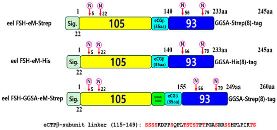
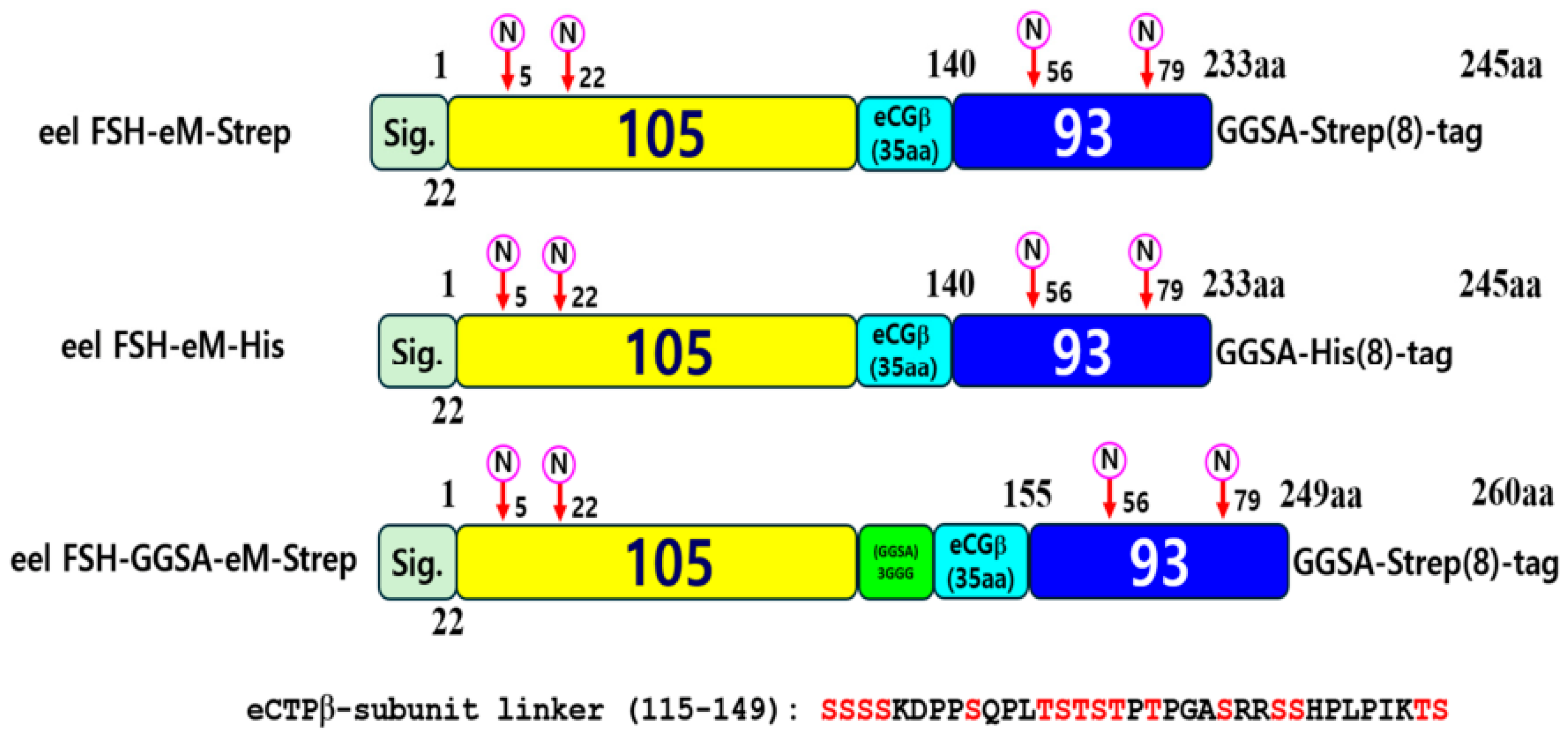
The cDNAs encoding the eel FSH α- and β-subunits were amplified from eel pituitary cDNA as previously described [7]. A wild-type single-chain eel FSH (FSH-wt), in which the α-subunit was fused to the C-terminus of the β-subunit, was generated using overlap extension PCR [2]. An eel FSH variant (FSH-eM) was subsequently constructed by inserting the eCG β-subunit carboxyl-terminal peptide (eCTP; amino acids 115–149) between the β- and α-subunits [14]. Strep- or His-tags were fused to the C-terminus of the α-subunit for recombinant protein purification, generating the FSH-eM-Strep and FSH-eM-His constructs. To further improve accessibility of the inserted eCG β-subunit CTP for O-linked glycosylation, a 15-amino-acid [(GGSA)3GGG] linker was inserted between the β-subunit and eCTP, generating the FSH-GGSA-eM-Strep construct. The resulting constructs were designated FSH-eM-Strep, FSH-eM-His, and FSH-GGSA-eM-Strep. Full-length PCR products were cloned into the pGEM-T Easy vector and verified by bidirectional DNA sequencing. The verified inserts were subcloned into the pOptiVEC expression vector, and their orientations were verified by restriction enzyme digestion. A schematic representation of the constructs is shown in Figure 1. The resulting plasmids were designated pOptiVEC-eel FSH-eM-Strep, pOptiVEC-eel FSH-eM-His, and pOptiVEC-eel FSH-GGSA-eM-Strep.
Figure 1.
Schematic representation of eel FSH-eM-Strep/His and FSH-GGSA-eM-Strep constructs. The tethered form of eel FSH (FSH-eM) was engineered by linking the FSH β-subunit and the common α-subunit sequences into a single-chain molecule. In the eel FSH-eM construct, the carboxyl-terminal peptide (CTP) linker derived from the equine chorionic gonadotropin (eCG) β-subunit was inserted between the β- and α-subunits by polymerase chain reaction. Finally, the FSH-GGSA-eM-Strep construct was generated by inserting the (GGSA)3GGG linker immediately upstream of the eCTP sequence. The eCG β-subunit CTP linker consists of a 35-amino acid sequence corresponding to the carboxyl-terminal region of the eCG β-subunit and contains approximately 12 potential O-linked oligosaccharide attachment sites. The numbers indicate the amino acid positions of the mature protein, excluding the signal peptide sequences. “N” indicates the N-linked glycosylation sites present in each subunit. The FSH β-subunit, α-subunit, and eCG CTP linker are indicated in yellow, blue, and light blue, respectively. The eCTPβ (115–149) sequence represents the amino acid sequence of the eCG β-subunit CTP linker. Red letters within the eCTP β-subunit (115–149) sequence indicate the predicted potential O-linked oligosaccharide attachment sites.
2.3. Transient and Stable Expression of Recombinant Eel FSH in CHO Cells
The mammalian expression vectors were transiently transfected into CHO-K1 and CHO-S cells using Lipofectamine 2000 and FreeStyle MAX Reagent, respectively, according to the manufacturers’ instructions. Briefly, CHO-K1 cells were cultured in 6-well plates until they reached 80–90% confluency. Diluted plasmid DNA was mixed with Lipofectamine reagent and incubated for 20 min at room temperature. The cells were washed with Opti-MEM, and the DNA–Lipofectamine complexes were added to each well. After 5 h, CHO growth medium containing 20% FBS was added to each well. At 24 h post-transfection, the medium was replaced with fresh CHO growth medium. The culture supernatant was collected at 72 h post-transfection. In CHO-S cells, 160 μg of DNA was gently mixed with 1.2 mL of OptiPRO™ SFM. Separately, 160 μL of FreeStyle MAX reagent was gently mixed with 1.2 mL of OptiPRO™ SFM. The diluted DNA and reagent solutions were combined and incubated at room temperature for 5 min to allow complex formation. The resulting 2.4 mL DNA–reagent complex was added to each cell suspension flask. The culture supernatant was collected on day 9 and subjected to lyophilization or concentration.
For recombinant protein production, the expression vectors were linearized using the PvuI restriction enzyme and transfected into CHO DG44 cells using the FreeStyle MAX Reagent according to the manufacturer’s protocol. For each transfection, CHO DG44 cells (1.5 × 10⁷ viable cells) were transferred to a new 125-mL spinner flask, and pre-warmed complete CD DG44 medium was added to a final volume of 30 mL. Plasmid DNA (18 μg) was diluted in 600 μL of OptiPRO™ SFM, and 15 μL of FreeStyle MAX Reagent was diluted separately in 600 μL of OptiPRO™ SFM. The diluted DNA and FreeStyle MAX Reagent solutions were gently mixed and incubated for 10 min at room temperature to allow complex formation. The DNA–FreeStyle MAX reagent complexes were then added dropwise to the cells while the flask was gently swirled.
2.4. Isolation of Single Cells Expressing Rec-Eel FSH-GGSA-eM-Strep Proteins
At 48 h after transfection, the cells were collected by centrifugation at 300× g for 5 min, and the medium was removed by aspiration. The cell pellet was resuspended in complete CD OptiCHO™ medium supplemented with 8 mM L-glutamine to obtain a final density of 5 × 10⁵ viable cells/mL for selection. The culture volume was maintained at 30 mL by adding fresh growth medium every 3–4 days for approximately 10–14 days until cell viability increased to over 90%. Cells were subsequently subjected to stepwise MTX amplification (500 nM, 2 μM, and 4 μM) to increase transgene copy number and recombinant protein production. Finally, the cell survival rate was greater than 95%. The selected cell pools by MTX amplification were frozen in aliquots at −80 °C. For limiting dilution cloning, MTX-amplified cells were expanded for at least two passages in fresh growth medium without 8 mM glutamine. Cells were then serially diluted to a final concentration of 1000 cells/mL and further diluted in complete cloning medium before being seeded into 96-well plates at approximately one cell per well. After 14–21 days, monoclonal colonies were identified microscopically and transferred sequentially to 24-well plates, 6-well plates, T-25 flasks, and finally 125-mL shaker flasks for expansion. Cells were cultured at 37 °C with 8% CO2, and shaker flasks were incubated at 130–135 rpm. Five clones exhibiting the highest recombinant protein expression were selected from 108 single-cell-derived clones, of which clones 5 and 96 were chosen as the final production cell lines.
2.5. Production of Rec-Eel FSH-GGSA-eM-Strep Protein
Selected single-cell-derived CHO DG44 clones were cultured at 37 °C in a humidified atmosphere containing 8% CO2 on an orbital shaker at 135 rpm. To evaluate recombinant protein production, cells were seeded at 3 × 10⁵ viable cells/mL in 30 mL of complete CD OptiCHO™ medium supplemented with 4 mM L-glutamine. Aliquots (1 mL) of the culture medium were collected on days 0, 1, 3, 5, 7, and 9 and analyzed for secreted recombinant eel FSH by ELISA and Western blotting. For protein purification, culture media collected on day 9 were centrifuged at 3000× g for 10 min at 4 °C to remove cell debris, and the clarified supernatants were collected. An aliquot of each supernatant was reserved for Western blotting, whereas the remaining sample was used for protein purification. For scale-up recombinant protein production, cultures were expanded in 1-L shake flasks under the same culture conditions.
2.6. ELISA for Rec-Eel FSH-GGSA-eM-Strep Protein Quantitation
The concentration of rec-eel FSH-GGSA-eM-Strep in the culture supernatants was determined by enzyme-linked immunosorbent assay (ELISA) using microplates coated with the monoclonal antibody 5A14, which specifically recognizes the eel FSH β-subunit, as previously described [7]. Briefly, the plates were blocked with 1% skim milk in PBS for 1 h and washed with PBS containing 0.05% Tween 20 (PBS-T). Culture supernatants (100 μL) were added to each well and incubated for 1 h at room temperature. After three washes with PBS-T, 100 μL of HRP-conjugated anti-Strep antibody diluted 1:1000 was added and incubated for 1 h at 37 °C. The plates were washed five times with PBS-T, followed by the addition of 100 μL of tetramethylbenzidine (TMB) substrate solution. The reaction was stopped after 20 min by adding 50 μL of 1 M H2SO4, and absorbance was measured at 450 nm using a Cytation 3 microplate reader (BioTek, Winooski, VT, USA). Protein concentrations were calculated from a standard curve generated using purified rec-eel FSH-GGSA-eM-Strep.
2.7. Purification of Rec-Eel FSH Proteins
Recombinant eel FSH variants were purified using Strep-Tactin® XT 4Flow, Strep-Tactin® XT High Capacity, or HisPur Ni-NTA Spin columns according to the manufacturers’ protocols. For purification using the Strep-Tactin XT columns, each column was equilibrated with two column volumes of washing buffer (100 mM Tris-HCl, pH 8.0, 150 mM NaCl, and 1 mM EDTA). Clarified culture supernatants containing recombinant eel FSH were applied to the columns and allowed to pass through by gravity flow. The columns were washed with five column volumes of washing buffer, and bound proteins were eluted with 0.5 column volumes of Buffer BXT (100 mM Tris-HCl, pH 8.0, 150 mM NaCl, 1 mM EDTA, and 50 mM biotin). Eluted fractions were collected for subsequent analyses. The flow-through fractions were collected to evaluate unbound proteins. For purification using HisPur Ni-NTA Spin columns, each column was equilibrated with two resin-bed volumes of Equilibration Buffer and centrifuged at 700× g for 2 min. Clarified culture supernatants were applied to the resin and incubated on an orbital shaker for 30 min. After removal of the bottom plug, the columns were centrifuged at 700× g for 2 min. His-tagged proteins were then eluted with one resin-bed volume of Elution Buffer followed by centrifugation under the same conditions.
2.8. Western Blot Analysis and Silver Staining of Rec-Eel FSH Proteins
For Western blot analysis, culture supernatants (20 µL) and purified protein samples (1 µL) were subjected to reducing 12% sodium dodecyl sulfate–polyacrylamide gel electrophoresis (SDS-PAGE). Following SDS-PAGE, proteins were transferred onto a polyvinylidene difluoride (PVDF) membrane (0.2 µm) using a Trans-Blot Turbo Transfer System (Bio-Rad, Hercules, CA, USA). The membrane was blocked with 5% skim milk in TBS-T (20 mM Tris-HCl, pH 7.6, 140 mM NaCl, and 0.1% Tween 20) and subsequently incubated overnight (13–15 h) with a polyclonal anti-Strep antibody or a monoclonal anti-eel FSH β antibody (5A14). After washing with TBS-T, the membrane was incubated with HRP-conjugated goat anti-rabbit and goat anti-mouse secondary antibodies for 2 h. The membrane was then incubated with 2 mL of Lumi-Light chemiluminescent substrate solution for 5 min, and protein signals were detected using an enhanced chemiluminescence (ECL) detection system.
Protein bands separated by SDS-PAGE were visualized using the Silver Stain Plus kit (Bio-Rad Laboratories, Hercules, CA, USA) according to the manufacturer’s instructions. Briefly, gels were fixed in fixing solution (50% methanol and 10% acetic acid) for 20 min, washed with distilled water, and stained with silver staining solution for 20 min. After brief rinsing, the gels were developed until the desired staining intensity was reached, and the reaction was stopped with 5% acetic acid. The stained gels were washed and imaged using a gel documentation system.
2.9. Analysis of N- and O-Linked Glycosylation by Enzymatic Deglycosylation
N-linked glycans were removed using PNGase F under denaturing conditions according to the manufacturer’s instructions. Briefly, protein samples (10–20 μg) were mixed with 1× Glycoprotein Denaturing Buffer (0.5% SDS and 40 mM DTT), heated at 100 °C for 10 min, and cooled to room temperature. Subsequently, 10× GlycoBuffer, 10% NP-40, and PNGase F (500 U) were added, and the reaction volume was adjusted to 20 μL with nuclease-free water. Samples were incubated at 37 °C for 1 h. For O-linked glycan removal, denatured samples were incubated with 10× GlycoBuffer, 10% NP-40, α2–3,6,8,9 neuraminidase, and O-glycosidase in a final volume of 20 μL at 37 °C for 4 h according to the manufacturer’s instructions. For selective removal of terminal sialic acid residues, denatured protein samples were incubated with α2–3,6,8,9 neuraminidase at 37 °C for 3 h. Following enzymatic digestion, samples were mixed with SDS-PAGE sample buffer, heated at 95 °C for 5 min, separated by SDS-PAGE, and analyzed by Western blotting.
2.10. Analysis of cAMP Levels by Homogeneous Time-Resolved Fluorescence Assays
Intracellular cAMP accumulation was measured in CHO-K1 cells transiently expressing eel FSHR 48–72 h after transfection using the cAMP Dynamic 2 HTRF Assay Kit (Cisbio, Codolet, France) according to the manufacturer’s instructions. Cells were seeded into 384-well plates at a density of 1 × 10⁴ cells/well and stimulated with various concentrations of recombinant eel FSH diluted in compound buffer for 30 min. Intracellular cAMP levels were quantified using a cryptate-labeled anti-cAMP antibody and d2-labeled cAMP reagent. HTRF signals were measured at 665 and 620 nm using a TriStar2 S LB942 microplate reader (Berthold Technologies, Bad Wildbad, Germany). The HTRF response was expressed as ΔF (%), and cAMP concentrations were calculated using GraphPad Prism version 6.0.
2.11. Analysis of Phospho-ERK1/2 Activation by Western Blot
For Western blot analysis, HEK293 cells were transiently transfected with the pCORON1000 SP VSV-G expression vector encoding eel FSHR. At 48 h after transfection, cells were serum-starved for 4 h and stimulated with recombinant eel FSH-GGSA-eM-Strep for the indicated times. For inhibitor experiments, cells were pretreated with H89 (20 μM) for 30 min before FSH stimulation. Cells were lysed in RIPA buffer supplemented with protease and phosphatase inhibitor cocktails. Equal amounts of protein (20–40 μg) were separated by 10% SDS-PAGE, transferred onto PVDF membranes, and subjected to Western blotting using antibodies against phospho-ERK1/2 (1:2000) and total ERK1/2 (1:3000). Immunoreactive bands were detected using HRP-conjugated secondary antibodies and SuperSignal™ West Pico chemiluminescent substrate. Phospho-ERK1/2 levels were quantified by densitometric analysis using Image Lab softwareversion 6.0 (Bio-Rad).
2.12. Data Analysis
Dose–response curves were fitted by nonlinear regression using a four-parameter logistic model, and EC50 values were calculated using GraphPad Prism version 6.0 (GraphPad Software, San Diego, CA, USA) after normalization to mock-transfected controls. Phospho-ERK1/2 data were analyzed using GraFit version 5 (Erithacus Software, Horley, Surrey, UK). cAMP data are presented as the mean ± SEM from two independent experiments, each performed in triplicate (n = 2). Statistical significance was determined by one-way analysis of variance (ANOVA) followed by Tukey’s post-hoc test, as indicated in the figure legends. Statistical analyses were performed using GraphPad Prism version 6.0, and differences were considered statistically significant at p < 0.05.
3. Results
3.1. Production and Purification of Rec-Eel FSH-eM-Strep and FSH-eM-His Proteins
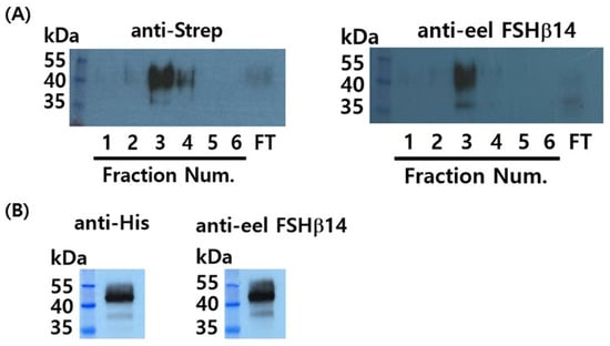
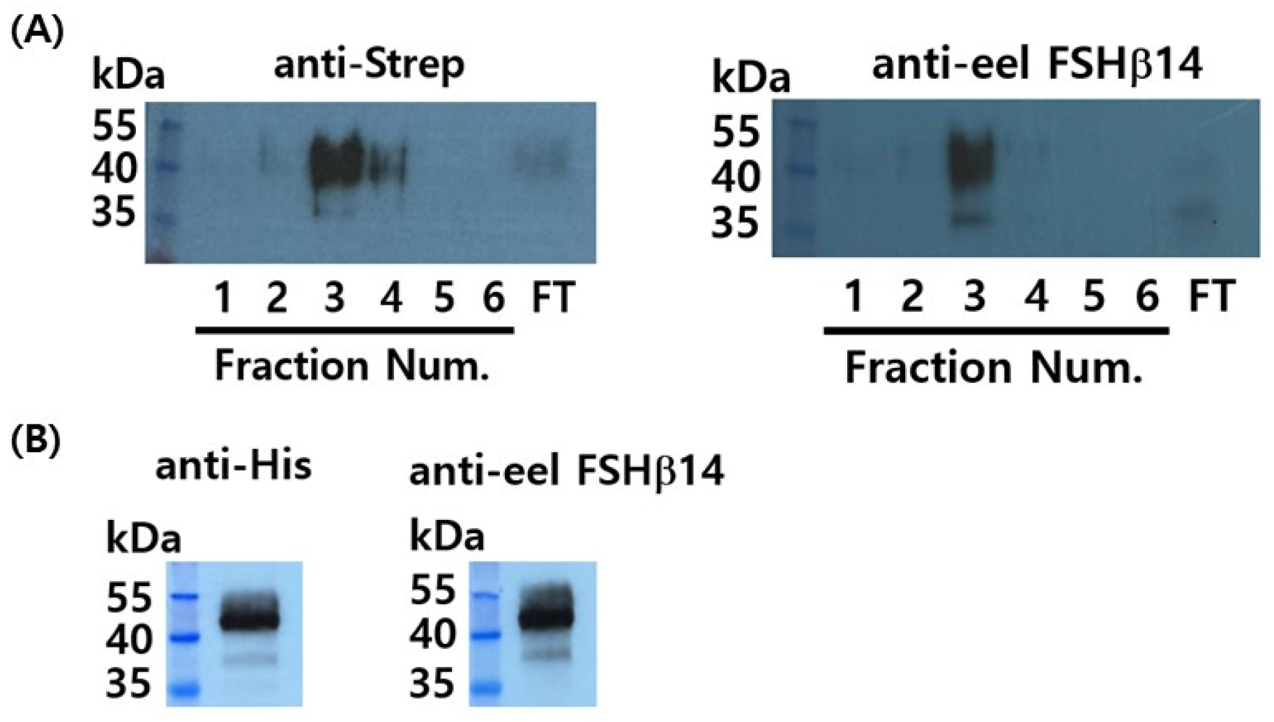
Recombinant eel FSH-eM-Strep and FSH-eM-His proteins were produced in transiently transfected CHO-K1 and CHO-S cells. Culture supernatants were collected 72 h after transfection from CHO-K1 cells and on day 9 from CHO-S cells. Recombinant proteins were purified using Strep-Tactin XT or HisPur Ni-NTA affinity chromatography. FSH-eM-Strep proteins were detected by both anti-Strep and anti-eel FSH β antibody (5A14), revealing broad immunoreactive bands between 48 and 53 kDa, with the strongest signal observed in fraction 3 (Figure 2A). Similarly, FSH-eM-His proteins were detected using anti-His and anti-eel FSH β antibodies (Figure 2B). These findings demonstrate that both recombinant eel FSH-eM-Strep and FSH-eM-His proteins were efficiently expressed and successfully purified by affinity chromatography.
Figure 2.
Production and purification of recombinant eel FSH-eM-Strep and FSH-eM-His proteins from CHO-S cells. Culture supernatants were collected on day 9 post-transfection. Recombinant eel FSH-eM-Strep and FSH-eM-His proteins were purified using Strep-Tactin XT 4 Flow columns and HisPur Ni-NTA spin columns, respectively. Aliquots (5 μL) of the purified proteins were separated by sodium dodecyl sulfate-polyacrylamide gel electrophoresis (SDS-PAGE), followed by transfer onto a PVDF membrane for Western blot analysis. (A) FSH-eM-Strep proteins were detected using a polyclonal anti-Strep antibody followed by a horseradish peroxidase (HRP)-conjugated goat anti-rabbit IgG secondary antibody (left panel). The same samples were also analyzed using a monoclonal anti-eel FSH antibody (eFSH5A14) followed by an HRP-conjugated goat anti-mouse IgG secondary antibody (right panel). (B) FSH-eM-His proteins were purified using a Ni-NTA affinity column and detected by Western blot analysis. FT: flow-through sample.
3.2. Deglycosylation of Recombinant Eel FSH-eM-Strep and FSH-eM-His Proteins
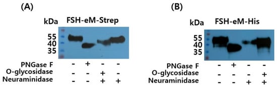
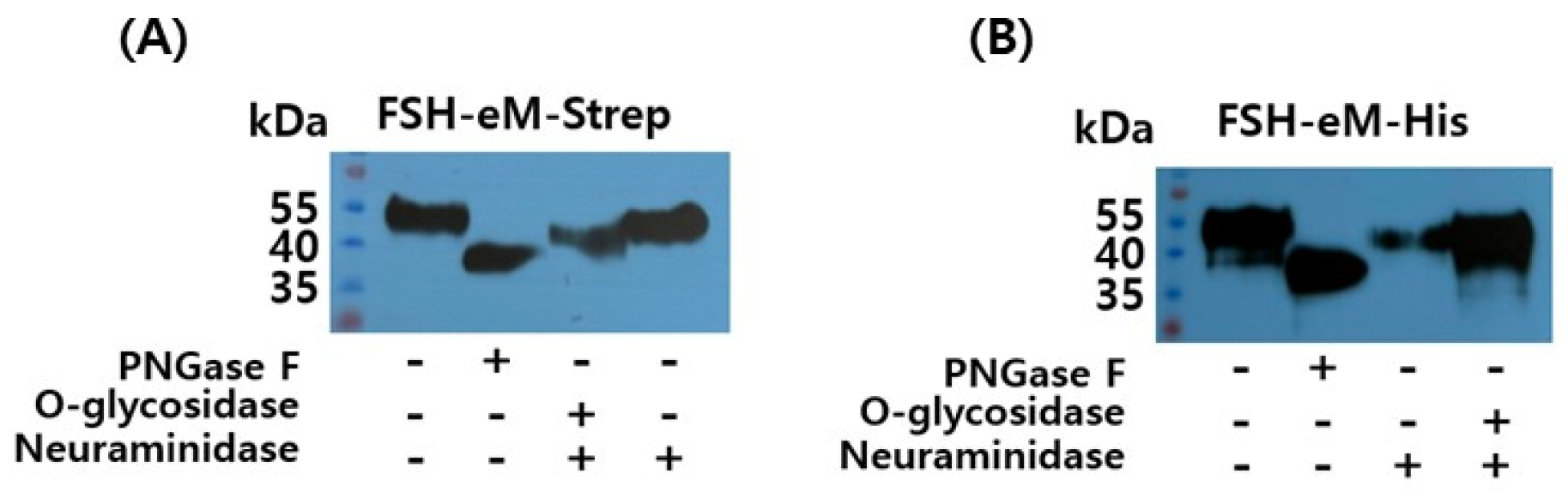
The molecular weights of rec-FSH-eM-Strep and rec-FSH-eM-His were reduced to approximately 38 kDa following PNGase F digestion (Figure 3A,B). In contrast, O-glycosidase treatment resulted in a smaller decrease in molecular weight, yielding proteins of approximately 41 kDa, indicating that O-linked glycans contributed less to the molecular weight than N-linked glycans. Neuraminidase treatment reduced the molecular weight to approximately 42 kDa, reflecting the removal of terminal sialic acid residues. These results indicate that N-linked glycans contributed more than 10 kDa to the molecular weight of the recombinant proteins and represented the major glycosylation component. Furthermore, the reduction in molecular weight following neuraminidase treatment demonstrated the presence of terminal sialic acid residues on the recombinant proteins.
Figure 3.
Deglycosylation analysis of recombinant eel FSH-eM-Strep and FSH-eM-His proteins. Recombinant proteins were treated with the peptide N-glycosidase F (PNGase F) to remove N-linked oligosaccharides and analyzed by Western blotting. PNGase F treatment reduced the apparent molecular weights by approximately 10–15 kDa. Subsequent digestion with O-glycosidase and neuraminidase yielded an apparent molecular weight of approximately 41 kDa, whereas neuraminidase treatment alone, which removes α2-3-, α2-6-, and α2-8-linked sialic acid residues, resulted in an apparent molecular weight of approximately 42 kDa. (A) FSH-eM-Strep proteins. (B) FSH-eM-His proteins.
3.3. Isolation of Single Cells and Western Blotting for Eel FSH-GGSA-eM-Strep Proteins
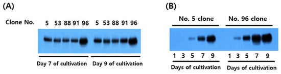
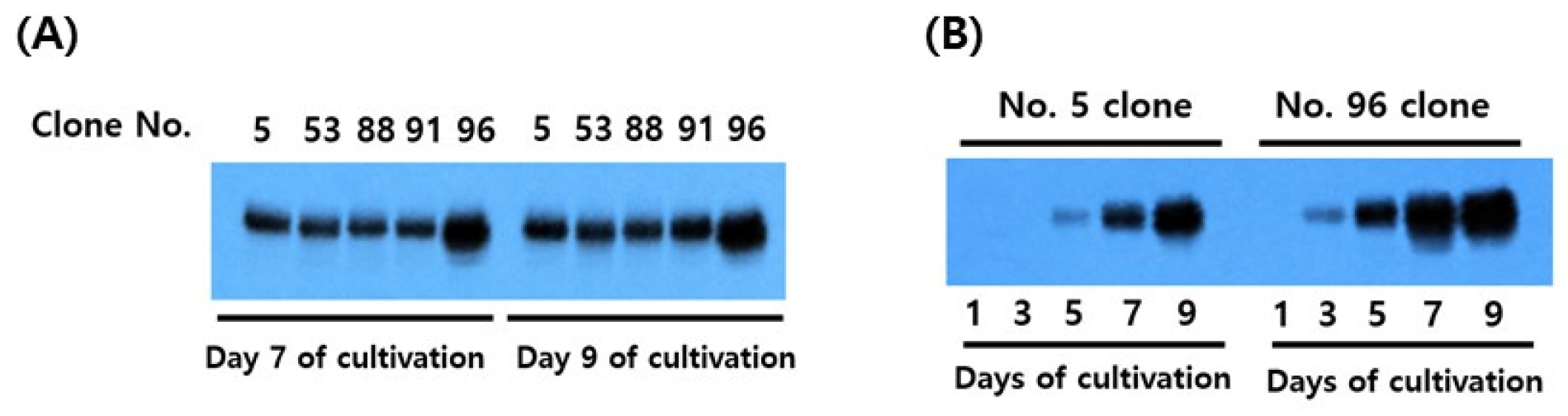
To enhance O-linked glycosylation, a construct containing a 15-amino-acid [(GGSA)3GGG] linker inserted between the FSH β-subunit and the eCTP region was generated, and stable single-cell clones were subsequently isolated. The transfected CHO DG44 cells were subjected to stepwise MTX amplification at increasing concentrations (500 nM, 2 µM, and 4 µM). The MTX-selected cells were cloned by limiting dilution in complete cloning medium in 96-well plates. A total of 108 single-cell-derived clones were obtained, from which five clones were selected for further analysis (Figure 4A). Based on Western blot analysis, clones #5 and #96 exhibited the strongest protein expression and were selected for subsequent experiments (Figure 4B). Detectable protein expression by Western blot in clone #5 was first observed on day 5 of cultivation, whereas clone #96 showed detectable expression on day 3, indicating an earlier onset of recombinant protein production. Western blot analysis further showed that clone #96 exhibited slightly higher recombinant protein expression than clone #5.
Figure 4.
Isolation of single cells expressing rec-eel FSH-GGSA-eM-Strep proteins. A total of five stable cell lines were finally selected from 108 clones. To evaluate recombinant protein expression, 20 µL of culture supernatant on days 7 and 9 was first analyzed by Western blotting (A). Among the five cell lines, clones #5 and #96 exhibited the highest expression levels. The secretion profiles of these two clones were further examined at different culture times (B). A faint protein band was first detected on day 5 in clone #5, whereas a weak band was already detectable on day 3 in clone #96. The highest protein expression was observed in both cell lines on day 9 of culture. A Strep-tag-specific antibody was used for the Western blot analysis.
3.4. Purification and Deglycosylation of Recombinant Eel FSH-GGSA-eM-Strep Protein
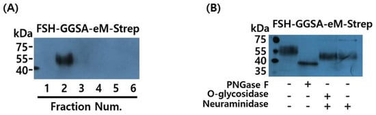
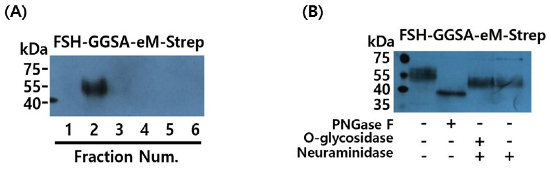
To evaluate the feasibility of scale-up production, rec-eel FSH-GGSA-eM-Strep was produced at an approximate culture volume of 5 L using five 1 L spinner flasks, with a final production yield of 10–12 mg/L. Following purification, approximately 6.5 mg/L of recombinant protein was recovered, corresponding to a recovery efficiency of 60%. Rec-eel FSH-GGSA-eM-Strep was successfully purified using a Strep-Tactin XT High-Capacity column. The majority of rec-eel FSH-GGSA-eM-Strep was recovered in fraction 2, whereas only negligible amounts were detected in the other fractions (Figure 5A). Western blot analysis of rec-eel FSH-GGSA-eM-Strep showed an increased apparent molecular weight with additional glycosylation of approximately 52–56 kDa (Figure 5B). Following PNGase F treatment, the molecular weight was reduced to approximately 38 kDa, indicating that N-linked glycans contributed approximately 14–18 kDa to the molecular weight of the recombinant protein. In contrast, O-glycosidase and neuraminidase treatments resulted in a reduction in the molecular weight to approximately 42 kDa, indicating contributions of O-linked glycans and terminal sialic acid residues to the observed molecular weight (Figure 5B). These results indicated that N-linked glycans represented the major glycosylation component of rec-eel FSH-GGSA-eM-Strep, whereas O-linked glycans and terminal sialic acid residues contributed to a lesser extent.
Figure 5.
Purification and deglycosylation of rec-eel FSH-GGSA-eM-Strep. (A) Recombinant protein was purified from 1 L of culture supernatant. Eluted fractions (5 mL each) were collected, and an aliquot (1 μL) of each fraction was subjected to SDS-PAGE and subsequently analyzed by Western blotting using an anti-Strep antibody to identify the recombinant eel FSH-GGSA-eM-Strep protein. A strong signal corresponding to eel FSH-GGSA-eM-Strep was detected only in fraction 2. (B) PNGase F treatment, which removes N-linked oligosaccharides, markedly decreased the molecular weight of both recombinant proteins to approximately 38 kDa. In contrast, digestion with O-glycosidase and neuraminidase resulted in similar molecular weights of approximately 42 kDa for both samples.
The molecular weight of FSH-eM-Strep/His proteins lacking the GGSA linker decreased to approximately 42 kDa following treatment with neuraminidase alone, whereas combined treatment with O-glycosidase and neuraminidase resulted in a further reduction of approximately 1 kDa (Figure 3A,B). These findings suggest that FSH-eM-Strep/His proteins undergo O-linked glycosylation, although the extent of this modification appears to be relatively limited. For the FSH-GGSA-eM-Strep protein containing the GGSA linker, it was difficult to accurately determine the extent of the molecular weight reduction based solely on the Western blot results obtained in the present study. Nevertheless, treatment with neuraminidase alone and combined treatment with O-glycosidase and neuraminidase resulted in similar apparent molecular weights of approximately 42 kDa, with no substantial difference observed between the two treatments (Figure 5B). These findings suggest that O-linked glycosylation may be minimal in the FSH-GGSA-eM-Strep protein.
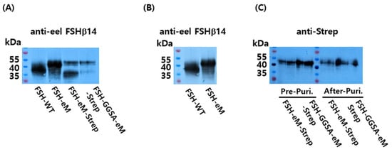
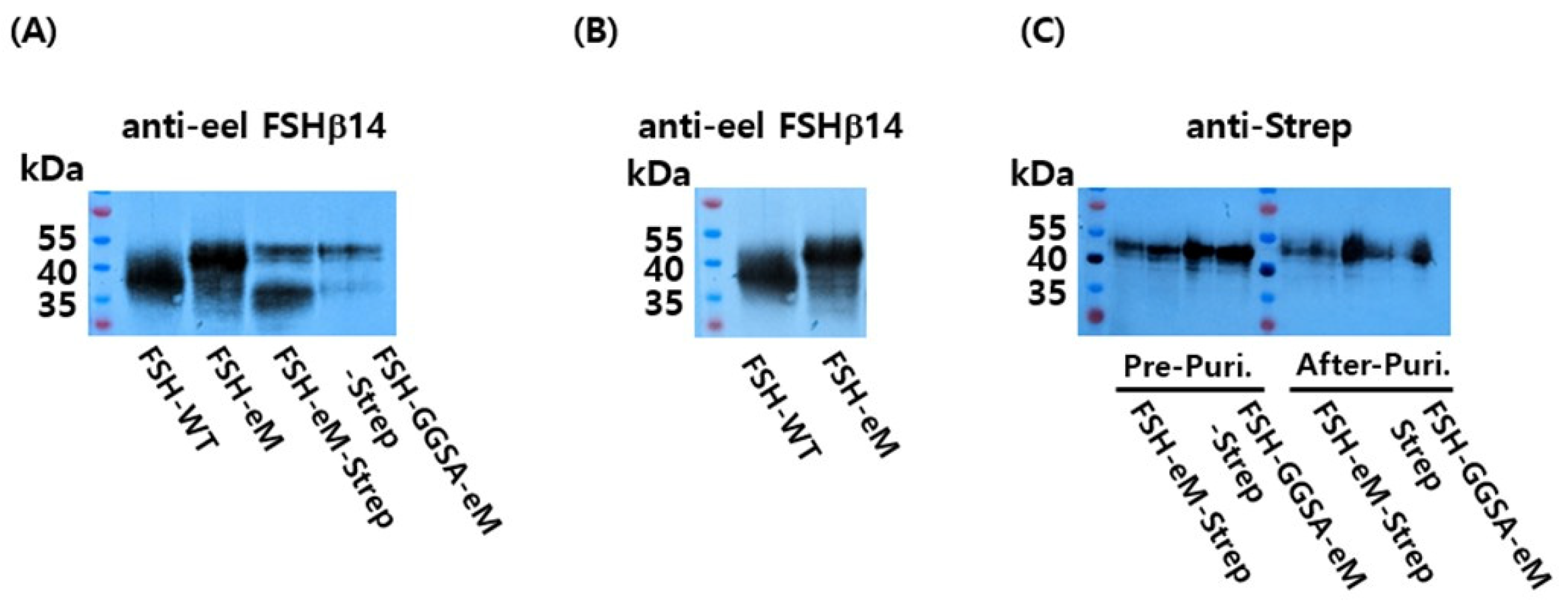
Western blot analysis of FSH-WT, FSH-eM, FSH-eM-Strep, and FSH-GGSA-eM-Strep was performed using the eel FSHβ5A14 antibody. Although equal amounts of protein were loaded, strong immunoreactive signals were detected for FSH-WT and FSH-eM, whereas relatively weaker bands were observed for FSH-eM-Strep and FSH-GGSA-eM-Strep (Figure 6A). Further comparison of FSH-WT and FSH-eM using the eel FSHβ5A14 antibody, which specifically recognizes the eel FSH β-subunit, revealed a substantial increase in the apparent molecular weight of FSH-eM compared with FSH-WT (Figure 6B). In addition, Western blot analysis of FSH-eM-Strep and FSH-GGSA-eM-Strep before and after purification, using an anti-Strep antibody, showed a slight increase in the apparent molecular weight of FSH-GGSA-eM-Strep relative to FSH-eM-Strep (Figure 6C). Repeated Western blot analyses of the four recombinant FSH variants using the same antibody consistently revealed differences in immunoreactive signal intensity. These findings suggest that the incorporation of the GGSA linker and Strep-tag may induce conformational changes or alter epitope accessibility, thereby affecting antibody recognition and binding efficiency.
Figure 6.
Western blot analysis of recombinant eel FSH variants. (A) Recombinant eel FSH variants, including FSH-WT, FSH-eM, FSH-eM-Strep, and FSH-GGSA-eM-Strep, were subjected to SDS-PAGE followed by Western blot analysis using the eel FSHβ5A14 antibody, which specifically recognizes the eel FSH β-subunit. (B) Western blot analysis of FSH-WT and FSH-eM using the eel FSHβ5A14 antibody revealed a marked increase in the apparent molecular weight of FSH-eM compared with FSH-WT. (C) Western blot analysis of FSH-eM-Strep and FSH-GGSA-eM-Strep recombinant proteins before and after purification using an anti-Strep antibody. Although equal amounts of the four recombinant FSH variants were loaded, comparative Western blot analysis revealed relatively weaker bands for the Strep-tagged constructs, FSH-eM-Strep and FSH-GGSA-eM-Strep, suggesting reduced recognition of these two recombinant proteins by the FSH β-subunit-specific antibody.
3.5. Silver Staining Results
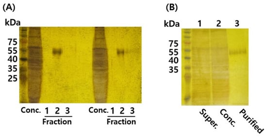
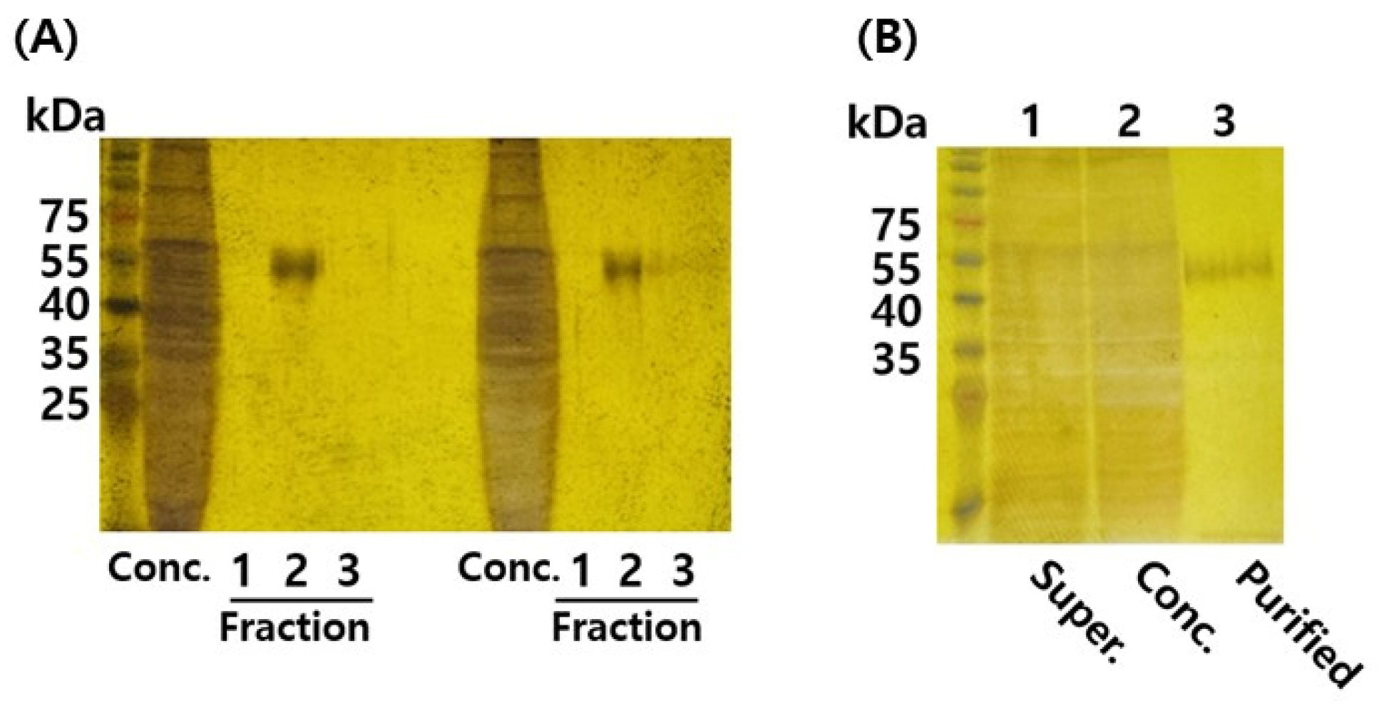
Silver staining analysis was performed to evaluate the recombinant protein expression and purification efficiency at different stages, including the culture supernatant, concentrated samples, and final purified products. A total of 20 µL of the culture supernatant from a 1 L-scale cultivation, 5 µL of the 10-fold concentrated sample, and 8 µL of the purified sample were loaded for SDS-PAGE and silver staining analysis. A host cell-derived band slightly above 55 kDa was detected in both the culture supernatant and concentrated samples. However, this band was not observed after purification, and a distinct recombinant protein band was detected at a slightly lower molecular weight position (Figure 7A). These results indicate that most nonspecific proteins were removed during purification, resulting in enrichment of the target recombinant protein. After purification using the Strep-Tactin XT column, fractions #1–#3 were further analyzed and compared with the concentrated sample prior to purification. Although the concentrated sample showed several nonspecific bands, fraction #2 after purification exhibited a strong and specific band corresponding to the recombinant protein, whereas other contaminating proteins were largely removed (Figure 7B). These results demonstrated the efficient purification of the recombinant protein using the Strep-Tactin XT column.
Figure 7.
Silver staining results of rec-eel FSH-GGSA-eM-Strep proteins. (A) Silver staining analysis of the 10-fold concentrated culture supernatant before purification and the samples purified using a Strep-Tactin XT High-Capacity resin column. A total of 5 μL of the concentrated sample and 8 μL of each purified fraction were loaded onto the gel. A strong protein signal was detected in fraction 2. (B) Comparison of the culture supernatant, concentrated sample, and purified protein by silver stain. The culture supernatant (20 μL), concentrated sample (5 μL), and purified sample (8 μL) were loaded onto an SDS-PAGE gel. Bands slightly above 55 kDa were detected in both the culture supernatant and concentrated sample, whereas the purified sample showed a band with a slightly lower molecular weight.
3.6. Biological and pERK1/2 Activities of Rec-Eel FSH-GGSA-eM-Strep Proteins
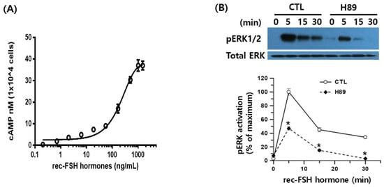
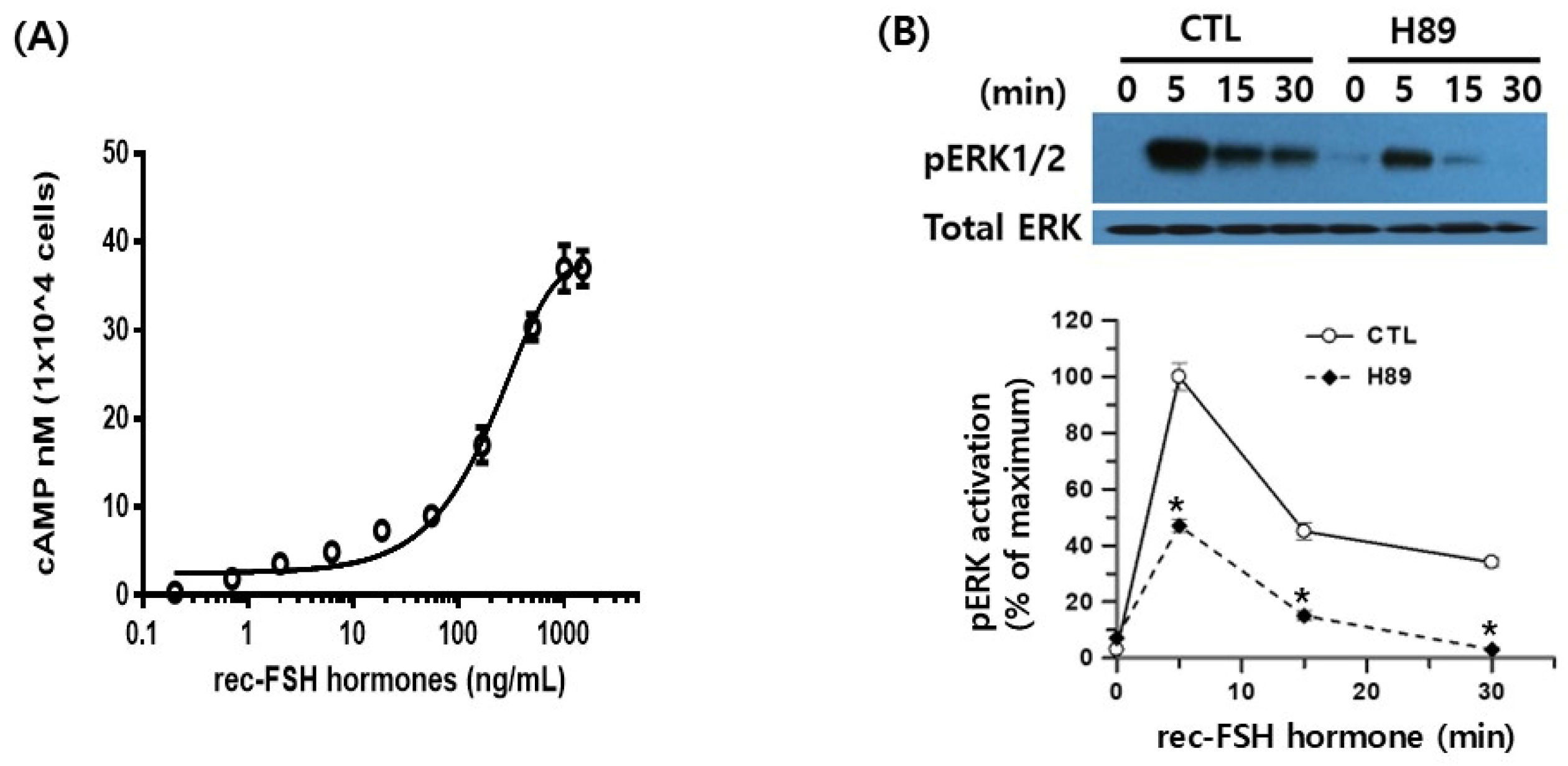
In vitro biological activity was evaluated using CHO-K1 cells transiently expressing the eel FSH receptor, as described previously [5]. Rec-eel FSH-GGSA-eM-Strep stimulated cAMP accumulation in a dose-dependent manner, as shown in Figure 8A. Rec-eel FSH-GGSA-eM-Strep exhibited an EC50 of 206.1 ng/mL, with a 95% confidence interval of 173.4–254.0 ng/mL, and a maximal response (Rmax) of 37.5 ± 0.9 nM/104 cells, based on at least three independent experiments (Table 1). We also examined ERK1/2 phosphorylation by Western blot analysis (Figure 8B). Following stimulation with rec-eel FSH-GGSA-eM-Strep, phosphorylated ERK1/2 (pERK1/2) reached its maximum level at 5 min and then rapidly declined by 30 min. However, pERK1/2 activation at 5 min was markedly inhibited by pretreatment with a PKA inhibitor. Only a faint pERK1/2 signal was detected at 15 min.
Figure 8.
Biological and pERK1/2 activities in cells expressing eel follicle-stimulating hormone receptor. (A) CHO-K1 cells transfected with eel FSH receptor (eel FSHR) were seeded into 384-well plates (10,000 cells per well) at 24 h post-transfection. The cells were incubated with rec-eel FSH-GGSA-eM-Strep for 30 min at room temperature. Intracellular cAMP production was measured using a homogeneous time-resolved fluorescence (HTRF) assay. Each data point represents the mean ± standard error of the mean (SEM) from two independent experiments, each performed in triplicate (n = 2). The mean values were fitted to sigmoidal concentration–response curves using a four-parameter logistic regression model to determine the half-maximal effective concentration (EC50) and maximal response (Emax). (B) Time-dependent pERK1/2 activation mediated by eel FSHR. HEK293 cells were transiently transfected with eel FSH receptor and serum-starved for 4–6 h before stimulation with rec-eel FSH-GGSA-eM-Strep agonists (400 ng/mL) for the indicated time periods. For PKA inhibition experiments, cells were pretreated with the PKA inhibitor H89 (20 μM) or DMSO (vehicle control, CTL) for 30 min prior to agonist stimulation. Whole-cell lysates were analyzed for phosphorylated ERK1/2 (pERK1/2) and total ERK levels. Twenty micrograms of protein was loaded per lane and subjected to Western blot analysis. The intensities of pERK1/2 and total ERK bands were quantified by densitometry, and pERK1/2 levels were normalized to total ERK expression. pERK1/2 was analyzed at 0, 5, 15, and 30 min following agonist stimulation. pERK1/2 levels were quantified by densitometric analysis using Image Lab software (Bio-Rad). The maximal pERK1/2 signal (typically at 5 min) was normalized to 100%. Representative Western blot images are shown, and the quantitative data are presented as the mean ± SEM from three independent experiments. * Statistical significance was determined by one-way ANOVA followed by Tukey’s post-hoc test (p < 0.05).
Treatment with the PKA inhibitor H89 markedly reduced ERK1/2 phosphorylation at the early time point (5 min), with pERK1/2 levels decreasing by approximately 58% compared with the control (CTL), corresponding to 42% of the control level. At 15 min, pERK1/2 levels in the CTL group decreased to approximately 45% of the initial response, whereas H89 treatment further reduced pERK1/2 levels to approximately 17%. By 30 min, pERK1/2 phosphorylation was almost undetectable in the H89-treated group. These results suggest that cAMP signaling and the ERK1/2 pathway are involved in eel FSH receptor-mediated signaling.
Table 1.
Bioactivity of rec-eel FSH-GGSA-eM protein in cells expressing eel FSH receptor.
4. Discussion
In the present study, we successfully engineered an eel FSH variant containing the eCG β-subunit CTP and a flexible GGSA linker, enabling efficient production of biologically active recombinant hormone in CHO DG44 cells. Deglycosylation analysis confirmed the presence of both N-linked and O-linked glycans in recombinant eel FSH containing eCTP, although their detailed compositions and structures were not characterized in this study. Functional assays further demonstrated receptor-mediated cAMP production in CHO-K1 cells and ERK1/2 activation in HEK293 cells.
The molecular weight of dimeric eel FSHα/β purified from the eel pituitary has been reported to be approximately 34–40 kDa [18]. Similarly, recombinant eel FSH consisting of a single-chain structure in which the β-subunit is linked to the α-subunit showed a comparable molecular weight [2]. However, the insertion of a 34-amino acid eCTP linker increased the molecular weight to approximately 39–46 kDa, representing an additional increase of 5–6 kDa [14]. In the present study, the molecular weight of eM-Strep was estimated to be approximately 48–53 kDa, indicating additional glycan modifications of approximately 13–14 kDa. Furthermore, GGSA-eM-Strep exhibited a higher molecular weight of approximately 52–56 kDa, corresponding to an additional 17–18 kDa of carbohydrate modification. A recent study employing a highly similar fusion model also demonstrated a broad molecular weight distribution ranging from approximately 30 to 49 kDa [13]. Our results suggest that the simultaneous incorporation of the [(GGSA)3GGG] linker and eCTP may facilitate O-linked glycosylation within the eCTP region, thereby influencing glycosylation-associated post-translational modification of recombinant eel FSH. Table 2 summarizes the constructs used in the present study, including the newly designed models and previously established constructs. Based on the structural characteristics of each construct, the corresponding molecular weights are also presented.
Table 2.
Structural features and design purpose of eel FSH variants.
The substantial reduction in molecular weight (approximately 14–15 kDa) following PNGase F digestion demonstrates that the four predicted N-linked glycosylation sites in GGSA-eM-Strep were extensively occupied by N-glycans. Moreover, the further decrease observed after O-Glycosidase and Neuraminidase treatment (approximately 10–14 kDa) indicates that O-linked glycosylation and terminal sialylation contribute significantly to the molecular heterogeneity of the recombinant protein. Our results were consistent with those of previous studies, suggesting that hCTP and eCTP linker attachments in hFSH [19], hTSH [20], hGH [21], and eel FSH and LH [14,22] remarkably increase the apparent molecular weights of recombinant glycoproteins.
Although the contribution of the GGSA linker to O-linked glycosylation appears to be minimal in the present study, the reduction in apparent molecular weight following neuraminidase treatment alone may be attributed to the removal of terminal sialic acid residues. Therefore, we cannot definitively conclude that O-linked glycosylation is negligible or absent based solely on these results. Our results are compatible with, but do not establish, a contribution of the linker to glycan modification. These findings suggest that the combined incorporation of eCTP and the [(GGSA)3GGG] linker may influence O-linked glycosylation and glycosylation-associated molecular heterogeneity in recombinant eel FSH. However, since precise molecular weight determination and detailed glycan structural analyses were not performed in the present study, we believe that it would be premature to draw a definitive conclusion regarding the extent of O-linked glycosylation. Therefore, further systematic investigations, including accurate molecular mass determination and comprehensive glycan structural characterization, are necessary to elucidate the glycosylation patterns of this recombinant protein.
Recent comprehensive glycan profiling studies of recombinant hCG [23] and eCG [24] produced in CHO DG44 cells demonstrated that the attached glycan structures were highly comparable to those of native hormones. These observations suggest that CHO DG44 cells possess the cellular machinery required for accurate glycan processing and maturation of complex glycoproteins. Therefore, the glycosylation characteristics observed in recombinant eel FSH-GGSA-eM-Strep, together with its demonstrated biological activity, further support the suitability of the CHO DG44 expression system for producing biologically active recombinant glycoprotein hormones. Glycosylation may also influence important properties of these hormones, including their stability and receptor-mediated signaling activity.
In the present study, we demonstrated that the rec-eel FSH-GGSA-eM-Strep model exhibited a dose-dependent increase in cAMP signaling, suggesting that the FSH analog was capable of activating receptor-mediated signaling. However, the EC50 and Rmax values for FSH-wt and FSH-eM-Strep reported in the present study cannot be directly compared at this stage because these values were not obtained through parallel experiments using the same quantitative analytical procedures under identical experimental conditions. Therefore, we acknowledge that the lack of appropriate parallel controls represents a limitation of the present study. In particular, it would be premature to draw definitive conclusions regarding differences in receptor-mediated signaling activity or biological potency among these recombinant FSH analogs based solely on the currently available data. These results are consistent with previous studies using recombinant human FSH [25] and eel FSH [22], which demonstrated that alterations in glycosylation patterns can significantly influence the biological activity, receptor interaction, and intracellular signaling potency of glycoprotein hormones. Collectively, these findings suggest that optimization of glycosylation structures represents an effective strategy for improving the functional properties of recombinant gonadotropins by enhancing their stability, receptor binding efficiency, and downstream cAMP/PKA signaling activation.
ERK1/2 phosphorylation occurs through the sequential activation of the Raf1–MEK1–ERK1/2 signaling cascade [26,27]. The influence of cAMP on ERK1/2 activation is highly context-dependent and differs among cell types [28]. In FSHR-mediated signaling, ERK activation has been reported to involve the coordinated contribution of both G protein-dependent and β-arrestin-dependent pathways, each displaying distinct temporal and spatial signaling profiles [29,30,31]. In the present study, ERK1/2 phosphorylation reached its maximum level within 5 min following stimulation with recombinant eel FSH and subsequently declined over time. This temporal pattern is consistent with previous studies of human FSH, in which ERK1/2 activation rapidly peaks at approximately 5–6 min before gradually decreasing [32].
Previous studies have demonstrated that structural alterations in FSHR can markedly influence downstream signaling. For example, substitution of six FSHR-specific amino acid residues within extracellular loop 2 (EL2) impairs receptor internalization and reduces cAMP signaling efficiency [33]. Likewise, mutations in extracellular loop 3 alter receptor internalization and attenuate ERK1/2 phosphorylation without significantly affecting cAMP production [34]. The robust increase in ERK1/2 phosphorylation was attenuated by approximately 60% following treatment with the PKA inhibitor H89, indicating that PKA activity contributes substantially to FSHR-mediated ERK1/2 activation under the experimental conditions used. These findings are in good agreement with previous reports demonstrating that FSHR-mediated ERK1/2 activation is regulated, at least in part, through a potential contribution of PKA signaling [30,35,36,37]. However, because FSHR-mediated ERK1/2 signaling has also been shown to involve β-arrestin-dependent pathways, further studies using β-arrestin 1- and β-arrestin 2-knockout cells will be necessary to clarify the contribution of β-arrestin-mediated signaling to ERK1/2 activation. These findings indicate that distinct structural domains of FSHR differentially regulate receptor trafficking and downstream signaling pathways, particularly ERK1/2 activation. Collectively, our results further support the concept that rapid and transient ERK1/2 phosphorylation is a conserved feature of FSHR signaling and suggest that the recombinant eel FSH produced in this study effectively preserves the signaling properties of the native hormone. The combined incorporation of a flexible [(GGSA)3GGG] linker and the eCTP may represent a potential glycoengineering strategy for recombinant eel FSH, supporting the production of proteins containing both N- and O-linked glycans while retaining receptor-mediated cAMP production in CHO-K1 cells and ERK1/2 activation in HEK293 cells.
5. Conclusions
In this study, we successfully developed recombinant eel FSH incorporating the eCG β-subunit CTP and a flexible (GGSA)3GGG linker using a CHO DG44 expression system. Deglycosylation analyses confirmed the presence of both N-linked and O-linked glycans, suggesting that the combined incorporation of eCTP and the flexible linker may influence glycosylation-associated post-translational modifications. Stable CHO DG44 clones enabled production and purification of biologically active recombinant eel FSH. Functional analyses demonstrated that the recombinant hormone retained its ability to stimulate receptor-mediated cAMP production and rapid ERK1/2 activation, with the latter occurring predominantly through a potential contribution of PKA signaling. The production of biologically active recombinant eel FSH provides a valuable tool for investigating reproductive endocrinology in teleost fish and may facilitate future applications in aquaculture, particularly in studies aimed at inducing and synchronizing gonadal maturation. Although EC50 and Rmax values were obtained for FSH-wt and FSH-eM-Strep, differences in the experimental and quantitative analytical conditions preclude a meaningful direct comparison of these parameters at present. Further studies involving parallel comparisons under standardized experimental conditions will be necessary to accurately evaluate their relative biological activities and determine whether the incorporation of the eCTP and GGSA linker influences receptor-mediated signaling efficiency. Taken together, these findings suggest that the combined incorporation of eCTP and the flexible [(GGSA)3GGG] linker represents a potential strategy for producing biologically active recombinant eel FSH containing both N-linked and O-linked glycans in CHO DG44 cells.
Author Contributions
Investigation, Formal Analysis, and Data Curation, M.B.; Data Curation, S.K. and H.K.; Formula Analysis, J.W.L. and G.Y.; Methodology and Visualization, M.-H.P.; Writing—Review and Editing, M.-H.K.; Conceptualization, Writing—Review and Editing, K.-S.M.; Funding Acquisition, H.W.K., M.G.S., Y.D. and K.-M.W. All authors have read and agreed to the published version of the manuscript.
Funding
This study was supported by a research grant from the National Institute of Fisheries Science (R2026023) and the Korean Research Foundation Program (Grant Number: RS202400335122) of the Republic of Korea.
Institutional Review Board Statement
Not applicable.
Informed Consent Statement
Not applicable.
Data Availability Statement
The original contributions presented in this study are included in the article. Further inquiries can be directed to the corresponding author.
Conflicts of Interest
M.-H.P. was employed at TNT Research. All authors declare that this research was conducted in the absence of any commercial or financial relationships that could be construed as potential conflicts of interest. The company had no role in the design of the study; in the data collection or interpretation of data; in the writing of the manuscript; or in the decision to publish the results.
References
- Pierce, J.C.; Parsons, T.F. Glycoprotein hormones: Structure and function. Annu. Rev. Biochem. 1981, 50, 465–495. [Google Scholar] [CrossRef] [Scilit] [PubMed]
- Kim, J.M.; Munkhuu, O.; Bambaragchaa, M.; Lee, B.I.; Kim, S.K.; Kang, M.H.; Kim, D.J.; Min, K.S. Site-specific roles of N-linked oligosaccharides in recombinant eel follicle-stimulating hormone for secretion and signal transduction. Gen. Comp. Endocrinol. 2019, 276, 37–44. [Google Scholar] [CrossRef] [Scilit] [PubMed]
- Min, K.S.; Park, J.J.; Byambaragchaa, M.; Kang, M.H. Characterization of tethered equine chorionic gonadotropin and its deglycosylated mutants by ovulation stimulation in mice. BMC Biotechnol. 2019, 19, 60. [Google Scholar] [CrossRef] [Scilit] [PubMed]
- Min, K.S.; Park, J.J.; Lee, S.Y.; Byambragchaa, M.; Kang, M.H. Comparative gene expression profiling of mouse ovaries upon stimulation with natural equine chorionic gonadotropin (N-eCG) and tethered recombinant-eCG (R-eCG). BMC Biotechnol. 2020, 20, 59. [Google Scholar] [CrossRef] [Scilit] [PubMed]
- Lee, S.Y.; Byambaragchaa, M.; Kang, H.J.; Choi, S.H.; Kang, M.H.; Min, K.S. Specific roles of N- and O-linked oligosaccharide sites on biological activity of equine chorionic gonadotropin (eCG) in cells expressing rat luteinizing hormone/chorionic gonadotropin receptor (LH/CGR) and follicle-stimulating hormone receptor (FSHR). BMC Biotechnol. 2021, 21, 52. [Google Scholar] [PubMed]
- Min, K.S.; Hiyama, T.; Seong, H.W.; Hattori, N.; Tanaka, S.; Shiota, K. Biological activities of tethered equine chorionic gonadotropin (eCG) and its deglycosylated mutants. J. Reprod. Dev. 2004, 50, 297–304. [Google Scholar] [CrossRef] [Scilit] [PubMed]
- Byambaragchaa, M.; Kim, D.J.; Kang, M.H.; Min, K.S. Site specificity of eel luteinizing hormone N-linked oligosaccharides in signal transduction. Gen. Comp. Endocrinol. 2018, 268, 50–56. [Google Scholar] [CrossRef] [Scilit] [PubMed]
- Joshi, H.J.; Narimatsu, Y.; Schjoldager, K.T.; Tytgat, H.L.P.; Aebi, M.; Clausen, H.; Halim, A. Snapshot: O-glycosylation pathways across Kingdoms. Cell 2018, 172, 632. [Google Scholar] [CrossRef] [Scilit] [PubMed]
- Magalhaes, A.; Duarte, H.O.; Reis, C.A. The role of O-glycosylation in human disease. Mol. Asp. Med. 2021, 79, 100946. [Google Scholar] [CrossRef] [Scilit] [PubMed]
- Fares, F. The role of O-linked and N-linked oligosaccharides on the structure-function of glycoprotein hormones: Development of agonists and antagonists. Biochim. Biophys. Acta 2006, 1760, 560–567. [Google Scholar] [CrossRef] [Scilit] [PubMed]
- Kazeto, Y.; Suzuki, H.; Ozaki, Y.; Gen, K. C-terminal peptide (hCTP) of human chorionic gonadotropin enhances in vivo biological activity of recombinant Japanese eel follicle-stimulating hormone and luteinizing hormone produced in FreeStyle 293-F cell lines. Gen. Comp. Endocrinol. 2021, 306, 113731. [Google Scholar] [CrossRef] [Scilit] [PubMed]
- Jazayeri, S.H.; Amiri-Yekta, A.; Gourabi, H.; Emami, B.A.; Halfinezhad, Z.; Abolghasemi, S.; Fatemi, N.; Daneshipour, A.; Ghahremani, M.H.; Sanati, M.H.; et al. Comparative assessment on the expression level of recombinant human follicle-stimulating hormone (FSH) in serum-containing versus protein-free culture media. Mol. Biotechnol. 2017, 59, 490–498. [Google Scholar] [CrossRef] [Scilit] [PubMed]
- Kazeto, Y.; Ito, R.; Tanaka, T.; Zuzuki, H.; Ozaki, Y.; Okuzawa, K.; Gen, K. Establishment of cell-lines stably expressing recombinant Japanese eel follicle-stimulating hormone and luteinizing hormone using CHO-DG44 cells: Fully induced ovarian development at different modes. Front. Endocrinol. 2023, 14, 1201250. [Google Scholar] [CrossRef] [Scilit] [PubMed]
- Byambaragchaa, M.; Park, S.H.; Kim, S.G.; Shin, M.G.; Kim, S.K.; Park, M.H.; Kang, M.H.; Min, K.S. Stable production of a recombinant single-chain eel follicle-stimulating hormone analog in CHODG44 cells. Int. J. Mol. Sci. 2024, 25, 7282. [Google Scholar] [CrossRef] [Scilit] [PubMed]
- Hershkovitz, O.; Bar-Ilan, A.; Guy, R.; Felikman, Y.; Moschcovich, L.; Hwa, V.; Rosenfeld, R.G.; Fima, E.; Hart, G. In vitro and in vivo characterization of MOD-4023, a long-acting carboxyl-terminal peptide (CTP)-modified human growth hormone. Mol. Pharm. 2016, 13, 631–639. [Google Scholar] [CrossRef] [Scilit] [PubMed]
- Strasburger, C.J.; Vanuga, P.; Payer, J.; Pfeifer, M.; Popovic, V.; Bajnok, L.; Goth, M.; Olsovska, V.; Trejbalova, L.; Vadasz, J.; et al. MOD-4023, a long-acting carboxyl-terminal peptide-modified human growth hormone: Results of a Phase 2 study in growth hormone-deficient adults. Eur. J. Endocrinol. 2017, 176, 283–294. [Google Scholar] [CrossRef] [Scilit] [PubMed]
- Zelinska, N.; Lotova, V.; Skorodok, J.; Maliesky, O.; Peterkova, V.; Samsonova, L.; Rosenfeld, R.G.; Zadik, Z.; Jaron-Mendelson, M.; Koren, R.; et al. Long-acting C-terminal peptide-modified hGH (MOD-4023): Results of a safety and dose-finding study in GHD children. J. Clin. Endocrinol. Metab. 2017, 102, 1578–1587. [Google Scholar] [CrossRef] [Scilit] [PubMed]
- Kamei, H.; Kawazoe, I.; Kanekko, T.; Aida, K. Purification of follicle-stimulating hormone from immature Japanese eel Anguilla japonica, and its biochemical properties and steroidogenic activities. Gen. Comp. Endocrinol. 2005, 143, 257–266. [Google Scholar] [CrossRef] [Scilit] [PubMed]
- Fares, F.; Suganuma, N.; Hishimori, K.; Lapolt, P.S.; Hsueh, A.J.W.; Boime, I. Design of a long-acting follitropin agonist by fusing the C-terminal sequence of the chorionic gonadotropin β subunit to the follitropin β subunit. Proc. Natl. Acad. Sci. USA 1992, 89, 4304–4308. [Google Scholar] [CrossRef] [Scilit] [PubMed]
- Fares, F.; Yamada, S.; Ben-Menahem, D.; Pixley, M.; Hsueh, A.J.W.; Boime, I. Conversion of thyrotropin heterodimer to a biologically active single-chain. Endocrinology 1998, 139, 2459–2468. [Google Scholar] [CrossRef] [Scilit]
- Fares, F.; Guy, R.; Bar-Ilan, A.; Felikman, Y.; Fima, E. Designing a long-acting human growth hormone (hGH) by fusing the carboxyl-terminal peptide of human chorionic gonadotropin β-subunit to the coding sequence of hGH. Endocrinology 2010, 151, 4410–4417. [Google Scholar] [CrossRef] [Scilit] [PubMed]
- Byambaragchaa, M.; Kim, S.G.; Park, S.H.; Shin, M.G.; Kim, S.K.; Kang, M.H.; Min, K.S. Production of recombinant single-chain eel luteinizing hormone and follicle-stimulating hormone analogs in Chinese hamster ovary suspension cell culture. Curr. Issues Mol. Biol. 2024, 46, 542–556. [Google Scholar] [CrossRef] [Scilit] [PubMed]
- Thennati, R.; Singh, S.K.; Nage, N.; Patel, Y.; Bose, S.K.; Burade, V.; Ranbhor, R.S. Analytical characterization of recombinant hCG and comparative studies with reference product. Biologics 2018, 12, 23–35. [Google Scholar] [CrossRef] [Scilit] [PubMed]
- Rodriguez, M.C.; Mussio, P.E.; Villarraza, J.; Tardivo, M.B.; Antuna, S.; Fontana, D.; Ceaglio, N.; Prieto, C. Physiochemical characterization of a recombinant eCG and comparative studies with PMSG commercial preparations. Protein J. 2023, 42, 24–36. [Google Scholar] [CrossRef] [Scilit] [PubMed]
- Sugahara, T.; Sato, A.; Kudo, M.; Ben-Menahem, D.; Pixley, M.R.; Hsueh, A.J.W.; Boime, I. Expression of biological active fusion genes encoding the common α subunit and the follicle-stimulating hormone β subunits. J. Biol. Chem. 1996, 271, 10445–10448. [Google Scholar] [CrossRef] [Scilit] [PubMed]
- Lefkowitz, R.J.; Shenoy, S.K. Transduction of receptor signals by β-arrestins. Science 2005, 308, 512–517. [Google Scholar] [PubMed]
- Miningou, N.; Blackwell, K.T. The road to ERK activation: Do neurons take alternate routes? Cell. Signal. 2020, 68, 109541. [Google Scholar] [CrossRef] [Scilit] [PubMed]
- Ascoli, M. Potential Leydig cell mitogenic signals generated by the wild-type and constitutively active mutants of the lutropin/choriogonadotropin receptor (LHR). Mol. Cell. Endocrinol. 2007, 260–262, 244–248. [Google Scholar] [CrossRef] [Scilit] [PubMed]
- Ahn, S.; Shenoy, K.K.; Wei, H.; Lefkowitz, R.J. Differential kinetic and spatial patterns of β-arrestin and G protein mediated ERK activation by the angiotensin II receptor. J. Biol. Chem. 2004, 279, 35518–35525. [Google Scholar] [CrossRef] [Scilit] [PubMed]
- Kara, E.; Crepieux, P.; Gauthier, C.; Martinat, N.; Piketty, V.; Guillou, F.; Reiter, E. A phosphorylation cluster of five serine and threonine residues in the C-terminus of the follicle-stimulating hormone receptor is important for desensitization but not for β-arrestin-mediated ERK activation. Mol. Endocrinol. 2006, 20, 3014–3026. [Google Scholar] [CrossRef] [Scilit] [PubMed]
- Reiter, E.; Lefkowitz, R.J. GRKs and β-arrestins: Roles in receptor silencing, trafficking and signaling. Trends Endocrinol. Metab. 2006, 17, 159–165. [Google Scholar] [CrossRef] [Scilit] [PubMed]
- Piketty, V.; Kara, E.; Guillou, F.; Reiter, E.; Crepiux, P. Follicle-stimulating hormone (FSH) activates extracellular signal-regulated kinase phosphorylation independently of beta-arrestin- and dynamin-mediated FSH receptor internalization. Reprod. Biol. Endocrinol. 2006, 4, 33. [Google Scholar] [CrossRef] [Scilit] [PubMed]
- Banerjee, A.A.; Dupakuntla, M.; Pathak, B.R.; Mahale, S.D. FSH receptor-specific residues L501 and I505 in extracellular loop 2 are essential for its function. J. Mol. Endocrinol. 2015, 54, 193–204. [Google Scholar] [CrossRef] [Scilit] [PubMed]
- Banerjee, A.A.; Mahale, S.D. Extracellular loop 3 substitutions K589N and A590S in FSH receptor increase FSH-induced receptor internalization and along with S588T substitution exhibit impaired ERK1/2 phosphorylation. Arch. Biochem. Biophys. 2018, 659, 57–65. [Google Scholar] [CrossRef] [Scilit] [PubMed]
- Shenoy, S.K.; Draka, M.K.; Nelson, C.D.; Houtz, D.A.; Xiao, K.; Madabushi, S.; Reiter, E.; Premont, R.T.; Lichtarge, O.; Lefkowitz, R.J. beta-arrestin-dependent, G protein-independent ERK1/2 activation by the beta2 adrenergic receptor. J. Biol. Chem. 2006, 281, 1261–1273. [Google Scholar] [CrossRef] [Scilit] [PubMed]
- Shenoy, S.K.; Barak, L.S.; Xiao, K.; Ahn, S.; Berthouze, M.; Shukla, A.K.; Luttrell, L.M.; Lefkowitz, R.J. Ubiquitination of β-arrestin links seven-transmembrane receptor endocytosis and ERK activation. J. Biol. Chem. 2007, 282, 29549–29562. [Google Scholar] [CrossRef] [Scilit] [PubMed]
- Gesty-Palmer, D.; Chen, M.; Reiter, E.; Ahn, S.; Nelson, C.D.; Wang, S.; Eckhardt, A.E.; Cowan, C.L.; Spurney, R.F.; Luttrell, L.M.; et al. Distinct β-arrestin- and G protein-dependent pathways for parathyroid hormone receptor-stimulated ERK1/2 activation. J. Biol. Chem. 2006, 281, 10856–10864. [Google Scholar] [CrossRef] [Scilit] [PubMed]

Disclaimer/Publisher’s Note: The statements, opinions and data contained in all publications are solely those of the individual author(s) and contributor(s) and not of MDPI and/or the editor(s). MDPI and/or the editor(s) disclaim responsibility for any injury to people or property resulting from any ideas, methods, instructions or products referred to in the content. |
© 2026 by the authors. Licensee MDPI, Basel, Switzerland. This article is an open access article distributed under the terms and conditions of the Creative Commons Attribution (CC BY) license.