


Abstract
MAP kinase-activated protein kinase 5 (MK5) plays an important role in cardiac fibroblast function. Although p38 MAPK and atypical MAPKs ERK3 and ERK4 have been identified as activators of MK5, the kinases that activate MK5 remain controversial. We examined the expression, subcellular distribution, and regulation of MK5 in cardiac ventricular myofibroblasts and myocytes. The copy numbers for MK5 and ERK4 mRNA were comparable in myocytes and myofibroblasts, whereas that of ERK3 was much higher in myofibroblasts. Interestingly, MK5 and ERK3 immunoreactivity was detected in myofibroblasts but not myocytes: treating myocytes with a proteasome inhibitor or hypertrophic agonists failed to rescue MK5 immunoreactivity. In myofibroblasts, MK5 and ERK3 immunoreactivity was predominantly nuclear and cytosolic, respectively. In serum-starved myofibroblasts, phosphothreonine-182 MK5 (pT182-MK5) immunoreactivity, representing phosphorylation of MK5 within its activation loop, was predominantly nuclear but increased in intensity and relocated to the cytoplasm in response to serum, sorbitol, angiotensin II, TGFβ, or H2O2: this was prevented upon inhibition of p38α/β. Phos-tag SDS-PAGE revealed multiple slower-migrating bands of MK5 immunoreactivity, indicating phosphorylation of MK5 at multiple sites. Phos-tag PAGE also revealed MK5 phosphorylation was increased with fibroblast activation and in hearts exposed to a chronic increase in afterload. These observations suggest that p38α and/or p38β are mediators of T182-MK5 phosphorylation in cardiac myofibroblasts.
1. Introduction
Mitogen-activated protein kinases (MAPKs) are protein serine/threonine kinases, expressed ubiquitously in eukaryotic cells including plants, fungi and animals [1,2], which regulate a wide range of cellular functions through phosphorylation of various substrates, including the MAPK-activated protein kinases (MKs) [3]. MAP kinase-activated protein kinase 5 (MK5) was initially identified and characterized as p38- or ERK2-regulated/activated kinase (PRAK or MAPKAPK5) [4,5]. Subsequent studies revealed atypical MAPKs ERK3 and ERK4 also activate MK5 [6,7,8]. The activation, substrates, and physiological role of MK5 remain poorly understood.
MK5 transcripts are in high abundance in the left ventricular myocardium of humans and mice [9,10]. Furthermore, both the left ventricle and ventricular myocytes of male MK5 haploinsufficient mice showed significantly less hypertrophy and a more restrictive filling pattern, relative to wild-type littermates, in response to a chronic increase in afterload induced by constriction of the transverse aorta (TAC) [11]. In addition, the TAC-induced increase in cardiac Col1a1 mRNA was significantly attenuated in MK5 haplodeficient mice relative MK5+/+ littermates and MK5 immunoreactivity was found to be in cardiac ventricular fibroblasts but not myocytes [11]. Following myocardial infarction induced by ligation of the left anterior descending coronary artery, scar size and collagen content were reduced in MK5-haploinsufficient mice compared with wild-type littermates [12]. Studies showed that MK5 deficiency or haplodeficiency altered the abundance of numerous transcripts for proteins involved in extracellular matrix homeostasis in cardiac ventricular myofibroblasts but not quiescent fibroblasts [13]. Reducing MK5 expression in cardiac myofibroblasts increased the abundance of Col1a1 mRNA and the secretion of type 1 collagen but attenuated the angiotensin II (Ang II)- and TGFβ-induced increase in type 1 collagen secretion and impaired myofibroblast migration, contraction, and proliferation [12,13]. Hence, MK5 plays an important role in mediating cardiac ventricular myofibroblast function.
The regulation of MK5 activity, mediated by phosphorylation at threonine-182 in the activation loop, remains controversial: p38α/β, ERK3, and ERK4 have been implicated [4,5,6,7,8]. In addition, PKA may activate MK5 via phosphorylation at serine-115 [14,15]. ERK3 immunoreactivity, but not that of ERK4 or p38α, was detected in MK5 immunoprecipitates from mouse heart [10,16]. In addition, as observed with MK5, the TAC-induced hypertrophy and increase in Col1a1 mRNA were attenuated in ERK3 haplodeficient mice [16]. In light of the role of MK5 in cardiac remodeling and its multiple possible mechanism(s) of activation, the present study was undertaken to examine the activation loop phosphorylation (T182) and subcellular localization of MK5 in adult cardiac ventricular myocytes and fibroblasts.
2. Materials and Methods
2.1. Materials
Medium 199, fungizone, and DNA stain 4′,6-diamidino-2-phenylindole (DAPI) were from Sigma-Aldrich (Oakville, ON, Canada), fetal bovine serum (FBS) and trypsin were from Gibco Laboratories (Life Technologies/Thermo Fisher Scientific, Mississauga, ON, Canada), and penicillin–streptomycin solution was from Multicell. All culture plates were from Sarstedt, Inc. (Montreal, QC, Canada). PCR Primers were from Invitrogen Canada Inc. (Burlington, ON, Canada). SDS-polyacrylamide gel electrophoresis reagents, nitrocellulose, and Bradford assay [17] reagents were from Bio-Rad Laboratories (Canada) Inc. (Mississauga, ON, Canada). Antibodies against MK5 were from Santa Cruz Biotechnology (PRAK A-7, #sc-46667; Mississauga, ON, Canada) and Cell Signaling Technology (MAPKAPK-5 #D70A10; New England Biolabs Ltd., Whitby, ON, Canada). Antibodies against phosphothreonine182-MK5 were from Abcam (pT182-MK5, ab138668; Waltham, MA, USA). ERK3 antibodies were from Santa Cruz Biotechnology (ERK3 D23, #sc-155; Mississauga, ON, Canada) and Thermo Fisher Scientific (ERK3 5E1, #MA1-101; Mississauga, ON, Canada). Antibodies to smooth muscle α-actin and GAPDH were from Sigma-Aldrich Canada (#A2547; Oakville, ON, Canada) and ThermoFisher Scientific (#AM 4300; Mississauga, ON, Canada), respectively. Antibodies against lamin A/C (#4777), phospho-TGY-p38 (#9211) and p38δ (#2380) were from Cell Signaling Technology (New England Biolabs Ltd., Whitby, ON, Canada). Antibodies against p38α (sc-535), p38β (sc-390984), and p38γ (sc-398546) were from Santa Cruz Biotechnology (Mississauga, ON, Canada). Horseradish peroxidase (HRP)-conjugated secondary antibodies were from Jackson ImmunoResearch Laboratories (Cedarlane, Burlington, ON, Canada). Alexa Fluor 555-conjugated donkey anti-mouse and Alexa Fluor 488-conjugated goat anti-mouse secondary antibodies were from Life Technologies/Thermo Fisher Scientific (Mississauga, ON, Canada). Lambda protein phosphatase and alkaline phosphatase were from Calbiochem (#539514-20KV; Mississauga, ON, Canada) and Sigma-Aldrich (#P6774-2KU; Oakville, ON, Canada), respectively. Collagen-coated hydrogel-bound (8 kPa) polystyrene 35 mm (PS35-COL-8) and 6-well (SW6-COL-8) plates were from Matrigen (Irvine, CA, USA). Phos-tag acrylamide was from Wako Chemicals (#AAL-107; Thermo Fisher Scientific, Mississauga, ON, Canada). All other reagents were of analytical grade or best grade available. Milli-Q type 1 (≥18.2 MΩ/cm) water was used throughout these studies.
2.2. Mice
The lines of constitutive MK5 and MK2 knockout mice employed in these studies have been described previously [11,18] and were on a mixed 129/Ola × C57BL background. Male 12- to 13-week-old wild-type (MK2+/+, MK5+/+) and knockout (MK2−/−, MK5−/−) littermate mice were used. All animal experiments were approved by the institutional ethics committee and performed according to the guidelines of the Canadian Council on Animal Care.
2.3. Isolation of Cardiac Ventricular Fibroblasts
Cardiac fibroblasts were isolated from 12–13-week-old mice (MK5+/+, MK5−/−, MK2+/+, MK2−/−) and young adult male Sprague-Dawley rats (151–175 g) as described earlier [11,19]. Briefly, mice were sacrificed by cervical dislocation. The heart was excised and placed in sterile phosphate-buffered saline (PBS; 137 mM NaCl, 2.7 mM KCl, 4.2 mM Na2HPO4·H2O, 1.8 mM KH2PO4, pH 7.4) at 37 °C. Hearts were trimmed and the atria were removed. The ventricular tissue was then macerated using scissors and subjected to a series of digestions in dissociation medium (116.4 mM NaCl, 23.4 mM HEPES, 0.94 mM NaH2PO4.H2O, 5.4 mM KCl, 5.5 mM dextrose, 0.4 mM MgSO4, 1 mM CaCl2, 1 mg/mL BSA, 0.5 mg/mL collagenase type IA, 1 mg/mL trypsin, and 20 μg/mL pancreatin, pH 7.4). Digestion was aided by gentle shaking on an orbital shaker maintained at 37 °C. Digests were centrifuged at 1500 rpm for 5 min and the pellet was resuspended in 4 mL of media M199 supplemented with 10% fetal bovine serum (FBS) and 2% penicillin–streptomycin. The cell suspension was plated onto two ‘compliant’ 35 mm collagen-coated soft hydrogel-bound polystyrene plates (elastic modulus 8 kPa) or ‘non-compliant’ 35 mm uncoated standard culture dishes (elastic modulus > 107 kPa) and incubated in a humidified incubator at 37 °C in a 5% CO2 atmosphere to obtain quiescent fibroblasts and activated fibroblasts, or myofibroblasts, respectively [19,20]. The medium was changed after 150 min, to remove unattached cells and debris, and every 48 h thereafter. Cultures from passages 0, 1, 2, and 3 were used at 80% confluency. Cells were washed with PBS and starved with serum-free M199 media for 16 h prior to treatment with 10% serum, 0.4 M sorbitol, 1 μM Ang II, 1 μM TGFβ, or 25 μM H2O2.
2.4. Isolation of Rat and Mouse Cardiac Ventricular Myocytes
Cardiac ventricular myocytes were isolated from 12–14 weeks-old MK5+/+ mice and young adult male Sprague-Dawley rats (151–175 g) as described previously [11,21] and used immediately. Briefly, rats and mice were anesthetized with an intraperitoneal injection of a combination of sodium pentobarbital (92 mg/kg body weight) and heparin (3.0 × 10³ U/kg body weight). The heart was exposed via sternotomy, rapidly excised, and immersed in ice-cold Tyrode solution (140 mM NaCl, 5.5 mM KCl, 1 mM MgCl2, 0.3 mM KH2PO4, 10 mM dextrose, 5 mM HEPES, 2 mM CaCl2, adjusted to pH 7.3 at room temperature with NaOH). The heart was mounted on an isolated heart perfusion system (Harvard Apparatus Canada, Montreal, QC, Canada) via cannulation of the ascending aorta and the coronary arteries were perfused with modified Tyrode solution (140 mM NaCl, 5.5 mM KCl, 1 mM MgCl2, 0.3 mM NaH2PO4, 5 mM HEPES and 10 mM dextrose adjusted to pH 7.3 at room temperature with NaOH) containing 200 μM CaCl2. After 3 min of perfusion, the buffer was changed to a nominally Ca2+-free Tyrode solution and the perfusion continued for an additional 6 min. The heart was then enzymatically digested by recirculation with Ca2+-free Tyrode solution containing 0.5 mg/mL collagenase type II (perfusion times: 25 min for mice, 40 min for rat). Solutions were constantly aerated with carbogen gas (95% O2/5% CO2). Solutions and cells were maintained at 37 °C throughout the isolation process. When the myocardial tissue had softened, the left ventricle (LV) was dissected away from the remainder of the heart and minced into small pieces in Kruftbrühe medium (100 mM potassium glutamate, 10 mM potassium aspartate, 25 mM KCl, 10 mM KH2PO4, 2 mM MgSO4, 20 mM taurine, 5 mM creatine, 0.5 mM EGTA, 20 mM glucose, 5 mM HEPES, and 0.1% (w/v) BSA, adjusted to pH 7.5 at room temperature with KOH). To promote cell dissociation, the solution and tissue fragments were gently triturated using a transfer pipette and the suspension was filtered through a 200 μm nylon mesh. The cell suspension was maintained at room temperature for 10 min to allow for gravitational separation of cardiomyocytes from non-cardiomyocytes. The supernatant (non-myocytes) was discarded or used for isolation of cardiac fibroblasts. The cell pellet, containing LV myocytes, was resuspended in Joklik’s minimal essential medium (25 mM NaHCO3, 1.2 mM MgSO4, 1 mM DL-carnitine, 1 mM CaCl2, adjusted to pH 7.5 with NaOH). Calcium-tolerant, rod-shaped ventricular cardiomyocytes (75–90% of all cells) were used for subsequent experiments.
2.5. Cardiomyocyte Stimulation
After calcium reintroduction, viable rod-shaped cardiomyocytes (105 cells) were plated onto laminin-coated culture dishes. Cardiomyocytes were incubated with angiotensin II (1 μM), endothelin-1 (50 nM), or norepinephrine (1 μM) for the following times: 0 min, 5 min, 10 min, 15 min, 30 min, 1 h, 2 h, 4 h, and 6 h. During this time, cells were maintained in a humidified incubator at 37 °C under 5% CO2.
2.6. Proteasome Inhibition
To examine the effects of proteasome inhibition on the abundance of MK5 immunoreactivity, Ca2+-tolerant, rod-shaped rat ventricular cardiomyocytes were incubated with or without the proteasome inhibitor, MG132 (Sigma-Aldrich, St Louis, MO, USA), at a final concentration of 10 μM or 20 μM for 1 and 24 h. Myocytes were lysed in ice-cold LB1 lysis buffer comprising 50 mM Tris, 20 mM b-glycerophosphate, 20 mM NaF, 5 mM EDTA, 10 mM EGTA, 1.0% (v/v) TX-100, 1 mM NaVO4, 1 μM microcystin LR, 5 mM DTT, 10 μg/mL leupeptin, 0.5 mM phenylmethylsulfonyl fluoride, and 10 mM benzamidine (pH 7.5 at 4 °C). Cell homogenates were centrifuged for 30 min at 40,000× g and 4 °C and the supernatants reserved for immunoblot assay.
2.7. siRNA-Mediated Knockdown
As MK5-deficient and -haplodeficient mice experience a chronic reduction in MK5 levels, additional compensatory long-term effects may have occurred. Hence, we also characterized myofibroblasts isolated from MK5+/+ mice following a knockdown of MK5 using small inhibitory RNA (siRNA), referred to herein as MK5-kd. We also characterized myofibroblasts from C57BL/6 mice following a knockdown of ERK3 using siRNA, referred to herein as ERK3-kd, and knockdown of p38 MAPK isoforms p38α, p38β, p38γ, and p38δ using isoform-specific siRNA, referred to herein as p38α-kd, p38β-kd, p38γ-kd, and p38δ-kd. These experiments were performed using mice with the same genetic background. Cells (8 × 10⁴/well) were seeded into 12-well plates. Twenty-four hour post sub-culture, the media was replaced with Opti-MEM containing Ambion Silencer-Select MK5 siRNA (5 pmol; catalog number: 4390771 ID: s69588 and s69586) or ERK3 siRNA (5 pmol; catalog number: 4390771 ID: s78451), or p38MAPK isoform-specific siRNAs (p38α siRNA (5 pmol; catalog number: 4390771 ID: s77115), p38β siRNA (5 pmol; catalog number: 4390771 ID: s72153), p38γ siRNA (5 pmol; catalog number: 4390771 ID: s78079), and p38δ siRNA (5 pmol; catalog number: 4390771 ID: s77110)), and Lipofectamine 2000 (2 μL) (Invitrogen) and cells were incubated for 19 h. Following an additional incubation in M199 with 10% FBS for 12 h, the cells were incubated in M199 with or without 10% FBS for subsequent experiments.
2.8. Transverse Aortic Constriction
Transverse aortic constriction (TAC) was done as described previously [11,18] in adult (12–13 weeks) male mice anesthetized with 2% isoflurane (in 100% O2, 1 L/min) plus buprenorphine (0.05 mg/kg, intraperitoneal injection). A 7-0 silk suture was used to constrict the transverse aorta, between the left and right carotid arteries, around a 27 Gauge piece of stainless-steel tubing. The suture was knotted, and the steel tube removed. The result was a constriction of the transverse aorta of approximately 60%. Sham animals underwent the identical surgical procedure, but the aorta was not constricted. Eight weeks after surgery, the hearts were removed, snap-frozen in liquid-nitrogen-chilled 2-methyl butane, and stored at −80 °C. Pentobarbital, rather than isoflurane, was always used prior to sacrifice as isoflurane activates p38 in the mouse heart.
Total RNA was extracted from cardiac myofibroblasts and myocytes using RNeasy® Micro kits (Qiagen Inc., Toronto, ON, Canada) following the manufacturer’s instructions. First-strand cDNA was synthesized from 20 μL reaction volume containing 500 ng of RNA, 100 ng of random primers, 1X First-Strand buffer (50 mM Tris-HCl pH 8.3, 75 mM KCl, 3 mM MgCl2), 0.5 mM dNTP, 10 mM DTT, 40 U RNaseOUT recombinant ribonuclease inhibitor, and 200 U of M-MLV reverse transcriptase using the RT2 First-Strand cDNA Synthesis Kit from (Invitrogen) according to the manufacturer’s protocol. To detect mRNA variants of MK5, SYBR green-based quantitative real-time PCR (qPCR) was performed using ABI StepOnePlus instrument as described previously [10]. Samples were assayed in triplicate with glyceraldehyde-3-phosphate dehydrogenase (GAPDH) as control. The expression relative to a reference sample was calculated using the ΔΔCT method. The forward and reverse primers used in this study are listed in Table 1.
Table 1.
Primers used during real-time qPCR and/or ddPCR.
Droplet digital PCR (ddPCR) was performed using a QX200 Droplet Digital PCR system from Bio-Rad Laboratories (Canada) Inc. (Mississauga, ON, Canada). Reaction mixtures comprised cDNA equivalent to 10 ng total RNA along with 100 nM of forward and reverse primers (MK5, ERK3, and ERK4). After the preparation of 25 μL of reaction mix using QX200 ddPCR EvaGreen Supermix (Bio-Rad Laboratories), 20 μL was loaded into a droplet generation cartridge (Bio-Rad Laboratories) along with 70 μL of droplet generation oil for EvaGreen. The resulting ddPCR droplets were transferred to a clean 96-well plate and the plate was sealed with a foil lid using a thermal plate sealer from Bio-Rad Laboratories prior to PCR amplification using a Bio-Rad C1000 touch thermal cycler with deep wells. The amplification parameters were: 95 °C for 5 min to activate the polymerase, followed by 40 cycles of 95 °C for 30 s and 60 °C for 1 min, ending with 95 °C for 10 min to deactivate the polymerase. After amplification, the plate was transferred to a Bio-Rad droplet reader where raw fluorescence amplitude data was extracted from the Quantasoft software v1.7.4 (Bio-Rad Laboratories Inc., Mississauga, ON, Canada) for downstream analysis. Each sample was run in duplicate.
2.9. Electrophoresis and Immunoblot Assay
Three different electrophoresis procedures were followed. One involving standard 10% SDS-PAGE gels, the second involving the in-gel combination of Phos-tag™ with Mn2+ (8% acrylamide gels supplemented with 50 μM Phos-tag reagent and 0.1 mM MnCl2 solution (Phos-tag gel)) and the third involving combination of Mn2+ Phos-tag gel with EDTA (Phos-tag gels supplemented with 10 mM EDTA (Phos-tag + EDTA gel)). For the standard SDS-PAGE, cells were lysed in ice-cold LB1 lysis buffer comprising 50 mM Tris-HCl, 20 mM β-glycerophosphate, 20 mM NaF, 5 mM EDTA, 10 mM EGTA, 1.0% TX-100, 1 mM Na3VO4, 1 μM microcystin LR, 5 mM DTT, 10 μg/mL leupeptin, 0.5 mM PMSF, and 10 mM benzamidine (pH 7.5 at 4 °C). For Phos-tag SDS-PAGE, cell and heart lysates were prepared in ice-cold EDTA-free lysis buffer comprising 50 mM Tris-HCl (pH 7.4), 150 mM NaCl, 1% NP-40, 0.5% sodium deoxycholate, 0.1% SDS with the protease and phosphatase inhibitors added as in LB1. For phosphatase treatment, quiescent fibroblasts and myofibroblasts were lysed in ice-cold EDTA-free lysis buffer comprising 50 mM Tris-HCl (pH 7.4), 150 mM NaCl, 1% NP-40 with protease inhibitors added as in LB1. The running buffer for both standard SDS-PAGE and Phos-tag SDS-PAGE was 25 mM Tris base, 192 mM glycine, and 0.1% w/v SDS. Standard SDS-PAGE was at 100 volt/gel for 90 min (constant voltage). However, importantly, for Phos-tag SDS-PAGE, gels were run at 12 mA/gel for 2 h (constant current). Protein loading on Phos-tag SDS-PAGE ± EDTA was exactly the same. EDTA chelates the Mn2+, preventing the formation of a Phos-tag-Mg2+ complex in the gel that would otherwise interact with phosphoryl groups and retard the motility of phosphorylated forms of MK5.
Following electrophoresis, proteins were transferred electrophoretically onto nitrocellulose membranes for 90 min at 100 volts in a buffer comprising 10 mM Na-CAPS (pH 11.0) and 10% methanol (for MK5 immunoblots) or 60 min at 100 volts in a buffer comprising 25 mM Tris base, 192 mM glycine and 20% methanol. Before transfer, Phos-tag SDS-PAGE gels were washed twice with Na-CAPS transfer buffer containing 10 mM EDTA for 10 min and once with Na-CAPS transfer buffer alone for 10 min with gentle agitation on an orbital shaker. Following the transfer, membranes were blocked for 1 h at room temperature in TBS containing 0.1% (v/v) Tween-20 (TBST) and 5% (w/v) non-fat dried milk. Membranes were then incubated with the indicated primary antibodies diluted 1:1000 in TBST containing 1% (w/v) BSA for 16 h at 4 °C, washed 3 times in TBST, and incubated with the appropriate horseradish peroxidase-conjugated secondary antibody (Jackson ImmunoResearch Laboratories, Inc., West Grove, PA, USA) diluted 1:10,000 in TBST containing 5% (w/v) non-fat dried milk. Immunoreactive bands were visualized using Western Lightning Plus ECL reagent (PerkinElmer BioSignal Inc., Montreal, QC, Canada) and Kodak BioMax Light film. Films were digitized using a Bio-Rad GS-800 densitometer and band intensities quantified using Quantity One software (Version 4.6.6 for Mac OS, Bio-Rad Laboratories Inc., Mississauga, ON, Canada).
2.10. Two-Dimensional IEF-SDS-PAGE Immunoblotting
Myofibroblast and heart lysates were precipitated and resuspended in rehydration buffer (see below). Briefly, ice-cold trichloroacetic acid (TCA) was added to a final concentration of 10% (w/v) using a 100% (w/v) TCA stock solution, samples were vortexed, incubated on ice for 60 min, and then centrifuged for 15 min at 18,300× g and 4 °C. The protein pellet was washed twice with ice-cold acetone and dried in SpeedVac for 5 min without heat. The protein pellets were solubilized in rehydration buffer (8 M Urea, 2% CHAPS, 25 mM DTT, 0.2% Bio- Lyte 3/10 ampholyte solution, bromophenol blue for faint color) at room temperature. Samples were resolved on 7 cm pH 3–10 immobilized pH-gradient (IPG) strips (Bio-Rad), under mineral oil, using a Bio-Rad Protean IEF cell with the following protocol: active rehydration at 50 V for 12 h, 250 V for 15 min, a linear gradient of 250-to-10,000 V over 3 h, 10,000 V for 4 h, followed by 500 V until the samples were removed from the IEF cell. After the run, the excess mineral oil was removed by gently touching the edges of the IPG strips to tissue paper. The strips were incubated in equilibration buffer I (375 mM Tris pH 8.8, 6 M urea, 2% SDS, 20% glycerol, and 130 mM DTT) and then equilibration buffer II (375 mM Tris pH 8.8, 6 M urea, 2% SDS, 20% glycerol, and 135 mM iodoacetamide) for 10 min each with gentle shaking on an orbital mixer. Strips were rinsed free of excess equilibration buffer by dipping 3–4 times in SDS-PAGE running buffer, positioned above a 1.5 mm 10% acrylamide gel, and overlaid with molten agar (1% agarose) in SDS-PAGE running buffer. Once the agar had set, the second dimension SDS-PAGE gels were run, transferred to nitrocellulose, and MK5 immunoreactivity revealed by immunoblotting as described above.
2.11. Immunocytofluorescence Microscopy
Cardiac myofibroblasts were cultured in 35 mm FluoroDish cell culture dishes. The cells were rinsed with PBS, fixed for 20 min at room temperature using PBS containing 2% (v/v) formaldehyde, and washed again with PBS. After permeabilization and blocking with PBS containing 0.2% (v/v) Triton X-100 and 2% (v/v) normal donkey serum, cells were incubated overnight at 4 °C with primary antibodies (1:100). The next morning, cells were washed three times with PBS and incubated in the dark with the appropriate Alexa Fluor 555-conjugated secondary antibody (1:500, Life Technologies) and DNA-binding stain DAPI (1:1000) for 1 h at room temperature, rinsed three times with PBS, and mounted on glass slides using 15 μL of DABCO/glycerol. Fluorescence imaging was accomplished using a Zeiss LSM 510 microscope with Plan-Apochromat 20×/0.8, 40×/1.3, or 63×/1.4 oil (UV) VIS-IR objectives. To visualize Alexa Fluor 555-conjugated secondary antibodies, a 543 nm HeNe laser was used and fluorescence emitted >560 nm was collected. To visualize the nucleus, DAPI was excited with a 405 nm/30 mW diode and fluorescence emitted between 430 nm and 480 nm was collected. Pixel size was set at 0.22 μm or 0.11 μm. The pinhole size was 132 μm for Fluor 555 and 232 μm for DAPI. Confocal z-stacks were deconvolved (Huygens Professional, Scientific Volume Imaging) using experimentally derived point-spread functions and 3-dimensional images rendered using Volocity Visualization software Volocity 8 (Quorum Technologies Inc., Puslinch, ON, Canada). Scale bars were derived from the LSM files using Fiji (ImageJ 1.54t). Area under the curve (AUC) were determined by defining a 10 × 100 μm region of interest and extracting the profile using Fiji. Numerical values were copied to Prism version 11.1 for the Mac OSX (GraphPad Software, Boston, MA, USA, www.graphpad.com), plotted, and the AUC for pT182-MK5 immunofluorescence determined. The mean of the first 5 datapoints for each dataset was used to subtract background. Where indicated, two channels in an LSM image file were overlayed in Fiji before exporting as an 8-bit RGB tiff file containing a ‘merged’ image.
2.12. Statistical Analysis
Results are reported as the mean ± standard error (SEM). Statistical differences between two means were tested using unpaired t-tests or two-way ANOVA followed by Tukey’s post hoc tests as indicated in the Figure legend. A value of p less than 0.05 was considered statistically significant. Statistical analyses were performed using GraphPad Prism version 11.1 for the Mac OSX (GraphPad Software, Boston, MA, USA, www.graphpad.com).
3. Results
3.1. The Abundance of MK5, ERK3, and ERK4 mRNA Differs Between Cardiac Myofibroblasts and Myocytes
Previous work from our laboratory showed the presence of MK5 mRNA in adult ventricular myocytes and myofibroblasts [22]. In this study, we examined the copy number of MK5 mRNA, as well as those of ERK3 and ERK4, in cardiac myofibroblasts and myocytes. The primers used for total MK5 mRNA (all known splice variants), ERK3, and ERK4 are listed in Table 1. All three transcripts were detected in myocytes and myofibroblasts. Droplet Digital PCR revealed the copy number of MK5 mRNA was similar in myocytes and myofibroblasts. In contrast, the copy number of ERK3 mRNA was significantly elevated in myofibroblasts, whereas the abundance of ERK4 mRNA was similar in myocytes and fibroblasts (Figure 1A). Hence, MK5, ERK3 and ERK4 are transcribed in cardiac myocytes and fibroblasts.
Figure 1.
MK5 and ERK3 mRNA and immunoreactivity in adult cardiac ventricular myofibroblasts and myocytes. (A) The abundance of MK5, ERK3, ERK4, and GAPDH mRNA was measured by droplet digital PCR in RNA isolated from MK5+/+ adult mouse cardiac ventricular myofibroblasts and myocytes. The graph incudes both individual values and mean ± SEM of three independent experiments. Each experiment was performed with myofibroblasts and myocytes isolated from a separate mouse. (B) MK5 variant mRNA levels were quantified by qPCR in RNA isolated from MK5+/+ cardiac myofibroblasts and myocytes and were normalized to the abundance of GAPDH mRNA. The graph incudes both individual values and mean ± SEM of three independent experiments. Each experiment was performed with myofibroblasts isolated from a separate mouse. Two-way ANOVA followed by Tukey’s post hoc tests was performed. Significance in panels (A,B) was indicated as follows: **** p < 0.0001, *** p < 0.001, and * p < 0.05. (C) Immunoblot assay of MK5 (Cell Signaling Technology #D70A10) and ERK3 (Santa Cruz Biotechnology #sc-155) in myofibroblasts and myocytes. Cardiac myofibroblasts and myocytes lysates (20 μg/lane) were prepared, resolved by SDS-PAGE, and transferred to nitrocellulose. Two MK5-immunoreactive bands (MK5.1, 48 kDa; MK5.6, 62 kDa) were detected in myofibroblast lysates (lane a), whereas no immunoreactivity was detected in myocyte lysates (lane b). Membranes were reprobed for ERK3 and GAPDH. An ERK3-immunoreactive band (100 kDa) was detected in myofibroblast lysates (lane a) but not myocyte lysates (lane b). GAPDH immunoreactivity was used as a loading control. Numbers at the right indicate molecular mass (in kDa). The original immunoblot images are shown in Figure S1. These results are qualitatively similar to those obtained from three separate cell preparations.
3.2. The Abundance of Transcripts for MK5 Splice Variants Is Similar in Cardiac Myofibroblasts and Myocytes
As previous work from our laboratory identified four novel splice variants of MK5 mRNA (MK5.2-MK5.5) in murine ventricular myocardium [10], we next sought to determine the relative quantity of each MK5 variant transcript in cardiac ventricular myofibroblasts and myocytes using qPCR. Primers (Table 1) for total MK5 (i.e., comprising all five variants), MK5.2+MK5.5, MK5.3, and MK5.4+MK5.5 were as described previously [10]. mRNA for MK5 splice variants were detected in both myofibroblasts and myocytes. The abundance of splice variant mRNAs was expressed relative to the abundance of total MK5 mRNA (Figure 1B). MK5.1 was the most abundant variant, whereas MK5.3 was the least abundant, in both myofibroblasts and myocytes. The variants MK5.2+MK5.5 represented 36% of the total pool of MK5 mRNA in cardiac myofibroblasts and 23% in myocytes. MK5.3 was 0.1% and 1.4% for myofibroblasts and myocytes, respectively. The abundance of MK5.4+MK5.5 was 2.9% in myofibroblasts and 5.7% in myocytes.
3.3. MK5 and ERK3 Immunoreactivity Is Detected in Myofibroblasts but Not Myocytes
As MK5 and ERK3 co-immunoprecipitate from heart extracts [10], we next examined the abundance of MK5 and ERK3 immunoreactivity in myofibroblast and myocyte lysates. Cardiac ventricular myocytes and myofibroblasts were isolated from adult mice, lysates prepared and resolved by SDS-PAGE. The four MK5 splice variant transcripts (MK5.2–MK5.5) result from exon skipping and, if translated, are predicted to produce proteins of lower molecular mass than MK5.1; however, as MK5.2 is only two amino acids (G406, K407) smaller than MK5.1, the band labeled MK5.1 may include both proteins [10]. As shown previously, two prominent bands of MK5 immunoreactivity were detected, at 48 kDa and 62 kDa, in cardiac fibroblasts, corresponding to MK5.1 and MK5.6, respectively. MK5 immunoreactivity was not detected in myocytes (Figure 1C) [11]. Similarly, ERK3 immunoreactivity was detected in the myofibroblasts but not in myocytes (Figure 1C) [16]. These results suggest that, although MK5 and ERK3 transcripts are present in both cardiac fibroblasts and myocytes, either their translation is cell-type-specific or they are rapidly degraded.
3.4. Proteasome Inhibition Did Not Rescue MK5 Immunoreactivity in Cardiac Myocytes
As proteasome-meditated degradation is an important mechanism for regulating in vivo protein levels, we sought to determine if the absence of detectable MK5 immunoreactivity in myocytes was a result of rapid, proteasome-meditated degradation. To address this, adult cardiac ventricular myocytes were treated with the proteasome inhibitor MG132 for 1 (Figure 2A) and 24 h (Figure 2B), lysed, and subjected to immunoblot assays for MK5. MG132 treatment failed to rescue MK5 immunoreactivity. Hence, the absence of MK5 immunoreactivity in cardiac myocytes is not due to its rapid proteasomal degradation.
Figure 2.
Proteasome inhibition did not rescue MK5 immunoreactivity in cardiomyocytes. (A) Ventricular myocytes, isolated from adult rat hearts, were treated with 10 or 20 μM MG132 for 1 h. Total cell lysates were then prepared for immunoblot assay. Lysates from human embryonic kidney 293 cells transfected with pIRES-MK5.1-V5 (40 ng) and rat cardiac myofibroblasts (20 μg) were applied to lane a and lane b, respectively. No MK5 (Cell Signaling Technology #D70A10) immunoreactivity was detected in myocyte lysates with or without MG132 treatment (lanes labeled c). GAPDH immunoreactivity was used as a loading control. (B) Ventricular myocytes, isolated from adult rat hearts, were treated with or without 10 μM MG132 for 24 h. Total cell lysates were then prepared for immunoblot assay. MK5-immunoreactive bands of 48 and 62 kDa were detected in cardiac myofibroblast lysates (20 μg/lane) from rat (lane a) whereas no MK5 immunoreactivity was detected in myocyte lysates (lanes labeled b). To maintain myocytes for 24 h, they were cultured in media containing CaCl2. Freshly isolated myocyte preparations were divided into 4 conditions, and one condition was lysed without addition of CaCl2-containing culture media. GAPDH immunoreactivity was used as a loading control. The original immunoblot images are shown in Figure S2. These results are qualitatively similar in 3 separate myocyte preparations.
3.5. Pro-Hypertrophic Stimuli Failed to Induce MK5 Translation in Cardiac Ventricular Myocytes
MK5 haplodeficiency is associated with an attenuation of cardiac hypertrophy evoked by ligation of the transverse aorta [11]. If this is due to a direct effect of MK5 on myocyte hypertrophy, one would expect MK5 to be translated in response to hypertrophic stimuli. Hence, we next determined if pro-hypertrophic agonists induce the translation of MK5 mRNA in adult ventricular myocytes. Angiotensin II (Ang II), endothelin-1 (ET-1), and norepinephrine (NE) are G protein-coupled receptor agonists that promote cardiac hypertrophy [23,24]. Adult rat ventricular myocytes were incubated with Ang II, ET-1, or NE for various times up to 6 h, lysed, and the presence of MK5 immunoreactivity was assessed by immunoblot assay. No MK5 immunoreactive bands were detected in myocyte lysates as a result of these treatments (Figure S3A–C). Myocytes are isolated in nominally Ca2+-free buffer, following which Ca2+ is re-introduced in a stepwise manner. The presence or absence of Ca2+ in the culture media did not alter the presence of MK5 immunoreactivity. Hence, pro-hypertrophic stimuli failed to induce the translation of MK5 transcripts in adult ventricular myocytes. Hence, all further studies focused on MK5 in cardiac ventricular fibroblasts.
3.6. Subcellular Localization of ERK3 and MK5 in Cardiac Myofibroblasts
Knowing the subcellular localization of ERK3 and MK5 in fibroblasts should help in understanding their function under physiological and pathophysiological conditions. Prior to activation, MK5 has been reported to be primarily located in the nucleus whereas ERK3 is both nuclear and cytoplasmic [14,25,26,27,28,29,30]. In the present studies, counterstaining with the dsDNA-binding dye DAPI was used to visualize nuclei. Consistent with previous reports [14,26,28], confocal immunofluorescence microscopy revealed MK5 immunoreactivity to be primarily in the nucleus but also in the perinuclear region in myofibroblasts (Figure S5A,B). In contrast, ERK3 immunoreactivity was predominantly cytoplasmic (Figure S6A,B). No changes in the subcellular localization of ERK3 or MK5 immunofluorescence was observed in myofibroblasts from passage numbers 0–3 (Figures S5A,B and S6A,B). Hence, in serum-starved, unstimulated myofibroblasts, ERK3 and MK5 were largely located in different subcellular compartments with some overlap.
Activation of MK5 involves its phosphorylation at threonine-182 (T182) in its activation loop. Our attempts to detect phospho-threonine-182 MK5 (pT182-MK5) immunoreactivity in fibroblast lysates by immunoblotting were unsuccessful. The available anti-pT182-MK5 antibodies revealed numerous non-specific bands in immunoblot assays. In serum-starved cardiac myofibroblasts, pT182-MK5 immunocytofluorescence was faint and predominantly nuclear (Figure 3A, time 0). However, following mitogen (e.g., 10% serum) stimulation, pT182-MK5 immunofluorescence increased and was observed in both the nucleus and cytoplasm (Figure 3A). As shown in Figure 3B, in actively dividing (continuously serum-fed) fibroblasts, pT182-MK5 immunofluorescence appeared to be associated with the cytoskeleton and pseudopodia in both passage 0 (Figure 3B, P0) and passage 3 (Figure 3B, P3) fibroblasts. As MK2 is also a substrate for p38α/β [31], and contains a residue analogous to MK5-T182 in its activation loop (MK2-T205 in mice), we assessed the possibility that pT182-MK5 antisera were cross-reacting with activated MK2. To achieve this, myofibroblasts isolated from MK2-deficient and wild-type littermate mice were employed [18]. pT182-MK5 immunocytofluorescence was unaffected by the absence of MK2 (Figure S7). Furthermore, no pT182-MK5 immunofluorescence was detected in actively dividing (continuously serum-fed) fibroblasts isolated from MK5‒/‒ mice (Figure 3C, Panel a). ERK3 immunofluorescence showed strong perinuclear localization, with faint staining colocalizing with that of DAPI, in unstimulated myofibroblasts; following reintroduction of 10% serum, ERK3 immunofluorescence also localized to membrane ruffles and/or lamellipodia (Figure 3D, arrows).
Figure 3.
Serum stimulation alters the subcellular distribution of pT182-MK5 and ERK3 immunoreactivity in fibroblasts. (A) Subconfluent passage 3 cardiac myofibroblasts in M199 were serum-starved overnight and then stimulated with 10% serum for the indicated times. The subcellular localization of phosphothreonine182-MK5 (pT182-MK5) immunoreactivity was revealed by decorating fibroblasts with an anti-pT182-MK5 antibody (Abcam #ab138668) followed by an Alexa Fluor 555-conjugated donkey anti-rabbit secondary antibody and visualized by confocal fluorescence microscopy. Nuclei were visualized using the DNA-binding fluorescent dye, DAPI (pseudo-colored blue). Merged refers to an overlay of pT182-MK5 and DAPI fluorescence. (B) Subconfluent, actively dividing (continuously serum-fed) MK5+/+ cardiac myofibroblasts from passage numbers 0 (P0) and 3 (P3) were fixed and the subcellular distribution of pT182-MK5 immunofluorescence examined as described for panel (A). (C) Subconfluent, actively dividing (continuously serum-fed) passage 2 MK5‒/‒ cardiac myofibroblasts were fixed and the subcellular distribution of pT182-MK5 immunofluorescence examined by incubating in the presence (a) or absence (b) of an anti-pT182-MK5 antibody (Abcam #ab138668) followed by an Alexa Fluor 555-conjugated donkey anti-rabbit secondary antibody and visualized by confocal fluorescence microscopy as described for panel (A). (D) Subconfluent cultures of cardiac passage 3 myofibroblasts in M199 were serum-starved and then stimulated with 10% serum for the indicated times. The subcellular distribution of ERK3 immunoreactivity was revealed by decorating fibroblasts with an anti-ERK3 antibody (Santa Cruz Biotechnology #sc-155) followed by an Alexa Fluor 555-conjugated anti-rabbit secondary antibody and visualized by confocal fluorescence microscopy. Nuclei were visualized using the DNA-binding fluorescent dye, DAPI (pseudo-colored blue). Merged refers to an overlay of two channels in Fiji prior to exporting as a tiff file. Results shown are representative of three qualitatively similar experiments performed using different fibroblast preparations. Bar = 50 μm.
We then examined if p38 or ERK3 activity was required for serum to increase pT182-MK5 immunocytofluorescence. Pretreating fibroblasts with an inhibitor of p38α/β activity, SB203580 (20 μM), attenuated the serum-dependent increase in pT182-MK5 immunofluorescence (Figure 4A,B). Note that p38δ and p38γ do not phosphorylate MK5.1 [10]. To confirm that the increase in pT182-MK5 immunofluorescence in the cytoplasm was due to translocation of MK5, rather than phosphorylation of MK5 already located within the cytoplasm, the location of total MK5 immunofluorescence was examined in serum-starved myofibroblasts treated with either SB203580 or serum (Figure S8). In cells treated with SB203580, MK5 immunofluorescence was nuclear whereas in the presence of serum it was in both nuclear and cytoplasmic compartments. To examine the role of ERK3 in T182A-MK5 phosphorylation, an siRNA approach was employed to interfere with ERK3 activity. Immunoblot assays using two different siRNA concentrations confirmed the knockdown was effective (Figure 4C, lanes a and b). Following the acute knockdown of ERK3, serum stimulation increased pT182-MK5 immunofluorescence, resulting in a diffuse, cytosolic distribution that resembled neither predominantly nuclear localization observed in control or serum plus SB203580 (Figure 4A,B) but also lacking the punctate perinuclear staining observed with serum alone. We next examined the effect of a diverse array of stimuli, including hyperosmotic stress (e.g., 0.4 M sorbitol), hypertrophic agonists (e.g., Ang II and TGFβ), and oxidative stress (e.g., H2O2) on the intensity and subcellular localization of pThr182-MK5 immunoreactivity (Figure 5). In each case, pT182-MK5 immunoreactivity both increased in intensity and relocated to the cytoplasm and the perinuclear region. Both the increase in intensity and redistribution of pT182-MK5 immunoreactivity were attenuated or prevented by SB203580. As SB203580 has reported off-target effects, the effects of BIRB0796 (Doramapimod), an inhibitor of p38 activity that is structurally distinct from SB203580, on pT182-MK5 immunoreactivity was also examined. Myofibroblasts were pretreated with BIRB0796 (10 μM) for 15 min prior to stimulation with serum, sorbitol, or Ang II. Similar to SB203580, both the increase in intensity and redistribution of pT182-MK5 immunoreactivity were prevented by BIRB0796 (Figure S9A,B).
Figure 4.
p38α/β inhibition and ERK3 knockdown alter the subcellular distribution of pT182-MK5 immunoreactivity in serum-stimulated cardiac myofibroblasts. (A) Myofibroblasts (passage 2) were transfected with ERK3-directed siRNA (column 4) or scrambled siRNA (columns 1–3), as described in Methods. After overnight serum deprivation, subconfluent cultures of cardiac myofibroblasts in M199 were stimulated with 10% serum for 2 h in presence or absence of SB203580 (20 μM). Where indicated, myofibroblasts were preincubated with SB203580 for 15 min prior to the addition of serum. The subcellular localization of pT182-MK5 immunoreactivity was determined by confocal fluorescence microscopy after decorating fibroblasts with a pT182-MK5-specific antibody (Abcam #ab138668) followed by an Alexa Fluor 555-conjugated donkey anti-rabbit secondary antibody. Nuclei were visualized using the DNA-binding fluorescent dye, DAPI (pseudo-colored blue). Merged refers to an overlay of pT182-MK5 and DAPI fluorescence. Myofibroblasts were visualized by differential interference contrast (DIC) microscopy (Bar = 20 μm). (B) Plots of the profile of pT182-MK5 immunocytofluorescence and DAPI fluorescence from panel A. The vertical dashed lines indicate the border of the nucleus based on DAPI fluorescence. Profiles and area under the curve (AUC) for pT182-MK5 immunocytofluorescence were determined from the images shown in panel A using Fiji and Prism (version 11.0 for Mac, GraphPad Software, Boston, Massachusetts USA), respectively, as described in Section 2. (C) Myofibroblasts were transiently transfected with either siRNA for ERK3 (si; a = 5 nM, b = 10 nM) or a scrambled RNA sequence (sc, 5 nM) as described under Methods. The abundance of ERK3 immunoreactivity was examined by immunoblot assay. ERK3 immunoreactive bands were detected in the myofibroblast lysates transfected with scrambled RNA sequence. The intensity of ERK3 bands was reduced, relative to scrambled RNA, upon ERK3 knockdown using a single siRNA. Numbers at the right indicate the position of molecular mass markers (in kDa). The original immunoblot images are shown in Figure S10. Results shown are representative of three qualitatively similar experiments performed using different fibroblast preparations.
Figure 5.
p38α/β inhibition prevents the subcellular relocalization of pT182-MK5 immunoreactivity induced by sorbitol, angiotensin II, TGFβ, and H2O2. After 16 h of serum deprivation, subconfluent cultures of cardiac myofibroblasts (passage 2) in M199 media were stimulated with 0.4 M sorbitol, 1 μM Ang II, 1 μM TGFβ, or 25 μM H2O2 for 2 h in the presence or absence of SB203580 (20 μM). Where indicated, cells were preincubated with SB203580 for 15 min prior to stimulation. The subcellular localization of pT182-MK5 immunoreactivity was determined by confocal fluorescence microscopy after decorating fibroblasts with a pT182-MK5-specific antibody (Abcam #ab138668) followed by an Alexa Fluor 555-conjugated donkey anti-rabbit secondary antibody. Nuclei were visualized using the DNA-binding fluorescent dye, DAPI (pseudo-colored blue). Merged refers to an overlay of pT182-MK5 and DAPI fluorescence. Myofibroblasts were visualized by differential interference contrast (DIC) microscopy. Results shown are representative of three qualitatively similar experiments performed using different fibroblast preparations. Bar = 50 μm.
To determine which p38 isoforms phosphorylate MK5 at threonine-182 in myofibroblasts, we used an siRNA-mediated approach to knock down p38α, p38β, p38δ, or p38γ individually before stimulation with serum or Ang II. Immunoblot assays revealed the effectiveness of knocking down the individual p38 isoforms to be approximately 50% (Figure S11). In fibroblasts following a p38α and p38β double knockdown (p38α/β-kd), both the intensity and redistribution of pT182-MK5 immunoreactivity were attenuated or prevented following serum or Ang II stimulation (Figure 6). However, the knockdown of individual p38 isoforms revealed that, whereas p38δ-kd or p38γ-kd were without effect on pT182-MK5 immunoreactivity, p38α-kd or p38β-kd resulted in a reduction in the intensity of serum-stimulated pT182-MK5 immunoreactivity without a noticeable effect on the redistribution of pT182-MK5 immunoreactivity (Figure 7). On the other hand, the effects of Ang II on the intensity and redistribution of pT182-MK5 immunoreactivity were attenuated in as a result of either p38α-kd or p38β-kd whereas p38δ-kd or p38γ-kd were without effect (Figure 8).
Figure 6.
p38α and p38β knockdown prevents the translocation of pT182-MK5 immunoreactivity by serum or angiotensin II in myofibroblasts. Myofibroblasts (passage 2) were transiently transfected with either siRNA directed for p38α and p38β (p38α/β-kd) or with a scrambled RNA sequence, as described in Methods. After 16 h of serum deprivation, subconfluent cultures of cardiac myofibroblasts in M199 were stimulated with media alone (control), 10% serum, or 1 μM Ang II for 2 h. The subcellular localization of pT182-MK5 immunoreactivity was determined by confocal fluorescence microscopy after decorating fibroblasts with a pT182-MK5-specific antibody (Abcam #ab138668) followed by an Alexa Fluor 555-conjugated donkey anti-rabbit secondary antibody. Nuclei were visualized using the DNA-binding fluorescent dye, DAPI (pseudo-colored blue). Merged refers to an overlay of pT182-MK5 and DAPI fluorescence. Results shown are representative of three qualitatively similar experiments performed using different fibroblast preparations. Bar = 100 μm.
Figure 7.
Knockdown of p38α or p38β attenuates the serum-stimulated pT182-MK5 immunoreactivity in cardiac myofibroblasts. Myofibroblasts (passage 2) were transiently transfected with siRNA directed against p38α (p38α-kd), p38β (p38β-kd), p38δ (p38δ-kd), p38γ (p38γ-kd), or with scrambled RNA sequence, as described in Methods. After 16 h of serum deprivation, subconfluent cultures of cardiac myofibroblasts in M199 were incubated without (control) or with 10% serum for 2 h. The subcellular localization of pT182-MK5 immunoreactivity was determined by confocal fluorescence microscopy after decorating myofibroblasts with a pT182-MK5-specific antibody (Abcam #ab138668) followed by an Alexa Fluor 555-conjugated donkey anti-rabbit secondary antibody. Nuclei were visualized using the DNA-binding fluorescent dye, DAPI (pseudo-colored blue). Merged refers to an overlay of pT182-MK5 and DAPI fluorescence. Results shown are representative of three qualitatively similar experiments performed using different myofibroblast preparations. Bar = 50 μm.
Figure 8.
Knockdown of p38α or p38β attenuates the angiotensin II-stimulated translocation of pT182-MK5 immunoreactivity in cardiac myofibroblasts. Myofibroblasts (passage 2) were transiently transfected with siRNA directed against p38α (p38α-kd), p38β (p38β-kd), p38δ (p38δ-kd), p38γ (p38γ-kd), or with scrambled RNA sequence, as described in Methods. After 16 h of serum deprivation, subconfluent cultures of cardiac myofibroblasts in M199 were incubated without (control) or with 1 μM Ang II for 2 h. The subcellular localization of pT182-MK5 immunoreactivity was determined by confocal fluorescence microscopy after decorating myofibroblasts with a pT182-MK5-specific antibody (Abcam #ab138668) followed by an Alexa Fluor 555-conjugated donkey anti-rabbit secondary antibody. Nuclei were visualized using the DNA-binding fluorescent dye, DAPI (pseudo-colored blue). Merged refers to an overlay of pT182-MK5 and DAPI fluorescence. Results shown are representative of three qualitatively similar experiments performed using different myofibroblast preparations. Bar = 50 μm.
Taken together, these results suggest p38α and p38β are the primary mediators of MK5 phosphorylation at Thr182 in cardiac myofibroblasts in response to the stimuli examined in this study, whereas ERK3 may be responsible for targeting activated MK5 to specific subcellular locations.
3.7. MK5 Is Phosphorylated at Multiple Sites in Myofibroblasts
As mentioned above, the available anti-pT182-MK5 antibodies revealed numerous non-specific bands in immunoblot assays using myofibroblast lysates. Furthermore, changes in pT182-MK5 immunoreactivity only provide information concerning phosphorylation at a single site. To have a more global view of changes in MK5 phosphorylation, two electrophoretic techniques that will separate phosphorylated forms of MK5, two-dimensional gel electrophoresis (2DE) and Phos-tag SDS-PAGE, were employed. As shown in Figure 1C, immunoblot analysis following SDS-PAGE MK5 revealed two bands of MK5 immunoreactivity, 48 and 62 kDa, referred to herein as MK5.1 and MK5.6, respectively. Total heart lysates were prepared from MK5+/+ and MK5−/− mice and resolved by 2DE comprising immobilized pH gradient (IPG) strips (pH 3–10) in the first dimension and SDS-PAGE in the second. Gels were then transferred to nitrocellulose and MK5 immunoreactive spots were revealed. MK5 immunoreactivity with an apparent molecular mass corresponding to that of MK5.1 was resolved into many spots by 2DE (Figure S13A) which were absent in lysates from MK5−/− hearts (Figure S13B). Multiple spots of total MK5 immunoreactivity in the 2DE profile suggested that MK5 was phosphorylated at sites in addition to threonine-182. Only weak immunoreactive spots were observed that would correspond to MK5.6. To further study the effect of stimuli on the phosphorylation status of MK5, we employed Phos-tag SDS-PAGE as: (1) it permitted multiple samples to be run on the same gel and (2) MK5.6 immunoreactivity was not lost. In the presence of a divalent cation, such as Mn2+, the Phos-tag reagent interacts with and reduces the electrophoretic mobility of phosphorylated proteins, whereas chelating Mn2+ using EDTA prevents this interaction and allows separation based upon apparent molecular mass. Myofibroblasts were isolated from mouse and rat left ventricle, lysed, treated with or without lambda and alkaline phosphatases, resolved on Phos-tag-SDS-PAGE gels cast with or without 10 mM EDTA, transferred to nitrocellulose, and total MK5 immunoreactivity revealed. In each case, protein loading was the same. In myofibroblast lysates from both mouse and rat heart, Phos-tag SDS-PAGE (-EDTA) resolved total MK5 immunoreactivity into multiple bands that included those identified as MK5.1 and MK5.6 on Figure 1C plus additional diffuse bands migrating slower than MK5.1 and MK5.6 (Figure 9, upper panel lanes 1 and 3). MK5.1 appeared to migrate as a doublet. Pretreating these myofibroblast lysates with phosphoprotein phosphatases (λ phosphatase followed by alkaline phosphatase) eliminated the lower mobility immunoreactive forms, yielding a profile of immunoreactivity resembling that observed following SDS-PAGE (Figure 1C) with MK5.1 and MK5.6 clearly visible (Figure 9, upper panel lanes 2 and 4) and mouse MK5.1 migrating as a single band. When the samples shown in the upper panel of Figure 9 were resolved on Phos-tag SDS-PAGE containing 10 mM EDTA (Figure 9, middle panel) no slower-migrating forms of MK5 immunoreactivity were observed. Both with or without phosphoprotein phosphatase pretreatment, only immunoreactive bands corresponding to MK5.1 and MK5.6 were observed. A comparison of the banding patterns in Figure 9 suggests that most of MK5.6 and a large proportion of MK5.1 were phosphorylated with a stoichiometry of at least 1 and that, in cardiac ventricular myofibroblasts, MK5 is phosphorylated at multiple sites.
Figure 9.
MK5 immunoreactivity migrates as multiple species on Phos-tag SDS-PAGE. Resolution of total MK5 immunoreactivity on Phos-tag SDS-PAGE in the presence or absence of EDTA. Lysates were prepared from mouse and rat passage 2 cardiac myofibroblasts. Where indicated, lysates were incubated with phosphoprotein phosphatases (λ phosphatase followed by alkaline phosphatase) to dephosphorylate MK5 prior to separation on Phos-tag SDS-PAGE. MK5 immunoreactive bands were detected by immunoblot assay with MK5-specific antibodies (Cell Signaling Technology #D70A10) as described in Methods. After probing for MK5 immunoreactivity, the Phos-tag + EDTA membrane was stripped and reprobed for GAPDH immunoreactivity, which was used as a loading control. Numbers at the right indicate the position of molecular mass markers (in kDa). The original immunoblot images are shown in Figure S15. A representative immunoblot is shown.
We next undertook a bioinformatic analysis of phosphorylation sites and their cognate protein kinases in mouse MK5.1 (NP_034895.1) using version 5.0 of the Group-based Prediction System (GPS5; http://gps.biocuckoo.cn) [32]. Using a high threshold, GPS5 predicted MK5 could be phosphorylated at 10 tyrosine, 28 serine, and 19 threonine residues (Table 2) by many protein serine/threonine, protein tyrosine, and dual-specificity kinases. As MK5 and ERK3 were found to co-immunoprecipitate from heart extracts when immune complexes were assessed by immunoblot assay [10], to experimentally determine sites phosphorylated in MK5, lysates were prepared from serum-fed myofibroblasts and either MK5 or ERK3 were immunoprecipitated. Immune complexes were submitted to a proteomics core facility for tryptic digest and LC-MS/MS. However, attempts to identify the phosphorylation sites in MK5 by LC-MS/MS have been unsuccessful to date and, hence, phosphorylation at these sites has not been validated experimentally.
Table 2.
Predicted phosphorylation sites in mouse MK5.1 and their cognate protein kinases.
The serum-induced increase in pT182-MK5 immunoreactivity was blocked by SB203580 (20 μM), so we next examined the role of p38α/β in the mobility shifts in MK5 immunoreactive bands observed on Phos-tag SDS-PAGE in lysates from serum-starved myofibroblasts with or without re-addition of serum (Figure 10A). In both serum-starved and serum-stimulated (2 h) myofibroblasts, no band was detected for the non-phosphorylated form of MK5.6. Inhibiting p38 did not prevent the phosphorylation of MK5.6 although two diffuse bands were observed to appear or increase in intensity in serum-starved and serum-stimulated myofibroblasts in the presence of SB203580 (indicated by asterisk). The phosphorylation state of MK5.1 differs from that of MK5.6. In serum-starved myofibroblasts, a band corresponding the unphosphorylated form of MK5.1 was detected (Figure 10A, upper panel) forming a doublet with a larger band of slightly lower electrophoretic mobility. In the presence of SB203580 (20 μM), the MK5 immunoreactivity appears to shift towards the lower band in this doublet. A similar pattern was observed for MK5.1 immunoreactivity in serum-stimulated myofibroblasts; however, the abundance of immunoreactivity in both bands was reduced relative to that from the serum-starved myofibroblasts. Immunoreactive bands corresponding to MK5.1 and MK5.6 were evident when the samples were resolved on Phos-tag SDS-PAGE containing 10 mM EDTA (Figure 10A, middle panel). Hence, p38α/β are not the only protein kinases that phosphorylate MK5.6 and MK5.1 in myofibroblasts.
Figure 10.
Phos-tag SDS-PAGE reveals lower-mobility forms of MK5 immunoreactivity in serum-, sorbitol-, angiotensin II-, TGFβ-, and H2O2-treated myofibroblasts. After 16 h of serum deprivation, subconfluent passage 2 myofibroblasts were treated with (A) 10% serum or (B) 0.4 M sorbitol, 1 μM Ang II, 1 μM TGFβ, or 25 μM H2O2 for 2 h in the presence or absence of SB203580 (20 μM). Where indicated, cells were preincubated with SB203580 for 15 min prior to stimulation. Lysates were prepared and each sample applied to both Phos-tag SDS-PAGE -EDTA and Phos-tag SDS-PAGE +EDTA followed by immunoblot assay for total MK5 immunoreactivity. Phosphatase (λ phosphatase followed by alkaline phosphatase)-treated rat cardiac myofibroblast lysate was used as a dephosphorylated control sample on Phos-tag SDS-PAGE -EDTA. After probing for MK5 immunoreactivity (Cell Signaling Technology #D70A10), the Phos-tag SDS-PAGE +EDTA membrane was stripped and reprobed for GAPDH immunoreactivity, which was used as a loading control. Numbers at the right indicate the position of molecular mass markers (in kDa). Asterisks in panel (A) indicate bands of MK5 immunoreactivity that increase in intensity with SB203580 treatment. (C) After 16 h of serum deprivation, subconfluent passage 2 cardiac myofibroblasts were treated with 10% serum, 0.4 M sorbitol, 1 μM Ang II, 1 μM TGFβ, or 25 μM H2O2 for 2 h in the presence or absence of SB203580 (20 μM). Where indicated, cells were preincubated with SB203580 for 15 min prior to the stimulation. Lysates were prepared, resolved on SDS-PAGE, followed by assessment by immunoblot assay for phospho-TGY-p38 and p38α. GAPDH was used as an internal loading control. The original immunoblot images are shown in Figure S16. Results shown are representative of three qualitatively similar experiments performed using different myofibroblast preparations.
To characterize the extent of MK5 phosphorylation in response to stimuli other than serum, serum-starved mouse cardiac myofibroblasts were exposed to osmotic stress (0.4 M sorbitol), Ang II, TGFβ, or H2O2, for 2 h, in the absence or presence of SB203580 (20 μM), lysed, and resolved on Phos-tag SDS-PAGE (Figure 10B). As described above, a basal level of MK5 phosphorylation was observed in serum-starved myofibroblasts with no band corresponding to MK5.6 detected, diffuse bands of lower electrophoretic mobility, and MK5.1 migrating as a doublet. In Figure 10B, two exposures (long, short) of the Phos-tag SDS-PAGE -EDTA immunoblot are provided to reveal both the faint, slower-migrating bands (long exposure) while also providing clear images of the MK5.1 doublet (short exposure). Each of the stimuli employed affected the profile of MK5 immunoreactivity on Phos-tag SDS-PAGE, relative to untreated serum-starved myofibroblasts (lane 2), albeit to different extents: they increased the abundance of various slower-migrating forms of MK5 immunoreactivity on Phos-tag PAGE. Although SB203580 affected some of the slower-migrating bands (Figure 10B, upper panel, L), it could not block phosphorylation of MK5.6. However, SB203580 clearly altered Ang II- and TGFβ-induced MK5.1 phosphorylation.
A shorter exposure to film allowed for a better assessment of MK5.1 (Figure 10B, upper panel, S). As with actively growing (constantly serum-fed) myofibroblasts (Figure 10C), in serum-starved myofibroblasts MK5.1 migrated as a doublet on Phos-tag SDS-PAGE. Looking at the effects of the stimuli examined in this study, hyperosmotic stress (0.4 M sorbitol) caused a reduced intensity of the upper band in the MK5.1 doublet, and sorbitol plus SB203580 increased the intensity of the lower band. Both Ang II and TGFβ increased the intensity of the upper band and reduced that of the lower band and this response was prevented by SB203580. The effects of H2O2 resembled those of osmotic stress whereas H2O2 plus SB203580 failed to produce the increase in the non-phosphorylated form of MK5.1 (lower band of the doublet) observed with sorbitol plus SB203580. Hence these stimuli differed in terms of their: (1) effects on the behavior of MK5 on Phos-tag SDS-PAGE and (2) sensitivities to inhibition of p38α/β, suggesting the involvement of additional protein kinases in phosphorylating MK5. Resolving these same samples on Phos-tag SDS-PAGE containing EDTA confirmed the presence of both MK5.1 and MK5.6 in each sample (Figure 10B, lower panel).
Figure 10C confirms that p38 MAPK was phosphorylated at the T-G-Y motif in the activation loop region in response to the stimuli employed herein; however, as these cells were exposed to stimuli for 2 h, and desensitization may have occurred in certain cases, the levels of phosphorylation shown in this figure do not represent the maximum extent of p38 activation. Furthermore, as SB203580 inhibits the activity of p38α/β, a decrease in phospho-T-G-Y immunoreactivity in the presence of SB203580 would indicate a stimulus acting via the non-canonical TAB1-mediated autoactivation of p38α [33,34]. Taken together, these results support the involvement of p38α/β in phosphorylating MK5 at threonine-182, while demonstrating that MK5 is phosphorylated at additional sites by kinases other than p38α/β in cardiac myofibroblasts.
3.8. Phosphorylation of MK5 Is Altered in Mouse Hearts Exposed to a Chronic Increase in Afterload
A chronic increase in afterload induced by constriction of the transverse aorta (TAC) is associated with fibroblast activation [35]. We have shown previously that MK5 haplodeficiency attenuates both the increase in Col1a1 mRNA and myocyte hypertrophy induced by TAC [11]. The abundance of total MK5 immunoreactivity in heart lysates is not altered by TAC [11]. Hence, we next used Phos-tag SDS-PAGE to determine if the phosphorylation status of MK5 was affected by TAC and observed a shift towards slower electrophoretic mobility bands in the whole heart lysates from mice sacrificed 8 wk after TAC relative to sham hearts (Figure 11A). This included reduced motility of both MK5.1 and MK5.6. It is also worthy of note that the band for MK5.6 was not observed in the absence of EDTA in either sham or TAC hearts, but was clearly visible in the presence of EDTA, indicating that MK5.6 was phosphorylated even in healthy sham-treated hearts. Furthermore, p38α/β phospho-T-G-Y immunoreactivity was more intense in TAC heart relative to sham heart (Figure 11B), consistent with previous reports of p38 being activated in response to a chronic increase in afterload [36]. These results establish that MK5 is phosphorylated in healthy hearts and the phosphorylation state of MK5 is altered in heart remodeling in response to increased afterload.
Figure 11.
Fibroblast activation and chronic elevations in cardiac afterload alter MK5 mobility on Phos-tag SDS-PAGE. (A) Heart lysates (20 μg/lane) from mice 8 wk after sham surgery (S) or constriction of the transverse aorta (T) were resolved on Phos-tag SDS-PAGE -EDTA and Phos-tag SDS-PAGE +EDTA followed by immunoblot assay for total MK5 immunoreactivity (Cell Signaling Technology #D70A10). Phosphatase (λ phosphatase followed by alkaline phosphatase)-treated rat cardiac myofibroblast lysate was used as a dephosphorylated control sample on Phos-tag SDS-PAGE -EDTA (lane a). After probing for MK5 immunoreactivity, the Phos-tag SDS-PAGE +EDTA membrane was stripped and reprobed for GAPDH immunoreactivity, which was used as a loading control. Results shown are representative of three qualitatively similar experiments performed using lysates prepared from different hearts. (B) Cardiac ventricular fibroblasts were cultured on ‘compliant’ collagen-coated, hydrogel-bound (8 kPa elastic modulus) polystyrene plates (c) or ‘non-compliant’ uncoated standard cell culture dishes (nc), resulting in quiescent fibroblasts or myofibroblasts, respectively. Lysates from passage 2 quiescent fibroblasts and myofibroblasts as well as from 8 wk sham (S) and TAC (T) hearts (20 μg/lane) were used for immunoblot assays. Representative immunoblots for phospho-TGY-p38, p38α, smooth muscle α-actin (αSMA), and GAPDH are shown. Two biological replicates were loaded per group (1,2). Numbers at the right indicate the position of molecular mass markers (in kDa). (C) Cardiac fibroblast and myofibroblast lysates (20 μg/lane) were subjected to Phos-tag SDS-PAGE ±EDTA followed by immunoblot assay for total MK5 immunoreactivity. Phosphatase (λ phosphatase followed by alkaline phosphatase)-treated rat cardiac myofibroblast lysate was used as a dephosphorylated control sample on Phos-tag SDS-PAGE -EDTA (lane a). After probing for MK5 immunoreactivity, the Phos-tag SDS-PAGE +EDTA membrane was stripped and reprobed for GAPDH immunoreactivity, which was used as a loading control. Numbers at the right indicate the position of molecular mass markers (in kDa). The original immunoblot images are shown in Figure S17. Results shown are representative of three qualitatively similar experiments performed using different myofibroblast preparations.
3.9. MK5 Is Phosphorylated in Both Quiescent and Activated Cardiac Fibroblasts
Fibroblasts maintained on a compliant (8 kPa elastic modulus) substrate remain quiescent whereas those cultured on a rigid, non-compliant (≈107 kPa elastic modulus; standard culture dishes) substrate become activated, a state referred to as a myofibroblast, and is associated with an increase in αSMA expression (Figure 11B) [19]. To examine the effect of fibroblast activation on MK5 phosphorylation, fibroblasts were isolated, cultured on rigid or compliant substrates, lysed, and the lysates resolved on Phos-tag SDS-PAGE. Multiple phospho-forms of MK5 were observed in lysates from both the quiescent fibroblasts and myofibroblasts, with an increase in intensity of lower mobility bands in myofibroblasts relative to quiescent fibroblasts. In both cases, no band corresponding to the non-phosphorylated form of MK5.6 was detected (Figure 11C). Interestingly, p38 phosphorylation at T-G-Y was not observed in either the actively growing quiescent fibroblasts or myofibroblasts (Figure 11B), consistent with MK5 being a substrate for other kinases in addition to p38.
4. Discussion
MK5 has been implicated in fibroblast function and cardiac remodeling [11,12,13]. Putative activators of MK5 include p38α/β, ERK3, ERK4, and PKA. ERK3, ERK4, and MK5 have been demonstrated at the mRNA and protein levels in a variety of tissues and cell lines [37]; however, the regulation, expression and subcellular distribution of ERK3, ERK4, and MK5 in cardiac cells remain poorly described. MK5 mRNA is ubiquitous in adult mammalian tissues including all the stages of embryogenesis and most abundant in brain, right ventricle, liver, skeletal muscle, and kidney [4,9]. Similar to MK5, ERK3 mRNA has been detected in all the tissues and cell lines examined; however, the level of expression varies widely with differentiation status and is of highest abundance in the brain, skeletal muscle, and gastrointestinal tract [37,38,39,40]. The present study was to analyze phosphorylation of the MK5 activation loop (T182) in primary cultures of adult cardiac ventricular fibroblasts and myocytes. MK5, ERK3, and ERK4 mRNAs were detected in both cardiac fibroblasts and myocytes. However, MK5 and ERK3 immunoreactivity was detected in myofibroblasts but not in myocytes. Despite comparable amounts of MK5 mRNA, and MK5 haploinsufficiency attenuating cardiac hypertrophy [11], no MK5 immunoreactivity was detected in myocytes, even following proteasome inhibition or incubation with pro-hypertrophic agonists. Further study revealed phosphorylation of MK5 at threonine-182 in the activation loop (pT182-MK5) was (1) low in serum-starved myofibroblasts, (2) increased in the cytoplasm in response to serum, osmotic stress, Ang II, TGFβ, or H2O2, and (3) attenuated or prevented in each case by pharmacological inhibition or siRNA-mediated knockdown of p38α/β. In contrast, knocking down ERK3 did not prevent T182A-MK5 phosphorylation but altered the subcellular localization of pT182-MK5 immunoreactivity. In addition, Phos-tag SDS-PAGE revealed that MK5 was phosphorylated at multiple sites, only some of which were sensitive to inhibition of p38α/β, in both serum-starved and -fed quiescent fibroblasts and myofibroblasts. The electrophoretic profile of MK5 immunoreactivity on Phos-tag SDS-PAGE changed in response to stimuli including osmotic stress, Ang II, TGFβ, or H2O2. Differences in the phosphorylation status of MK5 were also observed in hearts of mice 8 weeks after constriction of the transverse aorta (TAC), imposing an increase in afterload on the heart when compared with hearts from sham mice. Hence, in cardiac myofibroblasts, p38-mediated phosphorylation of MK5 at threonine-182 resulted in translocation of activated MK5 from the nuclear to the cytosolic compartment.
Studies in cell lines have localized ERK3 to both the nuclear and cytoplasmic compartments [25,27,30,41,42]. ERK3 has also been detected in the endoplasmic reticulum (ER), ER-Golgi intermediate compartment (ERGIC), and Golgi apparatus [43]. In cardiac myofibroblasts, ERK3 immunoreactivity was detected primarily in the cytoplasmic compartment. Unlike conventional MAP kinases, the subcellular localization of ERK3 is independent of activation loop phosphorylation or enzymatic activity and unaffected by common mitogenic or stress stimuli [6,25,27]. Although no functional NES motif has been identified in ERK3, previous studies have demonstrated that the cytoplasmic localization of ERK3 is mediated by a chromosome region maintenance 1 (CRM1)-dependent nuclear export mechanism with amino acids 297–542 in ERK3 comprising a CRM1-binding domain [27,44]. Alternatively, proteolytic cleavage within the carboxy-terminal of ERK3, a cell cycle-dependent event in HeLa cells, results in its translocation into the nucleus [43]. Upon stimulation of cardiac myofibroblasts with serum, ERK3 immunoreactivity was observed at the periphery of the cells, suggesting a possible interaction with proteins within the membrane or cytoskeleton. The cytoplasmic retention of ERK3 in cardiac myofibroblasts may, at least in part, be due to cell-type-specific differences in its post-translational modification or ERK3-binding proteins.
Subcellular localization may be a determinant of ERK3 and MK5 function [37,45]. In cell lines, both endogenous and ectopically expressed MK5 localized primarily to the nucleus. However, the presence of both nuclear export signal (NES) and nuclear localization signal (NLS) motifs enables a continuous shuttling of MK5 between the nucleus and cytoplasm [7,8,14,26,28,29,46,47]. Similarly, in serum-starved, unstimulated cardiac myofibroblasts, MK5 immunoreactivity was located predominantly in the nucleus with only a weak signal in the cytoplasm. Phosphorylation plays a critical role in the regulation of MK5 activity and subcellular localization with threonine-182 being the major regulatory phosphorylation site [29,48,49,50]. p38α/β, ERK3, and ERK4 phosphorylate MK5 at threonine-182 in vitro and in vivo whereas PKA has been reported to activate MK5 via phosphorylation at serine-115 [4,5,6,8,15,51]. In cardiac myofibroblasts, serum stimulation increased pT182-MK5 immunoreactivity in the cytoplasm, with a punctate distribution in the perinuclear region. Similarly, in response to sorbitol, Ang II, TGFβ, or H2O2, the overall abundance of pT182-MK5 immunoreactivity increased and localized primarily to the cytoplasm. Interestingly, the distribution of pT182-MK5 immunoreactivity within the cytoplasm depended on the nature of the stimuli. In serum and H2O2-stimulated myofibroblasts, pT182-MK5 immunoreactivity was concentrated in the perinuclear region. On the other hand, pT182-MK5 immunoreactivity was associated with the cytoskeleton in response to Ang II or TGFβ and showed a diffuse cytoplasmic distribution following sorbitol treatment. However, these studies examined the effects of a 2 h exposure to stimuli and, hence, may differ in terms of the timing of their effects on the subcellular localization of pT182-MK5 immunoreactivity because of temporal processes such as receptor desensitization and/or phosphoprotein phosphatase activation. In each case, inhibition of p38α/β activity with SB203580 or BIRB0796 attenuated both the increase in intensity and redistribution of pT182-MK5 immunoreactivity, suggesting that p38α/β MAPK activity may be necessary for the translocation of MK5 in cardiac myofibroblasts. Additionally, siRNA-mediated knockdown of p38 isoforms prior to serum or Ang II stimulation further confirmed the role of p38α and p38β, but not p38γ and p38δ, in mediating the phosphorylation of MK5 at threonine-182 and its cytoplasmic redistribution in myofibroblasts. When co-expressed, the cellular location of the p38α-MK5 complex was solely nuclear while the p38β-MK5 complex resides in the cytoplasm, suggesting that p38α and p38β may have distinct effects on the subcellular localization of MK5 [52]. In cardiac myofibroblasts, knocking down p38α or p38β had similar effects on the subcellular localization of pThr182-MK5 immunoreactivity; however, the relative contributions of p38α and p38β maybe dependent on both the nature of the stimulus and the relative abundance of each p38 isoform.
Bioinformatic analysis predicted numerous phosphorylation sites within MK5 (Table 2). In addition to threonine-182, phosphorylation of MK5 at threonine-142, serine-93, serine-115, serine-212, serine-472, tyrosine-188, and tyrosine-216 has been reported [4,14,53,54]. In actively dividing cardiac myofibroblasts, Phos-tag SDS-PAGE revealed several slower-migrating bands of MK5 immunoreactivity, indicating phosphorylation at multiple sites. Phos-tag SDS-PAGE also revealed MK5 was also phosphorylated in serum-deprived myofibroblasts where the pT182-MK5 immunoreactivity was minimal. Treatment of serum-starved myofibroblasts with serum, sorbitol, Ang II, TGFβ, or H2O2 increased the relative abundance of phosphorylated forms of MK5; however, stimulus-specific differences in the migration patterns of MK5 on Phos-tag SDS-PAGE were observed. Inhibition of p38α/β reduced the abundance of lower-mobility forms of MK5 on Phos-tag SDS-PAGE but failed to abolish the mobility shifts, confirming the involvement of other protein kinases and phosphorylation sites. Furthermore, the low abundance of a band corresponding to the unphosphorylated form of MK5.6 on Phos-tag SDS-PAGE under almost all conditions examined indicates it is constitutively phosphorylated at multiple sites in cardiac myofibroblasts.
ERK3 and ERK4 are atypical MAP kinases that possess structural properties that distinguish them from the conventional MAP kinases. Whereas conventional MAP kinases are activated by dual phosphorylation within a T-x-Y motif, ERK3 and ERK4 (1) are activated by phosphorylation at a serine residue within an S-E-G motif in their activation loop and (2) possess a C-terminal extension [55,56]. MK5 and the steroid receptor coactivator 3 (SRC-3) are well-studied interacting partners/substrates of ERK3 [6,8,57]. Similarly, ERK3 and ERK4 are well-characterized interacting partners of MK5 [6,7]. MK5 binds to the C-terminal of ERK3 or ERK4 via the D domain or FRIEDE motif, respectively [48]. In the case of ERK3, phosphorylation at serine-189 in the S-E-G motif, but not its actual catalytic activity, is necessary for its binding to MK5 [29,58,59]. MK5 or ERK3 haplodeficiency reduced the increase in Col1a1 mRNA and hypertrophy induced by a chronic increase in cardiac afterload [11,16] and their deficiency altered fibroblast function [12,13,16], suggesting that ERK3 and MK5 play important roles in cardiac remodeling. Phos-tag SDS-PAGE revealed MK5 was phosphorylated in both sham and TAC mouse hearts. However, a substantial decrease in the unphosphorylated form of MK5.1 and MK5.6 was observed in TAC hearts 8 weeks after surgery compared to sham hearts, suggesting that the MK5 phosphorylation was increased when the heart was exposed to a chronic increase in afterload. Similarly, MK5 phosphorylation was greater in myofibroblasts than quiescent fibroblasts. Although p38α/β, ERK3 and ERK4 have been proposed as activators of MK5, only ERK3 immunoreactivity is detected in MK5 immunoprecipitates from TAC and sham hearts [10,16], with the abundance of ERK3 immunoreactivity in MK5 immune complexes being greater in lysates from TAC hearts, suggesting ERK3 and MK5 interact to form a stable complex. Knocking down ERK3 did not prevent phosphorylation of MK5 at threonine-182 but altered the distribution of pT182-MK5 immunoreactivity in the cytoplasmic compartment. Based upon the findings of the present study, we propose that in cardiac myofibroblasts, stimuli such as stress or AngII activates p38α/β, which mediates the phosphorylation of MK5 at threonine-182, causing MK5 to relocate to the cytoplasm where it can form a complex with ERK3. In the cytoplasm, ERK3 may serve to target MK5 to specific subcellular locates and, possibly, phosphorylate or protect MK5 from dephosphorylation.
5. Conclusions
MK5 has been implicated in fibroblast function and cardiac remodeling [11,12,13]. This study examined the expression, subcellular localization, and regulation of MK5 in cardiac myofibroblasts and myocytes. Although MK5 and ERK3 transcripts were detected in myofibroblasts and myocytes, their immunoreactivity was detected in myofibroblasts. Under the conditions examined, phosphorylation of MK5 at threonine-182 in the activation loop was attenuated by pharmacological inhibition of p38α/β as was its relocalization from the nucleus to the cytoplasm. In contrast, Phos-tag SDS-PAGE demonstrated that MK5 was phosphorylated at multiple sites, in the absence or presence of various stimuli. Further work is needed to determine the significance of MK5 phosphorylation at sites other than threonine-182 and their relationship with other signaling pathways. These studies are also required to define the MK5 interactome and the role of the ERK3-MK5 interaction in cardiac fibroblast function in both healthy and remodeling hearts. A limitation of the present study is the exclusive use of male rodents, even though sex-dependent differences in reparative fibrosis have been reported [60].
Supplementary Materials
The following supporting information can be downloaded at https://www.mdpi.com/article/10.3390/cimb48111197/s1.
Author Contributions
Conceptualization, P.S. and B.G.A.; Formal Analysis, P.S., R.S.N. and B.G.A.; Funding acquisition, B.G.A.; Investigation, P.S., S.A.N., J.T., S.B., F.S., G.T.-J., R.S.N., S.N. and L.R.V.; Methodology, P.S., M.G. and J.-C.T.; Resources, M.G. and J.-C.T.; Supervision, B.G.A.; Visualization, P.S. and B.G.A.; Writing—original draft, P.S.; Writing—review and editing, P.S., M.G. and B.G.A. All authors have read and agreed to the published version of the manuscript.
Funding
This research was funded by the Grants-In-Aid from the Heart and Stroke Foundation of Canada, grant numbers G-14-0006060 (BGA) and G-18-0022227 (BGA) and the Canadian Institutes of Health Research grant number PJT-173466 (BGA).
Institutional Review Board Statement
All animal experiments were approved by the Montreal Heart Institute ethics committee and performed according to the guidelines of the Canadian Council on Animal Care. Protocol numbers 2012-10-03 (approved 24 September 2013), 2015-1954 (approved 22 March 2016), 2020-2617 (approved 21 January 2020).
Informed Consent Statement
Not applicable.
Data Availability Statement
The original contributions presented in this study are included in the article/Supplementary Material. Further inquiries can be directed to the corresponding author.
Acknowledgments
We thank Robert Parent and Adam Sanscartier for animal care and breeding. The authors would also like to thank Cindy Sutherland and Mike Walsh (University of Calgary) for their assistance in setting up the Phos-Tag SDS-PAGE system in the Allen lab.
Conflicts of Interest
The authors declare no conflicts of interest.
Abbreviations
The following abbreviations are used in this manuscript:
| 2DE | 2-dimensional electrophoresis |
| Ang II | angiotensin II |
| DAPI | 4′,6-diamidino-2-phenylindole |
| DIC | differential interference contrast |
| ERK3 | extracellular signal-regulated kinase 3 |
| ERK3-kd | ERK3 knockdown |
| ERK4 | extracellular signal-regulated kinase 4 |
| ET-1 | endothelin-1 |
| FBS | fetal bovine serum |
| GAPDH | glyceraldehyde-3-phosphate dehydrogenase |
| IEF | isoelectric focusing |
| MAPK | mitogen-activated protein kinase |
| MK2 | MAP kinase-activated protein kinase 2 |
| MK5 | MAP kinase-activated protein kinase 5 |
| MK5.x | splice variant ‘x’ of MK5 |
| PKA | protein kinase A |
| pT182 | phosphothreonine-182 |
| siRNA | small inhibitory RNA |
| T182 | threonine-182 |
| TAC | constriction of the transverse aorta |
| TCA | Trichloroacetic acid |
| TGFβ | transforming growth factor-beta |
References
- Li, M.; Liu, J.; Zhang, C. Evolutionary history of the vertebrate mitogen activated protein kinases family. PLoS ONE 2011, 6, e26999. [Google Scholar] [CrossRef] [Scilit] [PubMed]
- Widmann, C.; Gibson, S.; Jarpe, M.B.; Johnson, G.L. Mitogen-activated protein kinase: Conservation of a three-kinase module from yeast to human. Physiol. Rev. 1999, 79, 143–180. [Google Scholar] [CrossRef] [Scilit] [PubMed]
- Ronkina, N.; Gaestel, M. MAPK-Activated Protein Kinases: Servant or Partner? Annu. Rev. Biochem. 2022, 91, 505–540. [Google Scholar] [CrossRef] [Scilit] [PubMed]
- New, L.; Jiang, Y.; Zhao, M.; Liu, K.; Zhu, W.; Flood, L.J.; Kato, Y.; Parry, G.C.N.; Han, J. PRAK, a novel protein kinase regulated by the p38 MAP kinase. EMBO J. 1998, 17, 3372–3384. [Google Scholar] [CrossRef] [Scilit] [PubMed]
- Nia, H.; Wang, X.S.; Dienerb, K.; Yao, Z. MAPKAPK5, a novel mitogen-activated protein kinase (MAPK)-activated protein kinase, is a substrate of the extracellular-regulated kinase (ERK) and p38 kinase. Biochem. Biophys. Res. Commun. 1998, 243, 492–496. [Google Scholar] [CrossRef] [Scilit] [PubMed]
- Seternes, O.M.; Mikalsen, T.; Johansen, B.; Michaelsen, E.; Armstrong, C.G.; Morrice, N.A.; Turgeon, B.; Meloche, S.; Moens, U.; Keyse, S.M. Activation of MK5/PRAK by the atypical MAP kinase ERK3 defines a novel signal transduction pathway. EMBO J. 2004, 23, 4780–4791, Erratum in EMBO J. 2005, 24, 873. [Google Scholar] [CrossRef] [Scilit] [PubMed]
- Kant, S.; Schumacher, S.; Singh, M.K.; Kispert, A.; Kotlyarov, A.; Gaestel, M. Characterization of the atypical MAPK ERK4 and its activation of the MAPK-activated protein kinase MK5. J. Biol. Chem. 2006, 281, 35511–35519. [Google Scholar] [CrossRef] [Scilit] [PubMed]
- Schumacher, S.; Laass, K.; Kant, S.; Shi, Y.; Visel, A.; Gruber, A.D.; Kotlyarov, A.; Gaestel, M. Scaffolding by ERK3 regulates MK5 in development. EMBO J. 2004, 23, 4770–4779. [Google Scholar] [CrossRef] [Scilit] [PubMed]
- Gerits, N.; Shiryaev, A.; Kostenko, S.; Klenow, H.; Shiryaeva, O.; Johannessen, M.; Moens, U. The transcriptional regulation and cell-specific expression of the MAPK-activated protein kinase MK5. Cell. Mol. Biol. Lett. 2009, 14, 548–574. [Google Scholar] [CrossRef] [Scilit] [PubMed]
- Dingar, D.; Benoit, M.-J.; Mamarbachi, A.M.; Villeneuve, L.R.; Gillis, M.-A.; Grandy, S.; Gaestel, M.; Fiset, C.; Allen, B.G. Characterization of the expression and regulation of MK5 in the murine ventricular myocardium. Cell. Signal. 2010, 22, 1063–1075. [Google Scholar] [CrossRef] [Scilit] [PubMed]
- Nawaito, S.A.; Dingar, D.; Sahadevan, P.; Hussein, B.; Sahmi, F.; Shi, Y.; Gillis, M.A.; Gaestel, M.; Tardif, J.C.; Allen, B.G. MK5 haplodeficiency attenuates hypertrophy and preserves diastolic function during remodeling induced by chronic pressure overload in the mouse heart. Am. J. Physiol. Heart Circ. Physiol. 2017, 313, H46–H58, Erratum in Am. J. Physiol. Heart Circ. Physiol. 2017, 313, H458. https://doi.org/10.1152/ajpheart.00597.2017-corr.2017. [Google Scholar] [CrossRef] [Scilit] [PubMed]
- Nawaito, S.A.; Sahadevan, P.; Clavet-Lanthier, M.E.; Pouliot, P.; Sahmi, F.; Shi, Y.; Gillis, M.A.; Lesage, F.; Gaestel, M.; Sirois, M.G.; et al. MK5 haplodeficiency decreases collagen deposition and scar size during post-myocardial infarction wound repair. Am. J. Physiol. Heart Circ. Physiol. 2019, 316, H1281–H1296, Erratum in Am. J. Physiol. Heart Circ. Physiol. 2021, 321, H161. https://doi.org/10.1152/ajpheart.00532.2017_COR. [Google Scholar] [CrossRef] [Scilit] [PubMed]
- Nawaito, S.A.; Sahadevan, P.; Sahmi, F.; Gaestel, M.; Calderone, A.; Allen, B.G. Transcript levels for extracellular matrix proteins are altered in MK5-deficient cardiac ventricular fibroblasts. J. Mol. Cell. Cardiol. 2019, 132, 164–177. [Google Scholar] [CrossRef] [Scilit] [PubMed]
- Kostenko, S.; Shiryaev, A.; Gerits, N.; Dumitriu, G.; Klenow, H.; Johannessen, M.; Moens, U. Serine residue 115 of MAPK-activated protein kinase MK5 is crucial for its PKA-regulated nuclear export and biological function. Cell. Mol. Life Sci. 2011, 68, 847–862. [Google Scholar] [CrossRef] [Scilit] [PubMed]
- Gerits, N.; Mikalsen, T.; Kostenko, S.; Shiryaev, A.; Johannessen, M.; Moens, U. Modulation of F-actin rearrangement by the cyclic AMP/cAMP-dependent protein kinase (PKA) pathway is mediated by MAPK-activated protein kinase 5 and requires PKA-induced nuclear export of MK5. J. Biol. Chem. 2007, 282, 37232–37243. [Google Scholar] [CrossRef] [Scilit] [PubMed]
- Sahadevan, P.; Dingar, D.; Nawaito, S.A.; Nair, R.S.; Trépanier, J.; Sahmi, F.; Shi, Y.; Gillis, M.A.; Sirois, M.G.; Meloche, S.; et al. ERK3 is involved in regulating cardiac fibroblast function. Physiol. Rep. 2024, 12, e16108. [Google Scholar] [CrossRef] [Scilit] [PubMed]
- Bradford, M.M. A rapid and sensitive method for the quantitation of microgram quantities of protein utilizing the principle of protein-dye binding. Anal. Biochem. 1976, 72, 248–254. [Google Scholar] [CrossRef] [Scilit]
- Ruiz, M.; Khairallah, M.; Dingar, D.; Vaniotis, G.; Khairallah, R.J.; Lauzier, B.; Thibault, S.; Trépanier, J.; Shi, Y.; Douillette, A.; et al. MK2-Deficient Mice Are Bradycardic and Display Delayed Hypertrophic Remodeling in Response to a Chronic Increase in Afterload. J. Am. Heart Assoc. 2021, 10, e017791. [Google Scholar] [CrossRef] [Scilit] [PubMed]
- Sahadevan, P.; Allen, B.G. Isolation and culture of adult murine cardiac atrial and ventricular fibroblasts and myofibroblasts. Methods 2022, 203, 187–195. [Google Scholar] [CrossRef] [Scilit] [PubMed]
- Landry, N.M.; Rattan, S.G.; Dixon, I.M.C. Soft Substrate Culture to Mechanically Control Cardiac Myofibroblast Activation. Methods Mol. Biol. 2021, 2299, 171–179. [Google Scholar] [CrossRef] [Scilit] [PubMed]
- Rodrigues, B.; Severson, D.L. Preparation of cardiomyocytes. In Biochemical Techniques in the Heart; McNeill, J.H., Ed. Petralia, P., Eds.; Methods in Life Sciences, Methods in Pharmacology; CRC Press: New York, NY, USA, 1997; pp. 101–115. [Google Scholar]
- Sahadevan, P.; Allen, B.G. MK5: A novel regulator of cardiac fibroblast function? IUBMB Life 2017, 69, 785–794. [Google Scholar] [CrossRef] [Scilit] [PubMed]
- Zolk, O.; Kouchi, I.; Schnabel, P.; Bohm, M. Heterotrimeric G proteins in heart disease. Can. J. Physiol. Pharmacol. 2000, 78, 187–198. [Google Scholar] [CrossRef]
- Wang, J.; Gareri, C.; Rockman, H.A. G-Protein-Coupled Receptors in Heart Disease. Circ. Res. 2018, 123, 716–735, Erratum in Circ. Res. 2018, 123, e34. https://doi.org/10.1161/RES.0000000000000235. [Google Scholar] [CrossRef] [Scilit] [PubMed]
- Cheng, M.; Boulton, T.G.; Cobb, M.H. ERK3 is a constitutively nuclear protein kinase. J. Biol. Chem. 1996, 271, 8951–8958. [Google Scholar] [CrossRef] [Scilit] [PubMed]
- Seternes, O.M.; Johansen, B.; Hegge, B.; Johannessen, M.; Keyse, S.M.; Moens, U. Both binding and activation of p38 mitogen-activated protein kinase (MAPK) play essential roles in regulation of the nucleocytoplasmic distribution of MAPK-activated protein kinase 5 by cellular stress. Mol. Cell. Biol. 2002, 22, 6931–6945. [Google Scholar] [CrossRef] [Scilit] [PubMed]
- Julien, C.; Coulombe, P.; Meloche, S. Nuclear export of ERK3 by a CRM1-dependent mechanism regulates its inhibitory action on cell cycle progression. J. Biol. Chem. 2003, 278, 42615–42624. [Google Scholar] [CrossRef] [Scilit] [PubMed]
- New, L.; Jiang, Y.; Han, J. Regulation of PRAK subcellular location by p38 MAP kinases. Mol. Biol. Cell 2003, 14, 2603–2616. [Google Scholar] [CrossRef] [Scilit] [PubMed]
- Déléris, P.; Rousseau, J.; Coulombe, P.; Rodier, G.; Tanguay, P.L.; Meloche, S. Activation loop phosphorylation of the atypical MAP kinases ERK3 and ERK4 is required for binding, activation and cytoplasmic relocalization of MK5. J. Cell. Physiol. 2008, 217, 778–788. [Google Scholar] [CrossRef] [Scilit] [PubMed]
- Aredia, F.; Malatesta, M.; Veneroni, P.; Bottone, M.G. Analysis of ERK3 intracellular localization: Dynamic distribution during mitosis and apoptosis. Eur. J. Histochem. 2015, 59, 2571. [Google Scholar] [CrossRef] [Scilit] [PubMed]
- Tan, Y.; Rouse, J.; Zhang, A.; Cariati, S.; Cohen, P.; Comb, M.J. FGF and stress regulate CREB and ATF-1 via a pathway involving p38 MAP kinase and MAPKAP kinase-2. EMBO J. 1996, 15, 4629–4642. [Google Scholar] [CrossRef] [Scilit]
- Wang, C.; Xu, H.; Lin, S.; Deng, W.; Zhou, J.; Zhang, Y.; Shi, Y.; Peng, D.; Xue, Y. GPS 5.0: An Update on the Prediction of Kinase-specific Phosphorylation Sites in Proteins. Genom. Proteom. Bioinform. 2020, 18, 72–80. [Google Scholar] [CrossRef] [Scilit] [PubMed]
- Lu, G.; Kang, Y.J.; Han, J.; Herschman, H.R.; Stefani, E.; Wang, Y. TAB-1 modulates intracellular localization of p38 MAP kinase and downstream signaling. J. Biol. Chem. 2006, 281, 6087–6095. [Google Scholar] [CrossRef] [Scilit] [PubMed]
- Ge, B.; Gram, H.; di Padova, F.; Huang, B.; New, L.; Ulevitch, R.J.; Luo, Y.; Han, J. MAPKK-independent activation of p38 mediated by TAB1-dependent autophosphorylation of p38. Science 2002, 295, 1291–1294. [Google Scholar] [CrossRef] [Scilit] [PubMed]
- Nagalingam, R.S.; Chattopadhyaya, S.; Al-Hattab, D.S.; Cheung, D.Y.C.; Schwartz, L.Y.; Jana, S.; Aroutiounova, N.; Ledingham, D.A.; Moffatt, T.L.; Landry, N.M.; et al. Scleraxis and fibrosis in the pressure-overloaded heart. Eur. Heart J. 2022, 43, 4739–4750. [Google Scholar] [CrossRef] [Scilit] [PubMed]
- Dingar, D.; Merlen, C.; Grandy, S.; Gillis, M.A.; Villeneuve, L.R.; Mamarbachi, A.M.; Fiset, C.; Allen, B.G. Effect of pressure overload-induced hypertrophy on the expression and localization of p38 MAP kinase isoforms in the mouse heart. Cell. Signal. 2010, 22, 1634–1644. [Google Scholar] [CrossRef] [Scilit] [PubMed]
- Coulombe, P.; Meloche, S. Atypical mitogen-activated protein kinases: Structure, regulation and functions. Biochim. Biophys. Acta 2007, 1773, 1376–1387. [Google Scholar] [CrossRef] [Scilit] [PubMed]
- Boulton, T.G.; Nye, S.H.; Robbins, D.J.; Ip, N.Y.; Radziejewska, E.; Morgenbesser, S.D.; DePinho, R.A.; Panayotatos, N.; Cobb, M.H.; Yancopoulos, G.D. ERKs: A family of protein-serine/threonine kinases that are activated and tyrosine phosphorylated in response to insulin and NGF. Cell 1991, 65, 663–675. [Google Scholar] [CrossRef] [Scilit] [PubMed]
- Turgeon, B.; Saba-El-Leil, M.K.; Meloche, S. Cloning and characterization of mouse extracellular-signal-regulated protein kinase 3 as a unique gene product of 100 kDa. Biochem. J. 2000, 346, 169–175. [Google Scholar] [CrossRef]
- Di Benedetto, B.; Hitz, C.; Holter, S.M.; Kuhn, R.; Vogt Weisenhorn, D.M.; Wurst, W. Differential mRNA distribution of components of the ERK/MAPK signalling cascade in the adult mouse brain. J. Comp. Neurol. 2007, 500, 542–556. [Google Scholar] [CrossRef] [Scilit] [PubMed]
- Coulombe, P.; Rodier, G.; Pelletier, S.; Pellerin, J.; Meloche, S. Rapid turnover of extracellular signal-regulated kinase 3 by the ubiquitin-proteasome pathway defines a novel paradigm of mitogen-activated protein kinase regulation during cellular differentiation. Mol. Cell. Biol. 2003, 23, 4542–4558. [Google Scholar] [CrossRef] [Scilit] [PubMed]
- Déléris, P.; Trost, M.; Topisirovic, I.; Tanguay, P.L.; Borden, K.L.; Thibault, P.; Meloche, S. Activation loop phosphorylation of ERK3/ERK4 by group I PAKs defines a novel PAK-ERK3/4-MK5 signaling pathway. J. Biol. Chem. 2011, 286, 6470–6478. [Google Scholar] [CrossRef] [Scilit] [PubMed]
- Bind, E.; Kleyner, Y.; Skowronska-Krawczyk, D.; Bien, E.; Dynlacht, B.D.; Sanchez, I. A novel mechanism for mitogen-activated protein kinase localization. Mol. Biol. Cell 2004, 15, 4457–4466. [Google Scholar] [CrossRef] [Scilit] [PubMed]
- Alsaran, H.; Elkhadragy, L.; Shakya, A.; Long, W. L290P/V mutations increase ERK3’s cytoplasmic localization and migration/invasion-promoting capability in cancer cells. Sci. Rep. 2017, 7, 14979. [Google Scholar] [CrossRef] [Scilit] [PubMed]
- Moens, U.; Kostenko, S. Structure and function of MK5/PRAK: The loner among the mitogen-activated protein kinase-activated protein kinases. Biol. Chem. 2013, 394, 1115–1132. [Google Scholar] [CrossRef] [Scilit] [PubMed]
- Gong, X.; Ming, X.; Deng, P.; Jiang, Y. Mechanisms regulating the nuclear translocation of p38 MAP kinase. J. Cell. Biochem. 2010, 110, 1420–1429. [Google Scholar] [CrossRef] [Scilit] [PubMed]
- Gerits, N.; Van Belle, W.; Moens, U. Transgenic mice expressing constitutive active MAPKAPK5 display gender-dependent differences in exploration and activity. Behav. Brain Funct. 2007, 3, 58. [Google Scholar] [CrossRef] [Scilit] [PubMed]
- Aberg, E.; Torgersen, K.M.; Johansen, B.; Keyse, S.M.; Perander, M.; Seternes, O.M. Docking of PRAK/MK5 to the Atypical MAPKs ERK3 and ERK4 Defines a Novel MAPK Interaction Motif. J. Biol. Chem. 2009, 284, 19392–19401. [Google Scholar] [CrossRef] [Scilit] [PubMed]
- Zheng, M.; Wang, Y.-H.; Wu, X.-N.; Wu, S.-Q.; Lu, B.-J.; Dong, M.-Q.; Zhang, H.; Sun, P.; Lin, S.-C.; Guan, K.-L.; et al. Inactivation of Rheb by PRAK-mediated phosphorylation is essential for energy-depletion-induced suppression of mTORC1. Nat. Cell Biol. 2011, 13, 263–272. [Google Scholar] [CrossRef] [Scilit] [PubMed]
- Aberg, E.; Perander, M.; Johansen, B.; Julien, C.; Meloche, S.; Keyse, S.M.; Seternes, O.M. Regulation of MAPK-activated protein kinase 5 activity and subcellular localization by the atypical MAPK ERK4/MAPK4. J. Biol. Chem. 2006, 281, 35499–35510. [Google Scholar] [CrossRef] [Scilit] [PubMed]
- Dwyer, S.F.; Gelman, I.H. Cross-Phosphorylation and Interaction between Src/FAK and MAPKAP5/PRAK in Early Focal Adhesions Controls Cell Motility. J. Cancer Biol. Res. 2014, 2, 1045. [Google Scholar] [PubMed]
- Li, Q.; Zhang, N.; Zhang, D.; Wang, Y.; Lin, T.; Zhou, H.; Ye, Z.; Zhang, F.; Lin, S.C.; Han, J. Determinants that control the distinct subcellular localization of p38α-PRAK and p38β-PRAK complexes. J. Biol. Chem. 2008, 283, 11014–11023. [Google Scholar] [CrossRef] [Scilit] [PubMed]
- Marasa, B.S.; Srikantan, S.; Masuda, K.; Abdelmohsen, K.; Kuwano, Y.; Yang, X.; Martindale, J.L.; Rinker-Schaeffer, C.W.; Gorospe, M. Increased MKK4 abundance with replicative senescence is linked to the joint reduction of multiple microRNAs. Sci. Signal. 2009, 2, ra69. [Google Scholar] [CrossRef] [Scilit] [PubMed]
- Weber, C.; Schreiber, T.B.; Daub, H. Dual phosphoproteomics and chemical proteomics analysis of erlotinib and gefitinib interference in acute myeloid leukemia cells. J. Proteom. 2012, 75, 1343–1356. [Google Scholar] [CrossRef] [Scilit] [PubMed]
- English, J.M.; Vanderbilt, C.A.; Xu, S.; Marcus, S.; Cobb, M.H. Isolation of MEK5 and differential expression of alternatively spliced forms. J. Biol. Chem. 1995, 270, 28897–28902. [Google Scholar] [CrossRef] [Scilit] [PubMed]
- Robinson, M.J.; Cheng, M.; Khokhlatchev, A.; Ebert, D.; Ahn, N.; Guan, K.L.; Stein, B.; Goldsmith, E.; Cobb, M.H. Contributions of the mitogen-activated protein (MAP) kinase backbone and phosphorylation loop to MEK specificity. J. Biol. Chem. 1996, 271, 29734–29739. [Google Scholar] [CrossRef] [Scilit] [PubMed]
- Long, W.; Foulds, C.E.; Qin, J.; Liu, J.; Ding, C.; Lonard, D.M.; Solis, L.M.; Wistuba, I.I.; Tsai, S.Y.; Tsai, M.-J.; et al. ERK3 signals through SRC-3 coactivator to promote human lung cancer cell invasion. J. Clin. Investig. 2012, 122, 1869–1880. [Google Scholar] [CrossRef] [Scilit] [PubMed]
- De la Mota-Peynado, A.; Chernoff, J.; Beeser, A. Identification of the atypical MAPK Erk3 as a novel substrate for p21-activated kinase (Pak) activity. J. Biol. Chem. 2011, 286, 13603–13611. [Google Scholar] [CrossRef] [Scilit] [PubMed]
- Elkhadragy, L.; Alsaran, H.; Morel, M.; Long, W. Activation loop phosphorylation of ERK3 is important for its kinase activity and ability to promote lung cancer cell invasiveness. J. Biol. Chem. 2018, 293, 16193–16205. [Google Scholar] [CrossRef] [Scilit] [PubMed]
- Abual’anaz, B.M.; Cayer, L.G.J.; Haji Rasul, S.S.; Rattan, S.G.; Lambos, J.C.; Evans, T.I.M.; Rabinovich-Nikitin, I.; Kirshenbaum, L.A.; Kienesberger, P.C.; Wigle, J.T.; et al. Periostin contributes to differential sex-linked gene expression during infarct healing. Am. J. Physiol. Heart Circ. Physiol. 2026, 330, H927–H943. [Google Scholar] [CrossRef] [Scilit] [PubMed]
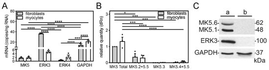
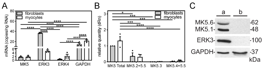
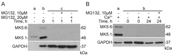
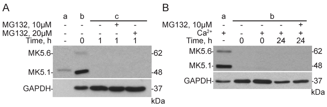
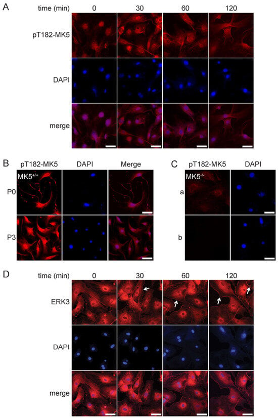
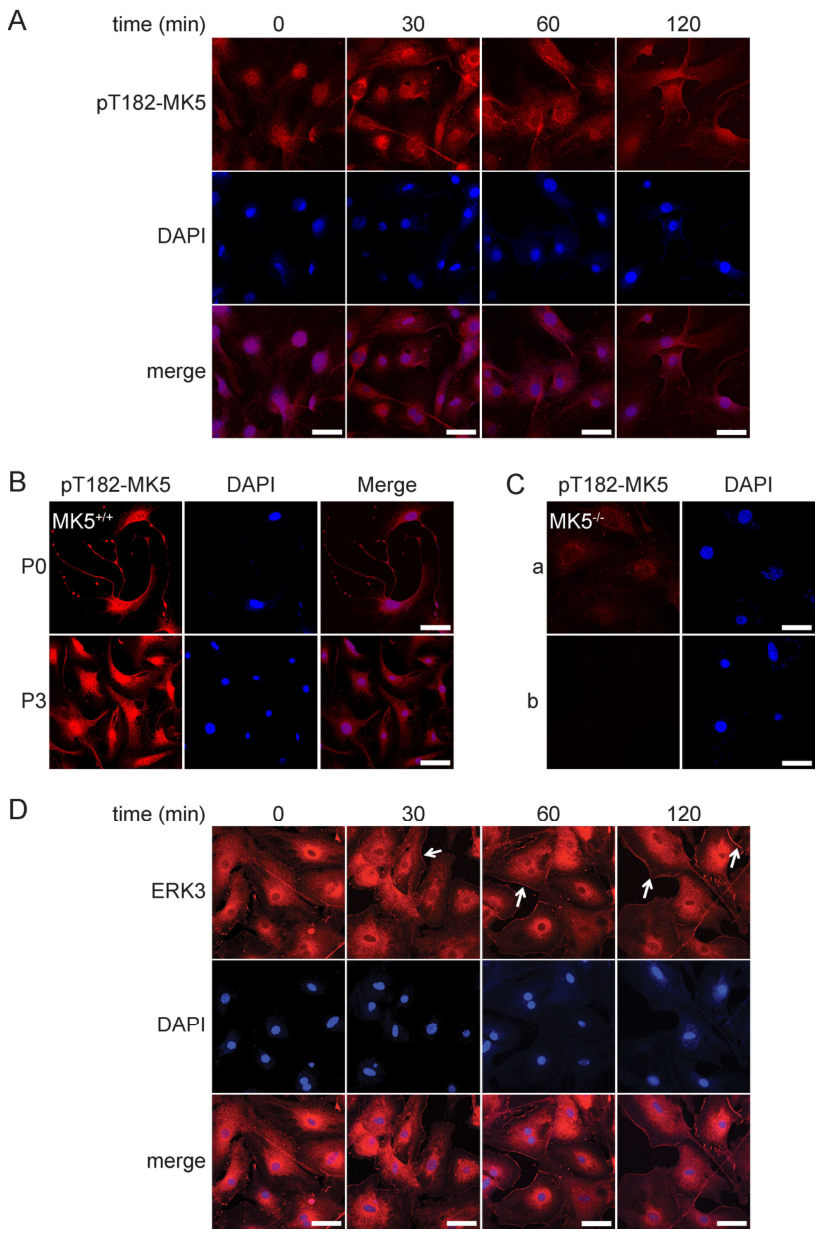
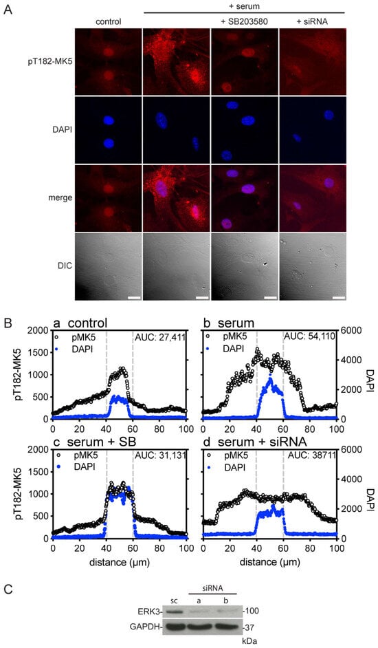
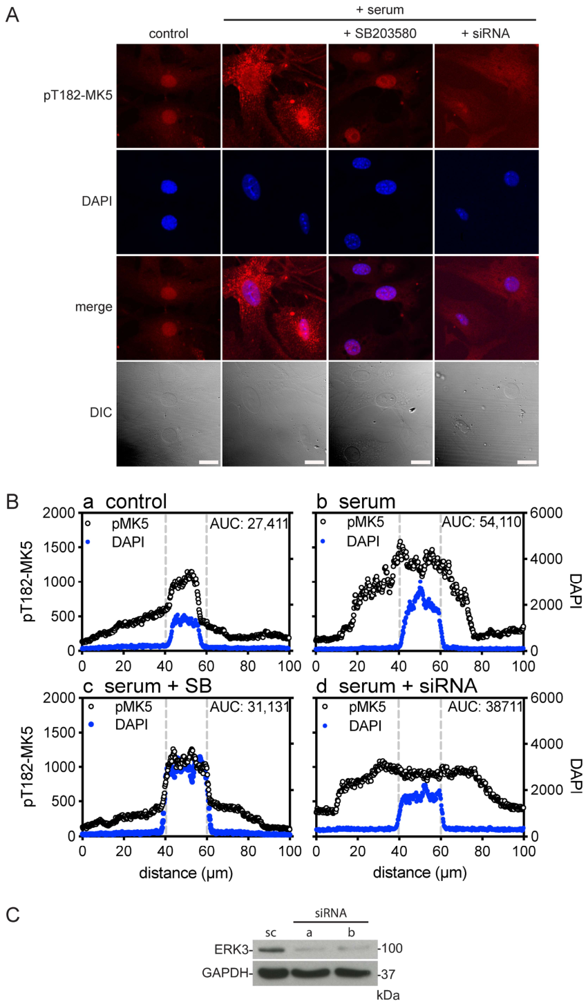
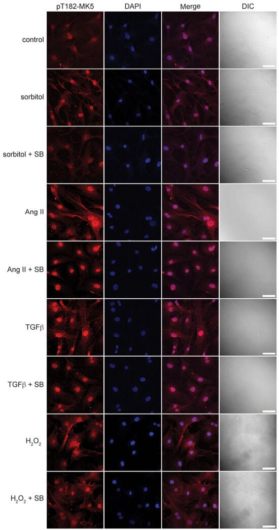
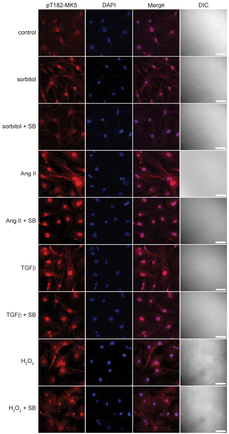
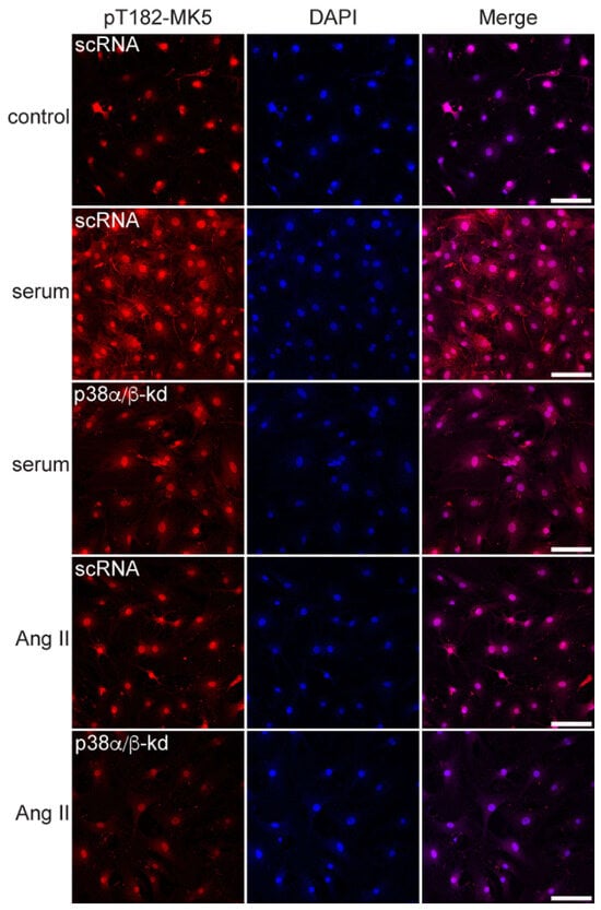
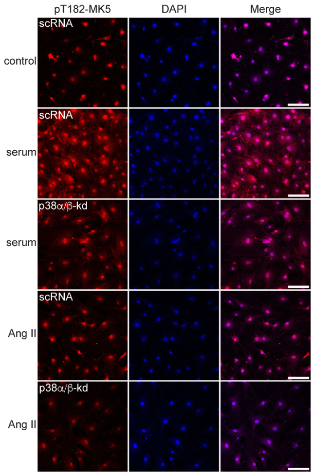
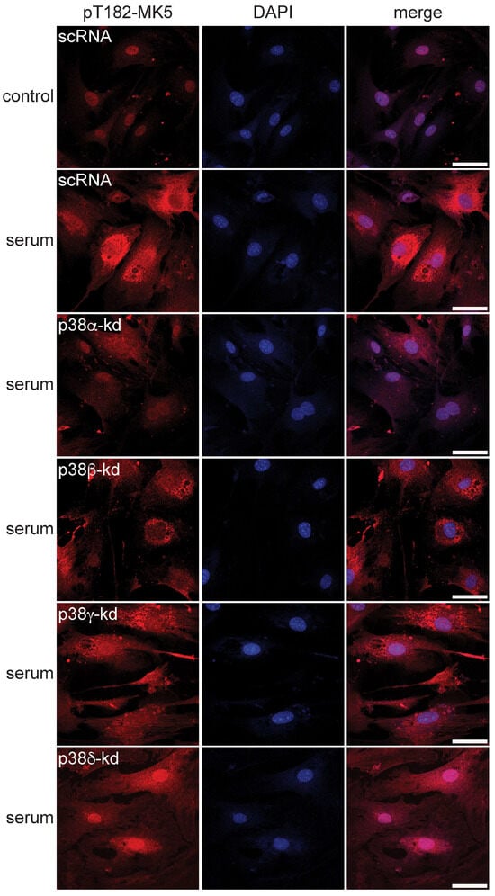
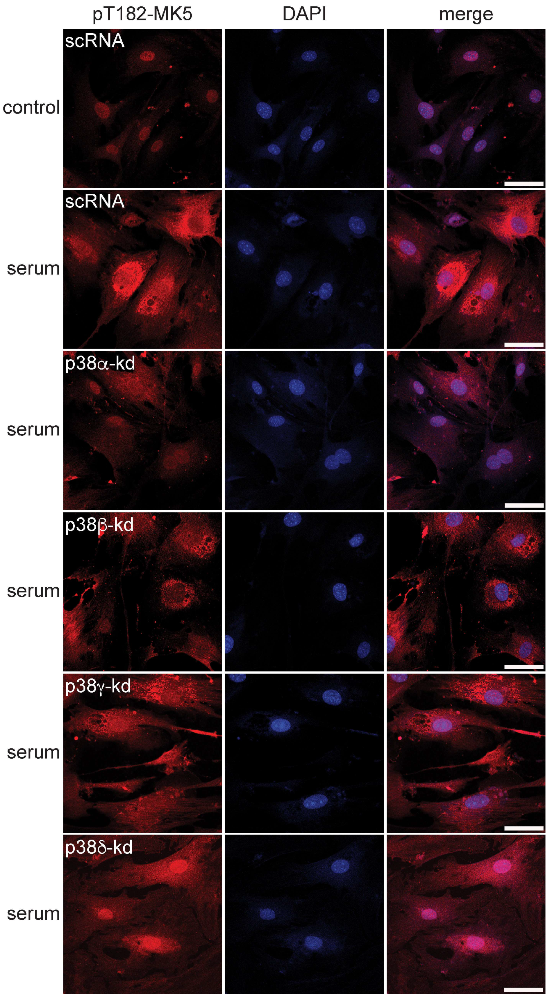
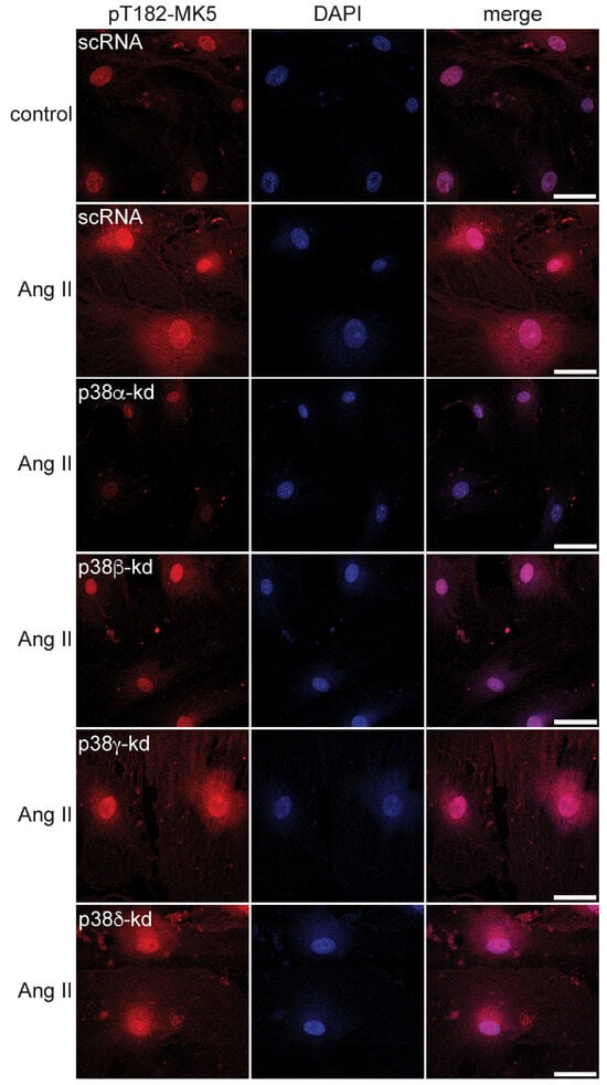
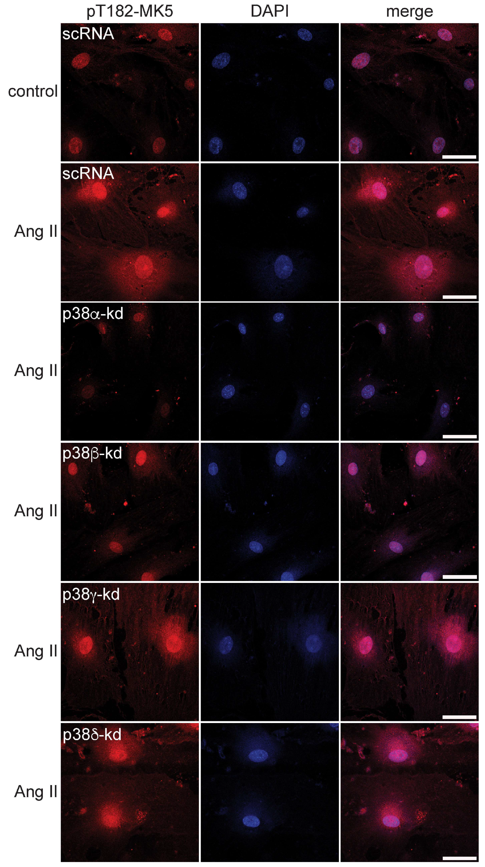
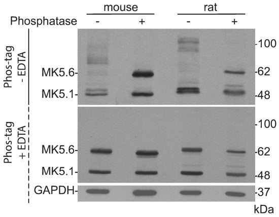
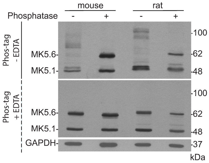
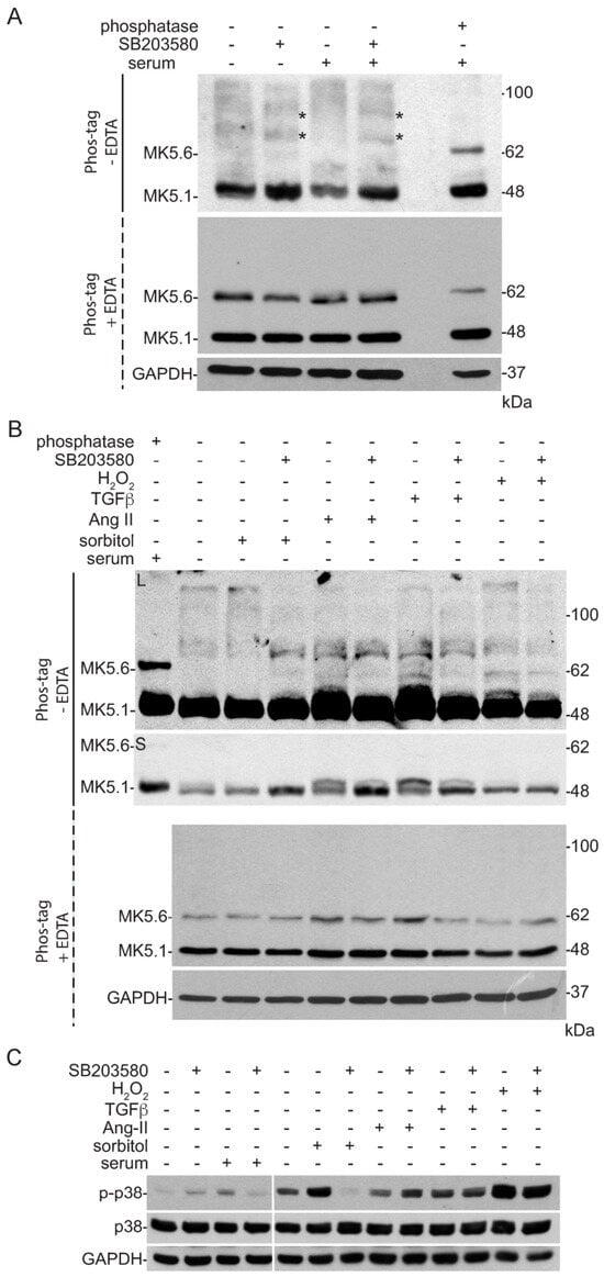
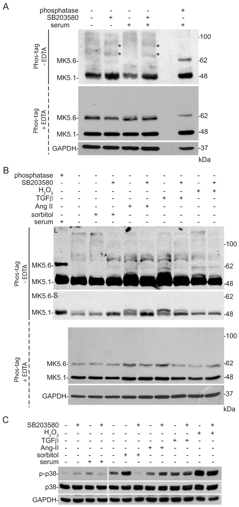
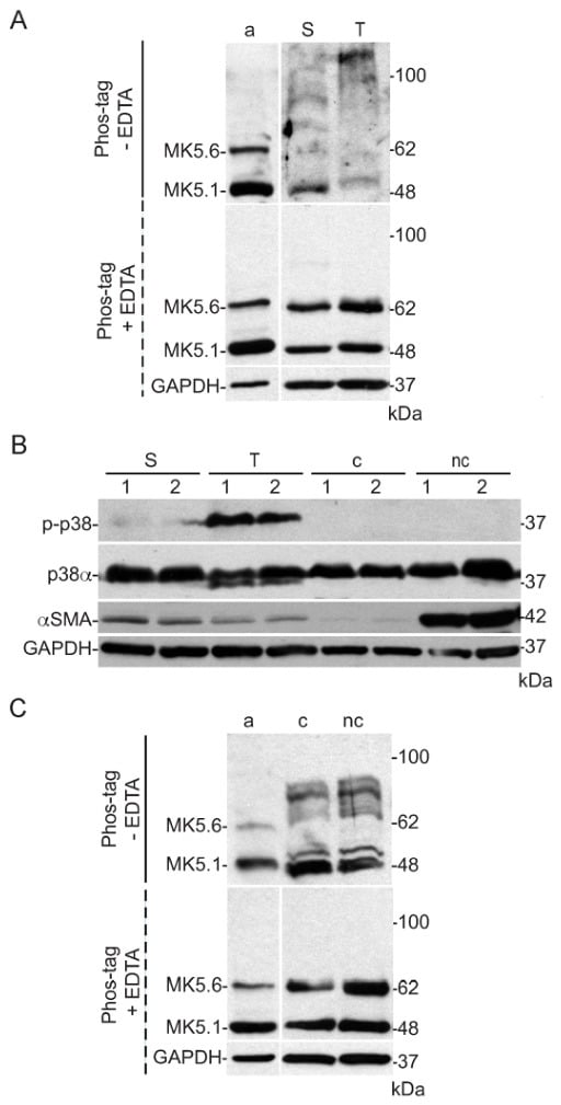
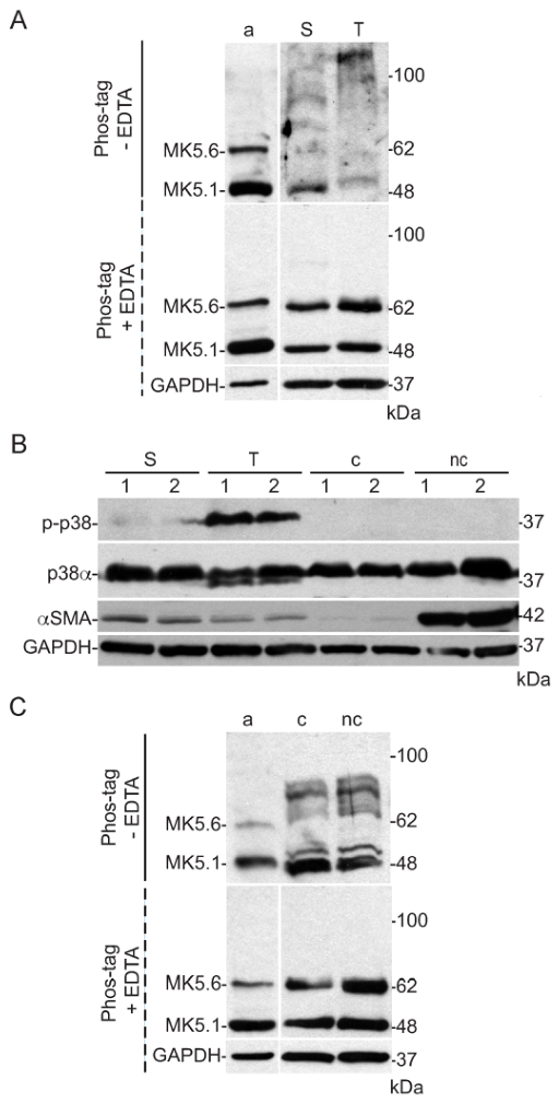

Disclaimer/Publisher’s Note: The statements, opinions and data contained in all publications are solely those of the individual author(s) and contributor(s) and not of MDPI and/or the editor(s). MDPI and/or the editor(s) disclaim responsibility for any injury to people or property resulting from any ideas, methods, instructions or products referred to in the content. |
© 2026 by the authors. Licensee MDPI, Basel, Switzerland. This article is an open access article distributed under the terms and conditions of the Creative Commons Attribution (CC BY) license.