


Abstract
This study characterised peptide abundance profiles in conventional colorectal adenomas and pT1 adenocarcinomas within the adenoma–carcinoma framework. An exploratory, cross-sectional secondary analysis was performed. Targeted proteomics assessed E-cadherin (CDH1), β-catenin (CTNNB1), p120-catenin (CTNND1) and ezrin (EZR) in 13 pairs of lesion tissue and adjacent non-lesional mucosa: three tubular adenomas, seven tubulovillous adenomas (TVAs) and three pT1 adenocarcinomas, with peptide-specific data availability. Median within-pair log2 differences were calculated. CTNNB1 medians were positive in all three categories, whereas CDH1 medians were negative in TVA. CTNND1 and EZR showed sequence-dependent patterns. Discovery proteomics yielded positive CTNNB1 differences in all three cases, consistent in direction with the targeted medians. The analysis of antigen-processing and human leucocyte antigen (HLA) proteins included one case per category. In case 05 (pT1), HLA proteins were identified, and antigen peptide transporters 1 (TAP1) and 2 (TAP2) and tapasin (TAPBP) were more abundant than in adjacent mucosa. Peptide-level information supports hypotheses about molecular heterogeneity. The small sample size, cross-sectional design and lack of spatial or functional measurements limit generalisability and mechanistic interpretation.
1. Introduction
The adenoma–carcinoma sequence is an established framework for colorectal carcinogenesis [1]. The transformation of tubulovillous adenoma into adenocarcinoma is documented clinically and histopathologically [2]. This framework supports the study of molecular differences between histological categories without assuming that every lesion must progress from tubular to tubulovillous adenoma and then to carcinoma.
Colorectal neoplasia provides a setting in which to examine the relationships between epithelial organisation, molecular signalling and protein homeostasis [3]. Reviews of WNT/β-catenin signalling describe its role in carcinogenesis, its association with cellular states and experimental approaches to its modulation [4,5,6]. Mutations in the adenomatous polyposis coli (APC) gene or the β-catenin gene (CTNNB1) can activate β-catenin-dependent transcription in colorectal tumours [7]. p120-catenin regulates cadherin turnover, and its depletion can reduce cadherin abundance and disrupt adhesion in experimental systems [8]. Related processes include the epithelial–mesenchymal transition [9], actin-mediated regulation of adherens junctions [10] and the contribution of ezrin to membrane and cytoskeletal organisation [11]. These findings support the investigation of adhesion and cytoskeletal proteins in colorectal lesions.
Targeted proteomics quantifies specific peptides and can verify abundance changes observed by discovery proteomics, provided that analytical quality criteria are appropriate to the study aims [12,13]. In workflows that involve protein digestion, the abundance of a protein or protein group is estimated from the signals of its constituent peptides. Methods such as MaxLFQ combine this information into an overall estimate even when individual peptides do not behave uniformly [14]. Discordance among some peptides therefore does not, by itself, invalidate the estimated protein abundance. Marked discordance nevertheless warrants caution, because a single protein-level value may obscure relevant differences between peptide sequences.
Retaining measurements for each sequence allows examination of heterogeneity that may reflect different proteoforms, post-translational modifications or proteolytic processing [15,16]. Digestion, recovery and matrix effects must also be considered, as their influence on quantification may vary by peptide and sample [17,18]. Combining the two approaches allows both overall abundance trends and individual sequence patterns to be examined, while distinguishing quantitative observations from the mechanisms that might explain them.
Discovery proteomics extends the characterisation to proteins involved in chromatin and protein homeostasis, or proteostasis [19]. In colorectal cancer cell models, the deubiquitinating enzyme ubiquitin-specific protease 7 (USP7) has been linked to the regulation of LIM domain-containing protein Ajuba (AJUBA) and to intercellular adhesion [20]. Ubiquitin-specific protease 14 (USP14), also a deubiquitinating enzyme, has been linked to cell growth and senescence [21]. Small ubiquitin-related modifiers 2 and 3 (SUMO2/3) have been associated with oxaliplatin resistance through SUMOylation of the DNA repair protein Ku80, encoded by XRCC5 [22]. These experimental findings support the study of proteostasis-related proteins alongside adhesion proteins. Whether these mechanisms operate in the lesions analysed requires specific investigation.
Antigen processing provides a further area for investigation. Studies of the human leucocyte antigen (HLA) ligandome and T-cell responses have identified naturally presented, immunogenic peptides, including a CTNNB1-derived neoantigen, in selected colorectal cancer models [23]. Integrated proteogenomic and immunopeptidomic analyses have also identified tumour-derived and bacterial peptides recognised by T cells [24]. Experimental recognition of an HLA complex bearing a mutated β-catenin peptide has been demonstrated using an antibody fragment engineered for this purpose [25]. These studies support the characterisation of antigen-processing and HLA components; identifying the ligands they present and establishing their immunogenicity require additional experiments.
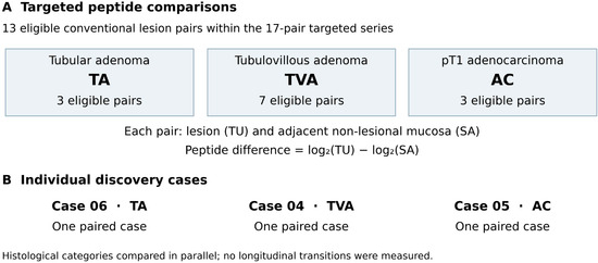
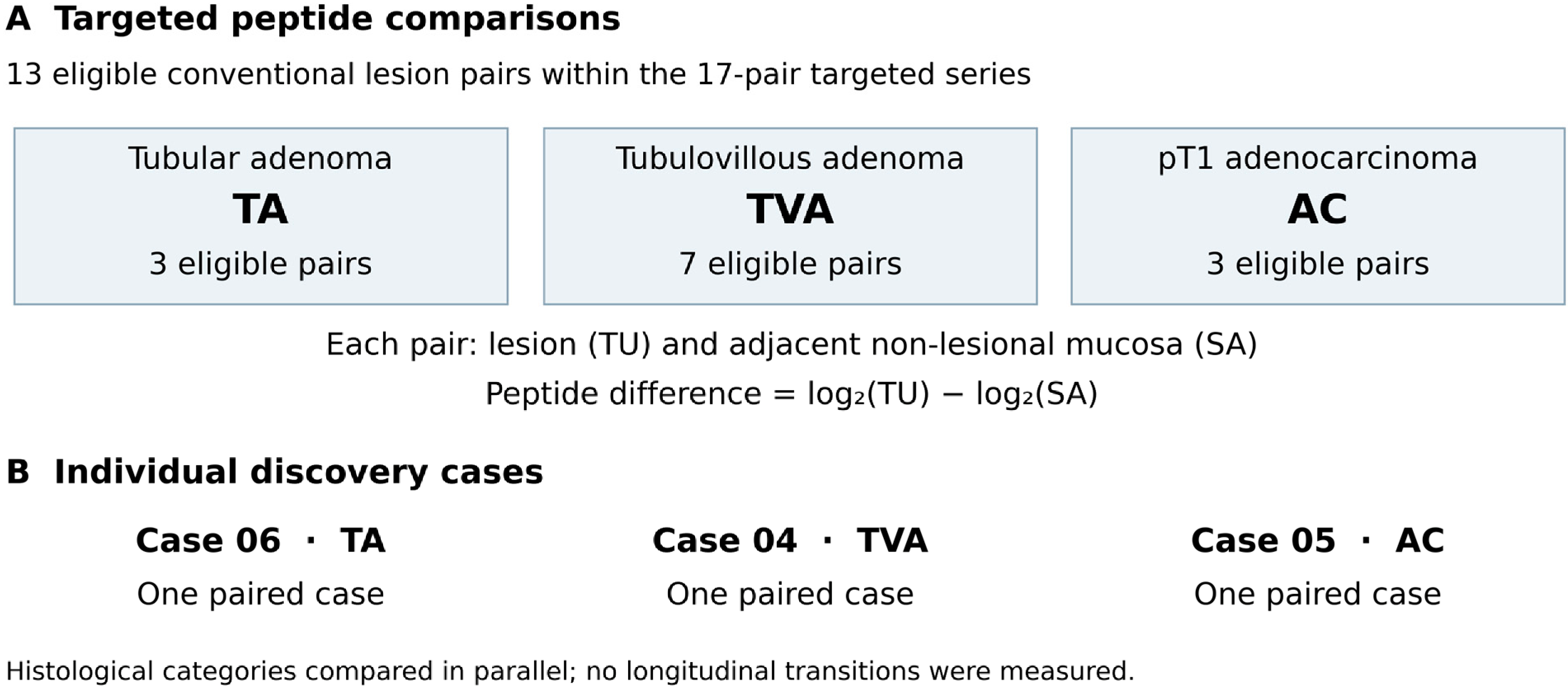
We conducted an exploratory, cross-sectional secondary analysis of an existing proteomic dataset. We characterised within-pair differences in peptides from E-cadherin (CDH1), β-catenin (CTNNB1), p120-catenin (CTNND1) and ezrin (EZR) between lesions and adjacent mucosa in tubular adenomas, tubulovillous adenomas and pT1 adenocarcinomas. We examined these findings descriptively alongside discovery proteomics and documented antigen-processing and HLA proteins in three individual cases. The aim was to describe relative abundance and peptide heterogeneity within the adenoma–carcinoma framework and to generate specific biological hypotheses. The histological categories comprised distinct lesions sampled cross-sectionally; Figure 1 summarises the design.
Figure 1.
Design of the cross-sectional secondary analysis. (A) Descriptive peptide comparison in 13 eligible lesion–mucosa pairs from conventional lesions, selected from the targeted proteomics series of 17 pairs: three tubular adenomas (TAs), seven tubulovillous adenomas (TVAs) and three pT1 adenocarcinomas arising in tubular adenomas (ACs). Each pair comprises lesion tissue (TU) and adjacent non-lesional mucosa (SA). The within-pair difference is expressed as Δlog2 = log2(TU) − log2(SA) = log2(TU/SA); positive values indicate higher abundance in the lesion and negative values indicate lower abundance. (B) Individual discovery proteomics observations for cases 06 (TA), 04 (TVA) and 05 (AC), with one pair per category. Eligible pair counts do not guarantee complete measurements for every peptide and should not be interpreted as confirmed counts of independent patients. The categories represent different lesions; individual transitions were not followed longitudinally.
2. Materials and Methods
2.1. Study Design and Patient Samples
This exploratory, cross-sectional secondary analysis examined human colorectal lesions detected through population-based colorectal cancer screening programmes. The categories were tubular adenoma (TA), tubulovillous adenoma (TVA) and pT1 adenocarcinoma arising in a tubular adenoma (AC). Lesion tissue (TU) and a matched sample of adjacent non-lesional colonic mucosa (SA) were obtained for each case. The comparison involved distinct lesions, with no longitudinal observation of their evolution. Adjacent mucosa served as a local tissue comparator.
Tissue samples were collected after endoscopic resection, frozen in liquid nitrogen and stored at −80 °C until processing. Histopathological classification was performed by pathologists experienced in gastrointestinal pathology, using established criteria.
The broader targeted proteomics collection included villous adenomas and serrated lesions, which were excluded from the main comparison of the conventional TA, TVA and AC categories in this analysis.
2.2. Protein Extraction and Sample Preparation
Frozen tissue samples were homogenised under cryogenic conditions and lysed in a denaturing buffer compatible with subsequent mass spectrometry analysis.
Proteins were reduced and alkylated, then enzymatically digested with trypsin according to established protocols. The resulting peptide mixtures were desalted by reversed-phase solid-phase extraction and vacuum-dried before mass spectrometry analysis.
2.3. Targeted Peptide-Level Proteomics
The targeted peptide-level proteomics dataset comprised 23 lesion records across six histological categories: 3 tubular adenomas (TA), 3 villous adenomas (VA), 7 tubulovillous adenomas (TVA), 4 adenomas containing pT1 adenocarcinoma (AC), 5 sessile serrated lesions (SSL) and 1 SSL with high-grade dysplasia.
Of these 23 records, 17 had paired samples of lesion tissue (TU) and adjacent mucosa (SA), corresponding to 34 tissue samples. These 17 pairs comprised 3 TA, 3 VA, 7 TVA, 3 AC and 1 SSL. The present comparison included the 13 pairs in the TA, TVA and AC categories: 3 TA, 7 TVA and 3 AC. These 13 pairs are a subset of the 17 available pairs and do not represent additional samples.
The availability of paired tissue samples did not ensure that every peptide had been quantified in both tissues. The number of evaluable pairs therefore depended on data availability for each peptide. Different peptides quantified in the same lesion were treated as measurements from that lesion, not as independent biological replicates.
Targeted proteomics quantified selected peptide sequences from β-catenin (CTNNB1), E-cadherin (CDH1), p120-catenin (CTNND1) and ezrin (EZR), proteins involved in intercellular adhesion, signalling and cytoskeletal organisation. Individual measurements were retained for each sequence to describe relative abundance and heterogeneity.
Heavy reference peptides containing 13C6,15N4-labelled arginine or 13C6,15N2-labelled lysine were added to each sample at identical concentrations to normalise the signal.
Peptides were selected on the basis of sequence specificity, reproducibility and suitability for quantitative mass spectrometry. Peptides in lesion tissue and paired adjacent mucosa were quantified using selected reaction monitoring or parallel reaction monitoring (PRM), as appropriate. For PRM, tandem mass spectrometry (MS/MS) data were acquired using an Orbitrap analyser (Mass spectrometer: EASY nLc100-Q Exactive), coupled with an Easy-nLC 1000 nano UPLSystem chromatography, System EEUU (Thermo Scientific, Waltham, MA USA) operating at a resolving power of 17,500 at m/z 200.
Peptide intensities were recorded on a log2 scale. For each peptide and histological category, results were summarised as the median within-pair log2 difference between lesion tissue (TU) and adjacent non-lesional mucosa (SA), defined as Δlog2 = log2(TU) − log2(SA), equivalent to log2(TU/SA). These differences were taken from the “Ratio TU–SA” columns in the “Peptide_ratio” worksheet and cross-checked against “Peptide_ratio_graficos”. Peptide sequences were not combined into a protein abundance estimate. The analysis yielded 154 differences from 13 eligible pairs: 3 pairs per peptide in TA, 7 in TVA and 3 in AC, except for CTNND1 NLSYQVHR and EZR IQVWHAEHR, each with 2 AC pairs because TU and SA measurements for these two sequences were unavailable in case 23. Missing values were not imputed. Relative abundance ratios were obtained by exponentiating the within-pair log2 differences to base 2. Analyses were descriptive; no confirmatory between-group tests were conducted and no clinical classification models were developed.
2.4. Discovery Proteomics
Discovery proteomics characterised relative abundance at the protein or protein-group level. Peptides were analysed by high-resolution nanoflow liquid chromatography–tandem mass spectrometry (nanoLC-MS/MS) on an Orbitrap platform. Identification was performed against the UniProt human proteome using MaxQuant, with a false discovery rate below 1% at both peptide and protein levels; label-free quantification (LFQ) was used. The processed matrix comprised 1916 protein entries, including histones and components of the ubiquitin–proteasome system.
Discovery data were analysed at the protein-group level using normalised LFQ intensities. These summaries were kept distinct from individual targeted peptide measurements. We did not assume that every peptide selected for targeted acquisition had contributed to each LFQ estimate. Post-translational modifications were not assessed in this analysis. The chromatin and proteostasis summary was considered separately from the case-specific reanalysis of selected antigen-processing and HLA proteins described in Section Exploratory Inventory of Antigen-Processing and HLA Proteins.
2.5. Data Interpretation and Visualisation
The two proteomic approaches were integrated descriptively by comparing the direction of lesion–mucosa differences in discovery protein groups with the medians for targeted peptide sequences. This comparison distinguishes individual discovery cases from summaries by histological category in the targeted analysis. No formal coefficient of agreement between methods was estimated, and no progression model was constructed. Figure 1 presents the study design; Figure 2 shows individual peptide differences and their medians.
Figure 2.
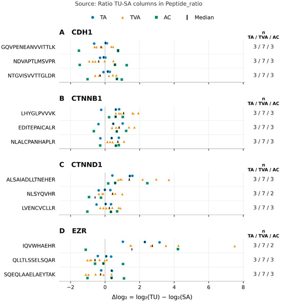
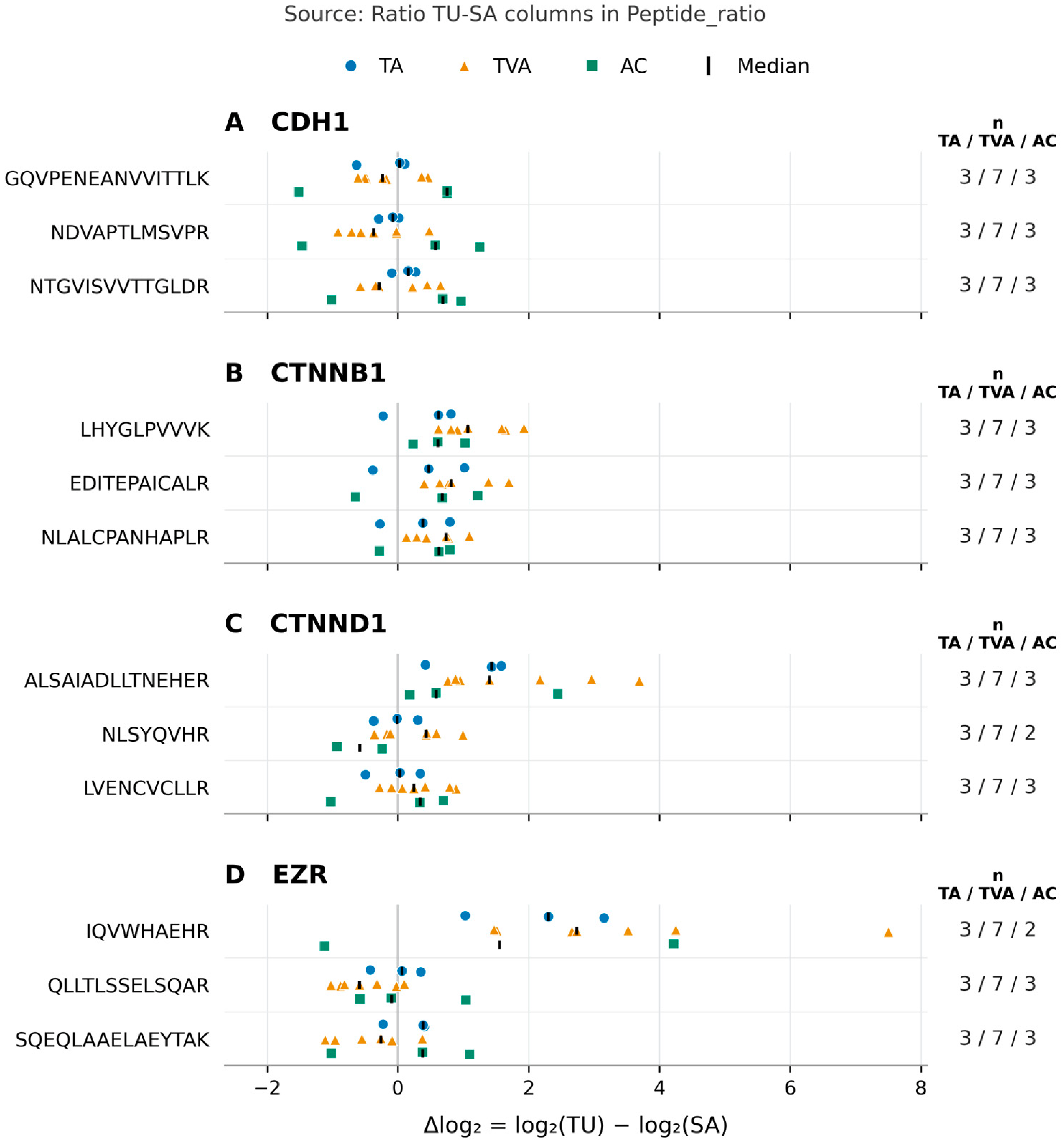
Individual peptide abundance differences by histological category. Panels show (A) E-cadherin (CDH1), (B) β-catenin (CTNNB1), (C) p120-catenin (CTNND1) and (D) ezrin (EZR), with three identified sequences per protein. Each point represents a within-pair difference for one peptide, Δlog2 = log2(TU) − log2(SA), where TU denotes the lesion and SA its adjacent non-lesional mucosa. Values were taken from the Ratio TU–SA columns in the Peptide_ratio worksheet and checked against Peptide_ratio_graficos. Positive values indicate higher abundance in the lesion and negative values indicate lower abundance. The zero line indicates no difference, and black bars show the medians. Blue circles denote TA (tubular adenoma), orange triangles denote TVA (tubulovillous adenoma) and green squares denote AC (pT1 adenocarcinoma arising in a tubular adenoma). The plot shows 154 paired differences across 12 sequences from 13 eligible pairs and 36 medians, one per sequence and histological category. The number of pairs per peptide is three in TA, seven in TVA and three in AC, except for CTNND1 NLSYQVHR and EZR IQVWHAEHR, for which two AC pairs were available because neither tissue measurement was available for case 23. Counts are shown to the right of each sequence. Missing values were not imputed. Peptides from the same lesion are not independent biological replicates. The display is descriptive and does not establish temporal progression or functional activity.
Exploratory Inventory of Antigen-Processing and HLA Proteins
A dedicated exploratory reanalysis examined individual LFQ protein-group results from three paired discovery proteomics cases: case 04, tubulovillous adenoma (TVA); case 05, pT1 adenocarcinoma arising in a tubular adenoma (AC); and case 06, tubular adenoma (TA). Within-pair differences for each protein group were described as Δlog2 = log2(TU) − log2(SA), equivalent to log2(TU/SA). With only one case per histological category, effects associated with histology could not be distinguished from variation between individual cases. No inferential pathway-level analyses were performed. HLA protein-group assignments from shotgun proteomics were treated as identifications, not as patient-specific HLA genotyping.
The selected exploratory module included CTNNB1 and CTNND1, together with proteasome subunits beta type-8 (PSMB8), type-9 (PSMB9) and type-10 (PSMB10), which form part of the immunoproteasome. It also included endoplasmic reticulum aminopeptidase 1 (ERAP1), antigen peptide transporters 1 and 2 (TAP1 and TAP2), tapasin (TAPBP), calreticulin (CALR), protein disulfide-isomerase A3 (PDIA3) and β2-microglobulin (B2M).
The module also included the HLA class I heavy chains HLA-A, HLA-B and HLA-C; the HLA-DR alpha chain (HLA-DRA), HLA-DP alpha 1 chain (HLA-DPA1) and HLA-DR beta 1 chain (HLA-DRB1); and the HLA class II invariant chain (CD74).
Detected proteins and their abundance differences were recorded for each case. The analysis characterised components of the antigen-processing machinery; it did not identify HLA-bound ligands or assess T-cell recognition.
2.6. Ethical Considerations
The study was conducted in accordance with the Declaration of Helsinki and approved by the relevant institutional clinical research ethics committee. Identifying committee and protocol details have been withheld for blinded peer review. A favourable opinion was issued on 28 January 2016. All participants provided written informed consent before enrolment, including authorisation to use their biological samples for research.
2.7. Generative AI Use in Graphical Abstract Preparation
OpenAI ChatGPT (GPT-5.6 Sol) with image generation (OpenAI; accessed 1 November 2026) was used to create the graphical abstract from scientific content and layout specifications defined by the authors. The tool was used only to generate illustrative artwork and was not used to generate, modify, analyse or interpret experimental data. The authors reviewed and edited the graphical abstract for scientific accuracy and take full responsibility for its content.
3. Results
3.1. Peptide-Level Characterisation of Cell Adhesion and Cytoskeletal Proteins in Colorectal Polyps
Targeted peptide-level proteomics was used to describe relative abundance differences between lesion tissue (TU) and adjacent non-lesional mucosa (SA) for proteins involved in intercellular adhesion and cytoskeletal organisation. The main analysis included three conventional histological categories: tubular adenoma (TA), tubulovillous adenoma (TVA) and adenoma containing pT1 adenocarcinoma (AC).
Results are expressed as within-pair peptide abundance differences, calculated as log2(TU) − log2(SA), equivalent to log2(TU/SA). Twelve sequences were retained, three for each of the four proteins. Figure 2 presents individual observations and 36 medians, one for each combination of sequence and histological category, together with the number of available pairs.
3.2. β-Catenin (CTNNB1)
Median within-pair differences for all three CTNNB1 sequences were positive in TA, TVA and AC. For LHYGLPVVVK, the median Δlog2 in TVA was 1.065 (n = 7). Positive medians do not imply that every individual value was positive.
The consistent direction of the medians across all three sequences describes a pattern of higher relative CTNNB1 peptide abundance in the categories analysed.
3.3. E-Cadherin (CDH1)
For CDH1 peptides in TA, the group medians of the within-pair differences were close to zero, with both positive and negative medians. In TVA, the medians for all three peptides were negative, ranging from −0.371 to −0.233 on the log2(TU/SA) scale, with seven pairs per peptide. These values correspond to ratios of approximately 0.773–0.851, equivalent to approximately 15–23% lower relative peptide abundance. This range spans the three peptide medians and is not a confidence interval.
In AC, median differences for all three CDH1 peptides were positive, with three pairs per peptide.
3.4. p120-Catenin (CTNND1)
CTNND1 measurements were discordant across peptide sequences. For ALSAIADLLTNEHER, median Δlog2 values were 1.429 in TA, 1.398 in TVA and 0.581 in AC, based on three, seven and three pairs, respectively.
For NLSYQVHR, median Δlog2 values were −0.011 in TA and 0.429 in TVA. In AC, the median was −0.584 and was calculated from two pairs because TU and SA measurements were unavailable for this sequence in case 23. The sequence-specific results were heterogeneous and were retained at the peptide level, without deriving a single CTNND1 protein abundance estimate from the targeted measurements.
3.5. Ezrin (EZR)
For the EZR peptide IQVWHAEHR, median Δlog2 values were 2.300 in TA, 2.733 in TVA and 1.549 in AC. The corresponding numbers of pairs were three, seven and two; the lesion and paired mucosa measurements were unavailable for this sequence in case 23.
The other two EZR peptides had negative medians in TVA: −0.585 for QLLTLSSELSQAR and −0.263 for SQEQLAAELAEYTAK, each based on seven pairs. Their contrasting direction relative to IQVWHAEHR shows a sequence-dependent pattern.
3.6. Peptide Patterns Across Histological Categories
Descriptive profiles varied across histological categories: CTNNB1 medians were positive, CDH1 medians were negative in TVA, and CTNND1 and EZR results depended on the sequence. Retaining individual peptide values allowed both concordance and heterogeneity in these patterns to be described.
Figure 2 shows the individual abundance differences for each peptide and category.
Table 1 summarises the principal observations and the scope of their interpretation.
Table 1.
Principal peptide abundance findings and scope of interpretation.
3.7. Discovery Proteomics of Histones and the Ubiquitin–Proteasome System
Discovery proteomics provided complementary information on linker histones, histone variants and components of the ubiquitin–proteasome system, using non-lesional tissue as the comparator. This overall summary describes the protein context of the samples and was not used to estimate differences between the TA, TVA and AC categories.
H1 histones, H2A variants, SUMO2/3, the 26S proteasome non-ATPase regulatory subunits 1 (PSMD1), 7 (PSMD7) and 14 (PSMD14), and the deubiquitinating enzymes ubiquitin-specific protease 5 (USP5), USP7 and USP14 were identified.
3.8. Antigen-Processing and HLA Protein Observations in Individual Cases
Selected antigen-processing proteins were identified in the LFQ results for individual cases. B2M had positive within-pair Δlog2 values in all three cases: +1.69, +0.47 and +0.90 for cases 04, 05 and 06, respectively. PSMB8 values were −0.21, +0.90 and +0.41, while PSMB9 values were −0.52, +1.66 and +1.43 in the same case order. PSMB10 values were −0.06, +0.25 and +0.19. These measurements are descriptive and refer to individual cases.
In case 05, a pT1 adenocarcinoma arising in a tubular adenoma, HLA proteins were identified and TAP1 (+0.71), TAP2 (+1.33) and TAPBP (+0.62) were increased relative to adjacent non-lesional mucosa, expressed as Δlog2. Corresponding changes were smaller or discordant in cases 04 and 06. The concurrent increase in these three peptide transport and loading components characterises the only adenocarcinoma in the discovery series. ERAP1, CALR and PDIA3 were detected, but their changes were not concordant in direction across the three cases. HLA-A, HLA-B and HLA-C protein groups were also identified; HLA allele genotyping was not performed.
HLA-DRA, HLA-DPA1, HLA-DRB1 and CD74 were detected, but their lesion–mucosa differences did not show a uniform direction. B2M abundance was higher in all three cases. The concomitant increases in TAP1, TAP2 and TAPBP characterised case 05. The paired antigen-processing/HLA matrix, UniProtKB identifiers and targeted CTNND1 data are provided in Supplementary Table S1; the corresponding processed discovery source records are presented in Supplementary Table S2.
3.9. Descriptive Integration of the Results
Discovery and targeted proteomics provided complementary information. For CTNNB1, within-pair LFQ differences were positive in all three discovery cases: +0.298 in case 04 (TVA), +0.077 in case 05 (AC) and +0.839 in case 06 (TA). This direction agrees with the positive medians for all three targeted sequences in the histological categories analysed. Agreement refers to the direction of change and compares individual cases with group summaries; it does not imply quantitative equivalence between methods or agreement across all proteins. Individual CTNND1 and EZR measurements retained sequence-dependent patterns.
4. Discussion
All three CDH1 peptides had negative median within-pair differences in TVA, whereas median CTNNB1 differences were positive across the histological categories examined. CTNND1 and EZR showed heterogeneous patterns across sequences. The direction of the CTNNB1 differences agreed between discovery and targeted proteomics, providing descriptive corroboration of higher relative abundance. Figure 2 and Table 1 present these findings alongside the variation between individual lesions.
The adenoma–carcinoma sequence, including the transformation of tubulovillous adenoma into adenocarcinoma, is established by clinical and histopathological evidence [1,2]. Our design allows histology-associated molecular patterns to be examined within this framework. Cross-sectional comparisons of different lesions cannot reconstruct the course of an individual lesion or establish that peptide changes determine its transformation. Greater molecular plasticity in TVA remains an interpretative hypothesis arising from these patterns and requires specific investigation. This uncertainty concerns the molecular interpretation of the present data: malignant transformation is well established, and TVA is not an obligatory stage in every progression pathway.
The median differences for the three CDH1 peptides in TVA ranged from −0.371 to −0.233 on the log2(TU/SA) scale, corresponding to approximately 15–23% lower relative abundance. Agreement in direction across the sequences supports this descriptive finding, although its functional significance remains to be established. Group medians summarise distributions of individual results and do not imply uniform behaviour across lesions. The heterogeneity of CTNND1 and EZR provides sequence-level information that should be retained when interpreting protein-level summaries.
Targeted proteomics provides selective measurements that complement discovery profiling [12,13]. Agreement in the direction of the CTNNB1 differences supports the quantitative observation within this series, although uniform agreement has not been demonstrated for all proteins. Corroborating abundance measurements is distinct from validation in an independent biological cohort and from spatial or functional characterisation.
Immunohistochemistry, immunofluorescence and other spatial methods could distinguish cellular distributions and membrane, cytoplasmic and nuclear localisation. WNT transcriptional activity and adhesive function require specific assessments. No such spatial or functional studies were performed. The increase in CTNNB1 is therefore interpreted as an abundance finding, while changes in CDH1, CTNND1 and EZR provide potential leads for investigating adhesion and the cytoskeleton. Experimental studies of β-catenin and p120-catenin provide a basis for these hypotheses [7,8].
Protein or protein-group abundance estimates integrate the peptide signals used for quantification and do not require every sequence to show changes of identical magnitude or direction. Robust estimators used in methods such as MaxLFQ accommodate variation between peptide ratios [14]. Discordance among some targeted peptides does not, by itself, invalidate a discovery-based protein estimate. Marked or systematic discordance does, however, limit the extent to which a single value can represent all molecular forms. Targeted peptide measurements were retained separately in this study.
Possible biological explanations include differences in isoforms, post-translational modifications and proteolytic processing, as illustrated by studies of proteoform heterogeneity [15,16]. For a single intact molecular form, the relative changes measured by different peptides would be expected to be similar if recovery and analytical response were stable. Tissue cell composition could also contribute if different cell populations expressed different molecular forms or processing products. These possibilities provide hypotheses to test without presuming that discordance is either an artefact or evidence of a particular biological mechanism.
Possible analytical explanations include variation in digestion and recovery, matrix effects, interference and peak integration [17,18]. A constant ionisation factor for a given sequence cancels in its TU/SA ratio and cannot, on its own, account for opposite directions of change in the ratios of different peptides. The contribution of each mechanism remains to be established. Reviewing the analytical records and peptide specificity, together with complementary measurements where appropriate, would help to test these explanations and clarify their functional implications.
The identification of histones, components of the ubiquitin–proteasome system and deubiquitinases broadens the molecular context of these lesions. Experimental findings on USP7, USP14 and SUMO2/3 [20,21,22] provide a rationale for investigating their relationships with adhesion and cellular responses. The present series documents abundance; functional coordination between these processes remains a hypothesis.
The therapeutic considerations are informed by external studies. These studies have examined WNT modulation and intestinal tumourigenesis [26,27,28,29], as well as molecular dependencies involving cytoplasmic acetyl-coenzyme A synthetase (ACSS2), the heat shock proteins HSPA1 and HSP90-alpha (HSP90AA1), nicotinamide phosphoribosyltransferase (NAMPT) and autophagy-related protein 7 (ATG7) [30,31,32,33]. These studies suggest questions for experimental investigation; neither the interventions nor the dependencies they describe were assessed in our samples.
The identification of β-catenin ligands and the development of β-catenin degraders [34,35,36,37,38], together with selective degradation of USP7 [39], provide external proof-of-concept evidence. Investigating these approaches in premalignant lesions would require demonstration of the relevant molecular dependency and evaluation of efficacy and safety. Abundance measurements in this series do not establish susceptibility to these treatments.
In the pT1 adenocarcinoma from case 05, HLA proteins were identified and TAP1, TAP2 and TAPBP abundance was higher than in adjacent mucosa; B2M abundance was higher in all three discovery cases. These observations characterise components of antigen processing. With one case per histological category, category-associated effects cannot be distinguished from individual variation. Identification of HLA proteins also does not establish which ligands are presented or whether T cells recognise them. These questions require immunopeptidomic and functional studies. The relationship between WNT and immune escape described in other systems, including lung cancer models [40], provides a rationale for investigation but does not demonstrate this mechanism in the lesions studied here.
Immunoprevention is considered in the context of external research. Mucin-1 (MUC1) vaccination has been studied in people with a history of advanced adenoma [41,42], although the randomised trial did not show a statistically significant reduction in overall recurrence [42]. Vaccination against achaete-scute homologue 2 (ASCL2) has been investigated in mice [43], and immunopreventive approaches have been proposed for familial adenomatous polyposis [44]. Our data neither identify vaccine candidates nor evaluate immunopreventive efficacy.
The groups are small and unbalanced: three eligible pairs in TA, seven in TVA and three in AC, with only two AC pairs available for NLSYQVHR and IQVWHAEHR. This composition limits the precision of the medians, estimation of variability and representation of histological diversity. In groups of three pairs, an individual case can substantially influence the summary pattern. Different peptides from the same lesion are not independent biological replicates. Pairing provides a local reference but cannot replace a larger series or a healthy control group from the general population. The differences are descriptive and were not subjected to confirmatory hypothesis testing; their direction and magnitude require evaluation in independent series before they can be generalised. Reuse of these measurements in other manuscripts does not constitute independent biological validation. The cross-sectional design and absence of spatial or functional measurements also limit inferences about mechanisms and lesion evolution.
The immediate value of this study lies in describing patterns of peptide abundance and heterogeneity that generate questions about the biology of these lesions. Further studies should assess reproducibility in independent series, characterise discordant sequences and examine localisation or function as required by the hypothesis. Potential clinical applications would involve adding information to histological assessment or risk estimation and would require specific prospective evaluation. The current findings do not support treatment selection or changes to clinical surveillance.
5. Conclusions
Combining discovery and targeted proteomics provides complementary characterisation of conventional colorectal lesions within the adenoma–carcinoma framework. The principal findings are agreement in the direction of CTNNB1 differences, negative CDH1 medians in TVA and sequence-dependent patterns in CTNND1 and EZR. Peptide heterogeneity provides information beyond a protein-level summary and generates hypotheses about its possible causes. In the only adenocarcinoma examined by discovery proteomics, case 05, HLA proteins were identified and TAP1, TAP2 and TAPBP abundance was higher than in adjacent mucosa. The sample size and cross-sectional design limit generalisability; localisation, functional mechanisms and clinical utility require specific investigation.
Supplementary Materials
The following supporting information can be downloaded at https://www.mdpi.com/article/10.3390/cimb48111175/s1.
Author Contributions
Conceptualization, J.L. and F.B.; methodology, J.L. and F.B.; software; validation, J.L. and F.B.; formal analysis, J.L.; investigation, F.B., and J.L.; resources, F.B., and J.L.; data curation, J.L.; writing—original draft preparation, J.L.; writing—review and editing, J.L., F.B. and P.G.; visualization, J.L.; supervision, P.G.; project administration, F.B.; funding acquisition, F.B. and J.L. All authors have read and agreed to the published version of the manuscript.
Funding
This work was supported by the Jesús de Gangoiti Barrera Foundation (Fundation Jesús de Gangoiti Barrena 16102026).
Institutional Review Board Statement
The study was conducted in accordance with the Declaration of Helsinki and approved by the relevant institutional clinical research ethics committee (Basque Country Clinical Research Ethics Committee Acta no. 01/2016 28012016; the Comité Ético de Investigación Clínica de Euskadi (CEIC-E; internal protocol code PI2016003)). A favourable opinion was issued on 28 January 2016 (Minutes 01/2016; protocol version 6 February 2016).
Informed Consent Statement
Written informed consent was obtained from all participants before enrolment, including permission to use colorectal lesion tissue, paired adjacent non-lesional mucosa and associated clinical data within the approved research project. Samples and data were coded to protect participant confidentiality.
Data Availability Statement
The mass spectrometry proteomics data have been deposited with the ProteomeXchange Consortium through its partner repository PRIDE. The repository accession and reviewer-access details have been provided separately to the editorial office and are withheld here to preserve blinded peer review. The dataset remains private pending publication. This study is a secondary analysis of a shared proteomic dataset also used in related manuscripts; identifying details of those manuscripts are withheld for blinded peer review. The three paired discovery cases 04–06, the discovery matrix comprising 1916 quantified proteins and the main series of 17 targeted proteomics pairs are part of this shared resource. The conventional subset described here comprises 13 eligible TA/TVA/pT1 pairs within the targeted proteomics dataset. CDH1, CTNNB1, CTNND1 and EZR are part of the shared panel. These samples and measurements were not newly generated and do not constitute independent validation data. This reanalysis describes paired abundance differences for 12 peptide sequences from CDH1, CTNNB1, CTNND1 and EZR in 13 eligible pairs from conventional lesions, including individual distributions and 36 medians, one per sequence and histological category (Figure 2; principal observations in Table 1). It also presents lesion–mucosa LFQ differences for selected antigen-processing/HLA protein groups in each of the three discovery cases, with one case per category (Section 3.8 and Supplementary Tables S1 and S2). Detection and network analysis of antigen-processing components had already been undertaken in related work; the analyses reported here are summaries derived from the shared experimental resource. The derived antigen-processing/HLA matrix, UniProtKB annotations and targeted CTNND1 measurements are provided in Supplementary Table S1. The corresponding processed discovery proteomics entries, as recorded in the source, are provided in Supplementary Table S2.
Acknowledgments
During preparation of the graphical abstract, the authors used OpenAI ChatGPT (GPT-5.6 Sol) with image generation (OpenAI; accessed 1 November 2026) to create an illustrative schematic from author-specified scientific content. Generative AI was used only to prepare the graphical abstract, as described in Section 2.7 and the Acknowledgements. AI-assisted tools were also used for language editing and formatting. No generative AI tool was used to generate, modify, analyse or interpret experimental data or results. The authors reviewed, edited and approved all output and take full responsibility for the content of the publication.
Conflicts of Interest
The authors declare no conflicts of interest relevant to this work.
References
- Fearon, E.R.; Vogelstein, B. A genetic model for colorectal tumorigenesis. Cell 1990, 61, 759–767. [Google Scholar] [CrossRef] [Scilit] [PubMed]
- Galandiuk, S.; Fazio, V.W.; Jagelman, D.G.; Lavery, I.C.; Weakley, F.A.; Petras, R.E.; Badhwar, K.; McGonagle, B.; Eastin, K.; Sutton, T. Villous and tubulovillous adenomas of the colon and rectum. A retrospective review, 1964–1985. Am. J. Surg. 1987, 153, 41–47. [Google Scholar] [CrossRef] [Scilit] [PubMed]
- Li, Q.; Geng, S.; Luo, H.; Wang, W.; Mo, Y.Q.; Luo, Q.; Wang, L.; Song, G.-B.; Sheng, J.-P.; Xu, B. Signaling pathways involved in colorectal cancer: Pathogenesis and targeted therapy. Signal Transduct. Target. Ther. 2024, 9, 266. [Google Scholar] [CrossRef] [Scilit] [PubMed]
- Song, P.; Gao, Z.; Bao, Y.; Chen, L.; Huang, Y.; Liu, Y.; Dong, Q.; Wei, X. Wnt/β-catenin signaling pathway in carcinogenesis and cancer therapy. J. Hematol. Oncol. 2024, 17, 46. [Google Scholar] [CrossRef] [Scilit] [PubMed]
- Wu, X.; Que, H.; Li, Q.; Wei, X. Wnt/β-catenin mediated signaling pathways in cancer: Recent advances, and applications in cancer therapy. Mol. Cancer 2025, 24, 171. [Google Scholar] [CrossRef] [Scilit] [PubMed]
- Katoh, M.; Katoh, M. WNT signaling and cancer stemness. Essays Biochem. 2022, 66, 319–331. [Google Scholar] [CrossRef] [Scilit] [PubMed]
- Morin, P.J.; Sparks, A.B.; Korinek, V.; Barker, N.; Clevers, H.; Vogelstein, B.; Kinzler, K.W. Activation of beta-catenin-Tcf signaling in colon cancer by mutations in beta-catenin or APC. Science 1997, 275, 1787–1790. [Google Scholar] [CrossRef] [Scilit] [PubMed]
- Davis, M.A.; Ireton, R.C.; Reynolds, A.B. A core function for p120-catenin in cadherin turnover. J. Cell Biol. 2003, 163, 525–534. [Google Scholar] [CrossRef] [Scilit] [PubMed]
- Nie, F.; Sun, X.; Sun, J.; Zhang, J.; Wang, Y. Epithelial-mesenchymal transition in colorectal cancer metastasis and progression: Molecular mechanisms and therapeutic strategies. Cell Death Discov. 2025, 11, 336. [Google Scholar] [CrossRef] [Scilit] [PubMed]
- James, J.; Winn, L.B.A.; Mottram-Epson, P.; Köster, D. Paths to stability—Actin regulation of adherens junction mechanics. J. Cell Sci. 2025, 138, jcs264055. [Google Scholar] [CrossRef] [Scilit] [PubMed]
- Lipreri da Silva, J.C.; Vicari, H.P.; Machado-Neto, J.A. Perspectives for targeting ezrin in cancer development and progression. Future Pharmacol. 2023, 3, 61–79. [Google Scholar] [CrossRef] [Scilit]
- Peterson, A.C.; Russell, J.D.; Bailey, D.J.; Westphall, M.S.; Coon, J.J. Parallel reaction monitoring for high resolution and high mass accuracy quantitative, targeted proteomics. Mol. Cell. Proteom. 2012, 11, 1475–1488. [Google Scholar] [CrossRef] [Scilit] [PubMed]
- Carr, S.A.; Abbatiello, S.E.; Ackermann, B.L.; Borchers, C.; Domon, B.; Deutsch, E.W.; Grant, R.P.; Hoofnagle, A.N.; Hüttenhain, R.; Koomen, J.M.; et al. Targeted peptide measurements in biology and medicine: Best practices for mass spectrometry-based assay development using a fit-for-purpose approach. Mol. Cell. Proteom. 2014, 13, 907–917. [Google Scholar] [CrossRef] [Scilit] [PubMed]
- Cox, J.; Hein, M.Y.; Luber, C.A.; Paron, I.; Nagaraj, N.; Mann, M. Accurate proteome-wide label-free quantification by delayed normalization and maximal peptide ratio extraction, termed MaxLFQ. Mol. Cell. Proteom. 2014, 13, 2513–2526. [Google Scholar] [CrossRef] [Scilit] [PubMed]
- Dermit, M.; Peters-Clarke, T.M.; Shishkova, E.; Meyer, J.G. Peptide correlation analysis (PeCorA) reveals differential proteoform regulation. J. Proteome Res. 2021, 20, 1972–1980. [Google Scholar] [CrossRef] [Scilit] [PubMed]
- Bludau, I.; Frank, M.; Dörig, C.; Cai, Y.; Heusel, M.; Rosenberger, G.; Picotti, P.; Collins, B.C.; Röst, H.; Aebersold, R. Systematic detection of functional proteoform groups from bottom-up proteomic datasets. Nat. Commun. 2021, 12, 3810. [Google Scholar] [CrossRef] [Scilit] [PubMed]
- Arnold, S.L.; Stevison, F.; Isoherranen, N. Impact of sample matrix on accuracy of peptide quantification: Assessment of calibrator and internal standard selection and method validation. Anal. Chem. 2016, 88, 746–753. [Google Scholar] [CrossRef] [Scilit] [PubMed]
- van den Broek, I.; Romijn, F.P.H.T.M.; Smit, N.P.M.; van der Laarse, A.; Drijfhout, J.W.; van der Burgt, Y.E.M.; Cobbaert, C.M. Quantifying protein measurands by peptide measurements: Where do errors arise? J. Proteome Res. 2015, 14, 928–942. [Google Scholar] [CrossRef] [Scilit] [PubMed]
- Zhang, C.; Li, J.; Tang, Q.; Li, L.; Cao, D. Targeting proteostasis for cancer therapy: Current advances, challenges, and future perspectives. Mol. Cancer 2025, 24, 265. [Google Scholar] [CrossRef] [Scilit] [PubMed]
- Al-Eidan, A.; Draper, B.; Wang, S.; Coke, B.; Skipp, P.; Wang, Y.; Ewing, R.M. Knockdown proteomics reveals USP7 as a regulator of cell-cell adhesion in colorectal cancer via AJUBA. Mol. Cell. Proteom. 2024, 23, 100878. [Google Scholar] [CrossRef] [Scilit] [PubMed]
- Gubat, J.; Sjöstrand, L.; Selvaraju, K.; Telli, K.; D’Arcy, P. Loss of the proteasomal deubiquitinase USP14 induces growth defects and a senescence phenotype in colorectal cancer cells. Sci. Rep. 2024, 14, 13037. [Google Scholar] [CrossRef] [Scilit] [PubMed]
- Feng, D.; He, J.; Yuan, M.; Chen, Q.; Zeng, X.; Zhou, Q.; Wu, J.; Han, B. SUMO2/3 promotes the progression and oxaliplatin resistance of colorectal cancer through facilitating the SUMOylation at Ku80-K307. Biofactors 2023, 49, 1158–1173. [Google Scholar] [CrossRef] [Scilit] [PubMed]
- Schwarz, S.; Schmitz, J.; Löffler, M.W.; Ghosh, M.; Rammensee, H.G.; Olshvang, E.; Markel, M.; Mockel-Tenbrinck, N.; Dzionek, A.; Krake, S.; et al. T cells of colorectal cancer patients’ stimulated by neoantigenic and cryptic peptides better recognize autologous tumor cells. J. Immunother. Cancer 2022, 10, e005651. [Google Scholar] [CrossRef] [Scilit] [PubMed]
- Yao, P.; Gao, M.; Hu, W.; Wang, J.; Wang, Y.; Wang, Q.; Ji, J. Proteogenomic analysis identifies neoantigens and bacterial peptides as immunotherapy targets in colorectal cancer. Pharmacol. Res. 2024, 204, 107209. [Google Scholar] [CrossRef] [Scilit] [PubMed]
- Miller, M.S.; Douglass, J.; Hwang, M.S.; Skora, A.D.; Murphy, M.; Papadopoulos, N.; Kinzler, K.W.; Vogelstein, B.; Zhou, S.; Gabelli, S.B. An engineered antibody fragment targeting mutant β-catenin via major histocompatibility complex I neoantigen presentation. J. Biol. Chem. 2019, 294, 19322–19334. [Google Scholar] [CrossRef] [Scilit] [PubMed]
- Wada, H.; Sato, Y.; Fujimoto, S.; Okamoto, K.; Bando, M.; Kawaguchi, T.; Miyamoto, H.; Muguruma, N.; Horimoto, K.; Matsuzawa, Y.; et al. Resveratrol inhibits development of colorectal adenoma via suppression of LEF1; comprehensive analysis with connectivity map. Cancer Sci. 2022, 113, 4374–4384. [Google Scholar] [CrossRef] [Scilit] [PubMed]
- Arimura, S.; Matsunaga, A.; Kitamura, T.; Aoki, K.; Aoki, M.; Taketo, M.M. Reduced level of Smoothened suppresses intestinal tumorigenesis by down-regulation of Wnt signaling. Gastroenterology 2009, 137, 629–638. [Google Scholar] [CrossRef] [Scilit] [PubMed]
- Noah, T.K.; Lo, Y.H.; Price, A.; Chen, G.; King, E.; Washington, M.K.; Aronow, B.J.; Shroyer, N.F. SPDEF functions as a colorectal tumor suppressor by inhibiting β-catenin activity. Gastroenterology 2013, 144, 1012–1023.e6. [Google Scholar] [CrossRef] [Scilit] [PubMed]
- Sasaki, Y.; Kamei, D.; Ishikawa, Y.; Ishii, T.; Uematsu, S.; Akira, S.; Murakami, M.; Hara, S. Microsomal prostaglandin E synthase-1 is involved in multiple steps of colon carcinogenesis. Oncogene 2012, 31, 2943–2952. [Google Scholar] [CrossRef] [Scilit] [PubMed]
- Budagyan, K.; Cannon, A.C.; Chatoff, A.; Benton, D.; Kurimchak, A.M.; Araiza-Olivera, D.; Gerasimova, A.; Snyder, N.W.; Duncan, J.S.; Uribe-Alvarez, C.; et al. KRAS G12V mutation-selective requirement for ACSS2 in colorectal adenoma formation. Cell Rep. 2025, 44, 115444. [Google Scholar] [CrossRef] [Scilit] [PubMed]
- Szczuka, I.; Wierzbicki, J.; Serek, P.; Szczęśniak-Sięga, B.M.; Krzystek-Korpacka, M. Heat shock proteins HSPA1 and HSP90AA1 are upregulated in colorectal polyps and can be targeted in cancer cells by anti-inflammatory oxicams with arylpiperazine pharmacophore and benzoyl moiety substitutions at thiazine ring. Biomolecules 2021, 11, 1588. [Google Scholar] [CrossRef] [Scilit] [PubMed]
- Lv, X.; Zhang, L.; Zhu, Y.; Said, H.M.; Shi, J.; Xu, G. Regulative effect of Nampt on tumor progression and cell viability in human colorectal cancer. J. Cancer 2015, 6, 849–858. [Google Scholar] [CrossRef] [Scilit] [PubMed]
- Scherr, A.L.; Jassowicz, A.; Pató, A.; Elssner, C.; Ismail, L.; Schmitt, N.; Hoffmeister, P.; Neukirch, L.; Gdynia, G.; Goeppert, B.; et al. Knockdown of Atg7 induces nuclear-LC3 dependent apoptosis and augments chemotherapy in colorectal cancer cells. Int. J. Mol. Sci. 2020, 21, 1099. [Google Scholar] [CrossRef] [Scilit] [PubMed]
- Kessler, D.; Mayer, M.; Zahn, S.K.; Zeeb, M.; Wöhrle, S.; Bergner, A.; Bruchhaus, J.; Ciftci, T.; Dahmann, G.; Dettling, M.; et al. Getting a grip on the undrugged: Targeting β-catenin with fragment-based methods. ChemMedChem 2021, 16, 1420–1424. [Google Scholar] [CrossRef] [Scilit] [PubMed]
- Liao, H.; Li, X.; Zhao, L.; Wang, Y.; Wang, X.; Wu, Y.; Zhou, X.; Fu, W.; Liu, L.; Hu, H.-G.; et al. A PROTAC peptide induces durable β-catenin degradation and suppresses Wnt-dependent intestinal cancer. Cell Discov. 2020, 6, 35. [Google Scholar] [CrossRef] [Scilit] [PubMed]
- Luo, P.; Shi, W.; Tian, L.; Li, Y.; Cheng, X.; Yang, J.; Li, Y.; Dong, J. C-Arg9-APCR3-VHL promotes β-catenin degradation via the VHL-mediated ubiquitin-proteasome system in APC-mutant colorectal cancer. Int. J. Biol. Macromol. 2025, 322, 146678. [Google Scholar] [CrossRef] [Scilit] [PubMed]
- Lu, X.; Jin, J.; Wu, Y.; Lin, J.; Zhang, X.; Lu, S.; Zhang, J.; Zhang, C.; Ren, M.; Chen, H.; et al. Self-assembled PROTACs enable protein degradation to reprogram the tumor microenvironment for synergistically enhanced colorectal cancer immunotherapy. Bioact. Mater. 2025, 43, 255–272. [Google Scholar] [CrossRef] [Scilit] [PubMed]
- Ye, T.; Alamgir, A.; Robertus, C.M.; Colina, D.; Monticello, C.; Donahue, T.C.; Bandi, C.; Hong, L.; Vincoff, S.; Goel, S.; et al. Programmable protein degraders enable selective knockdown of pathogenic β-catenin subpopulations in vitro and in vivo. Sci. Adv. 2026, 12, eaec8271. [Google Scholar] [CrossRef] [Scilit] [PubMed]
- Pei, Y.; Fu, J.; Shi, Y.; Zhang, M.; Luo, G.; Luo, X.; Song, N.; Mi, T.; Yang, Y.; Li, J.; et al. Discovery of a potent and selective degrader for USP7. Angew. Chem. Int. Ed. Engl. 2022, 61, e202204395. [Google Scholar] [CrossRef] [Scilit] [PubMed]
- Takeuchi, Y.; Tanegashima, T.; Sato, E.; Irie, T.; Sai, A.; Itahashi, K.; Kumagai, S.; Tada, Y.; Togashi, Y.; Koyama, S.; et al. Highly immunogenic cancer cells require activation of the WNT pathway for immunological escape. Sci. Immunol. 2021, 6, eabc6424. [Google Scholar] [CrossRef] [Scilit] [PubMed]
- Kimura, T.; McKolanis, J.R.; Dzubinski, L.A.; Islam, K.; Potter, D.M.; Salazar, A.M.; Schoen, R.E.; Finn, O.J. MUC1 vaccine for individuals with advanced adenoma of the colon: A cancer immunoprevention feasibility study. Cancer Prev. Res. 2013, 6, 18–26. [Google Scholar] [CrossRef] [Scilit] [PubMed]
- Schoen, R.E.; Boardman, L.A.; Cruz-Correa, M.; Bansal, A.; Kastenberg, D.; Hur, C.; Dzubinski, L.; Kaufman, S.F.; Rodriguez, L.M.; Richmond, E.; et al. Randomized, double-blind, placebo-controlled trial of MUC1 peptide vaccine for prevention of recurrent colorectal adenoma. Clin. Cancer Res. 2023, 29, 1678–1688. [Google Scholar] [CrossRef] [Scilit] [PubMed]
- Belnoue, E.; Leystra, A.A.; Carboni, S.; Cooper, H.S.; Macedo, R.T.; Harvey, K.N.; Colby, K.B.; Campbell, K.S.; Vanderveer, L.A.; Clapper, M.L.; et al. Novel protein-based vaccine against self-antigen reduces the formation of sporadic colon adenomas in mice. Cancers 2021, 13, 845. [Google Scholar] [CrossRef] [Scilit] [PubMed]
- Johnson, D.E.; Disis, M.L. Vaccines for cancer interception in familial adenomatous polyposis. Front. Immunol. 2025, 16, 1525157. [Google Scholar] [CrossRef] [Scilit] [PubMed]

Disclaimer/Publisher’s Note: The statements, opinions and data contained in all publications are solely those of the individual author(s) and contributor(s) and not of MDPI and/or the editor(s). MDPI and/or the editor(s) disclaim responsibility for any injury to people or property resulting from any ideas, methods, instructions or products referred to in the content. |
© 2026 by the authors. Licensee MDPI, Basel, Switzerland. This article is an open access article distributed under the terms and conditions of the Creative Commons Attribution (CC BY) license.