


Abstract
Successful human implantation depends on precise and adequate invasion of the extravillous trophoblast into maternal decidua. The role of the TWIST1 transcription factor in this process remains incompletely defined. In humans, deficiencies in EVT functionality are associated with great obstetrical syndromes. Understanding the TWIST1-based implantation impairment is limited by the lack of reliable in vitro models. To address this gap, we combined the ex vivo analysis of TWIST1 expression in healthy vs. aborted gestational tissues with in vitro Sw71 TWIST1-CRISPR/Cas9 knocked-out human blastocyst-like surrogate (Sw71TW1-KO BLS). We found strong expression of TWIST1 at the maternal–fetal interface of healthy but not of missed aborted pregnancy. The generated Sw71TW1-KO BLSs preserved blastocyst-like morphology but showed reduced expression of the essential HLA-G and HLA-C molecules. Sw71TW1-KO BLSs’ functionality was assessed by tests for migration and invasion. Sw71TW1-KO BLSs showed markedly impaired migration. Placed on Matrigel-coated endometrial stromal cell scaffold, Sw71TW1-KO BLSs failed to invade the scaffold. The results support that TWIST1 is an important component of the healthy decidua and placenta early in human pregnancy. The 3D model Sw71TW1-KO BLSs with suboptimal HLA expression and impaired function might serve as a tool for investigating TWIST1-associated trophoblast defects.
1. Introduction
Human reproduction is commonly described as an inefficient process [1,2]. The chance of a successful natural embryo implantation is around 30%, which makes it a rate-limiting step [3,4]. Over half of the miscarriages occur in the peri-implantation period, so they usually elude detection [1,5]. Implantation is a complex process that, unfortunately, is still poorly understood. This limits the clinical intervention and therapy of recurrent implantation failures in women undergoing assisted reproduction. Human embryo implantation is interstitial (within the endometrial stroma), associated with adequate trophoblast differentiation into three trophoblast subpopulations: syncytiotrophoblast (STB), cytotrophoblast (CTB), and extravillous trophoblast (EVT) [1]. STB and CTB are involved in the formation of placental villi, which are in direct contact with maternal blood in the intervillous spaces. The STB forms a multinucleated cell structure (syncytium) located above the cytotrophoblast (the inner proliferative layer), covering the placental villi. It is involved in producing many bioactive substances into the mother’s circulation, where they play essential roles in adjusting maternal metabolism and immune function to support fetal development [6]. EVTs, originating from the CTB cell columns of the anchoring placental villi, are highly invasive cells [7]. These migrate and invade deeply into the decidua and inner one-third of the myometrium to participate in the remodeling of maternal spiral arteries and glands, and the establishment of immune tolerance [8,9]. The remodeling of maternal spiral arteries is crucial for the effective perfusion of the definitive human placenta. EVTs replace the muscular and endothelial layers of the maternal spiral arteries, creating low-resistance channels that boost blood flow to the feto-placental unit [10]. Deficient trophoblast invasion causes poor spiral artery remodeling and restricts blood flow to the placenta. This placental insufficiency can trigger embryonic growth restriction, spontaneous abortion, or late-gestation preeclampsia (PE). PE is a hypertensive disorder of pregnancy, originating from shallow EVT invasion and improper spiral artery remodeling early in implantation, leading to prolonged placenta ischemia, which can impact the baby’s oxygen and nutrient supply [11]. Therefore, investigating the factors that regulate EVT differentiation and function, particularly their migration and invasion, is a key to clarifying the steps required to establish a healthy pregnancy.
The TWIST1 protein is a transcription factor, a member of the basic-helix-loop-helix protein family [12]. It is well-known for its key role in development and tumor biology. TWIST1 actively plays a role in mesoderm formation, neural crest cell migration and differentiation, as well as an inducer of epithelial–mesenchymal transition (EMT) and motility of the cells driving tumor cell metastasis [13,14] or physiological, non-oncogenic EVT invasion [15]. Insufficient invasion of EVTs, which results in reduced anchorage of the placenta to the endometrium and decreased vascular remodeling, was suggested to be the leading cause of PE [16]. It has been reported that the downregulation of TWIST1 leads to miscarriages as well [17].
It is difficult to study in vivo the processes of EVT migration and invasion. The in vitro modeling would allow insight into important signaling pathways related to trophoblast cell differentiation and function, to aid clinical intervention. The in vitro 3D models provide non-invasive and ethically acceptable research, allowing in-depth studies of the molecular mechanisms underlying (un)successful implantation. Such a standardized, validated, and reproducible 3D model is the Swan 71 blastocyst-like surrogate (Sw71 BLS). It shows morphological and functional characteristics as well as HLA profile, similar to the human hatched, ready-to-implant blastocyst [18,19,20]. It is a nearly perfect spheroid with a diameter between 300 and 400 µm with a blastocoel-like cavity, without inner cellular mass, and suitable for automated time-lapse monitoring [10,18,19,20]. It contains Sw71 EVT-like cells that effectively migrate and invade the extracellular matrix (ECM) and between endometrial stromal cells, like EVT in vivo [18,19,20,21]. Moreover, Sw71 EVT-like cells strongly express TWIST1 corresponding to their hybrid (epithelial/mesenchymal) phenotype and successful migration/invasion [21]. A restriction of the model is the lack of inner cell mass signaling. However, during early placentation, the EVT migration and invasion are governed primarily by intrinsic differentiation programs and tight interactions with the decidual environment [22,23]. In the present study, Sw71 BLS was used as a model for the functions of normal (healthy) trophoblast during implantation.
The objective of this study was to investigate the role of TWIST1 for the successful human pregnancy and its effect on the differentiation and function of human EVTs using the TWIST1-knocked-out 3D human blastocyst-like model.
2. Materials and Methods
2.1. Gestational Tissue Samples
Healthy pregnant women in early pregnancy (6–12 gestational weeks, gw, n = 7, mean age 30.67 ± 6.5 years), directed to elective pregnancy termination, along with age-matched pregnant women experiencing missed abortions (6–12 gw, n = 7, mean age 28.83 ± 8.7 years, p = 0.68), were enrolled in the present study. Gestational sacs and the absence of a heartbeat were confirmed by ultrasound for all women with missed abortions. This study was carried out in accordance with the Declaration of Helsinki and was approved by Human Research Ethics Committee at the University Obstetrics and Gynecology Hospital “Maichin Dom”, Sofia, Bulgaria (25-95-75/30.12.2021). The women signed a written informed consent form for the use of tissue samples. After obtaining, gestational tissues were processed and assessed by histology and immunohistochemical detection of TWIST1 protein.
2.2. Cell Lines, Culture Media, and Supplements
Sw71 trophoblast cell line (Sw71 WT, CVCL_D855) is a non-cancerous EVT-like cell line, immortalized by ectopic expression of the catalytic subunit of telomerase (hTERT) and isolated from a normal placenta at 7 gw [24]. Sw71 cells resemble EVT due to the specific simultaneous expression of some molecules, such as HLA-G and HLA-C, vimentin, cytokeratin 7, and human chorionic gonadotropin, as well as an invasion and migration capacity [24,25]. The culture media used for Sw71 cell propagation was DMEM/F12 (PM150312, Pricella, Elabscience, Houston, TX, USA) supplemented with 10% inactivated fetal bovine serum (FBS, F7524, Sigma-Aldrich, Burlington, MA, USA), 0.1 mmol/L MEM (M7145, Sigma-Aldrich, Burlington, MA, USA), 10 mmol/L HEPES (H0887, Sigma-Aldrich, Burlington, MA, USA), 1 mmol/L sodium pyruvate (11360-039, Gibco, Marlborough, MA, USA), and 100 U/mL penicillin/streptomycin (4458, Sigma-Aldrich, Burlington, MA, USA), referred to as DMEM/F12 complete. The Sw71 TWIST1-knocked-out cell clone (Sw71 TW1-KO) was generated in the lab of Dr. Mor, Yale University, by deletion of the TWIST1 gene in the Sw71 wildtype line using CRISPR/Cas9 [21]. The used guide RNA had high specificity with low predicted off-target scores proven by analysis with CRISPOR software. The analysis showed MIT specificity score ~ 86/100, considering it as highly specific with low risk, CFD score ~ 92/100, confirming a minimal probability of unintended cleavage, and perfect off-targets (0 mismatches), meaning that there are no sites in the genome other than our target within the target gene. Also, the guide RNA sequence has a favorable GC content (60%), ensuring stable RNA–DNA hybrid binding without the risk of forming overly complex secondary structures that could interfere with Cas9 activity [21].
The Sw71 TW1-KO variant was propagated in the same conditions as the wildtype (WT) Sw71 trophoblasts. Deleted TWIST1 gene was confirmed by sequencing, showing an overall efficiency of 90%, and protein expression was verified by Western blot [21]. The human endometrial stromal cell (HESC) line (CRL-4003 ATCC) is an immortalized cell line used for in vitro modeling of decidual stroma. Primary stromal cells were immortalized by transfecting with telomerase reverse transcriptase and retain a normal karyotype without structural or numerical chromosomal defects [26,27]. HESCs were propagated in the DMEM/F12 complete. Human endometrial epithelial cells (HEC-1A; HTB-112 ATCC) are a cell line of a human endometrial adenocarcinoma representing the uterine lining’s barrier that the trophoblast must traverse to achieve a successful implantation [27,28]. HEC-1A cells largely retain standard epithelial characteristics and produce the principal markers associated with implantation [29,30]. HEC-1A cells were grown in McCoy’s 5A medium (PM150710, Pricella Elabscience, Houston, TX, USA) with 10% FBS and the above-described supplements. The viability of cell suspensions was monitored using Trypan blue staining at each passage to ensure that more than 90% of the cells were vital.
2.3. DAPI Staining for Mycoplasma Contamination
Staining with DAPI (4’,6-diamidine-2’-phenylindole dihydrochloride; 10 236 276 001, Roche Diagnostics Corporation, Indianapolis, IN, USA) of antibiotic-free trophoblast or endometrial cell cultures at 70% confluence was performed. DAPI stock solution (1 mg/mL) was diluted 1:1000 in methanol to obtain a working concentration of 1 µg/mL. The culture medium was removed, and cells were washed once with DAPI-methanol working solution. Cells were subsequently incubated with DAPI-methanol (1 µg/mL) for 15 min at 37 °C. The staining solution was removed, and cells were washed once with methanol. Cell cultures were examined by an inverted microscope at high magnification. In the mycoplasma-free culture, only the large, well-defined host cell nuclei glow blue. In mycoplasma-contaminated culture, in addition to the host nuclei, small, discrete, fine-particulate or filamentous blue dots scattered across the cytoplasm containing mycoplasm nucleic acids or in the extracellular spaces surrounding the cells are observed.
2.4. Western Blot for TWIST1 Detection
Sw71 TW1-KO and WT cell lysates were obtained, as previously described [31]. In brief, cells were lysed on ice for 20 min using Cell Lysis Buffer (10X; 9803S, Cell Signaling Technology, Danvers, MA, USA) supplemented with protease inhibitor cocktail (11767900, Roche Diagnostics Corporation, USA) and phenylmethylsulfonyl fluoride (PMSF, 36978, Thermo Scientific, USA). Lysates were centrifuged at 13,000× g for 15 min at 4 °C and the protein concentration was measured using the Pierce BCA Protein Assay (23228 reagent A, 1859078 reagent B, Thermo Fisher Scientific, Waltham, MA, USA). Twelve percent sodium dodecyl sulfate–polyacrylamide gel electrophoresis (SDS-PAGE) was performed with 20 µg sample per start. Glyceraldehyde-3-phosphate dehydrogenase (GAPDH) was used as an internal loading control to normalize target protein signals and verify equal protein loading across the gel lanes. The probes were transferred to PVDF membranes (Millipore, Burlington, MA, USA). After blocking with 5% nonfat dry milk (DSM17200, DOT Scientific, Burton, MI, USA) in PBS containing 0.05% Tween20 (PBS-T) for 1 h at room temperature, the membranes were incubated overnight at 4 °C with primary anti-TWIST1 (sc-81417, clone: Twist2C1a, Santa Cruz Biotechnology, Santa Cruz, CA, USA) or anti-GAPDH (60004-1-Ig, Proteintech, Rosemont, IL, USA) antibody. The latter was diluted in 1% milk in PBS-T. The next day, membranes were washed and incubated with HRP-conjugated secondary antibody (horse anti-mouse, PI-2000, Vector Laboratories, Newark, CA, USA) diluted in 1% milk in PBS-T, for 1 h at room temperature. Detection was performed using Clarity Western ECL Substrate (1705061, Bio Rad, Hercules, CA, USA), and images were acquired using the Luminescent image analyzer (Model Image Quant LAS 500, Uppsala, Sweden).
2.5. Histology and Immunohistochemistry
Tissue samples (10 mm × 10 mm × 10 mm) of gestational tissues were fixed in Hope fixative (HOPE® I HL001R2500, HOPE® II L002C001, Innovative Diagnostic-System, Hamburg, Germany). Tissue samples were processed, embedded in paraffin wax, and sectioned at 5 μm. Hematoxylin and eosin staining was performed for histological analysis. Selected slides were stained for in situ detection of TWIST1 using the UltraTek biotin-polyvalent streptavidin-HRP visualization system (AFN600-IFU, ScyTek, Logan, UT, USA). Shortly, we deparaffinized tissue sections and rehydrated in a descending series of alcohol (100%, 96%, 80%, and 70%). Slides were washed, and then endogenous peroxidase activity was exhausted by incubation with 3% H2O2 for 30 min at 37 °C. Non-specific binding was blocked using SuperBlock (AAA-015, ScyTek Laboratories, Logan, UT, USA). Sections were incubated with primary rabbit polyclonal antibody against human TWIST1 (25465-1-AP, Proteintech) in a humidified chamber, overnight (ON), at 4 °C. After washing in phosphate-buffered saline (PBS, pH 7.4), the endogenous biotin was blocked using a commercial Biotin Blocking kit (10x diluted in PBS; BBK-IFU, ScyTek). Slides were washed three times in PBS and then incubated with biotinylated antibody for 10 min at room temperature (RT), followed by incubation with streptavidin-HRP, 10 min at RT. Signal detection was performed using a DAB (3,3-diaminobenzidine tetrahydrochloride) substrate. The nuclei were counterstained with Mayer’s acidic hematoxylin and dehydrated in an ascending series of alcohol (70%, 80%, 96%, and 100%). After xylene treatment, sections were covered. For the negative control, the primary antibody was omitted. The slides were analyzed and imaged with the Olympus BX51 microscope.
2.6. Immunohistochemical Scoring
Immunohistochemical staining for TWIST1 was evaluated semi-quantitatively by visual microscopic examination. To ensure objectivity, all slides were coded and analyzed independently by two observers blinded to the pregnant women groups. Discrepancies in scoring were resolved by re-evaluation on a multi-head microscope to reach a consensus. The expression of TWIST1 was assessed separately for maternal and fetal compartments, including decidual stromal cells (DSCs), decidual glands (DG), extravillous trophoblasts (EVT), villous cytotrophoblast (FV-CTB), and syncytiotrophoblast (FV-SYN). An immunoreactive semi-quantitative scoring system (IRS) was utilized, calculated by multiplication of the staining intensity and the proportion of positive cells. Staining intensity (IS) was graded on a four-point scale: 0 (negative), 1 (weak), 2 (moderate), and 3 (strong). Proportion score (PS) represented the percentage of positive cells and was classified into five categories: 0 (<5%), 1 (5–25%), 2 (26–50%), 3 (51–75%), and 4 (>75%). The final IRS for each representative microscopic field at 400× magnification, evaluating at least 5 random fields per section, ranged from 0 to 12 (IRS = IS × PS).
2.7. Differentiation of Blastocyst-like Surrogates
For the differentiation of Sw71 BLSs (spheroids), we applied a standardized protocol [20]. Briefly, after reaching at least 70% confluence, Sw71 WT or Sw71 TW1-KO cells were trypsinized and assessed for viability (over 90%). We transferred 4 × 10³ for both cell types per well in DMEM/F12 complete medium into an ultralow-attachment round-bottom 96-well plate (ULA, 7007, Costar, Amsterdam, The Netherlands) and centrifuged at 1500 rpm for 5 min. The differentiation of the spheroids was monitored for 48 h by time-lapse, life-cell imaging system OMNI (Axion Biosystems, Atlanta, GA, USA), and captured in detailed images at 0, 24, and 48 h using the ECHO Revolve microscope (RVL100-M, Echo, San Diego, CA, USA).
2.8. Migration Assay
For the migration assay we followed a standardized protocol [20]. In brief, the spheroids (WT or TW1-KO) were transferred into individual wells of a 96-well flat-bottom cell culture plate in Opti-MEM media with reduced serum concentration (51985034, Gibco) or serum-free DMEM/F12 (sfDMEM/F12) culture media for observation within 48 h using the OMNI time-lapse imaging system. Additionally, at 24 and 48 h, the spheroids were imaged with the ECHO Revolve hybrid microscope. The migration capacity was assessed by the percentage of migrated spheroids and the migration efficiency by the size of the migration zone. The migration zone was measured using ECHO Revolve software (ECHO Pro) and quantified by subtraction of the spheroid body and empty spaces (lacunae, if present) [20]. We set a criterion for the migration area to be at least 0.25 mm2 for the presence of migration. This is the minimal value for the migration area of a functional WT BLS. Spheroids with zone above 0.25 mm2 were listed as migrating (YES) and below as non-migrating (NO).
2.9. Invasion Assay
The invasion assay was also performed by a standardized protocol [19]. Briefly, HESC or HEC-1A cells were plated in flat-bottom 96-well plates until a confluent monolayer was formed. After that, the monolayers were layered with Matrigel (354228, Sigma Aldrich, Burlington, MA, USA), diluted in Opti-MEM (1:1, total volume 50 µL) to mimic the human extracellular matrix. The 96-well plate was returned to the 37 °C incubator until the Matrigel had solidified, and then the wells were filled with 150 μL of Opti-MEM. Single Sw71 TW1-KO or Sw71 WT spheroids were placed on the top. The invasion was followed for 6 days and imaged with the ECHO Revolve hybrid microscope every 48 h. The invasion capacity was assessed by the percentage of invaded spheroids and the invasion efficiency by the relative size of the invasion zone. To quantify the invasion zone in a two-dimensional (2D) setting, we performed image analysis using ECHO Revolve software to measure the total area covered by the main body of the spheroid plus protrusions and then subtracted the spheroid body. Since this 2D approach failed to calculate the invaded area beneath the spheroid, we referred to the zone as the relative invasion zone. We set a criterion for the invasion area to be at least 0.08 mm2 for the presence of invasion. This is the maximal value for invasion in non-promoting conditions (epithelial scaffold). Thus, spheroids with an invasion zone above 0.08 mm2 were listed as invading (YES) and below as non-invading (NO). Invasion quantification was performed blindly by three operators.
2.10. Immunocytochemistry
The expression of HLA-C and HLA-G was revealed by indirect immunofluorescent staining of migrating spheroids (at 48 h), cultured on chamber slides (354118, FALCON, Corning). Samples were washed twice with PBS/0.1% bovine serum albumin (BSA), fixed with ice-cold methanol for 5 min at −20°C, and then washed twice. After fixation, the cells were permeabilized with PBS/ 0.15% Triton X-100. The spheroids were washed twice and blocked with Super Block in PBS/0.1% Tween-20 (1:10). After washing, the spheroids were stained with anti-human primary antibodies, either mouse monoclonal anti-HLA-C (sc-166088, clone: C-8, Santa Cruz Biotechnology) or rabbit polyclonal anti-HLA-G (E-AB-18031, Elabscience), and incubated ON at 4 °C. After washing, the spheroids were incubated with secondary rat anti-mouse FITC-conjugated antibody (553443, BD Pharmingen) or goat anti-rabbit AF488-conjugated antibody (E-AB-1055, Elabscience), respectively, for 2 h at RT. The nuclei were counterstained with DAPI (10236276001, Roche). Appropriate negative controls omitting primary or secondary antibodies were run in parallel. The slides were imaged with the ECHO Revolve microscope and with the confocal microscope Leica TCS SPE (Leica Microsystems CMS GmbH). Three staining procedures, including three spheroids per group in each, were performed. The differential intensity of the signal in the migration zone of both types of spheroids was visually evaluated under microscope using the scores 0 = no signal, 1 = weak signal, 2 = moderate, and 3 = strong signal. Additionally, one spheroid per group (per stain) was subjected to measurement of mean fluorescence intensity (MFI). The MFI of the positive signal from migrating WT and TW1-KO Sw71 EVT-like cells was analyzed in eight zones (0.034 mm2) and quantified by the LAS X software. A positive signal represents the measured MFI after subtraction of the mean non-specific signal of the negative control.
2.11. Statistical Analysis
All data were statistically analyzed using GraphPad Prism v.8.0 software. A set of three normality tests (Anderson–Darling, Shapiro–Wilk, and d’Agostino and Pearson) were used to determine whether sample data in TW1-KO and WT groups were drawn from Gaussian distribution. In case of normal distribution, an independent t-test was used to compare two independent groups. Where there was no Gaussian distribution, the equivalent non-parametric Mann–Whitney U-test was run. The multiple comparisons between independent groups were analyzed by two-way mixed ANOVA. Binary data for migration/invasion capacity were tested as proportions with Fisher’s exact test. Data are presented as mean ± SD or median (IQR Q1–Q3) depending on the distribution normality. Statistical significance is defined as p < 0.05 (*), p < 0.01 (**), p < 0.001 (***), or p < 0.0001 (****).
3. Results
3.1. Strong Expression of TWIST1 at the Maternal–Fetal Interface of Healthy but Not of Missed Aborted Pregnancy
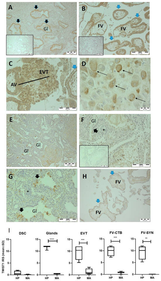
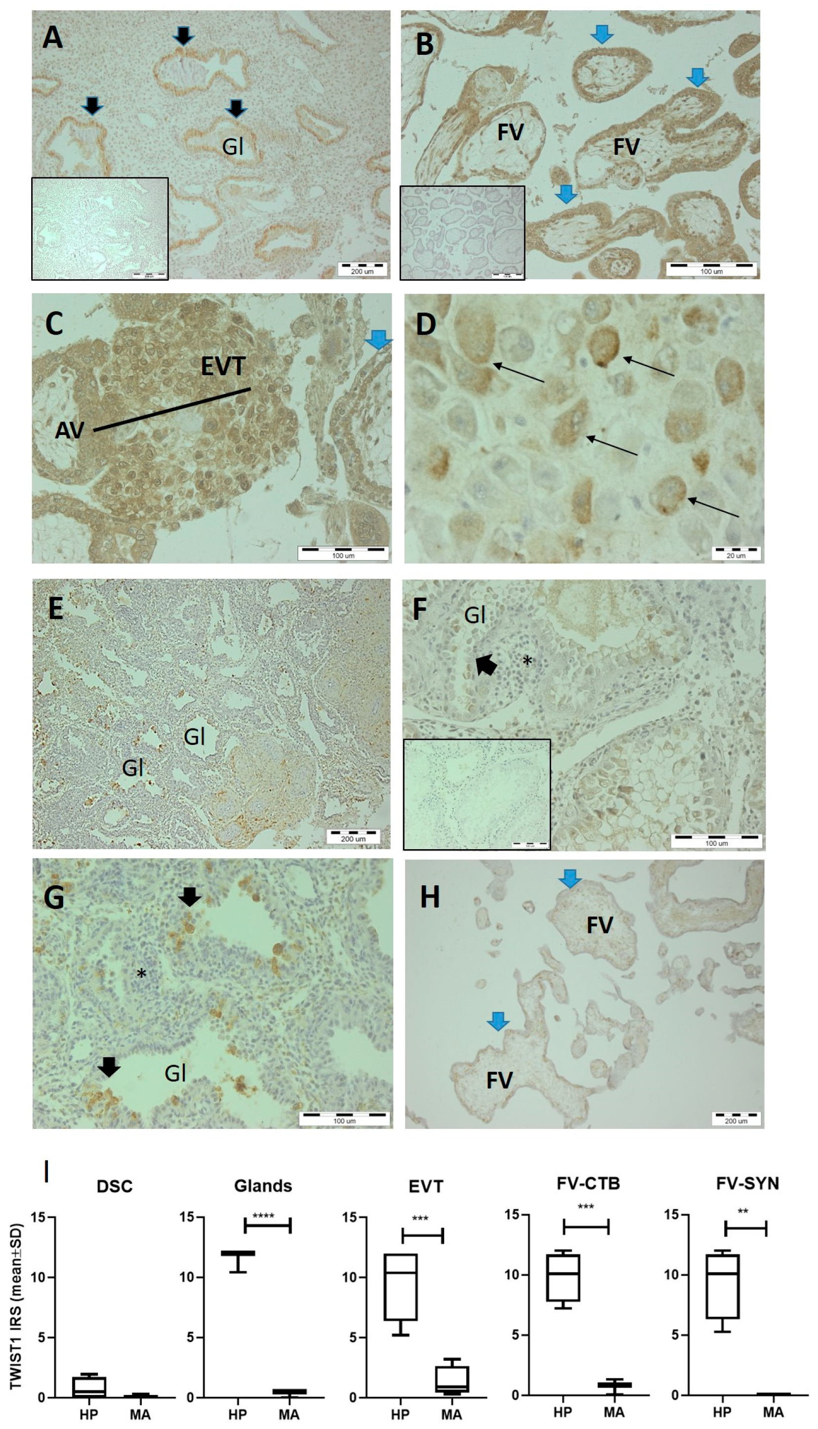
Our results showed an intensive signal for TWIST1 in the gestational tissues of normal pregnancy (Figure 1). The glandular epithelial cells of healthy decidua and the trophoblast cells of placental villi strongly expressed TWIST1 (Figure 1A,B,I). Trophoblast cells in cytotrophoblast columns of the anchoring placental villi and the EVT invading into decidua showed a particularly intense reaction for TWIST1 (Figure 1C,I). The cytoplasm of some decidual stromal cells was stained faintly for TWIST1 regardless of healthy or aborted tissue (Figure 1D,I, p = 0.17). In contrast, in the gestational tissues from missed aborted pregnancy—decidua and the placental villi—we revealed a weak reaction for TWIST1 (Figure 1E,I). Single glandular cells were TWIST1-positive (Figure 1F,G,I, p < 0.0001) and the immature and severely disrupted placental villi showed weak TWIST1 expression in both villous cytotrophoblast and syncytiotrophoblast (Figure 1H,I, p = 0.0008 and p = 0.003). Massive lymphocyte infiltration was observed in the aborted decidua (Figure 1G). All these data indicate that reduced TWIST1 expression strongly correlates with impaired placentation in the aborted tissues.
Figure 1.
TWIST1 expression in gestational tissues (decidua and placental villi) of healthy pregnancy (A–D) and missed abortions (E–H) with immunoreactive score (I). (A–D) An intense TWIST1 signal in the epithelial cells of the glands in normal decidua (A, black arrow) and in the trophoblast of the placental villi (B, blue arrow). Intermediate trophoblasts and EVTs in the cytotrophoblast columns (black line) of the anchoring placental villi are TWIST1-positive (C). Weak TWIST1 expression is shown by decidual stromal cells (D, black thin arrow). (E,F) Note the weak TWIST1 signal throughout the aborted decidua and uterine glands (black arrow), as compared to the strong TWIST1 signal in healthy decidua (A). (G) Single TWIST1-positive glandular cells in aborted decidua at higher magnification (black arrow). Note the massive lymphocyte infiltration, shown by an asterisk (*,F,G). Immature and severely destroyed placental villi weakly express TWIST1 (H, blue arrow). The magnification is shown by a scale bar (A,E,H—200 µm; B,C,F,G—100 µm; D—20 µm). (I) Graphs showing TWIST1 reaction determined in several decidual and placental compartments using the 12-point semi-quantitative immunoreactive score (IRS). Gl—gland, AV—anchoring villi, FV—floating villi, EVT—extravillous trophoblasts, BV—blood vessel, DSC—decidual stromal cell, FV-CTB—villous cytotrophoblast, FV-SYN—syncytiotrophoblast, HP—healthy pregnancy, MA—missed abortion. n = 10 donors per group.
3.2. Validation for the Sw71 TWIST1-KO Cell Line
Both Sw71 TW1-KO and Sw71 WT trophoblasts were cultured in healthy, mycoplasma-free cultures with numerous dividing cells (Figure S1). Before conducting all experiments with Sw71 TW1-KO cells, we performed proteomic validation by Western blot to confirm the complete absence of the TWIST1 protein in the TW1-KO clone against the wildtype parental line. Four Sw71 TW1-KO passages (p.7, p.8, p.9, and p.11) were tested for the presence of the TWIST1 transcription factor against coresponding Sw71 WT cells (p.7, p.8, p.9, and p.11). Western blot data indicated an absence of the TWIST1 protein in the knocked-out trophoblasts, confirming the loss of protein expression in the passages used for the current study. Western blot data for TW1-KO and WT Sw71 cells at p. 11 are shown in Figure S1.
3.3. Generation and Morphology of 3D TWIST1-Knocked-Out Sw71 BLS (TW1-KO Sw71 BLS)
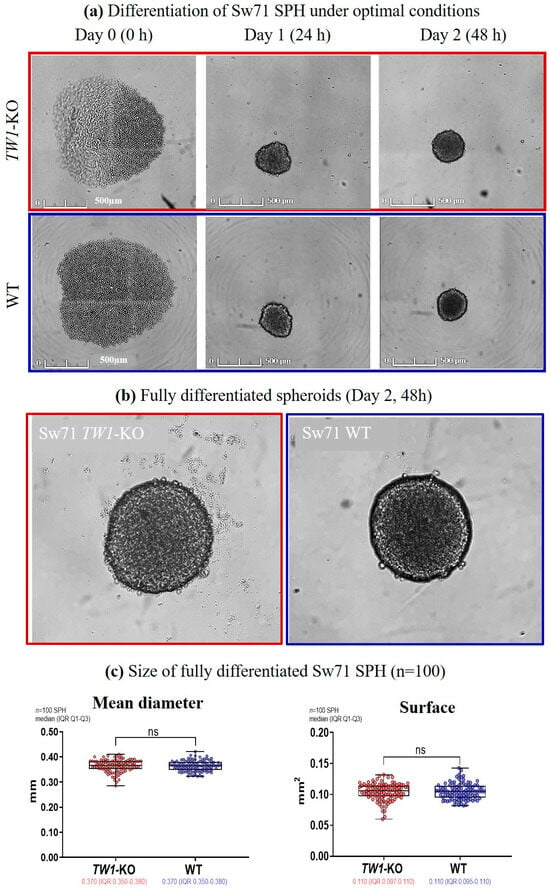
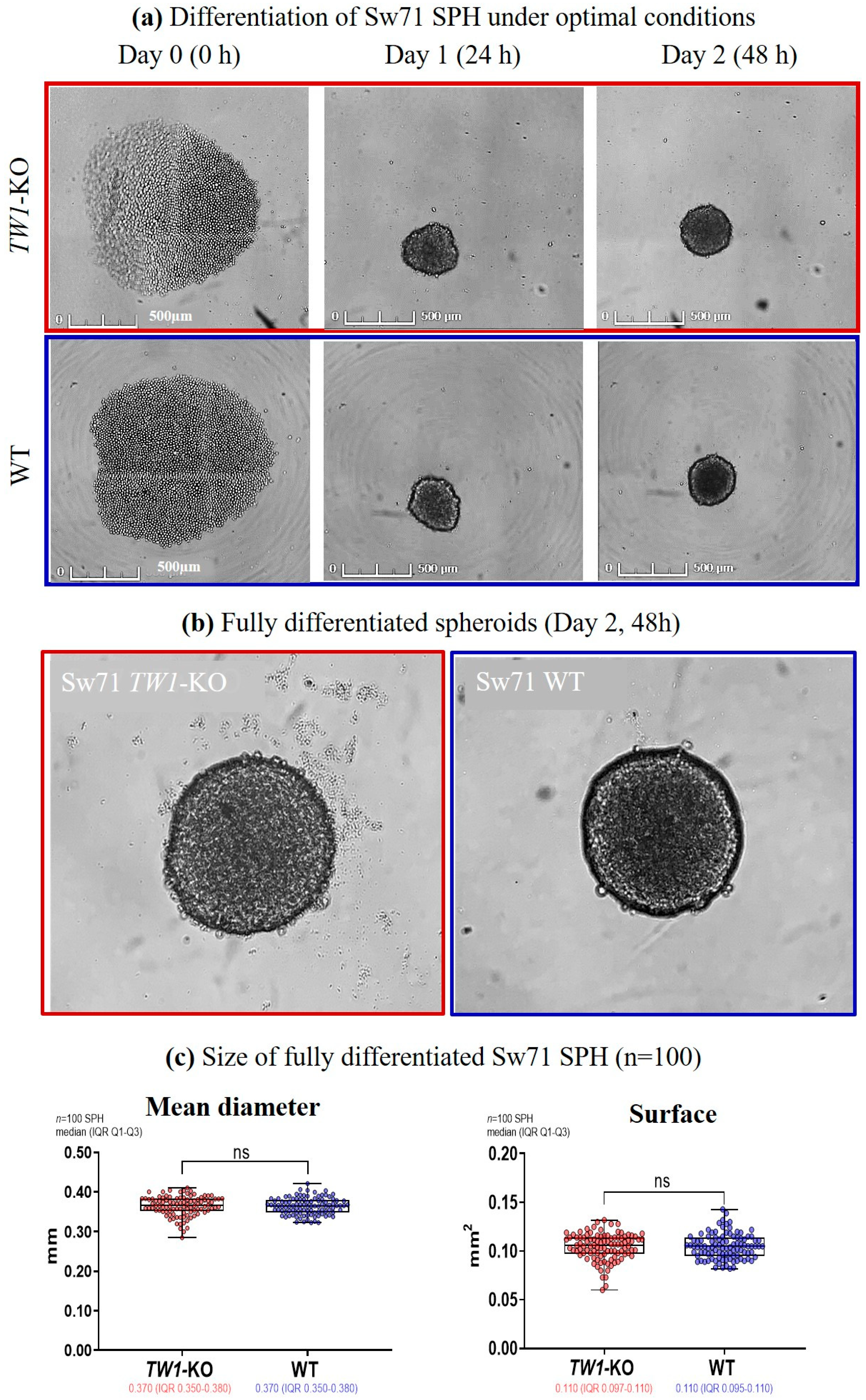
During propagation we found that the genetically modified TW1-KO Sw71 EVT-like cells multiply with retardation and were easier trypsinized than WT Sw71 trophoblasts. Of note, the differentiation of the epithelial Sw71 trophoblast cells into mesenchymal phenotype during spheroid formation was previously assessed [19]. Ten independent spheroid differentiation experiments were performed, each with 10 BLSs of both types (TW1-KO or WT). Thus, we finally obtained and morphologically characterized 100 spheroids of each type by measuring their mean diameter (mm) and spheroids’ surface (mm2), presented as median/IQR. We observed that Sw71 TW1-KO spheroids, like WT ones, were formed within 48 h as round multiple-cell-layered compact structures, where the presence of cell–cell junctions promotes the formation of a spheroid-like architecture (Figure 2; Videos S1 and S2). In both types, the strong compaction leads to significant reduction of the spheroids’ size (Figure 2). The statistical analysis showed that Sw71 TW1-KO spheroids had diameter (0.370 mm (IQR 0.350–0.380)) and surface (0.110 mm2 (IQR 0.097–0.110)) similar to those of control spheroids (0.370 mm (IQR 0.350–0.380) and 0.110 mm2 (IQR 0.095–0.110), Figure 2). The statistical analysis at the level of the independent experiment showed the same results—comparable morphology of both types of spheroids (Figure S2). Once formed, Sw71 TW1-KO spheroids are resistant to dissociation. Although Sw71 TW1-KO spheroids’ periphery is not as intact as that of WT BLSs, they were stable for transfer and further manipulation (Figure 2).
Figure 2.
Differentiation and morphological characterization of Sw71 TW1-KO compared to Sw71 WT spheroids. (a) Representative Sw71 TW1-KO (red frame) and WT (blue frame) spheroids shown on Days 0, 1, and 2 of differentiation. After strong compaction on Day 1, the size of the spheroid decreases significantly. The magnification is given by a scale bar (500 µm). (b) Representative images of fully differentiated Sw71 TW1-KO and WT BLSs. Images were digitally zoomed to be visualized in detail. (c) Comparable diameter and surface for both types of spheroids, presented as median (IQR Q1–Q3), n = 100 spheroids per group from 10 independent experiments, each with 10 speroids per group. ns- no significant difference.
3.4. HLA Profile of the Sw71 TW1-KO BLS Model
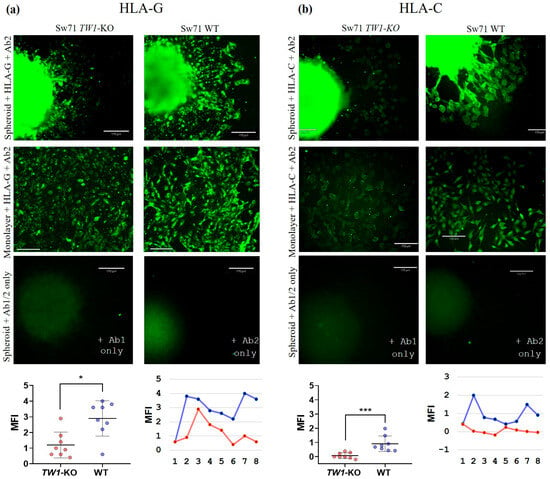
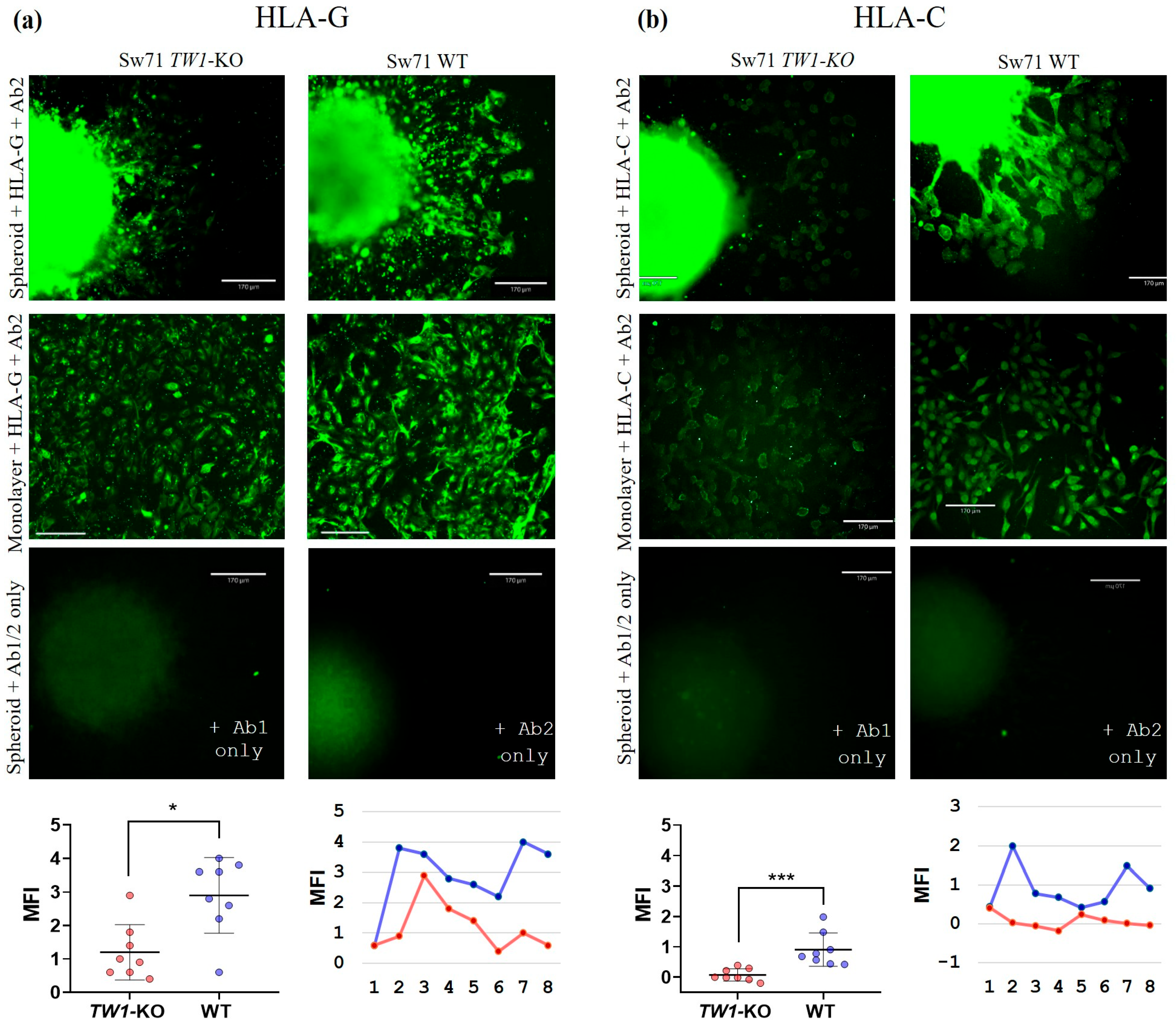
We have already shown the specific HLA-G+/HLA-C+ profile of Sw71 EVT-like cells resembling the EVT in vivo during human implantation [25]. Here, we checked the HLA profile of migrating Sw71 TW1-KO BLSs and in monolayer in the context of HLA-G and HLA-C expression. The results show significantly lower intensity of expression of both HLA-G and HLA-C molecules in Sw71 TW1-KO EVT-like migrating cells as compared to WT cells (Figure 3 and Figure S2). The MFI of the HLA-G signal of TW1-KO and WT cells was 1.20 ± 0.83 and 2.90 ± 1.13, respectively (p < 0.05). The MFI of the HLA-C signal was 0.07 ± 0.18 for TW1-KO cells and 0.91 ± 0.55 for WT ones (p < 0.001). It is important to note that immunofluorescent staining allowed us to demonstrate single-cell localization and intensity of the HLA molecules in functional (migrating) EVT-like cells of TW1-KO or WT BLS. Three independent immunocytochemistry stainings were performed with either monolayers or BLSs. The analysis at the level of the independent experiment confirmed these findings (Figure S2).
Figure 3.
HLA-G and HLA-C expression by Sw71 TW1-KO vs. Sw71 WT cells and spheroids, representative for 3 experiments. HLA-G (a) and HLA-C (b) expression by Sw71 TW1-KO vs. WT spheroids (1st row) and monolayers (2nd row), and appropriate controls by omitting the primary or secondary antibodies (3rd row). Lower HLA-G and HLA-C signals of migrating Sw71 TW1-KO cells than WT cells shown by analysis of MFI of migrating BLSs, representative for n = 3 in each group (4th row). Dots represent fields of measurement in a single spheroid (TW1-KO and WT). Note that these are not independent replicates. The magnification is given by a scale bar (170 µm).
3.5. Sw71 TW1-KO Spheroids Showed Markedly Impaired Migration
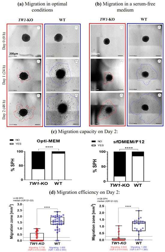
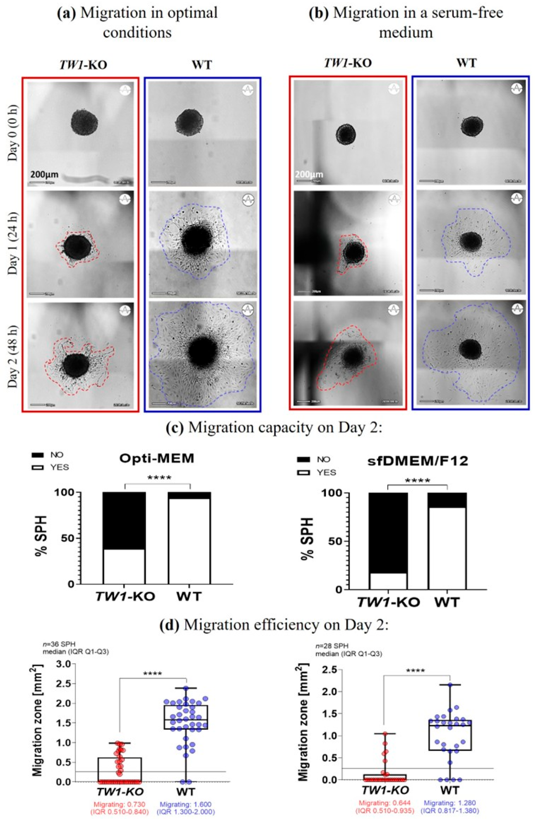
As mentioned previously, Sw71 EVT-like cells within BLSs successfully migrate, mimicking EVTs in vivo [20]. The migrating 3D Sw71 BLS is a dynamic structure. In the first 10–12 h after plating, the spheroid attaches to the bottom of the plate, and single Sw71 cells begin to detach from the periphery in a synchronized manner to form a zone of migrating trophoblast cells extending outward. The more cells migrate, the spheroid’s periphery becomes more diffuse. Since it is well-known that knock out of the TWIST1 gene leads to impaired cell motility, it was of interest to test the ability of Sw71 TW1-KO BLSs to migrate versus control WT ones. In addition to the most favorable conditions, the migration was monitored in a serum starvation-based condition as well, knowing that Sw71 WT BLSs successfully migrate even under starvation [20]. The capacity and efficiency of EVT migration were monitored for 48 h in Opti-MEM medium (optimal conditions; Figure 4a,c,d; Videos S3 and S4) and in sfDMEM/F12 complete (starving; Figure 4b–d). Nine independent experiments, each with 4 SPH per type, were performed. Our results showed that the Sw71 TW1-KO BLS had markedly impaired migration as capacity and efficiency, compared to WT spheroids. Under optimal conditions, only 39% of TW1-KO BLSs attempted to migrate, forming a smaller and less defined migration zone, 0.730 mm2 (IQR 0.510–0.840), vs. 94% of WT spheroids with an extensive and well-defined migration zone, 1.600 mm2 (IQR 1.300–2.00). Sixty one percent of TW1-KO spheroids vs. 6% of WT spheroids did not migrate at all (Figure 4c,d). The statistical analysis at the level of the independent experiment revealed the same results—lower migration efficiency of TW1-KO spheroids (Figure S2). As expected, the starvation stressed more Sw71 TW1-KO trophoblasts than WT ones. The proportion of migrated TW1-KO spheroids was 18%, showing a small and loose, often disrupted migration zone, 0.644 mm2 (IQR 0.510–0.935), in contrast to 86% migrated Sw71 WT BLSs with a larger migration zone, 1.280 mm2 (IQR 0.817–1.380; Figure 4c,d and Figure S2). The non-migrated TW1-KO and WT spheroids were 82% and 14%, respectively. When analyzed at the experiment level, we have a reduction of the p-value but the significance remains strong (Figure S2).
Figure 4.
Migration of Sw71 TW1-KO BLSs under optimal (a) and starvation (b) conditions. (a) Representative images of migrating TW1-KO (red frame) and WT (blue frame) spheroids in Opti-MEM over 48 h. Within 24 h, EVTs detach and form a migration zone, which is smaller and less defined in Sw71 TW1-KO BLSs (red dashed line) compared to the larger, clearly visible zone in Sw71 WT ones (blue dashed line). (b) Representative images of TW1-KO (red frame) and WT (blue frame) spheroids migrating in sf DMEM/F12 over 48 h. TW1-KO spheroids form a smaller and often disrupted migration zone (red dashed line), while WT BLSs show a more distinct and denser zone (blue dashed line). (c) Migration capacity of Sw71 TW1-KO BLSs in optimal conditions was impaired as compared to Sw71 WT BLSs (n = 36 spheroids from 9 independent experiments with 4 replicates). It was strongly affected/influenced by the removal of FBS from the media (n = 28 spheroids from 7 independent experiments with 4 replicates). (d) Coresponding migration efficiency of Sw71 TW1-KO BLSs was markedly reduced as compared to Sw71 WT BLSs in both optimal and starving conditions. The magnification is given by a scale bar (200 µm).
Altogether, the data demonstrated that the capacity and efficiency of migration by TW1-KO EVT-like cells were seriously compromised, most probably from the genetic modification.
3.6. Sw71 TW1-KO Surrogates Showed Blocked Invasion
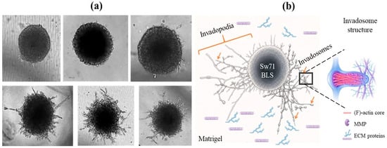
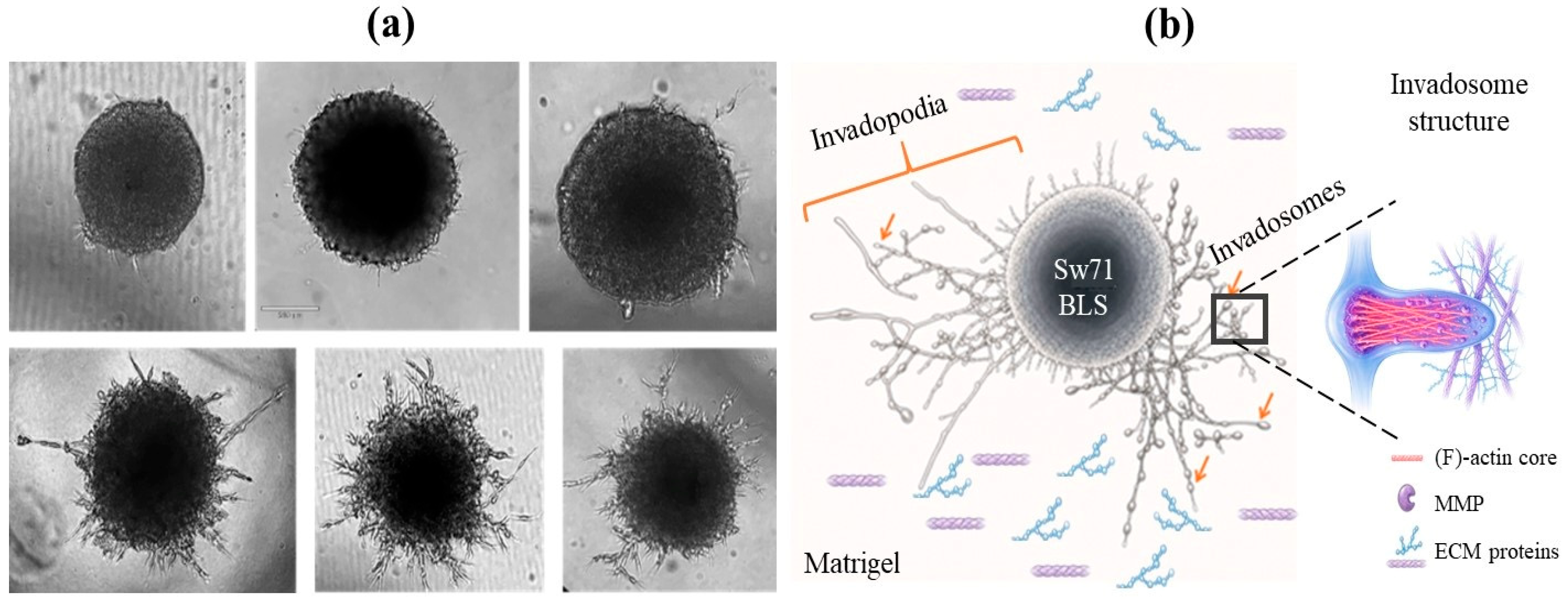
A key stage of in vivo implantation of human blastocyst is invasion of EVT into decidual stroma, glands, and uterine spiral arteries [1]. This process was simulated using Sw71 TW1-KO and WT BLSs invading in Matrigel (ECM), monitored for six days to follow the formation of long plasma membrane well-developed invadopodia-like outgrowths, the role of which is to support the sinking of the spheroid into ECM (Figure 5b). Matrigel is enriched in basement membrane proteins, such as laminin, entactin, and type IV collagen, which provide essential ligands needed for integrin adhesion during cell invasion [32]. The mature actin-rich invadopodia (schematically presented in Figure 5b) includes a dense filamentous (F)-actin core that contains proteins controlling actin assembly. Apart from the key scaffold components, the invadopodia are enriched in proteases, including matrix metalloproteases (MMPs), to degrade the ECM and ease trophoblast migration [33]. The invasion of Sw71 WT BLSs in Matrigel allowed the development of well-visible invadopodia-like outgrowths and clear delineating of the invasion zone and successfully served as a positive control. The monitoring of Sw71 TW1-KO BLSs showed few (thin and short) or no cell outgrowths at all, demonstrating blocked invasion (Figure 5a). Although classical, this test for invasion is a model system lacking some biological factors otherwise found in vivo.
Figure 5.
Invasion of Sw71 TW1-KO and WT BLSs in Matrigel. (a) Triplets of Sw71 TW1-KO (top) and WT (bottom) BLSs placed to invade and imaged at Day 6. Unlike the rare and short non-growing protrusions of the non-invading Sw71 TW1-KO BLS, WT spheroids acquired a distinctive brush-like (star-shaped) appearance due to numerous well-developed long invadopodia-like outgrowths. All images were digitally zoomed to be visualized in detail. (b) Scheme of a well-invaded Sw71 BLS and the structure of the invadopodia at closer view (orange arrows). Invasion zone outlines were extracted with https://illustrae.co from the microscopic image of the Sw71 BLS.
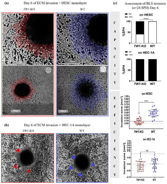
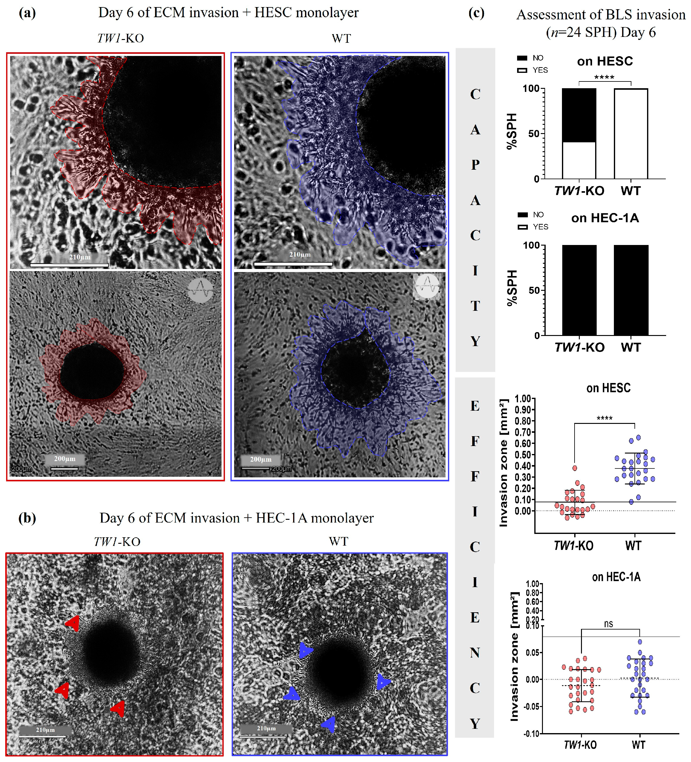
To mimic more physiologically the trophoblast invasion, we placed genetically modified Sw71 TW1-KO or Sw71 WT BLSs to invade into scaffold of endometrial stromal cells (HESC) layered with Matrigel (Figure 6a). These were monitored for 6 days with imaging and analysis on Days 2, 4, and 6. Unlike control Sw71 WT spheroids invading successfully and efficiently into Matrigel to reach the endometrial stromal cells, the invasion of Sw71 TW1-KO BLSs was impaired (Figure 6c and Figure S2). On Day 2, half of the spheroids in both groups started to form an invasion zone (p > 0.05). Between Days 4 and 6, only 30–40% of the tested Sw71 TW1-KO spheroids were able to release very few thin and short invadopodia-like structures, delineating a small, unclear, and non-growing invasion area on Day 6 (0.07 ± 0.11 mm2), often containing halo of numerous rounded dead EVTs. In contrast, all tested WT spheroids were functional, with many thick and long invadopodia-like outgrowths and developing well-visible invasion area into Matrigel, targeting the stromal cells (0.38 ± 0.14 mm2, p < 0.0001; Figure 6c and Figure S2). Of note, only the invasion zone of Sw71 WT BLSs increased progressively between Day 2 and Day 6 (p < 0.0001). The data about capacity and efficiency of the six-day invasion of TW1-KO vs. WT spheroids are shown in Figure 6c.
Figure 6.
Invasion of Sw71 TW1-KO and Sw71 WT spheroids under promoting (a) and non-promoting (b) conditions. (a) Representative images of Sw71 TW1-KO BLSs invading the ECM and endometrial stromal cells (HESCs), compared to WT spheroids (Day 6). The zone of EVT invasion with the established invadopodia-like outgrowths is delineated in red or blue. Note the few and small cell outgrowths and almost no invasion zone by Sw71 TW1-KO BLSs, unlike numerous and long invadopodia-like outgrowths and clear and the well-visible invasion zone by WT spheroids. The magnification is presented by a scale bar (210 µm up, 200 µm down). (b) Representative images of Sw71 TW1-KO and WT BLSs unable to invade the endometrial epithelial cells (HEC-1A) layered with Matrigel (Day 6), showing few thin and short invadopodia (arrowheads) and peripheral halo of dead EVT. Significant BLS destruction aggravated for Sw71 TW1-KO in non-promoting conditions. The magnification is presented by a scale bar (210 µm). (c) The spheroids’ invasion was assessed as capacity and efficiency on Day 6. Sw71 TW1-KO BLSs showed reduced invasion capacity compared to fully invasive WT BLSs. Sw71 TW1-KO EVT-like cells did not invade into Matrigel on the stromal cells, unlike WT spheroids showing a growing and well-developed area of invasion, n = 24 spheroids from 6 independent experiments with 4 spheroids per group in each.
It is well-known that trophoblast invasion could be modulated by the distinct microenvironments to support orthotopic vs. ectopic embryo growth. We decided to test the invasion of both types of EVTs under non-physiological contact with low receptive endometrial epithelial cells (HEC-1A) layered with Matrigel as a non-promoting condition. These formed few, non-growing invadopodia-like outgrowths, often disappearing before Day 6. Neither Sw71 TW1-KO (100% non-invasive, area: −0.012 ± 0.03 mm2) nor WT Sw71 EVTs (100% non-invasive, area: 0.003 ± 0.035 mm2) were able to invade the ECM with epithelial cells (p > 0.05, Figure 6b,c and Figure S2). We performed 6 independent experiments with 4 spheroids from both types in each (n = 24). The analysis at the level of the independent experiment confirmed the above-mentioned findings (Figure S2).
4. Discussion
Taken together, these findings are consistent with a role for TWIST1 in the human decidua and placenta early in pregnancy. TWIST1 expression is downregulated in missed abortion tissues compared to in healthy gestational tissues, and loss of TWIST1 impairs the migration and invasion of EVT-like cells in vitro.
It has been shown that the TWIST is involved in the preparation of the decidua for embryo implantation since it was detected with the induction of decidualization [34]. Several studies revealed the important role of TWIST in the trophoblasts syncytialization and function of syncytiotrophoblast [35,36]. The STB of the placental villi in healthy pregnancy shows higher TWIST1 expression than that of spontaneous pregnancy loss, and no TWIST1 was detected in the placenta of women with recurrent pregnancy loss [17] as well as in the placenta of PE patients [37]. In line with the above-mentioned studies, our results confirmed the weak TWIST1 expression in the placental villi of the women with missed abortion, unlike the strong signal in healthy human placenta. Although this observation could be a downstream consequence in missed abortion, there are published data showing a link between TWIST1 and inflammation (as a potential cause of missed abortion). It is well-known that a mild Th1-induced inflammation is required for the successful implantation. The inflammatory cytokines IFNγ and TNFα facilitate blood vessel remodeling and stimulate EVT invasion [38]. The inflammatory cytokines and TWIST1 expression are linked through a key biological feedback loop where pro-inflammatory signals turn on TWIST1, and TWIST1 then acts as an inflammatory brake by limiting IFNγ and TNFα production [39]. Accordingly, the insufficiency of TWIST1 could allow an excessive inflammation and lack of timely switch from Th1 to Th2 environment—a key requirement for successful pregnancy. Here, we observed that TWIST1 is highly expressed by EVTs within the healthy maternal decidua in situ. These cells play a pivotal role in the formation and function of human placenta and the maintenance of the normal development of the fetus, suggesting a primary role for TWIST1 in trophoblast function [17]. The study by Peng et al. reported that reproduction-related hormones like gonadotropin-releasing hormones I and II upregulate the expression of TWIST in human trophoblast cells [40]. It has been shown that TWIST increases the expression of mesenchymal markers vimentin and N-cadherin and significantly decreases the expression of E-cadherin in the EMT of the tumor cells [41] and healthy human EVTs [15,42]. Due to EMT, EVTs lose the polarity and acquire an invasive behavior. This allows them to break through extracellular matrices and migrate/invade the decidua basalis to transform the maternal spiral arteries into high-flow, low-resistance vessels, providing an adequate maternal blood flow to the developing placenta [37,42,43,44]. The 6th–8th week of pregnancy, when the EVTs invade the spiral artery and participate in the formation of the placenta, is considered to be the most crucial period for studying the pathophysiological changes of EVT function [45]. However, our histological study provided a snapshot of the cells and events that occurred later on, during early pregnancy, without insights into the dynamics of the processes and possible alterations earlier during implantation. The uniqueness of human implantation as a deep invasion of EVT in the endometrial stroma, glands, and vessels, providing early histotrophic nutrition and subsequent formation of hemochorial villous placenta, is not possible to study in vivo and ex vivo due to practical and ethical obstacles [10]. Thus, the advanced 3D models are valuable and very promising tools for fundamental research in peri-implantation and early placentation events and important biomarkers for subsequent pregnancy disorders [10]. Gene ablation techniques used in trophoblast cell lines have proven to be a powerful technique in elucidating TWIST1 gene function during implantation and particularly in EVT invasion [21]. In this study, we combined these two approaches to establish a 3D Sw71 TW1-KO in vitro model. There are few trophoblast cell lines suitable for TWIST1 research. Whereas JEG-3 and BeWo trophoblast cell lines poorly expressed TWIST1 [15], HTR-8/SVneo and Sw71 EVT-like cell lines strongly expressed TWIST1 and seem suitable for TWIST1 knockdown/out via transfection and/or transduction [15,21]. For example, a small interfering RNA strategy has been employed to determine the role of TWIST in trophoblast invasion using the HTR-8/SVneo cell line [15]. The authors showed that the TWIST downregulation decreases N-cadherin and reduces the invasion of HTR-8/SVneo cells as monolayer in a simple insert-based invasion assay using a porous membrane coated with Matrigel. Thus, TWIST is suggested as an upstream regulator of the mesenchymal marker N-cadherin-mediated invasion of human EVT cells [15]. In our study, the possible contribution of TWIST1 in the EVT function was revealed by using an advanced three-dimensional TWIST1-knocked-out blastocyst surrogate invading the endometrial stroma layered with ECM to mimic as closely as possible the maternal–fetal interface during embryo implantation. The non-cancerous endometrial stromal cell line (HESC) used as a scaffold in our invasion test promotes trophoblast invasion through the generation of a mild inflammatory environment modulated by TNF-α like in human implantation in vivo [28]. Moreover, to emphasize the specific effect of endometrial stromal cells on EVT invasion, we confirmed the lack of invasion when the EVTs were co-cultured with endometrial epithelial cells.
The main finding of the study is the analysis of the migration and invasion of genetically modified TW1-KO blastocyst-like surrogates. We clearly demonstrate that deletion of TWIST1 hinders the invasion of EVT-like cells and on-time formation of strong and growing invadopodia-like outgrowths. This prevents the breaking down of the extracellular matrix and contact with the endometrial stroma. Although TW1-KO blastocyst-like surrogates formed compact and stable structures in the same timeframe and manner as the normal Sw71 BLS, we could not provide evidence about different expression of the differentiation markers.
While in this study we functionally confirmed cell invasion through active extracellular matrix degradation, structural validation of the invadopodia-like outgrowths remains to be established. Complicating the invasion test in our study by bringing it closer to physiological conditions comes at the expense of accuracy in measuring the invasion area, since some of the invadopodia-like structures could be masked by the stromal cell scaffold. Together with the manual image assessment of the invasion zone, which is time- and human-resource-consuming, these could be considered as shortcomings. A perspective alternative to bypass this limitation is implantometrics, an automated deep learning analysis recently described by Gyunesh et al., which is significantly faster and more accurate [27].
Limitation of our study is the lack of a rescue experiment as a valuable control to rule out the off-target effects. The used guide RNA in CRISPR/Cas9 TWIST1 knockout of Sw71 cells had high specificity with low predicted off-target scores proven by analysis with CRISPOR software. As a robust alternative we verified the TWIST1 protein loss by Western blot to demonstrate that the phenotype is not an artifact. However, this validation does not rule out possible clonal or off-target effects.
It seems that an additional effect of TWIST1 removal was the diminished expression of the tolerogenic HLA molecules HLA-G, HLA-C, and HLA-F [21,31]. We confirmed that the deletion of the TWIST1 gene leads to lower expression of the essential HLA molecules HLA-G and HLA-C by the Sw71 TW1-KO BLS. These HLA molecules provide adequate immune recognition of the embryo and an establishment of the maternal–fetal immune tolerance via interaction with KIR on the decidual cytotoxic immune cells [46,47]. In line with this, Dunk et al. reported that both HLA-C and HLA-G are markedly reduced in placentas and maternal blood from pregnancies complicated by PE [48], recurrent miscarriage, and fetal growth restriction, suggesting unfavorable HLA-C/KIR combinations associated with these complications [49]. Moreover, the optimal HLA expression by EVT prompts the decidual NK cells to secrete vital pro-invasive chemokines and cytokines [50]. It has been shown that when HLA-C or HLA-F expression drops, decidual NK cells fail to receive an activation to produce the chemical signals that normally stimulate the trophoblasts to migrate and invade the uterine tissue [51]. Beyond interacting with maternal immune cells, molecules like HLA-G act directly on the trophoblasts themselves via autocrine pathways to assist with invasion [52]. Membrane-bound and soluble HLA-G promote the secretion of MMP-2 and MMP-9, specialized enzymes that digest the maternal extracellular matrix, carving out a physical path for EVTs to migrate into the uterus [53]. It seems that TWIST1 transcriptional factor acts via intermediate molecular networks when modulating the HLA molecules. The reduced HLA-G/HLA-C expression in TW1-KO cells is not due to direct binding of TWIST1 to suppress their transcription because these HLA molecules typically lack canonical E-box consensus sequences that TWIST1 binds directly [54]. TWIST1 probably modulates the immune landscape and suppresses these molecules indirectly by upregulating downstream repressors, coordinating with signaling pathways (such as STAT3, NF-κB, or TGF-β), or altering local cytokines and microRNAs known to downregulate or post-transcriptionally silence HLA-G and HLA-C expression [55]. We have already shown the indirect effect of TWIST1 on HLA-G/HLA-C expression via binding to IRF9 [21].
Interestingly, the study of Ma et al. revealed that TWIST1 promotes cell survival. Removing TWIST1 amplifies mitochondrial-driven cell death via apoptosis, while its restoration suppresses this pathway [12]. Also, we revealed previously that human trophoblast cells became vulnerable to Zika virus infection when TWIST1 was depleted [21]. Indeed, in our hands, the TW1-KO surrogates were less resistant to starving compared to WT ones. While differential viability was not formally assessed (a shortcoming of the study), we fully account for the possibility that lower viability of TW1-KO cells would indirectly affect their potential to migrate/invade. Furthermore, any potential impact of compromised cell growth or adhesion on the migration and invasion outcomes must be explicitly stated.
Numerous studies have shown that proper trophoblast invasion is essential for the remodeling of maternal spiral arteries to supply the growing placenta and fetus (references in [56]). In contrast, the stunted invasion process is a major underlying mechanism in GOS (great obstetrical syndrome), including pregnancy complications such as PE, fetal growth restriction, preterm birth, preterm premature rupture of membranes, placental abruption, and stillbirth (references in [57]). Therefore, we believe that the 3D Sw71 TW1-KO spheroid model, resembling in size and shape the human blastocyst in the peri-implantation period, but showing severely reduced migration and invasion capacity and efficiency, could be used to simulate the state of shallow EVT invasion—the hallmark of GOS. Different signaling pathways underlying TWIST1-associated EVT dysfunction could be explored to find prognostic markers for early detection of such late pregnancy disorders. Strengthening collaboration between basic research and clinical practice using such standardized models will be critical for translating findings into clinical applications to improve maternal and fetal health outcomes.
5. Conclusions
The transcription factor TWIST1 is an important component of the healthy decidua and placenta early in human pregnancy. The present study shows that the deletion of the TWIST1 gene in the Sw71 EVT-like cells preserves spheroids’ formation but leads to decreased expression of essential HLA molecules and affects the EVT function. The 3D Sw71 TWIST1-knocked-out human blastocyst surrogates provide new insight into the involvement of TWIST1 in trophoblast functionality and could be a potential in vitro model to study TWIST1-related signaling pathways involved in the compromised (shallow) EVT invasion.
Supplementary Materials
The following supporting information can be downloaded at: https://www.mdpi.com/article/10.3390/cimb48111146/s1.
Author Contributions
Conceptualization, T.D. and G.M.; methodology, Y.Y., M.I., M.A., and D.M.; data curation, M.I., M.A., and T.D.; formal analysis, M.I., M.A., D.M., and S.S.; validation, G.M.; funding acquisition, T.D.; investigation, M.I., M.A., D.M., T.D., E.S., and Y.Y.; project administration, T.D.; resources, S.S. and G.M.; visualization, M.A., M.I., and E.S.; writing—original draft, M.I. and M.A.; writing—review and editing, T.D. All authors have read and agreed to the published version of the manuscript.
Funding
The study is supported by the Bulgarian National Science Fund via NNP VIHREN, grant KP 06-DV3. The open access publishing is funded by Fulbright Bulgaria via the Alumni Grants Program.
Institutional Review Board Statement
The study was conducted following the Declaration of Helsinki and approved by the Human Research Ethics Committee at the University Obstetrics and Gynecology Hospital “Maichin Dom”, Sofia, Bulgaria (No. 25-95-75/30.12.2021), date: 30 December 2021.
Informed Consent Statement
All participants provided written informed consent for the use of the tissue samples.
Data Availability Statement
The datasets generated and analyzed during the current study are available from the corresponding author upon reasonable request.
Acknowledgments
We are grateful to Violeta Payakova, a midwife at University Obstetrics and Gynecology Hospital “Maichin Dom”.
Conflicts of Interest
The authors declare no conflicts of interest.
Abbreviations
The following abbreviations are used in this manuscript:
| BLS | Blastocyst-like surrogate |
| CTB | Cytotrophoblast |
| DAB | 3,3-diaminobenzidine tetrahydrochloride |
| ECM | Extracellular matrix |
| EMT | Epithelial–mesenchymal transition |
| EVT | Extravillous trophoblast |
| GOS | Great obstetrical syndrome |
| HEC-1A | Human endometrial epithelial cells |
| HESC | Human endometrial stromal cell |
| hCG | Human chorion gonadotropin |
| IFNγ | Interferon gamma |
| PBS | Phosphate-buffered saline |
| PE | Preeclampsia |
| sfDMEM/F12 | Serum-free DMEM/F12 |
| STB | Syncytiotrophoblast |
| TW1-KO | TWIST1-knocked out |
| TNFα | Tumor necrosis factor alfa |
| WT | Wildtype |
References
- Muter, J.; Lynch, V.J.; McCoy, R.C.; Brosens, J.J. Human embryo implantation. Development 2023, 150, dev201507. [Google Scholar] [CrossRef] [Scilit] [PubMed]
- Evers, J.L.H. Female subfertility. Lancet 2002, 360, 151–159. [Google Scholar] [CrossRef] [Scilit] [PubMed]
- Chard, T. Frequency of implantation and early pregnancy loss in natural cycles. Baillieres Clin. Obstet. Gynaecol. 1991, 5, 179–189. [Google Scholar] [CrossRef] [Scilit] [PubMed]
- Macklon, N.S.; Geraedts, J.P.M.; Fauser, B.C.J.M. Conception to ongoing pregnancy: The “black box” of early pregnancy loss. Hum. Reprod. Update 2002, 8, 333–343. [Google Scholar] [CrossRef] [Scilit] [PubMed]
- Wang, X.; Chen, C.; Wang, L.; Chen, D.; Guang, W.; French, J.; Xu, X. Conception, early pregnancy loss, and time to clinical pregnancy: A population-based prospective study. Fertil. Steril. 2003, 79, 577–584. [Google Scholar] [CrossRef] [Scilit] [PubMed]
- Renaud, S.J.; Jeyarajah, M.J. How trophoblasts fuse: An in-depth look into placental syncytiotrophoblast formation. Cell. Mol. Life Sci. 2022, 79, 433. [Google Scholar] [CrossRef] [Scilit] [PubMed]
- Moser, G.; Huppertz, B. Implantation and extravillous trophoblast invasion: From rare archival specimens to modern biobanking. Placenta 2017, 56 19. [Google Scholar] [CrossRef] [Scilit] [PubMed]
- DaSilva-Arnold, S.; James, J.L.; Al-Khan, A.; Zamudio, S.; Illsley, N.P. Differentiation of first trimester cytotrophoblast to extravillous trophoblast involves an epithelial–mesenchymal transition. Placenta 2015, 36, 1412–1418. [Google Scholar] [CrossRef] [Scilit] [PubMed]
- Li, X.; Zhou, J.; Fang, M.; Yu, B. Pregnancy immune tolerance at the maternal-fetal interface. Int. Rev. Immunol. 2020, 39, 247–263. [Google Scholar] [CrossRef] [Scilit] [PubMed]
- Dimova, T.; Alexandrova, M.; Vangelov, I.; You, Y.; Mor, G. The modeling of human implantation and early placentation: Achievements and perspectives. Hum. Reprod. Update 2024, 31 133. [Google Scholar] [CrossRef] [Scilit] [PubMed]
- Barbouti, A.; Varvarousis, D.N.; Kanavaros, P. The Role of Oxidative Stress-Induced Senescence in the Pathogenesis of Preeclampsia. Antioxidants 2025, 14, 529. [Google Scholar] [CrossRef] [Scilit] [PubMed]
- Ma, L.; Zhang, Z.; Dong, K.; Ma, Y. TWIST1 Alleviates Hypoxia-induced Damage of Trophoblast Cells by inhibiting mitochondrial apoptosis pathway. Exp. Cell Res. 2019, 385, 111687, Erratum in Exp. Cell Res. 2020, 387, 111777. https://doi.org/10.1016/J.YEXCR.2019.111687. [Google Scholar] [CrossRef] [Scilit] [PubMed]
- Tu, M.; Ge, B.; Li, J.; Pan, Y.; Zhao, B.; Han, J.; Wu, J.; Zhang, K.; Liu, G.; Hou, M.; et al. Emerging biological functions of Twist1 in cell differentiation. Dev. Dyn. 2025, 254, 8–25. [Google Scholar] [CrossRef] [Scilit] [PubMed]
- Gu, S.; Boyer, T.G.; Naski, M.C. Basic helix-loop-helix transcription factor Twist1 inhibits transactivator function of master chondrogenic regulator Sox9. J. Biol. Chem. 2012, 287, 21082–21092. [Google Scholar] [CrossRef] [Scilit] [PubMed]
- Ng, Y.H.; Zhu, H.; Leung, P.C.K. Twist modulates human trophoblastic cell invasion via regulation of N-cadherin. Endocrinology 2012, 153, 925–936. [Google Scholar] [CrossRef] [Scilit] [PubMed]
- Li, Z.; Wang, J.; Li, D.; Chen, H.; Meng, T. MiR-372-3p promotes preeclampsia progression by regulating twist1. Exp. Ther. Med. 2022, 24, 723. [Google Scholar] [CrossRef] [Scilit] [PubMed]
- Ahn, J.H.; Park, H.R.; Park, C.W.; Park, D.W.; Kwak-Kim, J. Expression of TWIST in the first-trimester trophoblast and decidual tissue of women with recurrent pregnancy losses. Am. J. Reprod. Immunol. 2017, 78. [Google Scholar] [CrossRef] [Scilit] [PubMed]
- Holmberg, J.C.; Haddad, S.; Wünsche, V.; Yang, Y.; Aldo, P.B.; Gnainsky, Y.; Granot, I.; Dekel, N.; Mor, G. An in vitro model for the study of human implantation. Am. J. Reprod. Immunol. 2012, 67, 169–178. [Google Scholar] [CrossRef] [Scilit] [PubMed]
- You, Y.; Stelzl, P.; Zhang, Y.; Porter, J.; Liu, H.; Liao, A.H.; Aldo, P.B.; Mor, G. Novel 3D in vitro models to evaluate trophoblast migration and invasion. Am. J. Reprod. Immunol. 2019, 81. [Google Scholar] [CrossRef] [Scilit] [PubMed]
- Alexandrova, M.; Manchorova, D.; You, Y.; Mor, G.; Dimitrova, V.; Dimova, T. Functional HLA-C expressing trophoblast spheroids as a model to study placental–maternal immune interactions during human implantation. Sci. Rep. 2022, 12. [Google Scholar] [CrossRef] [Scilit] [PubMed]
- You, Y.; Grasso, E.; Alvero, A.; Condon, J.; Dimova, T.; Hu, A.; Ding, J.; Alexandrova, M.; Manchorova, D.; Dimitrova, V.; et al. Twist1-IRF9 Interaction Is Necessary for IFN-Stimulated Gene Anti-Zika Viral Infection. J. Immunol. 2023, 210, 1899–1912. [Google Scholar] [CrossRef] [Scilit] [PubMed]
- Sato, Y.; Fujiwara, H.; Konishi, I. Mechanism of maternal vascular remodeling during human pregnancy. Reprod. Med. Biol. 2011, 11 27. [Google Scholar] [CrossRef] [Scilit] [PubMed]
- Knöfler, M.; Haider, S.; Saleh, L.; Pollheimer, J.; Gamage, T.K.J.B.; James, J. Human placenta and trophoblast development: Key molecular mechanisms and model systems. Cell. Mol. Life Sci. 2019, 76, 3479–3496. [Google Scholar] [CrossRef] [Scilit] [PubMed]
- Straszewski-Chavez, S.L.; Abrahams, V.M.; Alvero, A.B.; Aldo, P.B.; Ma, Y.; Guller, S.; Romero, R.; Mor, G. The Isolation and Characterization of a Novel Telomerase Immortalized First Trimester Trophoblast Cell Line. Swan 71, Placenta 2009, 30, 939–948. [Google Scholar] [CrossRef] [Scilit] [PubMed]
- Alexandrova, M.; Manchorova, D.; You, Y.; Terzieva, A.; Dimitrova, V.; Mor, G.; Dimova, T. Validation of the Sw71-spheroid model with primary trophoblast cells. Am. J. Reprod. Immunol. 2023, 90. [Google Scholar] [CrossRef] [Scilit] [PubMed]
- Krikun, G.; Mor, G.; Alvero, A.; Guller, S.; Schatz, F.; Sapi, E.; Rahman, M.; Caze, R.; Qumsiyeh, M.; Lockwood, C.J. A novel immortalized human endometrial stromal cell line with normal progestational response. Endocrinology 2004, 145, 2291–2296. [Google Scholar] [CrossRef] [Scilit] [PubMed]
- Gyunesh, A.A.; Rezk-Füreder, M.; Kapper, C.; Mor, G.; Shebl, O.; Oppelt, P.; Stelzl, P.; Arbeithuber, B. Multidimensional trophoblast invasion assessment by combining 3D in vitro modeling and deep learning analysis. npj Syst. Biol. Appl. 2025, 11, 116. [Google Scholar] [CrossRef] [Scilit] [PubMed]
- You, Y.; Stelzl, P.; Joseph, D.N.; Aldo, P.B.; Maxwell, A.J.; Dekel, N.; Liao, A.; Whirledge, S.; Mor, G. TNF-α Regulated Endometrial Stroma Secretome Promotes Trophoblast Invasion. Front. Immunol. 2021, 12. [Google Scholar] [CrossRef] [Scilit]
- Kurarmoto, H.; Hamano, M.; Imai, M. HEC-1 cells. Hum. Cell 2002, 15, 81–95. [Google Scholar] [CrossRef] [Scilit] [PubMed]
- Kuramoto, H.; Tamura, S.; Notake, Y. Establishment of a cell line of human endometrial adenocarcinoma in vitro. Am. J. Obstet. Gynecol. 1972, 114, 1012–1019. [Google Scholar] [CrossRef] [Scilit] [PubMed]
- Manchorova, D.; Ding, J.; Nguyen, A.T.; Dimova, T.; Slavov, S.; Djerov, L.; Zheng, R.; Mor, G. Type I Interferon Regulation of HLA-F Expression in Human Trophoblasts During Viral Infection. Viruses 2026, 18, 603. [Google Scholar] [CrossRef] [Scilit] [PubMed]
- Kleinman, H.K.; Martin, G.R. Matrigel: Basement membrane matrix with biological activity, Semin. Cancer Biol. 2005, 15, 378–386. [Google Scholar] [CrossRef] [Scilit] [PubMed]
- Paterson, E.K.; Courtneidge, S.A. Invadosomes are coming: New insights into function and disease relevance. FEBS J. 2018, 285, 8–27. [Google Scholar] [CrossRef] [Scilit] [PubMed]
- Schroeder, J.K.; Kessler, C.A.; Handwerger, S. Critical role for TWIST1 in the induction of human uterine decidualization. Endocrinology 2011, 152, 4368–4376. [Google Scholar] [CrossRef] [Scilit] [PubMed]
- Lu, X.; He, Y.; Zhu, C.; Wang, H.; Chen, S.; Lin, H.Y. Twist1 is involved in trophoblast syncytialization by regulating GCM1. Placenta 2016, 39, 45–54. [Google Scholar] [CrossRef] [Scilit] [PubMed]
- Ng, Y.H.; Zhu, H.; Leung, P.C.K. Twist regulates cadherin-mediated differentiation and fusion of human trophoblastic cells. J. Clin. Endocrinol. Metab. 2011, 96, 3881–3890. [Google Scholar] [CrossRef] [Scilit] [PubMed]
- Yang, S.; Tong, W.; Li, Y. The Mechanism of Downregulation of Twist1 Inhibiting Trophoblast Invasion and Aggravating the Development of Preeclampsia. Front. Surg. 2022, 9, 862716. [Google Scholar] [CrossRef] [Scilit]
- Wang, W.; Sung, N.; Gilman-Sachs, A.; Kwak-Kim, J. T Helper (Th) Cell Profiles in Pregnancy and Recurrent Pregnancy Losses: Th1/Th2/Th9/Th17/Th22/Tfh Cells. Front. Immunol. 2020, 11, 2025. [Google Scholar] [CrossRef] [Scilit] [PubMed]
- Niesner, U.; Albrecht, I.; Janke, M.; Doebis, C.; Loddenkemper, C.; Lexberg, M.H.; Eulenburg, K.; Kreher, S.; Koeck, J.; Baumgrass, R.; et al. Autoregulation of Th1-mediated inflammation by twist1. J. Exp. Med. 2008, 205, 1889. [Google Scholar] [CrossRef] [Scilit] [PubMed]
- Peng, B.; Zhu, H.; Leung, P.C.K. Gonadotropin-Releasing Hormone Regulates Human Trophoblastic Cell Invasion via TWIST-Induced N-cadherin Expression. J. Clin. Endocrinol. Metab. 2015, 100, E19–E29. [Google Scholar] [CrossRef] [Scilit] [PubMed]
- Yang, J.; Mani, S.A.; Donaher, J.L.; Ramaswamy, S.; Itzykson, R.A.; Come, C.; Savagner, P.; Gitelman, I.; Richardson, A.; Weinberg, R.A. Twist, a master regulator of morphogenesis, plays an essential role in tumor metastasis. Cell 2004, 117, 927–939. [Google Scholar] [CrossRef] [Scilit] [PubMed]
- Davies, J.E.; Pollheimer, J.; Yong, H.E.J.; Kokkinos, M.I.; Kalionis, B.; Knöfler, M.; Murthi, P. Epithelial-mesenchymal transition during extravillous trophoblast differentiation. Cell Adh. Migr. 2016, 10, 310. [Google Scholar] [CrossRef] [Scilit] [PubMed]
- Fang, L.; Wang, Z.; Wu, Z.; Yan, Y.; Gao, Y.; Li, Y.; Cheng, J.C.; Sun, Y.P. GDF-8 stimulates trophoblast cell invasion by inducing ALK5-SMAD2/3-mediated MMP2 expression. Reproduction 2021, 162, 331–338. [Google Scholar] [CrossRef] [Scilit] [PubMed]
- DaSilva-Arnold, S.C.; Zamudio, S.; Al-Khan, A.; Alvarez-Perez, J.; Mannion, C.; Koenig, C.; Luke, D.; Perez, A.M.; Petroff, M.; Alvarez, M.; et al. Human trophoblast epithelial-mesenchymal transition in abnormally invasive placenta. Biol. Reprod. 2018, 99, 409–421. [Google Scholar] [CrossRef] [Scilit] [PubMed]
- Staun-Ram, E.; Shalev, E. Human trophoblast function during the implantation process. Reprod. Biol. Endocrinol. 2005, 3. [Google Scholar] [CrossRef] [Scilit] [PubMed]
- Wasilewska, A.; Grabowska, M.; Moskalik-Kierat, D.; Brzoza, M.; Laudański, P.; Garley, M. Immunological Aspects of Infertility-The Role of KIR Receptors and HLA-C Antigen. Cells 2023, 13, 59. [Google Scholar] [CrossRef] [Scilit] [PubMed]
- Alexandrova, M.; Manchorova, D.; Dimova, T. Immunity at maternal-fetal interface: KIR/HLA (Allo)recognition. Immunol. Rev. 2022, 308, 55–76. [Google Scholar] [CrossRef] [Scilit] [PubMed]
- Dunk, C.E.; Bucher, M.; Zhang, J.; Hayder, H.; Geraghty, D.E.; Lye, S.J.; Myatt, L.; Hackmon, R. Human leukocyte antigen HLA-C, HLA-G, HLA-F, and HLA-E placental profiles are altered in early severe preeclampsia and preterm birth with chorioamnionitis. Am. J. Obstet. Gynecol. 2022, 227, 641.e1–641.e13. [Google Scholar] [CrossRef] [Scilit] [PubMed]
- Yang, X.; Meng, T. Killer-cell immunoglobulin-like receptor/human leukocyte antigen-C combination and ‘great obstetrical syndromes’ (Review). Exp. Ther. Med. 2021, 22, 1178. [Google Scholar] [CrossRef] [Scilit] [PubMed]
- Lin, X.X.; Xie, Y.M.; Zhao, S.J.; Liu, C.Y.; Mor, G.; Liao, A.H. Human leukocyte antigens: The unique expression in trophoblasts and their crosstalk with local immune cells. Int. J. Biol. Sci. 2022, 18, 4043. [Google Scholar] [CrossRef] [Scilit] [PubMed]
- Wang, F.; Qualls, A.E.; Marques-Fernandez, L.; Colucci, F. Biology and pathology of the uterine microenvironment and its natural killer cells. Cell. Mol. Immunol. 2021, 18, 2101–2113. [Google Scholar] [CrossRef] [Scilit] [PubMed]
- Zhuang, B.; Shang, J.; Yao, Y. HLA-G: An Important Mediator of Maternal-Fetal Immune-Tolerance. Front. Immunol. 2021, 12. [Google Scholar] [CrossRef] [Scilit] [PubMed]
- Liu, H.; Liu, X.; Jin, H.; Yang, F.; Gu, W.; Li, X. Proteomic analysis of knock-down HLA-G in invasion of human trophoblast cell line JEG-3. Int. J. Clin. Exp. Pathol. 2013, 6, 2451–2459. Available online: https://pmc.ncbi.nlm.nih.gov/articles/PMC3816814/ (accessed on 17 September 2026). [PubMed]
- Vollmers, S.; Lobermeyer, A.; Körner, C. The New Kid on the Block: HLA-C, a Key Regulator of Natural Killer Cells in Viral Immunity. Cells 2021, 10, 3108. [Google Scholar] [CrossRef] [Scilit] [PubMed]
- Velastegui, E.; Vera, E.; Vanden Berghe, W.; Muñoz, M.S.; Orellana-Manzano, A. HLA-C: Evolution, epigenetics, and pathological implications in the major histocompatibility complex. Front. Genet. 2023, 14. [Google Scholar] [CrossRef] [Scilit] [PubMed]
- Kim, S.-M.; Kim, J.-S. A Review of Mechanisms of Implantation. Dev. Reprod. 2017, 21, 351. [Google Scholar] [CrossRef] [Scilit] [PubMed]
- Hoffman, M.K. The Great Obstetrical Syndromes and the Placenta. BJOG 2023, 130, 8–15. [Google Scholar] [CrossRef] [Scilit] [PubMed]
Disclaimer/Publisher’s Note: The statements, opinions and data contained in all publications are solely those of the individual author(s) and contributor(s) and not of MDPI and/or the editor(s). MDPI and/or the editor(s) disclaim responsibility for any injury to people or property resulting from any ideas, methods, instructions or products referred to in the content. |
© 2026 by the authors. Licensee MDPI, Basel, Switzerland. This article is an open access article distributed under the terms and conditions of the Creative Commons Attribution (CC BY) license.