


Abstract
This study aimed to develop a glycoengineered recombinant eel follicle-stimulating hormone (rec-eel FSH) with modified O-linked glycosylation and to establish a stable mammalian cell-based production platform using Chinese hamster ovary DG44 (CHO DG44) cells. To introduce additional sites potentially accessible to O-linked glycosylation, the carboxyl-terminal peptide (CTP) region of the human chorionic gonadotropin (hCG) β-subunit (amino acids 115–145) was inserted between the β- and α-subunits of wild-type single-chain eel FSH (FSH-wt). In addition, a flexible 15-amino-acid linker, [(GGSA)3GGG], was incorporated between the FSH β-subunit and the hCG β-subunit CTP region to improve the structural accessibility of the CTP sequence, generating the FSH-GGSA-hM-Strep construct. FSH-GGSA-hM-Strep was stably expressed in CHO DG44 cells and purified using Strep-Tactin XT affinity chromatography. Western blotting and silver staining revealed a heterogeneous broad band corresponding to an apparent molecular mass of approximately 54–57 kDa, consistent with extensive post-translational modification. Enzymatic deglycosylation with PNGase F, O-glycosidase, and neuraminidase markedly reduced the apparent molecular mass, indicating that N-linked and O-linked glycans and terminal sialic acid residues contribute substantially to the increased molecular mass and molecular heterogeneity of the recombinant hormone. Functional characterization confirmed that FSH-GGSA-hM-Strep exhibited biological activity and elicited a dose-dependent response through the eel FSH receptor. Receptor stimulation induced rapid, transient ERK1/2 phosphorylation, peaking at 5 min, whereas PKA inhibition with H89 markedly attenuated this early response, supporting the involvement of cAMP/PKA-dependent signaling in ERK1/2 activation. In addition, β-arrestin 1 disruption altered the temporal pattern of ERK1/2 activation, with PKA inhibition abolishing the early response and revealing delayed ERK1/2 phosphorylation, suggesting temporally distinct PKA-dependent and β-arrestin-associated signaling mechanisms. These findings demonstrate that incorporation of the flexible [(GGSA)3GGG] linker together with the hCG β-subunit CTP sequence represents an effective strategy for generating a structurally modified recombinant eel FSH with engineered glycosylation-associated molecular heterogeneity. The establishment of stable CHO DG44 producer cells provides a potential platform for the scalable production of biologically active glycoengineered eel FSH.
1. Introduction
The molecular characteristics of large proteins can vary considerably depending on their complex structural organization and the extent of post-translational modifications, particularly glycosylation, which can substantially influence their molecular size, stability and heterogeneity. The complex structural conformation of proteins plays an important role in determining their specific biological activities [1]. Therefore, comprehensive characterization of the physicochemical and biochemical properties of recombinant proteins is essential for evaluating their quality, structural integrity, and biological potency [2]. Follicle-stimulating hormone (FSH) is a key reproductive glycoprotein hormone that plays pivotal roles in the regulation of gonadal development, maturation, and gametogenesis. FSH primarily promotes the recruitment, growth, and maturation of ovarian follicles and stimulates steroid hormone production. FSH acts mainly on Sertoli cells to support spermatogenesis and the development and maturation of germ cells [3,4].
The eel FSH α-subunit possesses two conserved consensus sites for N-linked glycosylation, located at Asn56 and Asn79, whereas two additional sites are present in the FSH β-subunit at Asn5 and Asn22 [5]. Glycosylation at these residues plays a critical role in maintaining the structural integrity and biological function of glycoprotein hormones [6]. Moreover, differences in glycan composition and structure can profoundly influence various pharmacological and physiological properties, including receptor interaction, signal transduction, metabolic clearance, and serum persistence, ultimately affecting the overall biological function of these hormones [7]. In addition to N-linked glycosylation, O-linked glycosylation is an important post-translational modification that contributes substantially to the structural and functional diversity of proteins. The addition of O-linked glycans can modulate various protein properties, including conformational stability, molecular interactions, and biological activity. Because O-glycan biosynthesis is strongly influenced by the cellular and tissue environment, considerable heterogeneity can arise in composition and structure, thereby affecting the functional characteristics of glycoproteins [8,9,10].
Recently, several studies have focused on developing novel FSH analogues through the introduction of additional O-linked glycosylation sites that are not naturally present in native FSH [3,4,11]. The potential of C-terminal peptide (CTP)-based glycoengineering has also been demonstrated in the development of long-acting protein therapeutics. For example, fusion of the CTP region derived from the human chorionic gonadotropin (hCG) β-subunit has been successfully applied to the development of long-acting protein therapeutic proteins, including the human growth hormone analogue MOD-4023, resulting in improved pharmacokinetic properties and prolonged systemic exposure [12,13,14]. These findings highlight the potential of O-linked glycosylation to modulate the structural, pharmacokinetic, and functional properties of therapeutic glycoproteins. In this context, the CTP regions of the hCG β-subunit and equine CG (eCG) β-subunit, which contain multiple potential sites for O-linked glycosylation, represent promising structural elements for the engineering of recombinant FSH analogues. In a previous study, recombinant Japanese eel FSH and LH produced in CHO-DG44 cells were reported to fully induce ovarian development [15]. Therefore, incorporation of O-glycosylation-rich CTP sequences into recombinant FSH may provide an effective strategy for developing novel analogues with modified molecular and functional properties and potentially improved biological performance.
In the present study, we aimed to establish a large-scale production system for recombinant Japanese eel FSH with modified O-linked glycosylation by incorporating the CTP region derived from the hCG β-subunit. To further promote O-linked glycosylation, a flexible GGSA linker was introduced upstream of the CTP region, and stable high-producing single-cell clones were established using CHO-DG44 cells for large-scale recombinant protein production and purification. The physicochemical and glycosylation characteristics of the purified recombinant eel FSH were subsequently evaluated, followed by assessment of its biological activity using cells expressing the Japanese eel FSH receptor (FSHR). Furthermore, the receptor-mediated signaling properties of recombinant eel FSH were investigated, with particular emphasis on cAMP/PKA-dependent ERK1/2 activation and the role of β-arrestin 1 (β-Arr1) in ERK1/2 phosphorylation.
2. Materials and Methods
2.1. Materials
The DNA ligation and polymerase chain reaction (PCR) reagents, as well as endonucleases and restriction enzymes, were obtained from Takara Bio (Shiga, Japan). The pOptiVEC-TOPO TA Cloning Kit, CHO-S cells, DG44 CHO cells, and cell culture media including CD OptiCHO medium were purchased from Life Technologies (Carlsbad, CA, USA). The Freestyle MAX Reagent, OptiMEM, CHO-S-SFMII medium, Ham’s F-12 medium, Strep-tag polyclonal antibody, His-tag monoclonal Antibody, SuperSignalTM chemiluminescent substrate and HisPur Ni-NTA Spin Columns were from Thermo Fisher Scientific (Waltham, MA, USA). The pCORON1000 SP VSV-G tag mammalian expression vector was from Amersham Biosciences (Piscataway, NJ, USA). A horseradish peroxidase (HRP)-conjugated anti-Strep antibody was purchased from GenScript Biotech Corporation (Piscataway, NJ, USA). A monoclonal antibody against eel FSH (clone 5A14) and an enzyme-linked immunosorbent assay (ELISA) system for the detection and quantification of recombinant eel FSH were developed previously and used in this study as described elsewhere [3]. The β-arrestin 1 (β-Arr1) XP monoclonal antibody, antibodies against phospho-p44/42 MAPK (ERK1/2) and total ERK1/2 were from Cell Signaling Technology (Beverly, MA, USA). β-Arr1 CRISPR/Cas9 KO plasmid (h), β-Arr1 HDR plasmid, and ERK1/2 (C-9) antibody were from Santa Cruz Biotechnology, Inc. (Dallas, TX, USA). PKA and PKC inhibitors, H-89, and GFX were from TOCRIS Bioscience (Bristol, UK). The glycosidases used for enzymatic deglycosylation, including PNGase F (P0704), O-Glycosidase (P0733), and α2–3,6,8,9-Neuraminidase (P0720), were purchased from New England Biolabs (Ipswich, MA, USA). The Silver Stain Plus kit and RIPA buffer [50 mM Tris-HCl, 150 mM NaCl, 1% NP-40, 0.5% sodium deoxycholate, 0.1% SDS, pH 7.4–8.0] were from Bio-Rad Laboratories (Hercules, CA, USA). The cAMP Dynamic 2 Immunoassay Kit and Strep-Tactin XT 4flow high-capacity column were purchased from Cisbio (Codolet, France) and IBA Lifesciences (Göttingen, Germany), respectively. Spinner flasks (CLS4500-1L) and Stirred Cell (UFSC40001) were purchased from Corning (Corning, NY, USA) and Amicon Bio Separation (Billerica, MA, USA), respectively. All other reagents were purchased from Sigma-Aldrich (St. Louis, MO, USA).
2.2. Construction of Eel FSH-GGSA-hM-Strep/His and FSH-GGSA-hGM-CSF-Strep Vectors
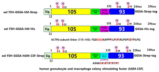
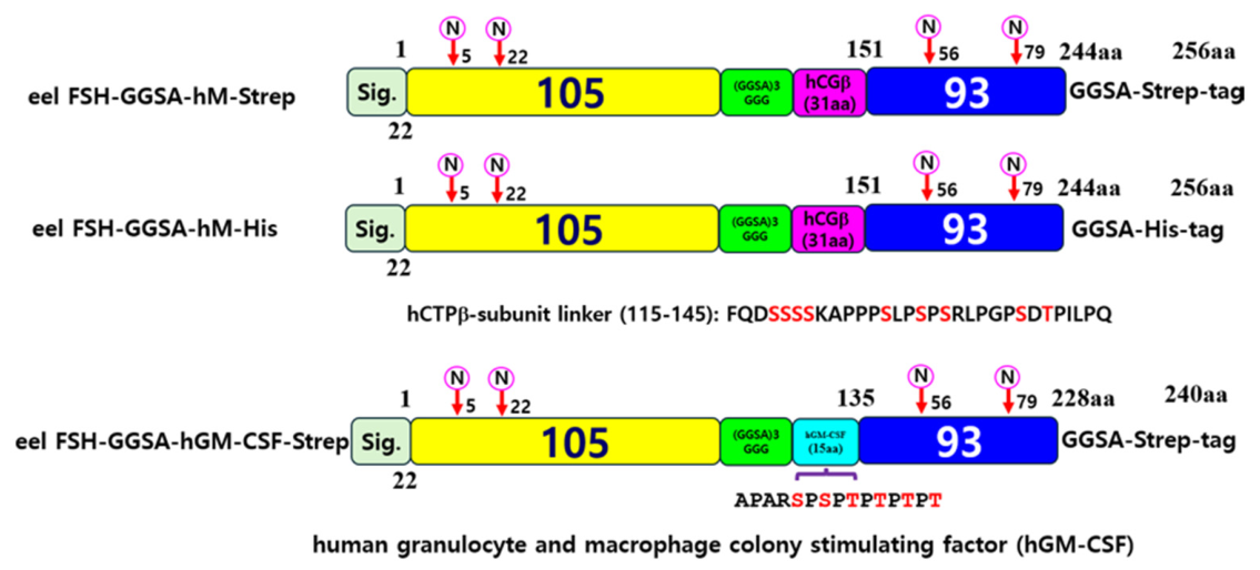
Eel FSH α- and β-subunit cDNAs were amplified from eel pituitary cDNA following a previously established procedure and subsequently used to generate a wild-type single-chain eel FSH (FSH-wt), in which the α-subunit was fused to the C-terminus of the β-subunit, by overlap extension PCR [4,5]. An eel FSH-hCG β-subunit region (FSH-hM) construct was subsequently generated by inserting the hCG β-subunit carboxyl-terminal peptide (hCTP; amino acids 115–145) between the β- and α-subunits of eel FSH, resulting in a single-chain FSH molecule in which the two subunits were linked by the hCTP region, with either a Strep- or His-tag fused to the C-terminus of the α-subunit to facilitate recombinant protein purification. To facilitate efficient O-linked glycosylation of the hCTP region, a 15-amino-acid (GGSA)3GGG linker was introduced between the β-subunit and hCTP. In addition, an alternative construct was generated by replacing the hCTP sequence with a modified 15-amino-acid sequence derived from a known O-linked glycosylation site of human granulocyte and macrophage colony-stimulating factor (hGM-CSF). The resulting constructs were designated FSH-GGSA-hM-Strep/His and FSH-GGSA-hGM-CSF-Strep. A schematic representation of the constructs is shown on Figure 1.
Figure 1.
Schematic representation of eel FSH-GGSA-hM-Strep, eel FSH-GGSA-hM-His, and eel FSH-GGSA-hGM-CSF-Strep constructs. Tethered eel FSH was engineered as a single-chain molecule consisting of the eel FSH β-subunit (105 amino acids) and the common α-subunit (93 amino acids). In the eel FSH-GGSA-hM constructs, a 31-amino acid carboxyl-terminal peptide (CTP) corresponding to residues 115–145 of the human chorionic gonadotropin β-subunit (hCGβ) was inserted between the FSH β- and common α-subunits. A flexible [(GGSA)3GGG] linker was positioned between the FSH β-subunit and the hCGβ CTP. In the eel FSH-GGSA-hGM-CSF-Strep construct, the hCGβ CTP was replaced with a 15-amino acid O-glycosylation-rich sequence derived from human granulocyte-macrophage colony-stimulating factor (hGM-CSF). Strep or His tags were fused to the C-terminus of the common α-subunit for affinity purification and detection. The numbers indicate amino acid positions in the mature proteins, excluding the signal peptide sequences. “N” indicates potential N-linked glycosylation sites in the FSH β- and common α-subunits. The FSH β-subunit, [(GGSA)3GGG] linker, hCGβ CTP, hGM-CSF-derived sequence, and common α-subunit are shown in yellow, green, magenta, cyan, and blue, respectively. Red letters in the hCGβ CTP and hGM-CSF-derived sequences indicate predicted potential O-linked glycosylation sites.
2.3. Transfection and Isolation of Single Cells Expressing Rec-Eel FSH-GGSA-hM-Strep
CHO-K1 and CHO-S cells were used to evaluate the initial expression of the recombinant constructs. Transfection was performed according to the manufacturer’s protocol for Lipofectamine 2000 reagent. For stable cell transfection, the expression construct was linearized with PvuI and subsequently transfected into CHO-DG44 cells as previously reported [3]. At 48 h post-transfection, cells were harvested and resuspended in complete CD OptiCHO™ medium containing 8 mM L-glutamine at a viable cell density of 5 × 10⁵ cells/mL for selection. Once cell viability exceeded 90%, gene amplification was performed by sequential exposure to increasing concentrations of MTX (500 nM, 2 μM, and 4 μM). Cell pools exhibiting >95% viability were subsequently cryopreserved. For single-cell cloning, MTX-amplified cells were first expanded in glutamine-free medium and then subjected to limiting dilution to achieve approximately one cell per well in 96-well plates. Following colony formation, approximately 100 individual clones were screened for recombinant protein expression. Five clones with high production levels were initially selected, and clones 7 and 20 were ultimately chosen for large-scale recombinant protein production.
2.4. Production and Quantitation of Rec-Eel FSH-GGSA-hM-Strep/His Protein
CHO-K1 and CHO-S cells were used to evaluate the initial expression of the recombinant constructs. Culture supernatants from transiently transfected CHO-K1 and CHO-S cells were harvested at the end of their respective culture periods, with 50 mL collected from each CHO-S culture. For single-cell-derived CHO-DG44 clones, 1 mL aliquots of culture supernatant were collected on culture days 1, 3, 5, 7, and 9. For large-scale production, selected clones were cultured in 1-L spinner flasks, and the entire culture supernatant was harvested on day 7 for clone #7 and day 9 for clone #20. Consequently, the final recovery yields of the two clones were comparable. The concentration of rec-eel FSH-GGSA-hM-Strep in culture supernatants was determined by ELISA as previously described [3]. In addition, the standard used in this study was prepared by purifying a 1-L production batch and subsequently freeze-drying the purified product. Microplates were coated with the monoclonal anti-eel FSH β-subunit antibody 5A14. Culture supernatants were incubated for 1 h, followed by HRP-conjugated anti-Strep antibody (1:1000). After the addition of TMB substrate, the reaction was stopped with 1 M H2SO4, and absorbance was measured at 450 nm using a Cytation 3 microplate reader.
2.5. Purification of Rec-Eel FSH-GGSA-hM-Strep/His Proteins
Recombinant eel FSH proteins were purified using Strep-Tactin® XT 4Flow, Strep-Tactin® XT High Capacity, or HisPur Ni-NTA Spin columns according to the manufacturers’ instructions. For Strep-Tactin XT purification, columns were equilibrated with washing buffer (100 mM Tris-HCl, pH 8.0, 150 mM NaCl, and 1 mM EDTA), loaded with clarified culture supernatants, washed, and eluted with Buffer BXT containing 50 mM biotin. Flow-through and eluted fractions were collected for subsequent analyses. The difference between the XT 4Flow and XT High-Capacity columns lies in the volume of elution buffer used during the final elution step. For HisPur Ni-NTA purification, columns were equilibrated, incubated with clarified supernatants for 30 min, washed by centrifugation at 700× g, and His-tagged proteins were eluted with Elution Buffer under the same centrifugation conditions.
2.6. Western Blot Analysis and Silver Staining of Rec-Eel FSH Proteins
For Western blot analysis, culture supernatants (20 µL) and purified protein samples (1 µL) were separated by reducing 12% SDS-PAGE and transferred onto 0.2-µm PVDF membranes. After blocking with 5% skim milk in TBS-T, membranes were incubated with anti-Strep, anti-His, and anti-eel FSH β (5A14) antibodies, followed by HRP-conjugated secondary antibodies. Protein signals were detected using an ECL detection system. SDS-PAGE gels were also stained using the Silver Stain Plus kit (Bio-Rad) according to the manufacturer’s instructions.
2.7. Analysis of Enzymatic Deglycosylation
N- and O-linked glycans were removed enzymatically according to the manufacturers’ instructions. Briefly, protein samples (10–20 μg) were denatured in Glycoprotein Denaturing Buffer (0.5% SDS and 40 mM DTT) at 100 °C for 10 min. For N-glycan removal, denatured samples were treated with PNGase F (500 U) in the presence of GlycoBuffer and NP-40 at 37 °C for 1 h. For O-glycan removal, samples were treated with α2–3,6,8,9 neuraminidase and O-glycosidase at 37 °C for 4 h. Terminal sialic acids were selectively removed by incubation with neuraminidase at 37 °C for 3 h. Following digestion, samples were subjected to SDS-PAGE and Western blot analysis.
2.8. cAMP Analysis Using Homogeneous Time-Resolved Fluorescence (HTRF)
The cAMP assay was performed using the cAMP Dynamic 2 Homogeneous Time-Resolved Fluorescence (HTRF) assay kit (Cisbio, Codolet, France) according to the manufacturer’s instructions and a method previously established in our laboratory [3]. Briefly, cells transiently expressing the appropriate receptor were resuspended and diluted in assay buffer containing 0.5 mM 3-isobutyl-1-methylxanthine (IBMX) to inhibit phosphodiesterase activity and prevent intracellular cAMP degradation. The cell suspension was then dispensed into 384-well microplates at a density of 10,000 cells per well. Subsequently, 5 μL of recombinant ligand was added to each well, and the plates were incubated for 30 min to allow receptor activation and intracellular cAMP accumulation.
Following ligand stimulation, cAMP-d2 conjugate and anti-cAMP cryptate were added to each well according to the manufacturer’s protocol. After incubation for 1 h at room temperature, time-resolved fluorescence signals were measured at emission wavelengths of 665 and 620 nm using a TriStar2 LB942 microplate reader (BERTHOLD Technologies, Wildbad, Germany). The HTRF signal was calculated from the ratio of fluorescence intensities measured at 665 and 620 nm and expressed as Delta F% (ΔF%), reflecting changes in intracellular cAMP levels. For quantitative determination of intracellular cAMP concentrations, the measured HTRF signals were compared with a standard curve generated using known concentrations of cAMP. Dose–response curves and cAMP concentrations were subsequently analyzed using GraphPad Prism version 6.0 (GraphPad Software, San Diego, CA, USA).
2.9. Construction of β-Arr1 Knockout Cell Lines
β-Arr1 knockout (KO) cells generated using CRISPR/Cas9 technology have been described previously [16]. Briefly, the knockout plasmid and β-Arr1 HDR template plasmid were co-transfected into HEK293 cells, followed by puromycin selection (1 μg/mL) starting 72 h after transfection for approximately 12–14 days. The surviving cells were then reseeded at a density of 100–500 cells per dish to allow colony formation. Individual colonies were subsequently isolated, transferred to 96-well plates, and expanded. A total of 19 clones were isolated and analyzed for β-Arr1 expression by Western blotting using an anti-β-Arr1 XP monoclonal antibody to confirm β-Arr1 knockout.
2.10. ERK1/2 Activation Assays
These procedures were performed as previously described [16]. Briefly, transfected cells cultured in 6-well plates at approximately 60–70% confluence were serum-starved for at least 4 h. To investigate the involvement of the PKA and PKC signaling pathways, the cells were pretreated with the PKA inhibitor H89 (20 μM) or the PKC inhibitor GF109203X (GFX; 2.5 μM) for 30 min before stimulation with recombinant FSH (rec-FSH; 400 ng/mL). The cells were subsequently lysed in RIPA buffer, and equal amounts of protein (10 μg) were subjected to immunoblot analysis to assess ERK1/2 phosphorylation. Phosphorylated ERK1/2 (pERK1/2) and total ERK1/2 were detected using a mouse monoclonal anti-phospho-p44/42 MAPK antibody and a rabbit polyclonal anti-MAPK1/2 antibody, respectively. Chemiluminescent signals were detected using SuperSignal West Pico reagent (Pierce). Immunoblot signals were quantified by densitometric analysis using Image Lab software version 6.0 (Bio-Rad, Hercules, CA, USA).
2.11. Data Analysis
Multiple sequence alignment was carried out using Multalin version 5.5.1. cAMP responses were calculated from the HTRF ΔF% data, and dose–response curves and EC50 value was analyzed using GraphPad Prism version 6.0 (San Diego, CA, USA). Relative changes in ERK1/2 phosphorylation were expressed as percentages or fold changes and analyzed using GraFit version 5 (Erithacus Software, Horley Surrey, UK). Statistical significance was evaluated by one-way ANOVA followed by Tukey’s post-hoc tests with GraphPad Prism 6.0. Statistical significance was defined as * p < 0.05 and ** p < 0.01.
3. Results
3.1. Production and Purification of Rec-Eel FSH-GGSA-hM-Strep/His Proteins
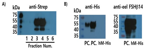
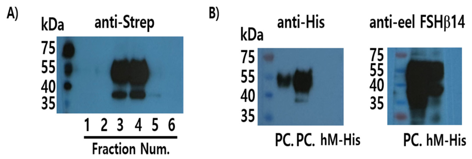
Recombinant proteins transiently transfected with eel FSH-hM-Strep/His plasmid were produced in CHO-K1 and CHO-S cells. The recombinant proteins were purified using Strep-Tactin XT or HisPur Ni-NTA affinity chromatography, depending on the affinity tag. FSH-GGSA-hM-Strep protein was detected as broad strong band at the fraction number 3 and 4 (Figure 2A). Although the positive control was detected as expected, the FSH-GGSA-hM-His protein was not detected using the anti-His antibody (Figure 2B, left). These results suggest that the low purification efficiency may be attributed to inefficient binding to the immobilized metal affinity resin, potentially due to limited accessibility of the His-tag. The potential contribution of structural changes resulting from GGSA insertion upstream of the His-tag cannot be excluded, although this possibility was not directly investigated. In contrast, a strong and distinct signal was observed when the protein was probed with an antibody recognizing the FSH β-subunit (Figure 2B, right). These results indicated that, although the Strep-tag was readily recognized, the His-tag was not efficiently detected by the anti-His antibody. FSH-GGSA-hGM-CSF-Strep protein was also produced using the same method, and both recombinant proteins were subsequently subjected to the following glycosidase digestion experiments.
Figure 2.
Production and purification of recombinant eel FSH-GGSA-hM-Strep and eel FSH-GGSA-hM-His proteins from CHO-S cells. Culture supernatants from transfected CHO-S cells were collected, and recombinant eel FSH-GGSA-hM-Strep and eel FSH-GGSA-hM-His proteins were purified using Strep-Tactin XT 4Flow and HisPur Ni-NTA affinity columns, respectively. Aliquots (5 μL) of the purified proteins were subjected to Western blot analysis. (A) Purified eel FSH-GGSA-hM-Strep was detected using an anti-Strep-tag antibody. (B) Eel FSH-GGSA-hM-His was purified using a Ni-NTA affinity column and analyzed by Western blotting with an anti-His-tag antibody. The FSH-GGSA-hM-His protein was not detected in the purified fraction using the anti-His-tag antibody (left). Therefore, the flow-through fraction was further analyzed using an anti-eel FSHβ14 antibody, which recognizes the eel FSH β-subunit, confirming the presence of FSH-GGSA-hM-His in the flow-through fraction (right). PC, positive control (purified FSH-GGSA-eM-His).
3.2. Deglycosylation of Recombinant Eel FSH-GGSA-hM-Strep and FSH-GGSA-hGM-CSF-Strep
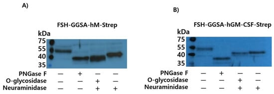
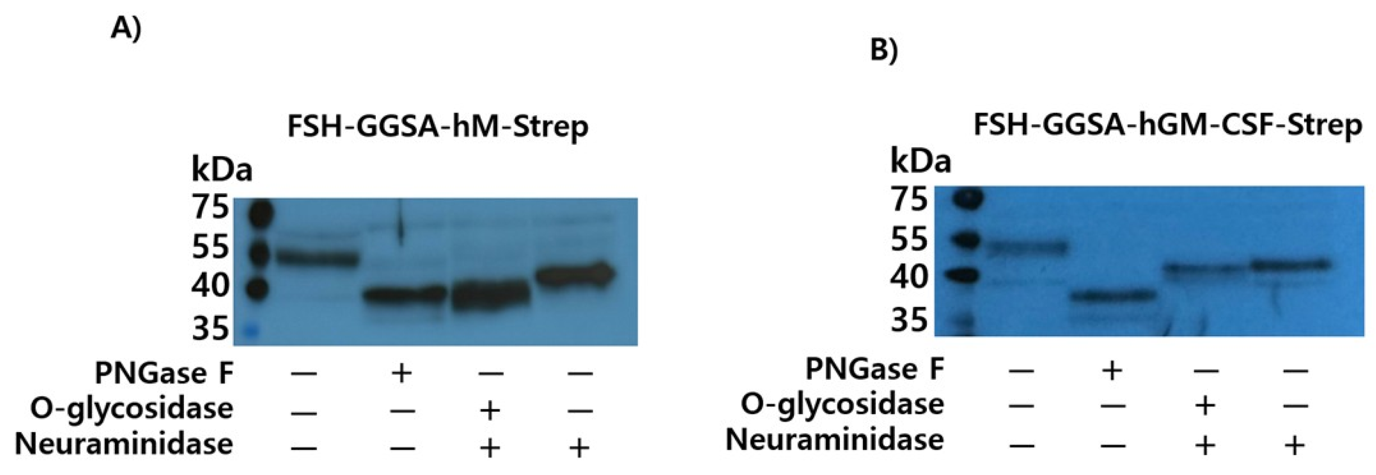
Western blot analysis of rec-eel FSH-GGSA-hM-Strep revealed a broad protein band with an apparent molecular weight of approximately 54–57 kDa (Figure 3A). To investigate the contribution of glycosylation to the molecular mass of the recombinant protein, enzymatic deglycosylation was subsequently performed. Following PNGase F treatment, which removes N-linked glycans, the apparent molecular weight was markedly reduced to approximately 40 kDa, indicating that N-linked glycans substantially contributed to the increased molecular mass of rec-eel FSH-GGSA-hM-Strep. Similarly, treatment with O-glycosidase resulted in a reduction in molecular weight, further suggesting the presence of O-linked glycans in the recombinant protein. In addition, neuraminidase treatment reduced the apparent molecular weight to approximately 42 kDa (Figure 3A), demonstrating that terminal sialic acid residues also contributed to the observed molecular mass. In contrast, FSH-GGSA-hGM-CSF-Strep exhibited a slightly lower apparent molecular weight of approximately 51–54 kDa. Following PNGase F treatment, the molecular weight was markedly reduced to approximately 37 kDa, indicating a substantial contribution of N-linked glycans to the overall molecular mass. However, treatment with O-glycosidase or neuraminidase resulted in a similar apparent molecular weight of approximately 40 kDa. These results suggest that, compared with FSH-GGSA-hGM-CSF-Strep, rec-eel FSH-GGSA-hM-Strep may undergo more extensive O-linked glycosylation. Collectively, these results indicate that rec-eel FSH-GGSA-hM-Strep undergoes extensive post-translational glycosylation, with both N- and O-linked glycans contributing to its increased molecular mass.
Figure 3.
Deglycosylation analysis of recombinant eel FSH-GGSA-hM-Strep and FSH-GGSA-hGM-CSF-Strep proteins. Recombinant proteins were treated with PNGase F, O-glycosidase, and/or neuraminidase to assess N- and O-linked glycosylation and subsequently analyzed by Western blotting. PNGase F treatment reduced the apparent molecular weights of both recombinant proteins by approximately 14–17 kDa. In FSH-GGSA-hM-Strep, combined treatment with O-glycosidase and neuraminidase resulted in a marked reduction in apparent molecular weight, with a mobility shift comparable to that observed following PNGase F treatment. Treatment with neuraminidase alone resulted in an apparent molecular weight of approximately 42 kDa, slightly higher than that observed after PNGase F or O-glycosidase treatment. For FSH-GGSA-hGM-CSF-Strep, combined treatment with O-glycosidase and neuraminidase produced a molecular-weight shift similar to that observed with neuraminidase treatment alone, suggesting a relatively lower degree of O-linked glycosylation. (A) FSH-GGSA-hM-Strep. (B) FSH-GGSA-hGM-CSF-Strep.
3.3. Isolation of Single Cells Expressing Eel FSH-GGSA-hM-Strep Proteins
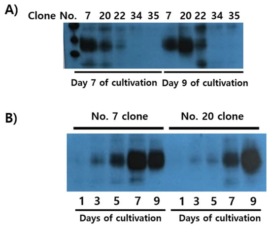
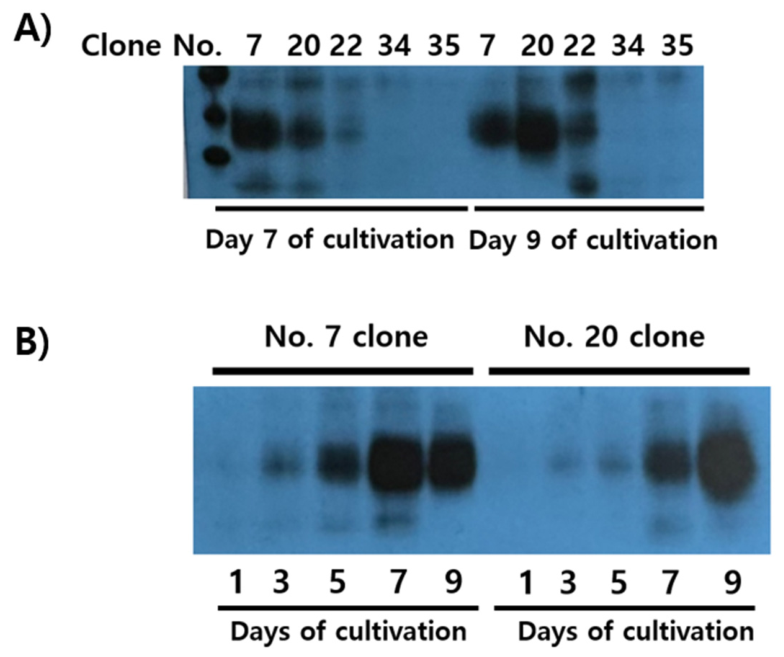
Single-cell clones were isolated from MTX-amplified CHO-DG44 cells, and five clones were ultimately selected. Recombinant protein expression in these clones was initially evaluated by Western blot analysis of culture supernatants collected on days 7 and 9 (Figure 4A). Among the five clones, clones #7 and #20 showed the highest levels of recombinant protein expression. These two clones were subsequently evaluated in a time-course experiment on culture days 1, 3, 5, 7, and 9. Clone #7 showed the strongest signal on day 7, whereas clone #20 exhibited the highest level of protein expression on day 9. Therefore, these two clones were further evaluated for their potential for large-scale production. Therefore, cultures of these two clones were expanded in 1-L spinner flasks to a total culture volume of up to 5 L. Culture supernatants were harvested on day 7 for clone #7 and day 9 for clone #20. The recombinant proteins were then purified, and their quantities were analyzed before and after purification. The recombinant proteins were produced at a final yield of 10–12 mg/L, with approximately 6.7 mg/L recovered, corresponding to a recovery efficiency of approximately 60%.
Figure 4.
Isolation and selection of single-cell clones expressing recombinant eel FSH-GGSA-hM-Strep. CHO-DG44 cells stably expressing recombinant eel FSH-GGSA-hM-Strep were subjected to stepwise MTX-mediated amplification at concentrations of 500 nM, 2 μM, and 4 μM, followed by single-cell cloning. Approximately 100 single-cell-derived clones were screened for recombinant protein production, and five stable clones were selected based on their levels of recombinant protein secretion. (A) Culture supernatants (20 μL) collected from the selected clones on days 7 and 9 were analyzed by Western blotting to compare the secretion levels of recombinant eel FSH-GGSA-hM-Strep. (B) Recombinant protein production was further evaluated over the culture period. Among the selected clones, clones #7 and #20 exhibited the highest levels of recombinant protein secretion. Clone #7 showed the highest secretion level on day 7, whereas clone #20 reached its highest secretion level on day 9. Based on their high levels of recombinant protein production, clones #7 and #20 were selected as candidate cell lines for large-scale production of recombinant eel FSH-GGSA-hM-Strep.
3.4. Purification and Silver Staining Results
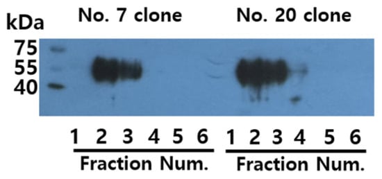
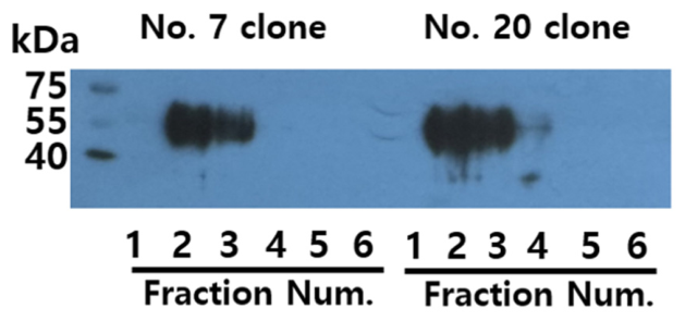
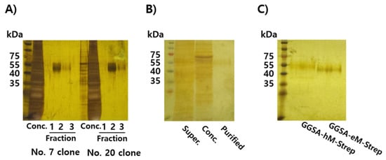
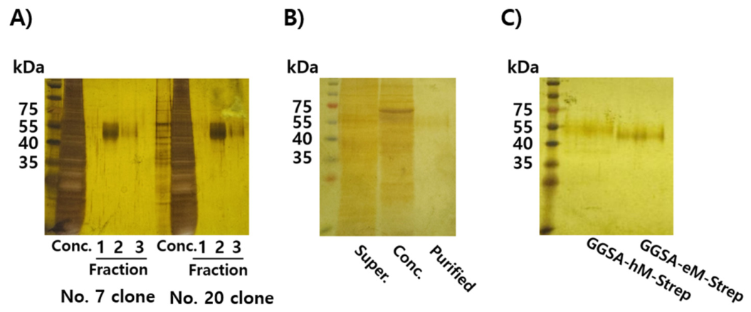
Based on quantitative analysis, clones #7 and #20 were selected for further culture, and approximately 5 L of culture supernatant was collected from each clone using spinner flasks. Following purification of the recombinant proteins using a Strep-Tactin XT 4Flow High-Capacity column, the strongest signal was detected in fraction 2, while a distinct but relatively weaker band was also observed in fraction 3. The purified recombinant proteins were detected as broad bands with an apparent molecular weight of approximately 54–57 kDa (Figure 5). Silver staining analysis of the purified samples showed that multiple protein bands were present in the concentrated culture supernatant before purification, whereas only the recombinant protein was detected in fractions 2 and 3 following purification (Figure 6A). Similarly, comparison of the culture supernatant, concentrated samples, and purified proteins by silver staining clearly demonstrated the presence of a distinct recombinant protein band in the purified fraction 2 samples (Figure 6B). Furthermore, comparison with the previously reported recombinant protein (GGSA-eCG β-subunit region-Strep; GGSA-eM-Strep) containing the eCTP linker instead of hCTP revealed that the incorporation of the hCTP sequence in the present construct resulted in a slightly increased apparent molecular weight.
Figure 5.
Purification of recombinant eel FSH-GGSA-hM-Strep from CHO-DG44 clones #7 and #20. Recombinant eel FSH-GGSA-hM-Strep was purified from 1 L of culture supernatant obtained from clones #7 and #20 using a Strep-Tactin XT high-capacity affinity column. Eluted fractions (5 mL each) were collected, and an aliquot (1 μL) of each fraction was analyzed by Western blotting using an anti-Strep-tag antibody to detect recombinant eel FSH-GGSA-hM-Strep. Strong immunoreactive bands corresponding to recombinant eel FSH-GGSA-hM-Strep were predominantly detected in elution fractions 2 and 3.
Figure 6.
Silver staining analysis of recombinant eel FSH-GGSA-hM-Strep during purification. Recombinant eel FSH-GGSA-hM-Strep was analyzed by SDS-PAGE followed by silver staining to evaluate the protein profile and purity during the purification process. (A) Recombinant eel FSH-GGSA-hM-Strep was produced from 1 L of culture supernatant from each of clones #7 and #20 and purified using a Strep-Tactin XT High-Capacity resin column. The 10-fold concentrated culture supernatants and eluted fractions were analyzed by silver staining. Aliquots of the concentrated culture supernatants (5 μL) and each eluted fraction (8 μL) were loaded onto the gel. A strong protein band at the expected molecular weight of recombinant eel FSH-GGSA-hM-Strep was predominantly observed in elution fraction 2, whereas only a faint band was detected in fraction 3. (B) Protein profiles before and after affinity purification were compared by silver staining. Aliquots of the original culture supernatant (20 μL), 10-fold concentrated culture supernatant (5 μL), and affinity-purified sample (8 μL) from fraction 2 were separated by SDS-PAGE. Multiple high-molecular-weight proteins (>60 kDa) were observed in the original and concentrated culture supernatants, whereas these bands were largely removed following affinity purification. The purified sample showed a distinct band at the expected apparent molecular weight of recombinant eel FSH-GGSA-hM-Strep. (C) Purified FSH-GGSA-hM-Strep was compared with the previously purified FSH-GGSA-eM-Strep by SDS-PAGE followed by silver staining. FSH-GGSA-hM-Strep migrated at a slightly higher apparent molecular weight than FSH-GGSA-eM-Strep. The difference in signal intensity between Figure 6A and Figure 6B is due to the shorter exposure time used for Figure 6B because the signal in Figure 6A was too strong.
3.5. Biological Activities of Rec-Eel FSH-GGSA-hM-Strep Proteins
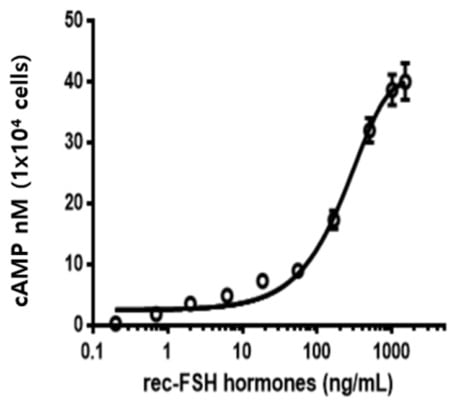
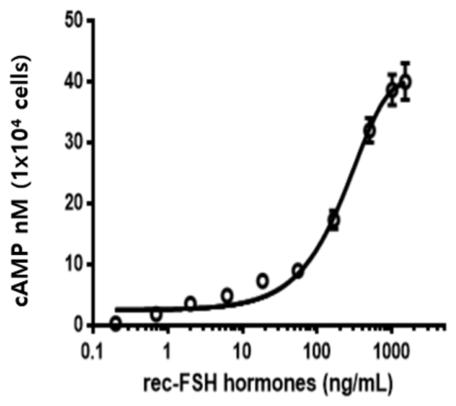
In vitro biological activity was evaluated using CHO-K1 cells transiently expressing the eel FSH receptor, as described previously [3]. Rec-eel FSH-GGSA-hM-Strep induced cAMP accumulation in a dose-dependent manner, with cAMP levels progressively increasing with increasing concentrations of the recombinant protein (Figure 7). The dose–response curve showed a clear concentration-dependent response, with an EC50 value of 220.7 ng/mL and a maximal response (Rmax) of 40.2 ± 0.9 nM/104 cells (Table 1). These results indicate that rec-eel FSH-GGSA-hM-Strep effectively activated cAMP production in CHO-K1 cells expressing the eel FSH receptor.
Figure 7.
Biological activity of recombinant eel FSH-GGSA-hM-Strep in cells expressing the eel follicle-stimulating hormone receptor. CHO-K1 cells transiently expressing the eel FSH receptor (eel FSHR) were seeded into 384-well plates at a density of 10,000 cells per well 24 h after transfection. Prior to stimulation, the cells were treated with 0.5 mM 3-isobutyl-1-methylxanthine (IBMX) to inhibit phosphodiesterase activity and prevent cAMP degradation. The cells were then stimulated with increasing concentrations of recombinant eel FSH-GGSA-hM-Strep for 30 min at room temperature. Total cAMP levels were measured using a homogeneous time-resolved fluorescence (HTRF) assay. The actual cAMP concentrations were first calculated from the cAMP Delta F% values. The EC50 values and 95% confidence intervals were then determined by fitting the data to a four-parameter dose–response stimulation model. Data are presented as the mean ± SEM from triplicate experiments.
Table 1.
Bioactivity of rec-eel FSH protein in cells expressing eel FSH receptor.
3.6. PKA-Dependent ERK1/2 Activation Mediated by Eel FSHR
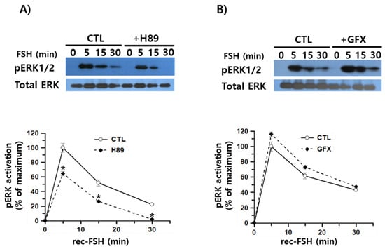
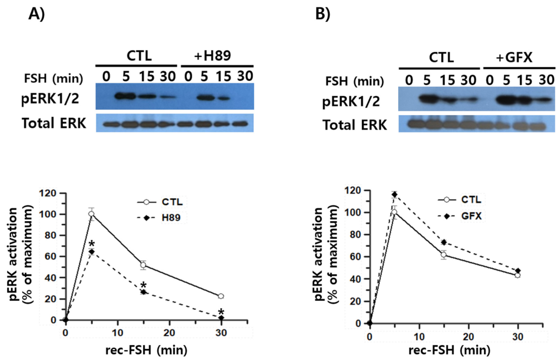
We next investigated the signaling pathway underlying eel FSHR-mediated ERK1/2 activation. In control cells transfected with eel FSHR, ERK1/2 phosphorylation rapidly increased, reaching a maximum at 5 min, followed by a gradual decline over the subsequent 30 min. At 15 and 30 min, pERK1/2 activity decreased to approximately 50% and 22%, respectively, of the level observed at 5 min. Pretreatment with H89, a PKA inhibitor, markedly attenuated ERK1/2 phosphorylation compared with the control. In H89-treated cells, pERK1/2 activity was approximately 64% and 50% of the corresponding control levels at 5 and 15 min, respectively, whereas ERK1/2 phosphorylation was almost completely abolished at 30 min (Figure 8A). In contrast, inhibition of PKC had little effect on ERK1/2 phosphorylation throughout the examined time course, although a slight increase in ERK1/2 phosphorylation was observed at each time point following GFX treatment (Figure 8B). These results indicate that PKA inhibition substantially suppresses eel FSHR-mediated ERK1/2 activation, whereas PKC inhibition has a minimal effect, suggesting that ERK1/2 activation downstream of eel FSHR is primarily associated with the cAMP/PKA signaling pathway.
Figure 8.
Effects of PKA and PKC inhibition on recombinant eel FSH-GGSA-hM-Strep-induced ERK1/2 activation. HEK293 cells were transiently transfected with the eel FSH receptor (eel FSHR) and serum-starved for 4–6 h before stimulation with recombinant eel FSH-GGSA-hM-Strep (400 ng/mL) for the indicated time periods. Whole-cell lysates (10 μg of protein per lane) were subjected to Western blot analysis. The band intensities of phosphorylated ERK1/2 (pERK1/2) and total ERK1/2 were quantified by densitometry, and pERK1/2 levels were normalized to the corresponding total ERK1/2 levels. The pERK1/2 response at 5 min in the control group was set to 100%, and all other values were expressed relative to this value. Representative Western blot images are shown, and quantitative data are represented as the mean ± S.E from at least three independent experiments. (A) Cells were pretreated with the PKA inhibitor H89 (20 μM) for 30 min before stimulation with recombinant eel FSH-GGSA-hM-Strep. (B) Cells were pretreated with the PKC inhibitor GF109203X (GFX; 2.5 μM) for 30 min before stimulation with recombinant eel FSH-GGSA-hM-Strep. Statistical comparison of the curves was performed by using a two-way ANOVA between PKA/PKC inhibitor groups and control cells. *, p < 0.05 vs. control curve.
3.7. Modulation of β-Arr 1-Dependent ERK Activation
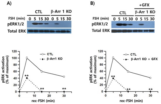
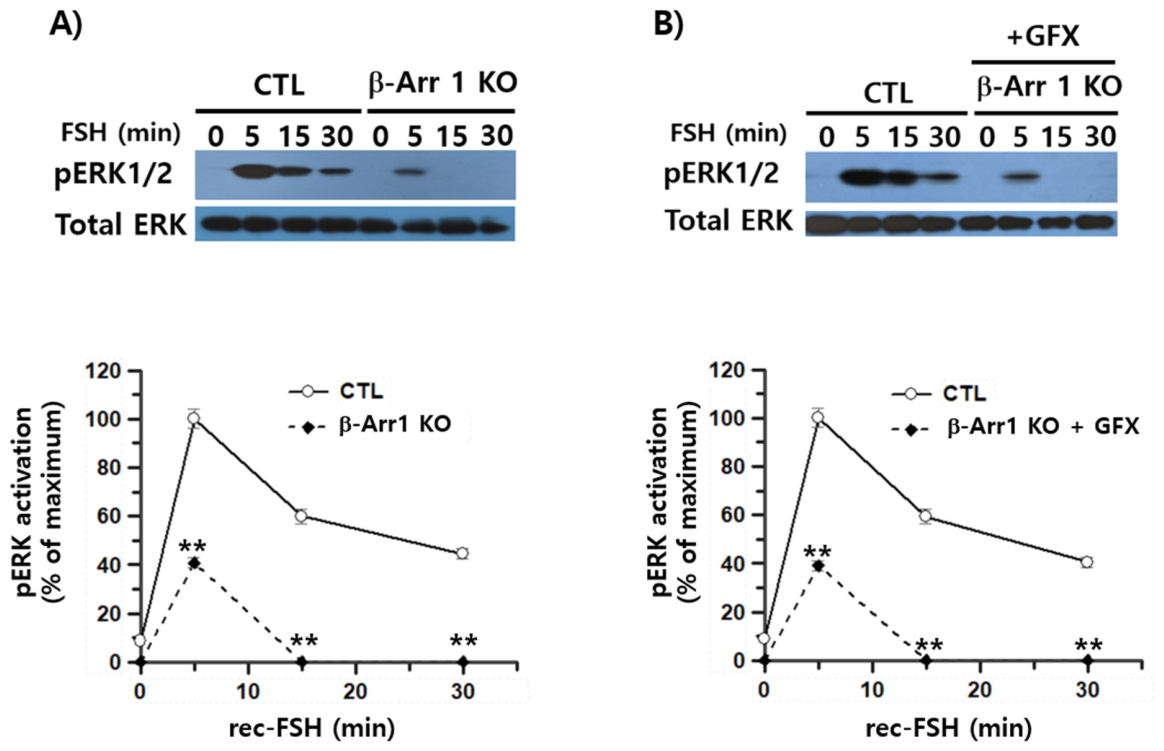
To further validate our finding that eel FSHR-mediated ERK activation can occur independently of β-Arr1, we generated β-Arr1 KO cells using the CRISPR/Cas9 system. Following puromycin selection, 19 individual cell clones were isolated. Western blot analysis using a β-Arr1-specific antibody confirmed the loss of β-Arr1 expression in 13 of the 19 clones, demonstrating successful β-Arr1 knockout in these cells (Figure 9). In β-Arr1 KO cells, eel FSHR-mediated ERK1/2 activation was markedly attenuated compared with that observed in control cells. The early ERK1/2 response at 5 min was reduced to approximately 40% of the control level, indicating that loss of β-Arr1 substantially impaired the rapid phase of ERK1/2 activation. Moreover, the sustained or late-phase ERK1/2 activity was almost completely abolished in β-Arr1 KO cells (Figure 10A). In contrast, treatment of β-Arr1 KO cells with a PKC inhibitor did not produce any appreciable additional reduction in ERK1/2 phosphorylation compared with untreated β-Arr1 KO cells (Figure 10B). Because parental HEK293 cells served as the control and the analysis was performed using only a single β-Arr1 KO cell clone, potential differences in eel FSHR transfection efficiency and expression levels between the control and knockout cells were not assessed, and clone-specific effects cannot be excluded.
Figure 9.
Isolation of β-arrestin 1 knockout cells using the CRISPR/Cas9 system. To generate β-arrestin 1 (β-Arr1) knockout cells, HEK293 cells were transfected with CRISPR/Cas9 vectors targeting the human β-Arr1 gene and selected with puromycin. Following selection and single-cell cloning, a total of 19 individual clones were selected. The isolated clones were screened for β-Arr1 protein expression by Western blotting using an anti-β-Arr1 antibody. Among the 19 clones examined, 13 showed complete loss of detectable β-Arr1 protein expression and were identified as β-Arr1 knockout clones.
Figure 10.
Effects of β-arrestin 1 knockout on eel FSHR-mediated ERK1/2 activation. HEK293 control and β-arrestin 1 (β-Arr1) knockout cells expressing the eel FSHR were stimulated with recombinant eel FSH-GGSA-hM-Strep for the indicated time periods, and ERK1/2 activation was analyzed by Western blotting. (A) Time-dependent ERK1/2 activation was compared between control and β-Arr1 knockout cells. (B) The effect of PKC inhibition on ERK1/2 activation was examined in β-Arr1 knockout cells. Cells were pretreated with the PKC inhibitor GF109203X (GFX; 2.5 μM) for 30 min before stimulation with recombinant eel FSH-GGSA-hM-Strep. The band intensities of phosphorylated ERK1/2 were normalized to the corresponding total ERK1/2 levels. The pERK1/2 response at 5 min after stimulation in the control group was set to 100%, and all other values were expressed relative to this value. Data are presented as the mean ± SEM from at least three independent experiments. Statistical differences between groups at each time point were analyzed by two-way ANOVA. ** p < 0.01; vs. control curve.
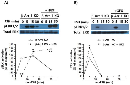
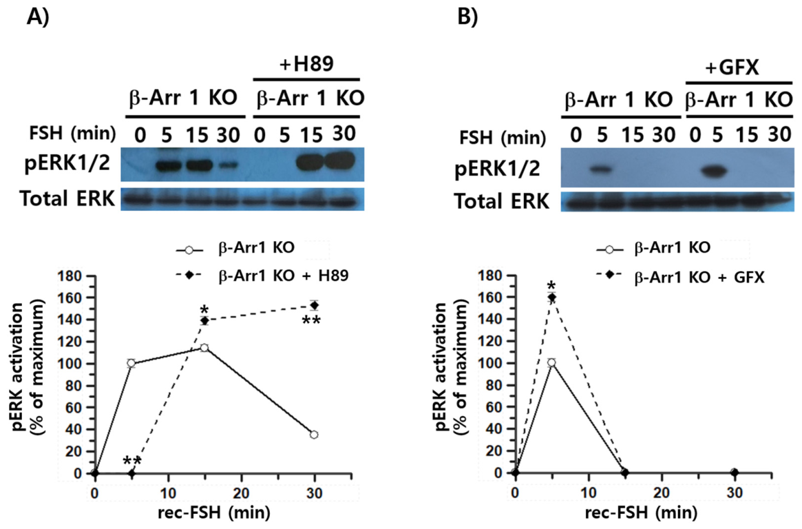
To further investigate the effects of PKA and PKC inhibition on eel FSHR-mediated ERK1/2 signaling in β-Arr1 KO cells, ERK1/2 phosphorylation was examined following treatment with the PKA inhibitor H89 and the PKC inhibitor GFX, with H89 completely abolishing ERK1/2 phosphorylation at 5 min. Interestingly, however, ERK1/2 phosphorylation re-emerged at later time points and was significantly increased at 15 and 30 min compared with the corresponding untreated control (Figure 11A). At 15 min, ERK1/2 phosphorylation increased by approximately 20% compared with the control, whereas at 30 min, it increased by more than 4-fold. In contrast, consistent with the results shown in Figure 8B, GFX treatment resulted in a significant increase in ERK1/2 phosphorylation at 5 min. The overall temporal pattern was largely comparable to that observed in control cells, indicating that PKC inhibition had little effect on the ERK1/2 response.
Figure 11.
Effects of β-arrestin 1 knockout on eel FSHR-mediated ERK1/2 activation by PKA and PKC inhibition treatment. (A) To examine the contribution of PKA signaling, β-Arr1 knockout cells were exposed to H89 (20 μM) for 30 min prior to stimulation with recombinant eel FSH-GGSA-hM-Strep. (B) The involvement of PKC signaling was evaluated by incubating β-Arr1 knockout cells with GF109203X (GFX; 2.5 μM) for 30 min before agonist stimulation. For quantitative analysis, the ERK1/2 phosphorylation level measured at 5 min after stimulation in β-Arr1 knockout cells was designated as 100%, and all other responses were calculated relative to this reference value. Values represent mean ± SEM from three independent experiments. Group differences across time were assessed by two-way ANOVA test. Statistical significance was defined as p < 0.05 (*), with ** indicating p < 0.01.
These findings reveal a distinct time-dependent pattern of ERK1/2 regulation in β-Arr1 KO cells, in which PKA inhibition suppresses the early ERK1/2 response but is accompanied by an enhanced ERK1/2 response at later point. Thus, in the absence of β-Arr1, the early phase of eel FSHR-mediated ERK1/2 activation appears to be PKA-dependent, whereas the late-phase ERK1/2 response may involve a PKA-independent signaling mechanism.
4. Discussion
In the present study, the hCTP region was incorporated into recombinant Japanese eel FSH as a strategy to potentially modify O-linked glycosylation, and a [(GGSA)3GGG] linker was introduced to potentially facilitate O-linked glycosylation within the hCTP region. This glycoengineering strategy facilitated the development of an efficient and scalable production platform, allowing the large-scale production of recombinant eel FSH from a single-cell-derived clone. The recombinant proteins produced in this study exhibited appropriate N-linked and O-linked glycosylation, as well as sialic acid modification, as demonstrated by enzymatic deglycosylation analysis. The recombinant proteins also showed a concentration-dependent increase in biological activity in FSHR-expressing cells and effectively induced ERK1/2 phosphorylation. Notably, treatment with the PKA inhibitor H89 and the use of β-arrestin 1 (β-Arr1) knockout cells significantly affected pERK1/2 activation, suggesting the involvement of both PKA- and β-Arr1-dependent mechanisms in FSHR-mediated ERK1/2 signaling.
Although a previous study successfully produced recombinant eel FSH in CHO-DG44 cells and demonstrated its ability to induce ovarian superovulation, the present study differs in several aspects of construct design and protein purification. Specifically, the previous study used a C-terminal His-tag, a [(GGGS)3GGG] linker, and Ni-NTA-based immobilized metal affinity chromatography (IMAC), whereas the present study employed a C-terminal GGSA-Strep-tag, a [(GGSA)3GGG] linker, and Strep-Tactin affinity chromatography [15]. These differences highlight the distinct construct designs and purification strategies used in the two studies.
Recently, increasing attention has been focused on the functional significance of O-linked glycans specifically present in the β-subunits of glycoprotein hormones. In particular, the importance of O-linked glycosylation in the production and functional properties of recombinant hCG [17] and eCG [18,19] has been increasingly recognized. Based on the findings of the present study, the incorporation of hCTP was associated with a greater apparent molecular mass than the addition of eCTP, despite the latter being reported to contain 12 potential O-linked glycosylation sites [6,20]. However, hCTP has been reported to contain four potential O-linked glycosylation sites at S121, S127, S132, and S138 [10,21]. Consistent with this, the silver staining results in the present study showed that GGSA-hM-Strep exhibited an approximately 2 kDa higher molecular weight than GGSA-eM-Strep. Nevertheless, despite the relatively small number of potential O-linked glycosylation sites in hCTP, hCTP appeared to promote glycosylation more efficiently than eCTP. Furthermore, this construct may have the potential to alter its pharmacokinetic properties in vivo compared with FSH-WT and GGSA-eM-Strep, although this possibility remains to be investigated experimentally. The underlying mechanisms responsible for this difference remain unclear and warrant further investigation. Furthermore, the GGSA-hM-His recombinant protein was not detectable by the anti-His antibody, possibly due to conformational changes associated with post-translational glycosylation and protein folding that may have affected antibody recognition of the His tag.
In the present study, eel FSH-GGSA-hM-Strep exhibited an apparent molecular weight of approximately 54–57 kDa, indicating a substantially higher degree of post-translational modification. Interestingly, hGM-CSF-Strep also exhibited detectable O-linked glycosylation, although the extent of modification appeared to be lower than that observed for the hM-containing construct. More notably, O-glycosidase treatment of GGSA-hM-Strep resulted in a pronounced reduction in apparent molecular weight, with the magnitude of the shift being comparable to that observed following PNGase F treatment. We acknowledge that the molecular weights inferred from Western blotting and silver staining are approximate and do not represent precise molecular weight measurements. Because the present findings are inferred from the results of enzymatic deglycosylation analyses, alterations in O-linked glycosylation can be suggested primarily on the basis of indirect biochemical evidence. Although the results suggest a possible alteration in O-linked glycosylation, the absence of an appropriate comparator makes it difficult to determine whether this change can be attributed specifically to the addition of the GGSA linker. Thus, the observed differences in apparent molecular weight may be attributed, at least in part, to O-linked glycosylation of the recombinant protein containing the GGSA linker and hCTP. This finding is consistent with our previously characterized GGSA-eM-Strep construct and suggests that incorporation of the hM sequence may be associated with differences in glycosylation of the recombinant eel FSH molecule.
These results are consistent with recent studies reporting the development of long-acting recombinant proteins in various mammalian species, including human FSH [22], bovine FSH [23], porcine FSH [24], human interferon-α2b [25], and erythropoietin [26]. These studies collectively highlight the potential of glycoengineering and fusion-based strategies to improve the pharmacological properties of recombinant proteins. In porcine FSH, fusion of the β-subunit with a fragment crystallizable (Fc) domain through a flexible [G4S]3 linker was reported to enhance biological activity and prolong the circulating half-life of the recombinant hormone [24]. Similarly, a recent study of human FSH incorporating the hCTP demonstrated O-linked glycosylation at six sites, rather than the four sites previously considered to be modified, further highlighting the substantial glycosylation potential of the hCTP sequence [22]. In light of these findings, our results suggest that incorporation of the flexible (GGSA)3GGG linker may facilitate efficient O-linked glycosylation of the adjacent hCTP sequence, potentially by increasing its structural flexibility and accessibility to the cellular O-glycosylation machinery. Notably, the hCTP-containing construct exhibited a slightly higher apparent molecular weight than the corresponding eCTP-containing construct in the present study. Although differences in apparent molecular weight alone cannot provide definitive evidence of the extent of glycosylation, this observation, together with the glycosidase digestion profiles, suggests that hCTP may influence the glycosylation characteristics of rec-eel FSH differently from eCTP.
As the functional significance of O-linked glycans in therapeutic proteins has been increasingly recognized, O-linked glycosylation has emerged as an important strategy for improving the pharmacological properties of recombinant glycoprotein therapeutics [27]. In this context, hCTP, which contains multiple potential O-glycosylation sites, may be particularly valuable for prolonging the circulating half-life and duration of action of recombinant glycoproteins.
ERK1/2 phosphorylation has been recognized as an important signaling event downstream of GPCR activation. In the present study, ERK1/2 activation mediated by the eel FSHR was markedly inhibited during the early phase by the PKA inhibitor H89, indicating an important role for PKA signaling in the early activation of ERK1/2 downstream of the eel FSHR. This finding is consistent with previous studies showing that H89 treatment significantly inhibits early-phase ERK1/2 activation mediated by the β2-adrenergic receptor (β2AR) [28,29] and human FSHR [30]. In contrast, eel FSHR-mediated ERK1/2 activation was largely unaffected by treatment with a PKC inhibitor. Together, these findings suggest that ERK1/2 activation downstream of the eel FSHR is predominantly mediated through a PKA-dependent rather than a PKC-dependent pathway.
In the present study, β-Arr1 KO resulted in a greater than 60% reduction in ERK1/2 activation during the early phase and a complete loss of ERK1/2 activation during the late phase. Interestingly, treatment of β-Arr1 KO cells with H89 completely abolished ERK1/2 activation during the early phase, whereas ERK1/2 activation was paradoxically increased during the late phase. These findings suggest that H89 exerts an inhibitory effect on PKA-dependent signaling during the early phase of the response, although its effects at later time points in β-arr1 KO cells remain unclear. In the β-Arr1 KO clone, differences in receptor expression and clone-specific effects cannot be excluded, particularly because only a single clone was analyzed. These observations are generally consistent with previous studies examining the involvement of β-Arr1 in ERK1/2 signaling mediated by human FSHR [30] and the parathyroid hormone receptor (PTHR) [31]. Comparative analyses using siRNA-mediated β-arrestin depletion and β-arrestin KO cells have further demonstrated receptor-specific differences in β-arrestin-dependent ERK1/2 signaling. For β2AR, ERK1/2 activation was slightly reduced following siRNA-mediated depletion of β-arrestins but was paradoxically increased in β-arrestin KO cells [32,33]. In contrast, ERK1/2 activation mediated by the β1AR and vasopressin V2 receptor (V2R) was markedly reduced following siRNA-mediated depletion but was largely unaffected in KO cells. Notably, human FSHR-mediated ERK1/2 activation was significantly reduced under both siRNA-mediated depletion and β-arrestin KO conditions [32]. These receptor-specific responses suggest that the contribution of β-arrestins to ERK1/2 signaling depends not only on the GPCR involved but also on whether β-arrestin function is acutely reduced by siRNA-mediated depletion or chronically eliminated by genetic knockout.
β-Arrestins are versatile regulators of GPCR signaling that interact with diverse signaling proteins and coordinate multiple downstream pathways following receptor activation. These dynamic interactions enable β-arrestins to adopt distinct conformational states depending on the activated receptor and cellular signaling context, thereby contributing to receptor-specific signaling responses [34]. In addition, the functional interplay between β-arrestins and G protein-coupled receptor kinases (GRKs) has been extensively investigated in several GPCRs, including the angiotensin II type 1A receptor [35], μ-opioid receptor [36], and β2AR [37,38]. Collectively, these studies indicate that the relative contributions of β-Arr1, β-Arr2, and different GRKs to ERK1/2 signaling can vary considerably depending on the GPCR and signaling context. Further studies are therefore warranted to elucidate the functional interplay among β-arrestins, GRKs, and ERK1/2 signaling downstream of the eel FSHR.
Taken together, the present findings suggest that eel FSHR-mediated ERK1/2 activation is temporally regulated through distinct signaling mechanisms. PKA-dependent signaling appears to make a major contribution during the early phase, whereas β-Arr1 may play a more prominent role in maintaining ERK1/2 activation during the late phase. In β-arrestin 1-knockout cells, H89 treatment unexpectedly increased FSH-induced ERK1/2 phosphorylation at 30 min. Although the underlying mechanism remains unclear, this increase may be associated with alterations in PKA-dependent feedback regulation or the activation of alternative/compensatory signaling pathways, such as those mediated by G-protein βγ subunits or receptor tyrosine kinase (RTK) transactivation. However, the potential off-target effects of H89 should also be considered when interpreting these findings. The paradoxical increase in late-phase ERK1/2 activation following H89 treatment in β-Arr1 KO cells may reflect the engagement of a possible compensatory signaling mechanism. Simultaneous loss of β-Arr1-mediated signaling and inhibition of the PKA pathway may redirect eel FSHR signaling toward alternative pathways capable of activating ERK1/2. This unexpected response raises the possibility of crosstalk among PKA-, β-arrestin-, and other GPCR-associated signaling pathways in the temporal regulation of ERK1/2 activation. Further studies employing selective pharmacological inhibitors and genetic approaches targeting alternative signaling components will be required to determine the molecular mechanisms responsible for this late-phase ERK1/2 activation.
5. Conclusions
In this study, biologically active recombinant eel FSH was successfully produced using a single-chain expression system incorporating a GGSA linker and the hCTP sequence. The recombinant proteins were efficiently purified by affinity chromatography, although the His-tag was not effectively recognized by the anti-His antibody. Deglycosylation analysis demonstrated that both N- and O-linked glycans substantially contributed to the increased molecular mass of rec-eel FSH-GGSA-hM-Strep. Stable, high-producing CHO-DG44 single-cell clones were successfully established and used for large-scale production of recombinant eel FSH. Purification using Strep-Tactin XT affinity chromatography yielded highly enriched recombinant protein with an apparent molecular weight of approximately 54–57 kDa. The purified recombinant eel FSH effectively activated eel FSHR and induced cAMP accumulation in a dose-dependent manner, confirming its biological activity. Eel FSHR stimulation also induced rapid ERK1/2 phosphorylation, with maximal activation occurring at 5 min. Pharmacological inhibition experiments indicated that the early ERK1/2 response was predominantly mediated through the cAMP/PKA signaling pathway, whereas PKC had little effect. Furthermore, β-Arr1 deficiency markedly impaired both the rapid and sustained phases of ERK1/2 activation, indicating an important role for β-Arr1 in eel FSHR-mediated ERK signaling. However, the H89-induced increase in ERK1/2 activation may reflect the activation of compensatory signaling pathways in β-Arr1 KO cells. Collectively, these findings demonstrate that the GGSA-hCTP-based expression strategy enables efficient production of glycosylated and biologically active recombinant eel FSH and provides new insights into the coordinated roles of PKA-dependent and β-Arr1-dependent signaling pathways in eel FSHR-mediated ERK1/2 activation.
Author Contributions
Investigation, Formal Analysis, Methodology and Visualization, M.B.; Data Curation, D.J.; E.P.; S.K.; Formula Analysis, H.K.; Writing—Review and Editing, M.-H.K.; Conceptualization, Writing—Review and Editing, K.-S.M.; Funding Acquisition, H.W.K., M.G.S., Y.D., and M.-H.P. All authors have read and agreed to the published version of the manuscript.
Funding
This study was supported by a research grant from the National Institute of Fisheries Science (R2026023) and the Korean Research Foundation Program (Grant Number: RS202400335122) of the Republic of Korea.
Institutional Review Board Statement
Not applicable.
Informed Consent Statement
Not applicable.
Data Availability Statement
The original contributions presented in this study are included in the article. Further inquiries can be directed to the corresponding author.
Conflicts of Interest
All authors declare that the research was conducted in the absence of any commercial or financial relationships that could be construed as a potential conflict of interest. M.-H.P. was employed at TNT Research. The authors declare that this research was conducted in the absence of any commercial or financial relationships that could be construed as potential conflicts of interest. The funders had no role in the design of the study; in the collection, analyses, or interpretation of data; in the writing of the manuscript; or in the decision to publish the results.
References
- Volkin, D.B.; Sanyal, G.; Burke, C.J.; Middaugh, C.R. Preformulation studies as an essential guide to formulation development and manufacture of protein pharmaceuticals. Pharm. Biotechnol. 2002, 14, 1–46. [Google Scholar] [CrossRef] [Scilit] [PubMed]
- Deechongkit, S.; Aoki, K.; Park, S.; Kerwin, B. Biophysical comparability of the same protein from different manufacturers: A Case Study using Epoetin Alfa from Epogen 1 and Eprex 1. Int. J. Drug Dev. Res. 2006, 95, 1931–1943. [Google Scholar] [CrossRef] [Scilit] [PubMed]
- Byambaragchaa, M.; Park, S.H.; Kim, S.G.; Shin, M.G.; Kim, S.K.; Park, M.H.; Kang, M.H.; Min, K.S. Stable production of a recombinant single-chain eel follicle-stimulating hormone analog in CHO DG44 cells. Intl. J. Mol. Sci. 2024, 25, 7282. [Google Scholar] [CrossRef] [Scilit] [PubMed]
- Byambaragchaa, M.; Kim, S.G.; Park, S.H.; Shin, M.G.; Kim, S.K.; Kang, M.H.; Min, K.S. Production of recombinant single-chain eel luteinizing hormone and follicle-stimulating hormone analogs in Chinese hamster ovary suspension cell culture. Curr. Issues Mol. Biol. 2024, 46, 542–556. [Google Scholar] [CrossRef] [Scilit] [PubMed]
- Kim, J.M.; Munkhuu, O.; Byambaragchaa, M.; Lee, B.I.; Kim, S.K.; Kang, M.H.; Kim, D.J.; Min, K.S. Site-specific roles of N-linked oligosaccharides in recombinant eel follicle-stimulating hormone for secretion and signal transduction. Gen. Comp. Endocrinol. 2019, 276, 37–44. [Google Scholar] [CrossRef] [Scilit] [PubMed]
- Lee, S.Y.; Byambaragchaa, M.; Kang, H.J.; Choi, S.H.; Kang, M.H.; Min, K.S. Roles of N-linked and O-linked glycosylation sites in the activity of equine chorionic gonadotropin in cells expressing rat luteinizing hormone/chorionic gonadotropin receptor and follicle-stimulating hormone receptor. BMC Biotechnol. 2021, 21, 52. [Google Scholar] [CrossRef] [Scilit] [PubMed]
- Byambaragchaa, M.; Kim, D.J.; Kang, M.H.; Min, K.S. Site specificity of eel luteinizing hormone N-linked oligosaccharides in signal transduction. Gen. Comp. Endocrinol. 2018, 268, 50–56. [Google Scholar] [CrossRef] [Scilit] [PubMed]
- Joshi, H.J.; Narimatsu, Y.; Schjoldager, K.T.; Tytgat, H.L.P.; Aebi, M.; Clausen, H.; Halim, A. Snapshot: O-glycosylation pathways across Kingdoms. Cell 2018, 172, 632. [Google Scholar] [CrossRef] [Scilit] [PubMed]
- Magalhaes, A.; Duarte, H.O.; Reis, C.A. The role of O-glycosylation in human disease. Mol. Asp. Med. 2021, 79, 100946. [Google Scholar] [CrossRef] [Scilit] [PubMed]
- Fares, F. The role of O-linked and N-linked oligosaccharides on the structure-function of glycoprotein hormones: Development of agonists and antagonists. Biochim. Biophys. Acta 2006, 1760, 560–567. [Google Scholar] [CrossRef] [Scilit] [PubMed]
- Kazeto, Y.; Suzuki, H.; Ozaki, Y.; Gen, K. C-terminal peptide (hCTP) of human chorionic gonadotropin enhances in vivo biological activity of recombinant Japanese eel follicle-stimulating hormone and luteinizing hormone produced in FreeStyle 293-F cell lines. Gen. Comp. Endocrinol. 2021, 306, 113731. [Google Scholar] [CrossRef] [Scilit] [PubMed]
- Hershkovitz, O.; Bar-Ilan, A.; Guy, R.; Felikman, Y.; Moschcovich, L.; Hwa, V.; Rosenfeld, R.G.; Fima, E.; Hart, G. In vitro and in vivo characterization of MOD-4023, a long-acting carboxyl-terminal peptide (CTP)-modified human growth hormone. Mol. Pharm. 2016, 13, 631–639. [Google Scholar] [CrossRef] [Scilit] [PubMed]
- Strasburger, C.J.; Vanuga, P.; Payer, J.; Pfeifer, M.; Popovic, V.; Bajnok, L.; Goth, M.; Olsovska, V.; Trejbalova, L.; Vadasz, J.; et al. MOD-4023, a long-acting carboxyl-terminal peptide-modified human growth hormone: Results of a Phase 2 study in growth hormone-deficient adults. Eur. J. Endocrinol. 2017, 176, 283–294. [Google Scholar] [CrossRef] [Scilit] [PubMed]
- Zelinska, N.; Lotova, V.; Skorodok, J.; Maliesky, O.; Peterkova, V.; Samsonova, L.; Rosenfeld, R.G.; Zadik, Z.; Jaron-Mendelson, M.; Koren, R.; et al. Long-acting C-terminal peptide-modified hGH (MOD-4023): Results of a safety and dose-finding study in GHD children. J. Clin. Endocrinol. Metab. 2017, 102, 1578–1587. [Google Scholar] [CrossRef] [Scilit] [PubMed]
- Kazeto, Y.; Ito, R.; Tanaka, T.; Suzuki, H.; Ozaki, Y.; Okuzawa, K.; Gen, K. Establishment of cell-lines stably expressing recombinant Japanese eel follicle-stimulating hormone and luteinizing hormone using CHO-DG44 cells: Fully induced ovarian development at different modes. Front. Endocrinol. 2023, 14, 1201250. [Google Scholar] [CrossRef] [Scilit] [PubMed]
- Park, S.H.; Byambaragchaa, M.; Yu, Y.R.; Lee, J.W.; Kwak, M.J.; Yoon, S.B.; Kim, J.S.; Kang, M.H.; Min, K.S. β-arrestin 1 differentially modulates cAMP and ERK pathways downstream of the FSH receptor. Curr. Issues Mol. Biol. 2025, 47, 1051. [Google Scholar] [CrossRef] [Scilit] [PubMed]
- Thennati, R.; Singh, S.K.; Nage, N.; Patel, Y.; Bose, S.K.; Burade, V.; Ranbhor, R.S. Analytical characterization of recombinant hCG and comparative studies with reference product. Biologics 2018, 12, 23–35. [Google Scholar] [CrossRef] [Scilit] [PubMed]
- Rodriguez, M.C.; Mussio, P.E.; Villarraza, J.; Tardivo, M.B.; Antuna, S.; Fontana, D.; Ceaglio, N.; Prieto, C. Physiochemical characterization of a recombinant eCG and comparative studies with PMSG commercial preparations. Protein J. 2023, 42, 24–36. [Google Scholar] [CrossRef] [Scilit] [PubMed]
- Losle, M.; Lin, C.W.; Beil-Wagner, J.; Aebi, M.; Buch, T. Comparison of pregnant mare serum gonadotropin products with surprising differences in protein content. Sci. Rep. 2025, 15, 6824. [Google Scholar] [CrossRef] [Scilit] [PubMed]
- Min, K.S.; Park, J.J.; Byambaragchaa, M.; Kang, M.H. Characterization of tethered equine chorionic gonadotropin and its deglycosylated mutants by ovulation stimulation in mice. BMC Biotechnol. 2019, 19, 60. [Google Scholar] [CrossRef] [Scilit] [PubMed]
- Zhang, Y.L.; Guo, K.P.; Ji, S.Y.; Liu, X.M.; Wang, P.; Wu, J.; Gao, L.; Jiang, T.Q.; Xu, T.; Fan, H.Y. Development and characterization of a novel long-acting recombinant follicle-stimulating hormone agonist by fusing Fc to an FSHβ-subunit. Hum. Reprod. 2016, 31, 169–182. [Google Scholar] [CrossRef] [Scilit] [PubMed]
- Hu, X.; Xiang, Y.; Zhang, X.; Sun, Y.; Li, Y.; Wang, L.; Lv, P.; Long, Z.; Liang, C.; Li, J. Analysis strategy for identifying the O-linked glycan profile and O-glycosylation sites on recombinant human follicle-stimulating hormone-C-terminal peptide (rhFSH-CTP). Molecules 2025, 30, 2141. [Google Scholar] [CrossRef] [Scilit] [PubMed]
- Villarraza, J.; Antuna, S.; Tardivo, M.B.; Rodriguez, M.C.; Diaz, P.U.; Notaro, U.S.; Ortega, H.H.; Prieto, C.; Ceaglio, N. Development of a biotechnology process for the production of a novel hyperglycosylation long-acting recombinant bovine follicle-stimulating hormone. Biotechnol. J. 2024, 19, e2400260. [Google Scholar] [CrossRef] [Scilit] [PubMed]
- Zhang, J.; Shi, L.; Zhong, X.; Bai, Y.; Dou, J.; Zhang, L.; Shi, X.; Wu, B.; Tan, Z.; Yan, L.; et al. Development of highly bioactive long-acting recombinant porcine FSH for batch production management of sows. Sci. Rep. 2025, 15, 4775. [Google Scholar] [CrossRef] [Scilit] [PubMed]
- de Los Milagros Sales, M.; Kratje, R.; Oggero, M.; Ceaglio, N. Bifunctional GM-CSF-derived peptides as tools for O-glycoengineering and protein tagging. J. Biotechnol. 2021, 327, 18–27. [Google Scholar] [CrossRef] [Scilit] [PubMed]
- Stubiger, G.; Marchetti, M.; Nagano, M.; Grimm, R.; Gmeiner, G.; Reichel, C.; Allmaier, G. Erythropoietin N-glycosylation of therapeutic formulations quantified and characterized: An interlab comparability study of high-throughput methods. Biomolecules 2024, 14, 125. [Google Scholar] [CrossRef] [Scilit] [PubMed]
- Thompson, N.; Wakarchuk, W. O-glycosylation and its role in therapeutic proteins. Biosci. Rep. 2022, 42, 0094. [Google Scholar] [CrossRef] [Scilit] [PubMed]
- Shenoy, S.K.; Draka, M.K.; Nelson, C.D.; Houtz, D.A.; Xiao, K.; Madabushi, S.; Reiter, E.; Premont, R.T.; Lichtarge, O.; Lefkowitz, R.J. beta-arrestin-dependent, G protein-independent ERK1/2 activation by the beta2 adrenergic receptor. J. Biol. Chem. 2006, 281, 1261–1273. [Google Scholar] [CrossRef] [Scilit] [PubMed]
- Shenoy, S.K.; Barak, L.S.; Xiao, K.; Ahn, S.; Berthouze, M.; Shukla, A.K.; Luttrell, L.M.; Lefkowitz, R.J. Ubiquitination of β-arrestin links seven-transmembrane receptor endocytosis and ERK activation. J. Biol. Chem. 2007, 282, 29549–29562. [Google Scholar] [CrossRef] [Scilit] [PubMed]
- Kara, E.; Crepieux, P.; Gauthier, C.; Martinat, N.; Piketty, V.; Guillou, F.; Reiter, E. A phosphorylation cluster of five serine and threonine residues in the C-terminus of the follicle-stimulating hormone receptor is important for desensitization but not for β-arrestin-mediated ERK activation. Mol. Endocrinol. 2006, 20, 3014–3026. [Google Scholar] [CrossRef] [Scilit] [PubMed]
- Gesty-Palmer, D.; Chen, M.; Reiter, E.; Ahn, S.; Nelson, C.D.; Wang, S.; Eckhardt, A.E.; Cowan, C.L.; Spurney, R.F.; Luttrell, L.M.; et al. Distinct β-arrestin- and G protein-dependent pathways for parathyroid hormone receptor-stimulated ERK1/2 activation. J. Biol. Chem. 2006, 281, 10856–10864. [Google Scholar] [CrossRef] [Scilit] [PubMed]
- Luttrell, L.M.; Wang, J.; Plouffe, B.; Smith, J.S.; Yamani, L.; Kaur, S.; Jean-Charles, P.Y.; Gauthier, C.; Lee, M.H.; Pani, B.; et al. Manifold roles of β-arrestins in GPCR signaling elucidated with siRNA and CRISPR/Cas9. Sci. Signal. 2018, 11, 549. [Google Scholar] [CrossRef] [Scilit] [PubMed]
- O’Hayre, M.; Eichel, K.; Avino, S.; Zhao, X.; Steffen, D.J.; Feng, X.; Kawakami, K.; Aoki, J.; Messer, K.; Sunahara, R.; et al. Genetic evidence that β-arrestins are dispensable for the initiation of β2-adrenergic receptor signaling to ERK. Sci. Signal. 2018, 10, 484. [Google Scholar] [CrossRef] [Scilit] [PubMed]
- Jean-Charles, P.Y.; Kaur, S.; Shenoy, S.K. GPCR signaling via β-arrestin-dependent mechanisms. J. Cardiovasc. Pharmacol. 2017, 70, 142–158. [Google Scholar] [PubMed]
- Kim, J.; Ahn, S.; Ren, X.R.; Whalen, E.J.; Reiter, E.; Wei, H.; Lefkowitz, R.J. Functional antagonism of different G protein-coupled receptor kinases for β-arrestin-mediated angiotension II receptor signaling. Proc. Natl. Acad. Sci. USA 2005, 102, 1142–1447. [Google Scholar] [CrossRef] [Scilit] [PubMed]
- Moller, T.C.; Pedersen, M.F.; van Senten, J.R.; Seiersen, S.D.; Mathiesen, J.M.; Bouvier, M.; Brauner-Osborne, H. Dissecting the roles of GRK2 and GRK3 in μ-opioid receptor internalization and β-arrestin2 recruitment using CRISPR/Cas9-edited HEK293 cells. Sci. Rep. 2020, 10, 17395. [Google Scholar] [CrossRef] [Scilit] [PubMed]
- Drube, J.; Haider, R.S.; Matthees, E.S.F.; Reichel, M.; Zeiner, J.; Fritzwanker, S.; Ziegler, C.; Barz, S.; Klement, L.; Filor, J.; et al. GPCR kinase knockout cells reveal the impact of individual GRKs on arrestin binding and GPCR regulation. Nat. Commun. 2022, 13, 540. [Google Scholar] [CrossRef] [Scilit] [PubMed]
- Burghi, V.; Paradis, J.S.; Officer, A.; Adame-Garci, S.R.; Wu, X.; Matthees, E.S.F.; Barsi-Rhyne, B.; Ramms, D.J.; Clubb, L.; Acosta, M.; et al. Gas is dispensable for β-arrestin coupling but dictates GRK selectivity and is predominant for gene expression regulation by β2-adrenergic receptor. J. Biol. Chem. 2023, 299, 205293. [Google Scholar] [CrossRef] [Scilit] [PubMed]



Disclaimer/Publisher’s Note: The statements, opinions and data contained in all publications are solely those of the individual author(s) and contributor(s) and not of MDPI and/or the editor(s). MDPI and/or the editor(s) disclaim responsibility for any injury to people or property resulting from any ideas, methods, instructions or products referred to in the content. |
© 2026 by the authors. Licensee MDPI, Basel, Switzerland. This article is an open access article distributed under the terms and conditions of the Creative Commons Attribution (CC BY) license.