


Abstract
Yixingensin (ombuin-3-O-glucoside) is a flavonoid glycoside previously reported to activate peroxisome proliferator-activated receptors in mammalian cells. Whether it affects lifespan in vivo remains unclear. In the present study, the effects of yixingensin were evaluated in wild-type Caenorhabditis elegans using bacterial growth assays, lifespan and healthspan phenotyping, a high-glucose-induced lipid accumulation model, transcriptome sequencing, and qRT-PCR validation. Yixingensin (25~100 μM) did not inhibit the growth of Escherichia coli OP50. Treatment with yixingensin significantly extended the mean lifespan of C. elegans without adversely affecting general physiological parameters and, at selected concentrations, improved pharyngeal pumping and body-bending activity. Yixingensin also reduced high-glucose-induced triglyceride accumulation. Consistently, imaging analyses revealed decreased lipofuscin autofluorescence and Oil Red O staining following yixingensin treatment. RNA sequencing identified 4677 differentially expressed genes, including 1313 upregulated and 3364 downregulated genes. These genes were significantly enriched in pathways related to xenobiotic metabolism, fatty acid metabolism, glutathione metabolism, and longevity regulation in C. elegans. qRT-PCR validation further showed that the stress-responsive genes hsp-12.3, hsp-16.2, and mtl-1 were upregulated, whereas sod-3 expression was downregulated. Overall, yixingensin treatment was associated with lifespan extension and remodeling of metabolic and stress-response gene programs.
1. Introduction
Global population aging is emerging as one of the most prominent public health challenges of the 21st century. According to United Nations estimates, the proportion of people aged 65 years or older worldwide is projected to rise from 9.3% in 2020 to 16% by 2050, accompanied by a sharp increase in the incidence of age-related diseases (such as cardiovascular diseases, neurodegenerative disorders, metabolic syndrome, and various cancers), thereby imposing a heavy socioeconomic burden [1]. Therefore, elucidating the molecular mechanisms of aging and developing effective interventions to delay aging have become urgent priorities.
Aging is accompanied by progressive remodeling of lipid metabolism as well as changes in proteostasis and redox balance. Fatty-acid turnover and composition influence energy production, membrane properties, signaling, and the capacity of cells to maintain homeostasis. Peroxisomes are particularly relevant because they couple fatty-acid oxidation with hydrogen-peroxide production and detoxification; age-associated impairment of peroxisomal function can therefore connect altered lipid handling with redox imbalance and mitochondrial stress [2]. In Caenorhabditis elegans, fatty-acid oxidation and desaturation are also linked to longevity through NHR-49-dependent metabolic programs. Loss of nhr-49 causes age-related lipid abnormalities, whereas NHR-49-dependent remodeling of fatty-acid oxidation and desaturation contributes to several long-lived states [3,4]. Thus, lipid and peroxisome-associated metabolism should not be viewed simply as passive biomarkers of aging: dysregulation can contribute to age-associated loss of metabolic homeostasis, while restoring balanced fatty-acid handling may mitigate such dysfunction. The present study therefore examines lipid homeostasis as one component of the aging phenotype rather than implying literal reversal of the aging process.
Caenorhabditis elegans (C. elegans) is widely used to study lifespan and to screen compounds that may modify aging-related phenotypes because of its short lifespan, transparent body, well-defined genetics, and conserved signaling pathways [5]. The signaling pathways related to aging, such as the insulin/insulin-like growth factor-1 (IIS) pathway, the FOXO transcription factor pathway, and the heat shock protein pathway, are highly conserved between C. elegans and humans. Therefore, findings obtained in nematodes can provide important clues for aging interventions in higher organisms and even in humans [6]. In recent years, studies using C. elegans as an in vivo model to screen for anti-aging bioactive compounds from natural products have shown a rapidly growing trend.
In the search for natural anti-aging compounds, plants of the genus Gynostemma have attracted considerable attention due to their rich pharmacological activities. Gynostemma pentaphyllum, a perennial herb of the Cucurbitaceae family, has long been used in traditional Chinese medicine for the treatment of inflammation, hyperlipidemia, diabetes, and various age-related diseases. Yixingensin (ombuin-3-O-β-D-glucopyranoside) is a flavonoid glycoside originally isolated from Gynostemma yixingense and subsequently reported from other Gynostemma species including Gynostemma pentaphyllum. Previous pharmacological studies have shown that yixingensin possesses dual PPARα/δ agonist activity, a unique property that fundamentally distinguishes it from ordinary flavonoids in terms of metabolic regulation [7]. These observations provide a rationale for examining yixingensin in an in vivo model but do not establish that the same receptor target exists in C. elegans.
C. elegans does not contain a strict one-to-one sequence ortholog of mammalian PPARα or PPARδ. NHR-49 is a nematode nuclear hormone receptor that is more closely related to the HNF4 family by sequence and structural criteria, while performing several PPARα-like functions in fatty-acid catabolism, desaturation, energy homeostasis, stress adaptation, and lifespan regulation [3]. NHR-49 also participates in α-linolenic-acid-associated lifespan extension [8]. Accordingly, the previously reported mammalian PPAR activity of yixingensin provides a hypothesis-generating link to NHR-49-regulated metabolism, but it does not demonstrate direct binding to NHR-49 or establish NHR-49 as a mediator of yixingensin action. Direct causal testing would require nhr-49 loss-of-function, RNAi, or rescue experiments. However, whether yixingensin can extend the lifespan of C. elegans, improve healthspan indicators, enhance resistance to oxidative stress, and whether it acts through PPAR/NHR-49-mediated lipid metabolism pathways have not yet been reported. Therefore, the anti-aging potential of yixingensin urgently requires systematic evaluation.
In recent years, a variety of plant-derived natural compounds have been shown to extend the lifespan of C. elegans by modulating the antioxidant defense system and lipid metabolism pathways. Dendrobine (DNLA) prolongs nematode lifespan by upregulating daf-16 and skn-1, while also significantly enhancing SOD activity; the leaf extract of dendrobium officinale extends mean lifespan through activation of the DAF-16/SOD-3 axis [9]; moreover, DNLA significantly prolongs nematode survival under both oxidative and heat stress conditions [10]. Jujube extract (JE) promotes longevity via the DAF-16/SOD-3 pathway, in which increased SOD-3 expression is key to delaying aging [11]. Quercetin-3-O-rhamnoside (quercitrin) exerts anti-aging effects by enhancing antioxidant enzyme activity, inhibiting ROS accumulation, extending lifespan, and improving heat stress resistance [12]. Collectively, these studies indicate that natural products delay aging through a dual mechanism involving antioxidant and metabolic regulation, a common pattern that also provides a rationale for the anti-aging potential of yixingensin.
This study systematically evaluates the lifespan-associated effects of yixingensin in C. elegans and its underlying molecular mechanisms. Through analyses of lifespan, physiological indicators, lipid metabolism, transcriptomics, qRT-PCR, we elucidate the molecular mechanisms by which yixingensin regulates peroxisomal and fatty acid metabolic pathways. The study is designed to identify associations rather than establish a definitive NHR-49-mediated mechanism or therapeutic efficacy. Through these systematic investigations, this study aims to clarify the biological effects and molecular mechanisms of yixingensin in delaying aging, thereby providing a basis for further investigation of yixingensin as a bioactive compound influencing lifespan-associated metabolic and stress-response processes.
2. Materials and Methods
2.1. Reagents and Materials
Yixingensin (CAS No. 158642-42-3, purity ≥ 98%, molecular formula C23H24O12, molecular weight 492.43) was purchased from Chengdu Desite Biological Technology Co., Ltd. (Chengdu, China). Dimethyl sulfoxide (DMSO) was obtained from Beijing Solarbio Science & Technology Co., Ltd. (Beijing, China). The triglyceride (TG) assay kit was purchased from Nanjing Jiancheng Bioengineering Institute (Nanjing, China). The Oil Red O staining kit (Cat. No. C0158S) was purchased from Shanghai Beyotime Biotechnology Co., Ltd. (Shanghai, China). All other reagents were of analytical grade. Escherichia coli OP50 and wild-type Caenorhabditis elegans (N2) strains were obtained from the Caenorhabditis Genetics Center (CGC, Minneapolis, MN, USA).
Yixingensin was dissolved in DMSO to prepare a 100 mM stock solution, which was aliquoted and stored at −20 °C protected from light. Working solutions were freshly diluted with M9 buffer as needed. The control group received an equal volume of DMSO, and the final concentration of DMSO in all groups did not exceed 0.1% (v/v). All NGM plates containing yixingensin were prepared by spreading the drug solution onto the surface after the medium had solidified, and the control plates were treated with the same volume of solvent.
2.2. C. elegans Culture and Treatment
C. elegans were cultured on nematode growth medium (NGM) plates seeded with E. coli OP50 as a food source at 20 °C. Synchronization was achieved by hypochlorite treatment or by selecting eggs laid within a short time window. Culture and maintenance were performed according to standard protocols described in WormBook. Wild-type N2 worms were characterized for multiple phenotypic parameters, including lifespan, lipofuscin accumulation, locomotion, lipid staining, and transcriptomic analysis.
Worms were cultured on NGM plates containing yixingensin, and OP50 was seeded onto the surface. Plates were incubated overnight at 20 °C before use. Unless otherwise specified, worm-based phenotypic experiments used 25, 50, and 100 μM yixingensin, with 100 μM as the maximum concentration applied to C. elegans. The 200 and 500 μM concentrations were used only in the bacterial growth screen. Synchronized C. elegans eggs were incubated on NGM plates containing OP50 and either vehicle or yixingensin at 20 °C until the required developmental stage for each assay. The control group was added with an equal amount of DMSO.
2.3. Bacterial Growth Inhibition Assay
OP50 suspensions containing 0, 25, 50, 100, 200, or 500 μM yixingensin were placed in conical flasks and cultured in a shaking incubator at 37 °C. The optical density at 600 nm (OD600) was measured every 2 h until 24 h using a microplate reader (Thermo Fisher Scientific, Waltham, MA, USA). This assay was intended to test direct effects of yixingensin on bacterial growth in liquid culture; it was not a direct measurement of worm food intake or bacterial nutritional quality on NGM plates.
2.4. Lifespan Assay
Synchronized L4-stage N2 larvae were transferred to NGM plates containing different concentrations of yixingensin (25, 50, 100 μM), with approximately 100 worms per concentration group. The day of synchronization was designated as day 0 of the experiment. To inhibit progeny production, 50 μM 5-fluoro-2′-deoxyuridine (FUdR) was added to the medium. Worms were transferred to fresh plates daily. Nematodes that did not respond to gentle mechanical stimulation were recorded as dead. Survival was recorded daily until all worms had died. Because all lifespan groups were exposed to FUdR, the current experiment cannot exclude a yixingensin-by-FUdR interaction. Lifespan assays were performed in three independent biological experiments, with approximately 100 synchronized L4-stage worms per treatment group in each experiment. Survival data were recorded separately for each independent experiment.
2.5. Body Length and Width Measurement
Synchronized L4-stage N2 larvae were transferred to NGM plates containing Yixingensin (25, 50, 100 μM) or solvent control. After 3 days of treatment, 10 randomly selected worms per group were anesthetized with 2 mM levamisole; images were captured under a stereomicroscope. Body length and width were measured using an Olympus SZX16 microscope (Olympus Corporation, Tokyo, Japan).
2.6. Pharyngeal Pumping Rate Assay
Synchronized L4-stage N2 larvae were transferred to NGM plates containing Yixingensin (25, 50, 100 μM) or solvent control. After 3 days of treatment, approximately 20 randomly selected worms per group were observed under a microscope, and the number of pharyngeal pumps was counted over 30 s. The rate was then converted to pumps per minute. There were 20 animals per group in each independent biological replicate, with three independent biological replicates. This endpoint was used as an indirect feeding-related physiological measure and was not treated as a direct measurement of food consumption.
2.7. Head and Body Bending Assay
Synchronized adult N2 worms were treated with yixingensin (25, 50, 100 μM) for 3 days. A body bend was defined as a sinusoidal wave-shaped S-curve movement. The number of head swings and body bends was measured under a microscope over a 30 s observation period. Approximately 20 adult worms per group were analyzed in each independent biological replicate, and three independent biological replicates were performed.
2.8. Oil Red O Staining
Synchronized adult N2 worms were first exposed to NGM containing 0.1 M glucose for 3 days to induce a high-glucose-associated metabolic/lipid-accumulation phenotype. Yixingensin treatment (25, 50, or 100 μM) or vehicle was then initiated for an additional 3 days (the NGM medium supplemented with high sugar content); worms on normal NGM served as the blank control. After treatment, worms were collected in M9 buffer. Ten worms from each group were selected to observe lipofuscin under a fluorescence microscope. Additionally, ten worms from each group were selected; worms were washed with M9 buffer, anesthetized with 10% levamisole, and immobilized for 10 min. After two additional washes with M9 buffer, the Oil Red O working solution was prepared and allowed to stand in the dark for 10 min. The worms were then washed with staining washing solution for 20 s. The washing solution was aspirated, and an appropriate volume of Oil Red O working solution was added to stain the worms for 10–20 min. The working solution was removed, and a suitable amount of washing solution was added for 30 s. After removing the washing solution, the worms were washed with M9 buffer for 20 s. Finally, images were captured under bright-field illumination.
2.9. Measurement of Lipofuscin Accumulation
Lipofuscin accumulation in C. elegans was evaluated by measuring the relative fluorescence intensity, according to a previously described method with minor modifications [13]. The culture procedure followed the high-glucose protocol described above. Three randomly selected worms per group were washed, anesthetized, mounted on 2% agarose pads, and imaged using fixed fluorescence settings (excitation 380 nm, emission 420 nm; violet filter). Fluorescence intensity was quantified using ImageJ 1.6.0. Because only three worms per group were analyzed, this endpoint is treated as exploratory and is not used as a direct measurement of ROS or oxidative damage.
2.10. Triglyceride Content Assay
Approximately 1000 worms treated with yixingensin (25, 50, 100 μM) or 0.1 mol/L glucose or solvent control for 3 days were collected per group. After washing three times with M9 buffer, the worms were lysed using an ultrasonic homogenizer. Triglyceride (TG) content was measured using a TG assay kit (Nanjing Jiancheng Bioengineering Institute, Nanjing, China) according to the manufacturer’s instructions. Total protein concentration was determined using a BCA protein assay kit (Beijing Lanbo Lide Biotechnology Co., Ltd., Beijing, China). TG content was expressed as mmol per mg of protein. The experiment was independently repeated three times (n = 3 biological worm pools).
2.11. Transcriptome Sequencing and Analysis
Total RNA was extracted from approximately 2000 synchronized adult C. elegans after 3 days of treatment with 100 μM yixingensin or solvent control (0.1% DMSO), using TRIzol® Reagent (Invitrogen, Carlsbad, CA, USA) following the manufacturer’s instructions. RNA quality was assessed using a NanoDrop 2000 spectrophotometer (Thermo Fisher Scientific, Waltham, MA, USA) and an Agilent 5300 Bioanalyzer (Agilent Technologies, Santa Clara, CA, USA). The 100 μM concentration was selected as the highest concentration used in worm phenotyping and as a phenotypically active dose that did not reduce body size. Only high-quality RNA samples (concentration ≥ 20 ng/μL, total RNA > 1 μg, RQN > 4.5) were used for library construction.
RNA-seq transcriptome libraries were prepared using the Illumina® Stranded mRNA Prep Ligation kit (Illumina, San Diego, CA, USA) according to the manufacturer’s protocol. Briefly, mRNA was isolated from total RNA using oligo(dT) magnetic beads and fragmented into short fragments. First-strand cDNA was synthesized with random hexamer primers, followed by second-strand cDNA synthesis incorporating dUTP. The second-strand cDNA was subsequently digested with uracil-DNA glycosylase (UDG) to retain strand specificity. The resulting cDNA fragments were end-repaired, A-tailed, and ligated to Illumina adapters. Library fragments of approximately 300 bp were size-selected, amplified by PCR, and quantified with a Qubit 4.0 fluorometer. Paired-end (PE150) sequencing was performed on a NovaSeq X Plus platform (Illumina).
Raw reads were quality-trimmed using fastp (version 0.23.2) and aligned to the C. elegans reference genome (WBcel235, Ensembl release 110) using HISAT2 (version 2.2.1). Gene-level abundance was quantified with RSEM (version 1.3.3). TPM values were used for descriptive expression visualization. Differential expression analysis between the yixingensin-treated group and the control group was performed using DESeq2 (version 1.38.3) in R (version 4.3.1). Genes with an absolute log2 fold change (|log2FC|) ≥ 1 and an adjusted p < 0.05 were considered significantly differentially expressed. Gene Ontology (GO) functional enrichment and Kyoto Encyclopedia of Genes and Genomes (KEGG) pathway enrichment analyses of the differentially expressed genes were performed using Goatools and the Python 3.12scipy package, respectively. A corrected p < 0.05 was considered statistically significant for enrichment.
2.12. Real-Time Quantitative PCR (qRT-PCR)
Total RNA was extracted from approximately 2000 synchronized adult C. elegans treated with 100 μM yixingensin or solvent control for 3 days using TRIzol reagent. RNA purity was assessed with a NanoDrop 2000 spectrophotometer. Reverse transcription of 1 μg of total RNA into cDNA was performed using the StarScript Pro All-in-one RT Mix with gDNA Remover (GenStar, Beijing, China) according to the manufacturer′s instructions. Using act-1 as the internal reference gene, qPCR was carried out with 2× RealStar Fast SYBR qPCR Mix (GenStar) on a QuantStudio 3 real-time PCR system (ABI, Los Angeles, CA, USA). The reaction mixture (10 μL) consisted of 5 μL of SYBR Master Mix, 0.4 μL each of forward and reverse primers (10 μM), 0.5 μL of diluted cDNA (10-fold dilution), and 3.7 μL of ddH2O. The thermal cycling protocol was as follows: initial denaturation at 95 °C for 30 s, followed by 40 cycles of 95 °C for 5 s, 57 °C for 30 s, and 72 °C for 30 s, and a subsequent melting curve analysis (60 °C to 95 °C with 0.5 °C increments every 15 s). Each sample was run in three technical replicates, and the experiment was independently repeated three times. The relative expression levels of target genes were calculated using the 2−ΔΔCt method, and data are presented as mean ± standard deviation (SD). The selected genes for validation included hsp-12.3, mtl-1, sod-3, and hsp-16.2. Primers were designed using Primer3 software and synthesized by Shenggong Biotech (Shanghai, China). Each sample was analyzed in three technical replicates, and the experiment was independently repeated three times. Relative gene expression was calculated using the 2−ΔΔCt method, with act-1 as the internal reference gene. For each sample, the mean Ct value of the three technical replicates was first calculated. The ΔCt value was then determined as ΔCt = Ct(target gene) − Ct(act-1). The ΔΔCt value was calculated as ΔΔCt = ΔCt-treatment − ΔCt-control, where the control group served as the calibrator. Relative expression was expressed as 2−ΔΔCt, with the mean expression level of the control group normalized to 1. Data are presented as the mean ± standard deviation (SD) of three independent biological replicates. Statistical significance was assessed using ΔCt values from independent biological replicates, whereas 2−ΔΔCt values were used to present relative gene expression level. Three independent biological replicates were analyzed per group, each measured in three technical replicates. The primer sequences are listed in Table 1.
Table 1.
Gene-specific primers used for qRT-PCR.
2.13. Statistical Analysis
Data are presented as mean ± standard deviation (SD), unless otherwise specified. For continuous phenotypic outcomes involving multiple treatment groups, one-way analysis of variance (ANOVA) followed by Dunnett’s multiple comparisons test was used to compare treatment groups with the corresponding control group. Survival distributions were estimated using the Kaplan–Meier method and compared using the log-rank (Mantel-Cox) test. For qRT-PCR analysis, statistical comparisons were performed using ΔCt values obtained from independent biological replicates, whereas relative expression fold changes calculated using the 2−ΔΔCt method were presented for visualization. A two-sided p < 0.05 was considered statistically significant.
3. Results
3.1. Effect of Yixingensin on E. coli OP50 Growth
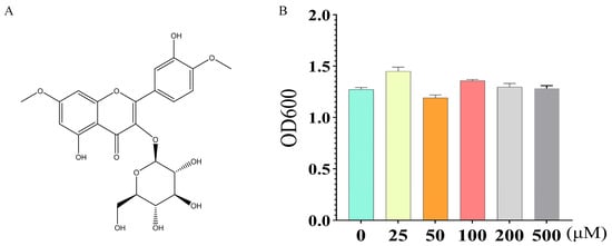
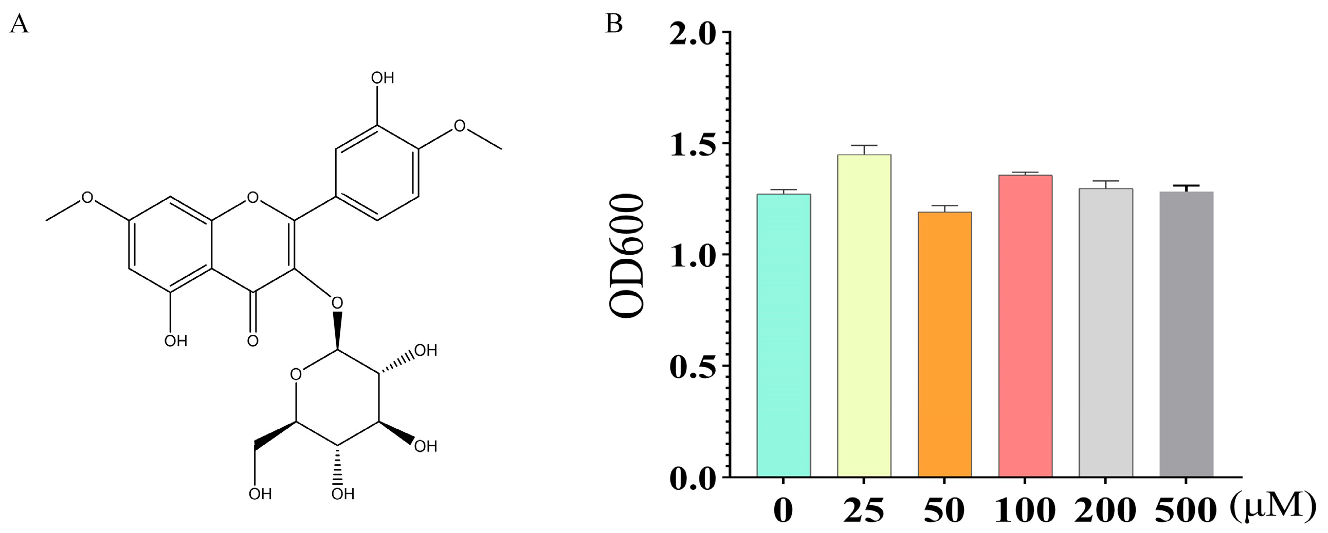
To exclude the possibility that yixingensin might indirectly induce a dietary restriction effect by inhibiting the food bacterium E. coli OP50, we examined the growth of OP50 in the presence of different concentrations of yixingensin. As shown in Figure 1, after co-culture of OP50 with 0, 25, 50, 100, 200, or 500 μM yixingensin for 24 h, no significant differences in OD600 values were observed among the concentration groups. Compared with the control group (OD600 = 1.27 ± 0.021), yixingensin at concentrations ranging from 25 to 500 μM had no significant effect on OP50 growth (p > 0.05). Since the highest concentration of yixingensin used in subsequent experiments was 200 μM, it can be concluded that within the tested effective concentration range, yixingensin does not interfere with normal OP50 growth. Therefore, the lifespan-extending effect of yixingensin is not indirectly achieved through reduced food intake or inhibition of bacterial growth. However, we do not establish that bacterial abundance, nutritional quality, or actual food intake by worms on NGM plates was unchanged.
Figure 1.
Chemical structure of yixingensin (A) and the effect of yixingensin on E. coli OP50 (B); Data are presented as mean ± SD (n = 3 independent experiments).
3.2. Effects of Yixingensin on Lifespan and Selected Physiological Phenotypes
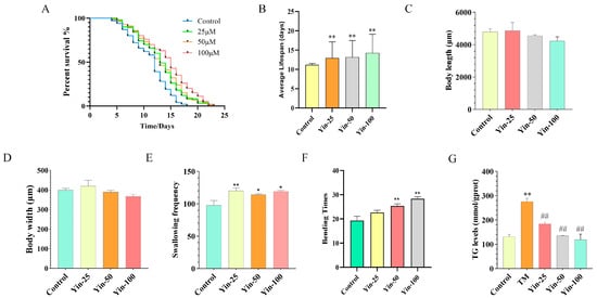
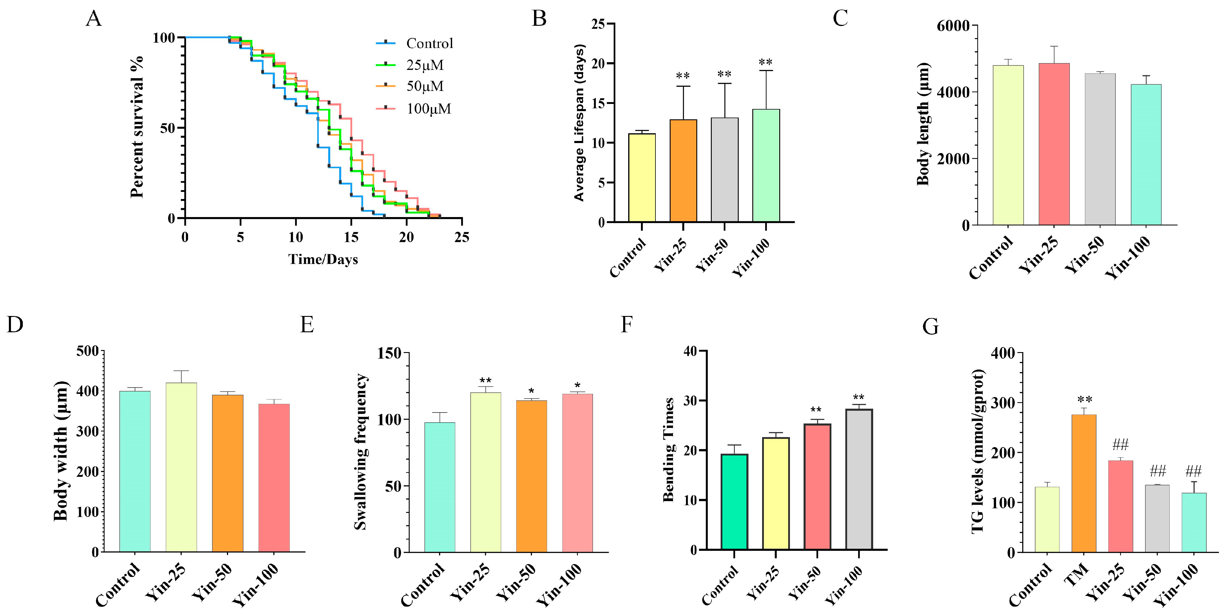
To assess whether yixingensin affects growth and feeding behavior, body length, body width, and pharyngeal pumping rate were measured after 3 days of treatment. As shown in Figure 2A,B and Table S2, Yixingensin significantly extended the lifespan of C. elegans. The mean lifespan of control animals was 11.20 days, whereas treatment with 25, 50, and 100 μM yixingensin increased the mean lifespan to 12.97, 13.19, and 14.28 days, corresponding to increases of 15.8%, 17.8%, and 27.5%, respectively. Kaplan–Meier analysis followed by pairwise log-rank (Mantel-Cox) tests showed significant differences between the control group and the 25 μM, 50 μM), and 100 μM groups. Median lifespan increased from 12 days in the control group to 13, 13, and 15 days at 25, 50, and 100 μM, respectively, representing increases of 8.3%, 8.3%, and 25.0%. The observed maximum lifespan increased from 18 days in the control group to 22, 23, and 23 days, corresponding to increases of 22.2%, 27.8%, and 27.8%, respectively. Although all three concentrations significantly shifted the survival curves relative to the control, the magnitude of the effect was similar at 25 and 50 μM, whereas 100 μM produced the largest increase in mean and median lifespan. Thus, the data do not support a strictly representative concentration-dependent lifespan extension across the entire tested range. Treatment with yixingensin results in extended lifespan in Caenorhabditis elegans (p < 0.05). No significant differences in body length or body width were observed between the control group and any yixingensin-treated group (25, 50, or 100 μM) (Figure 2C,D, p > 0.05). Similarly, the pharyngeal pumping rate was altered by yixingensin treatment compared with the control group (Figure 2E, p < 0.05). These results indicate that yixingensin neither inhibits growth nor induces food intake reduction, and its lifespan-extending effect is not mediated by decreased food consumption.
Figure 2.
Effects of yixingensin on lifespan and physiological indicators in C. elegans. (A) Survival curves of N2 worms treated with different concentrations of yixingensin (0, 25, 50, 100 μM). (B) Mean lifespan of worms in each treatment group. (C) Effect of yixingensin on body length, n = 3. (D) Effect of yixingensin on body width. (E) Effect of yixingensin on pharyngeal pumping rate with 20 worms/group/experiment. (F) Effect of yixingensin on body bend frequency in N2 worms with 20 worms/group/experiment. (G) Effect of yixingensin on TG content. n = 3 independent experiments, * p < 0.05, ** p < 0.01 vs. Control group; ## p < 0.01 vs. TM group.
The effect of yixingensin on age-related decline in locomotion was evaluated by counting the number of body bends within 30 s. As shown in Figure 2F, after 3 days of treatment, the body bend frequency was significantly increased in the 50 and 100 μM yixingensin groups compared with the control group (p < 0.05). The 25 μM group also showed a trend toward increased body bends, but the difference did not reach statistical significance. Thus, yixingensin treatment was associated with improved locomotor performance together with lifespan extension.
3.3. Effects of Yixingensin in a High-Glucose-Induced Metabolic/Lipid-Accumulation Model
To examine yixingensin under metabolic stress, N2 worms were exposed to 0.1 M glucose for 3 days and then treated with yixingensin (25, 50, or 100 μM) or vehicle for an additional 3 days. The worms were then treated with yixingensin (25, 50, or 100 μM) or solvent (model group) for an additional 3 days (containing 0.1 M glucose in NGM). TG content was used as the quantitative lipid endpoint, whereas lipofuscin autofluorescence and Oil Red O staining were treated as supportive imaging endpoints.
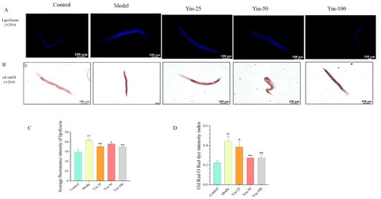
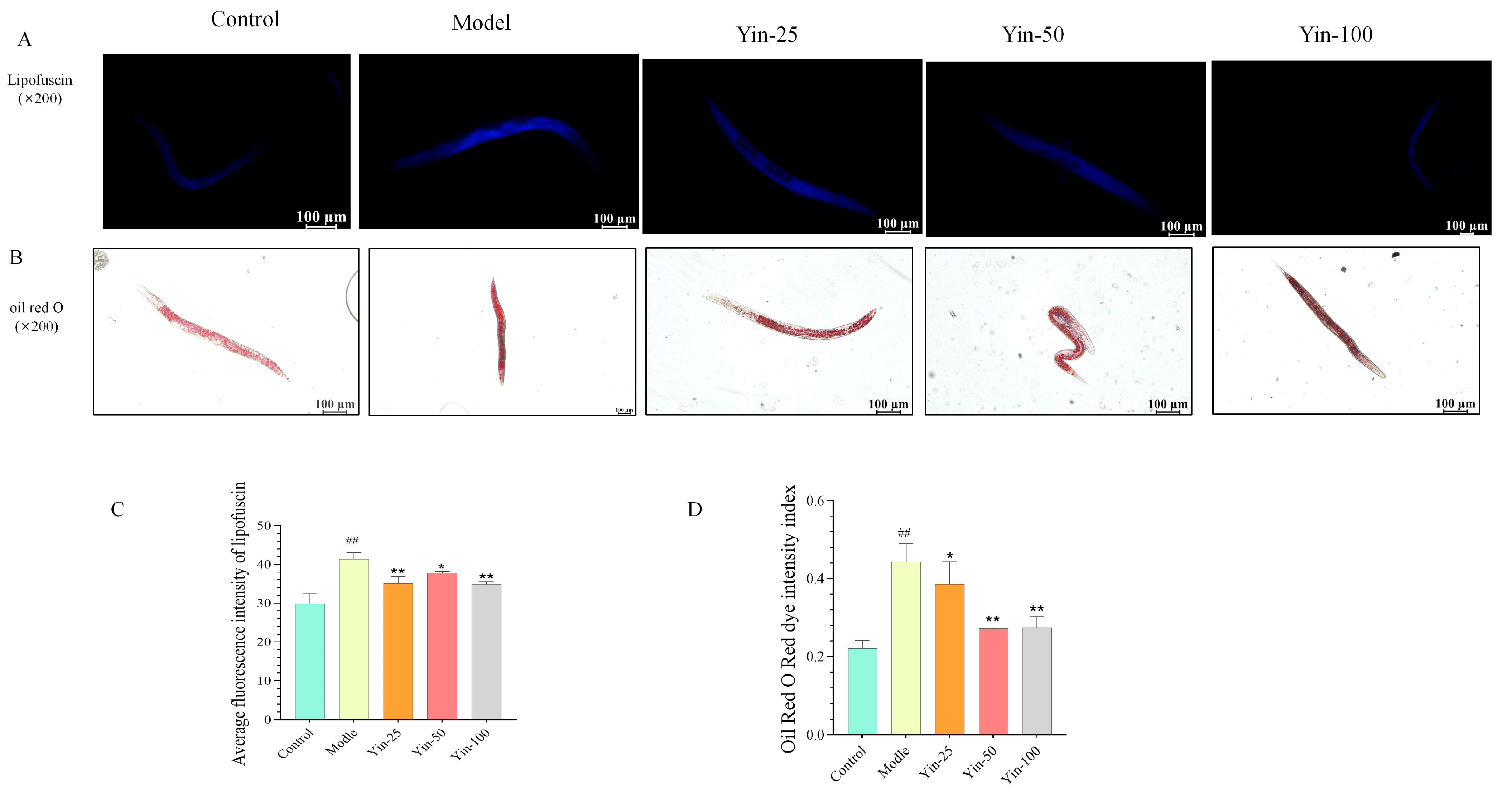
The high-glucose model group showed greater lipofuscin autofluorescence than the blank control, and representative images suggested lower fluorescence after yixingensin treatment (Figure 3A). Because the analysis included only three worms per group and no direct ROS or oxidative-damage assay was performed, these data are interpreted as changes in age-associated autofluorescence rather than direct evidence of reduced oxidative stress.
Figure 3.
Representative images of lipofuscin fluorescence ((A), ×200), Oil Red O staining ((B), ×200) and the quantitative chart (C,D) in C. elegans from the blank control, model (TM), and yixingensin-treated groups (25, 50, 100 μM). There are 10 nematodes in each group. n = 3 independent experiments, Scale bar = 100 μm. * p < 0.05, ** p < 0.01 vs. TM group; ## p < 0.01 vs. Control group.
Similarly, Figure 3B, Oil Red O staining revealed abundant lipid droplets in the model group, whereas yixingensin treatment decreased lipid staining in a concentration-dependent manner. The 50 and 100 μM yixingensin groups showed substantially lower Oil Red O intensity compared with the model group, approaching the level of the blank control. These results demonstrate that yixingensin alleviates both age-related lipofuscin accumulation and lipid deposition under high glucose conditions, suggesting its protective effects against oxidative stress and metabolic dysregulation.
3.4. Transcriptomic Analysis
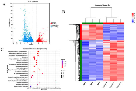
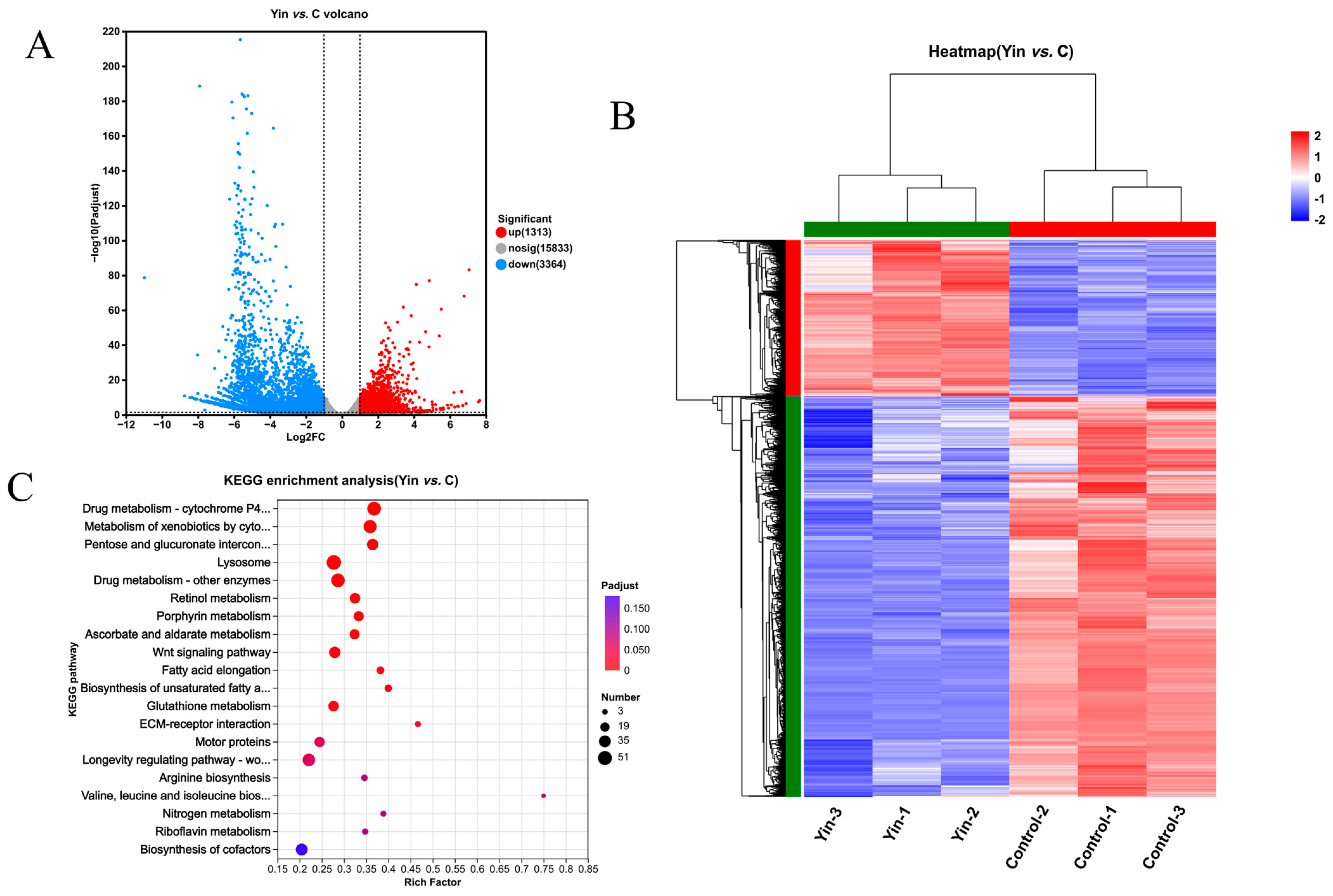
To elucidate the molecular mechanisms underlying the effects of yixingensin, RNA-sequencing analysis was performed on C. elegans treated with 100 μM yixingensin or solvent control for 3 days. Principal component analysis (PCA) revealed a clear separation between the control and yixingensin-treated groups (Figure S1B), indicating that yixingensin induced substantial transcriptomic alterations. The sample-to-sample correlation heatmap further demonstrated high consistency within each group (Figure S1A). A total of 4677 differentially expressed genes (DEGs) were identified between the yixingensin and control groups, including 1313 upregulated and 3364 downregulated genes (Figure 4A,B). This was a single-dose experiment in otherwise standard culture conditions and did not include a high-glucose group; accordingly, it characterizes the transcriptional response to yixingensin itself rather than the transcriptome of the high-glucose model.
Figure 4.
Transcriptomic analysis of C. elegans treated with yixingensin (Yin, 100 μM) vs. control. (A) Volcano plot of differentially expressed genes (DEGs). (B) Heatmap. (C) KEGG pathway enrichment bubble plot of DEGs. (n = 3).
KEGG pathway enrichment analysis of the DEGs revealed that the most significantly enriched pathways included “Drug metabolism-cytochrome P450”, “Metabolism of xenobiotics by cytochrome P450”, “Pentose and glucuronate interconversions”, “Lysosome”, and “Drug metabolism-other enzymes” (Figure 4C). Notably, several pathways related to lipid metabolism and longevity were also significantly enriched, including “Fatty acid elongation”, “Biosynthesis of unsaturated fatty acids”, “Glutathione metabolism”, and importantly, the “Longevity regulating pathway-worm” (p < 0.05, Figure 4C). These results suggest that yixingensin modulates both xenobiotic metabolism and conserved aging-associated pathways. This was a single-dose experiment in otherwise standard culture conditions and did not include a high-glucose group; accordingly, it characterizes the transcriptional response to yixingensin itself rather than the transcriptome of the high-glucose model.
Further analysis of the “longevity regulating pathway-worm” revealed that yixingensin significantly upregulated multiple stress-response genes, including the small heat shock proteins hsp-12.3 (upregulated approximately 2.4-fold), hsp-16.2 (approximately 2.3-fold), and hsp-16.41 (approximately 2.5-fold), as well as the metal detoxification genes mtl-1 (from 0.39 to 7.22) and mtl-2 (from 0 to 11.29) (Supplementary Materials Table S1). Conversely, multiple glutathione S-transferase genes (gst-5, gst-6, gst-13, gst-19, gst-22, gst-26, gst-27, gst-28, gst-34, gst-36, gst-38, and gst-39) were downregulated, with fold changes ranging from 1.0- to 3.7-fold. The fatty acid desaturase fat-6, a key enzyme in monounsaturated fatty acid synthesis, was significantly reduced (approximately 4.3-fold downregulation), while fat-7 was upregulated (approximately 2.3-fold), consistent with the observed decrease in triglyceride content and Oil Red O staining. Additionally, the insulin-like peptide genes ins-6 and ins-7 were downregulated (approximately 2.1- and 3.3-fold, respectively), whereas ins-2 was upregulated (approximately 5.2-fold). Collectively, the transcriptomic data indicate that yixingensin exerts its anti-aging effects by coordinately regulating genes involved in oxidative stress responses, lipid metabolism, and the insulin/IGF-1 signaling pathway, with a particular impact on the longevity-regulating pathway. These transcript changes are hypothesis-generating and do not establish NHR-49 activation, altered fatty-acid composition, β-oxidation flux, or DAF-16 dependence.
3.5. Validation of Key Differentially Expressed Genes by qRT-PCR
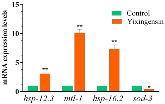
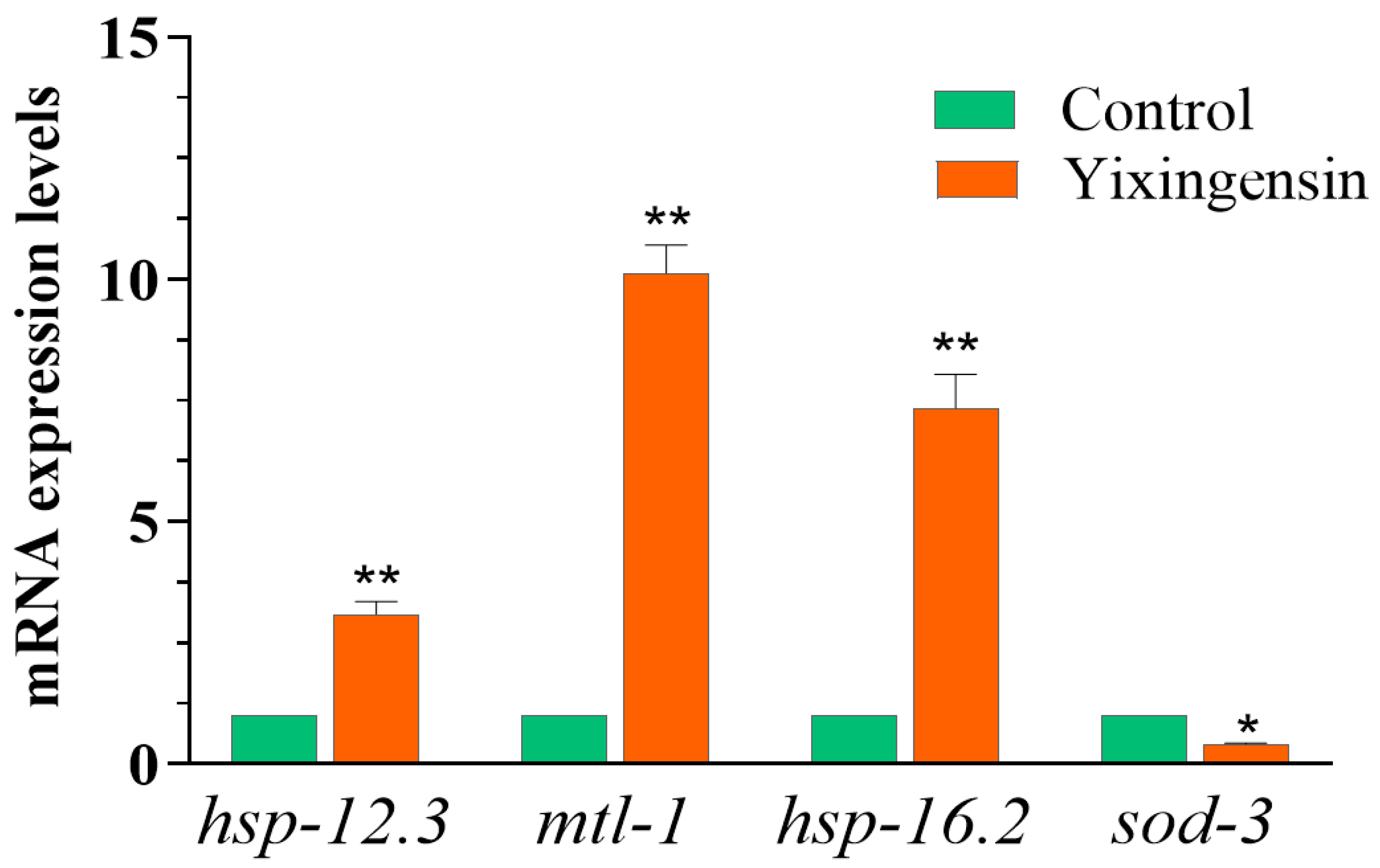
To validate the transcriptomic findings, we performed qRT-PCR analysis on selected genes involved in stress resistance, antioxidant defense, and longevity regulation, including hsp-12.3, mtl-1, hsp-16.2, and sod-3, using act-1 as the internal reference gene. As shown in Figure 5, the qPCR results were consistent with the expression direction of the RNA-seq data. Specifically, yixingensin treatment significantly upregulated the small heat shock protein genes hsp-12.3 (approximately 2.3-fold) and hsp-16.2 (approximately 5.3-fold). The metallothionein gene mtl-1 exhibited upregulation, with an approximately 7.0-fold increase in expression. In contrast, the superoxide dismutase gene sod-3 was downregulated by approximately 3.0-fold upon yixingensin treatment. These expression trends are in good agreement with the transcriptomic data, further supporting the conclusion that yixingensin activates stress-responsive pathways via heat shock proteins and metallothionein, while differentially modulating classical antioxidant enzymes. Examination of the RNA-seq dataset showed that hsp-16.2 was significantly upregulated following yixingensin treatment (log2FC = 1.1896, adjusted p = 0.000493), whereas mtl-1 was detected but did not exhibit statistically significant differential expression (log2FC = 0.1238, adjusted p = 0.6240). Thus, although mtl-1 was included in the targeted qRT-PCR analysis because of its relevance to stress-response regulation, its qRT-PCR expression change should not be interpreted as validation of a significant RNA-seq differential-expression result. These findings highlight the importance of distinguishing RNA-seq detection, differential-expression significance, and targeted qRT-PCR measurements. This targeted validation supports consistency for these genes only and does not independently validate the RNA-seq dataset as a whole. The qRT-PCR panel sampled stress-responsive genes; lipid-metabolism genes such as fat-6 and fat-7 were not independently validated and are therefore interpreted only at the transcriptomic-association level.
Figure 5.
Validation of key differentially expressed genes by qRT-PCR. Relative mRNA expression levels of hsp-12.3, mtl-1, hsp-16.2, and sod-3 in C. elegans treated with 100 μM yixingensin or solvent control for 3 days. Gene expression was normalized to act-1 using the 2−ΔΔCt method. The plotted values represent relative expression fold changes rather than raw Ct, ΔCt, or ΔΔCt values. Statistical comparisons were performed using ΔCt values from three independent biological replicates per group (n = 3), with three technical replicates per biological sample. * p < 0.05, ** p < 0.01, vs. control group.
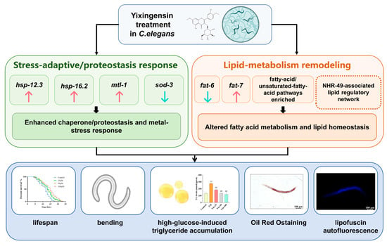
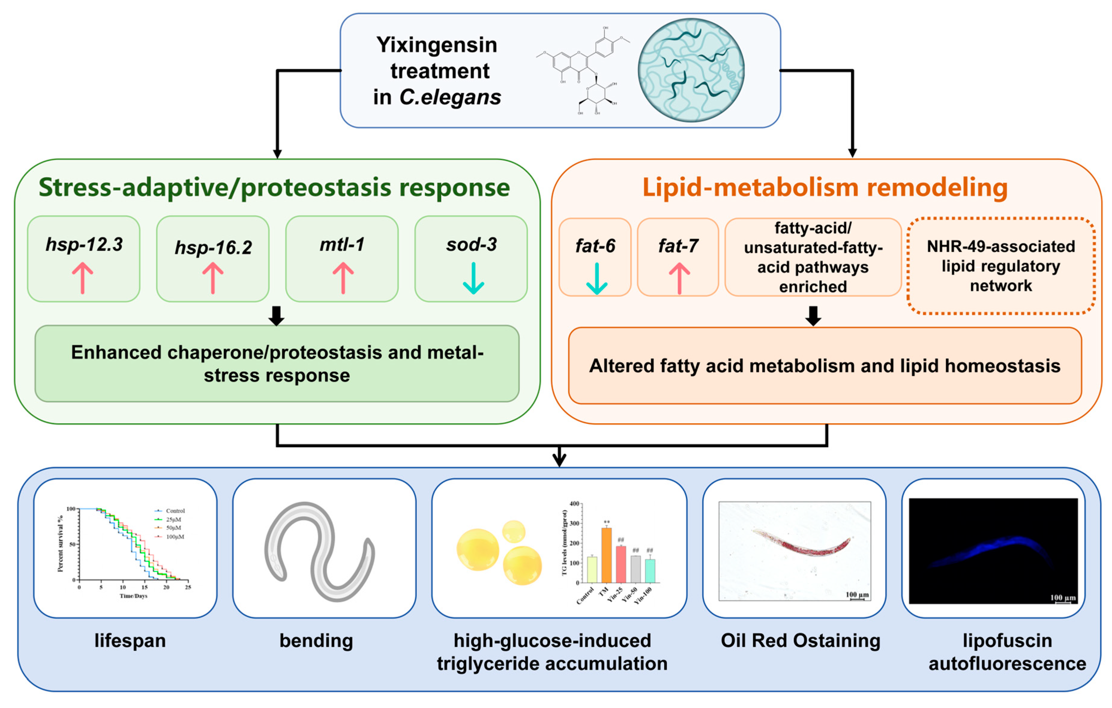
To integrate the phenotypic and gene-expression results, we constructed a working model (Figure 6). Solid arrows summarize observations measured in the present study, whereas dashed connections denote literature-supported hypotheses that were not genetically tested here. The model proposes that selective stress-adaptive/proteostasis responses and remodeling of lipid homeostasis may jointly contribute to the longevity phenotype; it does not assign a definitive causal pathway.
Figure 6.
Proposed working model for yixingensin-associated longevity in C. elegans. Solid arrows summarize measured findings from this study, including stress-response gene changes, lipid-metabolism remodeling, lifespan extension, increased body-bending activity, and reduced high-glucose-associated triglyceride accumulation. The dashed NHR-49 connection indicates a literature-supported candidate association that remains to be tested genetically. HSP induction is shown as a stress-adaptive response but may also reflect treatment-induced cellular stress. ** p < 0.01 vs. Control group; ## p < 0.01 vs. TM group.
4. Discussion
This study demonstrates the longevity-promoting activity of yixingensin in C. elegans and explores its mechanism of action through transcriptomic profiling. At concentrations ranging from 25 to 100 μM, the compound significantly extended mean lifespan without inhibiting the growth of the OP50 bacterial lawn or reducing body size. Pharyngeal pumping rate was maintained or even enhanced, and locomotor bending improved at 50 and 100 μM. Pharyngeal pumping is regulated by a conserved neuromuscular circuit involving serotonergic signaling, cholinergic motor neurons, and pharyngeal muscle activation. Serotonin released under food-associated conditions can stimulate pumping through SER-7 signaling in MC motor neurons, whereas octopaminergic pathways contribute to fasting-associated suppression. Therefore, the increased pumping observed after yixingensin treatment may reflect altered feeding-related neuromuscular regulation. However, neurotransmitter levels and receptor activities were not directly measured in the present study, and thus these pathways should be considered biological context rather than demonstrated targets of yixingensin. Locomotor decline during aging is closely associated with reduced neuromuscular transmission and motor-neuron function. Body bending requires coordinated activity of excitatory cholinergic and inhibitory GABAergic pathways at the neuromuscular junction. The increased bending frequency observed after yixingensin treatment suggests preservation of locomotor capacity, although the specific neuronal pathways involved remain to be experimentally determined. Concurrently, yixingensin attenuated high-glucose-induced triglyceride accumulation, and qualitative assessments via lipofuscin autofluorescence and Oil Red O staining consistently indicated lower levels of age-related and metabolic damage.
These phenotypes align with established findings in C. elegans, reinforcing the notion that natural products can modulate lifespan. Recent investigations into Dendrobium leaf extract, Dendrobium nobile alkaloids, quercetin, jujube extract, and plant-derived polysaccharides have all reported pleiotropic effects, including lifespan extension, improved motility or pumping, reduced lipofuscin or lipid deposition, and modulation of stress-response pathways [9,10,12,13,14,15]. The present results add to this body of evidence by introducing a chemically well-defined flavonoid glycoside. Moreover, yixingensin has been independently reported to exert lipid-regulatory pharmacological actions in mammalian cells [7].
The present findings are particularly relevant to the known biological functions of yixingensin. In mammalian systems, ombuin-3-O-β-D-glucopyranoside activates PPARα and PPARδ/β and alters the expression of genes involved in lipid metabolism [7]. Although C. elegans lacks strict one-to-one orthologs for all mammalian PPAR isoforms, NHR-49 has well-characterized roles in fatty acid β-oxidation, desaturation, fat depletion, stress adaptation, and lifespan regulation [3,4]. Furthermore, NHR-49 participates in α-linolenic acid-induced longevity, thereby linking lipid composition to extended lifespan [8]. Accordingly, the reduction in TG and the reciprocal fat-6/fat-7 transcript changes are therefore compatible with an NHR-49-associated metabolic network but do not demonstrate NHR-49 activation or necessity. No nhr-49 mutant, RNAi, rescue, receptor-binding, or classical target-gene dependency experiment was performed. NHR-49 should thus be considered a candidate mechanistic hypothesis for future testing rather than an established mediator of yixingensin action.
At the transcriptomic level, the most significantly enriched categories included cytochrome P450 and xenobiotic metabolism, along with pathways related to lysosomes, glutathione, fatty acids, and longevity. This pattern suggests that yixingensin functions not only as a metabolic modulator but also as a regulator of detoxification and stress responses. Such a response could contribute to hormetic protective effects; however, the current data do not permit discrimination between protective signaling and direct metabolic clearance of the compound. Future experiments incorporating targeted measurements of reactive oxygen species (ROS), glutathione status, phase I/II detoxification activities, and mitochondrial function will be necessary to clarify this issue. Several highly ranked DEGs may provide additional clues to the broader transcriptional response to yixingensin. Y75B8A.24 (pifk-1A) encodes a phosphatidylinositol 4-kinase, suggesting that phosphoinositide-dependent membrane or signaling processes may be affected. T05H10.3 (mtre-1) is a nematode-specific gene previously associated with hypodermal remodeling, whereas hil-7 encodes an H1 linker histone and may reflect changes in chromatin-associated transcriptional regulation. The altered expression of dpy-14, which encodes a cuticular collagen, may indicate changes related to extracellular-matrix or hypodermal biology. In addition, phat-5 encodes a ShKT-domain protein expressed in the pharyngeal glands and associated with pharyngeal gland/lumen function. Y110A2AL.4 remains poorly characterized, although its expression has been reported to vary in response to developmental and environmental conditions. These genes were not independently validated by qRT-PCR in the present study, and their direct relationships to lifespan extension or lipid metabolism remain unknown. Therefore, they should be regarded as hypothesis-generating candidates for future functional studies rather than as established mediators of yixingensin action.
The stress-response signature observed here also diverges from a simple “antioxidant enzyme activation” model. Expression of hsp-12.3, hsp-16.2, and mtl-1 was induced, whereas sod-3 expression was downregulated. Small heat shock proteins and metallothioneins contribute to protein homeostasis and tolerance to metal/redox stress, while sod-3 is commonly used as a DAF-16-responsive reporter in C. elegans. Many plant-based anti-aging interventions have been reported to increase SOD-3 [10,14]; in contrast, the present dataset reveals an opposite effect for yixingensin. Thus, the current results do not support a model in which yixingensin extends lifespan through canonical DAF-16/SOD-3 activation. Instead, the data point to a more complex stress-responsive program that will require genetic dissection of specific pathways for further validation. This is a selective stress-response remodeling.
The high-glucose experiments provide a second line of evidence for metabolic protection. Excess glucose shortens C. elegans lifespan through mechanisms involving ROS, mitochondrial dysfunction, and carbonyl stress [16]. In this study, high glucose elevated triglyceride (TG) levels, and yixingensin effectively attenuated this increase. Oil Red O staining and lipofuscin measurements were directionally consistent with the TG data. Nevertheless, quantitative fluorescence and staining intensity analyses with adequate biological replication are warranted. Yixingensin treatment began after a 3-day glucose pre-exposure, so the design is better described as post-exposure attenuation than as prevention. Oil Red O and lipofuscin images are supportive but not definitive because robust normalized intensity measurements across adequately replicated biological samples were not available. No direct ROS, oxidative-damage, fatty-acid-composition, or β-oxidation measurement was performed.
Several limitations should be acknowledged. First, only wild-type N2 animals were used, precluding assessment of whether NHR-49, DAF-16, SKN-1, or other regulators are necessary for the longevity effects. Second, FUdR was included in the lifespan assays. Although FUdR is commonly employed in C. elegans aging studies, it may influence lifespan and stress responses under certain genetic or environmental conditions [8,17]; a yixingensin-by-FUdR interaction cannot be excluded without repeating key survival experiments in the absence of FUdR. Third, bacterial growth in liquid culture and pharyngeal pumping do not directly measure food intake on NGM plates. Fourth, the imaging endpoints were limited by small sample size and/or lack of adequately replicated quantitative intensity analysis. Fifth, direct measurements of ROS/oxidative damage, fatty-acid composition or β-oxidation, yixingensin uptake/bioavailability, and receptor engagement were not performed.
Although the present study demonstrates lifespan extension and associated physiological and transcriptional changes in an intact C. elegans model, several limitations remain. First, additional independent lifespan cohorts and genetic validation experiments, such as nhr-49 loss-of-function or RNAi approaches, are required to establish whether the observed lipid-metabolism changes are causally involved in lifespan extension. Second, because all lifespan experiments were performed under FUdR exposure, potential interactions between FUdR and yixingensin cannot be completely excluded. Future studies using FUdR-free lifespan assays and additional stress-resistance models will be necessary to further validate the longevity-promoting activity of yixingensin.
5. Conclusions
Yixingensin extends lifespan in wild-type C. elegans and attenuates high-glucose-associated TG accumulation. At 100 μM, transcriptomic profiling identified changes in xenobiotic-response, stress-response, lipid-metabolism, and longevity-related gene programs, and qRT-PCR showed concordant expression directions for four selected genes. These findings provide proof-of-concept evidence in nematodes and support a working hypothesis that yixingensin engages metabolic and stress-response networks. They do not establish NHR-49 as a causal mediator, demonstrate generalized antioxidant activation, or establish therapeutic/functional-food efficacy in higher organisms.
Supplementary Materials
The following supporting information can be downloaded at: https://www.mdpi.com/article/10.3390/cimb48111095/s1.
Author Contributions
Writing-original draft preparation, conceptualization, supervision, J.L.; methodology, investigation, data curation, R.H.; visualization, J.S.; writing-review and editing, formal analysis, project administration, P.X. All authors have read and agreed to the published version of the manuscript.
Funding
This work was supported by the 2026 Henan Provincial Innovation and Entrepreneurship Training Program for Undergraduates (Grant No. 202612747016); the 2025 Chronic Disease Management Research Project of National Health Commission Capacity Building and Continuing Education Center (Grant No. GWJJMB202510024116).
Institutional Review Board Statement
Not applicable.
Informed Consent Statement
Not applicable.
Data Availability Statement
The original contributions presented in this study are included in the article. Further inquiries can be directed to the corresponding author.
Conflicts of Interest
The authors declare no conflicts of interest.
References
- Tu, W.J.; Zeng, X.; Liu, Q. Aging tsunami coming: The main finding from China’s seventh national population census. Aging Clin. Exp. Res. 2022, 4, 1159–1163. [Google Scholar] [CrossRef] [Scilit] [PubMed]
- Moriwaki, T.; Kato, S.; Kato, Y.; Hosoki, A.; Zhang-Akiyama, Q.M. Extension of lifespan and protection against oxidative stress by an antioxidant herb mixture complex (KPG-7) in Caenorhabditis elegans. J. Clin. Biochem. Nutr. 2013, 3, 81–88. [Google Scholar] [CrossRef] [Scilit] [PubMed]
- Doering, K.R.; Ermakova, G.; Taubert, S. Nuclear hormone receptor NHR-49 is an essential regulator of stress resilience and healthy aging in Caenorhabditis elegans. Front. Physiol. 2023, 14, 1241591. [Google Scholar] [CrossRef] [Scilit] [PubMed]
- Van Gilst, M.R.; Hadjivassiliou, H.; Jolly, A.; Yamamoto, K.R. Nuclear hormone receptor NHR-49 controls fat consumption and fatty acid composition in C. Elegans. PLoS Biol. 2005, 3, e53. [Google Scholar] [CrossRef] [Scilit] [PubMed]
- Shima, M.A.; Sayed, A.P.; Christian, S.L.; Nadine, S. Caenorhabditis elegans as in vivo model for the screening of natural plants-derived novel anti-aging compounds: A short introduction. J. Asian Nat. Prod. Res. 2025, 27, 577–590. [Google Scholar] [CrossRef] [Scilit] [PubMed]
- Zheng, H.; Su, C.; Wang, Z.; Zhao, L.; Li, W. Review on potential of Caenorhabditis elegans as an anti-aging evaluation model for polysaccharides. Int. J. Biol. Macromol. 2025, 322, 146921. [Google Scholar] [CrossRef] [Scilit] [PubMed]
- Malek, M.A.; Hoang, M.H.; Jia, Y.; Lee, J.H.; Jun, H.J.; Lee, D.H.; Lee, H.J.; Lee, C.; Lee, M.K.; Hwang, B.Y.; et al. Ombuin-3-O-β-D-glucopyranoside from Gynostemma pentaphyllum is a dual agonistic ligand of peroxisome proliferator-activated receptors α and δ/β. Biochem. Biophys. Res. Commun. 2013, 430, 1322–1328. [Google Scholar] [CrossRef] [Scilit] [PubMed]
- Qi, W.; Gutierrez, G.E.; Gao, X.; Dixon, H.; McDonough, J.A.; Marini, A.M.; Fisher, A.L. The ω-3 fatty acid α-linolenic acid extends Caenorhabditis elegans lifespan via NHR-49/PPARα and oxidation to oxylipins. Aging Cell 2017, 16, 1125–1135. [Google Scholar] [CrossRef] [Scilit] [PubMed]
- Gong, C.; Feng, S.; Chen, Y.F.; Luo, H.Q.; Zhou, S.Y.; Zhao, S.K.; Zhou, R.; Feng, L.; Ge, Y.F.; Yang, R.J.; et al. Dendrobium officinale leaf extract extends the mean lifespan in Caenorhabditis elegans via the DAF-16/SOD-3 axis. Biogerontology 2026, 27, 111. [Google Scholar] [CrossRef] [Scilit] [PubMed]
- Liu, J.; Zhang, J.; Cui, D.; Xu, Y.; Liu, B. Dendrobium nobile L. Alkaloids promote longevity and stress resistance of Caenorhabditis elegans via DAF-16 and skn-1. Arch. Gerontol. Geriatr. 2026, 149, 106308. [Google Scholar] [CrossRef] [Scilit] [PubMed]
- Zhang, Z.; Li, J.J.; Li, F.; Wang, T.; Luo, X.Y.; Li, B.; You, Y.L.; Wu, C.; Liu, X.M. Jujubae fructus extract prolongs lifespan and improves stress tolerance in Caenorhabditis elegans dependent on DAF-16/SOD-3. Sci. Rep. 2024, 14, 13713. [Google Scholar] [CrossRef] [Scilit] [PubMed]
- Kim, H.J.; Oh, S.H.; Kim, J.H. Antioxidant and anti-aging effects of quercetin 3-O-rhamnoside isolated from houttuynia cordata thunb extract on Caenorhabditis elegans. Free Radic. Biol. Med. 2026, 243, 143–156. [Google Scholar] [CrossRef] [Scilit] [PubMed]
- Liu, Q.; Yi, Y.L.; Liang, X.F.; Wu, M.T.; Li, J.; Chen, X.; Zhang, F.; Ruan, Q.L.; Liu, F.; Duan, J.A.; et al. Antioxidant and antiaging activities of the polysaccharide ZJP-04M from Ziziphus jujuba in Caenorhabditis elegans. Int. J. Biol. Macromol. 2025, 284, 138208. [Google Scholar] [CrossRef] [Scilit] [PubMed]
- Van Raamsdonk, J.M.; Hekimi, S. FUdR causes a twofold increase in the lifespan of the mitochondrial mutant gas-1. Mech. Ageing Dev. 2011, 132, 519–521. [Google Scholar] [CrossRef] [Scilit] [PubMed]
- Li, R.; Chen, X.; Wu, L.; Xie, L.; Chen, M.; Qiu, Y.; Liu, F.; Chen, J.; Tian, M. Antioxidant activity of Radix Cyathula officinalis kuan polysaccharides and their modulatory effects on the gut microbiota of Caenorhabditis elegans. Curr. Issues Mol. Biol. 2025, 47, 538. [Google Scholar] [CrossRef] [Scilit] [PubMed]
- Schlotterer, A.; Kukudov, G.; Bozorgmehr, F.; Hutter, H.; Du, X.; Oikonomou, D.; Ibrahim, Y.; Pfisterer, F.; Rabbani, N.; Sayed, A.A.; et al. Elegans as model for the study of high glucose mediated lifespan reduction Running title: Glucose regulates lifespan in C. Elegans. Diabetes 2009, 58, 2450–2456. [Google Scholar] [PubMed]
- Wang, H.; Zhao, Y.; Zhang, Z. Age-dependent effects of floxuridine (FUdR) on senescent pathology and mortality in the nematode Caenorhabditis elegans. Biochem. Biophys. Res. Commun. 2019, 509, 694–699. [Google Scholar] [CrossRef] [Scilit] [PubMed]

Disclaimer/Publisher’s Note: The statements, opinions and data contained in all publications are solely those of the individual author(s) and contributor(s) and not of MDPI and/or the editor(s). MDPI and/or the editor(s) disclaim responsibility for any injury to people or property resulting from any ideas, methods, instructions or products referred to in the content. |
© 2026 by the authors. Licensee MDPI, Basel, Switzerland. This article is an open access article distributed under the terms and conditions of the Creative Commons Attribution (CC BY) license.