


Abstract
Central nervous system (CNS) diseases, including ischemic stroke, Alzheimer’s disease, and Parkinson’s disease, impose a heavy socioeconomic burden worldwide. Current therapeutic strategies for CNS diseases primarily include modulating ion channels, inhibiting excitotoxicity, and applying anti-inflammatory, antioxidant, and anti-apoptotic approaches. However, existing drugs have not yet met the growing clinical demands. This paper summarizes novel lead compounds recently reported for CNS diseases and discusses the current challenges and emerging strategies in CNS drug development, aiming to provide a reference and scientific basis for future drug discovery and research.
1. Introduction
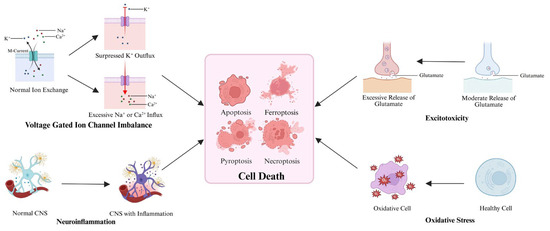
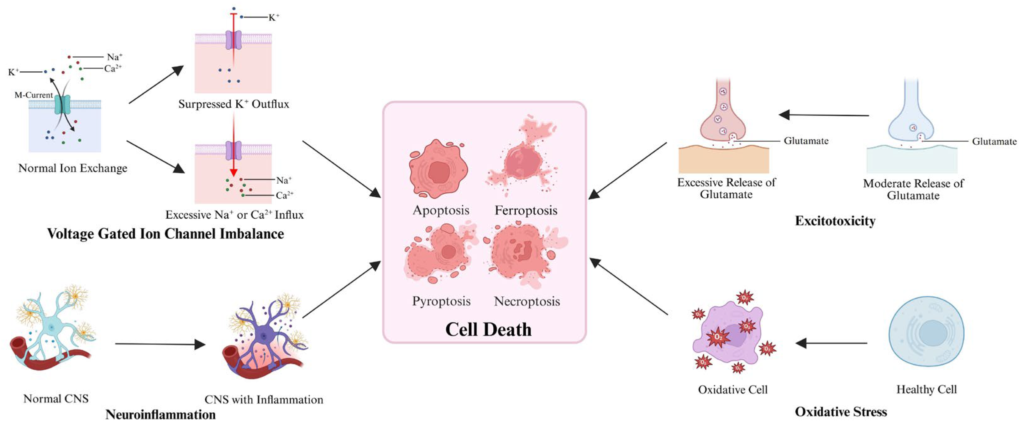
Central nervous system (CNS) diseases, including neurodegenerative diseases, ischemic stroke (IS), and traumatic brain injury (TBI), represent a major category of health issues primarily characterized by brain or spinal cord dysfunction. Their etiology is complex, involving multiple interactions among genetic, infectious, degenerative, and environmental factors [1,2,3]. The core molecular mechanisms underlying dysregulation of voltage-gated ion channels (VGICs), excitotoxicity, neuroinflammation, and oxidative stress ultimately lead to neuronal damage and various forms of cell death [4] (Figure 1). According to the World Health Organization, as of 2021, approximately one-third of the global population was affected by neurological diseases, and CNS diseases such as IS and Alzheimer’s disease (AD) ranked among the leading causes of disability adjusted life years [5]. Therefore, the development of drugs targeting CNS diseases is of critical importance. This review aims to systematically summarize recently reported novel lead compounds developed based on the core molecular mechanisms of CNS diseases, discuss the current challenges and emerging strategies in drug development, and provide a theoretical reference for the discovery of next-generation therapeutics. To achieve this goal, a systematic literature search was conducted in the Web of Science database, focusing on publications from the past three years. The lead compounds included in this review were selected based on their favorable pharmacological potential, including improvements in potency, selectivity, pharmacokinetic properties, or therapeutic efficacy. The most representative compounds are discussed in detail in the main text, whereas the remaining compounds are summarized in tables. Using the same database, this review further identifies and examines the major challenges in the field as well as the emerging and most representative strategies proposed to address them. Collectively, these findings provide an integrated overview of recent advances and future directions in CNS drug development.
Figure 1.
Core molecular mechanisms of CNS diseases. Created in Biorender. Yuhan Qiao (2026) https://BioRender.com/gpr0auo (accessed on 4 June 2026).
2. Novel Lead Compounds Developed Based on the Core Molecular Mechanisms of CNS Diseases
2.1. VGIC Modulators
2.1.1. VGICs and CNS Diseases
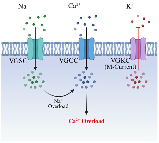
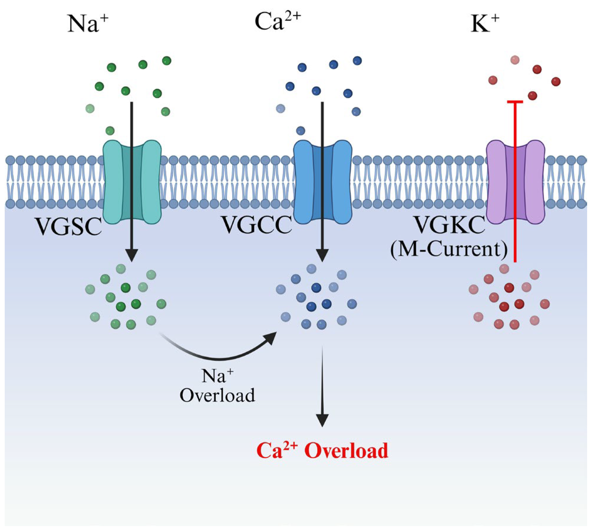
VGICs are a class of integral membrane proteins that sense changes in transmembrane potential and mediate the transmembrane flux of water-soluble ions, thereby regulating processes such as neurotransmitter release, muscle contraction, and hormone secretion. They primarily include voltage-gated sodium channels (VGSCs), voltage-gated potassium channels (VGKCs), and voltage-gated calcium channels (VGCCs) [6]. Under pathological conditions in the CNS, sustained membrane depolarization can trigger abnormal sodium influx via VGSCs, leading to intracellular sodium overload and maintenance of a depolarized state. This subsequently activates VGCCs, leading to calcium overload and downstream injuries [7]. Furthermore, the M-current mediated by the Kv7 subfamily of VGKCs can strongly inhibit membrane depolarization and neuronal firing. In pathological states, excessive suppression of Kv7 channel activity leads to neuronal hyperexcitability, thereby contributing to the pathogenesis of CNS diseases such as epilepsy [8] (Figure 2).
Figure 2.
Molecular mechanism of VGIC imbalance. Created in Biorender. Yuhan Qiao (2026) https://BioRender.com/5p5b6ts (accessed on 4 June 2026).
2.1.2. Novel VGIC Modulators
XPC-6591
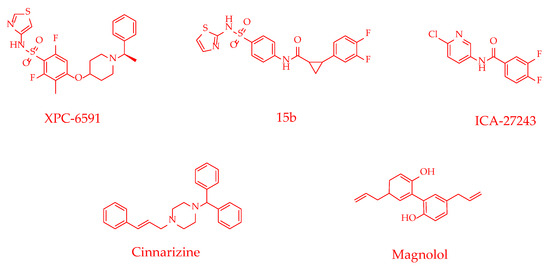
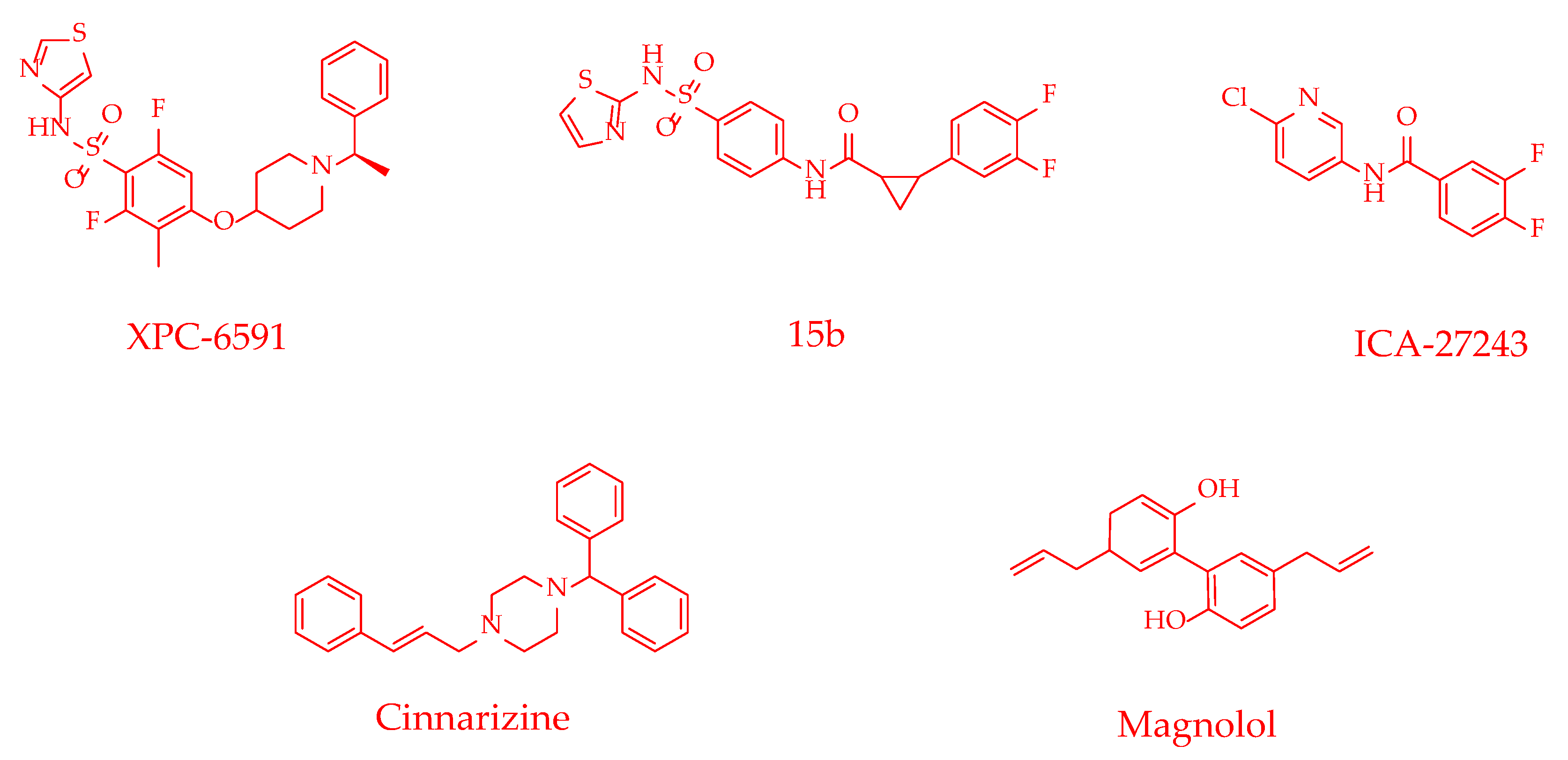
Early sodium channel modulators often exhibit limited subtype selectivity and are frequently associated with CNS and cardiovascular side effects. Moreover, the high homology among sodium channel subtypes presents a significant challenge for the development of highly selective inhibitors. Johnson et al. [9] found that XPC-6591 was a highly selective Nav1.6 inhibitor that bound with high affinity to the voltage-sensing domain 4 (VSD4) of the human Nav1.6 channel, thereby modulating channel function (IC50 = 0.0056 μM). In the Scn8aN1768D/+ gain-of-function mouse model and the direct current multielectrode shock (DC-MES) model in wild-type (WT) mice, XPC-6591 exhibited potent anti-seizure activity at nanomolar concentrations, with EC50 values of 0.0047 μM and 0.01 μM, respectively. Notably, XPC-6591 demonstrated over 700-fold selectivity for Nav1.1 and 100-fold selectivity for Nav1.5. Compared with currently available clinical sodium channel inhibitors, XPC-6591 showed higher selectivity, thereby significantly reducing the risk of potential central nervous system and cardiac side effects. Furthermore, XPC-6591 was a brain-penetrant Nav1.6 inhibitor that exerted anti-epileptic effects at very low cerebral concentrations. As one of the most potent Nav1.6 inhibitors reported to date, XPC-6591 highlights the therapeutic potential of the VSD4-targeting strategy. Future studies are required to further elucidate its long-term safety and efficacy in additional disease models, thereby facilitating its clinical translation.
ICA-27243
The M-current is a critical outward potassium current mediated by Kv7.2/7.3 heterodimers, and its functional homeostasis is essential for suppressing abnormal neuronal discharge. ICA-27243 initially gained attention as a potential anti-epileptic drug; however, Padilla et al. [10] found that this compound also exerted broader neuroprotective effects by targeting potassium channels. Mechanistic studies revealed that ICA-27243 allosterically activated Kv7.2/7.3 channels by binding to the S1-S4 voltage-sensing domain of the Kv7.2 channel, thereby alleviating neuronal hyperexcitability. Masegosa et al. [11] further demonstrated that ICA-27243 (10 μM) increased neuronal survival by 21% in an organotypic spinal cord culture model of acute excitotoxicity. In vivo, ICA-27243 (10 mg/kg) also significantly attenuated the decline in neuromuscular function, preserved motor coordination, and decreased glial reactivity in SOD1G93A transgenic mice. Moreover, following ICA-27243 treatment, the number of spinal motor neurons in SOD1G93A mice increased approximately 1.75-fold, reaching 58% of the level observed in WT mice, and the onset of motor impairment was delayed by at least two weeks. This study not only provides the first evidence of the neuroprotective effect of ICA-27243 on spinal motor neurons but also highlights the importance of allosteric modulation of Kv7 channels as an intervention strategy to counteract neuronal hyperexcitability. However, although ICA-27243 also exhibited a trend toward reducing glial reactivity in SOD1G93A transgenic mice during the experiment, this effect did not reach statistical significance. Therefore, its impact on glial cell activity warrants further investigation.
Magnolol
Recent studies have identified that cannabinoids possess antiepileptic properties. Magnolol, a cannabinoid-like compound derived from Magnolia officinalis, has attracted considerable attention due to its structural similarity to cannabinoids. Yip et al. [12] first reported that magnolol exerted anti-epileptic effects by inhibiting T-type calcium channel subtypes, with IC50 values of 4 μM, 5 μM, and 4 μM for Cav3.1, Cav3.2, and Cav3.3, respectively. This effect was achieved by directly inhibiting calcium currents rather than acting on cannabinoid CB1 or CB2 receptors. In Scn1a+/− mice, magnolol (100 mg/kg) significantly increased the threshold for febrile seizures; in the maximal electroshock (MES) test, the same dose protected 90% of mice from hindlimb extension; and in Gabrb3+/D120N mice, magnolol significantly reduced the frequency and duration of atypical absence seizures. Pharmacokinetic studies showed a brain-to-plasma concentration ratio of 3.55 for magnolol, indicating excellent brain permeability. This study demonstrates that magnolol is effective in both refractory epilepsy models described above. However, its efficacy against other seizure types, such as spontaneous generalized tonic–clonic seizures, has not yet been evaluated in these models. In addition, limitations including its low oral bioavailability remain to be addressed in future investigations.
Table 1 summarizes the effects of novel VGIC modulators, their molecular mechanisms, and their reported activities in various in vivo models for the treatment of CNS diseases.
Table 1.
Novel VGIC modulators for CNS diseases.
Figure 3.
Chemical structures of novel VGIC modulators.
2.2. Excitotoxicity Inhibitors
2.2.1. Excitotoxicity and CNS Diseases
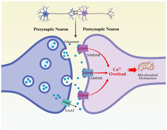
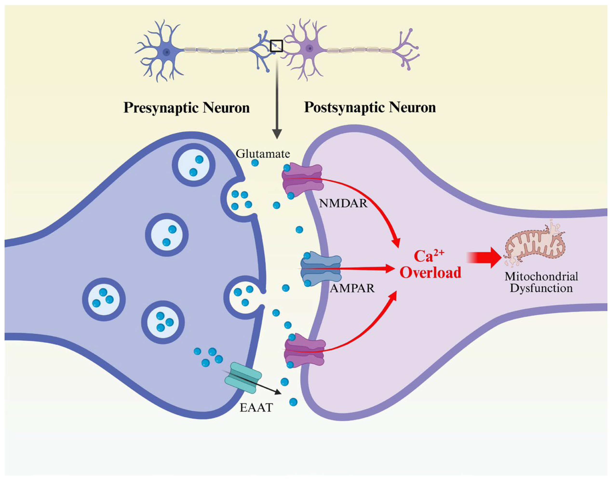
Excitotoxicity refers to the pathological mechanism by which excessive or sustained stimulation of postsynaptic neuronal receptors by excitatory neurotransmitters leads to neuronal injury or death [15]. Glutamate is one of the principal excitatory neurotransmitters in the CNS, participating in over 90% of neuronal activation processes by binding to glutamate receptors on the postsynaptic membrane. Under pathological conditions, sustained neuronal depolarization leads to a massive release of glutamate from synaptic vesicles; simultaneously, energy depletion may reverse the transport direction of excitatory amino acid transporters (EAATs), resulting in extensive non-vesicular glutamate efflux [16,17]. Excessive glutamate stimulation induces intracellular calcium overload, activates downstream oxidative stress signaling pathways and the p38 mitogen-activated protein kinase (MAPK) signaling pathway, disrupts mitochondrial stability, and ultimately causes progressive and sustained neuronal death [18]. (Figure 4) Among the glutamate receptor family, the roles of the ionotropic glutamate receptors, specifically the N-methyl-D-aspartate receptor (NMDAR) and the α-amino-3-hydroxy-5-methyl-4-isoxazolepropionic acid receptor (AMPAR), are particularly crucial [19].
Figure 4.
Molecular mechanism of excitotoxicity. Created in Biorender. Yuhan Qiao (2026) https://BioRender.com/zdvy1p5 (accessed on 5 June 2026).
The NMDAR is a ligand-gated, voltage-dependent ion channel widely distributed throughout the CNS, primarily consisting of two GluN1 subunits and two GluN2 subunits; the latter can be further classified into four subtypes: GluN2A, GluN2B, GluN2C, and GluN2D [20]. Although NMDARs contribute to excitotoxicity, differences in subunit composition and cellular localization confer dual functional effects. It has been proposed that the activation of GluN2A-containing NMDARs may exert neuroprotective effects, whereas the activation of GluN2B-containing NMDARs tends to induce neuronal death; however, inconsistencies remain among relevant research findings [21]. The AMPAR is a tetrameric ion channel composed of four subunits (GluA1-GluA4), and different combinations of these subunits confer its structural diversity [22]. Under certain conditions, AMPARs can mediate neurotoxic calcium influx, and their structural variations or functional overactivation are closely associated with the pathological progression of various CNS diseases.
2.2.2. Novel Excitotoxicity Inhibitors
2,3,5,4′-Tetrahydroxystilbene-2-O-β-D-Glucoside
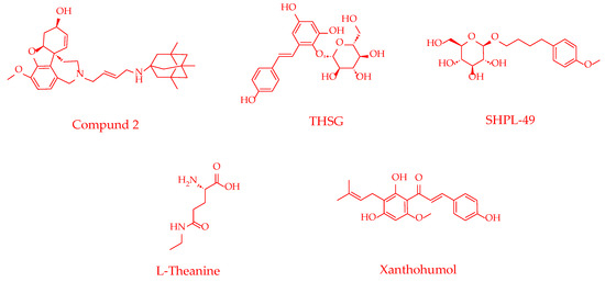
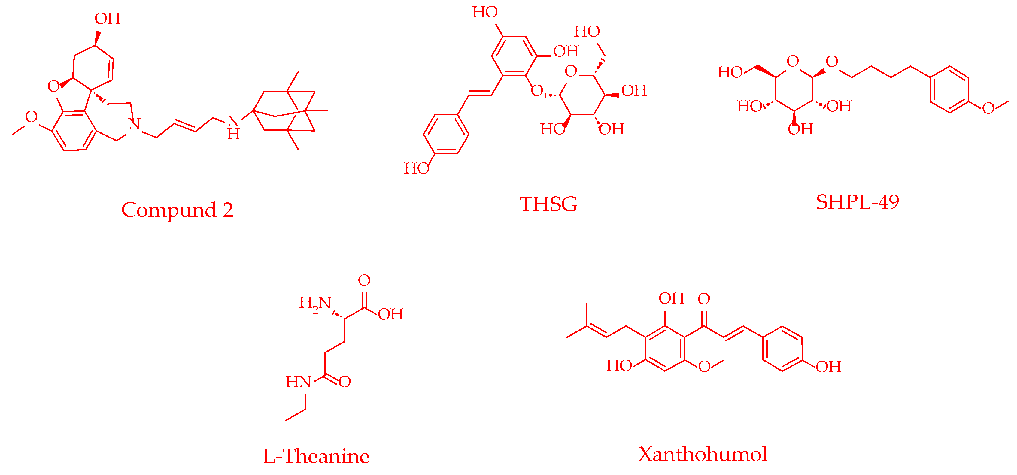
2,3,5,4′-Tetrahydroxystilbene-2-O-β-D-glucoside (THSG) is the primary active component of the traditional Chinese medicine Polygonum multiflorum, which has been used clinically as a tonic and anti-aging medicine for over a millennium. Liu et al. [21] demonstrated that THSG could form hydrogen bonds with residues such as Gly128 and Arg292 in the GluN2B subunit and engage in Pi-Alkyl interactions with Ala263 and Met132 residues, thereby stably occupying the docking pocket (binding energy = −5.2 kcal/mol). This binding subsequently activated the GluN2B–CaMKII–ERK1/2 signaling pathway, enhanced PINK1/PARK2-mediated mitophagy, inhibited neuronal apoptosis, and alleviated cerebral ischemia–reperfusion injury (CIRI) in transient middle cerebral artery occlusion/reperfusion (tMCAO/R) rats. Experiments confirmed that THSG (40 mg/kg) could significantly reduce cerebral infarct volume in tMCAO/R rats; compared with the positive control drug salvianic acid A sodium, rats in the THSG group exhibited lower neurological deficit scores and greater neurological functional improvements. This study reveals that THSG exerts neuroprotective effects through a GluN2B-dependent mechanism, providing a new strategy for developing NMDAR-targeted neuroprotectants based on natural products. However, the efficacy of this compound has thus far been validated only for a single type of neuronal mitophagy. Its effects on other mitophagy pathways and in other cell types remain to be investigated. Moreover, the pharmacokinetic characteristics and clinical translational potential of THSG warrant further evaluation.
L-Theanine
L-Theanine is a non-protein amino acid present in tea leaves that has attracted considerable attention for its potential to modulate nervous system functions. Park et al. [23] demonstrated that this compound exerted a central protective effect in TBI through multi-targeted mechanisms. As a structural analog of glutamate, L-theanine could bind to and inhibit AMPARs, specifically suppressing the overactivation of the GluA1 subunit, thereby directly antagonizing excitotoxicity. It exhibited therapeutic efficacy comparable to that of the AMPAR antagonist NBQX. Furthermore, L-theanine could enter astrocytes, promote glutathione synthesis, thereby enhancing endogenous antioxidant defense capacity. In a rat model of controlled cortical impact—traumatic brain injury (CCI-TBI), treatment with L-theanine (200 mg/kg) significantly attenuated zinc accumulation, neuronal death, and cognitive deficits in the hippocampus, accompanied by improvements in markers of oxidative stress and neuroinflammation. These results provide preclinical evidence for developing L-theanine as a neuroprotectant; its natural origin and favorable safety profile constitute distinct advantages. Nevertheless, future studies are required to elucidate its effects on neuron–astrocyte interactions in vivo and to explore additional underlying molecular mechanisms.
Table 2 summarizes the effects of novel excitotoxicity inhibitors, their molecular mechanisms, and their reported activities in various in vivo models for the treatment of CNS diseases.
Table 2.
Novel excitotoxicity inhibitors for CNS diseases.
Figure 5.
Chemical structures of excitotoxicity inhibitors.
2.3. Anti-Inflammatory Agents
2.3.1. Inflammation and CNS Diseases
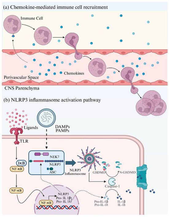
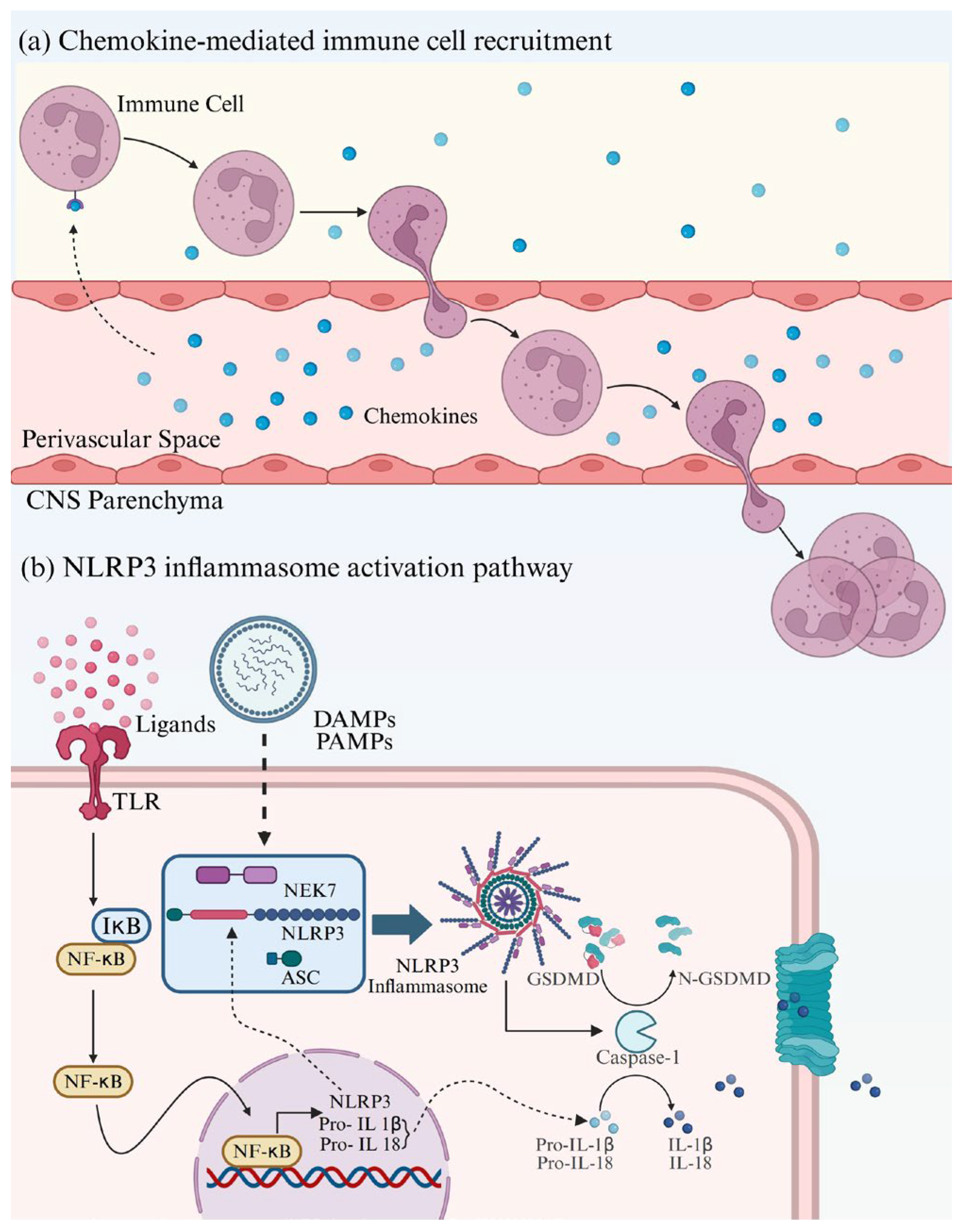
CNS Neuroinflammation refers to the process in which immune cells infiltrate the CNS and activate resident microglia and astrocytes [28]. A moderate inflammatory response is protective, contributing to pathogen clearance and tissue repair; however, when the inflammatory response becomes uncontrolled, chronic, and excessive, it drives various neurological diseases [29]. Under pathological conditions, NLRP3 inflammasomes are transcriptionally upregulated via the TLR/NF-κB signaling pathway and subsequently triggered by pathogen-associated molecular patterns (PAMPs) and damage-associated molecular patterns (DAMPs) to assemble and activate caspase-1. On the one hand, activated caspase-1 promotes the maturation of IL-1β and IL-18; on the other hand, it cleaves gasdermin D (GSDMD), allowing its N-terminal domain to translocate to the cell membrane and form pores, thereby mediating pyroptosis and releasing the pro-inflammatory cytokines IL-1β and IL-18 [30]. Simultaneously, during neuroinflammation, endothelial cells of the blood–brain barrier (BBB) upregulate adhesion molecules and transport chemokines, which recruit peripheral immune cells to migrate and infiltrate into the CNS, further exacerbating neuroinflammation [31] (Figure 6). Therefore, targeting the inhibition of NLRP3 inflammasome assembly and activation, blocking the release of proinflammatory cytokines, and modulating chemokines or their receptor signaling have emerged as key strategies for intervening in CNS neuroinflammation.
Figure 6.
Molecular mechanisms of CNS neuroinflammation. (a) Chemokine-mediated immune cell recruitment. Solid arrows represent the process of immune cell infiltration; dashed arrows represent cytokines crossing the perivascular space to bind to immune cells. (b) NLRP3 inflammasome activation pathway. Solid arrows represent direct effects; dashed arrows represent indirect effects. Created in Biorender. Yuhan Qiao (2026) https://BioRender.com/976pf4d (accessed on 4 June 2026).
2.3.2. Novel Anti-Inflammatory Agents
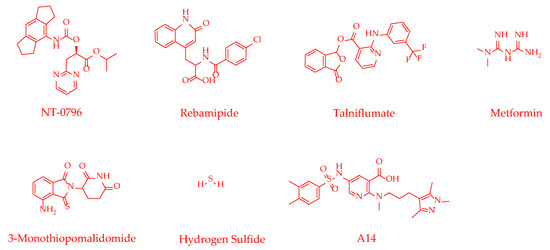
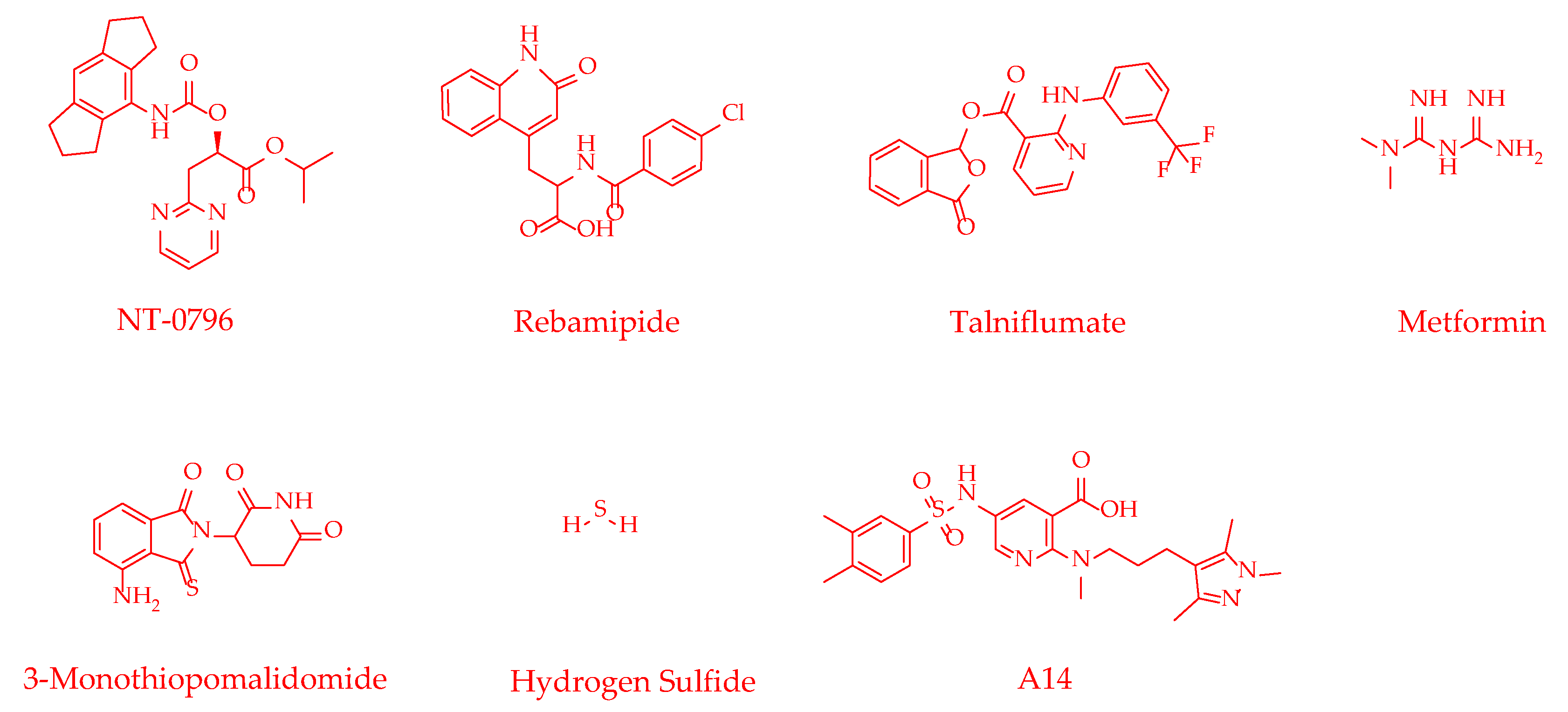
Talniflumate
In PD, upregulation of alanine-serine-cysteine transporter 2 (ASCT2) in astrocytes promotes its interaction with the NLRP3 inflammasome, thereby exacerbating neuroinflammation. Talniflumate was originally developed as an anti-inflammatory analgesic. Through virtual screening of an FDA-approved drug library, Liu et al. [32] identified that it could specifically target ASCT2. By specifically blocking the physical interaction between ASCT2 and NLRP3, this compound inhibited NLRP3 inflammasome assembly and activation, thereby alleviating neuroinflammation. In the MPTP-induced mouse model, talniflumate (50 mg/kg) efficiently crossed the BBB, attenuated neuroinflammation, protected dopaminergic neurons, and significantly improved the motor function. This study reveals a novel mechanism by which astrocytic ASCT2 participates in NLRP3 inflammasome activation, influencing the glial inflammatory response and PD progression. Moreover, target-based screening identified the ASCT2 inhibitor talniflumate, providing a new therapeutic strategy and a candidate drug for PD treatment. Nevertheless, further experimental validation is warranted to elucidate the precise binding sites between ASCT2 and NLRP3, as well as to assess the efficacy of Talniflumate in additional PD models.
Metformin
As a classic first-line oral hypoglycemic drug, metformin (MF) has been increasingly recognized for its novel pharmacological effects. In recent years, MF has shown neuroprotective potential in neurological diseases. Akter et al. [33] established a mouse stroke model with BBB disruption superimposed on tobacco smoke (TS) exposure to investigate the potential therapeutic effects of MF on TS-induced stroke-related neural damage. The results indicated that MF primarily activated the Nrf2-ARE antioxidant signaling pathway and inhibited the NF-κB inflammatory pathway, significantly reducing the release of pro-inflammatory factors such as TNF-α and IL-1β in primary cultured neurons and astrocytes in vitro. Further in vivo experiments confirmed that MF intervention (200 mg/kg) significantly attenuated the neuroinflammatory response in adolescent mice following stroke and TS exposure, downregulated NF-κB expression in brain tissue, and decreased the levels of inflammatory factors, including TNF-α, IL-1β, MCP-1, and MIP-2. This study is the first to systematically explore the direct anti-neuroinflammatory effects of MF on primary neurons and astrocytes under conditions of TS and IS. Furthermore, research on the impact of TS exposure on the Nrf2-ARE signaling pathway in adolescent mice has been rarely reported; this study fills the gap in this field, providing a new animal model basis for research on smoking-related neuroinflammation in adolescents. A limitation of this study is that neither the therapeutic time window for MF administration nor the optimal dosage required to maintain adequate cerebral concentrations has been established, warranting further experimental validation.
3-Monothiopomalidomide
3-Monothiopomalidomide (3-MP) is a novel immunomodulatory imide drug derived from pomalidomide. Hsueh et al. [34] found that it bound to cereblon (CRBN), a key adaptor protein of the E3 ubiquitin ligase complex, thereby interfering with CRBN-mediated inflammatory signal transduction (IC50 = 0.21 μM), demonstrating significantly superior efficacy compared to pomalidomide (IC50 = 2.38 μM), while not triggering the downstream degradation of SALL4 and Aiolos proteins, thus avoiding associated adverse reactions. 3-MP (10–60 μM) effectively reduced pro-inflammatory markers in LPS-stimulated murine macrophages and microglia. In in vivo experiments, 3-MP (26.5 mg/kg and 53 mg/kg) significantly reduced pro-inflammatory cytokine and chemokine levels in both plasma and brain of LPS-treated mice, without affecting the anti-inflammatory cytokine IL-10. Pharmacokinetic studies revealed that 3-MP readily crosses the BBB following intraperitoneal injection, with a brain-to-plasma concentration ratio of 0.44–0.47 (2.65–26.5 mg/kg), indicating good brain penetrability. In the CCI-TBI mouse model, 3-MP (13.23 mg/kg and 26.47 mg/kg) markedly alleviated behavioral deficits and inhibited astrocyte and microglial activation. In summary, 3-MP is a drug candidate possessing both brain penetrability and anti-inflammatory activity. Compared with pomalidomide, it exhibits lower toxicity and holds promise for the treatment of neuroinflammation-related diseases. Future studies are needed to evaluate its long-term safety profile and further assess its teratogenic potential across different experimental models.
Table 3 summarizes the effects of novel anti-inflammatory agents, their molecular mechanisms, and their reported activities in various in vivo models for the treatment of CNS diseases.
Table 3.
Novel anti-inflammatory agents for CNS diseases.
Figure 7.
Chemical structures of anti-inflammatory agents.
2.4. Antioxidants
2.4.1. Oxidative Stress and CNS Diseases
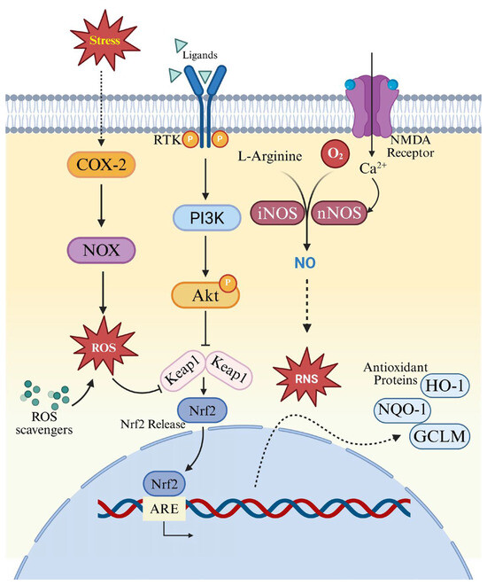
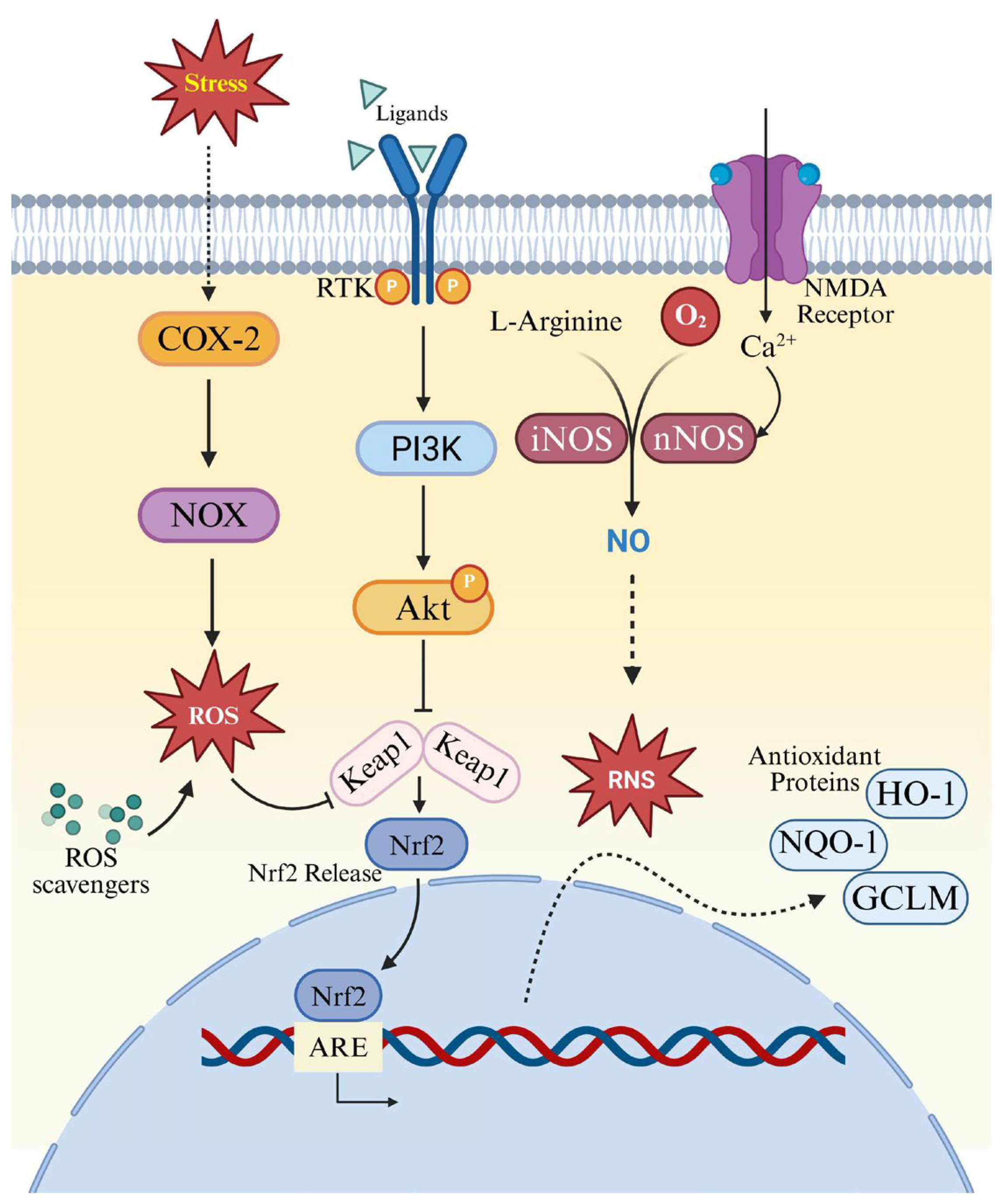
The essence of oxidative stress lies in an imbalance between pro-oxidant and antioxidant homeostasis, where the rate of reactive oxygen species (ROS) and reactive nitrogen species (RNS) generation exceeds the scavenging capacity of the antioxidant defense system. The CNS has a remarkably high energy demand, and its ATP is primarily generated through mitochondrial oxidative phosphorylation. While this process is essential for maintaining normal function, it inevitably generates ROS. Furthermore, superoxide anions produced via the activation of NADPH oxidase (NOX) can also be converted into ROS [40]. Low to moderate concentrations of ROS play important physiological roles, but under oxidative stress conditions, excessive ROS is highly detrimental [41]. Under pathological conditions, nitric oxide (NO) produced by certain nitric oxide synthases (NOSs) can trigger an explosive increase in RNS, constituting a critical mechanism that aggravates oxidative damage in the CNS [42]. When the rate of ROS/RNS production surpasses the clearance capability of endogenous enzymatic systems and small-molecule antioxidants, the resulting excess free radicals attack proteins, lipids, and DNA, ultimately leading to neuronal damage and functional decline [43]. To counteract oxidative stress, cells phosphorylate and activate the transcription factor Nrf2 via the PI3K/Akt signaling pathway, promoting its dissociation from Keap1 and subsequent nuclear translocation. This initiates the transcription of antioxidant enzyme genes, such as heme oxygenase-1 (HO-1), thereby constituting the core endogenous defense system. Simultaneously, small-molecule antioxidants can interrupt free radical chain reactions to decrease ROS formation [44,45] (Figure 8). Based on these mechanisms, targeted activation of the PI3K/Akt/Nrf2 pathway, inhibition of NOX and NOS activities, and direct ROS scavenging have emerged as vital strategies for developing central neuroprotective agents.
Figure 8.
Molecular mechanisms of the intracellular antioxidant system. Arrows indicate stimulatory interactions, with solid arrows representing direct effects and dashed arrows representing indirect effects; bars denote inhibitory effects. Created in Biorender. Yuhan Qiao (2026) https://BioRender.com/zdw19kw (accessed on 4 June 2026).
2.4.2. Novel Antioxidants
Gomisin N
Gomisin N (GN) is a natural lignan compound isolated from the traditional Chinese medicine Schisandra chinensis. It has attracted widespread attention due to its diverse pharmacological activities, including hepatoprotective, anti-inflammatory, and antioxidant effects. In recent years, its neuroprotective potential has also been investigated. Li et al. [44] found that in the mouse model of middle cerebral artery occlusion/reperfusion (MCAO/R), GN (30–90 mg/kg) could significantly attenuate neuronal damage, reduce cerebral infarct volume, and improve neurological deficits. Mechanistic analysis revealed that GN activated the PI3K/Akt/mTOR signaling pathway by promoting the phosphorylation of key proteins PI3K, AKT, and mTOR (binding energy of GN with these proteins: PI3K = −9.9 kcal/mol; Akt = −8.5 kcal/mol; mTOR = −8.0 kcal/mol), thereby inhibiting overactivated autophagy and reducing neuronal death. Molecular docking studies demonstrated a strong binding affinity between GN and PI3K, and the protective effect was reversed by the PI3K inhibitor LY294002, further confirming the critical role of this pathway. This study is the first to elucidate a novel neuroprotective mechanism of GN in cerebral ischemia by modulating autophagy, providing important preclinical evidence for its further development as a candidate anti-IS agent. Future research should further explore its multi-target synergistic mechanisms and in vivo pharmacokinetic properties.
Dl-3-n-Butylphthalide and Its Derivatives
Dl-3-n-butylphthalide (NBP) can exert neuroprotective effects in various CNS diseases by activating the Nrf2 signaling pathway to alleviate oxidative stress and neuroinflammation. Liu et al. demonstrated that in a nitroglycerin (NTG)-induced chronic migraine mouse model, NBP (30 mg/kg) significantly reduced oxidative stress and inflammation, and these therapeutic effects were markedly attenuated following administration of the Nrf2 inhibitor ML385 [46]. Compared with edaravone-dexborneol, NBP showed greater effects in reducing cerebral infarct volume, restoring cerebral blood flow, and scavenging oxidative stress factors [47]. Furthermore, Li et al. [48] synthesized a hybrid compound 8a by conjugating NBP with ligustrazine (TMP) via a triazole linkage. This hybrid, bound to Keap1, promoted Nrf2 dissociation and nuclear translocation and upregulated the production of downstream antioxidant factors, thereby reducing intracellular ROS levels and effectively protecting neuronal mitochondria. In the OGD/R-induced SH-SY5Y cell injury model, a low concentration of 8a (6.25 μM) achieved a protection rate of 75.6% without detectable cytotoxicity. It was demonstrated to activate the Nrf2 pathway and reduce markers of cell injury, oxidative stress, and apoptosis, showing greater efficacy than NBP. In the rat MCAO/R model, 8a (20 mg/kg) significantly reduced cerebral infarct volume, improved mNSSs, and enhanced cerebral blood flow, displaying superior therapeutic efficacy compared with NBP at the equivalent dose. In conclusion, NBP and its derivatives show promising potential for the treatment of various CNS diseases and warrant focused attention in future research, but further studies are required to characterize their pharmacokinetic profiles.
Crocetin
Crocetin is a natural carotenoid that can be extracted directly from saffron (Crocus sativus) and is also the intestinal metabolite of crocin, the principal water-soluble active constituent of saffron, exhibiting excellent BBB permeability. Wu et al. [49] found that this compound could antagonize parthanatos (PARP-1-dependent programmed cell death) during IS through a dual mechanism. In the early phase, crocetin inhibited the interaction between protein kinase Cζ (PKCζ) and the NOX subunit p47phox, thereby blocking the NOX2 complex assembly and activation, and markedly suppressing the ROS burst. In the late phase, crocetin directly bound to hexokinase-I (HK-I), specifically targeting key residues such as Leu31 and His373, which inhibited E3 ubiquitin ligase RNF146-mediated PARylation and ubiquitination degradation of HK-I, maintained the binding of HK-I to mitochondria, protected mitochondrial function, and prevented the nuclear translocation of apoptosis-inducing factor (AIF). The study demonstrated that crocetin pretreatment could elevate the neuronal survival to a level comparable to that achieved with the selective PARP inhibitor PJ34, indicating that crocetin effectively promoted cell survival through the anti-parthanatos pathway. In vivo experiments confirmed that crocetin (40 mg/kg) reduced the cerebral infarct volume in rats with pMCAO from 43.11% to 12.91%, and the mNSS decreased from 13.2 to 9.4. These findings not only reveal the potential of crocetin as a multi-target neuroprotective agent but also provide important insights for the development of novel anti-IS drugs targeting the NOX2-HK-PARP axis. However, the pharmacokinetic properties, clinically applicable dosage, and long-term safety of this compound still require further comprehensive investigation.
Table 4 summarizes the effects of novel antioxidants, their molecular mechanisms, and their reported activities in various in vivo models for the treatment of CNS diseases.
Table 4.
Novel antioxidants for CNS diseases.
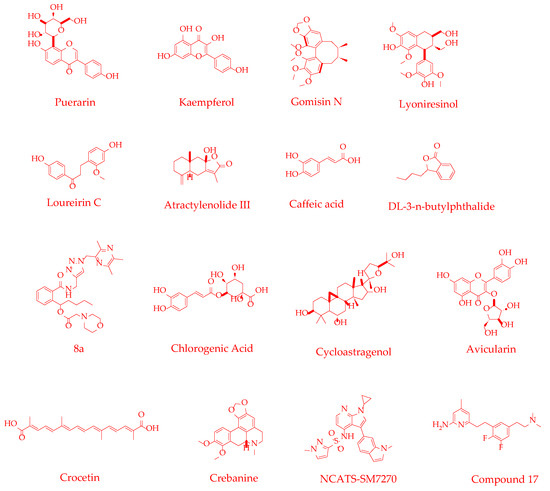
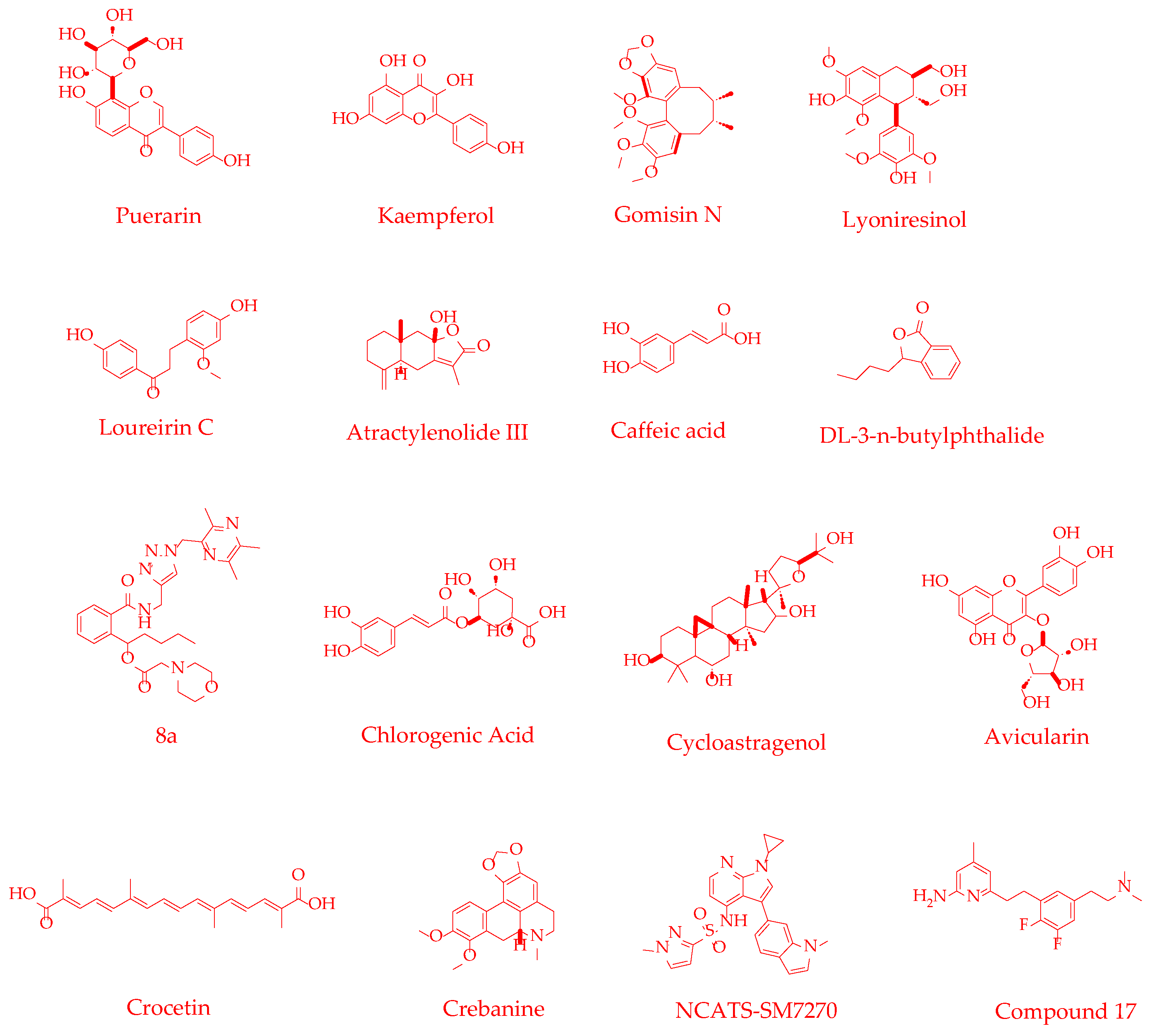
Figure 9.
Chemical structures of antioxidants.
2.5. Cell Death Inhibitors
2.5.1. Cell Death and CNS Diseases
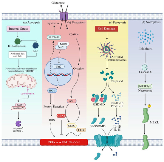
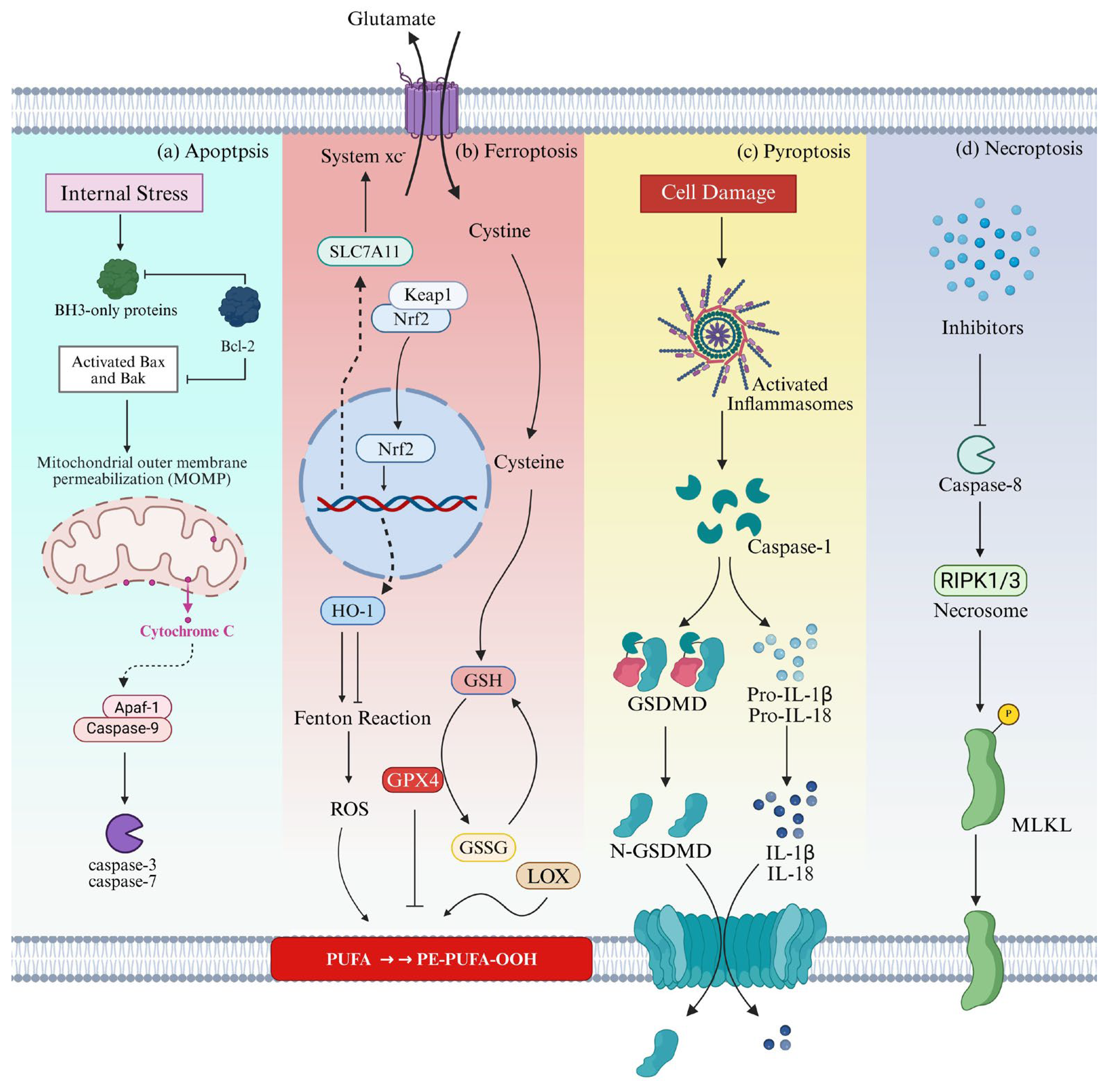
The development of the nervous system is inseparable from cell death [63]. Under normal conditions, programmed cell death eliminates redundant structures; however, under pathological conditions such as oxidative stress and inflammation, the CNS undergoes various forms of cell death, including apoptosis, pyroptosis, ferroptosis, and necroptosis, ultimately leading to neuronal injury and functional impairment. (Figure 10).
Figure 10.
Molecular mechanisms of cell death (a) Apoptosis, (b) Ferroptosis, (c) Pyroptosis, (d) Necroptosis. Arrows indicate stimulatory interactions, with solid arrows representing direct effects and dashed arrows representing indirect effects; bars denote inhibitory effects. Cyan background represents apoptosis; red background represents ferroptosis; yellow background represents pyroptosis; purple background represents necroptosis. Created in Biorender. Yuhan Qiao (2026) https://BioRender.com/38cy1ik (accessed on 4 June 2026).
In normal CNS cells, the anti-apoptotic protein Bcl-2, a member of the Bcl-2 family, can inhibit the pro-apoptotic effector proteins Bax and Bak, preventing them from initiating cell death. Under pathological conditions, the expression of Bcl-2 family proteins becomes dysregulated, and the loss of Bcl-2-mediated inhibition allows Bax and Bak to increase mitochondrial outer membrane permeability, leading to the release of cytochrome c and activation of the caspase cascade, with caspase-3 ultimately executing cellular disassembly [64,65]. Ferroptosis is driven by iron overload and lipid peroxidation. CNS cells, characterized by high oxygen consumption and abundant polyunsaturated fatty acids, are particularly susceptible to ferroptosis. Under physiological conditions, cells rely on the System Xc−-GSH-GPX4 axis to reduce lipid peroxides to non-toxic lipid alcohols and maintain iron homeostasis via iron chelation, thereby suppressing ROS accumulation. However, under pathological states, iron overload or the failure of the antioxidant defense axis allows intracellular free iron to catalyze the generation of massive amounts of ROS via the Fenton reaction, triggering a chain reaction of lipid peroxidation and ultimately leading to ferroptosis [66,67]. Pyroptosis is a pro-inflammatory form of programmed cell death mediated by the NLRP3 inflammasome signaling pathway (see Section 2.3.1), ultimately contributing to neuronal loss and disease progression in CNS diseases [68]. Furthermore, when Caspase-8 is inhibited, necroptosis is initiated by phosphorylation-driven activation of the RIPK1/RIPK3/MLKL signaling axis, resulting in loss of membrane integrity and the release of DAMPs [69]. Therefore, directly targeting the downstream effector molecules of these cell death pathways has emerged as the last line of defense to rescue damaged neurons and restore neural homeostasis.
2.5.2. Novel Cell Death Inhibitors
Metformin
Sritawan et al. [70] demonstrated that metformin could significantly antagonize the methotrexate (MTX)-induced decreases in neural stem cell markers Sox2 and nestin in the rat hippocampus, increase levels of the synaptic protein PSD95, and modulate the expression of apoptosis-related proteins, including downregulating caspase-3 and Bax while upregulating Bcl-2. Meanwhile, metformin enhanced the expression of the transcription factor CREB and its phosphorylated form pCREB, thereby promoting neurogenesis, inhibiting apoptosis, and enhancing synaptic plasticity through multiple pathways. In MTX-treated rats, metformin (200 mg/kg) treatment significantly restored PSD95 levels, reduced Caspase-3 content and Bax/Bcl-2 ratio, and restored the expression of proteins such as pCREB/CREB to near-normal levels. Moreover, it restored the number of Sox2-positive cells by approximately 1.94-fold and nestin-positive cells by approximately 1.97-fold in the rat hippocampus. These results strongly indicate that metformin exhibits superior neuroprotective activity in alleviating MTX chemotherapy-related neurotoxicity and memory impairment; however, this study does not include animal behavioral assessments, and the underlying molecular mechanisms, as well as the long-term safety profile, remain to be further elucidated.
Ferfluor-1
Ferrostatin-1 (Fer-1) is a classic ferroptosis inhibitor; however, it suffers from poor metabolic stability and insufficient BBB permeability. Based on its structure, Yan et al. [71] introduced a 1,3,4-thiadiazole ring to synthesize the derivative Ferfluor-1. In three cancer cell lines (HT1080, OS-RC-2, and SH-SY5Y), Ferfluor-1 exhibited nanomolar inhibitory activity against RSL3-induced ferroptosis, with EC50 values of 57 nM, 75 nM, and 2.3 nM, respectively. As a lipophilic radical-trapping antioxidant, Ferfluor-1 inhibited lipid peroxidation by scavenging phospholipid peroxyl radicals. Its mechanism of action was similar to that of Fer-1, presumably involving inhibition of the 15-LOX-2/PEBP1 complex to reduce lipid peroxidation product formation. Compared with Fer-1, Ferfluor-1 showed significantly improved metabolic stability. The stability of Ferfluor-1 in rat microsomes and plasma increased by approximately 8-fold and 40-fold over Fer-1, respectively, demonstrating favorable pharmacokinetic properties and superior BBB permeability (brain-to-plasma ratio = 0.33). In MCAO and MPTP-induced mouse models, Ferfluor-1 (5 mg/kg) significantly decreased MDA levels and restored dopaminergic neuronal function, thereby attenuating neurological damage. Furthermore, due to its unique excited-state intramolecular proton transfer properties, Ferfluor-1 could serve as a ratiometric photoluminescent probe that specifically responds to phospholipid hydroperoxides and peroxynitrite, enabling real-time visualization of ferroptosis in cells and zebrafish. This study not only provides a multifunctional tool compound with both inhibitory and detection capabilities for the diagnosis and treatment of CNS ferroptosis-related diseases, but also opens a new avenue for developing novel theragnostic probes based on ferroptosis inhibitors. Nevertheless, the pharmacokinetic properties of Ferfluor-1 still require further optimization. Moreover, when used as a probe, its near-infrared imaging performance and potential interference resulting from its inhibitory activity during long-term monitoring also warrant further refinement.
Biochanin A
Biochanin A (BCA) is a methoxy isoflavone with estrogen-like activity isolated from legumes such as chickpeas and Trifolium pratense L. Its development stems from the potential of traditional Chinese medicine-derived monomeric compounds in multi-targeted neuroprotective strategies. Li et al. [72] found that in the early stage of spinal cord injury (SCI), BCA attenuated inflammasome activation and oxidative stress by inhibiting the TLR4/NF-κB/NLRP3 inflammasome signaling pathway and activating the Nrf2/HO-1 antioxidant pathway, thereby promoting autophagy, reducing apoptosis and pyroptosis, and exerting neuroprotective effects. In an SD rat model of SCI, continuous intraperitoneal injection of BCA (40 mg/kg) for 14 days significantly improved motor function, alleviated spinal cord edema, decreased the levels of inflammatory factors and oxidative stress markers, and upregulated antioxidant enzyme activities. In the early stage of SCI, the therapeutic efficacy of BCA was comparable to that of the positive control methylprednisolone (30 mg/kg). These findings suggest that BCA offers a potential multi-target therapeutic strategy for early intervention in SCI. However, current research is still limited to the early stages in animal models. The direct molecular targets of BCA on signaling pathways, as well as its long-term efficacy and safety, remain unclear, and further mechanistic investigation is warranted.
Morin Hydrate
Morin hydrate (MH) is a natural flavonoid whose neuroprotective effects derive from its multi-target regulatory potential against neuroinflammation, apoptosis, and necroptosis, showing particularly strong efficacy in models of neurodegenerative diseases. Research by Elbaz et al. [73] revealed that MH (20 mg/kg) significantly ameliorated motor dysfunction, alleviated weight loss, and reversed striatal histopathological damage in a rat model of Huntington’s disease (HD) induced by 3-nitropropionic acid (3-NP), exhibiting comparable efficacy to the positive control drug necrosulfonamide (NSA). At the molecular level, it downregulated the 3-NP-induced TNF-α expression to 69%, reduced the expression of phosphorylated RIPK1, RIPK3, and MLKL by over 20%, and significantly inhibited caspase-3 activity, while increasing striatal succinate dehydrogenase (SDH) activity by 2.4-fold. Through these actions, it exerted multiple neuroprotective effects, including anti-necroptosis, anti-apoptosis, and anti-neuroinflammation. This study is the first to demonstrate that MH protects the striatum from 3-NP-induced neurotoxicity by modulating the necroptosis pathway involving RIPK1, RIPK3, and MLKL, thereby conferring protection against HD. Future studies should further investigate its pharmacokinetics, safety, and clinical translational potential.
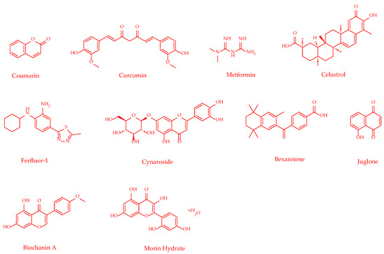
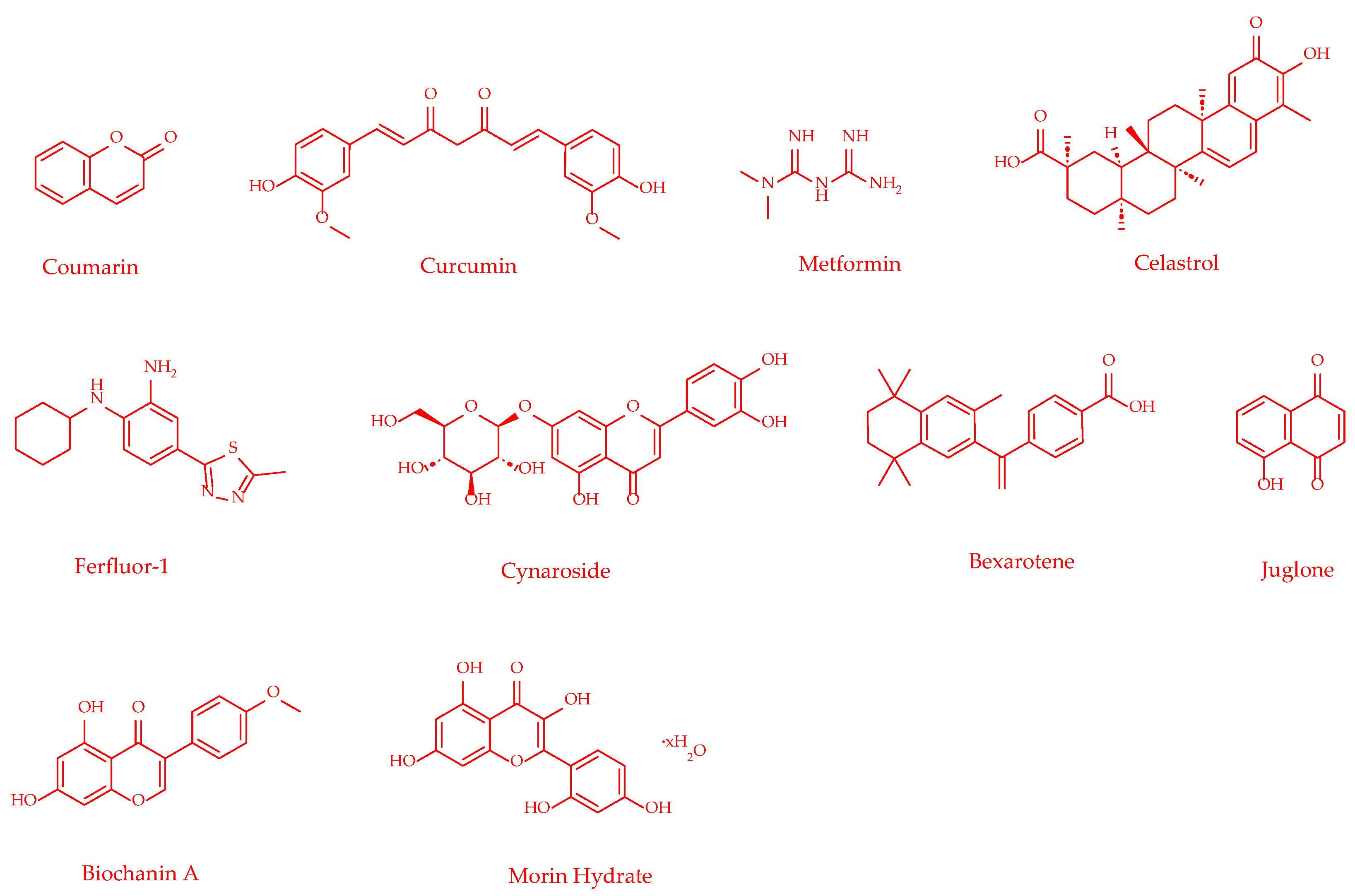
Table 5 summarizes the effects of novel anti-cell death agents, their molecular mechanisms, and their reported activities in various in vivo models for the treatment of CNS diseases.
Table 5.
Novel anti-cell death agents for CNS diseases.
Figure 11.
Chemical structures of anti-cell death agents.
3. Challenges and Emerging Strategies
As of 2021, the global number of patients with dementia reached 57 million, with nearly 10 million new cases emerging annually [79]; stroke, as the second leading cause of death and a primary cause of disability worldwide, also exhibits an upward trend in its incidence rate [80]. With the accelerating process of population aging, the incidence of CNS diseases continues to increase, posing severe challenges to the global public health system [81]. Despite the widespread impact of CNS diseases, the costs of corresponding drug research and development (R&D) remain persistently high. On average, it takes over a decade for a new drug to progress from discovery to market, with an estimated total cost of $2.87 billion (in 2013 US dollars) [82]. Due to the unique challenges faced in the treatment of CNS diseases, drug development in this field carries even higher risks and failure rates, and the actual costs far exceed this average.
The first major challenge in CNS drug development is the inadequacy of disease models. Traditionally, rodent models have been predominantly used for CNS drug discovery; however, their CNS differs significantly from that of humans in terms of cellular composition, morphology, and distribution. Furthermore, rodent neurons exhibit apparent resistance to neurodegeneration, thereby limiting their ability to fully recapitulate the pathological features of human neurodegenerative diseases. Although the CNS of non-human primates is more similar to that of humans, the time and financial costs associated with such research are prohibitively high [83]. The emergence of induced pluripotent stem cell (iPSC) technology has marked a turning point in addressing this dilemma. On the one hand, iPSCs can provide neurons and glial cells with specific human genetic backgrounds, enabling researchers to directly verify the therapeutic efficacy and potential toxicity of drugs in a human-derived system, thereby substantially enhancing the efficiency of compound screening. On the other hand, iPSCs themselves possess the therapeutic potential to differentiate into specific cells and replace diseased cells in patients, representing a promising strategy to overcome limitations in neuronal regeneration [84]. Furthermore, iPSC technology can be integrated with microfluidic chip technology to construct organ-on-a-chip models that more closely mimic physiological human conditions, overcoming the limitations of traditional animal models and two-dimensional cell cultures in CNS disease research [85]. Brain organoid technologies developed by researchers can induce iPSCs to self-organize in vitro into three-dimensional neural structures, simulating the cell types, tissue architecture, and partial functions during early human brain development. This technology has already been applied in drug screening and therapeutic exploration for various CNS diseases, such as AD and PD [86,87]. The widespread application of iPSC technology has opened new avenues for constructing more complex drug screening platforms and developing effective cell replacement therapies [84].
The second major challenge in CNS drug development is the low permeability of compounds across the BBB. This natural barrier prevents over 98% of small-molecule drugs from entering the brain, dramatically increasing the difficulty of CNS drug R&D [88]. In recent years, the development of nanodrug delivery systems has demonstrated tremendous potential in addressing this challenge. Nanoparticles within these systems can enrich drug molecules and protect their active functional groups, precisely delivering drugs across the BBB to specific sites for targeted therapy [89]. The use of nanoscale delivery systems can improve drug stability and loading capacity while enhancing targeting efficiency; this not only facilitates the better passage and functioning of compounds across the BBB, but also enables the co-delivery of drugs, imaging agents, and targeting ligands [90]. Regarding BBB permeability assessment, three-dimensional BBB models based on hydrogels, spheroids, and organoids, as well as microfluidic models, can be developed to screen the ability of drug delivery systems to cross the BBB, thereby accelerating the clinical translation of nanodrugs for brain-targeted delivery [91]. Additionally, intranasal administration is gaining increasing attention as an emerging strategy that bypasses the BBB and delivers drugs directly to the brain. Drugs absorbed through the nasal mucosa can directly enter the systemic circulation, effectively circumventing first-pass metabolism in the liver and gastrointestinal tract and significantly improving bioavailability, providing a promising solution to overcome BBB restrictions [92].
The third major challenge in CNS drug development is the high attrition rate and efficiency bottlenecks in translating laboratory lead compounds into clinical applications. Due to the high complexity of the CNS, the low selective permeability of the BBB, and the limitations of traditional pharmacokinetic evaluation models, a large number of candidate molecules with good in vitro activity often fail in the clinical stage because of poor permeability, off-target toxicity, or metabolic instability, leading to prolonged R&D cycle and persistently high costs. With the deeper integration of biology and computer science, artificial intelligence (AI) technology has emerged as a novel strategy to resolve this dilemma, injecting new momentum into the treatment of CNS diseases. At the compound screening stage, machine learning-based AI methods can rapidly screen massive compound libraries to identify molecules capable of modulating key CNS disease targets [93]. In the drug property evaluation phase, machine learning models such as graph neural networks (GNNs) are widely utilized to predict the absorption, distribution, metabolism, excretion, and toxicity of small molecules, thereby eliminating candidates with poor pharmacokinetic properties at an early stage [94]. Concurrently, AI-assisted design of delivery systems is gaining traction: by predicting the optimal size, surface charge, and ligand modifications of nanoparticles, machine learning models can optimize encapsulation efficiency of active molecules in carriers such as liposomes and micelles, achieving precise cross-barrier delivery and controlled release [95]. This full-process AI integration, spanning from molecular design to targeted delivery, is significantly shortening the R&D cycle for CNS drugs, paving the way for innovative neuroprotective therapies, and accelerating drug screening and clinical translation.
Author Contributions
Conceptualization, Y.Q.; writing—original draft preparation, Y.Q.; writing—review and editing, Y.Q. and J.C.; supervision, Y.Z. and W.S. All authors have read and agreed to the published version of the manuscript.
Funding
This research was supported by the University-Industry Collaborative Education Program (Project No. 240902690201453), the 2026 Educational Teaching Reform Research Project of Southwest University (Project No. 2026JY075), the Higher Education Research Project of the Year 2025–2026 of Chongqing Higher Education Society (Project No. cqgj25008C), and the Graduate Education Teaching Reform Research Projects of Southwest University (Project Nos. SWUYJS266215 and SWUYJS256210).
Institutional Review Board Statement
Not applicable.
Informed Consent Statement
Not applicable.
Data Availability Statement
No new data were created or analyzed in this study. Data sharing is not applicable.
Acknowledgments
Figure 1, Figure 2, Figure 4, Figure 6, Figure 8 and Figure 10 were created in https://BioRender.com. We confirm that the inspiration, conceptual framework, literature review, and initial drafts for this manuscript were entirely generated by human effort. However, as English is not our native language, we used AI tools solely for language polishing at the final stage of manuscript preparation (https://www.deepseek.com, accessed on 15 May 2026) to assist in checking grammar and spelling errors. No AI tools were used for scientific reasoning, literature analysis, or conclusions.
Conflicts of Interest
The authors declare no conflicts of interest.
Abbreviations
The following abbreviations are used in this manuscript:
| CNS | Central nervous system |
| IS | Ischemic stroke |
| TBI | Traumatic brain injury |
| AD | Alzheimer’s disease |
| VGIC | Voltage-gated ion channel |
| VGSC | Voltage-gated sodium channel |
| VGKC | Voltage-gated potassium channel |
| VGCC | Voltage-gated calcium channel |
| VSD4 | Voltage-sensing domain 4 |
| DE-MES | Direct current multielectrode shock |
| WT | Wild-type |
| MES | Maximal electroshock |
| ALS | Amyotrophic lateral sclerosis |
| PD | Parkinson’s disease |
| DA | Dopamine |
| EAAT | Excitatory amino acid transporter |
| MAPK | Mitogen-activated protein kinase |
| NMDAR | N-methyl-D-aspartate receptor |
| AMPAR | α-amino-3-hydroxy-5-methyl-4-isoxazolepropionic acid receptor |
| THSG | Tetrahydroxystilbene-2-O-β-D-glucoside |
| CI/RI | Cerebral ischemia–reperfusion injury |
| tMCAO/R | Transient middle cerebral artery occlusion/reperfusion |
| CCI-TBI | Controlled cortical impact—traumatic brain injury |
| mNSS | Modified neurological severity scores |
| pMCAO | Permanent middle cerebral artery occlusion |
| AIS | Acute ischemic stroke |
| PAMPs | Pathogen-associated molecular patterns |
| DAMPs | Damage-associated molecular patterns |
| GSDMD | Gasdermin D |
| BBB | Blood–brain barrier |
| ASCT2 | Alanine-serine-cysteine transporter 2 |
| MF | Metformin |
| TS | Tobacco smoke |
| 3-MP | 3-Monothiopomalidomide |
| CRBN | Cereblon |
| PTS | Photothrombotic stroke |
| ROS | Reactive oxygen species |
| RNS | Reactive nitrogen species |
| NOX | NADPH oxidase |
| NO | Nitric oxide |
| NOS | Nitric oxide synthases |
| HO-1 | Heme oxygenase-1 |
| GN | Gomisin N |
| MCAO/R | Middle cerebral artery occlusion/reperfusion |
| NBP | Dl-3-n-butylphthalide |
| NTG | Nitroglycerin |
| TMP | Ligustrazine |
| PKCζ | Protein kinase Cζ |
| HK-1 | Hexokinase-I |
| AIF | Apoptosis-inducing factor |
| tMCAO | Transient middle cerebral artery occlusion |
| MCAO | Middle cerebral artery occlusion |
| IR | Ischemia–reperfusion |
| mTBI | Mild traumatic brain injury |
| MTX | Methotrexate |
| Fer-1 | Ferrostatin-1 |
| BCA | Biochanin A |
| SCI | Spinal cord injury |
| MH | Morin hydrate |
| 3-MP | 3-nitropropionic acid |
| HD | Huntington’s disease |
| NSA | Necrosulfonamide |
| SDH | Succinate dehydrogenase |
| NFTs | Neurofibrillary tangles |
| GBE | Ginkgo biloba extract |
| CUMS | Chronic unpredictable mild stress |
| iPSC | Induced pluripotent stem cell |
| R&D | Research and development |
| AI | Artificial intelligence |
| GNN | Graph neural network |
References
- Krames, E.; Peckham, P.H.; Rezai, A.R. Neuromodulation, 1st ed.; Academic Press: Cambridge, MA, USA, 2009. [Google Scholar]
- Pardridge, W.M. Drug transport across the blood-brain barrier. J. Cereb. Blood Flow Metab. 2012, 32, 1959–1972. [Google Scholar] [CrossRef] [Scilit]
- National Institute of Neurological Disorders and Stroke (NINDS). A Phase 1 Open-Label Dose Escalation Safety Study of Convection Enhanced Delivery (CED) of Adeno-Associated Virus Encoding Glial Cell Line-Derived Neurotrophic Factor (AAV2-GDNF) in Subjects with Advanced Parkinson’s Disease. Clinicaltrials.gov. Clinical Trial Registration NCT01621581. April 2026. Available online: https://clinicaltrials.gov/study/NCT01621581 (accessed on 9 May 2026).
- Xu, X.; Zhang, Y. Regulation of Oxidative Stress by Long Non-coding RNAs in Central Nervous System Disorders. Front. Mol. Neurosci. 2022, 15, 931704. [Google Scholar] [CrossRef] [Scilit]
- Global, regional, and national burden of disorders affecting the nervous system, 1990–2021: A systematic analysis for the global burden of disease study 2021. Lancet Neurol. 2024, 23, 344–381. [CrossRef] [Scilit] [PubMed]
- Song, F.; Guo, H. Progress on structural biology of voltage-gated ion channels. J. Zhejiang Univ. (Med. Sci.) 2019, 48, 25–33. [Google Scholar] [CrossRef] [Scilit]
- Terlau, H.; Stühmer, W. Structure and Function of Voltage-Gated Ion Channels. Naturwissenschaften 1998, 85, 437–444. [Google Scholar] [CrossRef] [Scilit]
- Hoshi, N. M-Current Suppression, Seizures and Lipid Metabolism: A Potential Link Between Neuronal Kv7 Channel Regulation and Dietary Therapies for Epilepsy. Front. Physiol. 2020, 11, 513. [Google Scholar] [CrossRef] [Scilit]
- Johnson, J.P.; Focken, T.; Tari, P.K.; Dube, C.; Goodchild, S.J.; Andrez, J.; Bankar, G.; Burford, K.; Chang, E.; Chowdhury, S.; et al. The contribution of NaV1.6 to the efficacy of voltage-gated sodium channel inhibitors in wild type and NaV1.6 gain-of-function (GOF) mouse seizure control. Br. J. Pharmacol. 2024, 181, 3993–4011. [Google Scholar] [CrossRef] [Scilit] [PubMed]
- Padilla, K.; Wickenden, A.D.; Gerlach, A.C.; McCormack, K. The KCNQ2/3 selective channel opener ICA-27243 binds to a novel voltage-sensor domain site. Neurosci. Lett. 2009, 465, 138–142. [Google Scholar] [CrossRef] [Scilit]
- Masegosa, V.M.; Navarro, X.; Herrando-Grabulosa, M. ICA-27243 improves neuromuscular function and preserves motoneurons in the transgenic SOD1G93A mice. Neurotherapeutics 2024, 21, e00319. [Google Scholar] [CrossRef] [Scilit]
- Yip, K.L.; Udoh, M.; Sharman, L.A.; Harman, T.; Bedoya-Pérez, M.; Anderson, L.L.; Banister, S.D.; Arnold, J.C. Cannabinoid-like compounds found in non-cannabis plants exhibit antiseizure activity in genetic mouse models of drug-resistant epilepsy. Epilepsia 2024, 66, 303–314. [Google Scholar] [CrossRef] [Scilit]
- Zidar, N.; Tomašič, T.; Kikelj, D.; Durcik, M.; Tytgat, J.; Peigneur, S.; Rogers, M.; Haworth, A.; Kirby, R.W. New aryl and acylsulfonamides as state-dependent inhibitors of Nav1.3 voltage-gated sodium channel. Eur. J. Med. Chem. 2023, 258, 115530. [Google Scholar] [CrossRef] [Scilit]
- Lorenc-Koci, E.; Lenda, T.; Konieczny, J.; Jantas, D.; Domin, H. Cinnarizine, a Calcium Channel Blocker, Partially Prevents the Striatal Dopamine Decline and Loss of Nigral Dopamine Neurons in the Lactacystin-Induced Rat Model of Parkinson’s Disease. Int. J. Mol. Sci. 2025, 26, 8833. [Google Scholar] [CrossRef] [Scilit] [PubMed]
- Mehta, A.; Prabhakar, M.; Kumar, P.; Deshmukh, R.; Sharma, P.L. Excitotoxicity: Bridge to various triggers in neurodegenerative disorders. Eur. J. Pharmacol. 2013, 698, 6–18. [Google Scholar] [CrossRef] [Scilit]
- Luo, Y.; Tang, H.; Li, H.; Zhao, R.; Huang, Q.; Liu, J. Recent advances in the development of neuroprotective agents and therapeutic targets in the treatment of cerebral ischemia. Eur. J. Med. Chem. 2019, 162, 132–146. [Google Scholar] [CrossRef] [Scilit] [PubMed]
- Roman, J.Y.M.; González, C.C. Glutamate and excitotoxicity in central nervous system disorders: Ionotropic glutamate receptors as a target for neuroprotection. Neuroprotection 2024, 2, 137–150. [Google Scholar] [CrossRef] [Scilit] [PubMed]
- Rivera-Cervantes, M.C.; Torres, J.S.; Feria-Velasco, A.; Armendariz-Borunda, J.; Beas-Zárate, C. NMDA and AMPA receptor expression and cortical neuronal death are associated with p38 in glutamate-induced excitotoxicity in vivo. J. Neurosci. Res. 2004, 76, 678–687. [Google Scholar] [CrossRef] [Scilit]
- Song, P.; Ma, C.; Wang, S.; Zhang, Y.; Xu, L.; Fang, F.; Huang, X.; Zhang, Y.; Mao, Q.; Shi, C.; et al. Design, synthesis, and evaluation of the pharmacological activity of novel NMDA receptor antagonists based on the germacrone scaffold. Bioorg. Med. Chem. 2025, 119, 118063. [Google Scholar] [CrossRef] [Scilit]
- Hansen, K.B.; Yi, F.; Perszyk, R.E.; Menniti, F.S.; Traynelis, S.F. NMDA Receptors in the Central Nervous System. In NMDA Receptors; Burnashev, N., Szepetowski, P., Eds.; Methods in Molecular Biology; Humana Press: New York, NY, USA, 2017; Volume 1677. [Google Scholar] [CrossRef] [Scilit]
- Liu, T.; Shi, J.; Wu, D.; Li, D.; Wang, Y.; Liu, J.; Meng, P.; Hu, L.; Fu, C.; Mei, Z.; et al. THSG alleviates cerebral ischemia/reperfusion injury via the GluN2B–CaMKII–ERK1/2 pathway. Phytomedicine 2024, 129, 155595. [Google Scholar] [CrossRef] [Scilit]
- Qneibi, M.; Hawash, M.; Bdir, S.; Bdair, M.; Aldwaik, S.A. Assessing the Effects of Thiazole-Carboxamide Derivatives on the Biophysical Properties of AMPA Receptor Complexes as a Potential Neuroprotective Agent. Molecules 2024, 29, 3232. [Google Scholar] [CrossRef] [Scilit]
- Park, M.K.; Choi, B.Y.; Kho, A.R.; Lee, S.H.; Hong, D.K.; Kang, B.S.; Lee, C.J.; Yang, H.W.; Woo, S.Y.; Park, S.W.; et al. L-theanine ameliorates traumatic-brain-injury-induced hippocampal neuronal death in rats. Phytomedicine 2025, 139, 156457. [Google Scholar] [CrossRef] [Scilit]
- Basagni, F.; Ortega, J.A.; Bertozzi, S.M.; Armirotti, A.; Summa, M.; Bertorelli, R.; Bartolini, M.; Mellor, I.R.; Bedeschi, M.; Bottegoni, G.; et al. Galantamine-memantine hybrids for Alzheimer’s disease: The influence of linker rigidity in biological activity and pharmacokinetic properties. Eur. J. Med. Chem. 2023, 261, 115803. [Google Scholar] [CrossRef] [Scilit] [PubMed]
- Xie, D.; Zhang, P.; You, S.; Shen, Y.; Xu, W.; Zhan, C.; Zhang, J. Salidroside derivative SHPL-49 attenuates glutamate excitotoxicity in acute ischemic stroke via promoting NR2A-CAMKIIα-Akt/CREB pathway. Phytomed. Int. J. Phytother. Phytopharm. 2024, 134, 155583. [Google Scholar] [CrossRef] [Scilit] [PubMed]
- Zhang, P.; Xu, J.; Cui, Q.; Lin, G.; Wang, F.; Ding, X.; You, S.; Sang, N.; Tan, J.; Xu, W.; et al. Multi-pathway neuroprotective effects of a novel salidroside derivative SHPL-49 against acute cerebral ischemic injury. Eur. J. Pharmacol. 2023, 949, 175716. [Google Scholar] [CrossRef] [Scilit] [PubMed]
- Hu, F.F.; Pan, S.Y.; Chu, J.Y.; Liu, J.J.; Duan, T.T.; Luo, Y.; Zhou, W.; Wang, Z.M.; Liu, W.; Zeng, Y. Xanthohumol Protects Against Neuronal Excitotoxicity and Mitochondrial Dysfunction in APP/PS1 Mice: An Omics-Based Study. Nutrients 2024, 16, 3754. [Google Scholar] [CrossRef] [Scilit]
- Shi, F.D.; Yong, V.W. Neuroinflammation across neurological diseases. Science 2025, 388, eadx0043. [Google Scholar] [CrossRef] [Scilit]
- Zhang, H.M.; Luo, D.; Chen, R.; Wang, S.H.; Zhao, Y.J.; Li, J.X.; Zhou, M.F.; Yu, Z.M.; Zhang, J.L.; Liang, F.X. Research progress on acupuncture treatment in central nervous system diseases based on NLRP3 inflammasome in animal models. Front. Neurosci. 2023, 17, 1118508. [Google Scholar] [CrossRef] [Scilit]
- Xu, W.; Huang, Y.; Zhou, R. NLRP3 inflammasome in neuroinflammation and central nervous system diseases. Cell. Mol. Immunol. 2025, 22, 341–355. [Google Scholar] [CrossRef] [Scilit]
- Marchetti, L.; Engelhardt, B. Immune cell trafficking across the blood-brain barrier in the absence and presence of neuroinflammation. Vasc. Biol. 2020, 2, H1–H18. [Google Scholar] [CrossRef] [Scilit]
- Liu, Y.; Liu, T.; Zhou, Y.; Li, W.; Wang, M.; Song, N.; Zhang, W.; Jiang, J.; Yuan, S.; Ding, J.; et al. Impeding the combination of astrocytic ASCT2 and NLRP3 by talniflumate alleviates neuroinflammation in experimental models of Parkinson’s disease. Acta Pharm. Sin. B 2023, 13, 662–677. [Google Scholar] [CrossRef] [Scilit]
- Akter, K.A.; Sharma, S.; Sifat, A.E.; Zhang, Y.; Patel, D.K.; Cucullo, L.; Abbruscato, T.J. Metformin ameliorates neuroinflammatory environment for neurons and astrocytes during in vitro and in vivo stroke and tobacco smoke chemical exposure: Role of Nrf2 activation. Redox Biol. 2024, 75, 103266. [Google Scholar] [CrossRef] [Scilit]
- Hsueh, S.C.; Parekh, P.; Batsaikhan, B.; Vargesson, N.; Tweedie, D.; Luo, W.; Patel, C.N.; Liu, D.; McDevitt, R.A.; Baig, A.M.; et al. Targeting neuroinflammation: 3-monothiopomalidomide a new drug candidate to mitigate traumatic brain injury and neurodegeneration. J. Biomed. Sci. 2025, 32, 57. [Google Scholar] [CrossRef] [Scilit]
- Harrison, D.; Billinton, A.; Bock, M.G.; Doedens, J.R.; Gabel, C.A.; Holloway, M.K.; Porter, R.A.; Reader, V.; Scanlon, J.; Schooley, K.; et al. Discovery of clinical candidate NT-0796, a brain-penetrant and highly potent NLRP3 inflammasome inhibitor for neuroinflammatory disorders. J. Med. Chem. 2023, 66, 14897–14911. [Google Scholar] [CrossRef] [Scilit] [PubMed]
- Clarke, N.; Thornton, P.; Reader, V.; Lindsay, N.; Digby, Z.; Mullen, B.; Gorman, M.; Jacobson, E.; Langdon, G.; Johnstone, H.; et al. Anti-Neuroinflammatory and Anti-Inflammatory Effects of the NLRP3 Inhibitor NT-0796 in Subjects with Parkinson’s Disease. Mov. Disord. 2025, 40, 2199–2208. [Google Scholar] [CrossRef] [Scilit] [PubMed] [PubMed Central]
- Lim, H.-S.; Park, J.; Kim, E.; Lee, W.; Yun, H.-Y.; Lee, S.H.; Park, G. Rebamipide (Mucosta®), a clinically approved drug, alleviates neuroinflammation and dopaminergic neurodegeneration in a Parkinson’s disease model. J. Neuroinflamm. 2025, 22, 132. [Google Scholar] [CrossRef] [Scilit] [PubMed]
- Camarano, J.; Roque, M.; Gahn, G.; Whipple, S.G.; Terrell, D.; Ronkon, C.; Toms, J.; Sin, A.; Guthikonda, B.; Latha, K.; et al. Hydrogen sulfide inhibits recruitment of monocyte-derived tumor associated macrophages in glioblastoma by downregulating CXCL12. Redox Biol. 2025, 86, 103866. [Google Scholar] [CrossRef] [Scilit]
- Wu, Q.L.; Cui, L.Y.; Ma, W.Y.; Wang, S.S.; Zhang, Z.; Feng, Z.P.; Sun, H.S.; Chu, S.F.; He, W.B.; Chen, N.H. A novel small-molecular CCR5 antagonist promotes neural repair after stroke. Acta Pharmacol. Sin. 2023, 44, 1935–1947. [Google Scholar] [CrossRef] [Scilit]
- Vermot, A.; Petit-Härtlein, I.; Smith, S.M.E.; Fieschi, F. NADPH Oxidases (NOX): An Overview from Discovery, Molecular Mechanisms to Physiology and Pathology. Antioxidants 2021, 10, 890. [Google Scholar] [CrossRef] [Scilit] [PubMed]
- Li, J.; O, W.; Li, W.; Jiang, Z.-G.; Ghanbari, H.A. Oxidative Stress and Neurodegenerative Disorders. Int. J. Mol. Sci. 2013, 14, 24438–24475. [Google Scholar] [CrossRef] [Scilit]
- Xu, X.-X.; Shi, R.-X.; Fu, Y.; Wang, J.-L.; Tong, X.; Zhang, S.-Q.; Wang, N.; Li, M.-X.; Tong, Y.; Wang, W.; et al. Neuronal nitric oxide synthase/reactive oxygen species pathway is involved in apoptosis and pyroptosis in epilepsy. Neural Regen. Res. 2023, 18, 1277–1285. [Google Scholar] [CrossRef] [Scilit]
- Salim, S. Oxidative Stress and the Central Nervous System. J. Pharmacol. Exp. Ther. 2017, 360, 201–205. [Google Scholar] [CrossRef] [Scilit]
- Li, R.; Zheng, Y.; Zhang, J.; Zhou, Y.; Fan, X. Gomisin N attenuated cerebral ischemia-reperfusion injury through inhibition of autophagy by activating the PI3K/AKT/mTOR pathway. Phytomedicine 2023, 110, 154644. [Google Scholar] [CrossRef] [Scilit] [PubMed]
- Crisman, E.; Duarte, P.; Dauden, E.; Cuadrado, A.; Rodríguez-Franco, M.I.; López, M.G.; León, R. KEAP1-NRF2 protein–protein interaction inhibitors: Design, pharmacological properties and therapeutic potential. Med. Res. Rev. 2022, 43, 237–287. [Google Scholar] [CrossRef] [Scilit] [PubMed]
- Liu, Y.; Gong, Z.; Zhai, D.; Yang, C.; Lu, G.; Wang, S.; Xiao, S.; Li, C.; Chen, L.; Lin, X.; et al. Unveiling the therapeutic potential of Dl-3-n-butylphthalide in NTG-induced migraine mouse: Activating the Nrf2 pathway to alleviate oxidative stress and neuroinflammation. J. Headache Pain 2024, 25, 50. [Google Scholar] [CrossRef] [Scilit] [PubMed]
- Zhang, H.; Wang, L.; Zhu, B.; Yang, Y.; Cai, C.; Wang, X.; Deng, L.; He, B.; Cui, Y.; Zhou, W. A comparative study of the neuroprotective effects of dl-3-n-butylphthalide and edaravone dexborneol on cerebral ischemic stroke rats. Eur. J. Pharmacol. 2023, 951, 175801. [Google Scholar] [CrossRef] [Scilit]
- Li, G.; Xiao, H.; Zuo, C.; Xie, H.; Wang, X.; Wang, J.; Liu, Y.; Hou, Q.; Sun, G.; Tian, Y. N-butylphthalide (NBP) and ligustrazine (TMP) triazole hybrids target the KEAP1-NRF2 pathway to inhibit ferroptosis and exert brain neuroprotectivity. Redox Biol. 2025, 86, 103835, Corrigendum in Redox Biol. 2025, 87, 103860. https://doi.org/10.1016/j.redox.2025.103860.. [Google Scholar] [CrossRef] [Scilit]
- Wu, H.; Li, Y.; Zhang, Q.; Wang, H.; Xiu, W.; Xu, P.; Deng, Y.; Huang, W.; Wang, D.O. Crocetin antagonizes parthanatos in ischemic stroke via inhibiting NOX2 and preserving mitochondrial hexokinase-I. Cell Death Dis. 2023, 14, 50. [Google Scholar] [CrossRef] [Scilit]
- Xu, G.; Zhang, J.; Shao, C.; Wu, H.; Li, C.; Yu, L.; Zhou, H.; An, H.; Wang, T.; Pu, J.; et al. Puerarin alleviates cerebral ischemia/reperfusion (CIR)-induced neurocyte oxidative stress and apoptosis via DNA demethylation-mediated PI3K/Akt activation. Phytomedicine 2025, 145, 157094. [Google Scholar] [CrossRef] [Scilit]
- Zhang, Q.; Yao, M.; Qi, J.; Song, R.; Wang, L.; Li, J.; Zhou, X.; Chang, D.; Huang, Q.; Li, L.; et al. Puerarin inhibited oxidative stress and alleviated cerebral ischemia-reperfusion injury through PI3K/Akt/Nrf2 signaling pathway. Front. Pharmacol. 2023, 14, 1134380. [Google Scholar] [CrossRef] [Scilit]
- Liu, M.; Liu, D.N.; Shang, Y.F.; Zhang, W.F.; Feng, W.D.; Fang, L.H.; Du, G.H.; Wang, Y.H. Mechanism and potential targets of kaempferol alleviating photothrombotic stroke via integrating experimental validation, network pharmacology, and molecular docking. Phytomedicine 2025, 148, 157354. [Google Scholar] [CrossRef] [Scilit]
- Guo, X.H.; Pang, L.; Gao, C.Y.; Meng, F.L.; Jin, W. Lyoniresinol attenuates cerebral ischemic stroke injury in MCAO rat based on oxidative stress suppression via regulation of Akt/GSK-3β/Nrf2 signaling. Biomed. Pharmacother. 2023, 167, 115543. [Google Scholar] [CrossRef] [Scilit]
- Liu, Y.; Mi, Y.; Wang, Y.; Meng, Q.; Xu, L.; Liu, Y.; Zhou, D.; Wang, Y.; Liang, D.; Li, W.; et al. Loureirin C inhibits ferroptosis after cerebral ischemia reperfusion through regulation of the Nrf2 pathway in mice. Phytomedicine 2023, 113, 154729. [Google Scholar] [CrossRef] [Scilit]
- Guo, R.; Quan, S.; Yan, X.; Jia, Y.; Wang, N.; Bu, J.; Zhao, Y.; Su, L.; Yue, Z.; Wang, J.; et al. Atractylenolide III alleviates cerebral ischemia-reperfusion injury by targeting Keap1 to activate the Nrf2/HO-1 pathway. Phytomedicine 2025, 148, 157284. [Google Scholar] [CrossRef] [Scilit]
- Li, X.N.; Shang, N.Y.; Kang, Y.Y.; Sheng, N.; Lan, J.Q.; Tang, J.S.; Wu, L.; Zhang, J.L.; Peng, Y. Caffeic acid alleviates cerebral ischemic injury in rats by resisting ferroptosis via Nrf2 signaling pathway. Acta Pharmacol. Sin. 2024, 45, 248–267. [Google Scholar] [CrossRef] [Scilit]
- He, S.; Chen, Y.; Wang, H.; Li, S.; Wei, Y.; Zhang, H.; Gao, Q.; Wang, F.; Zhang, R. Neuroprotective effects of chlorogenic acid: Modulation of Akt/Erk1/2 signaling to prevent neuronal apoptosis in Parkinson’s disease. Free Radic. Biol. Med. 2024, 222, 275–287. [Google Scholar] [CrossRef] [Scilit]
- Feng, L.; Lo, H.; Zheng, J.; Weng, W.; Sun, Y.; Pan, X. Cycloastragenol reduces microglial NLRP3 inflammasome activation in Parkinson’s disease models by promoting autophagy and reducing Scrib-driven ROS. Phytomedicine 2024, 135, 156210. [Google Scholar] [CrossRef] [Scilit]
- Li, Z.; Lu, Y.; Zhen, Y.; Jin, W.; Ma, X.; Yuan, Z.; Liu, B.; Zhou, X.-L.; Zhang, L. Avicularin inhibits ferroptosis and improves cognitive impairments in Alzheimer’s disease by modulating the NOX4/Nrf2 axis. Phytomedicine 2024, 135, 156209. [Google Scholar] [CrossRef] [Scilit]
- Yang, Y.; Hao, T.; Yao, X.; Che, Y.; Liu, Y.; Fang, M.; Wang, Y.; Zhou, D.; Chai, H.; Li, N.; et al. Crebanine ameliorates ischemia-reperfusion brain damage by inhibiting oxidative stress and neuroinflammation mediated by NADPH oxidase 2 in microglia. Phytomedicine 2023, 120, 155044. [Google Scholar] [CrossRef] [Scilit]
- Mason, H.; Rai, G.; Kozyr, A.; De Jonge, N.; Gliniewicz, E.; Berg, L.J.; Wald, G.; Dorrier, C.; Henderson, M.J.; Zakharov, A.; et al. Development of an improved and specific inhibitor of NADPH oxidase 2 to treat traumatic brain injury. Redox Biol. 2023, 60, 102611. [Google Scholar] [CrossRef] [Scilit]
- Vasu, D.; Do, H.T.; Li, H.; Hardy, C.D.; Awasthi, A.; Poulos, T.L.; Silverman, R.B. Potent, selective, and membrane permeable 2-amino-4-substituted pyridine-based neuronal nitric oxide synthase inhibitors. J. Med. Chem. 2023, 66, 9934–9953. [Google Scholar] [CrossRef] [Scilit]
- Mercau, M.E.; Patwa, S.; Bhat, K.P.L.; Ghosh, S.; Rothlin, C.V. Cell death in development, maintenance, and diseases of the nervous system. Semin. Immunopathol. 2022, 44, 725–738. [Google Scholar] [CrossRef] [Scilit]
- Lessmann, V.; Gottmann, K.; Malcangio, M. Neurotrophin secretion: Current facts and future prospects. Prog. Neurobiol. 2003, 69, 341–374, Erratum in Prog. Neurobiol. 2004, 72, 165–166. https://doi.org/10.1016/j.pneurobio.2004.02.003.. [Google Scholar] [CrossRef] [Scilit]
- Chi, H.; Chang, H.-Y.; Sang, T.-K. Neuronal Cell Death Mechanisms in Major Neurodegenerative Diseases. Int. J. Mol. Sci. 2018, 19, 3082. [Google Scholar] [CrossRef] [Scilit]
- Dixon, S.J.; Lemberg, K.M.; Lamprecht, M.R.; Skouta, R.; Zaitsev, E.M.; Gleason, C.E.; Patel, D.N.; Bauer, A.J.; Cantley, A.M.; Yang, W.S.; et al. Ferroptosis: An iron-dependent form of nonapoptotic cell death. Cell 2012, 149, 1060–1072. [Google Scholar] [CrossRef] [Scilit]
- Stockwell, B.R.; Angeli, J.P.F.; Bayir, H.; Bush, A.I.; Conrad, M.; Dixon, S.J.; Fulda, S.; Gascón, S.; Hatzios, S.K.; Kagan, V.E.; et al. Ferroptosis: A Regulated Cell Death Nexus Linking Metabolism, Redox Biology, and Disease. Cell 2017, 171, 273–285. [Google Scholar] [CrossRef] [Scilit]
- Shi, J.; Zhao, Y.; Wang, K.; Shi, X.; Wang, Y.; Huang, H.; Zhuang, Y.; Cai, T.; Wang, F.; Shao, F. Cleavage of GSDMD by inflammatory caspases determines pyroptotic cell death. Nature 2015, 526, 660–665. [Google Scholar] [CrossRef] [Scilit]
- Moujalled, D.; Strasser, A.; Liddell, J.R. Molecular mechanisms of cell death in neurological diseases. Cell Death Differ. 2021, 28, 2029–2044. [Google Scholar] [CrossRef] [Scilit]
- Sritawan, N.; Sirichoat, A.; Aranarochana, A.; Pannangrong, W.; Wigmore, P.; Welbat, J.U. Protective effect of metformin on methotrexate induced reduction of rat hippocampal neural stem cells and neurogenesis. Biomed. Pharmacother. 2023, 162, 114613. [Google Scholar] [CrossRef] [Scilit]
- Yan, J.; Bao, L.; Liang, H.; Zhao, L.; Liu, M.; Kong, L.; Fan, X.; Liang, C.; Liu, T.; Han, X.; et al. A Druglike Ferrostatin-1 Analogue as a Ferroptosis Inhibitor and Photoluminescent Indicator. Angew. Chem. Int. Ed. Engl. 2025, 64, e202502195. [Google Scholar] [CrossRef] [Scilit]
- Li, X.; Fu, J.; Guan, M.; Shi, H.; Pan, W.; Lou, X. Biochanin A attenuates spinal cord injury in rats during early stages by inhibiting oxidative stress and inflammasome activation. Neural Regen. Res. 2023, 19, 2050–2056. [Google Scholar] [CrossRef] [Scilit]
- Elbaz, E.M.; Sayed, R.H.; Abdelkader, A.A.; Fahim, A.T. Neuroprotective role of morin hydrate on 3-nitropropionic acid-elicited huntington’s disease: In vivo investigation of RIPK1/RIPK3/MLKL necroptosis signaling pathway. Mol. Med. 2025, 31, 135. [Google Scholar] [CrossRef] [Scilit]
- Assi, A.-A.; Farrag, M.M.Y.; Badary, D.M.; Allam, E.A.H.; Nicola, M.A. Protective effects of curcumin and Ginkgo biloba extract combination on a new model of Alzheimer’s disease. Inflammopharmacology 2023, 31, 1449–1464. [Google Scholar] [CrossRef] [Scilit]
- Shen, W.; Li, C.; Liu, Q.; Cai, J.; Wang, Z.; Pang, Y.; Ning, G.; Yao, X.; Kong, X.; Feng, S. Celastrol inhibits oligodendrocyte and neuron ferroptosis to promote spinal cord injury recovery. Phytomedicine 2024, 128, 155380. [Google Scholar] [CrossRef] [Scilit]
- Zhou, Y.; Huang, Y.; Ye, W.; Chen, Z.; Yuan, Z. Cynaroside improved depressive-like behavior in CUMS mice by suppressing microglial inflammation and ferroptosis. Biomed. Pharmacother. 2024, 173, 116425. [Google Scholar] [CrossRef] [Scilit]
- Wang, X.; Shen, Z.; Zhang, H.; Zhang, H.-J.; Li, F.; Yu, L.; Chen, H.; Zhou, K.; Xu, H.; Sheng, S. Bexarotene improves motor function after spinal cord injury in mice. Neural Regen. Res. 2023, 18, 2733–2742. [Google Scholar] [CrossRef] [Scilit]
- Chen, L.; Chang, Y.; Zhang, S.; Wang, Z.; Liu, H.; Wang, H.; Shi, L.; Li, N.; Feng, S. Juglone promotes spinal cord injury recovery by suppressing pyroptosis and necroptosis through FOS/USP53/ubiquitination. Phytomedicine 2025, 148, 157341. [Google Scholar] [CrossRef] [Scilit]
- Dementia. World Health Organization. Available online: https://www.who.int/news-room/fact-sheets/detail/dementia (accessed on 3 June 2026).
- Campbell, B.C.V.; Khatri, P. Stroke. Lancet 2020, 396, 129–142. [Google Scholar] [CrossRef] [Scilit]
- Moreira, R.; Nóbrega, C.; de Almeida, L.P.; Mendonça, L. Brain-targeted drug delivery—Nanovesicles directed to specific brain cells by brain-targeting ligands. J. Nanobiotechnol. 2024, 22, 260. [Google Scholar] [CrossRef] [Scilit]
- DiMasi, J.A.; Grabowski, H.G.; Hansen, R.W. Innovation in the pharmaceutical industry: New estimates of R&D costs. J. Health Econ. 2016, 47, 20–33. [Google Scholar] [CrossRef] [Scilit]
- Pan, M.-T.; Zhang, H.; Li, X.-J.; Guo, X.-Y. Genetically modified non-human primate models for research on neurodegenerative diseases. Zool. Res. 2024, 45, 263–274. [Google Scholar] [CrossRef] [Scilit]
- Cerneckis, J.; Cai, H.; Shi, Y. Induced pluripotent stem cells (iPSCs): Molecular mechanisms of induction and applications. Signal Transduct. Target. Ther. 2024, 9, 112. [Google Scholar] [CrossRef] [Scilit]
- Amartumur, S.; Nguyen, H.; Huynh, T.; Kim, T.S.; Woo, R.-S.; Oh, E.; Kim, K.K.; Lee, L.P.; Heo, C. Neuropathogenesis-on-chips for neurodegenerative diseases. Nat. Commun. 2024, 15, 2219. [Google Scholar] [CrossRef] [Scilit]
- Wang, W.; Tan, S.; Zuo, X.; Gao, X.; Ma, L.; Sun, R.; Liang, G.; Yin, L.; Pu, Y.; Zhang, J. Brain Organoids in Neurodegenerative Disease Modeling: Advances, Applications and Future Perspectives. Mol. Neurobiol. 2025, 63, 142. [Google Scholar] [CrossRef] [Scilit]
- Kirkeby, A.; Main, H.; Carpenter, M. Pluripotent stem-cell-derived therapies in clinical trial: A 2025 update. Cell Stem Cell 2025, 32, 10–37, Erratum in Cell Stem Cell 2025, 32, P329–P331. [Google Scholar] [CrossRef] [Scilit]
- Yang, H.-M. Overcoming the Blood–Brain Barrier: Advanced Strategies in Targeted Drug Delivery for Neurodegenerative Diseases. Pharmaceutics 2025, 17, 1041. [Google Scholar] [CrossRef] [Scilit]
- Guo, S.; Yi, C.-X. Cell type-targeting nanoparticles in treating central nervous system diseases: Challenges and hopes. Nanotechnol. Rev. 2023, 12, 20230158. [Google Scholar] [CrossRef] [Scilit]
- Han, L.; Jiang, C. Evolution of blood–brain barrier in brain diseases and related systemic nanoscale brain-targeting drug delivery strategies. Acta Pharm. Sin. B 2021, 11, 2306–2325. [Google Scholar] [CrossRef] [Scilit]
- Pérez-López, A.; Torres-Suárez, A.I.; Martín-Sabroso, C.; Aparicio-Blanco, J. An overview of in vitro 3D models of the blood-brain barrier as a tool to predict the in vivo permeability of nanomedicines. Adv. Drug Deliv. Rev. 2023, 196, 114816. [Google Scholar] [CrossRef] [Scilit]
- Wu, X.; Zang, R.; Qiu, Y.; Zhang, Y.; Peng, J.; Cheng, Z.; Wei, S.; Liu, M.; Diao, Y. Intranasal Drug Delivery Technology in the Treatment of Central Nervous System Diseases: Challenges, Advances, and Future Research Directions. Pharmaceutics 2025, 17, 775. [Google Scholar] [CrossRef] [Scilit]
- Alghamdi, M.A. From Molecules to Medicines: The Role of AI-Driven Drug Discovery Against Alzheimer’s Disease and Other Neurological Disorders. Pharmaceuticals 2025, 18, 1041. [Google Scholar] [CrossRef] [Scilit]
- Komura, H.; Watanabe, R.; Mizuguchi, K. The Trends and Future Prospective of In Silico Models from the Viewpoint of ADME Evaluation in Drug Discovery. Pharmaceutics 2023, 15, 2619. [Google Scholar] [CrossRef] [Scilit]
- Haramshahi, S.M.A.; Hamblin, M.R.; Ravesh, R.K.; Sadr, H.; Ahmadirad, N.; Mehrabi, F.; Taherian, Z.; Hosseingolipour, S.; Barzegar, Z.; Mehrabi, S. Biomaterials for CNS disorders: A review of development from traditional methods to AI-assisted optimization. J. Mater. Sci. Mater. Med. 2025, 36, 85. [Google Scholar] [CrossRef] [Scilit] [PubMed]

Disclaimer/Publisher’s Note: The statements, opinions and data contained in all publications are solely those of the individual author(s) and contributor(s) and not of MDPI and/or the editor(s). MDPI and/or the editor(s) disclaim responsibility for any injury to people or property resulting from any ideas, methods, instructions or products referred to in the content. |
© 2026 by the authors. Licensee MDPI, Basel, Switzerland. This article is an open access article distributed under the terms and conditions of the Creative Commons Attribution (CC BY) license.