


Abstract
Background/Objectives: Systemic lupus erythematosus (SLE) is a chronic autoimmune disorder characterized by immune dysregulation that leads to widespread inflammation and damage across multiple organs. B lymphocytes play a vital role in SLE, with abnormal development and activation leading to autoreactive antibody production and immune complex formation, which damages tissues. Methods: The PPARα agonist WY14643 has anti-inflammatory effects in various inflammatory conditions, including CNS diseases. We investigated whether WY14643 decreases inflammatory mediator production in CD45R+ cells in the MRL/lpr mouse model of SLE. Flow cytometry was used to evaluate WY14643’s impact on the expression of IFN-γ, IL-6, iNOS, MCP-1, IL-1α, IL-2, Notch-1, Notch-3, GITR, and NF-κB p65 in splenic CD45R+ B cells. Additionally, we assessed the effect of WY14643 on the mRNA levels of these markers in the kidney using RT-PCR. Results: WY14643 decreased inflammatory markers such as CD45R+IFN-γ+, CD45R+IL-6+, CD45R+iNOS+, CD45R+MCP-1+, CD45R+IL-1α+, CD45R+IL-2+, CD45R+Notch1+, CD45R+Notch3+, CD45R+GITR+, and CD45R+NF-κB p65+ in splenic cells from MRL/lpr mice. Furthermore, WY14643 also lowered mRNA expression of IFN-γ, IL-6, iNOS, MCP-1, IL-2, IL-1α, Notch-1, Notch-3, GITR, and NF-κB p65 in the kidney. Conclusions: This study shows that WY14643 inhibits the production of inflammatory mediators and significantly reduces autoimmune features, including kidney inflammation, in MRL/lpr mice. Our results indicate that WY14643, a PPAR-α agonist, could be a potential therapy for lupus nephritis.
1. Introduction
Systemic lupus erythematosus (SLE) is a chronic autoimmune disease characterized by involvement of multiple systems and organs, recurrent episodes of remission and relapse, and numerous autoantibodies [1,2]. The etiology of SLE is complex and involves multiple factors, including heredity, sex hormones, and environmental factors (such as viral and bacterial infections) [3]. It is well established that autoantibody production, immune complex formation, and inflammatory responses driven by multiple cytokines contribute to the development of SLE [4]. B cells play a key role in the pathogenesis of autoimmune diseases [5]. Previous evidence indicates that B lymphocyte hyperactivity is a pathogenic event in SLE and contributes to the development of kidney disease [6]. B cells exhibit disrupted signaling pathways, leading to abnormal activation and differentiation in SLE [7]. Furthermore, there are imbalances in regulatory B cells, with a tendency toward pro-inflammatory B cells [8]. Additionally, B cells contribute to the progression of SLE by presenting antigens to self-reactive T cells and secreting inflammatory cytokines, which promote inflammation in multiple organs, including the kidneys, lungs, heart, and skin [9].
IFN-γ plays an important role in the pathogenesis of lupus nephritis [10]. In renal tissues, IFN-γ expression was significantly higher in patients with lupus nephritis and correlated with the activity of pathological lesions. IFN-γ promotes B cell class switching and stimulates the production of pathogenic autoantibodies in SLE [11]. Patients with SLE have higher levels of IFN-γ than controls [12]. IFN-γ has been implicated in the pathogenesis of SLE [13,14]. Among the immunological mediators implicated in SLE, IL-6 has attracted significant attention due to its diverse biological roles. In patients with SLE, IL-6 levels correlate with disease activity, renal involvement, and abnormal B and T cell function [15]. At the molecular level, IL-6 promotes B cell differentiation and autoantibody production, as well as Th17-cell expansion, a crucial immunopathological process in SLE [16,17]. Patients with SLE have higher serum IL-6 levels, and IL-6 is also found in cerebrospinal fluid [18].
Studies have shown that MCP-1 plays a significant role in fibrosis across multiple organs [19]. A recent study reported a substantial increase in MCP-1 in patients with lupus nephritis (LN) [20]. Alzawawy et al. previously reported that increased MCP-1 levels correlate with LN disease severity [21]. iNOS is closely linked to the initiation of inflammation [22]. Moreover, evidence indicates that iNOS is an inflammatory inducer that promotes LN progression [23,24]. Increased iNOS expression is linked to kidney damage in patients with SLE [25] and in mouse models [26].
Notch signaling is a highly conserved pathway across species that facilitates intercellular interactions and regulates the differentiation, growth, and development of various cell types [27,28]. Essential for tissue and organ development during embryogenesis, Notch signaling continues to influence developmental processes after birth and is associated with human diseases [29]. Research suggests that blocking Notch1 signaling could be an effective therapy for SLE [30]. One study also observed that patients with active SLE exhibit notably lower levels of Notch-1 mRNA and protein than healthy controls [31]. A recent study showed that abnormal Notch signaling has been implicated in several autoimmune conditions [32,33]. Another study demonstrated that disturbed Notch signaling plays a key role in the development of SLE [34]. A recent investigation further confirmed that inhibiting Notch1 signaling significantly reduces pristane-induced lupus in mice [35].
Research shows that the nuclear receptor PPAR-α influences inflammation across various tissues, including the vascular wall, heart, nervous tissue, lung, gut, and liver [36,37]. In one study, WY14643 significantly reduced the secretion of pro-inflammatory cytokines [38]. Activation of PPAR-α by WY14643 helps alleviate systemic LPS-induced acute lung injury [39] and protects cortical neurons from damage caused by pro-inflammatory mediators [40]. Additionally, WY14643 has been shown to lower inflammatory markers in experimental periodontitis [41]. It can also suppress pro-inflammatory responses in microglia [42]. Moreover, WY14643 has been found to reduce reactive oxygen species production triggered by perfluorododecanoic acid in rat liver [43]. WY14643 improves renal preservation in kidneys subjected to chronic perfusion [44].
The MRL/lpr strain is among the most well-established spontaneous models of SLE and is frequently used in lupus-related neuropsychiatric research. MRL/lpr mice naturally develop an autosomal recessive lymphoproliferative (lpr) mutation affecting the Fas gene [45]. A deletion in the Fas gene’s intron causes abnormal splicing of Fas mRNA [45] and results in the absence of Fas protein expression [46]. The disease in MRL/lpr mice closely resembles human SLE. As in humans, where a significant gender disparity exists (9:1 female-to-male ratio), female MRL/lpr mice tend to develop a more severe form of the disease. Hence, in the present study, we investigated the effects of WY14643 on immune response mediators in B cells and explored the potential mechanisms underlying its therapeutic effects in MRL/lpr mice. Clarifying the therapeutic potential of this innovative approach will advance the development of effective treatments for SLE-related kidney damage, ultimately improving patient care and outcomes.
2. Results
2.1. The PPAR-α Agonist WY14643 Decreases IFN-γ and IL-6 Expression
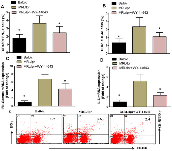
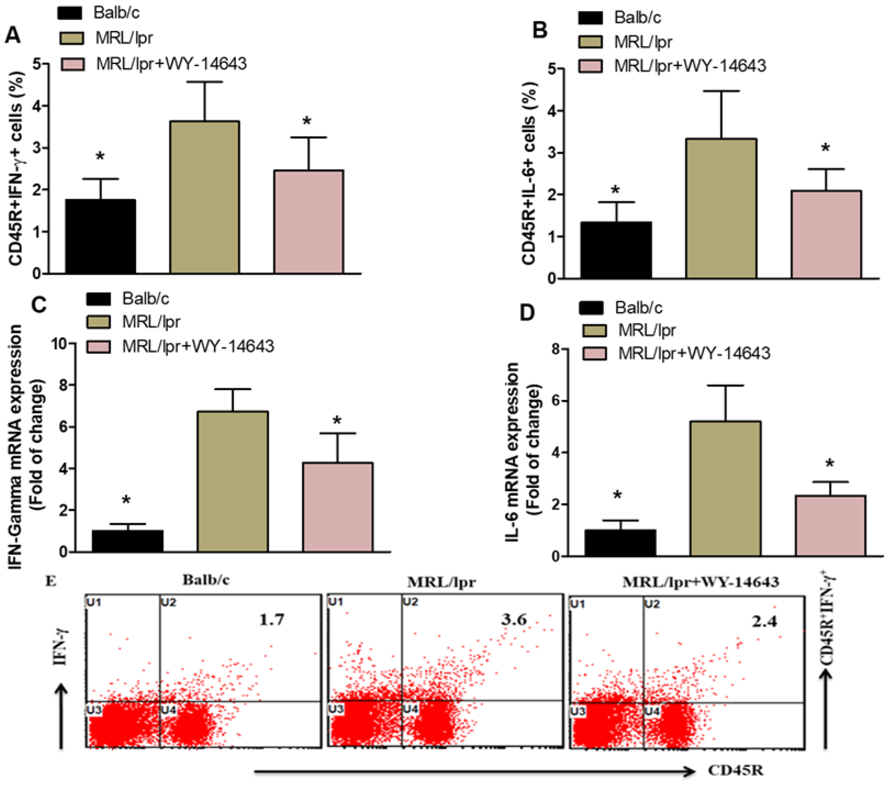
Flow cytometry analysis revealed that WY14643 decreases the expression of inflammatory markers in MRL/lpr mice. The number of CD45R+ cells producing IFN-γ and IL-6 was significantly lower in the spleens of WY14643-treated mice than in vehicle-treated controls (Figure 1A,B). Additionally, RT-PCR of kidney tissue showed reduced levels of IFN-γ and IL-6 mRNA in WY14643-treated mice compared with vehicle-treated mice (Figure 1C,D). These results suggest that WY14643 suppresses inflammatory mediators in MRL/lpr mice, indicating that activating PPAR-α may help reduce inflammation and cytokine production in this SLE model.
Figure 1.
(A,B). Effects of WY14643 on IFN-γ- and IL-6-expressing CD45R+ cells were analyzed by flow cytometry in splenocytes. (C,D) mRNA levels of IFN-γ and IL-6 were analyzed by RT-PCR in kidney tissue from MRL/lpr mice treated with WY14643. Cells were gated on forward- and side-scatter dot plots to determine the percentage of IFN-γ- and IL-6-expressing CD45R+ B cells in the spleen. (E) Representative Dot plots of a mouse from each group. MRL/lpr mice were treated with 10 mg/kg WY14643 intraperitoneally daily for eight weeks, whereas Balb/c mice (wild-type control) received a vehicle. The graphs represent the mean score ± SD of six mice per group. Statistical significance was determined as * p < 0.05.
2.2. PPAR-α Agonist WY14643 Treatment Decreases iNOS and MCP-1 Expression
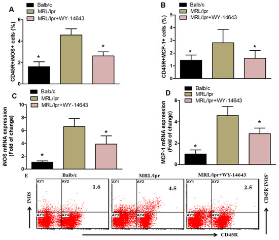
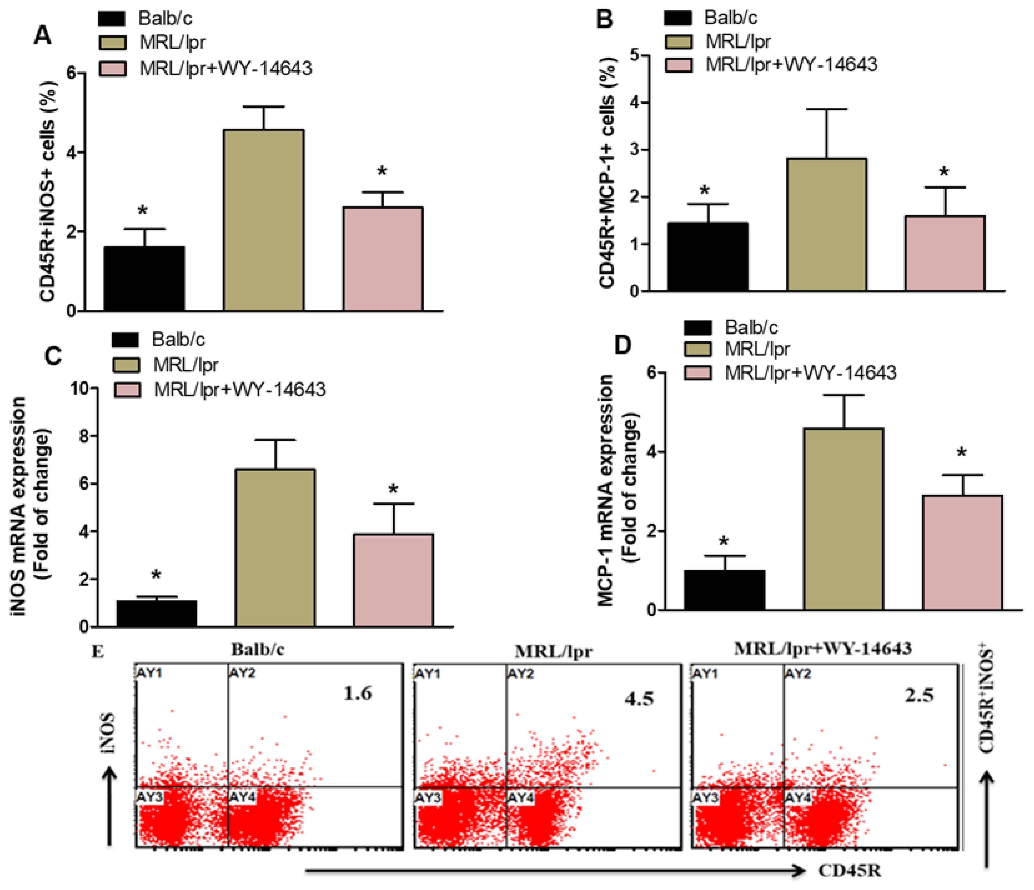
We investigated the effects of WY14643 on CD45R+ cells expressing iNOS and MCP-1 in MRL/lpr mice. The counts of CD45R+iNOS+ and CD45R+MCP-1+ cells were markedly lower in WY14643-treated MRL/lpr mice than in vehicle-treated mice (Figure 2A,B). RT-PCR results also showed reduced levels of iNOS and MCP-1 mRNA in the kidneys of WY14643-treated MRL/lpr mice compared with vehicle-treated mice (Figure 2C,D). These findings demonstrate that WY14643 treatment decreases iNOS and MCP-1 expression in MRL/lpr mice.
Figure 2.
(A,B). Effects of WY14643 on iNOS- and MCP-1-expressing CD45R+ cells were analyzed by flow cytometry in splenocytes. (C,D) mRNA levels of iNOS and MCP-1 were analyzed by RT-PCR in kidney tissue from MRL/lpr mice treated with WY14643. Cells were gated on forward- and side-scatter dot plots to determine the percentage of iNOS- and MCP-1-expressing CD45R+ B cells in the spleen. (E) Representative Dot plots of a mouse from each group. MRL/lpr mice were treated with 10 mg/kg WY14643 intraperitoneally daily for eight weeks, whereas Balb/c mice (wild-type control) received a vehicle. The graphs represent the mean score ± SD of six mice per group. Statistical significance was determined as * p < 0.05.
2.3. Effects of WY14643 on IL-1α- and IL-2-Expressing CD45R+ Cells
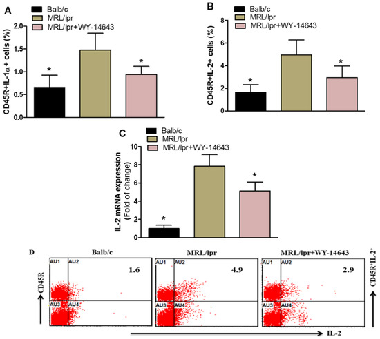
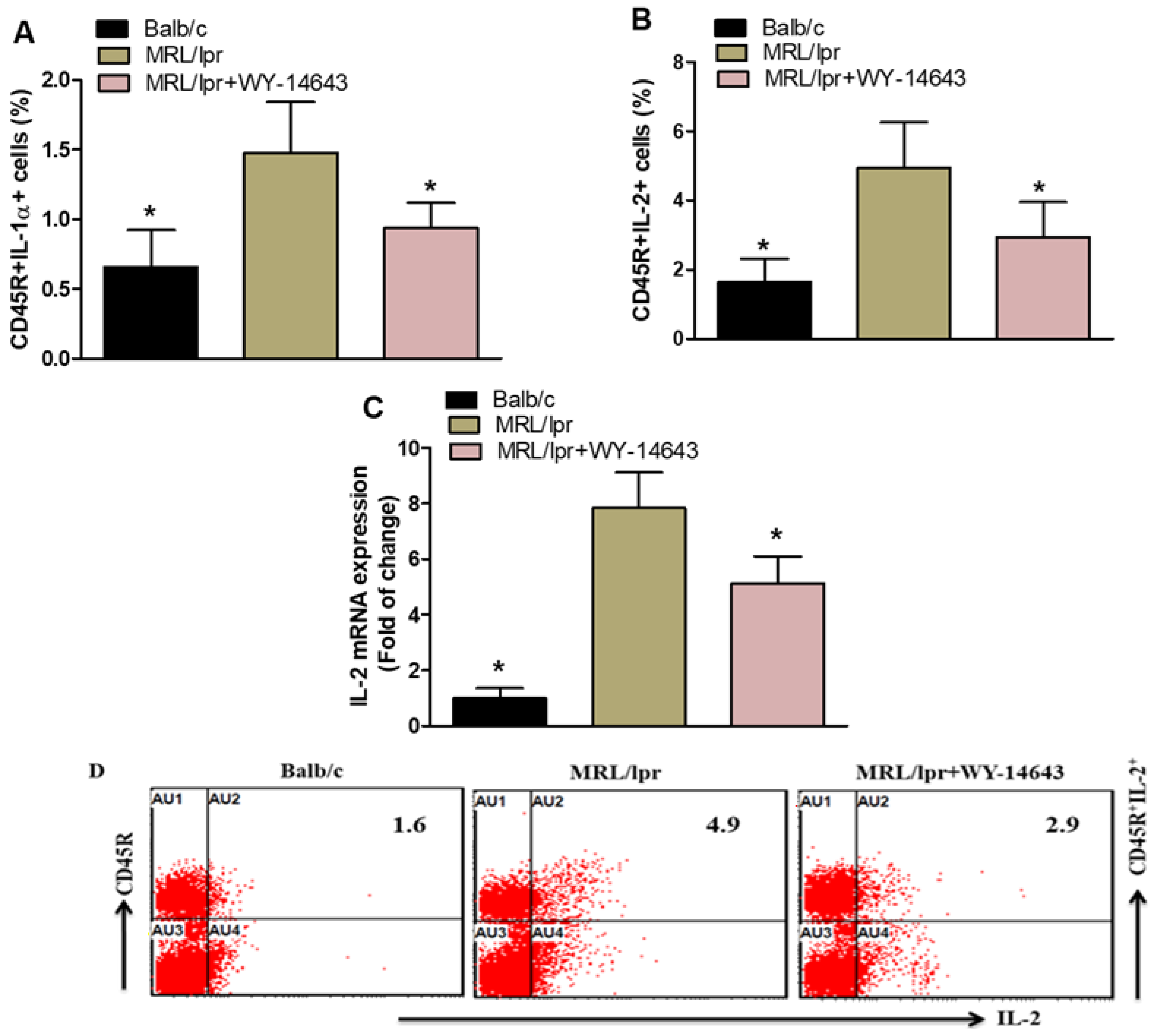
Flow cytometry revealed a significant decrease in CD45R+IL-1α+ and CD45R+IL-2+ cells in WY14643-treated mice compared with vehicle-treated controls (Figure 3A,B). Additionally, IL-2 mRNA levels in kidney tissue were markedly lower in WY14643-treated mice than in vehicle-treated controls (Figure 3C). These results suggest that WY14643 inhibits inflammatory mediator production in the SLE mouse model.
Figure 3.
(A,B). Effects of WY14643 on IL-1α- and IL-2-expressing CD45R+ cells were analyzed by flow cytometry in splenocytes. (C) mRNA levels of IL-1α and IL-2 were analyzed by RT-PCR in kidney tissue from MRL/lpr mice treated with WY14643. Cells were gated on forward- and side-scatter dot plots to determine the percentage of IL-1α- and IL-2-expressing CD45R+ B cells in the spleen. (D) Representative Dot plots of a mouse from each group. MRL/lpr mice were treated with 10 mg/kg WY14643 intraperitoneally daily for eight weeks, whereas Balb/c mice (wild-type control) received a vehicle. The graphs represent the mean score ± SD of six mice per group. Statistical significance was determined as * p < 0.05.
2.4. The PPAR-α Agonist WY14643 Inhibits the Notch-1 and Notch-3 Signaling
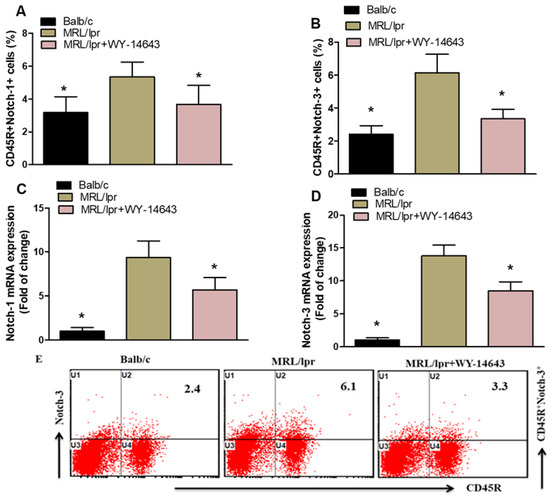
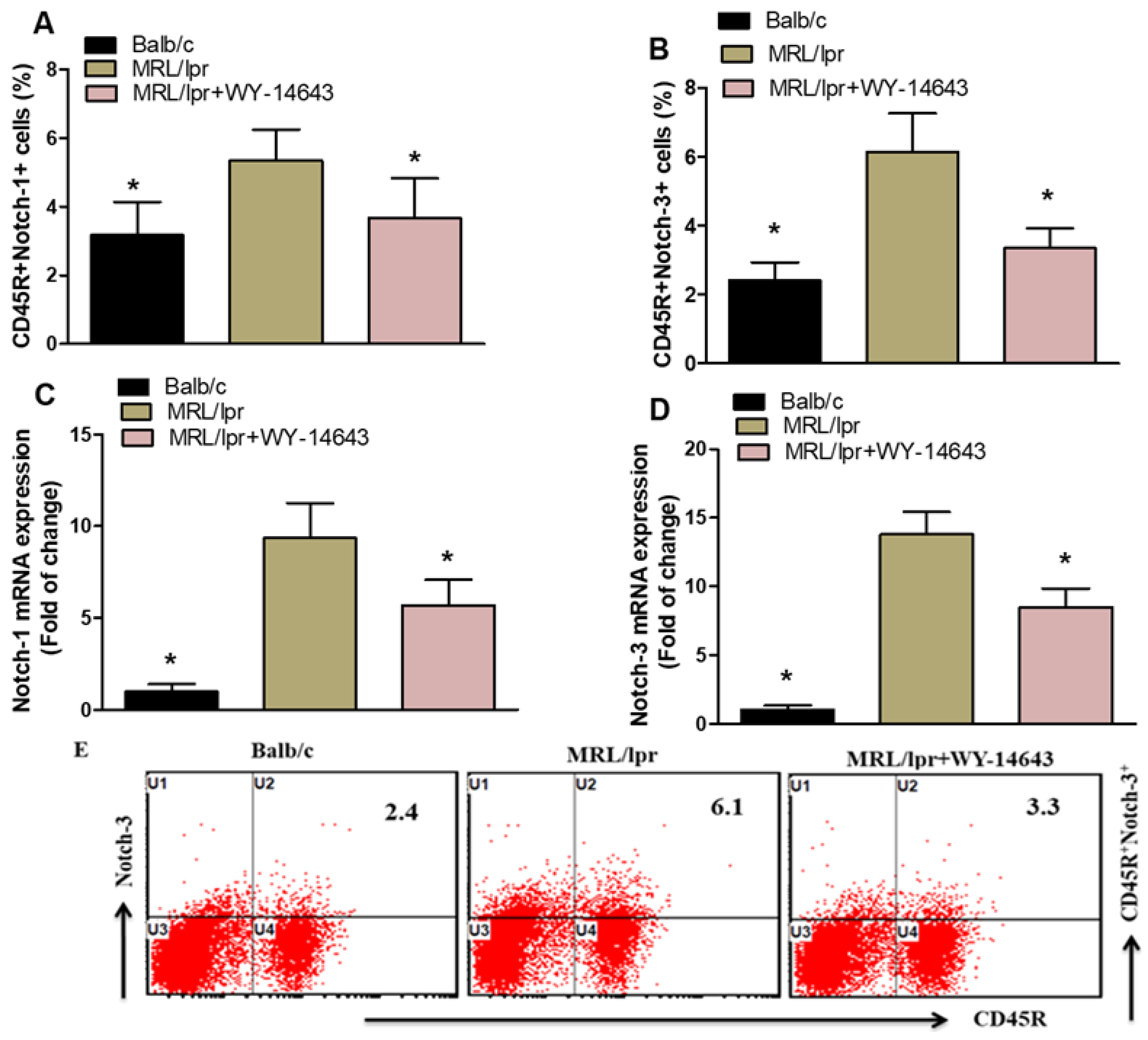
The study further analyzed Notch1 and Notch3 expression in splenic CD45R+ B cells. Treatment with WY14643 significantly reduced Notch1 and Notch3 levels in CD45R+ cells from MRL/lpr mice compared with vehicle-treated controls (Figure 4A,B). Consistent with the protein results, WY14643 also markedly decreased Notch1 and Notch3 mRNA levels in kidney tissue from MRL/lpr mice (Figure 4C,D). These results indicate that WY14643 effectively inhibits Notch1 and Notch3 signaling pathways.
Figure 4.
(A,B). Effects of WY14643 on Notch-1- and Notch-3-expressing CD45R+ cells were analyzed by flow cytometry in splenocytes. (C,D) mRNA levels of Notch-1 and Notch-3 were analyzed by RT-PCR in kidney tissue from MRL/lpr mice treated with WY14643. Cells were gated on forward- and side-scatter dot plots to determine the percentage of Notch-1- and Notch-3-expressing CD45R+ B cells in the spleen. (E) Representative Dot plots of a mouse from each group. Treated MRL/lpr mice received 10 mg/kg WY14643 intraperitoneally daily for eight weeks, whereas Balb/c mice (wild-type control) received a vehicle. The graphs represent the mean score ± SD of six mice per group. Statistical significance was determined as * p < 0.05.
2.5. WY14643 Decreases the Expression of GITR and NF-κB p65 in CD45R+ Cells
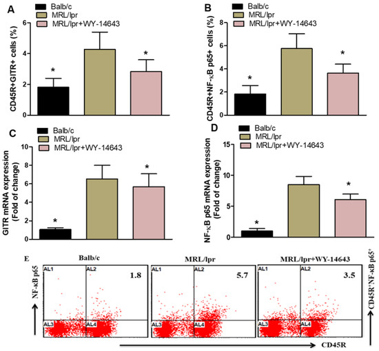
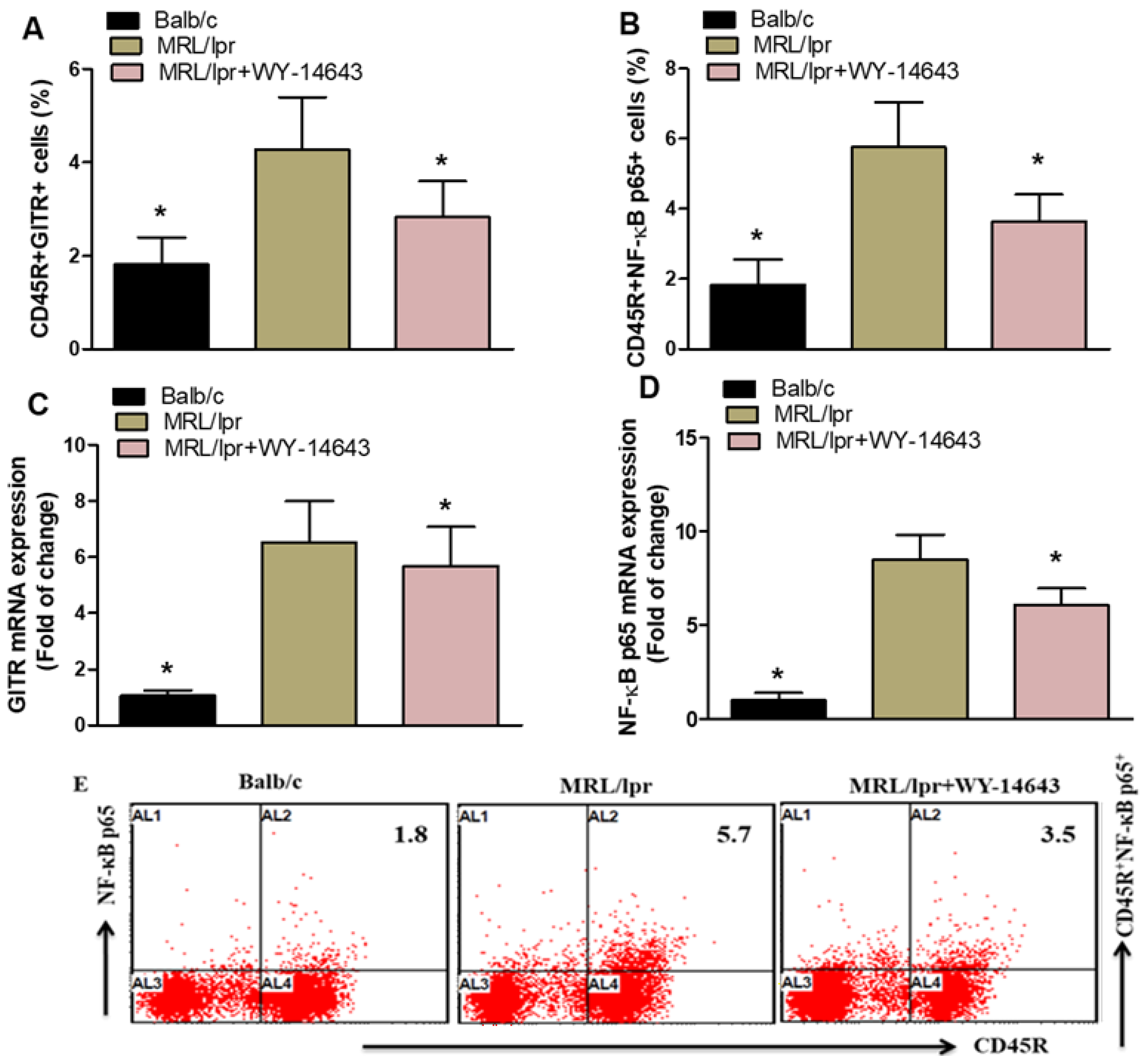
Treatment of MRL/lpr mice with WY-14643 significantly reduced the proportions of CD45R+GITR+ and CD45R+NF-κB p65+ cells compared with vehicle-treated controls (Figure 5A,B). Likewise, WY-14643 also lowered the mRNA levels of GITR and NF-κB p65 in these mice (Figure 5C,D). Overall, these findings suggest that WY-14643 has anti-inflammatory effects in MRL/lpr mice.
Figure 5.
(A,B). Effects of WY14643 on GITR- and NF-κB p65-expressing CD45R+ cells were analyzed by flow cytometry in splenocytes. (C,D) mRNA levels of GITR and NF-κB p65 were analyzed by RT-PCR in kidney tissue from MRL/lpr mice treated with WY14643. Cells were gated on forward- and side-scatter dot plots to determine the percentage of GITR- and NF-κB p65-expressing CD45R+ B cells in the spleen. (E) Representative Dot plots of a mouse from each group. MRL/lpr mice were treated with 10 mg/kg WY14643 intraperitoneally daily for eight weeks, whereas Balb/c mice (wild-type control) received a vehicle. The graphs represent the mean score ± SD of six mice per group. Statistical significance was determined as * p < 0.05.
3. Discussion
SLE is a long-term, multisystem autoimmune disease characterized by immune dysregulation, autoantibody production, and widespread inflammation [47,48]. Abnormal B cell activation, autoantibody secretion, and immune complex deposition in target organs are key pathological mechanisms of SLE [49]; therefore, targeting B cells is expected to be a treatment for SLE. Our previous research indicated that the PPAR-α agonist WY14643 has anti-inflammatory effects in SLE [50]. CD45R encodes a transmembrane protein-tyrosine phosphatase present on many immune cells [51]. Recent studies suggest that targeting CD45R can modulate immune responses; for example, anti-CD45R antibodies promote regulatory B cells and reduce inflammation [52]. Moreover, anti-CD45R therapy alleviated kidney damage in SLE models, improving BUN, Scr, dsDNA IgG levels, glomerular IgG and C3 deposits, and inflammatory cytokines [53]. Consistent with these findings, our study shows that WY14643 enhances immune regulation in MRL/lpr mice by reducing the levels of several inflammatory mediators.
IFN-γ signaling enhances B cell proliferation during the initial proliferative response after primary antigen exposure [54]. A previous study has shown that the IFN-γ gene signature appears early in SLE [34] and plays a crucial role in lupus nephritis [55]. Some studies indicate that levels of IFN-γ and its related genes are strongly associated with type I IFN activation in patients with SLE [56]. Another study highlights the significant role of IFN-γ in the early and active phases of SLE [13]. Another study has demonstrated that serum IFN-γ levels are higher in patients with SLE than in healthy individuals [57]. WY14643 significantly reduced IFN-γ and IL-6 levels in CD45R+ B cells, as measured by flow cytometry, and decreased their mRNA levels in kidney tissue. Since IFN-γ and IL-6 are crucial in lupus development, driving B cell activation, autoantibody production, and inflammation, this reduction suggests that activating PPAR-α may reduce systemic and renal inflammation. These results support earlier studies indicating that PPAR agonists inhibit cytokine production by repressing inflammatory gene transcription.
Both human and animal studies demonstrate that iNOS overexpression is associated with autoimmune diseases [58]. In MRL/lpr mice, an iNOS inhibitor has been shown to prevent glomerulonephritis [59], suggesting that iNOS contributes to the development and progression of SLE. Additionally, recent research in MRL/lpr mice indicates that iNOS promotes the proliferation of T follicular helper cells [60], which are believed to play a key role in SLE pathogenesis by stimulating B cells to produce more IgG [61,62]. Several chemokines participate in the development of lupus nephritis. MCP-1 is linked to kidney damage in SLE [63]. Research indicates that MCP-1-induced protein (IP-10) is significant in the pathogenesis of the disease [64]. Additionally, serum MCP-1 levels are elevated in patients with lupus nephritis [65]. The reductions in iNOS and MCP-1 support WY14643’s anti-inflammatory effects. iNOS promotes oxidative stress and tissue damage in lupus nephritis, while MCP-1 is a key chemokine that recruits monocytes to inflamed areas. The decreased levels of these mediators in splenic B cells and kidney tissue suggest that WY14643 may help prevent leukocyte infiltration and lessen kidney injury. This combined impact on cytokines and chemokines underscores the wide-ranging immunomodulatory potential of PPAR-α activation.
In SLE, abnormal immune activation drives chronic inflammation and organ damage [66]. IL-2 contributes to this harmful process by promoting immune responses that exacerbate autoimmunity. It promotes the growth and activation of autoreactive CD4+ T cells [67], which in turn drive B cell differentiation and autoantibody production, key aspects of SLE pathology. Moreover, IL-2 enhances the cytotoxic functions of CD8+ T cells [68,69], thereby contributing to tissue damage in organs such as the kidneys and skin. IL-1 has been linked to the development of several autoimmune inflammatory diseases, such as SLE [70]. WY14643 also decreased IL-1α+ and IL-2+CD45R+ cells, along with IL-2 mRNA levels in the kidney. IL-1α is a potent pro-inflammatory cytokine that causes tissue damage, whereas IL-2 has a complex regulatory role in T and B cells. In SLE, dysregulated IL-2 signaling impairs immune tolerance. The decline in these cytokines indicates that WY14643 might help restore immune balance and reduce inflammation in SLE.
Previous research has identified a pathogenic role for Notch signaling in SLE [71,72]. Notably, Zhang et al. showed that inhibiting Notch1 signaling improves murine lupus triggered by activated lymphocyte-derived DNA by preventing macrophage M2b polarization [30]. A recent study also demonstrated that blocking Notch1 signaling significantly reduces the development of pristane-induced murine lupus, underscoring the benefit of targeting Notch1 in SLE [35]. Notch3 regulates epithelial and inflammatory responses, facilitating the occurrence of acute kidney injury [73]. Activation of the Notch-3 receptor promotes inflammation and fibrosis after tubulointerstitial kidney damage [74]. The previously noted proinflammatory role of Notch-3 may therefore be linked to these acute kidney injuries [75]. WY14643 significantly inhibited the Notch1 and Notch3 signaling pathways. Notch signaling promotes B cell activation, differentiation, and survival, processes that are abnormally heightened in lupus. The suppression of Notch 1 and Notch 3 in both splenic B cells and kidney tissue suggests that activating PPAR-α could disrupt these harmful B cell signaling pathways. This is particularly significant because Notch signaling has been linked to autoantibody production and the development of lupus nephritis.
NF-κB activation promotes the production of inflammatory mediators that worsen disease symptoms [76]. The NF-κB signaling pathway is well established as being activated in SLE, and its excessive activation has been linked to disease development and progression [77]. The SLE group showed higher NF-κB expression than the control group [78]. GITR messenger RNA levels are elevated in peripheral blood mononuclear cells from patients with SLE [79,80]. Additionally, a study confirmed that patients with active SLE exhibit higher GITR expression [81,82]. Finally, WY14643 reduced the expression of GITR and NFκB p65, two molecules central to immune activation and the transcription of inflammatory genes. NFκB p65 is a master regulator of inflammatory responses, and its inhibition suggests a key mechanism through which WY14643 suppresses cytokine and chemokine expression. GITR, a co-stimulatory receptor, enhances B cell activation and survival; its downregulation further supports the immunosuppressive effects of PPAR-α agonism.
Overall, these findings demonstrate that WY14643 exerts multiple anti-inflammatory effects in MRL/lpr mice by reducing cytokine production, lowering chemokine levels, and disrupting key B cell activation pathways. The consistent reduction in inflammatory mediators in both spleen and kidney tissues indicates that PPAR-α agonists could be effective for managing systemic inflammation in SLE. By modulating pathways such as NF-κB and Notch signaling, WY14643 may offer a distinct mechanistic approach to controlling autoimmune responses. This study shows that WY14643 decreases inflammatory mediators in splenic CD45R+ B cells and reduces expression of cytokines and signaling molecules in the kidneys of MRL/lpr mice.
We acknowledge a few limitations in our research. First, the experiments rely solely on a pharmacological PPAR-α agonist. Although WY14643 is commonly used to activate PPAR-α, such agents can have off-target effects, so we cannot definitively say that the anti-inflammatory effects are solely due to PPAR-α signaling. Second, although we observe decreased levels of inflammatory mediators in CD45R+ B cells and kidney tissue, the study does not examine other immune cells involved in SLE development, such as T cells, dendritic cells, and macrophages. Another limitation of our study is that renal mRNA expression was assessed using whole-kidney homogenates. Therefore, modulation of renal B cells or other immune signaling pathways could not be specifically assessed using immunohistochemistry, immunofluorescence, or targeted cell isolation techniques. Therefore, future studies will use immunohistochemistry, immunofluorescence, or cell-specific isolation techniques.
4. Materials and Methods
4.1. Chemicals and Antibodies
WY14643 was purchased from Tocris Bioscience in Bristol, UK. Reagents such as RPMI-1640 medium, ionomycin, and Phorbol 12-myristate 13-acetate were purchased from Sigma-Aldrich (St. Louis, MO, USA). The FcR blocking reagent fluorescently labeled antibodies, including CD45R, IFN-γ, IL-6, iNOS, MCP-1, IL-1α, IL-2, Notch-1, Notch-3, GITR, and NF-κB p65; as well as buffers for RBC lysis, permeabilization, fixation, and the were purchased from BioLegend (San Diego, CA, USA); BD Biosciences (San Diego, CA, USA); Thermo Fisher Scientific (Branchburg, NJ, USA); and Miltenyi Biotech (Bergisch Gladbach, Germany). cDNA synthesis kit, and SYBR® Green were purchased from Applied Biosystems (Foster City, CA, USA) and TRIzol® reagent from Life Technologies (Carlsbad, CA, USA).
4.2. Animal Experiments
Female MRL/lpr mice and Balb/c mice (wild-type [WT]) were purchased from Jackson Laboratories (Bar Harbor, ME, USA); eight-week-old mice weighing 25–30 g were used. All animals were housed in a specific-pathogen-free environment and provided with ample water and food under a 12 h light–dark cycle, with 6 mice per cage. The laboratory environment was maintained at 21–23 °C with a relative humidity of 50–70%. All animal experiments were approved by the Laboratory Animal Ethics Committee of King Saud University (approval no: KSU-SE-23-02) and performed in accordance with the ARRIVE guidelines 2.0 and the King Saud University Animal Committee guidelines and adhered to the principles of laboratory animal care.
4.3. Drug Treatment of Animals
Eight-week-old MRL/lpr mice and WT mice were randomized into three groups (n = 6 per group). The sample size for the present investigation has been determined based on our prior knowledge and expertise with these experiments. In all experimental studies, the Balb/c group received vehicle, the MRL/lpr group received vehicle, and the MRL/lpr + WY14643 group received 10 mg/kg of WY14643 intraperitoneally daily for eight weeks. The dosage of WY14643 (10 mg/kg, i.p.) was chosen based on previous research [50,83,84,85]. The volume of drugs administered to each mouse was based on its body weight. Subsequently, the mice were euthanized by intraperitoneal injection of pentobarbital sodium (100 mg/kg), followed by cervical dislocation, and samples were collected for subsequent experiments, including flow cytometry staining and RT-PCR analysis.
4.4. Flow Cytometric Analysis
For flow cytometry analysis, conjugated antibodies targeting CD45R, IFN-γ, IL-6, iNOS, MCP-1, IL-1α, IL-2, Notch-1, Notch-3, GITR, and NF-κB p65 were used. The following conjugated antibodies (from BioLegend and Santa Cruz Biotechnology, Dallas, TX, USA) labeled spleen cells: FITC anti-mouse CD45R (BioLegend Cat. No. 103206), APC anti-mouse CD45R (BioLegend Cat. No. 103212), PE/Dazzle™ 594 anti-mouse IFN-γ (BioLegend Cat. No. 505845), APC anti-mouse IL-6 (BioLegend Cat. No. 504508), PE anti-mouse iNOS (BioLegend Cat. No. 696806), APC anti-mouse MCP-1 (BioLegend Cat. No. 505910), PE anti-mouse IL-1α (BioLegend Cat. No. 503203), APC anti-mouse IL-2 (BioLegend Cat. No. 503809), APC anti-mouse Notch-1 (BioLegend Cat. No. 130613), Alexa Fluor® 647 anti-mouse Notch-3 (BioLegend Cat. No. 130511), PE/Cyanine7 anti-mouse GITR (BioLegend Cat. No. 126317), and FITC anti-mouse NF-κB p65 (Santa Cruz Biotechnology Cat. No. SC-8008). Splenocytes were treated with PMA/ionomycin (Sigma-Aldrich) and Golgi-plug (BD Biosciences) for 4 h before labeling [86]. After washing, surface CD45R staining was performed, followed by fixation and permeabilization. Fluorescent antibodies against IFN-γ, IL-6, iNOS, MCP-1, IL-1α, IL-2, Notch-1, Notch-3, GITR, and NF-κB p65 labeled the spleen cells. The flow cytometry gating strategy involved first gating singlets based on FSC-A versus FSC-H, then removing dead cells and debris. Live lymphocytes were identified on FSC-SSC plots, and CD4+ T cells were gated. Within this subset, intracellular cytokine expression (IFN-γ, IL-17A, and IL-9) was assessed. To determine the different inflammatory mediators in lymphocytes, lymphocytes were separated from other immune cells (monocytes and granulocytes) using a conventional gating strategy based on physical properties (forward and side scatter). Lymphocytes were identified on FSC-SSC plots, and CD45R+ cells were gated. The percentages of other immunological markers were then analyzed in CD45R+ cells within their respective gates. The percentage of CD45R+IFN-γ+, CD45R+IL-6+, CD45R+iNOS+, CD45R+MCP-1+, CD45R+IL-1α+, CD45R+IL-2+, CD45R+Notch1+, CD45R+Notch3+, CD45R+GITR+, and CD45R+NF-κB p65+ cells were determined in the lymphocyte gate. A Beckman Coulter FC500 flow cytometer (Indianapolis, IN, USA) collected 10,000 cell events, and data were analyzed using CXP software version 2.0 [86].
4.5. RT-PCR Analysis
Total RNA was isolated from kidney tissue using TRIzol reagent (Life Technologies, Paisley, UK). cDNA synthesis was performed with a high-capacity cDNA reverse transcription kit, followed by real-time PCR with SYBR® Green PCR master mix (Applied Biosystems (Foster City, CA, USA), according to the manufacturer’s instructions. Primers were selected from PubMed. The assay primers included: IFN-γ (forward: 5′-CGGCACAGTCATTGAAAGCC-3′, reverse: 5′-TGCATCCTTTTTCGCCTTGC-3′), IL-6 (forward: 5′-GCCTTCTTGGGACTGATGCT-3′, reverse: 5′-GACAGGTCTGTTGGGAGTGG-3′), iNOS (forward: 5′-TCAGCCAAGCACTCCAATGT-3′, reverse: 5′-AGTGATGGAGGTGCCCTAGT-3′), MCP-1 (forward: 5′-CAAAGCCAGGGGCCTTTTTC-3′, reverse: 5′-TACCAGGAGCCAGGCATAGT-3′), IL-2 (forward: 5′-GGAACCTGAAACTCCCCAGG-3′, reverse: 5′-AATCCAGAACATGCCGCAGA-3′), Notch-1 (forward: 5′-GCCTCAAGCCCCTGAAGAAT-3′, reverse: 5′-GCGCTTTCGACGATCTGAAC-3′), Notch-3 (forward: 5′-AGGCCATGGTCTTCCCCTAT-3′, reverse: 5′-ACCTCCCCCATCAGACTCTC-3′), GITR (forward: 5ʹ-CCAAGCCAGACGCTACAAGA-3′, reverse: 5ʹ-CAGGGAAGGGTGCAGAACAT-3′), NF-κB p65 (forward: 5′-CTTCTCTATGGCGTCGTCCC-3′, reverse: 5′-AAGATGGCCTCCTTCACAGC-3′), and GAPDH (forward: 5′-GGCAAATTCAACGGCACAGT-3′, reverse: 5′-TGAAGTCGCAGGAGACAACC-3′). mRNA levels were normalized to GAPDH as an endogenous reference gene [87], and results are expressed as fold change.
4.6. Statistical Analysis
Data are presented as mean ± SD. The data undergo normality and homogeneity tests before analysis. Differences among the three groups were analyzed using one-way ANOVA, followed by Tukey’s post hoc test for multiple comparisons. All statistical analyses were conducted using GraphPad Prism 8; p < 0.05 indicated statistical significance.
Author Contributions
Conceptualization, H.A.A.-M., H.N.A., M.A.A., A.N., A.M.S.A., H.A.A. and S.F.A.; Methodology, H.A.A.-M., M.A.A., A.A.A. and S.F.A.; Software, H.N.A., A.M.S.A., A.A.A. and S.F.A.; Validation, H.N.A., H.A.A., A.A.A. and S.F.A.; Formal analysis, H.A.A.-M., H.N.A., A.N., A.M.S.A., H.A.A. and S.F.A.; Investigation, H.A.A.-M., S.A.B. and S.F.A.; Resources, H.N.A., M.A.A., A.M.S.A., H.A.A., A.A.A. and S.F.A.; Data curation, H.N.A., M.A.A., A.M.S.A. and S.F.A.; Writing – original draft, H.A.A.-M., A.N., S.M.A., S.A.B. and S.F.A.; Writing – review & editing, H.A.A.-M., A.N., S.M.A., S.A.B. and S.F.A.; Visualization, M.A.A., S.A.B. and S.F.A.; Supervision, A.N., S.M.A., S.A.B. and S.F.A.; Project administration, M.A.A., S.M.A. and S.F.A.; Funding acquisition, H.A.A.-M., M.A.A., S.M.A. and S.F.A. All authors have read and agreed to the published version of the manuscript.
Funding
This research was supported by the Ongoing Research Funding Program (ORF-2026-709) at King Saud University in Riyadh, Saudi Arabia.
Institutional Review Board Statement
All animal experiments were approved by the Laboratory Animal Ethics Committee of King Saud University (approval no: KSU-SE-23-02; approval date: 18 April 2023) and performed in accordance with the Institutes of Health Guide for the Care and Use of Laboratory Animals.
Informed Consent Statement
Not applicable.
Data Availability Statement
The original contributions presented in this study are included in the article. Further inquiries can be directed to the corresponding author.
Acknowledgments
The authors acknowledge and extend their appreciation to the Ongoing Re-search Funding Program (ORF-2026-709), King Saud University, Riyadh, Saudi Arabia, for funding this study.
Conflicts of Interest
The authors declare no conflicts of interest.
References
- Durcan, L.; O’Dwyer, T.; Petri, M. Management strategies and future directions for systemic lupus erythematosus in adults. Lancet 2019, 393, 2332–2343. [Google Scholar] [CrossRef]
- Fava, A.; Petri, M. Systemic lupus erythematosus: Diagnosis and clinical management. J. Autoimmun. 2019, 96, 1–13. [Google Scholar] [CrossRef]
- Murphy, G.; Lisnevskaia, L.; Isenberg, D. Systemic lupus erythematosus and other autoimmune rheumatic diseases: Challenges to treatment. Lancet 2013, 382, 809–818. [Google Scholar] [CrossRef]
- Wahren-Herlenius, M.; Dörner, T. Immunopathogenic mechanisms of systemic autoimmune disease. Lancet 2013, 382, 819–831. [Google Scholar] [CrossRef]
- Rosser, E.C.; Mauri, C. Regulatory B cells: Origin, phenotype, and function. Immunity 2015, 42, 607–612. [Google Scholar] [CrossRef] [PubMed]
- Iwata, S.; Tanaka, Y. B-cell subsets, signaling and their roles in secretion of autoantibodies. Lupus 2016, 25, 850–856. [Google Scholar] [CrossRef]
- Wen, L.; Zhang, B.; Wu, X.; Liu, R.; Fan, H.; Han, L.; Zhang, Z.; Ma, X.; Chu, C.Q.; Shi, X. Toll-like receptors 7 and 9 regulate the proliferation and differentiation of B cells in systemic lupus erythematosus. Front. Immunol. 2023, 14, 1093208. [Google Scholar] [CrossRef] [PubMed]
- Oleinika, K.; Mauri, C.; Salama, A.D. Effector and regulatory B cells in immune-mediated kidney disease. Nat. Rev. Nephrol. 2019, 15, 11–26. [Google Scholar] [CrossRef]
- Gallagher, S.; Yusuf, I.; McCaughtry, T.M.; Turman, S.; Sun, H.; Kolbeck, R.; Herbst, R.; Wang, Y. MEDI-551 Treatment Effectively Depletes B Cells and Reduces Serum Titers of Autoantibodies in Mice Transgenic for Sle1 and Human CD19. Arthritis Rheumatol. 2016, 68, 965–976. [Google Scholar] [CrossRef]
- Chen, X.; Zhu, L.; Xu, J.; Cheng, Q.; Dong, Y.; Xie, Y.; Hua, L.; Du, Y. Semaphorin 5A promotes Th17 differentiation via PI3K-Akt-mTOR in systemic lupus erythematosus. Arthritis Res. Ther. 2024, 26, 204. [Google Scholar] [CrossRef] [PubMed]
- Lee, S.K.; Silva, D.G.; Martin, J.L.; Pratama, A.; Hu, X.; Chang, P.-P.; Walters, G.; Vinuesa, C.G. Interferon-gamma excess leads to pathogenic accumulation of follicular helper T cells and germinal centers. Immunity 2012, 37, 880–992. [Google Scholar] [CrossRef]
- Shah, D.; Kiran, R.; Wanchu, A.; Bhatnagar, A. Oxidative stress in systemic lupus erythematosus: Relationship to Th1 cytokine and disease activity. Immunol. Lett. 2010, 129, 7–12. [Google Scholar] [CrossRef]
- Liu, W.; Li, M.; Wang, Z.; Wang, J. IFN-gamma mediates the development of systemic lupus erythematosus. BioMed Res. Int. 2020, 2020, 7176515. [Google Scholar] [CrossRef] [PubMed]
- Liu, W.; Zhang, S.; Wang, J. IFN-gamma, should not be ignored in SLE. Front. Immunol. 2022, 13, 954706. [Google Scholar]
- Richter, P.; Rezus, C.; Burlui, A.M.; Schreiner, T.G.; Rezus, E. Serum Interleukin-6 in Systemic Lupus Erythematosus: Insights into Immune Dysregulation, Disease Activity, and Clinical Manifestations. Cells 2025, 14, 1568. [Google Scholar] [CrossRef] [PubMed]
- Aliyu, M.; Zohora, F.T.; Anka, A.U.; Ali, K.; Maleknia, S.; Saffarioun, M.; Azizi, G. Interleukin-6 cytokine: An overview of the immune regulation, immune dysregulation, and therapeutic approach. Int. Immunopharmacol. 2022, 111, 109130. [Google Scholar] [CrossRef]
- Bacalao, M.A.; Satterthwaite, A.B. Recent Advances in Lupus B Cell Biology: PI3K, IFNγ, and Chromatin. Front. Immunol. 2021, 11, 615673. [Google Scholar] [CrossRef]
- Bassi, M.S.; Iezzi, E.; Drulovic, J.; Pekmezovic, T.; Gilio, L.; Furlan, R.; Finardi, A.; Marfia, G.A.; Sica, F.; Centonze, D.; et al. IL-6 in the Cerebrospinal Fluid Signals Disease Activity in Multiple Sclerosis. Front. Cell Neurosci. 2020, 14, 120. [Google Scholar]
- Lv, W.; Booz, G.W.; Wang, Y.; Fan, F.; Roman, R.J. Inflammation and renal fibrosis: Recent developments on key signaling molecules as potential therapeutic targets. Eur. J. Pharmacol. 2018, 820, 65–76. [Google Scholar] [CrossRef]
- Chougule, D.; Rajadhyaksha, A.; Jamale, T.; Mehta, K.; Jose, A.; Pawaskar, S.; Madkaikar, M.; Pradhan, V. Cytokine. A multifaceted approach to lupus nephritis: Deciphering the interplay between adipokines, cytokines and complement proteins in disease pathogenesis. Cytokine 2025, 194, 156992. [Google Scholar] [CrossRef]
- Alzawawy, A.; Zohary, M.; Ablordiny, M.; Eldalie, M. Estimation of monocyte-chemoattractant protein-1 (Mcp-1) level in patients with lupus nephritis. Int. J. Rheum. Dis. 2009, 12, 311–318. [Google Scholar] [CrossRef]
- Yang, Y.; Wei, Z.; Teichmann, A.T.; Wieland, F.H.; Wang, A.; Lei, X.; Zhu, Y.; Yin, J.; Fan, T.; Zhou, L.; et al. Development of a novel nitric oxide (NO) production inhibitor with potential therapeutic effect on chronic inflammation. Eur. J. Med. Chem. 2020, 193, 112216. [Google Scholar] [CrossRef]
- Cho, Y.C.; Park, J.; Cho, S. Anti-Inflammatory and Anti-Oxidative Effects of luteolin-7-O-glucuronide in LPS-Stimulated Murine Macrophages through TAK1 Inhibition and Nrf2 Activation. Int. J. Mol. Sci. 2020, 21, 2007. [Google Scholar] [CrossRef] [PubMed]
- Peairs, A.; Radjavi, A.; Davis, S.; Li, L.; Ahmed, A.; Giri, S.; Reilly, C.M. Activation of AMPK inhibits inflammation in MRL/lpr mouse mesangial cells. Clin. ExpImmunol. 2009, 156, 542–551. [Google Scholar] [CrossRef] [PubMed]
- Wang, J.; Chen, Q.; Zhang, Z.; Wang, S.; Wang, Y.; Xiang, M.; Liang, J.; Xu, J. Azithromycin alleviates systemic lupus erythematosus via the promotion of M2 polarisation in lupus mice. Cell Death Discov. 2021, 7, 82. [Google Scholar] [CrossRef] [PubMed]
- Njoku, C.; Self, S.E.; Ruiz, P.; Hofbauer, A.F.; Gilkeson, G.S.; Oates, J.C. Inducible nitric oxide synthase inhibitor SD-3651 reduces proteinuria in MRL/lpr mice deficient in the NOS2 gene. J. Investig. Med. 2008, 56, 911–919. [Google Scholar] [CrossRef]
- MacGrogan, D.; Munch, J.; de la Pompa, J.L. Notch and interacting signalling pathways in cardiac development, disease, and regeneration. Nat. Rev. Cardiol. 2018, 15, 685–704. [Google Scholar] [CrossRef]
- Siebel, C.; Lendahl, U. Notch signaling in development, tissue homeostasis, and disease. Physiol. Rev. 2017, 97, 1235–1294. [Google Scholar] [CrossRef]
- Talora, C.; Campese, A.F.; Bellavia, D.; Felli, M.P.; Vacca, A.; Gulino, A.; Screpanti, I. Notch signaling and diseases: An evolutionary journey from a simple beginning to complex outcomes. Biochim. Biophys. Acta 2008, 1782, 489–497. [Google Scholar] [CrossRef]
- Zhang, W.; Xu, W.; Xiong, S. Blockade of notch1 signaling alleviates murine lupus via blunting macrophage activation and m2b polarization. J. Immunol. 2010, 184, 6465–6478. [Google Scholar] [CrossRef]
- Rauen, T.; Grammatikos, A.P.; Hedrich, C.M.; Floege, J.; Tenbrock, K.; Ohl, K.; Kyttaris, V.C.; Tsokos, G.C. Camp-responsive element modulator alpha (cremalpha) contributes to decreased notch-1 expression in t cells from patients with active systemic lupus erythematosus (sle). J. Biol. Chem. 2012, 287, 42525–42532. [Google Scholar] [CrossRef] [PubMed]
- Jin, K.; Wen, Z.; Wu, B.; Zhang, H.; Qiu, J.; Wang, Y.; Warrington, K.J.; Berry, G.J.; Goronzy, J.J.; Weyand, C.M. NOTCH-induced rerouting of endosomal trafficking disables regulatory T cells in vasculitis. J. Clin. Investig. 2021, 131, e136042. [Google Scholar] [CrossRef]
- Wei, K.; Korsunsky, I.; Marshall, J.L.; Gao, A.; Watts, G.F.M.; Major, T.; Croft, A.P.; Watts, J.; Blazar, P.E.; Lange, J.K.; et al. Notch signalling drives synovial fibroblast identity and arthritis pathology. Nature 2020, 582, 259–264. [Google Scholar] [CrossRef]
- Munroe, M.E.; Lu, R.; Zhao, Y.D.; A Fife, D.; Robertson, J.M.; Guthridge, J.M.; Niewold, T.B.; Tsokos, G.C.; Keith, M.P.; Harley, J.B.; et al. Altered type II interferon precedes autoantibody accrual and elevated type I interferon activity prior to systemic lupus erythematosus classification. Ann. Rheum. Dis. 2016, 75, 2014–2021. [Google Scholar] [CrossRef]
- Li, X.; Fei, F.; Yao, G.; Yang, X.; Geng, L.; Wang, D.; Gao, Y.; Hou, Y.; Sun, L. Notch1 signalling controls the differentiation and function of myeloid-derived suppressor cells in systemic lupus erythematosus. Immunology 2023, 168, 170–183. [Google Scholar] [CrossRef] [PubMed]
- Gervois, P.; Mansouri, R.M. PPARalpha as a therapeutic target in inflammation associated diseases. Expert. Opin. Ther. Targets 2012, 16, 1113–1125. [Google Scholar] [CrossRef]
- Hecker, M.; Behnk, A.; Morty, R.E.; Sommer, N.; Vadász, I.; Herold, S.; Seeger, W.; Mayer, K. PPAR-alpha activation reduced LPS-induced inflammation in alveolar epithelial cells. Exp. Lung Res. 2015, 41, 393–403. [Google Scholar] [CrossRef]
- Huang, D.; Zhao, Q.; Liu, H.; Guo, Y.; Xu, H. PPAR-alpha Agonist WY-14643 Inhibits LPS-Induced Inflammation in Synovial Fibroblasts via NF-kB Pathway. J. Mol. Neurosci. 2016, 59, 544–553. [Google Scholar] [CrossRef]
- Yoo, S.H.; Abdelmegeed, M.A.; Song, B.J. Activation of PPARalpha by Wy-14643 ameliorates systemic lipopolysaccharide-induced acute lung injury. Biochem. Biophys. Res. Commun. 2013, 436, 366–371. [Google Scholar] [CrossRef] [PubMed]
- Gray, E.; Ginty, M.; Kemp, K.; Scolding, N.; Wilkins, A. Peroxisome proliferator-activated receptor-alpha agonists protect cortical neurons from inflammatory mediators and improve peroxisomal function. Eur. J. Neurosci. 2011, 33, 1421–1432. [Google Scholar] [CrossRef]
- Briguglio, E.; Di Paola, R.; Paterniti, I.; Mazzon, E.; Oteri, G.; Cordasco, G.; Cuzzocrea, S. WY-14643, a Potent Peroxisome Proliferator Activator Receptor-α PPAR-α Agonist Ameliorates the Inflammatory Process Associated to Experimental Periodontitis. PPAR Res. 2010, 2010, 193019. [Google Scholar] [CrossRef]
- Wang, G.; Namura, S. Effects of chronic systemic treatment with peroxisome proliferator-activated receptor α activators on neuroinflammation induced by intracerebral injection of lipopolysaccharide in adult mice. Neurosci. Res. 2011, 70, 230–237. [Google Scholar] [CrossRef]
- Liu, H.; Zhang, H.; Cui, R.; Guo, X.; Wang, D.; Dai, J. Activation of peroxisome proliferator-activated receptor α ameliorates perfluorododecanoic acid-induced production of reactive oxygen species in rat liver. Arch. Toxicol. 2016, 90, 1383–1397. [Google Scholar] [CrossRef] [PubMed]
- Jackson, T.C.; Mi, Z.; Bastacky, S.I.; McHale, T.; Melhem, M.F.; Sonalker, P.A.; Tofovic, S.P.; Jackson, E.K. PPAR alpha agonists improve renal preservation in kidneys subjected to chronic in vitro perfusion: Interaction with mannitol. Transpl. Int. 2007, 20, 277–2790. [Google Scholar] [CrossRef]
- Watson, M.L.; Rao, J.K.; Gilkeson, G.S.; Ruiz, P.; Eicher, E.M.; Pisetsky, D.S.; Matsuzawa, A.; Rochelle, J.M.; Seldin, M.F. Genetic analysis of MRL-lpr mice: Relationship of the Fas apoptosis gene to disease manifestations renal disease-modifying loci. J. Exp. Med. 1992, 176, 1645–1656. [Google Scholar] [CrossRef]
- Drappa, J.; Brot, N.; Elkon, K.B. The Fas protein is expressed at high levels on CD4+CD8+ thymocytes and activated mature lymphocytes in normal mice but not in the lupus-prone strain, MRL lpr/lpr. Proc. Natl. Acad. Sci. USA 1993, 90, 10340–10344. [Google Scholar] [CrossRef] [PubMed]
- Hoi, A.; Igel, T.; Mok, C.C.; Arnaud, L. Systemic lupus erythematosus. Lancet 2024, 403, 2326–2338. [Google Scholar] [CrossRef] [PubMed]
- Siegel, C.H.; Sammaritano, L.R. Systemic Lupus Erythematosus: A Review. JAMA 2024, 331, 1480–1491. [Google Scholar] [CrossRef]
- Caielli, S.; Wan, Z.; Pascual, V. Systemic Lupus Erythematosus Pathogenesis: Interferon and Beyond. Annu. Rev. Immunol. 2023, 41, 533–560. [Google Scholar] [CrossRef]
- Al-Mazroua, H.A.; Nadeem, A.; Attia, S.M.; Bakheet, S.A.; Ahmad, A.; Ansari, M.A.; Ibrahim, K.E.; Alomar, H.A.; Almutairi, M.M.; Algarzae, N.K.; et al. The PPAR-α selective agonist WY14643 improves lupus nephritis via the downregulation of the RORγT/STAT3 signaling pathway in MRL/lpr mice. Int. Immunopharmacol. 2025, 145, 113787. [Google Scholar] [CrossRef]
- Preußner, M.; Schreiner, S.; Hung, L.H.; Porstner, M.; Jäck, H.M.; Benes, V.; Rätsch, G.; Bindereif, A. HnRNP L and L-like cooperate in multiple-exon regulation of CD45 alternative splicing. Nucleic Acids Res. 2012, 40, 5666–5678. [Google Scholar] [CrossRef]
- Fang, T.; Koo, T.Y.; Lee, J.-G.; Jang, J.Y.; Xu, Y.; Hwang, J.H.; Park, S.; Yan, J.-J.; Ryu, J.-H.; Ryu, Y.-M.; et al. Anti-CD45RB Antibody Therapy Attenuates Renal Ischemia-Reperfusion Injury by Inducing Regulatory B Cells. J. Am. Soc. Nephrol. 2019, 30, 1870–1885. [Google Scholar] [CrossRef]
- Lv, J.; Chen, L.; Zhao, L. Renoprotective anti-CD45RB antibody induces B cell production in systemic lupus erythematosus based on single-cell RNA-seq analysis. J. Autoimmun. 2023, 134, 102949. [Google Scholar] [CrossRef] [PubMed]
- Szelinski, F.; Lino, A.C.; Dörner, T. B cells in systemic lupus erythematosus. Curr. Opin. Rheumatol. 2022, 34, 125–132. [Google Scholar] [CrossRef]
- Fava, A.; Buyon, J.; Mohan, C.; Zhang, T.; Belmont, H.M.; Izmirly, P.; Clancy, R.; Trujillo, J.M.; Fine, D.; Zhang, Y.; et al. Integrated urine proteomics and renal single-cell genomics identify an IFN-g response gradient in lupus nephritis. JCI Insight 2020, 5, 138345. [Google Scholar] [CrossRef]
- Greene, J.A.; DeVecchio, J.L.; Gould, M.P.; Auletta, J.J.; Heinzel, F.P. In vivo and in vitro regulation of type I IFN synthesis by synergistic effects of CD40 and type II IFN. J. Immunol. 2006, 176, 5995–6003. [Google Scholar] [CrossRef] [PubMed]
- Paradowska-Gorycka, A.; Wajda, A.; Stypinska, B.; Walczuk, E.; Rzeszotarska, E.; Walczyk, M.; Haladyj, E.; Romanowska-Prochnicka, K.; Felis-Giemza, A.; Lewandowska, A.; et al. Variety of endosomal TLRs and interferons (IFN-a, IFN-b, IFNg) expression profiles in patients with SLE, SSc and MCTD. Clin. Exp. Immunol. 2021, 204, 49–63. [Google Scholar] [CrossRef] [PubMed]
- Karpuzoglu, E.; Ahmed, S.A. Estrogen regulation of nitric oxide and inducible nitric oxide synthase (iNOS) in immune cells: Implications for immunity, autoimmune diseases, and apoptosis. Nitric Oxide 2006, 15, 177–186. [Google Scholar] [CrossRef]
- Mishra, N.; Reilly, C.M.; Brown, D.R.; Ruiz, P.; Gilkeson, G.S. Histone deacetylase inhibitors modulate renal disease in the MRL-lpr/lpr mouse. J. Clin. Investig. 2003, 111, 539–552. [Google Scholar] [CrossRef]
- Zhang, Z.; Feng, R.; Niu, L.; Huang, S.; Deng, W.; Shi, B.; Yao, G.; Chen, W.; Tang, X.; Gao, X.; et al. Human umbilical cord mesenchymal stem cells inhibit T follicular helper cell expansion through the activation of iNOS in lupus-prone B6.MRL-Fas(lpr) mice. Cell Transplant. 2017, 26, 1031–1042. [Google Scholar] [CrossRef]
- Crotty, S. T follicular helper cell differentiation, function, and roles in disease. Immunity 2014, 41, 529–542. [Google Scholar] [CrossRef]
- Ueno, H.; Banchereau, J.; Vinuesa, C.G. Pathophysiology of T follicular helper cells in humans and mice. Nat. Immunol. 2015, 16, 142–152. [Google Scholar] [CrossRef]
- DE Lema, G.P.; Maier, H.; Nieto, E.; Vielhauer, V.; Luckow, B.; Mampaso, F.; Diaeresis Ndorff, D.S. Chemokine expression precedes inflammatory cell infiltration and chemokine receptor and cytokine expression during the initiation of murine lupus nephritis. J. Am. Soc. Nephrol. 2001, 12, 1369–1382. [Google Scholar] [CrossRef]
- Abujam, B.; Cheekatla, S.; Aggarwal, A. Urinary CXCL-10/IP-10 and MCP-1 as markers to assess activity of lupus nephritis. Lupus 2013, 22, 614–623. [Google Scholar] [CrossRef]
- Deng, T.; Lei, F.; Wang, Z.; Wang, Y.; Li, G.; Zhu, Y.; Du, B.; Xi, X. MCP-1/CCR2 axis is involved in the regulation of gammadeltaT cells in lupus nephritis. Scand. J. Immunol. 2023, 98, e13305. [Google Scholar] [CrossRef]
- Dai, X.; Fan, Y.; Zhao, X. Systemic lupus erythematosus: Updated insights on the pathogenesis, diagnosis, prevention and therapeutics. Signal Transduct. Target. Ther. 2025, 10, 102. [Google Scholar] [CrossRef] [PubMed]
- Kato, H.; Perl, A. Double-Edged Sword: Interleukin-2 Promotes T Regulatory Cell Differentiation but Also Expands Interleukin-13- and Interferon-γ-Producing CD8+ T Cells via STAT6-GATA-3 Axis in Systemic Lupus Erythematosus. Front. Immunol. 2021, 12, 635531. [Google Scholar] [CrossRef]
- D’Souza, W.N.; Schluns, K.S.; Masopust, D.; Lefrançois, L. Essential role for IL-2 in the regulation of antiviral extralymphoid CD8 T cell responses. J. Immunol. 2002, 168, 5566–5572. [Google Scholar] [CrossRef]
- Rollings, C.M.; Sinclair, L.V.; Brady, H.J.M.; Cantrell, D.A.; Ross, S.H. Interleukin-2 shapes the cytotoxic T cell proteome and immune environment-sensing programs. Sci. Signal. 2018, 11, eaap8112. [Google Scholar] [CrossRef] [PubMed]
- Voronov, E.; Dayan, M.; Zinger, H.; Gayvoronsky, L.; Lin, J.P.; Iwakura, Y.; Apte, R.N.; Mozes, E. IL-1 beta-deficient mice are resistant to induction of experimental SLE. Eur. Cytokine Netw. 2006, 17, 109–116. [Google Scholar] [PubMed]
- Liu, S.; Liu, D.; Chen, C.; Hamamura, K.; Moshaverinia, A.; Yang, R.; Liu, Y.; Jin, Y.; Shi, S. MSC Transplantation Improves Osteopenia via Epigenetic Regulation of Notch Signaling in Lupus. Cell Metab. 2015, 22, 606–618. [Google Scholar] [CrossRef]
- Li, X.; Liu, F.; Zhang, X.; Shi, G.; Ren, J.; Ji, J.; Ding, L.; Fan, H.; Dou, H.; Hou, Y. Notch-Hes-1 axis controls TLR7-mediated autophagic death of macrophage via induction of P62 in mice with lupus. Cell Death Dis. 2016, 7, e2341. [Google Scholar] [CrossRef]
- Kavvadas, P.; Keuylian, Z.; Prakoura, N.; Placier, S.; Dorison, A.; Chadjichristos, C.E.; Dussaule, J.C.; Chatziantoniou, C. Notch3 orchestrates epithelial and inflammatory responses to promote acute kidney injury. Kidney Int. 2018, 94, 126–138. [Google Scholar] [CrossRef]
- Djudjaj, S.; Chatziantoniou, C.; Raffetseder, U.; Guerrot, D.; Dussaule, J.-C.; Boor, P.; Kerroch, M.; Hanssen, L.; Brandt, S.; Dittrich, A.-M.; et al. Notch-3 receptor activation drives inflammation and fibrosis following tubulointerstitial kidney injury. J. Pathol. 2012, 228, 286–299. [Google Scholar] [CrossRef] [PubMed]
- Radić, M.; Martinović Kaliterna, D.; Radić, J. Drug-induced vasculitis: A clinical and pathological review. Neth. J. Med. 2012, 70, 12–17. [Google Scholar] [PubMed]
- Zhang, H.; Sun, S.C. NF-κB in inflammation and renal diseases. Cell Biosci. 2015, 5, 63. [Google Scholar] [CrossRef]
- Zhang, J.; Tabush, N.; Wei, C.; Luo, L. Regulatory effect of IL-38 on NF-κB pathway in systemic lupus erythematosus. Immunobiology 2023, 228, 152322. [Google Scholar] [CrossRef] [PubMed]
- Yoo, E.J.; Oh, K.-H.; Piao, H.; Kang, H.J.; Jeong, G.W.; Park, H.; Lee, C.J.; Ryu, H.; Yang, S.H.; Kim, M.-G.; et al. Macrophage transcription factor TonEBP promotes systemic lupus erythematosus and kidney injury via damage-induced signaling pathways. Kidney Int. 2023, 104, 163–180. [Google Scholar] [CrossRef]
- Hu, S.; Xiao, W.; Kong, F.; Ke, D.; Qin, R.; Su, M. Regulatory T cells and their molecular markers in peripheral blood of the patients with systemic lupus erythematosus. J. Huazhong Univ. Sci. Technol. Med. Sci. 2008, 28, 549–552. [Google Scholar] [CrossRef]
- Hussein, H.Y.; Hasan, A.H.; Hussein, A.J.; Ayoob, M.M.; Samad, M.K.; Hussen, N.H.; Hawaiz, F.E.; Shakya, S.; Muzaffar, S.; Jamalis, J. Novel pyrazoline-thiazole hybrids containing azo group as antibacterial agents: Design, synthesis, in vitro bioactivity, in silico molecular docking, ADME profile and DFT studies. Res. Chem. Intermed. 2024, 50, 4551–4578. [Google Scholar] [CrossRef]
- Sun, J.; Yu, N.; Li, X.; Wang, L.; Pan, Y.; Li, X.; Tao, J.; Chen, Z.; Wang, G. Aberrant GITR expression on different T cell subsets and the regulation by glucocorticoid in systemic lupus erythematosus. Int. J. Rheum. Dis. 2016, 19, 199–204. [Google Scholar] [CrossRef] [PubMed]
- Shakya, B.; Shakya, S.; Hasan Siddique, Y. Effect of geraniol against arecoline induced toxicity in the third instar larvae of transgenic Drosophila melanogaster (hsp70-lacZ) Bg9. Toxicol. Mech. Methods 2019, 29, 187–202. [Google Scholar] [CrossRef] [PubMed]
- Jiang, B.; Huang, C.; Zhu, Q.; Tong, L.J.; Zhang, W. WY14643 produces anti-depressant-like effects in mice via the BDNF signaling pathway. Psychopharmacology 2015, 232, 1629–1642. [Google Scholar] [CrossRef] [PubMed]
- Xu, H.; You, Z.; Wu, Z.; Zhou, L.; Shen, J.; Gu, Z. WY14643 Attenuates the Scopolamine-Induced Memory Impairments in Mice. Neurochem. Res. 2016, 41, 2868–2879. [Google Scholar] [CrossRef]
- Yang, R.; Wang, P.; Chen, Z.; Hu, W.; Gong, Y.; Zhang, W.; Huang, C. WY-14643, a selective agonist of peroxisome proliferator-activated receptor-α, ameliorates lipopolysaccharide-induced depressive-like behaviors by preventing neuroinflammation and oxido-nitrosative stress in mice. Pharmacol. Biochem. Behav. 2017, 153, 97–104. [Google Scholar] [CrossRef]
- Ahmad, S.F.; Ansari, M.A.; Nadeem, A.; Bakheet, S.A.; Al-Ayadhi, L.Y.; Attia, S.M. Toll-like receptors, NF-κB, and IL-27 mediate adenosine A2A receptor signaling in BTBR T+ Itpr3tf/J mice. Prog. Neuropsychopharmacol. Biol. Psychiatry 2017, 79, 184–191. [Google Scholar] [CrossRef]
- Livak, K.J.; Schmittgen, T.D. Analysis of relative gene expression data using real-time quantitative PCR and the 2(-Delta Delta C(T)) Method. Methods 2001, 25, 402–408. [Google Scholar] [CrossRef]
Disclaimer/Publisher’s Note: The statements, opinions and data contained in all publications are solely those of the individual author(s) and contributor(s) and not of MDPI and/or the editor(s). MDPI and/or the editor(s) disclaim responsibility for any injury to people or property resulting from any ideas, methods, instructions or products referred to in the content. |
© 2026 by the authors. Licensee MDPI, Basel, Switzerland. This article is an open access article distributed under the terms and conditions of the Creative Commons Attribution (CC BY) license.