


Abstract
Background: Taraxacum antungense Kitag., a perennial herb belonging to the genus Taraxacum in the family Asteraceae, has been traditionally used as a medicinal plant and is rich in bioactive constituents, including flavonoids and phenolic acids. Previous studies have demonstrated that Taraxacum-derived compounds possess antioxidant, anti-inflammatory, and antitumor activities. However, the anti-melanoma effects and potential cellular mechanisms of flavonoid-enriched extract from T. antungense remain largely unexplored. This study aimed to investigate the anti-melanoma effects of flavonoid-enriched extract from T. antungense and explore the potential mechanisms by integrating phytochemical analysis, network pharmacology, molecular docking, and experimental validation. Methods: Flavonoids in the flowers, leaves, and roots of T. antungense were characterized by UPLC-ESI-MS/MS. Network pharmacology was used to predict candidate bioactive constituents, potential anti-melanoma targets, and associated signaling pathways, and molecular docking was performed to assess predicted ligand–target interactions. The anti-melanoma activity of the flavonoid-enriched extract was evaluated using cell viability, Live/Dead staining, apoptosis, and mitochondrial membrane potential assays. Results: A total of 16 flavonoids were identified across the different organs of T. antungense, with 12 detected in the flowers, 11 in the leaves, and 12 in the roots. Network pharmacology analysis identified 172 potential anti-melanoma targets, with 17 predicted core targets including AKT1, EGFR, BCL2, PTGS2, CDK2, and GSK3B. Enrichment analysis suggested that these targets were mainly associated with cancer-related pathways and apoptosis-related biological processes. Molecular docking suggested favorable predicted interactions between candidate flavonoids (chrysin, luteolin, quercetin, apigenin, and kaempferide) and predicted core targets. In vitro experiments showed that flavonoid-enriched extract from T. antungense inhibited melanoma cell viability in a concentration-dependent manner, induced apoptosis, and induced mitochondrial membrane potential disruption. Conclusions: Our study demonstrates that flavonoid-enriched extract from T. antungense exerts anti-melanoma effects by suppressing melanoma cell viability and promoting apoptosis accompanied by mitochondrial membrane potential disruption. Network pharmacology and molecular docking further suggest that multiple targets and signaling pathways may contribute to these effects.
1. Introduction
Skin cancer is among the most common malignancies worldwide and includes malignant melanoma (MM), basal cell carcinoma (BCC) and squamous cell carcinoma (SCC). Its global incidence and mortality continue to rise, largely because of excessive ultraviolet exposure, population aging, and lifestyle changes [1]. Melanoma is one of the most aggressive forms of skin cancer and is characterized by a strong propensity for invasion and metastasis. Current therapeutic strategies include surgical resection, targeted therapy, and immunotherapy [2,3]. Although surgery remains the primary treatment for localized melanoma, advanced or metastatic disease generally requires systemic therapy. The development of molecularly targeted therapies and immune checkpoint inhibitors has markedly improved the outcomes of patients with advanced melanoma; however, therapeutic resistance, limited efficacy in certain molecular subtypes, and treatment-related toxicities remain important clinical challenges [4,5]. Accordingly, natural products have attracted considerable attention as promising sources of novel antitumor agents with high efficacy and low toxicity.
Flavonoids are a class of polyphenolic secondary metabolites widely distributed in plants, fruits, and vegetables [6]. Numerous flavonoids isolated from traditional Chinese medicinal plants exhibit significant anticancer effects in both in vitro and in vivo models, including activities against non-small-cell lung cancer [7], hepatocellular carcinoma [8], skin cancer [9]. Several flavonoid monomers also demonstrate notable anti-melanoma potential. For example, luteolin inhibits the proliferation of melanoma A375 cells [10]. Kaempferol induces apoptosis in A375 and B16F10 cells and suppresses melanoma cell migration [11]. Quercetin inhibits the proliferation of melanoma A375SM cells [12], whereas naringenin induces melanoma cell apoptosis through intracellular ROS accumulation [13]. Collectively, these findings highlight the potential of flavonoid-rich medicinal plants as promising sources for developing anti-melanoma agents.
Dandelion, also known as Taraxacum spp., is a well-known Asteraceae plant that is distributed worldwide [14]. It has traditionally been used to treat inflammatory swelling, breast carbuncles, abscesses, sores, and injuries caused by insect or snake bites, and is included in the official Chinese Pharmacopoeia [15]. Taraxacum antungense Kitag. is endemic to China and mainly distributed in Dandong, Dalian and other regions of Liaoning Province. Dandelion is rich in flavonoids, phenolic acids, terpenoids, polysaccharides and other bioactive constituents, and exhibits antibacterial, anti-inflammatory, antioxidant, antitumor, hypotensive and hypoglycemic activities [16]. Previous studies have reported that dandelion root extract can induce apoptosis in melanoma cells [17], and earlier work has demonstrated that its flavonoids possess pronounced bioactivity. However, investigations into the anti-melanoma effects of flavonoid-enriched extract from T. antungense remain limited. The active constituents, potential molecular targets, and underlying mechanisms require further investigation.
Network pharmacology, a novel branch of pharmacology rooted in systems biology and network biology, aims to elucidate the complex interactions among drugs, targets, and diseases, thereby providing insights into the molecular mechanisms underlying therapeutic interventions [18]. In recent years, this approach has attracted considerable attention for elucidating the mechanisms underlying traditional Chinese medicine in the treatment of complex diseases such as cancer and cardiovascular disorders [19]. Network pharmacology analysis has predicted the active ingredients and signaling pathways of Xianling Lianxia Formula, revealing that quercetin, kaempferol, and stigmasterol suppress angiogenesis in triple-negative breast cancer by blocking the VEGF/MMPs axis [20]. These studies highlight the utility of network pharmacology in elucidating the multi-component and multi-target mechanisms of traditional herbal medicines. However, although flavonoid-enriched extract from T. antungense may possess anti-melanoma potential, their major candidate bioactive constituents, potential anti-melanoma targets, and underlying potential molecular mechanisms remain poorly understood. In particular, systematic experimental evidence linking the predicted molecular targets to the anti-melanoma effects of T. antungense flavonoids is still lacking.
The present study aimed to characterize the flavonoid composition of T. antungense and investigate the anti-melanoma activity of its flavonoid-enriched extract and the potential molecular mechanisms underlying its effects. To this end, UPLC–ESI–MS/MS analysis, network pharmacology, molecular docking, and in vitro validation using human and murine melanoma cell models were integrated to identify candidate bioactive constituents, predict potential therapeutic targets and signaling pathways, and evaluate the extract’s effects on melanoma cell viability, apoptosis, and mitochondrial membrane potential.
2. Results
2.1. Characterization of Flavonoid Profiles in T. antungense
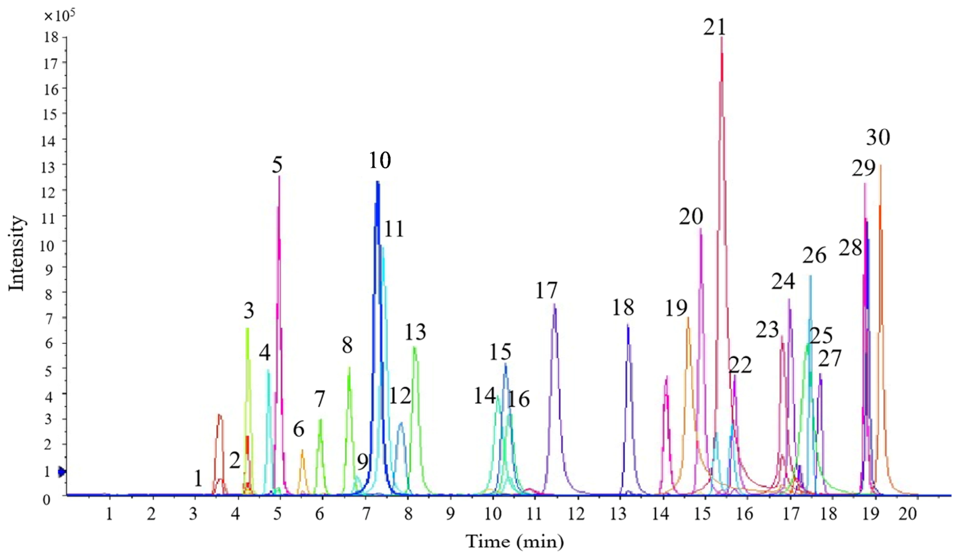
To characterize the flavonoid composition of T. antungense, a UPLC-ESI-MS/MS method was established using a mixture of flavonoid reference standards (Figure 1). Each compound exhibited a distinct extracted ion chromatogram (XIC) with sharp peak shapes and stable retention times, indicating good chromatographic resolution and system stability. Representative XIC chromatograms for flavonoids detected in the flowers, leaves, and roots of T. antungense are presented in the Supplementary Material S1 (Figures S1–S3). Representative extracted ion chromatograms (EICs) of flavonoid reference standards and analytes (Supplementary Material S2). In addition, calibration curves for all reference flavonoids were established, showing good linearity within the tested concentration ranges, with correlation coefficients (r2) greater than 0.995. The retention times, linear equations, linear ranges and limits of quantification (LOQs) are summarized in Table 1. UPLC-ESI-MS/MS was used to analyze the flavonoids in different organs of T. antungense, and a total of 16 flavonoids were identified. Twelve flavonoids were detected in the flowers: taxifolin, vitexin, cynaroside, rutin, isoquercitrin, diosmin, astragalin, daidzein, quercetin, naringenin, luteolin and apigenin. Eleven flavonoids were found in the leaves: cynaroside, rutin, isoquercitrin, diosmin, astragalin, daidzein, naringenin, luteolin, apigenin, formononetin and chrysin. Twelve flavonoids were quantified in the roots: isovitexin, cynaroside, rutin, isoquercitrin, diosmin, astragalin, quercetin, naringenin, luteolin, apigenin, chrysin and kaempferide. The quantitative contents of these flavonoids in each organ are listed (Table 2). Based on the specified pharmacokinetic screening criteria, nine candidate flavonoids were retained from the 16 flavonoids identified in the organ-specific profiles of T. antungense (Table 3). The PubChem compound IDs (CIDs) and complete SwissADME prediction results for all 16 identified flavonoids are provided in Supplementary Materials S3, S4 and S5, respectively.
Figure 1.
Extracted ion chromatograms (XICs) of 30 flavonoid reference standards analyzed by UPLC-ESI-MS/MS. The standard compounds corresponding to peaks 1–30 are listed in the order of elution as follows: Catechin, Dihydromyricetin, Puerarin, Daidzin, Glycitein, Taxifolin, Vitexin, Isovitexin, Cynaroside, Rutin, Isoquercitrin, Naringin, Genistin, Quercitrin, Diosmin, Astragalin, Liquiritigenin, Daidzein, Quercetin, Naringenin, Luteolin, Genistein, Kaempferol, Apigenin, Baicalin, Icariin, Formononetin, Biochanin A, Chrysin, Kaempferide.
Table 1.
Retention times, calibration equations, linear ranges, correlation coefficients (r2), and limits of quantification (LOQs) of flavonoid reference standards.
Table 2.
Flavonoid contents in different organs of T. antungense (mg/g).
Table 3.
Candidate flavonoids from T. antungense selected based on pharmacokinetic criteria.
2.2. Screening of Candidate Targets for Flavonoid Compounds from T. antungense
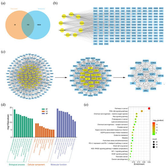
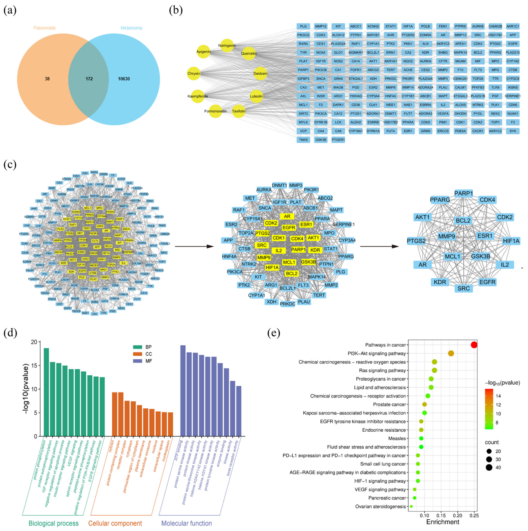
The SwissTargetPrediction, GeneCards, OMIM, and Therapeutic Target Database (TTD) databases were used to predict the potential targets of flavonoid compounds identified from T. antungense and to collect melanoma-associated targets. A total of 210 putative targets corresponding to the flavonoids were predicted, while 10,802 melanoma-associated targets were retrieved. Venn diagram analysis identified 172 overlapping targets between flavonoid-related targets and melanoma-associated targets (Figure 2a). These overlapping targets were considered potential anti-melanoma targets associated with flavonoid compounds from T. antungense. Subsequently, a flavonoid–melanoma target network was constructed using Cytoscape version 3.10.3 to visualize the interactions between the identified flavonoids and overlapping targets (Figure 2b). The complete target prediction lists for the identified flavonoids are provided in Supplementary Material S6.
Figure 2.
Network pharmacology analysis of potential targets of flavonoid-enriched extract from T. antungense against melanoma. (a) Venn diagram showing the overlapping targets between flavonoid-enriched extract from T. antungense. and melanoma-associated targets. (b) Compound-target network of candidate flavonoids and their associated targets. (c) Protein–protein interaction (PPI) network analysis and identification of predicted core targets. (d) GO enrichment analysis of the overlapping targets, including biological process (BP), cellular component (CC), and molecular function (MF). (e) KEGG pathway enrichment analysis presented as a bubble plot.
2.3. Network Analysis
In total, 172 potential targets were imported into the STRING database to construct a protein–protein interaction (PPI) network. After removing isolated nodes, the resulting PPI network consisted of 170 nodes and 1864 edges. Subsequently, network topology was analyzed using the CytoNCA plug-in in Cytoscape. The screening process was based on the distribution characteristics of betweenness centrality (BC), closeness centrality (CC), and degree centrality (DC). Specifically, the median values of BC, CC, and DC were calculated for the corresponding network at each screening round, and only nodes with values exceeding the respective medians for all three parameters were retained for further analysis. In the first round of screening, the thresholds were BC > 63.1890, CC > 0.4774, and DC > 18, yielding a subnetwork comprising 57 nodes and 756 edges. Based on this subnetwork, a second round of screening was performed using the median values of its topological parameters, with thresholds of BC > 189.8018, CC > 0.5265, and DC > 36. Finally, a core PPI network containing 17 core targets and 124 edges was obtained (Figure 2c). The 17 predicted core targets were serine/threonine-protein kinase AKT (AKT1), epidermal growth factor receptor erbB1 (EGFR), apoptosis regulator Bcl-2 (BCL2), estrogen receptor alpha (ESR1), tyrosine-protein kinase SRC (SRC), hypoxia-inducible factor 1-alpha (HIF1A), cyclooxygenase-2 (PTGS2), matrix metalloproteinase 9 (MMP9), peroxisome proliferator-activated receptor gamma (PPARG), glycogen synthase kinase-3 beta (GSK3B), poly [ADP-ribose] polymerase-1 (PARP1), vascular endothelial growth factor receptor 2 (KDR), myeloid cell leukemia-1 (MCL1), cyclin-dependent kinase 2 (CDK2), interleukin-2 (IL2), androgen receptor (AR), and cyclin-dependent kinase 4 (CDK4). These network-prioritized targets may be relevant to the potential anti-melanoma effects of the flavonoid-enriched extract from T. antungense (Table 4).
Table 4.
Characteristic parameters of target network node of main candidate compounds of flavonoid-enriched extract from T. antungense.
2.4. Enrichment Analysis
The 172 shared targets were submitted to DAVID for GO and KEGG enrichment analyses (p < 0.05). GO analysis yielded 406 biological process (BP), 68 cellular component (CC), and 184 molecular function (MF) terms. The enriched BP terms mainly included protein phosphorylation (GO:0006468), protein autophosphorylation (GO:0046777), insulin-like growth factor receptor signaling pathway (GO:0048009), and negative regulation of apoptotic process (GO:0043066). Enriched CC terms mainly included cytosol (GO:0005829), protein-containing complex (GO:0032991), and receptor complex (GO:0043235). Enriched MF terms were primarily related to ATP binding (GO:0005524), protein serine kinase activity (GO:0106310), protein kinase activity (GO:0004672), and protein serine/threonine kinase activity (GO:0004674) (Figure 2d). KEGG enrichment analysis mapped the 172 shared targets to 140 signaling pathways. Among them, pathways in cancer (hsa05200), the phosphoinositide 3-kinase-protein kinase B (PI3K-Akt) signaling pathway (hsa04151), chemical carcinogenesis-reactive oxygen species (hsa05208), EGFR tyrosine kinase inhibitor resistance (hsa01521), and endocrine resistance (hsa01522) were identified as significantly enriched pathways potentially associated with the biological functions of these targets. (Figure 2e), The complete KEGG pathway enrichment results are provided in Supplementary Material S7.
2.5. Molecular Docking Analysis of Candidate Flavonoids and Predicted Core Targets
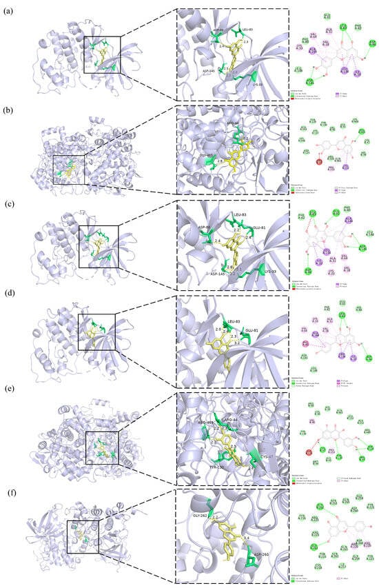
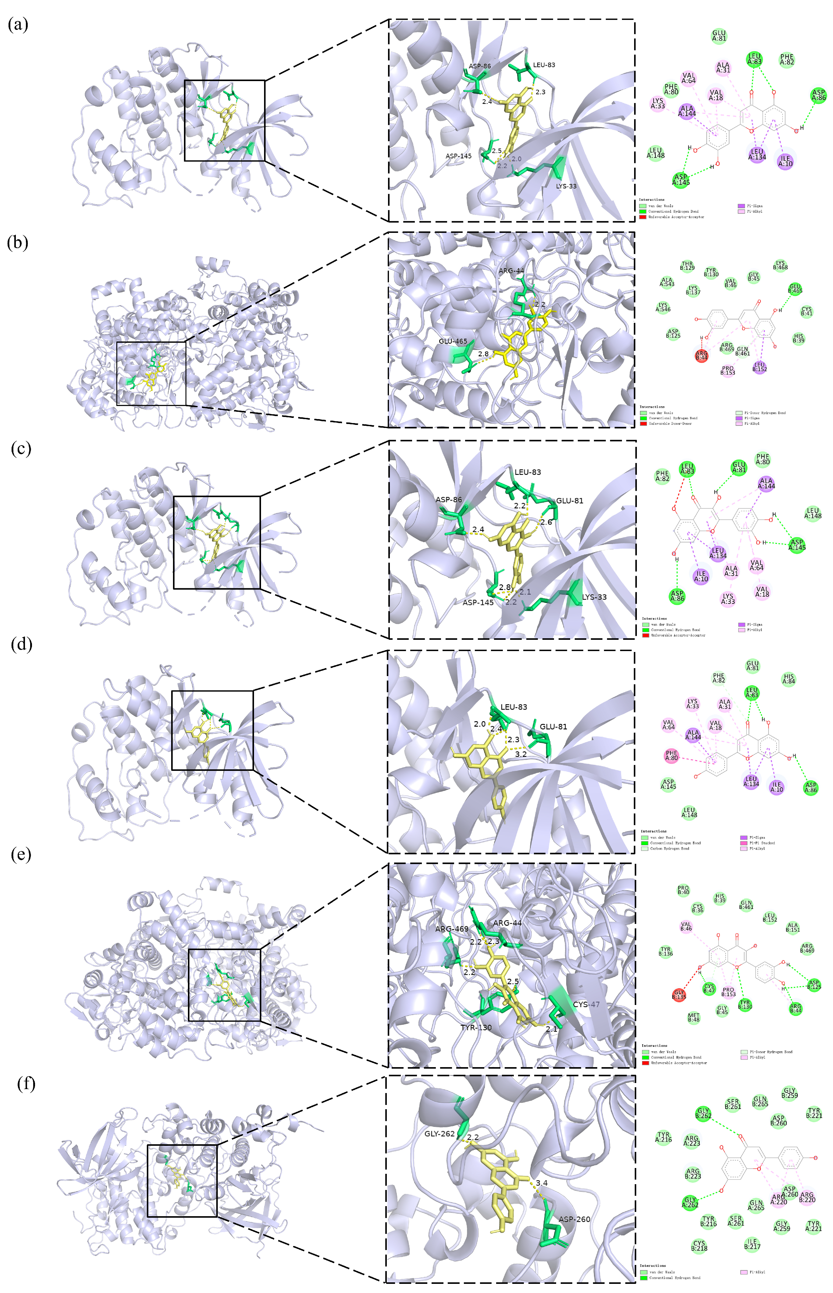
Among the nine candidate flavonoids retained after SwissADME screening, the five compounds with the highest degree values in the flavonoid–melanoma common-target network were chrysin (degree = 86), luteolin (degree = 83), quercetin (degree = 83), apigenin (degree = 82), and kaempferide (degree = 81). These compounds were selected as representative ligands for molecular docking against the 17 predicted core targets. The docking results indicated that all compounds exhibited favorable predicted binding affinities toward the target receptors (Table 5). The six pairs with the lowest calculated binding energies were selected for visualization. Molecular docking analysis revealed predicted hydrogen-bonding patterns between luteolin, quercetin, and apigenin and the target proteins CDK2, PTGS2, and GSK3B. Luteolin, which contains hydroxyl groups at the C5 and C7 positions of the A ring and at the C3′ and C4′ positions of the B ring, formed hydrogen bond interactions with key polar residues in the ATP-binding pocket of CDK2, including ASP-86, ASP-145 and GLU-81 (Figure 3a). In PTGS2, hydrogen bond interactions of luteolin were mainly mediated by its B-ring hydroxyl groups, forming hydrogen bonds with ARG-469 and GLU-465 (Figure 3b). Quercetin, possessing hydroxyl groups at the C5 and C7 positions of the A ring, the C3 position of the C ring, and the C3′ and C4′ positions of the B ring, exhibited the most extensive hydrogen bonding interactions. In the CDK2–quercetin complex, hydroxyl groups located on the A and C rings formed stable hydrogen bonds with ASP-86, ASP-145 and GLU-81, while B-ring hydroxyl groups contributed additional hydrogen bond interactions (Figure 3c). In PTGS2, quercetin formed multiple hydrogen bonds through its B-ring and C-ring hydroxyl groups with residues including ARG-469, ARG-44, CYS-47 and TYR-130 (Figure 3e). Apigenin, which contains hydroxyl groups at the C5 and C7 positions of the A ring and the C4′ position of the B ring, showed relatively fewer hydrogen bond interactions. In CDK2, hydrogen bonds were mainly mediated by A-ring hydroxyl groups and involved residues such as GLU-81 (Figure 3d), whereas in the GSK3B–apigenin complex, A-ring hydroxyl groups formed key hydrogen bonds with ASP-260 and GLY-262 located in the catalytic region of the protein (Figure 3f).
Table 5.
Predicted binding energies of candidate flavonoids with 17 predicted core targets.
Figure 3.
Molecular docking results of representative ligand–target complexes. (a) CDK2–luteolin; (b) PTGS2–luteolin; (c) CDK2–quercetin; (d) CDK2–apigenin; (e) PTGS2–quercetin; (f) GSK3B–apigenin. Note: Different colors and line styles represent different types of molecular interactions. The yellow sticks represent the docked ligand, the green sticks indicate selected amino acid residues, green dashed lines represent conventional hydrogen bonds, and pink dashed lines indicate π-alkyl interactions. The protein structure is shown as a light purple ribbon.
2.6. Flavonoid-Enriched Extract from T. antungense Reduces Melanoma Cell Viability and Induces Apoptosis In Vitro
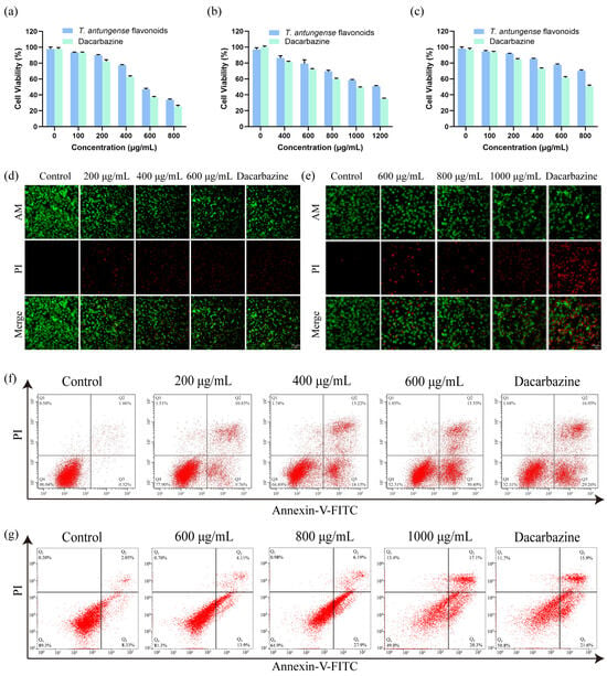
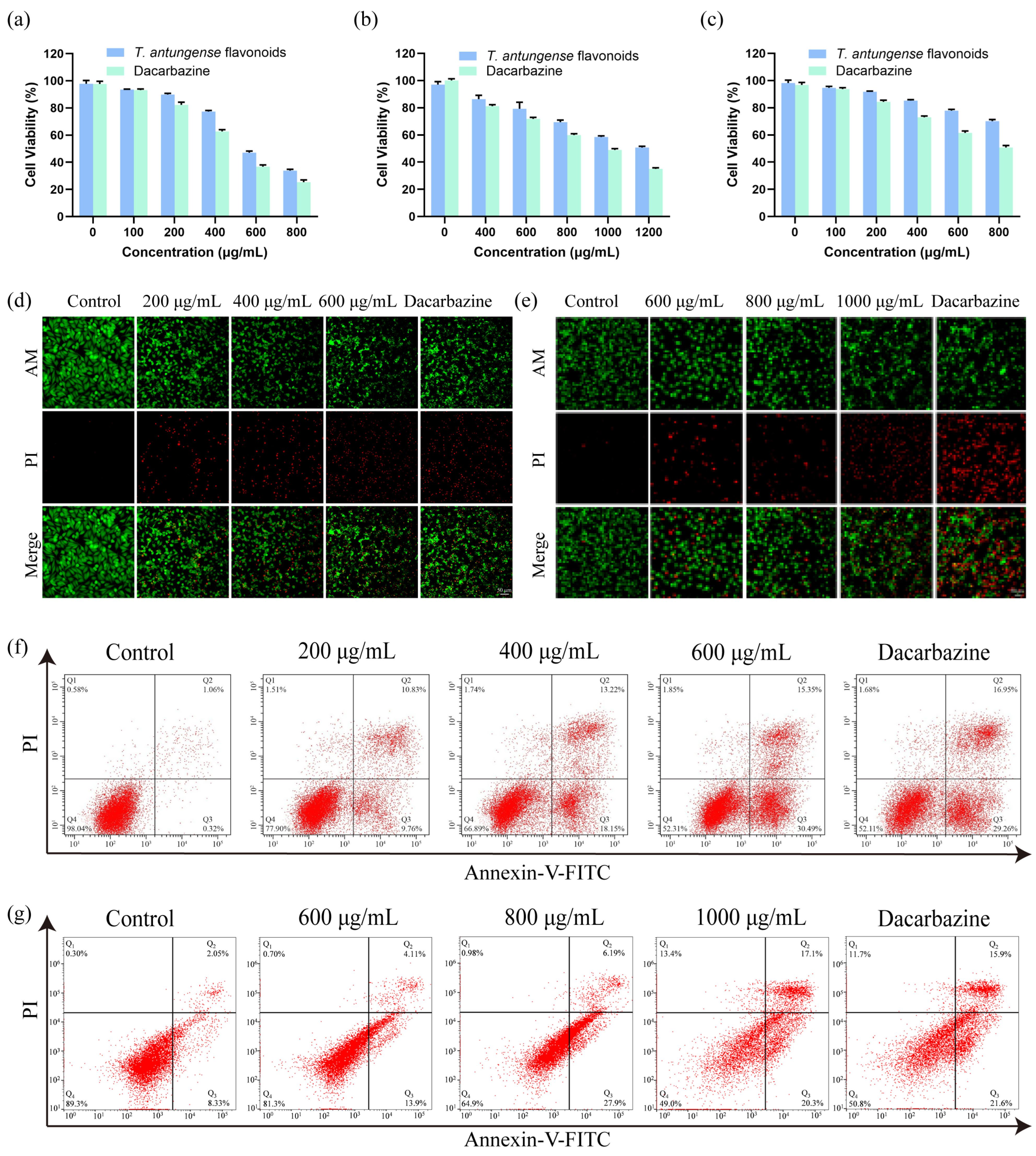
MTT assays showed that flavonoid-enriched extract from T. antungense and the positive control dacarbazine reduced A375 cell viability in a concentration-dependent manner (Figure 4a). The half-maximal inhibitory concentration (IC50) values of the flavonoid-enriched extract and dacarbazine against A375 cells were 600.5 μg/mL (95% CI: 569.1–634.9 μg/mL) and 474.9 μg/mL (95% CI: 452.2–498.3 μg/mL), respectively. Similarly, both treatments reduced B16F10 cell viability in a concentration-dependent manner (Figure 4b), with IC50 values of 1159 μg/mL (95% CI: 1112–1214 μg/mL) and 894.6 μg/mL (95% CI: 841.3–949.0 μg/mL) for the flavonoid-enriched extract and dacarbazine, respectively. In comparison, HaCaT cells exhibited lower sensitivity to the flavonoid-enriched extract than the melanoma cell lines under the tested conditions (Figure 4c).
Figure 4.
Flavonoid-enriched extract from T. antungense reduces melanoma cell viability, induces apoptosis. (a–c) Cell viability of A375, B16F10, and HaCaT cells treated with different concentrations of flavonoid-enriched extract from T. antungense or dacarbazine for 24 h, as determined by the MTT assay. (d,e) Representative fluorescence images of Calcein-AM/PI double staining in A375 and B16F10 cells after treatment with flavonoid-enriched extract. Green fluorescence indicates viable cells, whereas red fluorescence indicates dead cells. Scale bar = 50 μm. Live cells were stained green by Calcein-AM, while dead cells were stained red by PI. (f,g) Flow cytometric analysis of apoptosis in A375 and B16F10 cells using Annexin V-FITC/PI staining after treatment with different concentrations of flavonoid-enriched extract.
To further evaluate the effects of flavonoid-enriched extract from T. antungense on melanoma cell viability and death, Calcein-AM/PI double staining was performed in A375 and B16F10 cells. As shown in Figure 4d,e, control cells exhibited strong green fluorescence with minimal red fluorescence, indicating high cell viability. Following treatment with increasing concentrations of flavonoid-enriched extract, the intensity of green fluorescence gradually decreased, whereas PI-positive red fluorescence markedly increased in both A375 and B16F10 cells. Compared with the control group, cells treated with 600 μg/mL flavonoid-enriched extract showed decreased Calcein-AM-positive fluorescence and increased PI-positive fluorescence. Similar effects were observed in B16F10 cells treated with higher concentrations (1000 μg/mL). These results demonstrated that flavonoid-enriched extract from T. antungense induced melanoma cell death in a concentration-dependent manner.
To further investigate whether the reduced melanoma cell viability was accompanied by apoptosis, Annexin V-FITC/PI staining followed by flow cytometric analysis was performed in A375 and B16F10 cells. As shown in Figure 4f,g, control cells exhibited a low proportion of apoptotic cells, whereas treatment with increasing concentrations of flavonoid-enriched extract increased the percentage of apoptotic cells. In A375 cells, the proportion of apoptotic cells increased from 1.38% in the control group to 20.59%, 31.37%, and 45.84% after treatment with 200, 400, and 600 μg/mL flavonoid-enriched extract, respectively. Similarly, B16F10 cells treated with 600, 800, and 1000 μg/mL flavonoid-enriched extract showed a concentration-dependent increase in the proportion of apoptotic cells. Dacarbazine treatment also markedly increased apoptosis in both cell lines. These results demonstrated that flavonoid-enriched extract from T. antungense induced apoptosis in melanoma cells in a concentration-dependent manner.
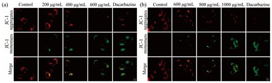
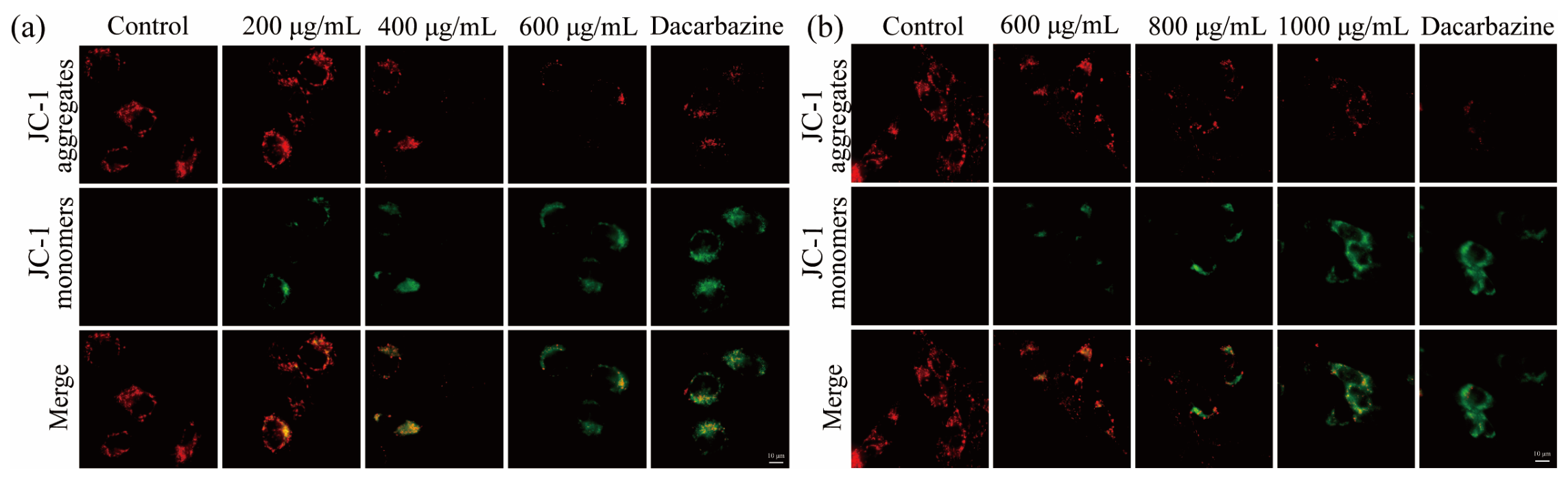
To evaluate mitochondrial functional changes associated with flavonoid-enriched extract-induced apoptosis, JC-1 staining was performed to assess mitochondrial membrane potential in A375 and B16F10 cells. As shown in Figure 5a,b, control cells exhibited strong red fluorescence derived from JC-1 aggregates and weak green fluorescence from JC-1 monomers, indicating normal mitochondrial membrane potential. After treatment with flavonoid-enriched extract, red fluorescence gradually decreased, whereas green fluorescence increased in a concentration-dependent manner. In A375 cells, treatment with 200, 400, and 600 μg/mL flavonoid-enriched extract resulted in a marked reduction in JC-1 aggregate fluorescence and an increase in JC-1 monomer fluorescence compared with the control group. Similar changes were observed in B16F10 cells following treatment with 600, 800, and 1000 μg/mL flavonoid-enriched extract. These results indicated that flavonoid-enriched extract from T. antungense caused a loss of mitochondrial membrane potential, suggesting that mitochondrial dysfunction was associated with the observed apoptotic response.
Figure 5.
Flavonoid-enriched extract from T. antungense disrupts mitochondrial membrane potential. (a,b) JC-1 staining analysis of mitochondrial membrane potential changes in A375 and B16F10 cells following flavonoid-enriched extract treatment. Red fluorescence represents JC-1 aggregates with intact mitochondrial membrane potential, whereas green fluorescence represents JC-1 monomers indicating mitochondrial membrane depolarization. Scale bar = 10 μm.
3. Discussion
Natural products and their derivatives have long served as important sources for anticancer drug discovery [21]. Medicinal plants contain structurally diverse bioactive constituents, including polyphenols, alkaloids, and terpenoids, many of which exhibit a broad range of pharmacological activities. Among these compounds, flavonoids have attracted considerable attention because of their potential antitumor properties [22]. Dandelion is a medicinal and edible herb that possesses notable antitumor properties [23,24]. Flavonoids are the key bioactive compounds responsible for its efficacy [25]. Flavonoids suppress multiple myeloma cell proliferation and induce apoptosis via the PI3K/AKT/mTOR pathway [26]. Dandelion root extract has also been reported to induce apoptosis in human melanoma cells while exhibiting relatively limited cytotoxicity toward noncancerous cells [17]. Nevertheless, the antitumor potential of flavonoid-enriched extract from T. antungense, particularly against melanoma, has not been systematically explored. In the present study, flavonoid-enriched extract from T. antungense reduced the viability of both A375 and B16F10 melanoma cells in a concentration-dependent manner. Compared with A375 and B16F10 cells, HaCaT cells exhibited lower sensitivity to the flavonoid-enriched extract under the tested conditions. However, this observation does not establish melanoma-selective cytotoxicity or demonstrate safety in normal skin, as HaCaT cells are immortalized keratinocytes rather than normal primary skin cells. Additional studies using primary skin cells are needed to assess the potential selectivity and safety of the extract. The relatively high concentrations required to inhibit melanoma cell viability also warrant caution when considering the extract’s therapeutic potential. Further studies are needed to identify the active constituents and evaluate the extract’s efficacy and safety in vivo.
UPLC-ESI-MS/MS analysis revealed distinct flavonoid composition in different organs of T. antungense. Luteolin and cynaroside were abundant in the flowers, leaves, and roots. Notably, some compounds showed tissue-specific distribution: kaempferide and isovitexin were detected exclusively in the roots, formononetin only in the leaves, and taxifolin and vitexin solely in the flowers. Previous studies have shown that these compounds possess anti-melanoma activity through diverse pathways. Kaempferide inhibits melanoma proliferation and invasion and induces ferroptosis by inactivating the Wnt/β-catenin pathway [27]. Formononetin reduces melanin synthesis, decreases tyrosinase activity, arrests B16F10 cells at G2/M and induces apoptosis [28]. Taxifolin promotes apoptosis in melanoma cells by modulating the PI3K/AKT signaling pathway [29]. Vitexin and isovitexin show cytotoxicity toward both human and mouse melanoma cells [30]. These findings suggest that multiple flavonoid constituents may contribute to the potential anti-melanoma activity of the extract. However, the individual contributions of these compounds were not directly assessed in the present study.
Network pharmacology analysis highlighted chrysin, luteolin, quercetin, apigenin, and kaempferide as candidate flavonoids of the flavonoid-enriched extract from T. antungense. Previous studies have reported that these individual flavonoids possess anti-melanoma activities in different experimental models. For example, chrysin inhibits tumor growth in murine B16F10 melanoma cells in a dose-dependent manner by inducing apoptosis and cell-cycle arrest at the G2/M phase [31]. Luteolin exhibits significant anti-proliferative, anti-adhesive, anti-migratory, and anti-invasive activities against melanoma cells in vitro [32]. Quercetin induces mitochondrial apoptosis in melanoma cells [33]. Apigenin exhibits antiproliferative and proapoptotic effects against A375 human melanoma cells [34]. Together with previous reports, our compositional and cellular findings suggest that multiple flavonoid constituents may collectively contribute to the growth-inhibitory and pro-apoptotic effects of flavonoid-enriched extract from T. antungense in melanoma cells.
Among the identified pathways, the KEGG “Pathways in cancer” pathway was closely associated with melanoma progression. Previous studies have demonstrated that melanoma development involves complex regulatory networks, including MAPK, PI3K/Akt, and apoptosis-related signaling pathways [35]. KEGG analysis revealed that 43 identified targets play roles in cancer-related pathways, including PTGS2, PPARG, ESR1, AR, IL2, etc. PTGS2 is frequently overexpressed in malignant melanoma and has been associated with patient survival [36]. PPARG ligands have also been reported to exert antiproliferative and apoptosis-inducing effects in human melanoma cells [37]. ESR1 and AR signaling have also been implicated in melanoma development and progression [38,39]. IL-2 is a crucial cytokine that plays a key role in the activation, proliferation and functional regulation of multiple immune cells [40]. IL-2 is an important regulator of antitumor immune responses; however, its potential contribution could not be evaluated in the present cell-based models. Collectively, these findings suggest that the predicted targets may be associated with various tumor-related processes. However, network pharmacology and KEGG enrichment analyses provide computational predictions based on target overlap and pathway annotation. Because neither the expression nor the functional activity of these targets was directly examined in the present study, their involvement in the observed anti-melanoma effects requires experimental verification.
The PI3K/AKT signaling pathway has been implicated in melanoma progression [41,42]. In the present study, KEGG enrichment analysis identified PI3K/AKT-related signaling among the predicted pathways, and molecular docking suggested potential interactions between selected flavonoids and candidate targets. These results provide hypotheses regarding the possible molecular basis of the extract’s activity but do not establish direct target binding or pathway regulation in cells. Separately, Annexin V-FITC/PI flow cytometry showed an increased proportion of apoptotic cells, while JC-1 staining indicated a loss of mitochondrial membrane potential in A375 and B16F10 cells following treatment. These experimental findings support the involvement of apoptosis and mitochondrial dysfunction in the cellular response to the extract under the tested conditions. However, whether PI3K/AKT signaling or the specific computationally predicted targets mediate these effects remains unknown and requires further protein-level and functional validation.
Previous studies have reported the anti-melanoma activity of dandelion root extract [17]. Although current clinical treatments, including dacarbazine-based chemotherapy, targeted therapy, and immunotherapy, have improved melanoma management, drug resistance and adverse effects remain major challenges [4]. We expanded on earlier work by characterizing and quantifying the flavonoid composition of the flowers, leaves, and roots of T. antungense and evaluating the in vitro anti-melanoma activity of its flavonoid-enriched extract in human A375 and murine B16F10 cells. Under the tested conditions, the extract reduced cell viability, increased apoptosis, and decreased mitochondrial membrane potential in both cell lines. By integrating phytochemical characterization, network pharmacology, molecular docking, and cellular assays, this study provides new experimental evidence supporting the potential anti-melanoma activity of the flavonoid-enriched extract from T. antungense and identifies candidate flavonoids, potential molecular targets, and associated signaling pathways for further investigation. Although these cellular findings are consistent with the apoptosis-related predictions from network pharmacology, the individual contributions of the identified flavonoids and the involvement of the predicted molecular targets and signaling pathways remain to be experimentally validated. Further in vivo studies are also needed to evaluate the extract’s antitumor efficacy and safety.
This study has several limitations. First, the biological experiments evaluated the flavonoid-enriched extract rather than its individual constituents. Therefore, the observed anti-melanoma effects cannot be attributed directly to the five flavonoids selected for molecular docking—chrysin, luteolin, quercetin, apigenin, and kaempferide. Further studies using isolated compounds are needed to clarify their individual contributions to the extract’s biological activity. Second, although network pharmacology and molecular docking predicted multiple potential targets and signaling pathways, including PI3K/AKT-related signaling, these molecular targets and pathways were not directly validated by protein-level or functional assays. The present cellular experiments mainly evaluated apoptosis and mitochondrial membrane potential changes, and further studies are required to verify the predicted molecular mechanisms. Finally, all efficacy data were obtained from in vitro cell-based experiments, and in vivo validation is still lacking. Further in vivo studies are required to evaluate the antitumor efficacy, safety, and biological relevance of the flavonoid-enriched extract.
4. Materials and Methods
4.1. Plant Materials
T. antungense samples were collected in May 2018 from the Northeast Wild Vegetable Resource Garden, Shenyang Agricultural University, Liaoning Province, China. The plant was taxonomically identified by Prof. Jiyun Li. A voucher specimen (No. IFP0244946) was deposited in the Herbarium of the Institute of Applied Ecology, Chinese Academy of Sciences (IFP), Shenyang, China. Healthy flowers, leaves, and roots were washed and dried, a portion was immediately frozen in liquid nitrogen and stored at −80 °C until analysis. The remaining samples were vacuum-dried at 45 °C to constant weight.
4.2. Preparation of Flavonoid-Enriched Extract from T. antungense
The crude flavonoid extract from the whole herb of T. antungense was prepared using ultrasound-assisted ethanol extraction [43]. Briefly, the dried whole plant was ground and passed through an 80-mesh sieve. The powdered material was extracted with 80% (v/v) ethanol at a solid-to-solvent ratio of 1:35 (g/mL) under ultrasonic treatment at 60 °C for 55 min. The crude extract was subsequently enriched and purified using D-101 macroporous adsorption resin [44]. Briefly, the sample solution (1.20 mg/mL, pH 3) was loaded onto the resin column at 2 bed volumes (BV) with a flow rate of 2.0 BV/h until adsorption saturation. The adsorbed compounds were eluted with 70% ethanol (4 BV) at a flow rate of 2 BV/h. The eluate was concentrated by rotary evaporation and vacuum-dried to obtain a flavonoid-enriched powder. The total flavonoid content of the obtained powder was determined to be 80.26%. The powder was sealed and stored for subsequent experiments. A total of 121.6 mg of flavonoid-enriched extract powder was accurately weighed, dissolved in dimethyl sulfoxide (DMSO), and further diluted with complete culture medium to prepare drug-containing media at the desired concentrations for subsequent cell viability assays.
4.3. UPLC-ESI-MS/MS Analysis of Flavonoid-Enriched Extract from T. antungense
Chromatographic separation was performed using an ACQUITY UPLC® BEH C18 column (2.1 × 100 mm, 1.7 μm; Waters, Milford, MA, USA). The mobile phase consisted of solvent A (0.1% formic acid in water) and solvent B (methanol). The injection volume was 5 μL, the column temperature was maintained at 40 °C, and the flow rate was set at 0.25 mL/min.
The gradient elution program was as follows: 0–1 min, 10% B; 1–3 min, 10–33% B; 3–10 min, 33% B; 10–15 min, 33–50% B; 15–20 min, 50–90% B; 20–21 min, 90% B; 21–22 min, 90–10% B; 22–25 min, 10% B.
The mass spectrometric (QE-HF-X, Thermo, Waltham, MA, USA) conditions were as follows: negative ion mode with an electrospray ionization (ESI) source. The ion spray temperature was set at 500 °C, and the ion spray voltage was set at −4500 V. Collision gas, curtain gas, nebulizer gas, and auxiliary gas were set at 6, 30, 50, and 50 psi, respectively. The scan mode was multiple reaction monitoring (MRM).
4.4. Prediction of Potential Targets of Flavonoid-Enriched Extract from T. antungense
The canonical SMILES of the chemical constituents of the flavonoid-enriched extract from T. antungense were obtained from the PubChem database (https://pubchem.ncbi.nlm.nih.gov/, accessed on 13 October 2025) and entered into SwissADME (https://www.swissadme.ch/, accessed on 13 October 2025) to generate the full output for each compound [45]. The compounds were screened based on two criteria: compliance with Lipinski’s rule of five (indicated by “Yes; 0 violation”) and high gastrointestinal (GI) absorption (indicated by “High”) [46]. Nine flavonoids meeting both criteria were selected as candidate flavonoids. Their potential targets were then predicted using the SwissTargetPrediction database (SwissTargetPrediction, accessed on 13 October 2025) [47]. The predicted targets were standardized using the UniProt database (https://www.uniprot.org/, accessed on 15 October 2025), with target names converted to official gene/protein symbols, and duplicate targets removed before further analysis.
4.5. Identification of Melanoma-Related Targets
Melanoma-related targets were retrieved from the GeneCards (https://www.genecards.org/, accessed on 21 October 2025), OMIM (https://www.omim.org/, accessed on 21 October 2025), and Therapeutic Target Database (TTD) databases (https://ttd.idrblab.cn/, accessed on 21 October 2025) using the keyword “Melanoma.” The obtained targets from different databases were integrated, standardized using the UniProt database by converting target names into official gene/protein symbols, and duplicate entries were removed. The resulting target set was used for subsequent intersection analysis with predicted flavonoid targets.
4.6. Construction of Flavonoid-Enriched Extract-Target Network
Venn analysis was performed with the online bioinformatics platform (http://www.bioinformatics.com.cn/, accessed on 21 October 2025) to identify the common targets between flavonoid-enriched extract from T. antungense and melanoma. These common targets are considered as potential targets for the treatment of melanoma with flavonoid-enriched extract from T. antungense, and the flavonoid-target network was constructed using Cytoscape 3.10.3 software (accessed on 1 November 2025).
4.7. Protein–Protein Interaction (PPI) Network Analysis
The overlapping targets were imported into the STRING database (https://string-db.org/, accessed on 21 October 2025) [48] to generate a protein–protein interaction (PPI) network, with the “hide disconnected nodes in the network” option enabled and all other parameters set to default. The PPI data were then imported into Cytoscape 3.10.3 for visualization. Finally, the topological parameters of the network were analyzed using the CytoNCA plug-in (version 2.1.6, accessed on 1 November 2025) to identify potential core targets. Betweenness centrality (BC), closeness centrality (CC), and degree centrality (DC) were selected as the main topological parameters for node screening. BC reflects the extent to which a node participates in the shortest paths connecting other nodes, CC indicates the relative proximity of a node to other nodes in the network, and DC represents the number of direct connections associated with a node.
4.8. GO and KEGG Pathway Enrichment Analysis
The overlapping targets were imported into the DAVID database (https://davidbioinformatics.nih.gov/, accessed on 21 October 2025) for Gene Ontology (GO) and Kyoto Encyclopedia of Genes and Genomes (KEGG) pathway enrichment analyses. The top 10 GO terms and top 20 KEGG pathways with p < 0.05 were selected and uploaded to the online bioinformatics platform for visualization.
4.9. Molecular Docking Analysis
Receptor protein structures were retrieved from UniProt and the RCSB PDB (https://www.rcsb.org/, accessed on 21 October 2025). Ligand structures were downloaded from PubChem, optimized in PyMOL (version 3.0.3, accessed on 21 October 2025), processed in Chem3D (version 22.0.0.22, accessed on 21 October 2025) for structural minimization, and saved in mol2 format. Molecular docking was conducted using AutoDock Vina (version 1.1.2, accessed on 29 October 2025) [49]. Blind docking was employed to explore potential binding sites across the target proteins without restricting the search to predefined binding pockets. For each receptor protein, the grid center coordinates and box dimensions were determined individually to encompass the intended search region. The docking scores were presented as predicted binding energies generated by AutoDock Vina. More negative values indicated stronger predicted interactions between ligands and target proteins. For each ligand–target pair, the lowest-energy docking pose was selected. The six most stable complexes were visualized using PyMOL, and ligand–receptor interactions were analyzed with Discovery Studio 2019 Client (version 19.1.0, accessed on 10 November 2025).
4.10. Cell Culture
HaCaT, A375, and B16F10 cells were purchased from BeNa Culture Collection (Beijing, China) and cultured in Dulbecco’s modified Eagle’s medium (DMEM, Invitrogen), supplemented with 1% penicillin–streptomycin and 10% fetal bovine serum (FBS). All cells were maintained at 37 °C in a humidified incubator atmosphere containing 5% CO2 and 95% air. The cells were routinely passaged to maintain them in the exponential growth phase.
4.11. Cell Viability Assay
HaCaT, A375, and B16F10 cells were separately seeded into 96-well plates and cultured for 24 h at 37 °C in a humidified atmosphere containing 5% CO2. Subsequently, the original culture medium was removed and replaced with fresh complete DMEM containing different concentrations of flavonoid-enriched extract from T. antungense or dacarbazine (positive control). The concentration ranges were adjusted according to the sensitivity of each cell line. HaCaT and A375 cells were treated with 0, 100, 200, 400, 600, and 800 μg/mL, while B16F10 cells were treated with 0, 400, 600, 800, 1000, and 1200 μg/mL. After a further 24 h of incubation, 10 μL of 3-(4,5-dimethylthiazol-2-yl)-2,5-diphenyltetrazolium bromide (MTT, 5 mg/mL) (G4101-200T, Servicebio, Wuhan, China) solution was added to each well and incubated for 4 h. The supernatant was discarded, and 150 μL of DMSO was added to each well to dissolve the formazan crystals. The plates were gently shaken at room temperature for 10 min, and the absorbance at 490 nm was measured using a microplate reader. The MTT assay was performed in three independent biological experiments (n = 3), with three technical replicates included in each experiment. Cell viability was calculated as follows:
In the above formula, ODsample refers to the absorbance value of the test well, ODblank refers to the absorbance value of the blank well, and ODcontrol refers to the absorbance value of the control well.
4.12. Live/Dead Staining
A375 and B16F10 cells were seeded into 20-mm glass-bottom dishes and cultured for 24 h at 37 °C in a humidified atmosphere containing 5% CO2. Based on the results of the cell viability assay, cells were divided into different treatment groups. A375 cells were treated with 200, 400, and 600 μg/mL flavonoid-enriched extract from T. antungense, while B16F10 cells were treated with 600, 800, and 1000 μg/mL flavonoid-enriched extract. Cells without treatment were used as the control group. Dacarbazine-treated cells were used as the positive control group, with A375 cells treated with 500 μg/mL dacarbazine and B16F10 cells treated with 1000 μg/mL dacarbazine. After incubation for 24 h, the culture medium was removed, and cells were washed twice with PBS. Subsequently, cells were stained with Calcein-AM/PI (G1707-100T, Servicebio, Wuhan, China) staining solution for 20 min in the dark. Cell viability and death were evaluated using an inverted fluorescence microscope (IX83, Olympus Corporation, Hachioji, Japan). Fluorescence images were acquired using the following parameters: Calcein-AM (green channel), excitation at 488 nm and emission collection at 493–556 nm; PI (red channel), excitation at 561 nm and emission collection at 593–725 nm. The Calcein-AM/PI Live/Dead staining assay was performed in one independent biological experiment (n = 1).
4.13. Apoptosis Assay by Flow Cytometry
A375 and B16F10 cells were seeded into 60-mm cell culture dishes and cultured for 24 h at 37 °C in a humidified atmosphere containing 5% CO2. Cells were divided into different treatment groups. A375 cells were treated with 200, 400, and 600 μg/mL flavonoid-enriched extract from T. antungense, while B16F10 cells were treated with 600, 800, and 1000 μg/mL flavonoid-enriched extract. Cells without treatment were used as the control group. Dacarbazine-treated cells were used as the positive control, with A375 cells treated with 500 μg/mL dacarbazine and B16F10 cells treated with 1000 μg/mL dacarbazine. After incubation for 24 h, cells were stained with an Annexin V-FITC/PI apoptosis detection kit (G1511-100T, Servicebio, Wuhan, China) according to the manufacturer’s instructions and subsequently analyzed using a BD flow cytometer (FACSCelesta™, BD Biosciences, San Jose, CA, USA). The Annexin V-FITC/PI apoptosis assay was performed in one independent biological experiment (n = 1).
4.14. JC-1 Staining
A375 and B16F10 cells were seeded into 20-mm glass-bottom dishes and cultured for 24 h at 37 °C in a humidified atmosphere containing 5% CO2. Cells were divided into different treatment groups. A375 cells were treated with 200, 400, and 600 μg/mL flavonoid-enriched extract from T. antungense, while B16F10 cells were treated with 600, 800, and 1000 μg/mL flavonoid-enriched extract. Cells without treatment were used as the control group. Dacarbazine-treated cells were used as the positive control, with A375 cells treated with 500 μg/mL dacarbazine and B16F10 cells treated with 1000 μg/mL dacarbazine. After incubation for 24 h, the culture medium was removed, and 1 mL of JC-1 staining working solution (C2003S, Beyotime, Shanghai, China) was added to each dish and incubated for 30 min. The staining solution was then removed, and the cells were washed twice with JC-1 staining buffer (C2003S-2, Beyotime, Shanghai, China). Subsequently, 1 mL of culture medium was added to each dish, and fluorescence images were captured using a fluorescence microscope (IX83, Olympus Corporation, Hachioji, Japan). Fluorescence imaging was performed using the following parameters: for the green channel (JC-1 monomers), excitation at 488 nm and emission collected at 500–550 nm; for the red channel (JC-1 aggregates), excitation at 561 nm and emission collected at 570–630 nm. The JC-1 mitochondrial membrane potential assay was performed in one independent biological experiment (n = 1).
4.15. Statistical Analysis
All MTT assay data were analyzed using SPSS (version 19.0) and GraphPad Prism (version 10.6.0). Data are expressed as the mean ± standard deviation (SD). Differences among groups were analyzed using one-way analysis of variance (ANOVA), followed by Duncan’s multiple range test. The IC50 values were determined by nonlinear regression analysis of concentration–response curves using a four-parameter logistic model with a variable slope in GraphPad Prism. The corresponding 95% confidence intervals (CIs) were calculated using the profile likelihood method. A p-value < 0.05 was considered statistically significant.
5. Conclusions
In summary, this study demonstrates that flavonoid-enriched extract from T. antungense exhibits anti-melanoma activity by reducing the viability of A375 and B16F10 melanoma cells and inducing apoptosis. Integrated network pharmacology and molecular docking analyses suggest that multiple potential targets and signaling pathways, including PI3K/AKT-related signaling, may be involved in the predicted mechanisms underlying the anti-melanoma effects of flavonoid-enriched extract. In vitro experiments further demonstrated that flavonoid-enriched extract treatment induced apoptosis and mitochondrial membrane potential loss in both A375 and B16F10 cells, indicating that the apoptotic response was accompanied by mitochondrial dysfunction. Collectively, these findings provide evidence for the anti-melanoma potential of flavonoid-enriched extract from T. antungense and provide a basis for further molecular and in vivo investigations.
Supplementary Materials
The following supporting information can be downloaded at https://www.mdpi.com/article/10.3390/ph19111712/s1. Supplementary Material S1: Representative XIC chromatograms for flavonoids detected in the flowers, leaves, and roots of T. antungense. Supplementary Material S2: Extracted ion chromatograms (EICs) of flavonoid reference standards and analytes; Supplementary Material S3: PubChem Compound IDs (CIDs) of the 16 identified flavonoids; Supplementary Material S4: Complete SwissADME prediction results for the 16 identified flavonoids; Supplementary Material S5: Chemical information, UPLC–ESI–MS/MS parameters, Calibration ranges, and Organ-Specific contents of the 16 identified flavonoids. Supplementary Material S6: Complete predicted target list of the nine candidate flavonoids; Supplementary Material S7: Complete results of KEGG pathway enrichment analysis.
Author Contributions
Conceptualization, L.L. and X.W.; methodology, L.L. and X.W.; software, L.L. and X.W.; validation, L.L. and X.W.; formal analysis, L.L. and X.W.; investigation, L.L. and X.W.; resources, L.L.; data curation, L.L. and X.W.; writing—original draft preparation, L.L., X.W.; writing—review and editing, L.L., X.W. and X.L.; visualization, L.L., X.W. and D.X.; supervision, L.L. and Y.Z.; project administration, L.L. and X.J.; funding acquisition, L.L. and X.J. All authors have read and agreed to the published version of the manuscript.
Funding
This research was funded by the National Natural Science Foundation of China (22578280, 22078201) and Basic Research Project of Liaoning Province (JYTMS20231499).
Institutional Review Board Statement
Not applicable.
Informed Consent Statement
Not applicable.
Data Availability Statement
The original contributions presented in this study are included in the article/Supplementary Materials. Further inquiries can be directed to the corresponding author.
Conflicts of Interest
The authors declare no conflicts of interest.
Abbreviations
The following abbreviations are used in this manuscript:
| AR | Androgen Receptor |
| BC | Betweenness Centrality |
| BV | Bed Volumes |
| CC | Closeness Centrality |
| CDK | Cyclin-Dependent Kinase |
| DMSO | Dimethyl Sulfoxide |
| DC | Degree Centrality |
| EGFR | Epidermal Growth Factor Receptor |
| GSK3B | Glycogen Synthase Kinase-3 Beta |
| GI | Gastrointestinal Absorption |
| GO | Gene Ontology |
| HIF1A | Hypoxia-Inducible Factor 1-Alpha |
| IC50 | Half-maximal inhibitory concentration |
| IL2 | Interleukin-2 |
| KEGG | Kyoto Encyclopedia of Genes and Genomes |
| MTT | 3-(4,5-Dimethylthiazol-2-yl)-2,5-Diphenyltetrazolium Bromide |
| MMP9 | Matrix Metalloproteinase 9 |
| MM | Malignant Melanoma |
| MCL1 | Myeloid Cell Leukemia-1 |
| PPARG | Peroxisome Proliferator-Activated Receptor Gamma |
| PARP1 | Poly [ADP-Ribose] Polymerase-1 |
| PI3K-AKT | Phosphoinositide 3-Kinase-Protein Kinase B |
| PPI | Protein–Protein Interaction |
| PBS | Phosphate-Buffered Saline |
| PI | Propidium Iodide |
| SRC | Proto-Oncogene Tyrosine-Protein Kinase SRC |
| T. antungense | Taraxacum antungense Kitag. |
| UPLC-ESI-MS/MS | Ultra-Performance Liquid Chromatography-Electrospray Ionization-Tandem Mass Spectrometry |
| VEGF | Vascular Endothelial Growth Factor |
References
- Bastos, M.; Saraiva, L.; Marques, A.C.; Amaral, M.H. Skin cancer chemoprevention: An overview. Drug Discov. Today 2025, 30, 104493. [Google Scholar] [CrossRef] [Scilit] [PubMed]
- Curti, B.D.; Faries, M.B. Recent Advances in the Treatment of Melanoma. N. Engl. J. Med. 2021, 384, 2229–2240. [Google Scholar] [CrossRef] [Scilit] [PubMed]
- Tasdogan, A.; Sullivan, R.J.; Katalinic, A.; Lebbe, C.; Whitaker, D.; Puig, S.; van de Poll-Franse, L.V.; Massi, D.; Schadendorf, D. Cutaneous melanoma. Nat. Rev. Dis. Primers 2025, 11, 23. [Google Scholar] [PubMed]
- Liang, C.; Wang, P.; Li, M.; Li, R.; Lai, K.P.; Chen, J. Anti-cancer mechanisms of natural isoflavones against melanoma. Heliyon 2024, 10, e28616. [Google Scholar] [CrossRef] [Scilit] [PubMed]
- Samieefar, S.; Arasteh, O.; Yeganeh Khorasanii, N.; Abedi, F.; Hosseinzadeh, H.R.; Hosseinzadeh, H. Natural compounds as immune checkpoint inhibitors in melanoma: A systematic review. Naunyn Schmiedebergs Arch. Pharmacol. 2026, 399, 11055–11071. [Google Scholar] [CrossRef] [Scilit] [PubMed]
- López-Lázaro, M. Distribution and biological activities of the flavonoid luteolin. Mini Rev. Med. Chem. 2009, 9, 31–59. [Google Scholar] [CrossRef] [Scilit] [PubMed]
- Shi, P.; Wang, L.; Qiu, X.; Yu, X.; Hayakawa, Y.; Han, N.; Yin, J. The flavonoids from the fruits of Psoralea corylifolia and their potential in inhibiting metastasis of human non-small cell lung cancers. Bioorg Chem. 2024, 150, 107604. [Google Scholar] [CrossRef] [Scilit] [PubMed]
- Tripathi, R.; Yadav, A.; Biharee, A.; Gupta, J.K.; Singh, B.; Pathak, P.; Otuechere, C.A.; Verma, A. Therapeutic potential and molecular mechanisms of natural flavonoids in liver cancer. Pharmacol. Res.-Nat. Prod. 2025, 8, 100288. [Google Scholar] [CrossRef] [Scilit]
- Thamrongwatwongsa, J.; Chusrisom, J.; Katemala, K.; Tantasirin, S.; Jumnongjit, P.; Nateerom, P.; Sonjaroon, W.; Tongkok, P.; Pichaiyotinkul, P.; Paemanee, A.; et al. Determination of flavonoid content in Grammatophyllum speciosum and in vitro evaluation of their anti-skin cancer and antibacterial activities. Heliyon 2024, 10, e33330. [Google Scholar] [CrossRef] [Scilit] [PubMed]
- Yao, X.; Jiang, W.; Yu, D.; Yan, Z. Luteolin inhibits proliferation and induces apoptosis of human melanoma cells in vivo and in vitro by suppressing MMP-2 and MMP-9 through the PI3K/AKT pathway. Food Funct. 2019, 10, 703–712. [Google Scholar] [CrossRef] [Scilit] [PubMed]
- Zheng, X.; Pan, Y.; Yang, G.; Liu, Y.; Zou, J.; Zhao, H.; Yin, G.; Wu, Y.; Li, X.; Wei, Z.; et al. Kaempferol impairs aerobic glycolysis against melanoma metastasis via inhibiting the mitochondrial binding of HK2 and VDAC1. Eur. J. Pharmacol. 2022, 931, 175226. [Google Scholar] [CrossRef] [Scilit] [PubMed]
- Kim, S.H.; Yoo, E.S.; Woo, J.S.; Han, S.H.; Lee, J.H.; Jung, S.H.; Kim, H.J.; Jung, J.Y. Antitumor and apoptotic effects of quercetin on human melanoma cells involving JNK/P38 MAPK signaling activation. Eur. J. Pharmacol. 2019, 860, 172568. [Google Scholar] [CrossRef] [Scilit] [PubMed]
- Fernando, P.; Zhen, A.X.; Piao, M.J.; Herath, H.; Kang, K.A.; Yoon, S.P.; Boo, H.J.; Hyun, C.L.; Hyun, J.W. Naringenin Induces Cellular Apoptosis in Melanoma Cells via Intracellular ROS Generation. Anticancer. Res. 2024, 44, 1079–1086. [Google Scholar] [CrossRef] [Scilit] [PubMed]
- Martinez, M.; Poirrier, P.; Chamy, R.; Prüfer, D.; Schulze-Gronover, C.; Jorquera, L.; Ruiz, G. Taraxacum officinale and related species-An ethnopharmacological review and its potential as a commercial medicinal plant. J. Ethnopharmacol. 2015, 169, 244–262. [Google Scholar] [CrossRef] [Scilit] [PubMed]
- Gao, L.; Jiang, X.; Zhang, R.; Deng, Z.; Huang, K.; Gu, K.; Lu, X.; Li, Y.; Shen, Y. Dandelion a lleviates Helicobacter pylori associated gastritis by inhibiting proteolytic activity of HtrA. J. Ethnopharmacol. 2026, 359, 121081. [Google Scholar] [CrossRef] [Scilit] [PubMed]
- Yan, Q.; Xing, Q.; Liu, Z.; Zou, Y.; Liu, X.; Xia, H. The phytochemical and pharmacological profile of dandelion. Biomed. Pharmacother. 2024, 179, 117334. [Google Scholar] [CrossRef] [Scilit] [PubMed]
- Chatterjee, S.J.; Ovadje, P.; Mousa, M.; Hamm, C.; Pandey, S. The efficacy of dandelion root extract in inducing apoptosis in drug-resistant human melanoma cells. Evid.-Based Complement. Altern. Med. 2011, 2011, 129045. [Google Scholar] [CrossRef] [Scilit] [PubMed]
- Kibble, M.; Saarinen, N.; Tang, J.; Wennerberg, K.; Mäkelä, S.; Aittokallio, T. Network pharmacology applications to map the unexplored target space and therapeutic potential of natural products. Nat. Prod. Rep. 2015, 32, 1249–1266. [Google Scholar] [CrossRef] [Scilit] [PubMed]
- Yang, H.Y.; Liu, M.L.; Luo, P.; Yao, X.S.; Zhou, H. Network pharmacology provides a systematic approach to understanding the treatment of ischemic heart diseases with traditional Chinese medicine. Phytomedicine 2022, 104, 154268. [Google Scholar] [CrossRef] [Scilit] [PubMed]
- Gupta, M.; Ahmad, J.; Ahamad, J.; Kundu, S.; Goel, A.; Mishra, A. Flavonoids as promising anticancer therapeutics: Contemporary research, nanoantioxidant potential, and future scope. Phytother. Res. 2023, 37, 5159–5192. [Google Scholar] [CrossRef] [Scilit] [PubMed]
- Chen, Y.; Tang, X.; Zou, Y.; Zhu, D.; Muthusamy, S.; Deshpande, M.; Kiruthiga, N.; Theivendren, P.; Rajalakshmi, K.; Wu, S.; et al. Flavonoids as promising natural therapeutics in ovarian cancer treatment: A comprehensive review. J. Steroid Biochem. Mol. Biol. 2026, 265, 107116. [Google Scholar] [CrossRef] [Scilit] [PubMed]
- Fontana, F.; Raimondi, M.; Di Domizio, A.; Moretti, R.M.; Montagnani Marelli, M.; Limonta, P. Unraveling the molecular mechanisms and the potential chemopreventive/therapeutic properties of natural compounds in melanoma. Semin. Cancer Biol. 2019, 59, 266–282. [Google Scholar] [CrossRef] [Scilit] [PubMed]
- Qu, J.; Ke, F.; Liu, Z.; Yang, X.; Li, X.; Xu, H.; Li, Q.; Bi, K. Uncovering the mechanisms of dandelion against triple-negative breast cancer using a combined network pharmacology, molecular pharmacology and metabolomics approach. Phytomedicine 2022, 99, 153986. [Google Scholar] [CrossRef] [Scilit] [PubMed]
- Rhayem, L.; Boussetta, N.; Kubo, M.T.K.; Merlier, F.; Vorobiev, E.; Lebovka, N. Bioactive Compounds from Dandelion (Taraxacum officinale): Advances in Extraction Techniques and Applications. Foods 2026, 15, 782. [Google Scholar] [CrossRef] [Scilit] [PubMed]
- Zhang, Y.; Hu, Y.F.; Li, W.; Xu, G.Y.; Wang, K.R.; Li, L.; Luo, H.; Zou, L.; Wu, J.S. Updates and advances on pharmacological properties of Taraxacum mongolicum Hand.-Mazz and its potential applications. Food Chem. 2022, 373, 131380. [Google Scholar] [CrossRef] [Scilit] [PubMed]
- Gui, H.; Fan, X. Anti-tumor effect of dandelion flavone on multiple myeloma cells and its mechanism. Discov. Oncol. 2024, 15, 215. [Google Scholar] [CrossRef] [Scilit] [PubMed]
- Xu, Y.; Zhao, R.; Huang, Z.; Zhang, Y.; Yang, R.; Zhang, J. Kaempferide Inhibits the Proliferation and Invasion and Induces Ferroptosis in Melanoma by Inactivating Wnt/β-Catenin Pathway. J. Biochem. Mol. Toxicol. 2025, 39, e70535. [Google Scholar] [CrossRef] [Scilit] [PubMed]
- Han, N.R.; Park, H.J.; Ko, S.G.; Moon, P.D. The Mixture of Natural Products SH003 Exerts Anti-Melanoma Effects through the Modulation of PD-L1 in B16F10 Cells. Nutrients 2023, 15, 2790. [Google Scholar] [CrossRef] [Scilit] [PubMed]
- Wang, Y.; Chen, S.J.; Ma, T.; Long, Q.; Chen, L.; Xu, K.X.; Cao, Y. Promotion of apoptosis in melanoma cells by taxifolin through the PI3K/AKT signaling pathway: Screening of natural products using WGCNA and CMAP platforms. Int. Immunopharmacol. 2024, 138, 112517. [Google Scholar] [CrossRef] [Scilit] [PubMed]
- Ganesan, K.; Xu, B. Molecular targets of vitexin and isovitexin in cancer therapy: A critical review. Ann. N. Y Acad. Sci. 2017, 1401, 102–113. [Google Scholar] [CrossRef] [Scilit] [PubMed]
- Sassi, A.; Maatouk, M.; El Gueder, D.; Bzéouich, I.M.; Abdelkefi-Ben Hatira, S.; Jemni-Yacoub, S.; Ghedira, K.; Chekir-Ghedira, L. Chrysin, a natural and biologically active flavonoid suppresses tumor growth of mouse B16F10 melanoma cells: In vitro and in vivo study. Chem. Biol. Interact. 2018, 283, 10–19. [Google Scholar] [CrossRef] [Scilit] [PubMed]
- Shi, M.L.; Chen, Y.F.; Wu, W.Q.; Lai, Y.; Jin, Q.; Qiu, W.L.; Yu, D.L.; Li, Y.Z.; Liao, H.F. Luteolin inhibits the proliferation, adhesion, migration and invasion of choroidal melanoma cells In Vitro. Exp. Eye Res. 2021, 210, 108643. [Google Scholar] [CrossRef] [Scilit] [PubMed]
- Seo, S.Y.; Ju, W.S.; Kim, K.; Kim, J.; Yu, J.O.; Ryu, J.S.; Kim, J.S.; Lee, H.A.; Koo, D.B.; Choo, Y.K. Quercetin Induces Mitochondrial Apoptosis and Downregulates Ganglioside GD3 Expression in Melanoma Cells. Int. J. Mol. Sci. 2024, 25, 5146. [Google Scholar] [CrossRef] [Scilit] [PubMed]
- Li, F.; Shi, Y.; Zhang, Y.; Yang, X.; Wang, Y.; Jiang, K.; Hua, C.; Wu, C.; Sun, C.; Qin, Y.; et al. Investigating the mechanism of Xian-ling-lian-xia-fang for inhibiting vasculogenic mimicry in triple negative breast cancer via blocking VEGF/MMPs pathway. Chin. Med. 2022, 17, 44. [Google Scholar] [CrossRef] [Scilit] [PubMed]
- Berthenet, K.; Castillo Ferrer, C.; Fanfone, D.; Popgeorgiev, N.; Neves, D.; Bertolino, P.; Gibert, B.; Hernandez-Vargas, H.; Ichim, G. Failed Apoptosis Enhances Melanoma Cancer Cell Aggressiveness. Cell Rep. 2020, 31, 107731. [Google Scholar] [CrossRef] [Scilit] [PubMed]
- Panza, E.; De Cicco, P.; Ercolano, G.; Armogida, C.; Scognamiglio, G.; Anniciello, A.M.; Botti, G.; Cirino, G.; Ianaro, A. Differential expression of cyclooxygenase-2 in metastatic melanoma affects progression free survival. Oncotarget 2016, 7, 57077–57085. [Google Scholar] [CrossRef] [Scilit] [PubMed]
- Freudlsperger, C.; Moll, I.; Schumacher, U.; Thies, A. Anti-proliferative effect of peroxisome proliferator-activated receptor gamma agonists on human malignant melanoma cells In Vitro. Anticancer. Drugs 2006, 17, 325–332. [Google Scholar] [CrossRef] [Scilit] [PubMed]
- Bechmann, N.; Calsina, B.; Richter, S.; Pietzsch, J. Therapeutic Potential of Nitric Oxide—Releasing Selective Estrogen Receptor Modulators in Malignant Melanoma. J. Investig. Dermatol. 2022, 142, 2217–2227. [Google Scholar] [CrossRef] [Scilit] [PubMed]
- Liu, Q.; Adhikari, E.; Lester, D.K.; Fang, B.; Johnson, J.O.; Tian, Y.; Mockabee-Macias, A.T.; Izumi, V.; Guzman, K.M.; White, M.G.; et al. Androgen drives melanoma invasiveness and metastatic spread by inducing tumorigenic fucosylation. Nat. Commun. 2024, 15, 1148. [Google Scholar] [CrossRef] [Scilit] [PubMed]
- Yang, L.T.; Zhou, H.; Li, S.; Huang, X.S.; Abudureheman, T.; Zhu, H.; Zhu, J.M.; Chen, J.; Duan, C.W.; Chen, K.M. Development of a Cell Membrane-Anchored Aptamer Sensor for IL-2 Detection In Situ. Anal. Chem. 2025, 97, 11685–11694. [Google Scholar] [CrossRef] [Scilit] [PubMed]
- Shen, Q.; Han, Y.; Wu, K.; He, Y.; Jiang, X.; Liu, P.; Xia, C.; Xiong, Q.; Liu, R.; Chen, Q.; et al. MrgprF acts as a tumor suppressor in cutaneous melanoma by restraining PI3K/Akt signaling. Signal Transduct. Target. Ther. 2022, 7, 147. [Google Scholar] [CrossRef] [Scilit] [PubMed]
- Long, J.; Pi, X. Polyphyllin I Promoted Melanoma Cells Autophagy and Apoptosis via PI3K/Akt/mTOR Signaling Pathway. Biomed. Res. Int. 2020, 2020, 5149417. [Google Scholar] [CrossRef] [Scilit] [PubMed]
- Chen, S.; Wang, X.; Cheng, N. Ultrasound-assisted ethanol extraction of Actinidia arguta pollen possesses antioxidant activity and protects DNA from oxidative damage. J. Food Biochem. 2021, 45, e13603. [Google Scholar] [CrossRef] [Scilit] [PubMed]
- Zhu, X.; Ru, Y.; Yan, K.; Zhang, S.; Sun, Y.; Du, F.; Liu, Y.; Niu, Y.; Xi, J.; Liu, K. Ultrasound-assisted and resin-based purification of bioactive polyphenols from peony (Paeonia ostii) pods: Process optimization and α-glucosidase inhibitory activity. Ultrason. Sonochem. 2026, 127, 107775. [Google Scholar] [CrossRef] [Scilit] [PubMed]
- Daina, A.; Michielin, O.; Zoete, V. SwissADME: A free web tool to evaluate pharmacokinetics, drug-likeness and medicinal chemistry friendliness of small molecules. Sci. Rep. 2017, 7, 42717. [Google Scholar] [CrossRef] [Scilit] [PubMed]
- Maitisha, G.; Zhou, J.; Zhao, Y.; Liu, G.; Zhao, Y.; Zheng, Y.; Li, L.; Han, S.; Peng, L.; Abliz, A. Exploring the pharmacological action mechanism of chamomile essential oil on the treatment of breast cancer based on network pharmacology. BMC Complement. Med. Ther. 2025, 25, 382. [Google Scholar] [CrossRef] [Scilit] [PubMed]
- Gfeller, D.; Grosdidier, A.; Wirth, M.; Daina, A.; Michielin, O.; Zoete, V. SwissTargetPrediction: A web server for target prediction of bioactive small molecules. Nucleic Acids Res. 2014, 42, W32–W38. [Google Scholar] [CrossRef] [Scilit] [PubMed]
- Huang, D.W.; Sherman, B.T.; Lempicki, R.A. Systematic and integrative analysis of large gene lists using DAVID bioinformatics resources. Nat. Protoc. 2009, 4, 44–57. [Google Scholar] [CrossRef] [Scilit] [PubMed]
- Eberhardt, J.; Santos-Martins, D.; Tillack, A.F.; Forli, S. AutoDock Vina 1.2.0: New Docking Methods, Expanded Force Field, and Python Bindings. J. Chem. Inf. Model. 2021, 61, 3891–3898. [Google Scholar] [CrossRef] [Scilit] [PubMed]

Disclaimer/Publisher’s Note: The statements, opinions and data contained in all publications are solely those of the individual author(s) and contributor(s) and not of MDPI and/or the editor(s). MDPI and/or the editor(s) disclaim responsibility for any injury to people or property resulting from any ideas, methods, instructions or products referred to in the content. |
© 2026 by the authors. Licensee MDPI, Basel, Switzerland. This article is an open access article distributed under the terms and conditions of the Creative Commons Attribution (CC BY) license.