


Abstract
Opioid peptide hybrids are multifunctional molecules in which an opioid pharmacophore is covalently integrated with a second opioid or non-opioid pharmacophore within a single molecular entity. Their development is driven by a major challenge in analgesic pharmacotherapy: although μ-opioid receptor (MOR) agonists remain among the most effective agents for the treatment of moderate-to-severe pain, their therapeutic use is limited by adverse effects and risks associated particularly with prolonged or high-dose exposure, including tolerance, constipation, respiratory depression, physical dependence, and abuse liability. Especially, chronic and neuropathic pain involves complex interactions among multiple receptor systems, neurotransmitter networks, and intracellular signaling pathways. Accordingly, hybrid peptide ligands provide a rational multitarget strategy aimed at achieving analgesia through the coordinated modulation of complementary pharmacological targets rather than selective modulation of a single receptor system. This approach may enhance analgesic efficacy and potentially improve the therapeutic profile of opioid-based treatments by reducing some of the dose-limiting adverse effects associated with conventional opioid analgesics. In this review, we discuss the functional and pharmacological interactions between opioid receptors and other receptor systems involved in pain perception and modulation, with emphasis on those for which peptide-based hybrid ligands with demonstrated antinociceptive activity have been developed.
1. Introduction
Opioid receptors are the primary targets for the most potent antinociceptive drugs available in medicine. They belong to the massive family of G protein-coupled receptors (GPCRs) and are distributed extensively throughout the central nervous system (particularly in the brainstem and spinal cord) and in peripheral tissues. There are three main types of opioid receptor: μ-, δ-, and κ-opioid receptors, MOR, DOR, and KOR, respectively. Activation of the MOR is responsible for strongest antinociceptive effect; however, its prolonged stimulation leads to receptor desensitization, development of tolerance, dependence, respiratory depression, constipation, opioid-induced hyperalgesia (OIH), and withdrawal due to compensatory cellular adaptations. Classical opioids such as morphine and fentanyl are strong MOR agonists and remain the most effective analgesics available, yet their clinical use is hampered by those serious side effects [1,2,3].
A key pharmacological insight from the past three decades indicates that these adverse effects may be dissociated from analgesia, as they stem from specific intracellular signaling consequences triggered by activation of the MOR (β-arrestin 2 recruitment, adenylyl cyclase superactivation upon chronic treatment, ERK/MAPK signaling, receptor internalization, and desensitization) and from cross-talk with other systems involved in pain perception including nociceptin/orphanin FQ (N/OFQ, NOP) receptor, substance P/neurokinin-1 (NK1), neurotensin, cholecystokinin (CCK), melanocortin and neuropeptide FF (NPFF). Opioid hybrids are specific types of compounds which fuse an opioid pharmacophore with a second pharmacophore engaging another opioid subtype, or a non-opioid modulatory receptor.
Therefore, the concept behind the design of opioid hybrids involves simultaneously targeting two receptors whose signaling pathways converge to produce stronger analgesic effect but diverge regarding the pathways responsible for adverse effects [4,5,6,7]. The term “hybrid” is used in several related ways. A bifunctional ligand contains two pharmacophores in one molecule and is intended to interact with two pharmacological targets. A bivalent ligand contains two recognition elements connected by a spacer and may bridge receptor protomers or receptor heteromers. A multifunctional compound may activate or block more than one receptor without necessarily being long enough to physically bridge receptors. A merged pharmacophor contains overlapping structural elements that contribute to activity at both targets. Therefore, hybrid ligands may simply co-activate two independent receptors, bias one receptor toward a signaling pathway, stabilize receptor heteromers, or alter receptor trafficking [8,9].
From a signaling perspective, opioid hybrids should not be understood simply as “stronger opioids.” Their biological profile depends on how the opioid component engages Gi/o proteins, β-arrestins, receptor phosphorylation, desensitization, internalization, ERK/MAPK, PI3K/AKT and ion channel regulation, and on how the second pharmacophore modifies pronociceptive or anti-opioid systems.
Here we focus on opioid hybrid compounds of peptide structure. Peptides are attractive models for drug design because their scaffolds offer a structural solution to the biggest problem in opioid pharmacology: separating potent pain relief from dangerous side effects. Unlike traditional small-molecule opioids (like morphine or fentanyl), which are rigid and tend to bind deeply within the pocket of a single receptor type, peptide hybrids are modular. Therefore, the peptide structures may be designed to simultaneously target different specific interactions across multiple biological systems [10,11].
The opioid peptide pharmacophore can be derived from endogenous ligands, like enkephalins, endomorphins, dermorphin, deltorphin, dynorphin-like motifs, biphalin-like structures, or synthetic opioid peptides with unnatural or D-amino acids incorporated in their structure. A second pharmacophore can then be attached through a flexible, rigid, or overlapping linker to modulate another receptor system involved in pain perception. This strategy aims to combine analgesic mechanisms, reduce effective opioid dose, block anti-opioid pronociceptive pathways, alter receptor trafficking, or create receptor-specific signaling profiles [6,9,12,13,14,15,16].
Understanding the intracellular mechanisms responsible for therapeutic effect and the side effects of molecules which influence pain perception is crucial for detailing these specific metrics and providing the “blueprint” for drug design. This review provides the rational framework to translate chemical structure into physiological response, with a focus on current knowledge about the intracellular signaling of these molecules.
2. Opioid Receptor Signaling
Opioid receptors (MOR, DOR, KOR, and the nociceptin/orphanin FQ receptor) are class A of the G protein-coupled receptor family. Their activation produces analgesia mainly through Gi/o-mediated inhibition of adenylyl cyclase, reduction of cAMP signaling, modulation of Ca2+ and K+ channels, reduced neurotransmitter release, and downstream regulation of kinases such as ERK/MAPK. In parallel, activated opioid receptors can be phosphorylated by GPCR kinases and recruit β-arrestins, which contribute to desensitization, internalization, recycling and additional signaling outputs [13,17].
The canonical opioid signaling pathway begins when an agonist stabilizes an active receptor conformation that couples to Gi/o proteins. The Gαi/o subunit inhibits adenylyl cyclase and lowers cAMP, while Gβγ subunits regulate ion channels, including inhibition of voltage-gated Ca2+ channels and activation of G protein-coupled inwardly rectifying K+ channels. These actions decrease neuronal excitability and neurotransmitter release in nociceptive pathways [17,18]. After receptor phosphorylation by GRKs, β-arrestins can uncouple receptors from G proteins, promote internalization, scaffold kinase pathways and regulate receptor recycling or degradation [19,20].
Nevertheless, β-arrestin signaling is often oversimplified as they also act as scaffolds and regulators of additional intracellular signaling pathways [21,22]. Early models proposed that opioid-induced analgesia is associated with activation of G protein-dependent mechanisms and the suppression of neuronal excitability, whereas β-arrestin signaling was proposed to contribute to opioid tolerance, dependence, and other adverse effects [23]. For example, administration of morphine to β-arrestin knockout mice produced enhanced and prolonged MOR-mediated antinociception and markedly reduced tolerance [24,25]. While both G protein and β-arrestin signaling pathways can be activated by “balanced” ligands that equally target both pathways (such as morphine), they can also be differentially activated by so-called biased ligands, which stabilize distinct receptor conformations and preferentially activate G protein signaling over β-arrestin recruitment, or vice versa.
TRV 130 (Oliceridine) is a non-peptide MOR agonist initially characterized as G protein-biased, with less gastrointestinal dysfunction and respiratory suppression, relative to morphine, in rodents [26]. It was approved for medical use in the United States in 2020. PZM21 and SR-17018 are other MOR ligands initially characterized as G protein-biased. Both produce prolonged antinociceptive effects in mice; however, subsequent studies demonstrated that PZM21 can induce tolerance and withdrawal, whereas the development of tolerance to SR-17018 appears to depend on the experimental pain model [27,28,29,30,31].
On the other hand, fentanyl, which is one of the most addictive drugs and became one of the leading causes of opioid overdose deaths, exhibits higher efficacy and stronger receptor phosphorylation and β-arrestin recruitment than morphine [32,33].
However, later studies have made this model more nuanced. Current reviews emphasize that many adverse effects may also be G protein-dependent and that the improved pharmacological profiles of some “G protein-biased” ligands may, at least partly, result from their low intrinsic efficacy rather than true pathway selectivity [14,34,35,36]. Those studies indicated that biased agonism alone is insufficient to completely dissociate analgesia from opioid-induced adverse effects. Opioid receptors can also activate kinase cascades, such as the ERK1/2, p38 MAPK, JNK, Src, PKC, and PI3K/AKT pathways, which are relevant not only to acute nociceptive modulation but also to longer-term neuronal plasticity, tolerance, opioid-induced hyperalgesia (OIH), and changes in gene expression [13,37,38,39,40].
This caution is especially important for peptide hybrids. Some compounds may show reduced β-arrestin recruitment in a cell line overexpressing the receptor but still produce receptor desensitization, ERK activation, tolerance, or respiratory effects in vivo through other mechanisms. Conversely, efficient receptor internalization and recycling may, in some contexts, preserve opioid responsiveness rather than promote tolerance development. Intracellular signaling profiles and possible interactions between opioid receptors and other types of GPCRs involved in pain perception are summarized in Figure 1.
Figure 1.
Possible interactions between opioid receptors and other types of GPCRs involved in pain perception.
3. NOP Receptor Signaling
The nociceptin/orphanin FQ peptide receptor (NOP) is the fourth member of the opioid receptor family, but a poor target for endogenous opioid peptides. N/OFQ itself has very low affinity at MOR/DOR/KOR, and the classical opioid peptides have correspondingly low affinity at NOP [41]. NOP couples canonically to PTX-sensitive Gi/o, reduces Ca2+ conductance, stimulates inwardly rectifying K+ currents, and engages kinase cascade, including PLC, PLA2, PKC and the MAPKs ERK1/2, p38 and JNK, with GRK-mediated receptor phosphorylation driving β-arrestin recruitment, internalization and arrestin-dependent signaling components [41].
NOP receptor activation has a clear modulatory role on MOR-mediated analgesia, tolerance, reward and motor function, and the two receptors are co-expressed and can be functionally and physically coupled throughout the central nervous system [41,42]. Studies described by Bird et al. [43] suggest that MOR and NOP are not pharmacologically independent at the cellular level. In an HEK293 dual-expression system, MOP ligands displaced NOP binding and vice versa, fluorescent dermorphin and N/OFQ probes colocalized by FRET in both HEK and hippocampal tissue, and NOP stimulation produced a rightward shift in the MOR concentration–response curve in both [35S]GTPγS binding and cAMP inhibition assays [43].
Single-particle tracking and lipid-raft immunocytochemistry show that morphine, but not buprenorphine, enhances MOR-NOP colocalization on lipid rafts and that morphine stimulates ERK1/2 phosphorylation similarly in MOR-only and MOR + NOP cells, whereas buprenorphine activates ERK1/2 only via the NOP receptor in the co-expression system [44].
In non-human primates, selective NOP agonists produce antinociception without the cardinal MOR liabilities (respiratory depression, itch, and reinforcement) and synergize with MOR-mediated analgesia.
The bifunctional MOR/NOP partial agonists AT-121, BU08028 and BU10038 were also tested in rhesus monkeys. All produced morphine-comparable analgesia without reinforcing effects or respiratory or cardiovascular depression at antinociceptive and even 10–30× higher doses. Interestingly, no measurable physical dependence or tolerance development was observed after repeated administration of tested compounds [45,46,47].
Opioid/NOP Peptide Hybrids
Several peptide-based strategies have been used to obtain ligands with combined classical opioid and NOP receptor pharmacology. Early bivalent chimeras linked MOP agonist sequences such as dermorphin or YRFB (Tyr-D-Arg-Phe-β-Ala-NH2) to the NOP-recognizing hexapeptide Ac-Arg-Tyr-Tyr-Arg-Ile-Lys-NH2. The obtained compounds showed affinity for both MOR and NOP receptors and evoked antinociceptive activity after intrathecal administration in rodents [48].
Peptide-based opioid agonist/NOP antagonist hybrids such as KGNOP-type, in which the synthetic opioid tetrapeptide H-Dmt-D-Arg-Aba-β-Ala-NH2 is combined with nociceptin or its analogs showed low-nanomolar receptor binding. In in vivo studies, the compounds alleviated allodynia and hyperalgesia in neuropathic pain models, and one hybrid (13a, H-Dmt-D-Arg-Aba-β-Ala-Arg-Tyr-Tyr-Arg-Ile-Lys-NH2) did not produce significant respiratory depression, compared with an equipotent analgesic dose of morphine [49].
DeNo is a bivalent dermorphin–N/OFQ peptide designed as a mixed MOR/NOP agonist, and in vitro it retained high affinity and agonist activity at both receptors, broadly reflecting the pharmacology of its parent peptides. Functionally, DeNo activated MOP- and NOP-mediated responses in recombinant systems, including G protein-related assays, but its in vivo spinal antinociceptive efficacy was weak, suggesting that simple covalent linkage of two active pharmacophores does not necessarily translate into improved analgesic activity [50].
De101 is a bivalent dermorphin–UFP-101 peptide designed as a mixed MOR agonist/NOP antagonist, in which the dermorphin moiety provides MOP agonism and the UFP-101 moiety provides high-affinity NOP antagonism. In binding and functional assays, De101 showed affinity for both MOP and NOP receptors and stimulated MOP-mediated G protein activation and inhibition of forskolin-induced cAMP formation, but was inactive as a NOP agonist and antagonized N/OFQ responses. Unlike the MOP/NOP agonist DeNo, De101 did not show the same loss of potency in MOP/NOP co-expression systems, suggesting that blocking rather than activating NOP may preserve MOP signaling in this experimental context [43].
The YGGF/RYYRIK-based hybrids BA55, BA61 and BA62 are other examples of opioid–nociceptin chimeric peptides. BA55 showed preferential KOP/NOP interaction and BA62 mainly DOP/KOP-related activity, with the weakest effect in mouse vas deferens. Unexpectedly, BA61, with lack of an N-terminal opioid “message” sequence, behaved as the most potent analog in the functional bioassay, with activity involving mainly DOP and NOP receptors. In [35S]GTPγS binding assays, BA55 and BA62 effectively stimulated G protein activation, while BA61 showed weaker/partial agonist-like activity, indicating that linker length, orientation and exposure of the YGGF and RYYRIK motifs strongly determine the pharmacological profile of these opioid/NOP hybrids [51].
The RP-170/RYYRIK chimeras KW-495 and KW-496 were designed by conjugating the cyclic MOP/KOP opioid peptide RP-170 with the NOP ligand fragment RYYRIK-NH2, either directly in KW-495 or through a Gly3 spacer in KW-496. KW-495 showed a mixed opioid/NOP profile and was selected for in vivo testing, where intrathecal administration produced antinociception in the mouse hot-plate test that was sensitive to both naloxone and the selective NOP antagonist SB-612111, indicating involvement of both classical opioid and NOP receptors. KW-496, in contrast, shifted mainly toward a dual KOP/MOP agonist profile, showing that insertion of the Gly3 spacer strongly altered receptor selectivity and functional activity [52]. The structures and activity profiles of opioid/NOP hybrid peptides are summarized in Table 1.
Table 1.
Opioid/NOP hybrid structures and activity profiles.
4. Neurokinin Receptor Signaling
Neurokinin receptors comprise three tachykinin GPCR subtypes, NK1, NK2 and NK3, which are seven-transmembrane receptors preferentially activated by substance P, neurokinin A and neurokinin B, respectively. They are widely distributed in the central and peripheral nervous systems as well as in gastrointestinal, respiratory, urogenital, vascular, immune, and inflammatory tissues, where they regulate nociceptive transmission, neurogenic inflammation, smooth-muscle contraction, vascular permeability, emesis, stress-related responses and neuroendocrine functions [57].
Substance P is the major tachykinin ligand of NK1 receptor. Its function is to facilitate nociceptive transmission, neurogenic inflammation and central sensitization [58,59].
4.1. Neurokinin and Opioid Receptor Cross-Talk
NK1 receptor activation engages Gq/11-dependent PLC/IP3/Ca2+/PKC signaling, but can also recruit Gs and β-arrestin-dependent pathways, leading to MAPK/ERK activation, receptor internalization and longer-term transcriptional effects [60].
In contrast, opioid receptors primarily couple to Gi/o proteins, inhibit adenylyl cyclase and cAMP formation, reduce voltage-gated calcium-channel activity, activate GIRK potassium channels and modulate kinase cascades such as ERK and AKT [61]. At the spinal level, opioid receptor activation counteracts tachykinin signaling by inhibiting stimulus-evoked substance P release from primary afferent terminals [62]. This presynaptic inhibition limits activation of postsynaptic NK1-positive dorsal horn neurons, thereby reducing amplification of nociceptive signals [58,62].
Anatomical studies show that MOR and NK1 receptors can be co-localized in pain-processing regions, creating a structural basis for receptor cross-talk [59]. Direct receptor-level interaction has also been reported, because MOR and NK1 receptors can form heterodimers that undergo co-internalization and display altered trafficking and resensitization properties [63].
Under sustained opioid receptor stimulation, peripheral nerve injury, or persistent inflammatory input, the physiological inhibitory influence of opioid signaling on nociceptive neurotransmission may be progressively counterbalanced by recruitment of pronociceptive tachykininergic mechanisms. In particular, increased substance P release and/or enhanced neurokinin-1 receptor (NK1R) expression and activation within the spinal dorsal horn have been implicated in central sensitization, reduced opioid analgesic efficacy, OIH, and the development of antinociceptive tolerance. These adaptations may function as part of an endogenous opioid-opposing, or “anti-opioid,” system, in which sustained opioid exposure paradoxically facilitates excitatory nociceptive transmission through substance P/NK1R-dependent mechanisms [58,64,65].
Interestingly, neuropathic injury may impair the ability of MOR activation to inhibit substance P release, which may partly explain the reduced opioid efficacy in some neuropathic pain states [64].
Therefore, NK1 activation often functionally opposes opioid analgesia in spinal pain circuits, whereas opioid receptor activation can suppress NK1-mediated transmission by reducing substance P release. Substance P/NK1 signaling in supraspinal regions may also influence stress, reward, affective pain processing and region-specific analgesic responses [58].
This relationship has encouraged the development of multifunctional ligands combining opioid receptor agonism with NK1 receptor antagonism, aiming to preserve analgesia while limiting substance P-driven pronociceptive plasticity [66].
4.2. Opioid/NK Peptide Hybrids
While traditional MOR agonists are the gold standard for treating severe acute pain, their efficacy may be reduced in chronic and neuropathic pain conditions [66]. This loss of efficacy is associated with neuroplastic changes in the central nervous system during prolonged pain states. Specifically, sustained nociceptive input or chronic opioid use can upregulate pronociceptive (pain-enhancing) systems, which may counteract the pain-relieving effects of opioids [67,68].
Because SP can oppose opioid analgesia and contributes to central sensitization (the hyper-excitability of pain pathways), it was hypothesized that hybrid compounds able simultaneously to activate opioid receptors while blocking the NK1 receptor could be highly potent antinociceptive agents [69]. One of representative examples is TY005, a peptidic opioid agonist/NK1R antagonist with the sequence Tyr-D-Ala-Gly-Phe-Met-Pro-Leu-Trp-O-3,5-Bn(CF3)2. TY005 produced dose-dependent antinociception in uninjured animals and reduced nerve injury-induced thermal and tactile hypersensitivity more effectively than morphine in a rodent neuropathic pain model [70]. TY005 did not produce detectable motor impairment in the rotarod test at antihyperalgesic doses or even at higher doses, and repeated spinal administration did not lead to antihyperalgesic tolerance [70]. These findings support the hypothesis that simultaneous opioid receptor activation and NK1R antagonism may improve analgesic efficacy in neuropathic pain while reducing the tendency toward tolerance development.
Further optimization of opioid/NK1R antagonist peptides focused on conformational restriction, metabolic stability and receptor selectivity. Yamamoto et al. introduced cyclic and topological constraints using penicillamine residues and identified a constrained bifunctional compound with potent opioid agonist activity, marked DOR selectivity and NK1R antagonist activity [71]. Guillemyn and co-workers developed conformationally constrained opioid agonist/NK1R antagonist peptidomimetics based on the opioid pharmacophore Dmt-D-Arg-Aba-Gly and an NK1 antagonist component. These compounds were designed to retain potent opioid activity while incorporating an antagonist element capable of blocking SP/NK1R-mediated pronociceptive signaling. Although some analogs displayed promising in vitro profiles, these studies also demonstrated a key challenge in hybrid design: the two pharmacophores must be arranged so that neither domain sterically or conformationally compromises the other. Linker length, rigidity, charge distribution and C-terminal substitution can markedly influence receptor affinity, functional efficacy and in vivo antinociceptive activity [68].
The potential value of opioid/NK1R antagonist hybrids appears especially under neuropathic pain conditions. Starnowska et al. evaluated bifunctional opioid agonist/NK1R antagonist hybrids in naïve mice and in mice subjected to chronic constriction injury (CCI), a model of neuropathic pain. Biochemical analysis confirmed elevated SP mRNA and protein levels in the lumbar spinal cord of CCI mice, consistent with enhanced pronociceptive tachykininergic tone. In naïve mice, the opioid agonist parent compounds produced acute antinociception more strongly than the hybrids. However, under neuropathic conditions, the opioid ligands showed poor efficacy. In contrast, the best hybrid produced robust and long-lasting alleviation of both tactile and thermal hypersensitivity [66]. This observation is pharmacologically significant because it suggests that NK1R antagonism becomes especially valuable when SP/NK1R signaling is pathologically upregulated.
In another type of opioid/neurokinin hybrid, Spantide and Spantide II were used as NK1R antagonist components derived from substance P. Spantide-like pharmacophores retain structural similarity to SP but contain D-amino acid substitutions and other modifications that convert the tachykinin sequence into an antagonist. This feature makes them attractive for hybridization with opioid peptides, because the C-terminal region of SP and its analogs contains key determinants of NK1R recognition. Wtorek et al. synthesized a series of opioid/neurokinin hybrids in which a cyclic endomorphin-2-derived opioid pharmacophore, Tyr-c[D-Lys-Phe-Phe-Asp]NH2, was combined either with SP fragments or with C-terminal fragments of Spantide II [12].
This cyclic opioid fragment displayed mixed MOR/KOR affinity, enhanced enzymatic stability and strong antinociceptive activity. In the Spantide II-containing hybrids, the C-terminal penta- or hexapeptide antagonist fragments were incorporated.
Among these compounds, the opioid agonist/NK1R antagonist hybrid Tyr-[D-Lys-Phe-Phe-Asp]-Asn-D-Trp-Phe-D-Trp-Leu-Nle-NH2 produced significant antinociception in the mouse writhing test. Notably, repeated administration did not induce detectable antinociceptive tolerance over the seven-day treatment, and the compound did not reduce stool mass, in contrast to the parent cyclic opioid peptide [12].
Although NK1R antagonism is the most intuitive approach for reducing pronociceptive SP signaling, several studies have shown that substance P-derived agonist pharmacophores can also be incorporated into opioid hybrids with favorable antinociceptive profiles. This apparently paradoxical strategy is based on the observation that SP/NK1R activation can exert different effects depending on dose, anatomical site, receptor population, and temporal context. While high or sustained SP/NK1R activity is generally pronociceptive, controlled coactivation of SP receptors with opioid receptors may maintain opioid responsiveness and reduce tolerance development [72,73].
The prototypical compound in this category is ESP7 (Tyr-Pro-Phe-Phe-Gly-Leu-Met-NH2), a substance P/opioid chimeric peptide containing overlapping N-terminal endomorphin-2 and C-terminal SP-derived pharmacophores [73]. Intrathecal administration of ESP7 produced opioid-dependent analgesia in rats, without loss of potency over repeated treatments. Interestingly, when ESP7 was administered together with an SP receptor antagonist, the analgesic effect progressively declined, consistent with tolerance development. Moreover, in morphine-tolerant animals, subsequent ESP7 administration restored a substantial analgesic [73]. These findings suggest that coincident activation of MOR and SP receptors may maintain opioid responsiveness rather than simply amplify acute analgesia.
A closely related chimera, ESP6, differs from ESP7 by a Gly-to-Pro substitution within the overlapping region. ESP6 alone showed reduced analgesic efficacy compared with ESP7, likely due to altered conformational flexibility and lower MOR potency. However, intrahecal co-administration of ESP6 and morphine resulted in prolonged morphine analgesia over a 5-day period. This effect was opioid receptor-dependent and was blocked by an opioid antagonist, naltrexone [74].
Moreover, concurrent NK1R blockade abolished the protective effect of ESP6 against morphine tolerance, supporting the idea that controlled activation of SP/NK1R-dependent mechanisms can participate in anti-tolerance opioid modulation [74].
Wtorek et al. [12] also compared the activities of an opioid/NK1R agonist within the same structural framework. The hybrid compound contained a C-terminal SP hexapeptide fragment, Tyr-[D-Lys-Phe-Phe-Asp]-Gln-Phe-Phe-Gly-Leu-Met-NH2, and was directly compared with a Spantide II-derived opioid/NK1R antagonist hybrid. The SP-fragment hybrid retained opioid receptor activity and behaved as an NK1R agonist in calcium mobilization assays. In in vivo studies, it produced significant antinociception in the writhing test, did not induce tolerance and did not cause constipation. Interestingly, no major differences in antinociceptive profile were observed between the NK1R agonist and antagonist hybrids bearing the same opioid fragment [12].
This finding suggests that both NK1R blockade and controlled NK1R activation can, under specific structural and pharmacological conditions, improve opioid-like antinociception. The structures and activity profiles of opioid/NK1 agonist/antagonist hybrid peptides are summarized in Table 2.
Table 2.
Opioid/NK1 hybrids containing NK1 agonist or substance-P-like pharmacophores.
5. Neurotensin Receptors Signaling
Neurotensin (NT) is an endogenous 13-amino-acid neuropeptide that modulates nociceptive processing predominantly through neurotensin receptor type 1 (NTS1/NTSR1) and type 2 (NTS2/NTSR2), both of which are G protein-coupled receptors. Neurotensin and its receptors are distributed at several levels of pain-processing pathways, including primary sensory structures, the spinal dorsal horn and supraspinal regions. NT-induced antinociception is largely naloxone-insensitive, indicating that neurotensin receptors can engage opioid-independent mechanisms of pain control [86,87].
5.1. NTS1 Signaling
NTS1 preferentially couples to Gq/11, resulting in phospholipase Cβ (PLCβ) activation and hydrolysis of phosphatidylinositol-4,5-bisphosphate (PIP2) into inositol-1,4,5-trisphosphate (IP3) and diacylglycerol (DAG). IP3 promotes Ca2+ release from intracellular stores, whereas DAG, together with Ca2+, activates conventional protein kinase C (PKC) isoforms. These pathways can influence neuronal excitability, neurotransmitter release, and synaptic transmission and therefore provide potential mechanisms through which NTS1 may modulate nociceptive processing.
However, NTS1 signaling is not restricted to Gq/11. In recombinant human NTS1-expressing cells, neurotensin also activates Gαi1, GαoA and Gα13, modulates cAMP signaling and recruits β-arrestin 1 and β-arrestin 2 [88].
ERK1/2 constitutes an important point of convergence of these pathways. In CHO-K1 cells expressing human NTS1, full ERK1/2 activation required both a pertussis toxin-sensitive Gi/o-c-Src pathway and a Gq-PLCβ-DAG-PKC-Raf-1 pathway [88]. Thus, NTS1-induced ERK1/2 phosphorylation results from integrated signaling rather than from a simple linear consequence of Gq activation. ERK1/2 signaling may be involved in regulation of neuronal excitability, synaptic plasticity and activity-dependent transcription. Interestingly, persistent spinal ERK activation is frequently associated with sensitization, whereas receptor-restricted and temporally restricted signaling may have different functional consequences. NTS1-dependent ERK1/2 activation should therefore not be regarded as intrinsically pro- or antinociceptive [89].
Following activation, NTS1 can be phosphorylated by GPCR kinases and recruit β-arrestins, promoting receptor desensitization and internalization. The β-arrestins can additionally act as signaling scaffolds, demonstrating that this pathway is not limited to termination of G protein signaling [90,91]. The importance of β-arrestin-dependent NTS1 signaling for pain modulation has recently been demonstrated using SBI-810, an arrestin-biased NTS1 allosteric modulator. SBI-810 produced antinociception in postoperative, inflammatory and neuropathic pain models through mechanisms requiring NTS1 and β-arrestin 2. At the cellular level, it reduced excitatory synaptic transmission and NMDA receptor/ERK signaling in spinal nociceptive neurons, decreased Nav1.7 surface expression and action-potential firing in primary sensory neurons, and attenuated C-fiber responses [92].
These findings support a role for β-arrestin 2-biased NTS1 signaling in reducing peripheral nociceptor excitability and central nociceptive transmission in the investigated experimental models.
5.2. NTS2 Signaling
NTS2 was initially characterized as a low-affinity neurotensin-binding receptor but is now recognized as a signaling-competent GPCR involved in pain modulation. Its intracellular signaling is less comprehensively defined than that of NTS1 and should not be assumed to reproduce the canonical NTS1 Gq/PLC/Ca2+ pathway. In CHO cells expressing rat NTS2, neurotensin and NTS2-active ligands induced receptor internalization and ERK1/2 phosphorylation. Inhibition of receptor endocytosis prevented ERK1/2 activation, indicating that NTS2-mediated ERK signaling is dependent on receptor internalization in this experimental system [93]. In the same experimental system, NTS2 stimulation did not elicit a characteristic receptor-dependent Ca2+ mobilization response. These findings distinguish NTS2 signaling from the prominent PLC/Ca2+ pathway associated with NTS1.
A direct causal link between NTS2-induced ERK1/2 activation and antinociception has not yet been established. Nevertheless, in vivo studies provide strong evidence for the involvement of NTS2 in spinal pain modulation. NTS2 is present in dorsal root ganglia and superficial dorsal horn regions associated with nociceptive processing, and intrathecal administration of NTS2-preferring ligands such as JMV-431 and levocabastine produces antinociceptive effects [94,95].
In the formalin model, these compounds reduced nociceptive behavior and decreased pain-evoked c-Fos expression in superficial dorsal horn neurons, consistent with reduced activation of spinal nociceptive circuits [95]. Several NTS2-selective ligands have additionally produced antinociception without the prominent hypothermia and hypotension often associated with NTS1 activation in preclinical studies, making NTS2 an attractive target for analgesic drug development [96].
Overall, NTS1 and NTS2 appear to modulate pain through partially distinct intracellular mechanisms. NTS1 engages a complex signaling network involving Gq/PLCβ/Ca2+/PKC, additional G protein pathways, ERK1/2 and β-arrestins. Recent evidence supports a role for β-arrestin 2-biased NTS1 signaling in reducing Nav1.7-dependent nociceptor excitability and reduced spinal NMDA/ERK-mediated transmission. In contrast, NTS2 signaling remains less well-characterized but includes internalization-dependent ERK1/2 activation and is associated in vivo with suppression of spinal nociceptive neuronal activity. Further studies are required to determine which intracellular NTS2 pathways are directly responsible for its antinociceptive effects.
5.3. Neurotensin Receptor Signaling and Opioid-Independent Analgesia
NT and NT analogs produce analgesic responses that are not abolished by naloxone in several experimental paradigms, indicating that NTSs can directly modulate pain transmission without the activation of opioid receptors [89,95].
NT(8–13) analogs such as JMV2009 and JMV5296 support this concept. JMV2009 activates NTS1 and NTS2, induces sustained p42/p44 MAPK activation and receptor internalization in vitro, and produces antinociceptive activity in acute, tonic, visceral, inflammatory and neuropathic pain models without inducing constipation, tolerance or hypothermia [89]. An NTS2-preferring analog, JMV5296, also produces similar long-lasting antinociception without hypothermia [97].
Interestingly, simultaneous activation of opioid receptors and NTSRs has been shown to enhance antinociception under inflammatory conditions [98]. The neurotensin and opioid systems are functionally interconnected at several levels. Both systems modulate spinal nociceptive input and supraspinal descending pain control. At the cellular level, opioid receptors mainly suppress excitability through Gi/o-dependent inhibition of cAMP/PKA signaling and regulation of ion channels, whereas NTS1 and NTS2 can engage multiple intracellular pathways, including Gq-, Gi/o-, β-arrestin- and ERK-related pathways. As these mechanisms may be complementary, simultaneous activation of opioid and neurotensin receptors could produce additive or potentially synergistic antinociception, possibly reducing the level of opioid receptor activation or the opioid dose required for pain relief.
Experimental data showed that co-administration of morphine with a brain-penetrant NT(8–13) analog improved the analgesic/adverse-effect ratio; at equianalgesic doses, the combination reduced morphine-associated inhibition of gastrointestinal transit compared with morphine alone [98]. Direct receptor-level crosstalk may also occur. Heterodimerization between KOR and NTS1 has been reported to generate a β-arrestin 2-biased signaling pathway [13]. Although the full relevance of this mechanism to analgesia remains to be clarified, it illustrates that opioid and neurotensin receptors can not only interact at the circuit level but may also may influence receptor trafficking, desensitization, MAPK activation and ligand-specific signaling bias.
5.4. Opioid–Neurotensin Hybrid Ligands and Intracellular Complementarity
The development of opioid–neurotensin hybrid ligands is based on the same design rationale. These compounds combine opioid pharmacophores with NT-like fragments to engage opioid and neurotensin receptors within a single molecule. PK23 is an example of an opioid–neurotensin hybrid peptide; it produces potent spinal antinociception and shows a more favorable behavioral profile than intrathecally administered morphine, including delayed tolerance development [99].
BNT12, a more recent hybrid peptide containing opioid and NT-like fragments, produces central antinociception in acute, neuropathic, inflammatory, visceral and formalin pain models. Its effects are mediated by MOR and DOR as well as NTS1 and NTS2, and it shows reduced or absent opioid-like side effects in assays of conditioned place preference, withdrawal, locomotor activation, gastrointestinal transit and cardiovascular responses [100].
Opioid–neurotensin hybrids may be useful tools in the development of compounds that can produce analgesia without NTS-mediated hypothermia, hypotension or motor effects. From a signaling perspective, these hybrid ligands may be advantageous because they distribute analgesic efficacy across multiple intracellular systems. The opioid component engages Gi/o-mediated inhibition of cAMP/PKA signaling, Ca2+ influx and neuronal excitability, whereas the neurotensin component can recruit NTS1/NTS2-dependent PLC/PKC, ERK1/2, β-arrestin and receptor-internalization-dependent mechanisms. Such multitarget signaling could contribute to potent antinociception without requiring maximal MOR activation and may potentially be associated with reduced opioid-like adverse effects, although the precise intracellular mechanisms underlying these pharmacological outcomes remain to be established. The structures and activity profiles of hybrid peptides targeting opioid–neurotensin receptors are summarized in Table 3.
Table 3.
Hybrid peptides targeting opioid–neurotensin receptors.
6. NPFF Receptor Signaling
Neuropeptide FF (NPFF) is an octapeptide (Phe-Leu-Phe-Gln-Pro-Gln-Arg-Phe-NH2) which belongs to the RF-amide family of mammalian neuropeptides. Initially isolated from bovine brain tissue, NPFF has become widely recognized as a critical endogenous modulator of nociceptive signaling and opioid homeostasis [106]. The NPFF system consists mainly of two class A G protein-coupled receptors, NPFF receptor 1 (NPFFR1; formerly GPR147) and NPFF receptor 2 (NPFFR2; formerly GPR74), which are activated by NPFF-related RF-amide peptides, including NPFF, neuropeptide AF (NPAF), neuropeptide SF (NPSF/RFRP-1), and RF-amide-related peptide-3 (RFRP-3/NPVF). Both receptors are expressed in central nervous system regions involved in nociceptive processing, including spinal and supraspinal pain-modulatory circuits.
The best-established intracellular mechanism downstream of NPFF receptors is Gi/o-mediated inhibition of adenylyl cyclase. By reducing cAMP production, NPFF receptor activation may decrease PKA-mediated phosphorylation of ion channels and synaptic proteins, thereby altering neuronal excitability and neurotransmitter release. In pain pathways, cAMP/PKA signaling contributes to peripheral and central sensitization by facilitating voltage-gated Ca2+ channels, sodium channels, glutamatergic transmission, and transcriptional regulators such as CREB. Therefore, NPFF receptor-dependent suppression of cAMP/PKA signaling could theoretically exert antinociceptive effects by reducing sensitization-related kinase activity.
However, this interpretation is complicated by the anti-opioid role of the NPFF system. Opioid receptors, particularly MOR, are also Gi/o-coupled and produce analgesia through inhibition of adenylyl cyclase, suppression of presynaptic Ca2+ influx, and activation of K+ conductance. NPFF receptor activation can counteract opioid receptor function despite partial convergence on Gi/o-associated pathways. This suggests that NPFF does not simply mimic opioid receptor signaling; instead, it can reorganize opioid receptor coupling, receptor trafficking, or effector access in a cell-specific manner [107,108].
6.1. Regulation of Ca2+ Signaling and Voltage-Gated Calcium Channels
NPFF receptor signaling also intersects with Ca2+-dependent mechanisms. In neuronal models, NPFF receptor activation can modulate voltage-gated N-type Ca2+ channels, which are essential for neurotransmitter release from primary afferent terminals and central nociceptive synapses. Opioid agonists typically inhibit N-type Ca2+ channels through Gi/o βγ-subunit signaling, thereby reducing release of glutamate, substance P, and CGRP. NPFF receptor activation may antagonize this opioid-mediated inhibition of Ca2+ conductance, providing a plausible cellular basis for anti-opioid activity [107,109].
In SH-SY5Y neuroblastoma-derived models expressing NPFF receptors, NPFF receptor activation attenuated opioid-induced inhibition of N-type Ca2+ channels and modified opioid receptor coupling to downstream effectors [110,111,112].
Presynaptic Ca2+ influx is a central mechanism involved in regulation of nociceptive synaptic transmission, because activation of voltage-gated Ca2+ channels, particularly CaV2.2/N-type channels located on primary afferent terminals of the spinal dorsal horn, couples the action of potential invasion to vesicular release of pronociceptive transmitters, including glutamate, substance P and CGRP [113].
Consequently, modulation of presynaptic Ca2+ channel activity can markedly influence the strength of excitatory nociceptive input. This mechanism is directly relevant to opioid analgesia, since MOR activation inhibits N-type Ca2+ currents in sensory and neuronal models, thereby reducing Ca2+-dependent transmitter release [61,114].
The NPFF system appears to intersect with this process at the level of Ca2+ channel regulation: NPFF receptor activation can modulate voltage-gated N-type Ca2+ channels, and NPFF/NPFF analogs have been shown to counteract opioid-induced inhibition of N-type Ca2+ channels in rat spinal ganglion neurons and in NPFF receptor-transfected SH-SY5Y cells [115,116].
Thus, depending on receptor subtype, neuronal population and pain state, NPFF receptor activation may either reduce Ca2+-dependent excitatory transmission and contribute to antinociception, or attenuate opioid-mediated inhibition of Ca2+ influx, thereby potentially facilitating excitatory neurotransmission and reducing opioid analgesic efficacy. This bidirectional regulation of presynaptic Ca2+ signaling provides a plausible cellular explanation for the dual pharmacological profile of NPFF-related peptides, which have been associated with both antinociceptive and anti-opioid/pro-nociceptive effects.
6.2. MAPK/ERK Signaling and Receptor Regulation
MAPK/ERK signaling represents another important intracellular pathway linked to NPFF and opioid modulation. In opioid receptor-expressing cells, NPFF analogs can modify opioid-induced ERK phosphorylation, indicating that NPFF-related peptides influence not only second-messenger production but also kinase-dependent signaling dynamics [108]. ERK1/2 activation is highly relevant in nociceptive biology because it regulates synaptic plasticity, transcriptional responses, and long-term changes in dorsal horn excitability. Studies on NPFFR2 have identified agonist-induced receptor phosphorylation sites involved in receptor desensitization and/or internalization, suggesting that kinase-dependent receptor regulation may shape the duration and intensity of NPFF signaling [117,118].
Therefore, it can be assumed that persistent or repeated NPFF receptor activation may produce different outcomes from transient activation, particularly in chronic pain states or during prolonged opioid exposure.
6.3. The Neuropeptide FF System as a Target for Multifunctional Opioid Ligands
The NPFF system is an important modulator of opioid tolerance and OIH. Chronic opioid exposure can recruit endogenous NPFF-dependent anti-opioid mechanisms that reduce analgesic efficacy and promote pain hypersensitivity. RF9, originally characterized as an NPFFR1/NPFFR2 antagonist, prevented OIH and the associated development of tolerance in rodents [119]. Subsequent studies, in which more selective pharmacological and genetic approaches were used, confirmed that NPFFR1 contributes to fentanyl-induced hyperalgesia, whereas spinal NPFFR2 can limit opioid analgesia [120,121].
NPFFR1 and NPFFR2 are predominantly Gi/o-coupled GPCRs, but their anti-opioid effects involve cross-regulation of opioid receptors rather than simple opposition of canonical intracellular signaling. NPFFR2 activation can attenuate MOR-mediated inhibition of voltage-gated Ca2+ conductance and induce Gi/o- and GRK2-dependent heterologous phosphorylation of MOR at Ser377, followed by β-arrestin 2 recruitment and reduced MOR responsiveness in recombinant cellular models [122].
MOR and NPFFR2 can also physically associate, and NPFFR2 activation alters MOR membrane organization and mobility [111,123]. In addition, NPFFR2 activation has been linked to increased CGRP expression and release and to hyperalgesia, although this represents a broader pronociceptive mechanism rather than a specifically established pathway of OIH [124].
These findings provide a rationale for bifunctional MOR agonist/NPFFR antagonist ligands. KGFF09 combines G protein-biased MOR agonism with NPFFR antagonism and produces potent antinociception with markedly reduced development of hyperalgesia, tolerance and physical dependence during repeated treatments [125].
More recently developed MOR agonist/NPFFR antagonist hybrids have similarly produced potent antinociception with reduced opioid-related adverse effects in preclinical models [126]. The opioid component suppresses nociceptive transmission through Gi/o-dependent inhibition of adenylyl cyclase and voltage-gated Ca2+ channels, whereas NPFFR antagonism may limit the recruitment of NPFF-dependent anti-opioid adaptations. Opioid/NPFFR agonist hybrids represent an alternative strategy. EN-9, BN-9, DN-9 and related cyclic or stapled analogs have produced potent and prolonged antinociception, in several studies, with limited tolerance and other opioid-related adverse effects [127,128,129,130,131]. However, NPFFR blockade can enhance the acute antinociceptive effect of DN-9, indicating that NPFFR activation may partially oppose its opioid-mediated analgesia [129].
Thus, NPFFR agonism should not be regarded as intrinsically analgesic or protective against tolerance. Overall, both opioid agonist/NPFFR antagonist and opioid/NPFFR agonist hybrids are promising preclinical strategies, although the available evidence more directly links NPFFR antagonism to the prevention of OIH and tolerance in certain experimental models, while the intracellular mechanisms underlying the favorable pharmacological profiles of NPFFR agonist hybrids remain to be fully elucidated. The structures and activity profiles of opioid/neuropeptide FF hybrid peptides are summarized in Table 4.
Table 4.
Opioid/neuropeptide FF hybrid peptides and peptide-derived ligands.
7. Cholecystokinin Receptor Signaling
Cholecystokinin (CCK) is a biologically active peptide that functions both as a gastrointestinal hormone and as a neuromodulator in the central and peripheral nervous systems. Its biological effects are mediated mainly by two class A G protein-coupled receptors: cholecystokinin receptor type 1 (CCK1R/CCKAR), originally characterized as the peripheral “A” receptor, and cholecystokinin receptor type 2 (CCK2R/CCKBR), historically referred to as the central “B” receptor. Although both receptor subtypes may participate in nociceptive modulation, CCK2R is the receptor most consistently implicated in pain facilitation, anti-opioid activity, opioid tolerance and OIH [142,143,144,145].
CCK1R and CCK2R are primarily coupled to Gq/11 proteins. Upon agonist binding, receptor activation stimulates phospholipase Cβ (PLCβ), which hydrolyses phosphatidylinositol 4,5-bisphosphate into inositol 1,4,5-trisphosphate (IP3) and diacylglycerol (DAG). IP3 promotes Ca2+ release from intracellular stores, whereas DAG activates protein kinase C (PKC). Thus, the classical intracellular signaling axis activated by CCK receptors can be summarized as Gq/PLCβ/IP3/DAG/Ca2+/PKC [143].
This pathway is highly relevant to nociceptive processing, as intracellular Ca2+ and PKC regulate neuronal excitability, neurotransmitter release, receptor phosphorylation, membrane-channel function and activity-dependent plasticity [143,144,145].
In addition to Gq/11-dependent signaling, CCK receptors can activate several kinase-associated pathways, including ERK1/2, JNK, p38 MAPK, PI3K/Akt, Src-family kinases, focal adhesion kinase-related pathways and other tyrosine kinase-dependent signaling modules [143,144].
7.1. Ca2+-Dependent Signaling and Nociceptive Excitability
The CCK receptor-induced increase in intracellular Ca2+ is one of the most important cellular events linking receptor activation to altered neuronal function. Therefore, CCK receptor-dependent Ca2+ mobilization provides a plausible mechanism through which CCK may facilitate nociceptive transmission, especially under conditions of persistent peripheral input, inflammatory sensitization, nerve injury or prolonged opioid exposure [143,144,145].
Especially, CCK2R activation has been associated with neuronal depolarization and activation of Ca2+-dependent intracellular cascades, which may enhance neuronal responsiveness to nociceptive input, particularly through interaction with other neuromodulatory systems, e.g., the opioid system [144,145].
7.2. PKC, MAPK and PI3K/Akt Signaling as Modulators of Pain-Related Plasticity
PKC is a central downstream effector of CCK receptor activation. In nociceptive circuits, PKC can phosphorylate ion channels, glutamatergic receptors and other signaling proteins that regulate neuronal excitability and synaptic efficacy. Therefore, CCK receptor-mediated PKC activation may contribute to increased responsiveness of pain-processing neurons and to the maintenance of activity-dependent plasticity. This mechanism is consistent with the broader role of Gq-coupled receptor signaling in enhancing excitatory neurotransmission and sensitization-related intracellular responses [143,144,145].
CCK receptors may also activate MAPK pathways, including ERK1/2, JNK and p38 MAPK. However, the evidence directly connecting CCK receptor-triggered MAPK activation to pain facilitation is not equally strong for all cell types and pain models [143,144,145].
Similarly, CCK receptor-associated PI3K/Akt signaling may participate in cellular plasticity, survival signaling, receptor regulation and modulation of synaptic function. Although PI3K/Akt is not the most specific hallmark of CCK-mediated nociception, it belongs to the broader network of intracellular kinase pathways activated downstream of CCK and may contribute to pain-related adaptations, although the specific involvement of Akt in CCK-mediated nociceptive signaling remains to be fully established [143,144].
7.3. CCK2R as an Endogenous Anti-Opioid System
A subset of brainstem neurons co-expresses MOR and CCK2R, providing an anatomical and cellular substrate for functional opioid–CCK interactions in descending pain control [146]. Cholecystokinin octapeptide, CCK-8, has been shown to antagonize opioid analgesia mediated by MOR and KOR in the rat spinal cord [147]. Moreover, the blockade of CCK receptors with antagonists such as proglumide potentiated morphine analgesia in preclinical models, supporting the hypothesis that CCK-evoked signaling can limit opioid antinociception [148]. Selective CCK2R antagonists like L-365,260 or CI988 enhanced morphine analgesia and prevented morphine tolerance in rats [149,150]. In a rat model of peripheral neuropathic pain, co-administration of L-365,260 prevented tolerance to the antinociceptive effects of morphine [151]. Therefore, CCK2R-mediated Gq/Ca2+/PKC and kinase signaling may functionally counterbalance opioid-induced neuronal inhibition [143,147].
Since opioid receptors are primarily Gi/o-coupled receptors that inhibit adenylyl cyclase, reduce Ca2+ influx and suppress transmitter release, CCK2R-mediated Gq/Ca2+/PKC and kinase signaling may functionally counterbalance opioid-induced neuronal inhibition [143,147]. A major site of CCK-mediated pain facilitation is the rostral ventromedial medulla (RVM), a key brainstem structure controlling descending modulation of spinal nociceptive transmission. CCK in the RVM contributes to opioid-induced hyperalgesia and antinociceptive tolerance. Xie and colleagues demonstrated that CCK signaling in the RVM is involved in the pronociceptive consequences of sustained opioid exposure, supporting the concept that CCK can recruit descending facilitatory pathways that oppose opioid analgesia [152]. It was also reported that activation of descending pain-facilitatory pathways from the RVM by CCK elicits an increase in spinal prostaglandin E2 levels. This provides a mechanistic link between supraspinal CCK2R activation and spinal sensitization: CCK signaling in the brainstem may increase descending facilitation, which in turn promotes spinal prostaglandin signaling and enhances nociceptive processing [153].
Sustained opioid administration may recruit endogenous CCK signaling as a compensatory anti-opioid mechanism. Once activated, CCK2R-dependent signaling may increase excitatory Ca2+/PKC and kinase-dependent pathways, strengthen descending pain facilitation and reduce the net inhibitory effect of opioid receptor activation, which may contribute to antinociceptive tolerance and opioid-induced hyperalgesia [147,149,150,151,152]. This interaction was also observed in mice with deletion of the CCK2R gene, in which it resulted in upregulation of components of the endogenous opioid system [154].
These findings support the view that CCK2R is not only a pain-facilitating receptor, but also a homeostatic modulator of endogenous opioid signaling and opioid responsiveness [144,145,154]. However, clinical translation has been difficult. Although proglumide potentiated morphine analgesia in an experimental human pain study, later clinical work failed to demonstrate potentiation of morphine analgesia by proglumide in a randomized double-blind postoperative patient-controlled analgesia setting [155,156].
These mixed outcomes suggest that the therapeutic benefit of CCK receptor antagonism may depend on antagonist selectivity, CNS penetration, pain etiology, opioid dose, timing of administration and the degree to which endogenous CCK signaling is recruited in a given clinical condition.
Another therapeutic direction is the development of multifunctional analogs that combine opioid receptor agonist pharmacophore with CCK2R antagonist pharmacophore. This strategy remains less developed than opioid–neurokinin or opioid–NPFF hybrid pharmacology, but may be justified by the involvement of CCK2R signaling in the modulation of opioid analgesia, morphine tolerance, opioid-induced hyperalgesia and descending pain facilitation [146,147,149,150,151,152,153]. The structures and activity profiles of opioid agonist/CCK2R antagonist hybrid peptides are summarized in Table 5.
Table 5.
Opioid agonist/CCK2R antagonist hybrid peptides and peptide-derived ligands.
8. Melanocortin Receptor-Associated Intracellular Signaling
The melanocortin system is composed of peptide ligands derived from proopiomelanocortin (POMC), including α-, β- and γ-melanocyte-stimulating hormone (MSH) and adrenocorticotropic hormone (ACTH), and five melanocortin receptors, MC1R-MC5R. These receptors belong to the class A G protein-coupled receptor (GPCR) family and are associated with Gs-mediated stimulation of adenylyl cyclase, intracellular cAMP accumulation and activation of protein kinase A (PKA). Although MC4R is most widely recognized for its role in regulating energy homeostasis and feeding behavior, increasing evidence indicates that it also contributes to the control of glucose metabolism, stress responses, reward processing, sexual behavior, inflammatory modulation, and nociceptive signaling [165,166].
Melanocortin receptor signaling is not limited to the Gs/cAMP/PKA axis. Depending on receptor subtype, ligand structure, receptor expression level and accessory proteins, melanocortin receptors may also engage ERK1/2, p38 MAPK, JNK, PI3K/Akt, Ca2+-dependent signaling and β-arrestin-associated regulatory mechanisms [165,166].
Among melanocortin receptor subtypes, MC4R has the strongest preclinical association with pronociceptive signaling, especially in neuropathic pain. Pharmacological studies in rodent nerve-injury models indicate that blockade of the spinal or peripheral melanocortin system reduces mechanical allodynia and thermal hyperalgesia, whereas melanocortin receptor agonists can enhance nociceptive sensitivity [167,168,169,170,171].
Nevertheless, frequently used ligands such as SHU9119 and MTII are not absolutely selective for MC4R and can interact with both MC3R and MC4R. Therefore, the available data should be more broadly interpreted, as in some experimental settings mixed MC3R/MC4R melanocortin signaling has contributed to pathological pain sensitization [168,169,170,171,172].
8.1. cAMP/PKA-Dependent Mechanisms
The classical intracellular pathway activated by melanocortin receptors involves Gs-dependent stimulation of adenylyl cyclase, increased intracellular cAMP, and activation of PKA [165,166]. PKA phosphorylates multiple downstream substrates, including ion channels, transcription factors and proteins involved in synaptic regulation. In neuronal systems, this pathway can alter membrane excitability, neurotransmitter release and activity-dependent plasticity. In immune and glial cells, cAMP/PKA signaling may suppress pro-inflammatory transcriptional programs, partly through inhibition of NF-κB-dependent responses and promotion of anti-inflammatory mediators [173,174].
Nevertheless, melanocortin receptor modulation cannot be classified as uniformly pro- or antinociceptive, as activation of MC1R or MC3R in immune and inflammatory cells is generally associated with anti-inflammatory and pro-resolving responses, whereas MC4R activation within pain-processing neuronal circuits may facilitate nociception [173,174,175,176,177,178,179].
8.2. MC4R, P38 MAPK and Neuropathic Pain
A substantial body of preclinical evidence links MC4R signaling with MAPK activation in neuropathic pain. In a rat chronic constriction injury model, Chu and colleagues reported increased MC4R expression and elevated phosphorylated p38 MAPK in the lumbar spinal cord. Intrathecal administration of the MC4R antagonist HS014 reduced thermal hyperalgesia and prevented p38 MAPK activation, whereas pharmacological inhibition of p38 MAPK reduced hyperalgesia without preventing MC4R upregulation [172].
These findings support a model in which p38 MAPK acts downstream of MC4R activation in the spinal cord during neuropathic pain.
A related study implicated MC4R-dependent p38 MAPK activation in dorsal root ganglia after chronic constriction injury [175].
This suggests that MC4R may contribute to neuropathic hypersensitivity not only at the spinal dorsal horn level but also within primary sensory neurons. Since p38 MAPK is known to regulate the production of inflammatory mediators, transcriptional plasticity, and nociceptor sensitization, MC4R-dependent p38 activation provides a plausible intracellular route through which melanocortin signaling may amplify neuropathic pain. However, because p38 MAPK can be activated in neurons, microglia, astrocytes and peripheral sensory neurons, the cellular source of this signaling may differ across pain models.
8.3. MC4R and JNK/c-Jun Signaling
MC4R has also been linked to JNK signaling in neuropathic pain. Zhao and colleagues demonstrated that chronic constriction injury increased MC4R expression and activated the JNK pathway, as indicated by increased JNK phosphorylation and c-Jun expression. Pharmacological blockade of MC4R with HS014 attenuated behavioral hypersensitivity and reduced activation of JNK-related signaling [180]. JNK/c-Jun signaling participates in stress-induced transcriptional responses, glial activation and long-term nociceptive plasticity. Therefore, MC4R-associated JNK signaling may contribute to the maintenance of neuropathic pain by promoting persistent changes in pain-related gene expression and cellular excitability.
8.4. PI3K-Dependent Modulation of Sensory Neuronal Excitability
Beyond transcriptional and MAPK-dependent mechanisms, MC4R can directly influence sensory neuron excitability through ion channel modulation. In trigeminal ganglion neurons, α-MSH-mediated activation of MC4R suppresses transient outward A-type K+ currents through a PI3K-dependent mechanism [181]. A-type K+ currents normally oppose membrane depolarization and limit repetitive firing. Their inhibition can lower the threshold for action potential generation, increase neuronal firing and enhance mechanical pain sensitivity [181].
Thus, MC4R activation may contribute to nociceptive sensitization not only through inflammatory or transcriptional pathways but also through acute modulation of membrane excitability. This mechanism provides a direct cellular explanation for melanocortin-mediated pain facilitation. If MC4R activation reduces potassium-channel-mediated repolarizing control in sensory neurons, nociceptive neurons become more responsive to peripheral stimuli, thereby amplifying nociceptive input to the spinal cord.
8.5. Functional Interaction Between Melanocortin and Opioid Systems
The formalin test has provided additional evidence for the contribution of spinal melanocortin signaling to nociceptive sensitivity. The intrathecal administration of the melanocortin receptor agonist MTII increased nociceptive behavior in both phases of the mouse formalin test. In contrast, melanocortin receptor antagonists, including SHU9119, HS014 and JKC-363, as well as endogenous Agouti-related protein, reduced nociception in the late inflammatory phase [182].
These findings support the concept that spinal melanocortin receptor activation can facilitate inflammatory nociceptive processing, particularly during the second phase of the formalin response, which reflects inflammatory and central sensitization-related mechanisms. It can be concluded that activation of spinal melanocortin signaling can increase nociceptive sensitivity, whereas blockade of melanocortin receptors can reduce inflammatory pain-like behavior in this model.
MC1R is best known for its role in pigmentation, but it is also expressed in immune and non-melanocytic cells and contributes to inflammatory regulation. Activation of MC1R by α-MSH increases cAMP/PKA signaling and can inhibit NF-κB-dependent transcription of pro-inflammatory mediators. This anti-inflammatory profile includes reduced production of cytokines and inflammatory mediators and increased protective or pro-resolving responses [173,174]. Delaney and colleagues showed that MC1R is involved in acute pain and inflammatory pain, but not neuropathic pain, in the studied experimental models, with sex-dependent effects [176]. The relationship between MC1R, melanocortins and opioid tone is further supported by studies of red-hair-associated MC1R loss-of-function biology. In red-haired mice, reduced MC1R signaling in melanocytes decreases circulating melanocortin tone and indirectly reduces MC4R-mediated opposition to MOR signaling in the periaqueductal gray. This mechanism was proposed to contribute to altered nociceptive thresholds associated with red hair phenotypes [177].
These data provide an important physiological link between peripheral melanocortin biology, central MC4R signaling and endogenous opioid-mediated pain control.
MC3R was not as extensively studied in pain models as MC4R, but it has important anti-inflammatory functions that may be relevant to inflammatory pain. MC3R is expressed in immune cells, including macrophages, and melanocortin signaling through MC3R can reduce inflammatory cell recruitment and suppress cytokine-driven inflammatory responses [178,179].
One of the potential therapeutic strategies emerging from melanocortin pain biology is MC4R antagonism. In rodent neuropathic pain models, melanocortin receptor antagonists reduce mechanical allodynia and thermal hyperalgesia after spinal, central or peripheral administration [167,168,169,170,171].
Therefore, bifunctional opioid agonist/MC4R antagonist ligands gained the scientists’ interest as a strategy to engage two complementary mechanisms involved in pain modulation. These compounds are designed to combine opioid receptor-mediated inhibition of nociceptive transmission with blockade of MC4R-mediated pronociceptive signaling. In preclinical neuropathic pain models, hybrid compounds produced potent antinociceptive effects [183,184]. Later work on bifunctional opioid/melanocortin peptidomimetics further demonstrated that linker type and linker length influence pharmacological activity and efficacy in neuropathic pain models [9].
Although the strategy of combining an opioid agonist pharmacophore with an MC4R antagonist pharmacophore appears promising, the pharmacological activity of those compounds is not fully determined. Their pharmacokinetic properties, blood–brain barrier penetration, receptor selectivity, long-term safety, tolerance liability, respiratory effects and abuse potential require further systematic evaluation before translational conclusions can be drawn. The structures and activity profiles of opioid/melanocortin hybrid peptides are summarized in Table 6.
Table 6.
Opioid/melanocortin hybrid peptides, peptidomimetics and peptide-derived multitarget ligands.
9. Discussion
9.1. The Rationale for Pharmacological Hybridization
The development of opioid peptide hybrids containing additional pharmacophores directed toward non-opioid peptide receptors represents a rational strategy for improving analgesic efficacy while reducing the limitations associated with classical opioid receptor activation. Conventional MOR agonists remain highly effective analgesics, but their clinical utility is limited by tolerance, OIH, constipation, respiratory depression, physical dependence, and abuse liability. In this context, multifunctional peptide ligands should not be regarded simply as “stronger opioids,” but rather as pharmacological tools designed to redistribute analgesic efficacy across several pain-modulatory systems. By combining an opioid pharmacophore with a second peptide or peptidomimetic motif, these compounds may either activate an additional antinociceptive pathway or block an endogenous pronociceptive/anti-opioid mechanism that limits opioid efficacy [70,99,100,125,126].
This concept is particularly significant as persistent pain is not driven by a single receptor system. Neuropathic, inflammatory, visceral, and cancer-related pain states involve plastic changes in primary afferent neurons, spinal dorsal horn neurons, glial cells, descending modulatory pathways, and supraspinal circuits. These changes include altered opioid receptor function, increased excitatory neuropeptide signaling, enhanced kinase-dependent sensitization, recruitment of anti-opioid systems, and neuroimmune activation. Therefore, hybrid opioid peptides targeting NOP, neurokinin, neurotensin, NPFF, cholecystokinin, melanocortin, and other GPCRs may be therapeutically advantageous when their second pharmacophore addresses a defined biological limitation of opioid pharmacology [100,125,126,150,151,183,184].
9.2. Dual Analgesic Activation: NOP and Neurotensin Hybrids
An important direction is the design of opioid/NOP receptor hybrids. The nociceptin/orphanin FQ peptide receptor signals mainly through Gi/o-dependent mechanisms similar to classical opioid receptors, but its functional role in pain depends on the anatomical site of action and the specific pain state. The NOP receptor modulation may produce antinociception, modify opioid reward, alter tolerance development, or even oppose classical opioid effects. Some compounds have been successfully designed as opioid receptor agonist/NOP receptor antagonist ligands for the dual treatment of acute and neuropathic pain, whereas others, such as KGNOP1, act as bifunctional opioid/NOP receptor agonists with robust antinociceptive activity in neuropathic pain models [49,55]. These studies suggest that the optimal NOP component may not always be simple full agonism; rather, future compounds should be optimized for pain-state-specific efficacy and well-defined NOP intrinsic activity.
Opioid–neurotensin hybrids represent a complementary strategy based on the ability of neurotensin receptors to mediate opioid-independent or opioid-sparing antinociception. Neurotensin receptors (NTS1 and NTS2) are coupled to intracellular pathways involving Gq/PLC/Ca2+/PKC cascades, Gi/o-dependent signaling, and ERK1/2 activation. NTS2-preferring activation is especially attractive because it preserves antinociceptive efficacy while limiting NTS1-associated adverse effects, such as hypothermia and hypotension. Opioid–neurotensin hybrids such as PK23 and BNT12 support this thesis. PK23 produced long-lasting spinal antinociception and delayed tolerance development in preclinical studies, while BNT12 produced potent central antinociception with a reduced side-effect profile in several rodent assays [99,100]. These data demonstrate that neurotensin pharmacophores can act as independent analgesic modules capable of expanding the therapeutic window of opioid-based peptides.
9.3. Antagonizing Pronociceptive and Anti-Opioid Systems
Alternatively, hybrids can be designed to block systems that actively oppose opioid analgesia. Opioid/neurokinin hybrids constitute one of the most developed examples of this “opioid plus anti-pronociceptive blockade” strategy. Substance P/NK1 receptor signaling is strongly associated with nociceptive transmission, central sensitization, and opioid-opposing neuroadaptations. Opioid agonist/NK1 receptor antagonist hybrids, such as TY005, are designed to preserve opioid-mediated inhibition while blocking tachykininergic pronociceptive signaling. TY005 significantly attenuated pain in preclinical models, with reduced tolerance development compared with classical opioid treatment [70].
However, the neurokinin field illustrates an important nuance: some opioid/substance P-derived NK-1 agonist chimeras may modulate tolerance through controlled co-activation rather than receptor blockade. This indicates that the second pharmacophore should not be classified as intrinsically “beneficial” without considering receptor efficacy, dose, anatomical site, and pain state.
The neuropeptide FF (NPFF) system provides another highly relevant target for blocking endogenous anti-opioid mechanisms. NPFF receptors participate in opioid modulation and are heavily implicated in OIH and tolerance. Therefore, several advanced hybrids combine MOR agonism with NPFF receptor antagonism. KGFF09, a bifunctional biased MOR agonist/NPFF receptor antagonist, showed potent antinociceptive activity with improved acute and chronic side-effect profiles [125]. More recently, optimized DP-series analogs produced robust antinociception with reduced adverse effects in mice [126].
These findings support the broader hypothesis that blocking anti-opioid peptide systems may be as therapeutically important as activating additional analgesic receptor pathways.
Cholecystokinin (CCK) and melanocortin (MC4R) receptor pharmacology support a similar conceptual framework. CCK-2 receptors actively oppose opioid analgesia and contribute to morphine tolerance. While selective CCK-2 receptor antagonists (e.g., CI988 and L-365,260) prevent morphine tolerance development, fully optimized opioid/CCK peptide hybrids with comprehensive in vivo safety profiling remain a plausible but less mature strategy [150,151].
Opioid agonist/MC4R antagonist hybrids have recently been shown to produce potent antinociceptive effects in mouse and rat neuropathic pain models by combining opioid receptor-mediated inhibition with the simultaneous blockade of melanocortin-driven pain facilitation [183,184].
Additional receptor systems may further expand this framework. For example, opioid/cannabinoid bifunctional ligands (e.g., OCP002) have demonstrated potent antinociception with minimized side effects in mouse models [188]. Development and optimization of opioid hybrids simultaneously targeting bradykinin receptors or neuropeptide Y (NPY) receptors, particularly Y1 and Y2, also represent attractive future targets, although those compounds remain less developed than their NK1 or NPFF counterparts [189,190,191,192].
9.4. Preclinical Limitations and Future Directions
Future development of opioid peptide hybrids should prioritize several pharmacological principles. First, receptor efficacy, not just affinity, must be optimized (e.g., utilizing partial MOR agonism, NTS2 preference, or NPFF antagonism). Second, intracellular signaling bias (G protein activation vs. β-arrestin recruitment) should be systematically characterized, as it heavily influences the development of the adverse-effect profile. Third, linker structure (length, flexibility, and metabolic stability) is critical for ensuring both pharmacophores retain biological activity. Finally, pharmacokinetics must be considered early, as systemic utility is often limited by enzymatic degradation and restricted blood–brain barrier penetration.
The main limitation within the development of opioid hybrid peptides is that favorable side-effect profiles are frequently based on incomplete preclinical assessments, while their enzymatic stability and pharmacokinetic properties remain insufficiently characterized. Proteolytic degradation may limit the therapeutic efficacy of peptide compounds due to limited bioavailability and duration of action [193]. For example, EMs are susceptible to degradation by dipeptidyl peptidase IV and aminopeptidase M, whereas neurotensin is inactivated by zinc metallopeptidases [194,195]. Structural optimization strategies, including cyclization, incorporation of D- or non-natural amino acids and introduction of other modifications such as glycosylation, can improve resistance to enzymatic degradation [196]. However, enhanced in vitro stability does not necessarily translate into favorable in vivo pharmacokinetics or prolonged analgesic efficacy. Hybridization itself may influence susceptibility to enzymatic cleavage by altering the accessibility of protease-sensitive peptide bonds, as suggested by studies of opioid–neurotensin hybrids [105]. Further structural modifications may improve enzymatic stability, as demonstrated by opioid–neurotensin peptidomimetics exhibiting half-lives exceeding 48 h in rat plasma in vitro and the disulfide-cyclized opioid–NK1R hybrid TY038, which showed a plasma half-life exceeding 24 h in vitro [83,105]. Glycosylation also enhanced the stability of an opioid–NK1R hybrid, with 70 ± 9% remaining intact after 24 h in rat plasma [82]. Nevertheless, enzymatic stability remains insufficiently characterized for many opioid hybrid peptides reported to date, limiting our understanding of their susceptibility to proteolytic degradation and its potential impact on pharmacological efficacy. Therefore, comprehensive evaluation of both metabolic stability and opioid-related adverse effects is essential for assessing the therapeutic potential of these multifunctional ligands.
In parallel, a more detailed understanding of how intracellular signaling pathways engaged by individual pharmacophores interact within a single hybrid molecule is essential to determine how signaling cross-talk, pathway convergence and functional bias contribute to overall analgesic efficacy and adverse-effect profiles. Reduced tolerance in a tail-flick or von Frey paradigm does not guarantee the absence of respiratory depression, reward liability or physical dependence. Future studies should therefore combine comprehensive assessment of enzymatic stability and pharmacokinetics with overall characterization of receptor signaling and standardized side-effect evaluations, including respiratory function, gastrointestinal transit, reward/aversion and the consequences of repeated administration.
10. Conclusions
Opioid peptide hybrids targeting additional neuropeptide or peptide-sensitive receptor systems provide a promising framework for the development of next-generation analgesics. Their primary value lies not simply in increased potency, but in their capacity to functionally reshape pain-related signaling networks. Although most of these compounds remain in experimental stages, they provide a coherent medicinal chemistry strategy for complex pain states where single-target opioid pharmacology often remains insufficient.
Author Contributions
Conceptualization, I.C., J.W. and J.P.-C.; methodology, I.C., J.W., J.P.-C., A.D. and D.P.; software, I.C., J.W. and D.P.; validation, I.C., J.W., D.P. and J.P.-C.; formal analysis, I.C., J.W., K.W. and D.P.; investigation, I.C., J.W., J.P.-C., A.D., D.P. and K.W.; resources, I.C., J.W., J.P.-C., A.D., D.P. and K.W.; data curation, I.C., J.W., J.P.-C., A.D., D.P. and K.W.; writing—original draft preparation, I.C., J.W., J.P.-C., A.D., D.P. and K.W.; writing— review and editing, I.C., J.W. and J.P.-C.; visualization, I.C., J.W. and J.P.-C.; supervision, J.P.-C.; project administration, I.C., J.W. and J.P.-C. All authors have read and agreed to the published version of the manuscript.
Funding
Financial support was obtained from the Medical University of Lodz (Poland), Grant No. 503/1-156-02/503-11-001.
Institutional Review Board Statement
Not applicable.
Informed Consent Statement
Not applicable.
Data Availability Statement
No new data were created or analyzed in this study. Data sharing is not applicable to this article.
Conflicts of Interest
The authors declare no conflicts of interest.
Abbreviations
The following abbreviations are used in this manuscript:
| Aba | 4-amino-1,2,4,5-tetrahydro-2-benzazepin-3-one scaffold |
| AC | adenyl cyclase |
| ACTH | adrenocorticotropic hormone |
| Adoc | 1-adamantyloxycarbonyl |
| Akt | protein kinase B |
| BBB | blood–brain barrier |
| beta3hArg | beta3-homoarginine |
| Bn | benzyl |
| Bpa | 4-benzoyl-L-phenylalanine |
| BRET | bioluminescence resonance energy transfer |
| CaV2.2 | voltage-gated calcium channel subtype 2.2 (N-type calcium channel) |
| cAMP | cyclic adenosine monophosphate |
| CCI | chronic constriction injury |
| CCK | cholecystokinin |
| CCK1R/CCK-A | cholecystokinin receptor type 1 |
| CCK2R/CCK-B | cholecystokinin receptor type 2 |
| CFA | complete Freund’s adjuvant |
| c-Fos | cellular Jun proto-oncogene protein |
| CGRP | calcitonin gene-related peptide |
| CHO-K1 | Chinese hamster ovary K1 cells |
| CNS | central nervous system |
| CREB | cAMP response element-binding protein |
| DAG | diacylglycerol |
| DAMGO | [D-Ala2, N-MePhe4, Gly-ol]-enkephalin |
| D-Nal | D-naphthylalanine |
| Dmt | 2,6-dimethyl-L-tyrosine |
| DOR | delta-opioid receptor |
| EC50 | half-maximal effective concentration |
| ERK | extracellular signal-regulated kinase |
| GABA | gamma-aminobutyric acid |
| Gα | G alpha subunit |
| Gα13 | G protein alpha subunit 13 |
| Gαi1 | G protein alpha subunit i1 |
| GαoA | G protein alpha subunit oA |
| Gαs | G protein alpha subunit s |
| Gβ | G beta subunit |
| Gγ | G gamma subunit |
| Gi/o | inhibitory G protein family |
| GPI/LMMP | guinea pig ileum/longitudinal muscle-myenteric plexus |
| GPCR | G protein-coupled receptor |
| GRKs | G protein-coupled receptor kinases |
| GTPγS | guanosine 5′-O-[γ-thio]triphosphate |
| Gq/11 | G protein subfamily Gq/11 |
| HEK293 | human embryonic kidney 293 cells |
| i.c.v. | intracerebroventricular |
| IC50 | half-maximal inhibitory concentration |
| i.t. | intrathecal |
| IP3 | inositol 1,4,5-trisphosphate |
| JNK | c-Jun N-terminal kinase |
| Ki | inhibition constant |
| KOR | kappa-opioid receptor |
| MAPK | mitogen-activated protein kinase |
| MOR | mu-opioid receptor |
| MC1-5R | melanocortin 1-5 receptors |
| MVD | mouse vas deferens |
| N-MeNle | N-methyl-norleucine |
| Nav1.7 | voltage-gated sodium channel NaV1.7 |
| NF-κB | nuclear factor kappa B |
| NK1R | neurokinin-1 receptor |
| NK2R | neurokinin-2 receptor |
| NK3R | neurokinin-3 receptor |
| Nle | norleucine |
| NMDA | N-methyl-D-aspartate |
| NOP | nociceptin/orphanin FQ peptide receptor |
| NPAF | neuropeptide AF |
| NPFF | neuropeptide FF |
| NPFFR1/NPFF1R | neuropeptide FF receptor 1 |
| NPFFR2/NPFF2R | neuropeptide FF receptor 2 |
| NPSF | neuropeptide SF |
| NPVF | neuropeptide VF |
| NT | neurotensin |
| NTS1/NTSR1 | neurotensin receptor type 1 |
| NTS2/NTSR2 | neurotensin receptor type 2 |
| OIH | opioid-induced hyperalgesia |
| p38 MAPK | p38 mitogen-activated protein kinase |
| pEC50 | negative logarithm of EC50 |
| PI3K | phosphoinositide 3-kinase |
| PK | protein kinase |
| PKA | protein kinase A |
| PKC | protein kinase C |
| PLA2 | phospholipase A2 |
| PLC | phospholipase C |
| PLCβ | phospholipase C beta |
| POMC | proopiomelanocortin |
| PTX | pertussis toxin |
| RFRP-1 | RFamide-related peptide-1 |
| RFRP-3 | RFamide-related peptide-3 |
| RVM | rostral ventromedial medulla |
| SAR | structure–activity relationship |
| SH-SY5Y | human neuroblastoma SH-SY5Y cells |
| SNI | spared nerve injury |
| SP | substance P |
| Tic(6-OH) | 6-hydroxy-1,2,3,4-tetrahydroisoquinoline-3-carboxylic acid |
| Tle | tert-leucine |
References
- Adhikary, S.; Williams, J.T. Cellular Tolerance Induced by Chronic Opioids in the Central Nervous System. Front. Syst. Neurosci. 2022, 16, 937126. [Google Scholar] [CrossRef] [Scilit] [PubMed]
- Christie, M.J. Cellular Neuroadaptations to Chronic Opioids: Tolerance, Withdrawal and Addiction. Br. J. Pharmacol. 2008, 154, 384–396. [Google Scholar] [CrossRef] [Scilit] [PubMed]
- Trang, T.; Al-Hasani, R.; Salvemini, D.; Salter, M.W.; Gutstein, H.; Cahill, C.M. Pain and Poppies: The Good, the Bad, and the Ugly of Opioid Analgesics. J. Neurosci. 2015, 35, 13879–13888. [Google Scholar] [CrossRef] [Scilit] [PubMed]
- Kleczkowska, P.; Lipkowski, A.W.; Tourwé, D.; Ballet, S. Hybrid Opioid/Non-Opioid Ligands in Pain Research. Curr. Pharm. Des. 2013, 19, 7435–7450. [Google Scholar] [CrossRef] [Scilit] [PubMed]
- Anand, J.P.; Montgomery, D. Multifunctional Opioid Ligands. In Delta Opioid Receptor Pharmacology and Therapeutic Applications; Jutkiewicz, E.M., Ed.; Springer International Publishing: Cham, Switzerland, 2018; pp. 21–51. [Google Scholar] [CrossRef] [Scilit] [PubMed]
- Breault, É.; Desgagné, M.; Neve, J.D.; Côté, J.; Barlow, T.M.A.; Ballet, S.; Sarret, P. Multitarget Ligands That Comprise Opioid/Nonopioid Pharmacophores for Pain Management: Current State of the Science. Pharmacol. Res. 2024, 209, 107408. [Google Scholar] [CrossRef] [Scilit] [PubMed]
- Rehrauer, K.J.; Cunningham, C.W. IUPHAR Review—Bivalent and Bifunctional Opioid Receptor Ligands as Novel Analgesics. Pharmacol. Res. 2023, 197, 106966. [Google Scholar] [CrossRef] [Scilit] [PubMed]
- Agnes, R.S.; Lee, Y.S.; Davis, P.; Ma, S.W.; Badghisi, H.; Porreca, F.; Lai, J.; Hruby, V.J. Structure–Activity Relationships of Bifunctional Peptides Based on Overlapping Pharmacophores at Opioid and Cholecystokinin Receptors. J. Med. Chem. 2006, 49, 2868–2875. [Google Scholar] [CrossRef] [Scilit] [PubMed]
- Witkowska, E.; Godlewska, M.; Osiejuk, J.; Gątarz, S.; Wileńska, B.; Kosińska, K.; Starnowska-Sokół, J.; Piotrowska, A.; Lipiński, P.F.J.; Matalińska, J.; et al. Bifunctional Opioid/Melanocortin Peptidomimetics for Use in Neuropathic Pain: Variation in the Type and Length of the Linker Connecting the Two Pharmacophores. Int. J. Mol. Sci. 2022, 23, 674. [Google Scholar] [CrossRef] [Scilit] [PubMed]
- Eliasof, A.; Liu-Chen, L.Y.; Li, Y. Peptide-Derived Ligands for the Discovery of Safer Opioid Analgesics. Drug Discov. Today 2024, 29, 103950. [Google Scholar] [CrossRef] [Scilit] [PubMed]
- Hochrainer, N.; Serafin, P.; D’Ingiullo, S.; Mollica, A.; Granica, S.; Brytan, M.; Kleczkowska, P.; Spetea, M. In Vitro and In Vivo Pharmacological Profiles of LENART01, a Dermorphin–Ranatensin Hybrid Peptide. Int. J. Mol. Sci. 2024, 25, 4007. [Google Scholar] [CrossRef] [Scilit] [PubMed]
- Wtorek, K.; Adamska-Bartłomiejczyk, A.; Piekielna-Ciesielska, J.; Ferrari, F.; Ruzza, C.; Kluczyk, A.; Piasecka-Zelga, J.; Calo’, G.; Janecka, A. Synthesis and Pharmacological Evaluation of Hybrids Targeting Opioid and Neurokinin Receptors. Molecules 2019, 24, 4460. [Google Scholar] [CrossRef] [Scilit] [PubMed]
- Che, T.; Roth, B.L. Molecular Basis of Opioid Receptor Signaling. Cell 2023, 186, 5203–5219. [Google Scholar] [CrossRef] [Scilit] [PubMed]
- Kelly, E.; Conibear, A.; Henderson, G. Biased Agonism: Lessons from Studies of Opioid Receptor Agonists. Annu. Rev. Pharmacol. Toxicol. 2023, 63, 491–515. [Google Scholar] [CrossRef] [Scilit] [PubMed]
- Turnaturi, R.; Chiechio, S.; Salerno, L.; Rescifina, A.; Pittalà, V.; Cantarella, G.; Tomarchio, E.; Parenti, C.; Pasquinucci, L. Progress in the Development of More Effective and Safer Analgesics for Pain Management. Eur. J. Med. Chem. 2019, 183, 111701. [Google Scholar] [CrossRef] [Scilit] [PubMed]
- Faouzi, A.; Varga, B.R.; Majumdar, S. Biased Opioid Ligands. Molecules 2020, 25, 4257. [Google Scholar] [CrossRef] [Scilit] [PubMed]
- Lamberts, J.T.; Traynor, J.R. Opioid Receptor Interacting Proteins and the Control of Opioid Signaling. Curr. Pharm. Des. 2013, 19, 7333–7347. [Google Scholar] [CrossRef] [Scilit] [PubMed]
- Milligan, G.; Kostenis, E. Heterotrimeric G-Proteins: A Short History. Br. J. Pharmacol. 2006, 147, S46–S55. [Google Scholar] [CrossRef] [Scilit] [PubMed]
- Dang, V.C.; Chieng, B.; Azriel, Y.; Christie, M.J. Cellular Morphine Tolerance Produced by βArrestin-2-Dependent Impairment of μ-Opioid Receptor Resensitization. J. Neurosci. 2011, 31, 7122–7130. [Google Scholar] [CrossRef] [Scilit] [PubMed]
- Goodman, O.B., Jr.; Krupnick, J.G.; Santini, F.; Gurevich, V.V.; Penn, R.B.; Gagnon, A.W.; Keen, J.H.; Benovic, J.L. β-Arrestin Acts as a Clathrin Adaptor in Endocytosis of the β2-Adrenergic Receptor. Nature 1996, 383, 447–450. [Google Scholar] [CrossRef] [Scilit] [PubMed]
- Ma, L.; Pei, G. β-Arrestin Signaling and Regulation of Transcription. J. Cell Sci. 2007, 120, 213–218. [Google Scholar] [CrossRef] [Scilit] [PubMed]
- Defea, K. β-Arrestins and Heterotrimeric G-Proteins: Collaborators and Competitors in Signal Transduction. Br. J. Pharmacol. 2008, 153, S298–S309. [Google Scholar] [CrossRef] [Scilit] [PubMed]
- Raehal, K.M.; Schmid, C.L.; Groer, C.E.; Bohn, L.M. Functional Selectivity at the μ-Opioid Receptor: Implications for Understanding Opioid Analgesia and Tolerance. Pharmacol. Rev. 2011, 63, 1001–1019. [Google Scholar] [CrossRef] [Scilit] [PubMed]
- Bohn, L.M.; Lefkowitz, R.J.; Gainetdinov, R.R.; Peppel, K.; Caron, M.G.; Lin, F.T. Enhanced Morphine Analgesia in Mice Lacking βArrestin 2. Science 1999, 286, 2495–2498. [Google Scholar] [CrossRef] [Scilit] [PubMed]
- Bohn, L.M.; Lefkowitz, R.J.; Caron, M.G. Differential Mechanisms of Morphine Antinociceptive Tolerance Revealed in β-Arrestin-2 Knock-Out Mice. J. Neurosci. 2002, 22, 10494–10500. [Google Scholar] [CrossRef] [Scilit] [PubMed]
- DeWire, S.M.; Yamashita, D.S.; Rominger, D.H.; Liu, G.; Cowan, C.L.; Graczyk, T.M.; Chen, X.-T.; Pitis, P.M.; Gotchev, D.; Yuan, C.; et al. A G Protein-Biased Ligand at the μ-Opioid Receptor Is Potently Analgesic with Reduced Gastrointestinal and Respiratory Dysfunction Compared with Morphine. J. Pharmacol. Exp. Ther. 2013, 344, 708–717. [Google Scholar] [CrossRef] [Scilit] [PubMed]
- Manglik, A.; Lin, H.; Aryal, D.K.; McCorvy, J.D.; Dengler, D.; Corder, G.; Levit, A.; Kling, R.C.; Bernat, V.; Hübner, H.; et al. Structure-Based Discovery of Opioid Analgesics with Reduced Side Effects. Nature 2016, 537, 185–190. [Google Scholar] [CrossRef] [Scilit] [PubMed]
- Grim, T.W.; Schmid, C.L.; Stahl, E.L.; Pantouli, F.; Ho, J.H.; Acevedo-Canabal, A.; Kennedy, N.M.; Cameron, M.D.; Bannister, T.D.; Bohn, L.M. A G Protein Signaling-Biased Agonist at the μ-Opioid Receptor Reverses Morphine Tolerance While Preventing Morphine Withdrawal. Neuropsychopharmacology 2020, 45, 416–425. [Google Scholar] [CrossRef] [Scilit] [PubMed]
- Schmid, C.L.; Kennedy, N.M.; Ross, N.C.; Lovell, K.M.; Yue, Z.; Morgenweck, J.; Cameron, M.D.; Bannister, T.D.; Bohn, L.M. Bias Factor and Therapeutic Window Correlate to Predict Safer Opioid Analgesics. Cell 2017, 171, 1165–1175.e13. [Google Scholar] [CrossRef] [Scilit] [PubMed]
- Kudla, L.; Bugno, R.; Skupio, U.; Wiktorowska, L.; Solecki, W.; Wojtas, A.; Golembiowska, K.; Zádor, F.; Benyhe, S.; Buda, S.; et al. Functional Characterization of a Novel Opioid, PZM21, and Its Effects on the Behavioural Responses to Morphine. Br. J. Pharmacol. 2019, 176, 4434–4445. [Google Scholar] [CrossRef] [Scilit] [PubMed]
- Pantouli, F.; Grim, T.W.; Schmid, C.L.; Acevedo-Canabal, A.; Kennedy, N.M.; Cameron, M.D.; Bannister, T.D.; Bohn, L.M. Comparison of Morphine, Oxycodone and the Biased MOR Agonist SR-17018 for Tolerance and Efficacy in Mouse Models of Pain. Neuropharmacology 2021, 185, 108439. [Google Scholar] [CrossRef] [Scilit] [PubMed]
- Ciccarone, D. The Rise of Illicit Fentanyls, Stimulants and the Fourth Wave of the Opioid Overdose Crisis. Curr. Opin. Psychiatry 2021, 34, 344–350. [Google Scholar] [CrossRef] [Scilit] [PubMed]
- Podlewska, S.; Bugno, R.; Kudla, L.; Bojarski, A.J.; Przewlocki, R. Molecular Modeling of μ Opioid Receptor Ligands with Various Functional Properties: PZM21, SR-17018, Morphine, and Fentanyl—Simulated Interaction Patterns Confronted with Experimental Data. Molecules 2020, 25, 4636. [Google Scholar] [CrossRef] [Scilit] [PubMed]
- Madariaga-Mazón, A.; Marmolejo-Valencia, A.F.; Li, Y.; Toll, L.; Houghten, R.A.; Martinez-Mayorga, K. Mu-Opioid Receptor Biased Ligands: A Safer and Painless Discovery of Analgesics? Drug Discov. Today 2017, 22, 1719–1729. [Google Scholar] [CrossRef] [Scilit] [PubMed]
- Gurevich, V.V.; Gurevich, E.V. GPCR Signaling Regulation: The Role of GRKs and Arrestins. Front. Pharmacol. 2019, 10, 125. [Google Scholar] [CrossRef] [Scilit] [PubMed]
- Gillis, A.; Christie, M.J. Opioid Overdose and Tolerance: Is the Recruitment of β-Arrestin to the μ-Receptor Involved? Neuropsychopharmacology 2021, 46, 2226–2227. [Google Scholar] [CrossRef] [Scilit] [PubMed]
- Bruchas, M.R.; Chavkin, C. Kinase Cascades and Ligand-Directed Signaling at the Kappa Opioid Receptor. Psychopharmacology 2010, 210, 137–147. [Google Scholar] [CrossRef] [Scilit] [PubMed]
- Madishetti, S.; Schneble, N.; König, C.; Hirsch, E.; Schulz, S.; Müller, J.P.; Wetzker, R. PI3Kγ Integrates cAMP and Akt Signalling of the μ-Opioid Receptor. Br. J. Pharmacol. 2014, 171, 3328–3337. [Google Scholar] [CrossRef] [Scilit] [PubMed]
- Williams, J.T.; Christie, M.J.; Manzoni, O. Cellular and Synaptic Adaptations Mediating Opioid Dependence. Physiol. Rev. 2001, 81, 299–343. [Google Scholar] [CrossRef] [Scilit] [PubMed]
- Colvin, L.A.; Bull, F.; Hales, T.G. Perioperative opioid analgesia-when is enough too much? A review of opioid-induced tolerance and hyperalgesia. Lancet 2019, 393, 1558–1568. [Google Scholar] [CrossRef] [Scilit] [PubMed]
- Toll, L.; Bruchas, M.R.; Calo’, G.; Cox, B.M.; Zaveri, N.T. Nociceptin/Orphanin FQ Receptor Structure, Signaling, Ligands, Functions, and Interactions with Opioid Systems. Pharmacol. Rev. 2016, 68, 419–457. [Google Scholar] [CrossRef] [Scilit] [PubMed]
- Donica, C.L.; Awwad, H.O.; Thakker, D.R.; Standifer, K.M. Cellular Mechanisms of Nociceptin/Orphanin FQ (N/OFQ) Peptide (NOP) Receptor Regulation and Heterologous Regulation by N/OFQ. Mol. Pharmacol. 2013, 83, 907–918. [Google Scholar] [CrossRef] [Scilit] [PubMed]
- Bird, M.F.; McDonald, J.; Horley, B.; O’Doherty, J.P.; Fraser, B.; Gibson, C.L.; Guerrini, R.; Caló, G.; Lambert, D.G. MOP and NOP Receptor Interaction: Studies with a Dual Expression System and Bivalent Peptide Ligands. PLoS ONE 2022, 17, e0260880. [Google Scholar] [CrossRef] [Scilit] [PubMed]
- Zhuo, G.Y.; Chen, M.C.; Lin, T.Y.; Lin, S.T.; Chen, D.T.L.; Lee, C.W.S. Opioid-Modulated Receptor Localization and Erk1/2 Phosphorylation in Cells Coexpressing μ-Opioid and Nociceptin Receptors. Int. J. Mol. Sci. 2023, 24, 1048. [Google Scholar] [CrossRef] [Scilit] [PubMed]
- Ding, H.; Kiguchi, N.; Yasuda, D.; Daga, P.R.; Polgar, W.E.; Lu, J.J.; Czoty, P.W.; Kishioka, S.; Zaveri, N.T.; Ko, M.-C. A Bifunctional Nociceptin and Mu Opioid Receptor Agonist Is Analgesic without Opioid Side Effects in Nonhuman Primates. Sci. Transl. Med. 2018, 10, eaar3483. [Google Scholar] [CrossRef] [Scilit] [PubMed]
- Kiguchi, N.; Ding, H.; Cami-Kobeci, G.; Sukhtankar, D.D.; Czoty, P.W.; DeLoid, H.B.; Hsu, F.-C.; Toll, L.; Husbands, S.M.; Ko, M.-C. BU10038 as a Safe Opioid Analgesic with Fewer Side-Effects after Systemic and Intrathecal Administration in Primates. Br. J. Anaesth. 2019, 122, e146–e156. [Google Scholar] [CrossRef] [Scilit] [PubMed]
- Ding, H.; Czoty, P.W.; Kiguchi, N.; Cami-Kobeci, G.; Sukhtankar, D.D.; Nader, M.A.; Husbands, S.M.; Ko, M.-C. A Novel Orvinol Analog, BU08028, as a Safe Opioid Analgesic without Abuse Liability in Primates. Proc. Natl. Acad. Sci. USA 2016, 113, E5511–E5518. [Google Scholar] [CrossRef] [Scilit] [PubMed]
- Kawano, S.; Ito, R.; Nishiyama, M.; Kubo, M.; Matsushima, T.; Minamisawa, M.; Ambo, A.; Sasaki, Y. Receptor Binding Properties and Antinociceptive Effects of Chimeric Peptides Consisting of a μ-Opioid Receptor Agonist and an ORL1 Receptor Antagonist. Biol. Pharm. Bull. 2007, 30, 1260–1264. [Google Scholar] [CrossRef] [Scilit] [PubMed]
- Guillemyn, K.; Starnowska, J.; Lagard, C.; Dyniewicz, J.; Rojewska, E.; Mika, J.; Chung, N.N.; Utard, V.; Kosson, P.; Lipkowski, A.W.; et al. Bifunctional Peptide-Based Opioid Agonist–Nociceptin Antagonist Ligands for Dual Treatment of Acute and Neuropathic Pain. J. Med. Chem. 2016, 59, 3777–3792. [Google Scholar] [CrossRef] [Scilit] [PubMed]
- Bird, M.F.; Cerlesi, M.C.; Brown, M.; Malfacini, D.; Vezzi, V.; Molinari, P.; Micheli, L.; Mannelli, L.D.C.; Ghelardini, C.; Guerrini, R.; et al. Characterisation of the Novel Mixed Mu-NOP Peptide Ligand Dermorphin-N/OFQ (DeNo). PLoS ONE 2016, 11, e0156897. [Google Scholar] [CrossRef] [Scilit] [PubMed]
- Erdei, A.I.; Borbély, A.; Magyar, A.; Taricska, N.; Perczel, A.; Zsíros, O.; Garab, G.; Szűcs, E.; Ötvös, F.; Zádor, F.; et al. Biochemical and Pharmacological Characterization of Three Opioid-Nociceptin Hybrid Peptide Ligands Reveals Substantially Differing Modes of Their Actions. Peptides 2018, 99, 205–216. [Google Scholar] [CrossRef] [Scilit] [PubMed]
- Wtorek, K.; Ghidini, A.; Gentilucci, L.; Adamska-Bartłomiejczyk, A.; Piekielna-Ciesielska, J.; Ruzza, C.; Sturaro, C.; Calò, G.; Pieretti, S.; Kluczyk, A.; et al. Synthesis, Biological Activity and Molecular Docking of Chimeric Peptides Targeting Opioid and NOP Receptors. Int. J. Mol. Sci. 2022, 23, 12700. [Google Scholar] [CrossRef] [Scilit] [PubMed]
- Kawano, S.; Ambo, A.; Sasaki, Y. Synthesis and Receptor Binding Properties of Chimeric Peptides Containing a μ-Opioid Receptor Ligand and Nociceptin/Orphanin FQ Receptor Ligand Ac-RYYRIK-Amide. Bioorg. Med. Chem. Lett. 2006, 16, 4839–4841. [Google Scholar] [CrossRef] [Scilit] [PubMed]
- Lagard, C.; Chevillard, L.; Guillemyn, K.; Risède, P.; Laplanche, J.L.; Spetea, M.; Ballet, S.; Mégarbane, B. Bifunctional Peptide-Based Opioid Agonist/Nociceptin Antagonist Ligand for Dual Treatment of Nociceptive and Neuropathic Pain. Pain 2017, 158, 505–515. [Google Scholar] [CrossRef] [Scilit] [PubMed]
- Starnowska, J.; Guillemyn, K.; Makuch, W.; Mika, J.; Ballet, S.; Przewłocka, B. Bifunctional Opioid/Nociceptin Hybrid KGNOP1 Effectively Attenuates Pain-Related Behaviour in a Rat Model of Neuropathy. Eur. J. Pharm. Sci. 2017, 104, 221–229. [Google Scholar] [CrossRef] [Scilit] [PubMed]
- Dumitrascuta, M.; Bermudez, M.; Trovato, O.; De Neve, J.; Ballet, S.; Wolber, G.; Spetea, M. Antinociceptive Efficacy of the μ-Opioid/Nociceptin Peptide-Based Hybrid KGNOP1 in Inflammatory Pain without Rewarding Effects in Mice: An Experimental Assessment and Molecular Docking. Molecules 2021, 26, 3267. [Google Scholar] [CrossRef] [Scilit] [PubMed]
- Steinhoff, M.S.; von Mentzer, B.; Geppetti, P.; Pothoulakis, C.; Bunnett, N.W. Tachykinins and Their Receptors: Contributions to Physiological Control and the Mechanisms of Disease. Physiol. Rev. 2014, 94, 265–301. [Google Scholar] [CrossRef] [Scilit] [PubMed]
- Xiao, J.; Zeng, S.; Wang, X.; Babazada, H.; Li, Z.; Liu, R.; Yu, W. Neurokinin 1 and Opioid Receptors: Relationships and Interactions in Nervous System. Transl. Perioper. Pain Med. 2016, 3, 11–21. [Google Scholar] [CrossRef] [Scilit]
- Aicher, S.A.; Punnoose, A.; Goldberg, A. μ-Opioid Receptors Often Colocalize with the Substance P Receptor (NK1) in the Trigeminal Dorsal Horn. J. Neurosci. 2000, 20, 4345–4354. [Google Scholar] [CrossRef] [Scilit] [PubMed]
- Valentin-Hansen, L.; Frimurer, T.M.; Mokrosinski, J.; Holliday, N.D.; Schwartz, T.W. Biased Gs Versus Gq Proteins and β-Arrestin Signaling in the NK1 Receptor Determined by Interactions in the Water Hydrogen Bond Network. J. Biol. Chem. 2015, 290, 24495–24508. [Google Scholar] [CrossRef] [Scilit] [PubMed]
- Al-Hasani, R.; Bruchas, M.R. Molecular Mechanisms of Opioid Receptor-Dependent Signaling and Behavior. Anesthesiology 2011, 115, 1363–1381. [Google Scholar] [CrossRef] [Scilit] [PubMed]
- Beaudry, H.; Dubois, D.; Gendron, L. Activation of Spinal μ- and δ-Opioid Receptors Potently Inhibits Substance P Release Induced by Peripheral Noxious Stimuli. J. Neurosci. 2011, 31, 13068–13077. [Google Scholar] [CrossRef] [Scilit] [PubMed]
- Pfeiffer, M.; Kirscht, S.; Stumm, R.; Koch, T.; Wu, D.; Laugsch, M.; Schröder, H.; Höllt, V.; Schulz, S. Heterodimerization of Substance P and μ-Opioid Receptors Regulates Receptor Trafficking and Resensitization. J. Biol. Chem. 2003, 278, 51630–51637. [Google Scholar] [CrossRef] [Scilit] [PubMed]
- Chen, W.; McRoberts, J.A.; Marvizón, J.C.G. μ-Opioid Receptor Inhibition of Substance P Release from Primary Afferents Disappears in Neuropathic Pain but Not Inflammatory Pain. Neuroscience 2014, 267, 67–82. [Google Scholar] [CrossRef] [Scilit] [PubMed]
- King, T.; Gardell, L.R.; Wang, R.; Vardanyan, A.; Ossipov, M.H.; Malan, P.T.; Vanderah, T.W.; Hunt, S.P.; Hruby, V.J.; Lai, J.; et al. Role of NK-1 Neurotransmission in Opioid-Induced Hyperalgesia. Pain 2005, 116, 276–288. [Google Scholar] [CrossRef] [Scilit] [PubMed]
- Starnowska, J.; Costante, R.; Guillemyn, K.; Popiolek-Barczyk, K.; Chung, N.N.; Lemieux, C.; Keresztes, A.; Van Duppen, J.; Mollica, A.; Streicher, J.; et al. Analgesic Properties of Opioid/NK1 Multitarget Ligands with Distinct in Vitro Profiles in Naive and Chronic Constriction Injury Mice. ACS Chem. Neurosci. 2017, 8, 2315–2324. [Google Scholar] [CrossRef] [Scilit] [PubMed]
- Ossipov, M.H.; Lai, J.; King, T.; Vanderah, T.W.; Porreca, F. Underlying Mechanisms of Pronociceptive Consequences of Prolonged Morphine Exposure. Pept. Sci. Orig. Res. Biomol. 2005, 80, 319–324. [Google Scholar] [CrossRef] [Scilit] [PubMed]
- Guillemyn, K.; Kleczkowska, P.; Lesniak, A.; Dyniewicz, J.; Van der Poorten, O.; Van den Eynde, I.; Keresztes, A.; Varga, E.; Lai, J.; Porreca, F.; et al. Synthesis and Biological Evaluation of Compact, Conformationally Constrained Bifunctional Opioid Agonist–Neurokinin-1 Antagonist Peptidomimetics. Eur. J. Med. Chem. 2015, 92, 64–77. [Google Scholar] [CrossRef] [Scilit] [PubMed]
- Yamamoto, T.; Nair, P.; Davis, P.; Ma, S.W.; Navratilova, E.; Moye, S.; Tumati, S.; Lai, J.; Vanderah, T.W.; Yamamura, H.I.; et al. Design, Synthesis, and Biological Evaluation of Novel Bifunctional C-Terminal-Modified Peptides for δ/μ Opioid Receptor Agonists and Neurokinin-1 Receptor Antagonists. J. Med. Chem. 2007, 50, 2779–2786. [Google Scholar] [CrossRef] [Scilit] [PubMed]
- Largent-Milnes, T.; Yamamoto, T.; Nair, P.; Moulton, J.; Hruby, V.; Lai, J.; Porreca, F.; Vanderah, T. Spinal or Systemic TY005, a Peptidic Opioid Agonist/Neurokinin 1 Antagonist, Attenuates Pain with Reduced Tolerance. Br. J. Pharmacol. 2010, 161, 986–1001. [Google Scholar] [CrossRef] [Scilit] [PubMed]
- Yamamoto, T.; Nair, P.; Jacobsen, N.E.; Kulkarni, V.; Davis, P.; Ma, S.W.; Navratilova, E.; Yamamura, H.I.; Vanderah, T.W.; Porreca, F.; et al. Biological and Conformational Evaluation of Bifunctional Compounds for Opioid Receptor Agonists and Neurokinin 1 Receptor Antagonists Possessing Two Penicillamines. J. Med. Chem. 2010, 53, 5491–5501. [Google Scholar] [CrossRef] [Scilit] [PubMed]
- Kream, R.M.; Liu, N.L.; Zhuang, M.; Esposito, P.L.; Esposito, T.R.; Stefano, G.B.; Witmeyer, J.J. Synthesis and Pharmacological Analysis of a Morphine/Substance P Chimeric Molecule with Full Analgesic Potency in Morphine-Tolerant Rats. Med. Sci. Monit. 2007, 13, BR25–BR31. [Google Scholar] [PubMed]
- Foran, S.E.; Carr, D.B.; Lipkowski, A.W.; Maszczynska, I.; Marchand, J.E.; Misicka, A.; Beinborn, M.; Kopin, A.S.; Kream, R.M. A Substance P-Opioid Chimeric Peptide as a Unique Nontolerance-Forming Analgesic. Proc. Natl. Acad. Sci. USA 2000, 97, 7621–7626. [Google Scholar] [CrossRef] [Scilit] [PubMed]
- Foran, S.E.; Carr, D.B.; Lipkowski, A.W.; Maszczynska, I.; Marchand, J.E.; Misicka, A.; Beinborn, M.; Kopin, A.S.; Kream, R.M. Inhibition of Morphine Tolerance Development by a Substance P-Opioid Peptide Chimera. J. Pharmacol. Exp. Ther. 2000, 295, 1142–1148. [Google Scholar] [CrossRef] [Scilit]
- Varamini, P.; Hussein, W.M.; Mansfeld, F.M.; Toth, I. Synthesis, Biological Activity and Structure–Activity Relationship of Endomorphin-1/Substance P Derivatives. Bioorg. Med. Chem. 2012, 20, 6335–6343. [Google Scholar] [CrossRef] [Scilit] [PubMed]
- Kowalczyk, A.; Kleczkowska, P.; Rękawek, M.; Kulik, K.; Lesniak, A.; Erdei, A.; Borics, A.; Martin, C.; Pawlik, K.; Lipkowski, A.W.; et al. Biological Evaluation and Molecular Docking Studies of AA3052, a Compound Containing a μ-Selective Opioid Peptide Agonist DALDA and d-Phe-Phe-d-Phe-Leu-Leu-NH2, a Substance P Analogue. Eur. J. Pharm. Sci. 2016, 93, 11–20. [Google Scholar] [CrossRef] [Scilit] [PubMed]
- Wojciechowski, P.; Szereda-Przestaszewska, M.; Lipkowski, A.W. Cardiorespiratory Action of Opioid/Tachykinin Agonist Peptide Hybrid in Anaesthetized Rats: Transduction Pathways. Eur. J. Pharmacol. 2017, 810, 9–14. [Google Scholar] [CrossRef] [Scilit] [PubMed]
- Bonney, I.M.; Foran, S.E.; Marchand, J.E.; Lipkowski, A.W.; Carr, D.B. Spinal Antinociceptive Effects of AA501, a Novel Chimeric Peptide with Opioid Receptor Agonist and Tachykinin Receptor Antagonist Moieties. Eur. J. Pharmacol. 2004, 488, 91–99. [Google Scholar] [CrossRef] [Scilit] [PubMed]
- Matalińska, J.; Lipiński, P.F.J.; Kosson, P.; Kosińska, K.; Misicka, A. In Vivo, In Vitro and In Silico Studies of the Hybrid Compound AA3266, an Opioid Agonist/NK1R Antagonist with Selective Cytotoxicity. Int. J. Mol. Sci. 2020, 21, 7738. [Google Scholar] [CrossRef] [Scilit] [PubMed]
- Matalińska, J.; Lipiński, P.F.J.; Kotlarz, A.; Kosson, P.; Muchowska, A.; Dyniewicz, J. Evaluation of Receptor Affinity, Analgesic Activity and Cytotoxicity of a Hybrid Peptide, AWL3020. Int. J. Pept. Res. Ther. 2020, 26, 2603–2617. [Google Scholar] [CrossRef] [Scilit]
- Nair, P.; Yamamoto, T.; Largent-Milnes, T.M.; Cowell, S.; Kulkarni, V.; Moye, S.; Navratilova, E.; Davis, P.; Ma, S.-W.; Vanderah, T.W.; et al. Truncation of the peptide sequence in bifunctional ligands with mu and delta opioid receptor agonist and neurokinin 1 receptor antagonist activities. Bioorg. Med. Chem. Lett. 2013, 23, 4975–4978. [Google Scholar] [CrossRef] [Scilit] [PubMed]
- Yamamoto, T.; Nair, P.; Jacobsen, N.E.; Vagner, J.; Kulkarni, V.; Davis, P.; Ma, S.-W.; Navratilova, E.; Yamamura, H.I.; Vanderah, T.W.; et al. Improving Metabolic Stability by Glycosylation: Bifunctional Peptide Derivatives That Are Opioid Receptor Agonists and Neurokinin 1 Receptor Antagonists. J. Med. Chem. 2009, 52, 5164–5175. [Google Scholar] [CrossRef] [Scilit] [PubMed]
- Yamamoto, T.; Nair, P.; Ma, S.W.; Davis, P.; Yamamura, H.I.; Vanderah, T.W.; Porreca, F.; Lai, J.; Hruby, V.J. The Biological Activity and Metabolic Stability of Peptidic Bifunctional Compounds That Are Opioid Receptor Agonists and Neurokinin-1 Receptor Antagonists with a Cystine Moiety. Bioorg. Med. Chem. 2009, 17, 7337–7343. [Google Scholar] [CrossRef] [Scilit] [PubMed]
- Yamamoto, T.; Nair, P.; Largent-Milnes, T.M.; Jacobsen, N.E.; Davis, P.; Ma, S.W.; Yamamura, H.I.; Vanderah, T.W.; Porreca, F.; Lai, J.; et al. Discovery of a Potent and Efficacious Peptide Derivative for δ/μ Opioid Agonist/Neurokinin 1 Antagonist Activity with a 2′,6′-Dimethyl-L-Tyrosine: In Vitro, In Vivo, and NMR-Based Structural Studies. J. Med. Chem. 2011, 54, 2029–2038. [Google Scholar] [CrossRef] [Scilit] [PubMed]
- Guillemyn, K.; Kleczkowska, P.; Novoa, A.; Vandormael, B.; Van den Eynde, I.; Kosson, P.; Asim, M.F.; Schiller, P.W.; Spetea, M.; Lipkowski, A.W.; et al. In Vivo Antinociception of Potent Mu Opioid Agonist Tetrapeptide Analogues and Comparison with a Compact Opioid Agonist–Neurokinin 1 Receptor Antagonist Chimera. Mol. Brain 2012, 5, 4. [Google Scholar] [CrossRef] [Scilit] [PubMed]
- Feng, Y.P.; Wang, J.; Dong, Y.L.; Wang, Y.Y.; Li, Y.Q. The Roles of Neurotensin and Its Analogues in Pain. Curr. Pharm. Des. 2015, 21, 840–848. [Google Scholar] [CrossRef] [Scilit] [PubMed]
- Kleczkowska, P.; Lipkowski, A.W. Neurotensin and Neurotensin Receptors: Characteristic, Structure–Activity Relationship and Pain Modulation—A Review. Eur. J. Pharmacol. 2013, 716, 54–60. [Google Scholar] [CrossRef] [Scilit] [PubMed]
- Besserer-Offroy, É.; Brouillette, R.L.; Lavenus, S.; Froehlich, U.; Brumwell, A.; Murza, A.; Longpré, J.-M.; Marsault, É.; Grandbois, M.; Sarret, P.; et al. The Signaling Signature of the Neurotensin Type 1 Receptor with Endogenous Ligands. Eur. J. Pharmacol. 2017, 805, 1–13. [Google Scholar] [CrossRef] [Scilit] [PubMed]
- Tétreault, P.; Besserer-Offroy, É.; Brouillette, R.L.; René, A.; Murza, A.; Fanelli, R.; Kirby, K.; Parent, A.J.; Dubuc, I.; Beaudet, N.; et al. Pain Relief Devoid of Opioid Side Effects Following Central Action of a Silylated Neurotensin Analog. Eur. J. Pharmacol. 2020, 882, 173174. [Google Scholar] [CrossRef] [Scilit] [PubMed]
- Huang, W.; Masureel, M.; Qu, Q.; Janetzko, J.; Inoue, A.; Kato, H.E.; Robertson, M.J.; Nguyen, K.C.; Glenn, J.S.; Skiniotis, G.; et al. Structure of the Neurotensin Receptor 1 in Complex with β-Arrestin 1. Nature 2020, 579, 303–308. [Google Scholar] [CrossRef] [Scilit] [PubMed]
- Inagaki, S.; Ghirlando, R.; Vishnivetskiy, S.A.; Homan, K.T.; White, J.F.; Tesmer, J.J.G.; Gurevich, V.V.; Grisshammer, R. G Protein-Coupled Receptor Kinase 2 (GRK2) and 5 (GRK5) Exhibit Selective Phosphorylation of the Neurotensin Receptor in Vitro. Biochemistry 2015, 54, 4320–4329. [Google Scholar] [CrossRef] [Scilit] [PubMed]
- Guo, R.; Chen, O.; Zhou, Y.; Bang, S.; Chandra, S.; Li, Y.; Chen, G.; Xie, R.-G.; He, W.; Xu, J.; et al. Arrestin-Biased Allosteric Modulator of Neurotensin Receptor 1 Alleviates Acute and Chronic Pain. Cell 2025, 188, 4332–4349.e21. [Google Scholar] [CrossRef] [Scilit] [PubMed]
- Gendron, L.; Perron, A.; Payet, M.D.; Gallo-Payet, N.; Sarret, P.; Beaudet, A. Low-Affinity Neurotensin Receptor (NTS2) Signaling: Internalization-Dependent Activation of Extracellular Signal-Regulated Kinases 1/2. Mol. Pharmacol. 2004, 66, 1421–1430. [Google Scholar] [CrossRef] [Scilit] [PubMed]
- Sarret, P.; Esdaile, M.J.; Perron, A.; Martinez, J.; Stroh, T.; Beaudet, A. Potent Spinal Analgesia Elicited through Stimulation of NTS2 Neurotensin Receptors. J. Neurosci. 2005, 25, 8188–8196. [Google Scholar] [CrossRef] [Scilit] [PubMed]
- Roussy, G.; Dansereau, M.A.; Baudisson, S.; Ezzoubaa, F.; Belleville, K.; Beaudet, N.; Martinez, J.; Richelson, E.; Sarret, P. Evidence for a Role of NTS2 Receptors in the Modulation of Tonic Pain Sensitivity. Mol. Pain 2009, 5, 38. [Google Scholar] [CrossRef] [Scilit] [PubMed]
- Previti, S.; Desgagné, M.; Tourwé, D.; Cavelier, F.; Sarret, P.; Ballet, S. Opening the Amino Acid Toolbox for Peptide-Based NTS2-Selective Ligands as Promising Lead Compounds for Pain Management. J. Pept. Sci. 2023, 29, e3471. [Google Scholar] [CrossRef] [Scilit] [PubMed]
- Brouillette, R.L.; Besserer-Offroy, É.; Mona, C.E.; Chartier, M.; Lavenus, S.; Sousbie, M.; Belleville, K.; Longpré, J.-M.; Marsault, É.; Grandbois, M.; et al. Cell-Penetrating Pepducins Targeting the Neurotensin Receptor Type 1 Relieve Pain. Pharmacol. Res. 2020, 155, 104750. [Google Scholar] [CrossRef] [Scilit] [PubMed]
- Eiselt, E.; Côté, J.; Longpré, J.M.; Blais, V.; Sarret, P.; Gendron, L. The Combination of Opioid and Neurotensin Receptor Agonists Improves Their Analgesic/Adverse Effect Ratio. Eur. J. Pharmacol. 2019, 848, 80–87. [Google Scholar] [CrossRef] [Scilit] [PubMed]
- Frączek, K.; Ferraiolo, M.; Hermans, E.; Bujalska-Zadrozny, M.; Kasarello, K.; Erdei, A.; Kulik, K.; Kowalczyk, A.; Wojciechowski, P.; Sulejczak, D.; et al. Novel Opioid-Neurotensin-Based Hybrid Peptide with Spinal Long-Lasting Antinociceptive Activity and a Propensity to Delay Tolerance Development. Acta Pharm. Sin. B 2020, 10, 1440–1452. [Google Scholar] [CrossRef] [Scilit] [PubMed]
- Wang, S.Y.; Zhang, Y.Z.; Liu, X.H.; Guo, X.C.; Wang, X.F.; Wang, J.R.; Liu, B.J.; Han, F.T.; Zhang, Y.; Wang, C.L. BNT12, a Novel Hybrid Peptide of Opioid and Neurotensin Pharmacophores, Produces Potent Central Antinociception with Limited Side Effects. Eur. J. Pharmacol. 2024, 978, 176775. [Google Scholar] [CrossRef] [Scilit] [PubMed]
- Kleczkowska, P.; Kosson, P.; Ballet, S.; Van den Eynde, I.; Tsuda, Y.; Tourwé, D.; Lipkowski, A.W. PK20, a New Opioid-Neurotensin Hybrid Peptide That Exhibits Central and Peripheral Antinociceptive Effects. Mol. Pain 2010, 6, 86. [Google Scholar] [CrossRef] [Scilit] [PubMed]
- Kleczkowska, P.; Hermans, E.; Kosson, P.; Kowalczyk, A.; Lesniak, A.; Pawlik, K.; Bojnik, E.; Benyhe, S.; Nowicka, B.; Bujalska-Zadrozny, M.; et al. Antinociceptive Effect Induced by a Combination of Opioid and Neurotensin Moieties vs. Their Hybrid Peptide [Ile9]PK20 in an Acute Pain Treatment in Rodents. Brain Res. 2016, 1648, 172–180. [Google Scholar] [CrossRef] [Scilit] [PubMed]
- Gonzalez, S.; Dumitrascuta, M.; Eiselt, E.; Louis, S.; Kunze, L.; Blasiol, A.; Vivancos, M.; Previti, S.; Dewolf, E.; Martin, C.; et al. Optimized Opioid-Neurotensin Multitarget Peptides: From Design to Structure–Activity Relationship Studies. J. Med. Chem. 2020, 63, 12929–12941. [Google Scholar] [CrossRef] [Scilit] [PubMed]
- De Neve, J.; Breault, É.; Previti, S.; Vangeloven, E.; Loranger, B.; Chartier, M.; Brouillette, R.; Lanoie, A.; Holleran, B.J.; Longpré, J.-M.; et al. Design, Synthesis, and In Vitro Characterization of Proteolytically-Stable Opioid-Neurotensin Hybrid Peptidomimetics. ACS Pharmacol. Transl. Sci. 2024, 7, 2784–2798. [Google Scholar] [CrossRef] [Scilit] [PubMed]
- Breault, É.; Neve, J.D.; Ranjbaran, A.; Tremblay, A.; Robberechts, I.; Lussier, F.; Bélair, F.; Tremblay, V.; Holleran, B.J.; Brochu, I.; et al. Dual-Target Opioid/Neurotensin Ligands Drive Potent and Safer Analgesia In Vivo. Biomed. Pharmacother. 2026, 199, 119488. [Google Scholar] [CrossRef] [Scilit] [PubMed]
- Ayachi, S.; Simonin, F. Involvement of Mammalian RF-Amide Peptides and Their Receptors in the Modulation of Nociception in Rodents. Front. Endocrinol. 2014, 5, 158. [Google Scholar] [CrossRef] [Scilit] [PubMed]
- Kersanté, F.; Mollereau, C.; Zajac, J.M.; Roumy, M. Anti-Opioid Activities of NPFF1 Receptors in a SH-SY5Y Model. Peptides 2006, 27, 980–989. [Google Scholar] [CrossRef] [Scilit] [PubMed]
- Änkö, M.L.; Panula, P. Functional Modulation of Human Delta Opioid Receptor by Neuropeptide FF. BMC Neurosci. 2005, 6, 21. [Google Scholar] [CrossRef] [Scilit] [PubMed]
- Kersanté, F.; Moulédous, L.; Zajac, J.M.; Mollereau, C. Modulation by Neuropeptide FF of the Interaction of Mu-Opioid (MOP) Receptor with G-Proteins. Neurochem. Int. 2010, 56, 768–773. [Google Scholar] [CrossRef] [Scilit] [PubMed]
- Nguyen, T.; Marusich, J.; Li, J.X.; Zhang, Y. Neuropeptide FF and Its Receptors: Therapeutic Applications and Ligand Development. J. Med. Chem. 2020, 63, 12387–12402. [Google Scholar] [CrossRef] [Scilit] [PubMed]
- Carayon, K.; Moulédous, L.; Combedazou, A.; Mazères, S.; Haanappel, E.; Salomé, L.; Mollereau, C. Heterologous Regulation of Mu-Opioid (MOP) Receptor Mobility in the Membrane of SH-SY5Y Cells. J. Biol. Chem. 2014, 289, 28697–28706. [Google Scholar] [CrossRef] [Scilit] [PubMed]
- Mollereau, C.; Zajac, J.M.; Roumy, M. Staurosporine Differentiation of NPFF2 Receptor-Transfected SH-SY5Y Neuroblastoma Cells Induces Selectivity of NPFF Activity towards Opioid Receptors. Peptides 2007, 28, 1125–1128. [Google Scholar] [CrossRef] [Scilit] [PubMed]
- Zamponi, G.W.; Lewis, R.J.; Todorovic, S.M.; Arneric, S.P.; Snutch, T.P. Role of Voltage-Gated Calcium Channels in Ascending Pain Pathways. Brain Res. Rev. 2009, 60, 84–89. [Google Scholar] [CrossRef] [Scilit] [PubMed]
- Seward, E.; Hammond, C.; Henderson, G. μ-Opioid-Receptor-Mediated Inhibidon of the N-Type Calcium-Channel Current. Proc. R. Soc. B 1991, 244, 129–135. [Google Scholar] [CrossRef] [Scilit] [PubMed]
- Rebeyrolles, S.; Zajac, J.M.; Roumy, M. Neuropeptide FF Reverses the Effect of μ-Opioid on Ca2+ Channels in Rat Spinal Ganglion Neurones. NeuroReport 1996, 7, 2979–2982. [Google Scholar] [CrossRef] [Scilit] [PubMed]
- Mollereau, C.; Mazarguil, H.; Zajac, J.M.; Roumy, M. Neuropeptide FF (NPFF) Analogs Functionally Antagonize Opioid Activities in NPFF2 Receptor-Transfected SH-SY5Y Neuroblastoma Cells. Mol. Pharmacol. 2005, 67, 965–975. [Google Scholar] [CrossRef] [Scilit] [PubMed]
- Bray, L.; Froment, C.; Pardo, P.; Candotto, C.; Burlet-Schiltz, O.; Zajac, J.M.; Mollereau, C.; Moulédous, L. Identification and Functional Characterization of the Phosphorylation Sites of the Neuropeptide FF2 Receptor. J. Biol. Chem. 2014, 289, 33754–33766. [Google Scholar] [CrossRef] [Scilit] [PubMed]
- Änkö, M.-L.; Panula, P. Regulation of Endogenous Human NPFF2 Receptor by Neuropeptide FF in SK-N-MC Neuroblastoma Cell Line. J. Neurochem. 2006, 96, 573–584. [Google Scholar] [CrossRef] [Scilit] [PubMed]
- Simonin, F.; Schmitt, M.; Laulin, J.P.; Laboureyras, E.; Jhamandas, J.H.; MacTavish, D.; Matifas, A.; Mollereau, C.; Laurent, P.; Parmentier, M.; et al. RF9, a Potent and Selective Neuropeptide FF Receptor Antagonist, Prevents Opioid-Induced Tolerance Associated with Hyperalgesia. Proc. Natl. Acad. Sci. USA 2006, 103, 466–471, Correction in Proc. Natl. Acad. Sci. USA 2006, 103, 16058. https://doi.org/10.1073/pnas.0608112103. [Google Scholar] [CrossRef] [Scilit] [PubMed]
- Quillet, R.; Schneider, S.; Utard, V.; Drieu la Rochelle, A.; Elhabazi, K.; Henningsen, J.B.; Gizzi, P.; Schmitt, M.; Kugler, V.; Simonneaux, V.; et al. Identification of an N-Acylated-DArg-Leu-NH2 Dipeptide as a Highly Selective Neuropeptide FF1 Receptor Antagonist That Potently Prevents Opioid-Induced Hyperalgesia. J. Med. Chem. 2021, 64, 7555–7564. [Google Scholar] [CrossRef] [Scilit] [PubMed]
- Chen, D.; Zhang, M.; Zhang, Q.; Wu, S.; Yu, B.; Zhang, X.; Hu, X.; Zhang, S.; Yang, Z.; Kuang, J.; et al. The Blockade of Neuropeptide FF Receptor 1 and 2 Differentially Contributed to the Modulating Effects on Fentanyl-Induced Analgesia and Hyperalgesia in Mice. Eur. J. Pharmacol. 2024, 969, 176457. [Google Scholar] [CrossRef] [Scilit] [PubMed]
- Moulédous, L.; Froment, C.; Dauvillier, S.; Burlet-Schiltz, O.; Zajac, J.M.; Mollereau, C. GRK2 Protein-Mediated Transphosphorylation Contributes to Loss of Function of μ-Opioid Receptors Induced by Neuropeptide FF (NPFF2) Receptors. J. Biol. Chem. 2012, 287, 12736–12749. [Google Scholar] [CrossRef] [Scilit] [PubMed]
- Roumy, M.; Lorenzo, C.; Mazères, S.; Bouchet, S.; Zajac, J.M.; Mollereau, C. Physical Association between Neuropeptide FF and μ-Opioid Receptors as a Possible Molecular Basis for Anti-Opioid Activity. J. Biol. Chem. 2007, 282, 8332–8342. [Google Scholar] [CrossRef] [Scilit] [PubMed]
- Lin, Y.T.; Liu, H.L.; Day, Y.J.; Chang, C.C.; Hsu, P.H.; Chen, J.C. Activation of NPFFR2 Leads to Hyperalgesia through the Spinal Inflammatory Mediator CGRP in Mice. Exp. Neurol. 2017, 291, 62–73. [Google Scholar] [CrossRef] [Scilit] [PubMed]
- la Rochelle, A.D.; Guillemyn, K.; Dumitrascuta, M.; Martin, C.; Utard, V.; Quillet, R.; Schneider, S.; Daubeuf, F.; Willemse, T.; Mampuys, P.; et al. A Bifunctional-Biased Mu-Opioid Agonist-Neuropeptide FF Receptor Antagonist as Analgesic with Improved Acute and Chronic Side Effects. Pain 2018, 159, 1705–1718. [Google Scholar] [CrossRef] [Scilit] [PubMed]
- De Neve, J.; Elhabazi, K.; Gonzalez, S.; Herby, C.; Schneider, S.; Utard, V.; Fellmann-Clauss, R.; Petit-Demouliere, N.; Lecat, S.; Kremer, M.; et al. Multitarget μ-Opioid Receptor Agonists–Neuropeptide FF Receptor Antagonists Induce Potent Antinociception with Reduced Adverse Side Effects. J. Med. Chem. 2024, 67, 7603–7619. [Google Scholar] [CrossRef] [Scilit] [PubMed]
- Wang, Z.L.; Li, N.; Wang, P.; Tang, H.H.; Han, Z.L.; Song, J.J.; Li, X.-H.; Yu, H.-P.; Zhang, T.; Zhang, R.; et al. Pharmacological Characterization of EN-9, a Novel Chimeric Peptide of Endomorphin-2 and Neuropeptide FF That Produces Potent Antinociceptive Activity and Limited Tolerance. Neuropharmacology 2016, 108, 364–372. [Google Scholar] [CrossRef] [Scilit] [PubMed]
- Li, N.; Han, Z.; Wang, Z.; Xing, Y.; Sun, Y.; Li, X.; Song, J.; Zhang, T.; Zhang, R.; Zhang, M.; et al. BN-9, a Chimeric Peptide with Mixed Opioid and Neuropeptide FF Receptor Agonistic Properties, Produces Nontolerance-Forming Antinociception in Mice. Br. J. Pharmacol. 2016, 173, 1864–1880. [Google Scholar] [CrossRef] [Scilit] [PubMed]
- Wang, Z.; Xu, B.; Jiang, C.; Zhang, T.; Zhang, M.; Li, N.; Zhang, Q.; Xu, K.; Chen, D.; Xiao, J.; et al. Spinal DN-9, a Peptidic Multifunctional Opioid/Neuropeptide FF Agonist Produced Potent Nontolerance Forming Analgesia with Limited Side Effects. J. Pain 2020, 21, 477–493. [Google Scholar] [CrossRef] [Scilit] [PubMed]
- Zhang, M.; Xu, B.; Li, N.; Zhang, R.; Zhang, Q.; Shi, X.; Xu, K.; Xiao, J.; Chen, D.; Niu, J.; et al. Development of Multifunctional and Orally Active Cyclic Peptide Agonists of Opioid/Neuropeptide FF Receptors That Produce Potent, Long-Lasting, and Peripherally Restricted Antinociception with Diminished Side Effects. J. Med. Chem. 2021, 64, 13394–13409. [Google Scholar] [CrossRef] [Scilit] [PubMed]
- Zhang, M.; Xu, B.; Li, N.; Zhang, Q.; Chen, D.; Wu, S.; Yu, B.; Zhang, X.; Hu, X.; Zhang, S.; et al. All-Hydrocarbon Stapled Peptide Multifunctional Agonists at Opioid and Neuropeptide FF Receptors: Highly Potent, Long-Lasting Brain Permeant Analgesics with Diminished Side Effects. J. Med. Chem. 2023, 66, 17138–17154. [Google Scholar] [CrossRef] [Scilit] [PubMed]
- Mudgal, A.; Kumar, K.; Mollereau, C.; Pasha, S. NPYFa, a Chimeric Peptide of Met-Enkephalin, and NPFF Induces Tolerance-Free Analgesia. Chem. Biol. Drug Des. 2016, 87, 885–894. [Google Scholar] [CrossRef] [Scilit] [PubMed]
- Zhang, R.; Xu, B.; Zhang, Q.; Chen, D.; Zhang, M.; Zhao, G.; Xu, K.; Xiao, J.; Zhu, H.; Niu, J.; et al. Spinal Administration of the Multi-Functional Opioid/Neuropeptide FF Agonist BN-9 Produced Potent Antinociception without Development of Tolerance and Opioid-Induced Hyperalgesia. Eur. J. Pharmacol. 2020, 880, 173169. [Google Scholar] [CrossRef] [Scilit] [PubMed]
- Wang, Z.L.; Pan, J.X.; Song, J.J.; Tang, H.H.; Yu, H.P.; Li, X.H.; Li, N.; Zhang, T.; Zhang, R.; Zhang, M.-N.; et al. Structure-Based Optimization of Multifunctional Agonists for Opioid and Neuropeptide FF Receptors with Potent Nontolerance Forming Analgesic Activities. J. Med. Chem. 2016, 59, 10198–10208. [Google Scholar] [CrossRef] [Scilit] [PubMed]
- Xu, B.; Zhang, M.; Shi, X.; Zhang, R.; Chen, D.; Chen, Y.; Wang, Z.; Qiu, Y.; Zhang, T.; Xu, K.; et al. The Multifunctional Peptide DN-9 Produced Peripherally Acting Antinociception in Inflammatory and Neuropathic Pain via μ- and κ-Opioid Receptors. Br. J. Pharmacol. 2020, 177, 93–109. [Google Scholar] [CrossRef] [Scilit] [PubMed]
- Zhang, M.; Xu, B.; Li, N.; Liu, H.; Shi, X.; Zhang, Q.; Shi, Y.; Xu, K.; Xiao, J.; Chen, D.; et al. Synthesis and Biological Characterization of Cyclic Disulfide-Containing Peptide Analogs of the Multifunctional Opioid/Neuropeptide FF Receptor Agonists That Produce Long-Lasting and Nontolerant Antinociception. J. Med. Chem. 2020, 63, 15709–15725. [Google Scholar] [CrossRef] [Scilit] [PubMed]
- Zhang, M.; Xu, B.; Li, N.; Zhang, R.; Zhang, Q.; Chen, D.; Rizvi, S.F.A.; Xu, K.; Shi, Y.; Yu, B.; et al. OFP011 Cyclic Peptide as a Multifunctional Agonist for Opioid/Neuropeptide FF Receptors with Improved Blood–Brain Barrier Penetration. ACS Chem. Neurosci. 2022, 13, 3078–3092. [Google Scholar] [CrossRef] [Scilit] [PubMed]
- Zhang, T.; Han, Z.; Shi, X.; Zhao, W.; Wang, Z.; Zhang, R.; Xu, B.; Zhang, M.; Zhang, Q.; Xiao, J.; et al. Discovery of Two Novel Branched Peptidomimetics Containing Endomorphin-2 and RF9 Pharmacophores: Synthesis and Neuropharmacological Evaluation. Bioorg. Med. Chem. 2019, 27, 630–643. [Google Scholar] [CrossRef] [Scilit] [PubMed]
- Dumitrascuta, M.; Martin, C.; Ballet, S.; Spetea, M. Bifunctional Peptidomimetic G Protein-Biased Mu-Opioid Receptor Agonist and Neuropeptide FF Receptor Antagonist KGFF09 Shows Efficacy in Visceral Pain without Rewarding Effects after Subcutaneous Administration in Mice. Molecules 2022, 27, 8785. [Google Scholar] [CrossRef] [Scilit] [PubMed]
- Li, M.; Zhou, L.; Ma, G.; Dong, S. Analgesic Properties of Chimeric Peptide Based on Morphiceptin and PFRTic-Amide. Regul. Pept. 2012, 179, 23–28. [Google Scholar] [CrossRef] [Scilit] [PubMed]
- He, C.; Wang, X.; Zhang, J.; Wang, H.; Zhao, Y.; Shah, J.N.; Ma, C.; Li, H.; Su, W.; Zhang, Z.; et al. MCRT, a Multifunctional Ligand of Opioid and Neuropeptide FF Receptors, Attenuates Neuropathic Pain in Spared Nerve Injury Model. Basic Clin. Pharmacol. Toxicol. 2021, 128, 731–740. [Google Scholar] [CrossRef] [Scilit] [PubMed]
- Rehfeld, J.F. Cholecystokinin—From Local Gut Hormone to Ubiquitous Messenger. Front. Endocrinol. 2017, 8, 47. [Google Scholar] [CrossRef] [Scilit] [PubMed]
- Dufresne, M.; Seva, C.; Fourmy, D. Cholecystokinin and Gastrin Receptors. Physiol. Rev. 2006, 86, 805–847. [Google Scholar] [CrossRef] [Scilit] [PubMed]
- Bernard, A.; Danigo, A.; Bourthoumieu, S.; Mroué, M.; Desmoulière, A.; Sturtz, F.; Rovini, A.; Demiot, C. The Cholecystokinin Type 2 Receptor, a Pharmacological Target for Pain Management. Pharmaceuticals 2021, 14, 1185. [Google Scholar] [CrossRef] [Scilit] [PubMed]
- LaVigne, J.E.; Alles, S.R.A. CCK2 Receptors in Chronic Pain. Neurobiol. Pain 2022, 11, 100092. [Google Scholar] [CrossRef] [Scilit] [PubMed]
- Zhang, W.; Gardell, S.; Zhang, D.; Xie, J.Y.; Agnes, R.S.; Badghisi, H.; Hruby, V.J.; Rance, N.; Ossipov, M.H.; Vanderah, T.W.; et al. Neuropathic Pain Is Maintained by Brainstem Neurons Co-Expressing Opioid and Cholecystokinin Receptors. Brain 2009, 132, 778–787. [Google Scholar] [CrossRef] [Scilit] [PubMed]
- Wang, X.J.; Wang, X.H.; Han, J.S. Cholecystokinin Octapeptide Antagonized Opioid Analgesia Mediated by μ- and κ- but Not δ-Receptors in the Spinal Cord of the Rat. Brain Res. 1990, 523, 5–10. [Google Scholar] [CrossRef] [Scilit] [PubMed]
- Watkins, L.R.; Kinscheck, I.B.; Mayer, D.J. Potentiation of Morphine Analgesia by the Cholecystokinin Antagonist Proglumide. Brain Res. 1985, 327, 169–180. [Google Scholar] [CrossRef] [Scilit] [PubMed]
- Dourish, C.T.; O’Neill, M.F.; Coughlan, J.; Kitchener, S.J.; Hawley, D.; Iversen, S.D. The Selective CCK-B Receptor Antagonist L-365,260 Enhances Morphine Analgesia and Prevents Morphine Tolerance in the Rat. Eur. J. Pharmacol. 1990, 176, 35–44. [Google Scholar] [CrossRef] [Scilit] [PubMed]
- Xu, X.J.; Wiesenfeld-Hallin, Z.; Hughes, J.; Horwell, D.C.; Hökfelt, T. CI988, a Selective Antagonist of CholecystokininB Receptors, Prevents Morphine Tolerance in the Rat. Br. J. Pharmacol. 1992, 105, 591–596. [Google Scholar] [CrossRef] [Scilit] [PubMed]
- Idänpään-Heikkilä, J.J.; Guilbaud, G.; Kayser, V. Prevention of Tolerance to the Antinociceptive Effects of Systemic Morphine by a Selective Cholecystokinin-B Receptor Antagonist in a Rat Model of Peripheral Neuropathy. J. Pharmacol. Exp. Ther. 1997, 282, 1366–1372. [Google Scholar] [CrossRef] [Scilit]
- Xie, J.Y.; Herman, D.S.; Stiller, C.O.; Gardell, L.R.; Ossipov, M.H.; Lai, J.; Porreca, F.; Vanderah, T.W. Cholecystokinin in the Rostral Ventromedial Medulla Mediates Opioid-Induced Hyperalgesia and Antinociceptive Tolerance. J. Neurosci. 2005, 25, 409–416. [Google Scholar] [CrossRef] [Scilit] [PubMed]
- Marshall, T.M.; Herman, D.S.; Largent-Milnes, T.M.; Badghisi, H.; Zuber, K.; Holt, S.C.; Lai, J.; Porreca, F.; Vanderah, T.W. Activation of Descending Pain-Facilitatory Pathways from the Rostral Ventromedial Medulla by Cholecystokinin Elicits Release of Prostaglandin-E2 in the Spinal Cord. Pain 2012, 153, 86–94. [Google Scholar] [CrossRef] [Scilit] [PubMed]
- Pommier, B.; Beslot, F.; Simon, A.; Pophillat, M.; Matsui, T.; Dauge, V.; Roques, B.P.; Noble, F. Deletion of CCK2 Receptor in Mice Results in an Upregulation of the Endogenous Opioid System. J. Neurosci. 2002, 22, 2005–2011. [Google Scholar] [CrossRef] [Scilit] [PubMed]
- Price, D.D.; von der Gruen, A.; Miller, J.; Rafii, A.; Price, C. Potentiation of Systemic Morphine Analgesia in Humans by Proglumide, a Cholecystokinin Antagonist. Anesth. Analg. 1985, 64, 801–806. [Google Scholar] [CrossRef] [Scilit]
- Lehmann, K.A.; Schlüsener, M.; Arabatsis, P. Failure of Proglumide, a Cholecystokinin Antagonist, to Potentiate Clinical Morphine Analgesia—A Randomized Double-Blind Postoperative Study Using Patient-Controlled Analgesia (PCA). Anesth. Analg. 1989, 68, 51–56. [Google Scholar] [PubMed]
- Slaninova, J.; Knapp, R.J.; Wu, J.J.; Fang, S.N.; Kramer, T.; Burks, T.F.; Hruby, V.J.; Yamamura, H.I. Opioid Receptor Binding Properties of Analgesic Analogues of Cholecystokinin Oceapeptide. Eur. J. Pharmacol. 1991, 200, 195–198. [Google Scholar] [CrossRef] [Scilit] [PubMed]
- Williams, C.L.; Rosenfeld, G.C.; Dafny, N.; Fang, S.N.; Hruby, V.J.; Bowden, G.; Cullinan, C.A.; Burks, T.F. SNF9007: A Novel Analgesic That Acts Simultaneously at Delta 1, Delta 2 and Mu Opioid Receptors. J. Pharmacol. Exp. Ther. 1994, 269, 750–755. [Google Scholar] [CrossRef] [Scilit]
- Hruby, V.J.; Agnes, R.S.; Davis, P.; Ma, S.W.; Lee, Y.S.; Vanderah, T.W.; Lai, J.; Porreca, F. Design of Novel Peptide Ligands Which Have Opioid Agonist Activity and CCK Antagonist Activity for the Treatment of Pain. Life Sci. 2003, 73, 699–704. [Google Scholar] [CrossRef] [Scilit] [PubMed]
- Lee, Y.S.; Agnes, R.S.; Badghisi, H.; Davis, P.; Ma, S.W.; Lai, J.; Porreca, F.; Hruby, V.J. Design and Synthesis of Novel Hydrazide-Linked Bifunctional Peptides as δ/μ Opioid Receptor Agonists and CCK-1/CCK-2 Receptor Antagonists. J. Med. Chem. 2006, 49, 1773–1780. [Google Scholar] [CrossRef] [Scilit] [PubMed]
- Lee, Y.S.; Agnes, R.S.; Davis, P.; Ma, S.W.; Badghisi, H.; Lai, J.; Porreca, F.; Hruby, V.J. Partial Retro-Inverso, Retro, and Inverso Modifications of Hydrazide Linked Bifunctional Peptides for Opioid and Cholecystokinin (CCK) Receptors. J. Med. Chem. 2007, 50, 165–168. [Google Scholar] [CrossRef] [Scilit] [PubMed]
- Agnes, R.S.; Ying, J.; Kövér, K.E.; Lee, Y.S.; Davis, P.; Ma, S.W.; Badghisi, H.; Porreca, F.; Lai, J.; Hruby, V.J. Structure-Activity Relationships of Bifunctional Cyclic Disulfide Peptides Based on Overlapping Pharmacophores at Opioid and Cholecystokinin Receptors. Peptides 2008, 29, 1413–1423. [Google Scholar] [CrossRef] [Scilit] [PubMed]
- Zheng, Y.; Akgün, E.; Harikumar, K.G.; Hopson, J.; Powers, M.D.; Lunzer, M.M.; Miller, L.J.; Portoghese, P.S. Induced Association of μ Opioid (MOP) and Type 2 Cholecystokinin (CCK2) Receptors by Novel Bivalent Ligands. J. Med. Chem. 2009, 52, 247–258. [Google Scholar] [CrossRef] [Scilit] [PubMed]
- Lee, Y.S.; Fernandes, S.; Kulkarani, V.; Mayorov, A.; Davis, P.; Ma, S.W.; Brown, K.; Gillies, R.J.; Lai, J.; Porreca, F.; et al. Design and Synthesis of Trivalent Ligands Targeting Opioid, Cholecystokinin, and Melanocortin Receptors for the Treatment of Pain. Bioorg. Med. Chem. Lett. 2010, 20, 4080–4084. [Google Scholar] [CrossRef] [Scilit] [PubMed]
- Rodrigues, A.R.; Almeida, H.; Gouveia, A.M. Intracellular signaling mechanisms of the melanocortin receptors: Current state of the art. Cell. Mol. Life Sci. 2015, 72, 1331–1345. [Google Scholar] [CrossRef] [Scilit] [PubMed]
- Laiho, L.; Murray, J.F. The Multifaceted Melanocortin Receptors. Endocrinology 2022, 163, bqac083. [Google Scholar] [CrossRef] [Scilit] [PubMed]
- Vrinten, D.H.; Gispen, W.H.; Groen, G.J.; Adan, R.A.H. Antagonism of the Melanocortin System Reduces Cold and Mechanical Allodynia in Mononeuropathic Rats. J. Neurosci. 2000, 20, 8131–8137. [Google Scholar] [CrossRef] [Scilit] [PubMed]
- Vrinten, D.H.; Adan, R.A.H.; Groen, G.J.; Gispen, W.H. Chronic Blockade of Melanocortin Receptors Alleviates Allodynia in Rats with Neuropathic Pain. Anesth. Analg. 2001, 93, 1572–1577. [Google Scholar] [CrossRef] [Scilit] [PubMed]
- Bertorelli, R.; Fredduzzi, S.; Tarozzo, G.; Campanella, M.; Grundy, R.; Beltramo, M.; Reggiani, A. Endogenous and Exogenous Melanocortin Antagonists Induce Anti-Allodynic Effects in a Model of Rat Neuropathic Pain. Behav. Brain Res. 2005, 157, 55–62. [Google Scholar] [CrossRef] [Scilit] [PubMed]
- Starowicz, K.; Mousa, S.A.; Obara, I.; Chocyk, A.; Przewłocki, R.; Wedzony, K.; Machelska, H.; Przewłocka, B. Peripheral Antinociceptive Effects of MC4 Receptor Antagonists in a Rat Model of Neuropathic Pain—A Biochemical and Behavioral Study. Pharmacol. Rep. 2009, 61, 1086–1095. [Google Scholar] [CrossRef] [Scilit] [PubMed]
- Korczeniewska, O.A.; Kohli, D.; Katzmann Rider, G.; Zaror, C.; Iturriaga, V.; Benoliel, R. Effects of Melanocortin-4 Receptor (MC4R) Antagonist on Neuropathic Pain Hypersensitivity in Rats—A Systematic Review and Meta-Analysis. Eur. J. Oral Sci. 2021, 129, e12786. [Google Scholar] [CrossRef] [Scilit] [PubMed]
- Chu, H.; Xia, J.; Xu, H.; Yang, Z.; Gao, J.; Liu, S. Melanocortin 4 Receptor Mediates Neuropathic Pain through p38MAPK in Spinal Cord. Can. J. Neurol. Sci. 2012, 39, 458–464. [Google Scholar] [CrossRef] [Scilit] [PubMed]
- Maaser, C.; Kannengiesser, K.; Kucharzik, T. Role of the Melanocortin System in Inflammation. Ann. N. Y. Acad. Sci. 2006, 1072, 123–134. [Google Scholar] [CrossRef] [Scilit] [PubMed]
- Brzoska, T.; Luger, T.A.; Maaser, C.; Abels, C.; Böhm, M. α-Melanocyte-Stimulating Hormone and Related Tripeptides: Biochemistry, Antiinflammatory and Protective Effects In Vitro and In Vivo, and Future Perspectives for the Treatment of Immune-Mediated Inflammatory Diseases. Endocr. Rev. 2008, 29, 581–602. [Google Scholar] [CrossRef] [Scilit] [PubMed]
- Chu, H.; Xia, J.; Yang, Z.; Gao, J. Melanocortin 4 Receptor Induces Hyperalgesia and Allodynia after Chronic Constriction Injury by Activation of p38 MAPK in DRG. Int. J. Neurosci. 2012, 122, 74–81. [Google Scholar] [CrossRef] [Scilit] [PubMed]
- Delaney, A.; Keighren, M.; Fleetwood-Walker, S.M.; Jackson, I.J. Involvement of the Melanocortin-1 Receptor in Acute Pain and Pain of Inflammatory but Not Neuropathic Origin. PLoS ONE 2010, 5, e12498. [Google Scholar] [CrossRef] [Scilit] [PubMed]
- Robinson, K.C.; Kemény, L.V.; Fell, G.L.; Hermann, A.L.; Allouche, J.; Ding, W.; Yekkirala, A.; Hsiao, J.J.; Su, M.Y.; Theodosakis, N.; et al. Reduced MC4R Signaling Alters Nociceptive Thresholds Associated with Red Hair. Sci. Adv. 2021, 7, eabd1310. [Google Scholar] [CrossRef] [Scilit] [PubMed]
- Lam, C.W.; Getting, S.J. Melanocortin Receptor Type 3 as a Potential Target for Anti-Inflammatory Therapy. Curr. Drug Targets-Inflamm. Allergy 2004, 3, 311–315. [Google Scholar] [CrossRef] [Scilit] [PubMed]
- Getting, S.J.; Lam, C.W.; Chen, A.S.; Grieco, P.; Perretti, M. Melanocortin 3 Receptors Control Crystal-Induced Inflammation. FASEB J. 2006, 20, 2234–2241. [Google Scholar] [CrossRef] [Scilit] [PubMed]
- Zhao, Y.; Xin, Y.; Chu, H. MC4R Is Involved in Neuropathic Pain by Regulating JNK Signaling Pathway after Chronic Constriction Injury. Front. Neurosci. 2019, 13, 919. [Google Scholar] [CrossRef] [Scilit] [PubMed]
- Zhang, Y.; Jiang, D.; Li, H.; Sun, Y.; Jiang, X.; Gong, S.; Qian, Z.; Tao, J. Melanocortin Type 4 Receptor-Mediated Inhibition of A-Type K+ Current Enhances Sensory Neuronal Excitability and Mechanical Pain Sensitivity in Rats. J. Biol. Chem. 2019, 294, 5496–5507. [Google Scholar] [CrossRef] [Scilit] [PubMed]
- Bellasio, S.; Nicolussi, E.; Bertorelli, R.; Reggiani, A. Melanocortin Receptor Agonists and Antagonists Modulate Nociceptive Sensitivity in the Mouse Formalin Test. Eur. J. Pharmacol. 2003, 482, 127–132. [Google Scholar] [CrossRef] [Scilit] [PubMed]
- Starnowska-Sokół, J.; Piotrowska, A.; Bogacka, J.; Makuch, W.; Mika, J.; Witkowska, E.; Godlewska, M.; Osiejuk, J.; Gątarz, S.; Misicka, A.; et al. Novel Hybrid Compounds, Opioid Agonist+Melanocortin 4 Receptor Antagonist, as Efficient Analgesics in Mouse Chronic Constriction Injury Model of Neuropathic Pain. Neuropharmacology 2020, 178, 108232. [Google Scholar] [CrossRef] [Scilit] [PubMed]
- Piotrowska, A.; Starnowska-Sokół, J.; Makuch, W.; Mika, J.; Witkowska, E.; Tymecka, D.; Ignaczak, A.; Wilenska, B.; Misicka, A.; Przewłocka, B. Novel Bifunctional Hybrid Compounds Designed to Enhance the Effects of Opioids and Antagonize the Pronociceptive Effects of Nonopioid Peptides as Potent Analgesics in a Rat Model of Neuropathic Pain. Pain 2021, 162, 432–445. [Google Scholar] [CrossRef] [Scilit] [PubMed]
- Lee, Y.S.; Agnes, R.S.; Cain, J.P.; Kulkarni, V.; Cai, M.; Salibay, C.; Ciano, K.; Petrov, R.; Mayorov, A.; Vagner, J.; et al. Opioid and Melanocortin Receptors: Do They Have Overlapping Pharmacophores? Biopolymers 2008, 90, 433–438. [Google Scholar] [CrossRef] [Scilit] [PubMed]
- Kulkarni, V.V.; Lee, Y.S.; Salibay, C.; Davis, P.; Ma, S.W.; Porreca, F.; Hruby, V.J. Novel Analogues of Bifunctional Ligands for Opioid and Melanocortin 4 Receptor; Advances in Experimental Medicine and Biology; Springer: New York, NY, USA, 2009; Volume 611, pp. 195–196. [Google Scholar] [CrossRef] [Scilit] [PubMed]
- Misicka-Kesik, A.; Witkowska, E.; Wilenska, B.; Przewlocka, B.; Mika, J.; Starnowska-Sokol, J.; Piotrowska-Murzyn, A. Hybrid Peptidomimetics for Use in Neuropathic Pain. U.S. Patent US11041010B2, 22 June 2021. Available online: https://patents.google.com/patent/US11041010B2/en (accessed on 30 July 2026).
- Xu, B.; Zhang, Q.; Chen, D.; Zhang, M.; Zhang, R.; Zhao, W.; Qiu, Y.; Xu, K.; Xiao, J.; Niu, J.; et al. OCP002, a Mixed Agonist of Opioid and Cannabinoid Receptors, Produces Potent Antinociception with Minimized Side Effects. Anesth. Analg. 2023, 136, 373–386. [Google Scholar] [CrossRef] [Scilit] [PubMed]
- Diaz-delCastillo, M.; Woldbye, D.P.D.; Heegaard, A.M. Neuropeptide Y and Its Involvement in Chronic Pain. Neuroscience 2018, 387, 162–169. [Google Scholar] [CrossRef] [Scilit] [PubMed]
- Cayla, C.; Labuz, D.; Machelska, H.; Bader, M.; Schäfer, M.; Stein, C. Impaired Nociception and Peripheral Opioid Antinociception in Mice Lacking Both Kinin B1 and B2 Receptors. Anesthesiology 2012, 116, 448–457. [Google Scholar] [CrossRef] [Scilit] [PubMed]
- Ferreira, R.C.M.; de Sousa Fonseca, F.C.; de Almeida, D.L.; Freitas, A.C.N.; Peigneur, S.; Romero, T.R.L.; Amaral, F.A.; Duarte, I.D.G. Bradykinin Induces Peripheral Antinociception in PGE2-Induced Hyperalgesia in Mice. Biochem. Pharmacol. 2022, 198, 114965. [Google Scholar] [CrossRef] [Scilit] [PubMed]
- Deekonda, S.; Rankin, D.; Davis, P.; Lai, J.; Porreca, F.; Hruby, V.J. Design, Synthesis and Biological Evaluation of Multifunctional Ligands Targeting Opioid and Bradykinin 2 Receptors. Bioorg. Med. Chem. Lett. 2015, 25, 4148–4152. [Google Scholar] [CrossRef] [Scilit] [PubMed]
- Fichna, J.; Perlikowska, R.; Wyrębska, A.; Gach, K.; Piekielna, J.; do-Rego, J.C.; Toth, G.; Kluczyk, A.; Janecki, T.; Janecka, A. Effect of 2′,6′-dimethyl-L-tyrosine (Dmt) on pharmacological activity of cyclic endomorphin-2 and morphiceptin analogs. Bioorg. Med. Chem. 2011, 19, 6977–6981. [Google Scholar] [CrossRef] [Scilit] [PubMed]
- Janecka, A.; Staniszewska, R.; Gach, K.; Fichna, J. Enzymatic degradation of endomorphins. Peptides 2008, 29, 2066–2073. [Google Scholar] [CrossRef] [Scilit] [PubMed]
- Kitabgi, P. Inactivation of neurotensin and neuromedin N by Zn metallopeptidases. Peptides 2006, 27, 2515–2522. [Google Scholar] [CrossRef] [Scilit] [PubMed]
- Perlikowska, R.; Janecka, A. Bioavailability of endomorphins and the blood-brain barrier-a review. Med. Chem. 2014, 10, 2–17. [Google Scholar] [CrossRef] [Scilit] [PubMed]
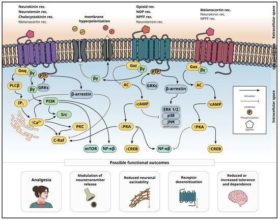
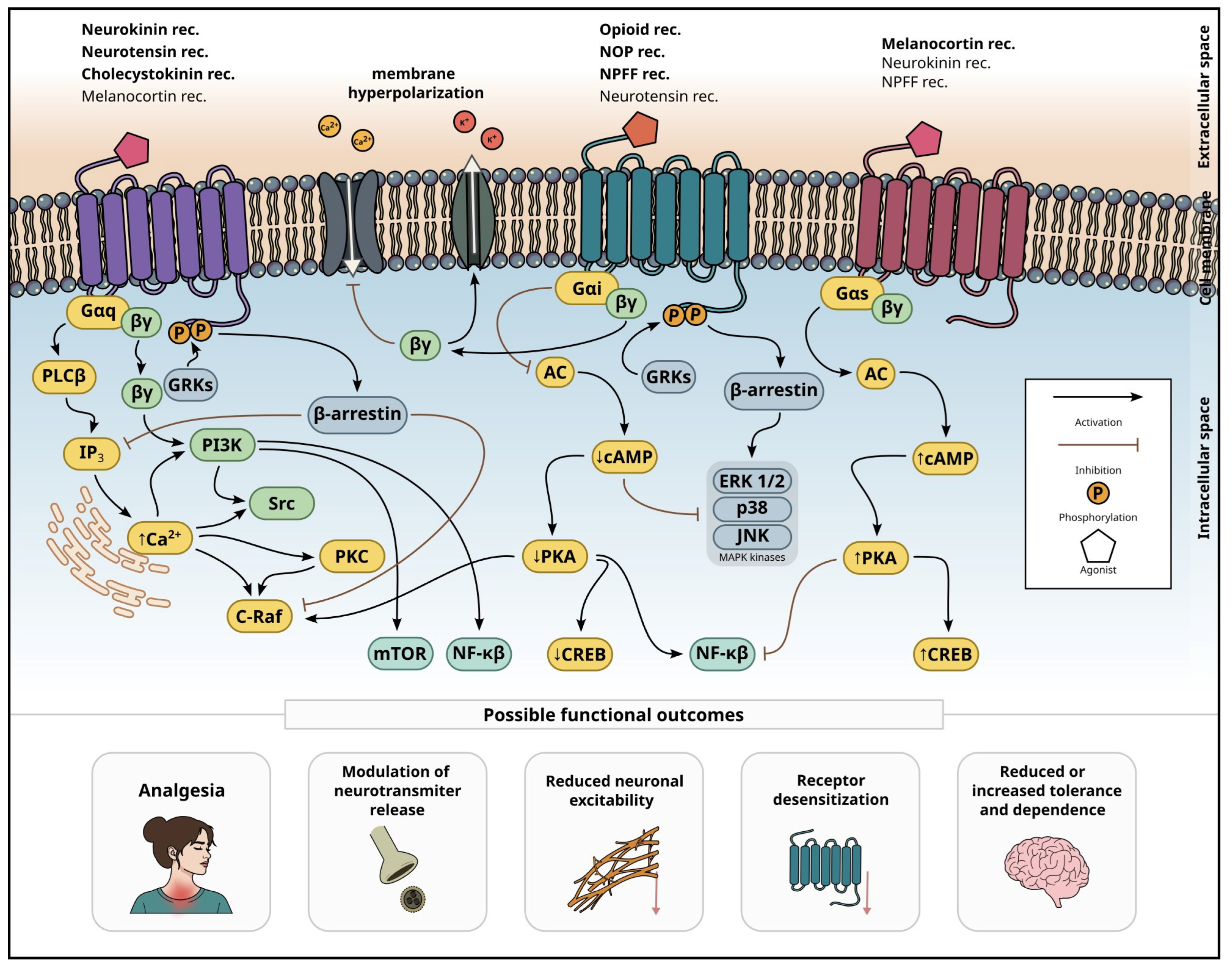

Disclaimer/Publisher’s Note: The statements, opinions and data contained in all publications are solely those of the individual author(s) and contributor(s) and not of MDPI and/or the editor(s). MDPI and/or the editor(s) disclaim responsibility for any injury to people or property resulting from any ideas, methods, instructions or products referred to in the content. |
© 2026 by the authors. Licensee MDPI, Basel, Switzerland. This article is an open access article distributed under the terms and conditions of the Creative Commons Attribution (CC BY) license.