


Abstract
In the Mediterranean and Himalayan regions, the genus Mandragora (family Solanaceae), sometimes called mandrake, is widely utilized in herbal therapy and is well-known for its mythical associations. Objective: To compile up-to-date information on M. autumnalis’s therapeutic properties. Its pharmacological properties and phytochemical composition are particularly covered in managing several illnesses, including diabetes, cancer, and heart disease. Methods: Articles on the review topic were found by searching major scientific literature databases, such as PubMed, Scopus, ScienceDirect, SciFinder, Chemical Abstracts, and Medicinal and Aromatic Plants Abstracts. Additionally, general online searches were conducted using Google Scholar and Google. The time frame for the search included items released from 1986 to 2023. Results: Mandragora has been shown to contain a variety of phytochemicals, including coumarins, withanolides, and alkaloids. The pharmacological characteristics of M. autumnalis, such as increasing macrophage anti-inflammatory activity, free radicals inhibition, bacterial and fungal growth inhibition, cytotoxic anticancer activities in vivo and in vitro against cancer cell lines, and enzyme-inhibitory properties, are attributed to these phytochemicals. Furthermore, M. autumnalis also inhibits cholinesterase, tyrosinase, α-amylase, α-glucosidase, and free radicals. On the other hand, metabolic risk factors, including the inhibition of diabetes-causing enzymes and obesity, have been treated using dried ripe berries. Conclusions: Investigations into the pharmacological and phytochemical characteristics of M. autumnalis have revealed that this plant is a rich reservoir of new bioactive substances. This review aims to provide insight into the botanical and ecological characteristics of Mandragora autumnalis, including a summary of its phytochemical components and antioxidant, antimicrobial, antidiabetic, anticancer, enzyme-inhibitory properties, as well as toxicological implications, where its low cytotoxic activity against the normal VERO cell line has been shown. More research on this plant is necessary to ensure its efficacy and safety. Still, it is also necessary to understand the molecular mechanism of action behind the observed effects to clarify its therapeutic potential.
1. Introduction
Since prehistoric times, plants have been used extensively in medicine to treat various illnesses. Population growth, the scarcity of some treatments, their expensive cost, and the unfavorable side effects of many synthetic drugs have all contributed to a rise in commercial and scientific interest in medicinal plants [1,2]. Researchers may identify promising opportunities for medicinal development by working with indigenous tribes and examining the traditional usage of botanicals. The process of finding new drugs is streamlined when traditional knowledge and contemporary scientific methods are combined [3]. Atropine, cocaine, ephedrine, colchicine, caffeine, digitoxin, morphine, quinine, scopolamine, theobromine, and taxol are plant-derived natural products of medical importance that were previously obtained from herbal sources but are now produced economically primarily through synthesis [4]. For instance, a significant class of drugs called atropine derivatives is made from a primary constituent found in natural products [5]. This ongoing research emphasizes how important nature is as a source of bioactive molecules that support the development of novel drugs and therapies in contemporary medicine. Herbal treatments have so far been standardized using drug development techniques to find analytical marker biomolecules [6]. The innovative use of biotechnology on plants to create medications derived from natural products that the medical community can then use to treat life-threatening conditions like asthma, influenza, cancer, tuberculosis, diabetes mellitus, coronary artery disease, and diarrhea is known as plant-made pharmaceuticals [7]. However, many herbal products are still untested, and their use is either poorly or not monitored at all, even though therapies involving these agents have demonstrated promising potential and the effectiveness of many of them has been established.
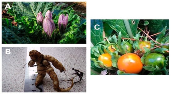
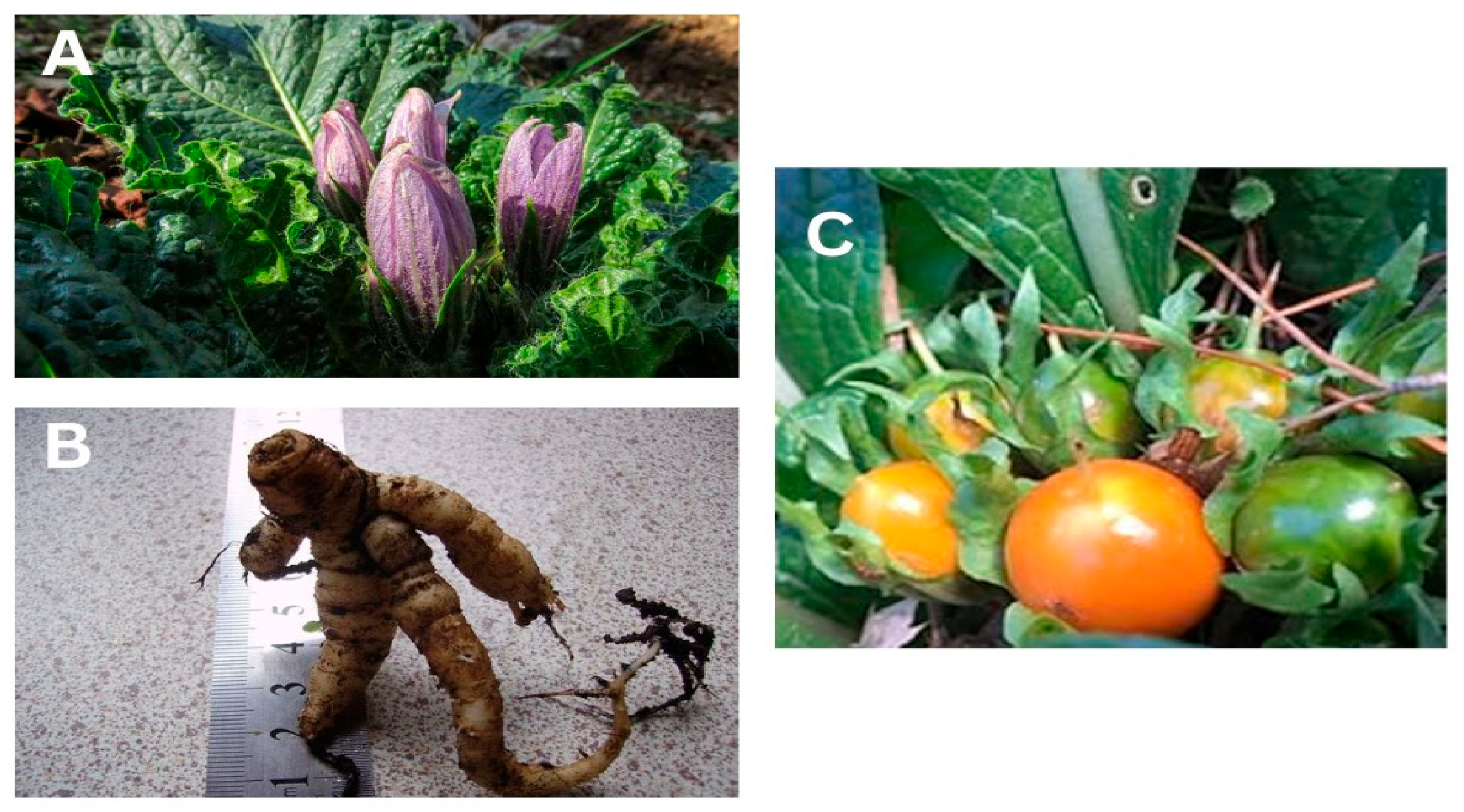
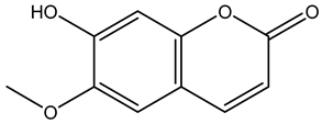
One of these botanicals is Mandragora autumnalis, also known as the mandrake plant or witch’s or devil’s herb, which has long been valued as one of the most important medicinal plants and as an herb with great cultural significance [8,9]. Mandragora autumnalis is a thick-rhizome-bearing annual herb with oblong-ovate leaves and blue-violet flowers that bloom from September through October (rarely in April) [10,11]. This plant was originally believed to have aphrodisiac and magical qualities because of the “human body”-shaped root (Figure 1) [12]. Traditionally, this species has been used to treat a wide range of conditions, including depression, melancholy, rheumatic pain, hemorrhoids, insomnia, and dysuria. Studies conducted in vitro have validated the biological characteristics of crude extracts from various parts of Mandragora, including their ability to modulate immunity, inhibit bacterial growth, prevent free radical-induced tissue damage, and inhibit several enzymes [13]. Indeed, Mandragora has a wide range of uses, including medicinal, hallucinogenic, and fertility-boosting applications, as well as the treatment of a variety of illnesses, including ulcers, inflammation, sleeplessness, and eye disorders. Specifically, Mandragora autumnalis has been shown to have antibacterial, antioxidant, and antitumor properties in addition to exhibiting a narcotic effect [14]. A variety of phytochemicals have been isolated from Mandragora species, including lipid-like compounds (β-sitosterol), coumarins (umbelliferone and scopoletin), alkaloids (atropine and scopolamine), and withanolides (salpichrolide C). These plant compounds are known to have biologically significant attributes, such as anticholinergic, antidepressant, antioxidant, and anti-inflammatory properties [15]. Most published publications related to Mandragora autumnalis concentrate on the unique ecological role of the mandrake plant despite the apparent increase in interest in its pharmaceutical effects. The discovery of novel bioactive metabolites with potential medical applications is necessary due to the ongoing need for alternative treatments derived from Mandragora species. This review attempts to gather all the previous research and provide a foundation for further studies to help identify M. autumnalis’s possible use as a medicinal agent. Considering the paucity of research on the phytochemical makeup and pharmacological characteristics of Mandragora autumnalis, this review paper aims to describe the plant’s botanical and ecological traits, provide an updated, thorough compilation of its phytochemical composition, and discuss its pharmacological activities and toxicological effects. Our review primarily concentrates on gathering all available information, both current and historical, regarding the pharmacological characteristics of this species, laying the groundwork for further research on this important species in the medical domain.
Figure 1.
(A) Mandragora autumnalis leaves and flowers, (B) Mandragora autumnalis roots, and (C) Mandragora autumnalis fruits.
2. Methods
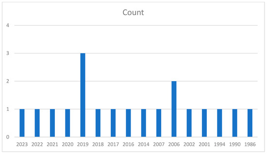
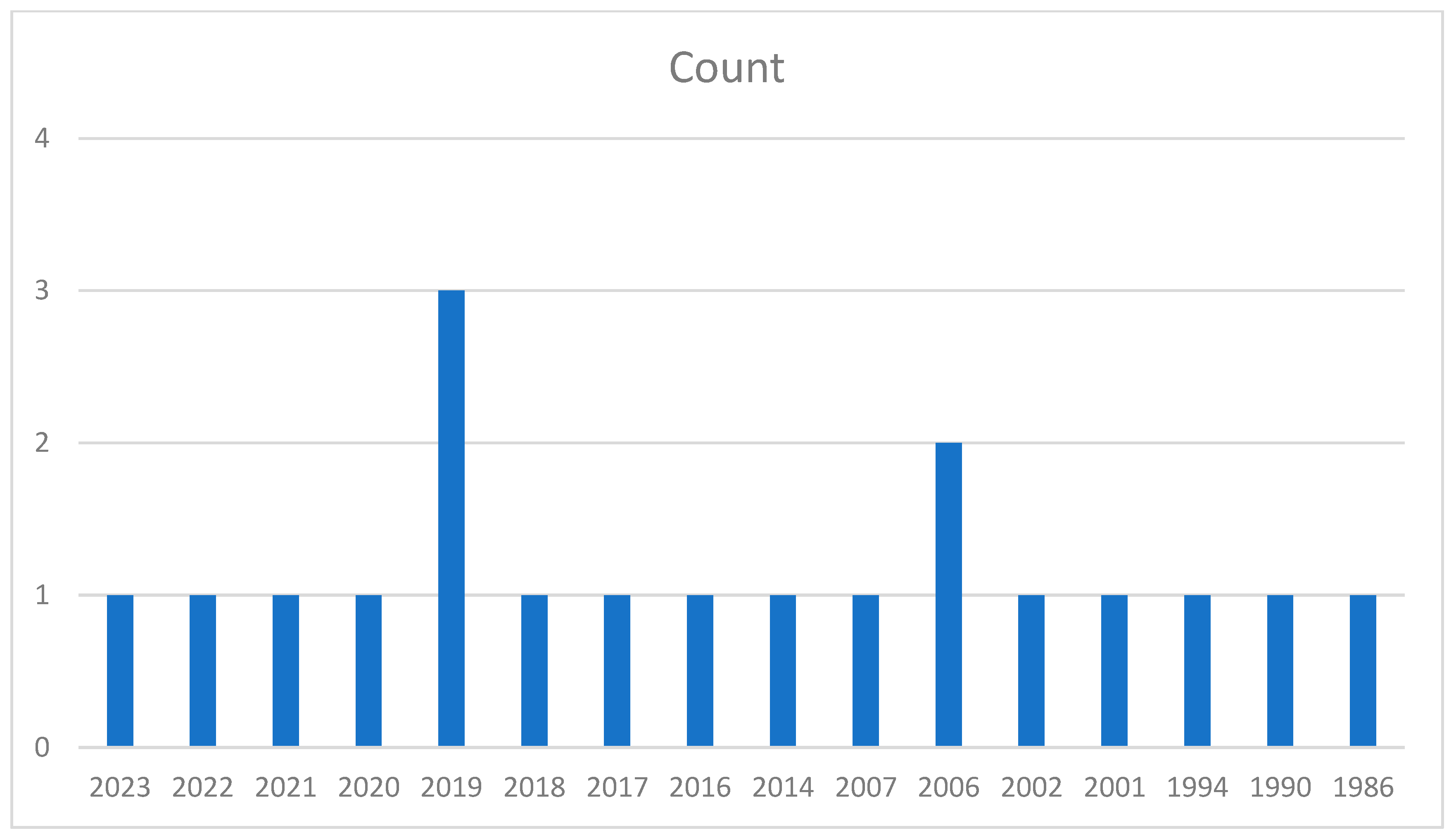
Articles on the review topic were found and retrieved using major scientific literature databases, such as PubMed, ScienceDirect, Scopus, Chemical Abstracts, Henriette’s Herbal Homepage, and Medicinal and Aromatic Plants Abstracts. The general internet Google Scholar and Google were also used for searches. Based on PubMed, Figure 2 shows that the search period encompassed the number of publications released from 1986 to 2023. The keywords used are “Mandrake plant”, “Mandragora species”, “Mandragora autumnalis”, “phytochemistry”, “extraction”, “ethnopharmacology”, “antioxidant activities”, “antibacterial”, “anti-enzymatic”, “anticancer activities”, “clinical trials”, and “toxicological activities”.
Figure 2.
A bar graph showing the number of articles considering Mandragora autumnalis from the year 1986 to 2023.
3. Taxonomical Classification of Mandragora autumnalis
The Mandragora genus belongs to the family Solanaceae, which has six species that are native to the Eurasian continent, including M. turcomanica, M. officinarum, M. caulescens, M. autumnalis, M. acaulis, and M. vernalis [12]. The taxonomy of Mandragora autumnalis is shown in Table 1.
Table 1.
Taxonomic classification of Mandragora autumnalis.
4. Geographical and Botanical Characteristics of Mandragora L.
4.1. Geographical Characteristics
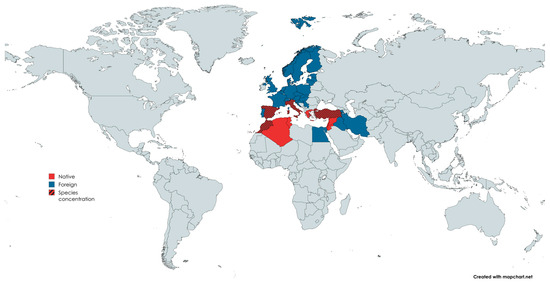
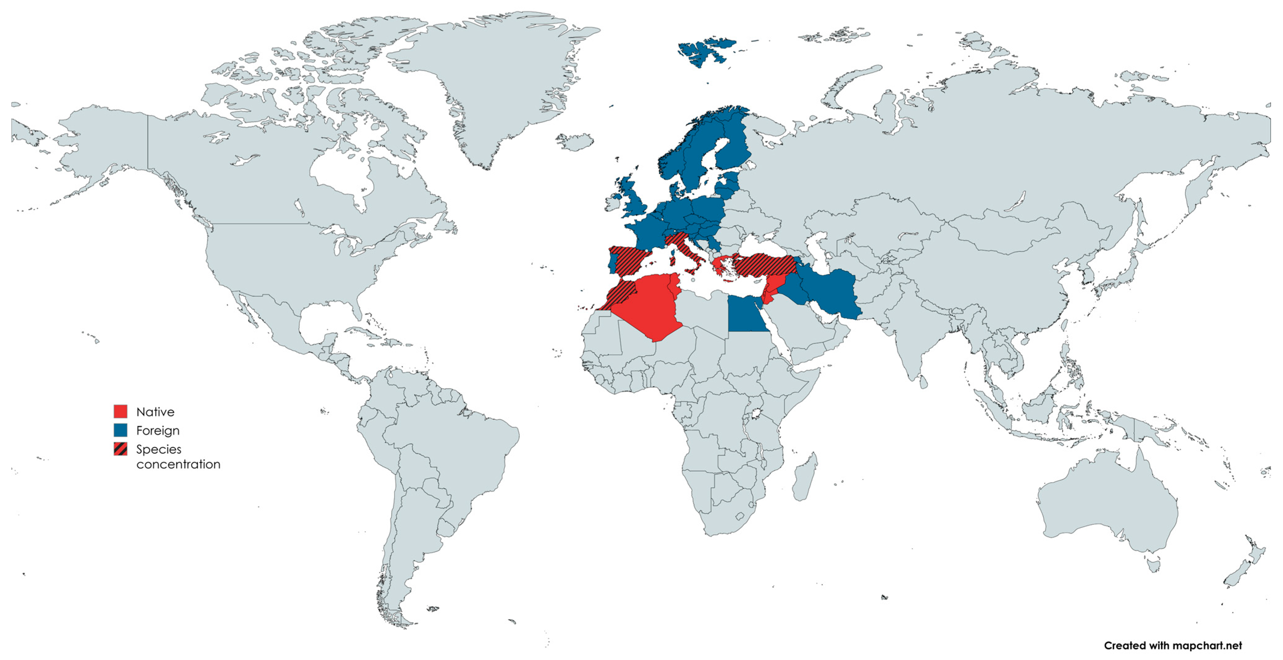
Mandragora L. is found throughout northern Africa and Eurasia. The genus has a traditional disjunction pattern in the biogeography of the Old-World flora, occurring discontinuously in the Tibetan Plateau, the Mediterranean area, and the Turanian region [16]. This plant is native to the following regions: Turkey, Palestine (Israel), Jordan, Syria, Lebanon, Tunisia, Algeria, and Morocco; on the other side, the plant is not native and is not ubiquitous according to the Flora Croatica Database, 2022, to Western, Central, and Northern Europe, Iran, Iraq, Serbia, Croatia, Armenia, and Egypt [17], as shown in the map in Figure 3.
Figure 3.
Geographic distribution of native and grown (foreign) Mandragora L.; the lined legend corresponds to the regions where Mandragora autumnalis species are concentrated. The map was designed using https://www.mapchart.net/world.html. Accessed online on 25 February 2025.
4.2. Botanical Characteristics of Mandragora autumnalis
The wild medicinal plant Mandragora autumnalis is a perennial herbaceous plant belonging to the Solanaceae family [8]. The specific name autumnalis refers to the season in which these plants typically bloom. Mandrake has a ground-hugging rosette of large shiny green leaves up to 40 cm long and densely packed mauve or violet five-petalled flowers that are 4 to 5 cm across [18]. Its ripe fruits (berries) are orange or yellow, and its blossoms are purple or violet. Unripe fruits, leaves, and roots are known locally as Tufah Almajan (Satan’s Apple), love apples, and Beed Aljin (Eggs of the Jinn) because of their propensity to stimulate the senses and produce mental disorders [19]. There are 55 main odoriferous components in the relatively strange chemical makeup of mandrake scent [20].
5. Phytochemical Constituents of Mandragora autumnalis
In addition to its ecological value, the mandrake plant has been utilized in traditional folk medicine to cure several illnesses, such as skin issues, inflammatory symptoms, and digestive difficulties. Additionally, it has been demonstrated that extracts from Mandragora autumnalis exhibit various pharmacological properties, including antibacterial, anti-fungal, anti-enzymatic, and anticancer properties. Throughout the years, different parts of the mandrake plant have been used to treat various ailments, as shown in Table 2. Numerous secondary metabolites from other parts of M. autumnalis are thought to be responsible for these characteristics.
Table 2.
The traditional medical and modes of usage of different parts of M. autumnalis.
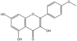
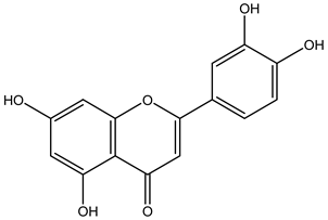
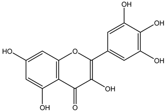
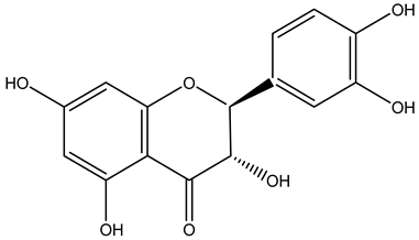
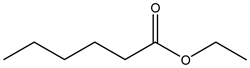
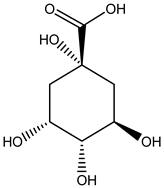
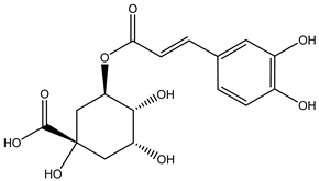
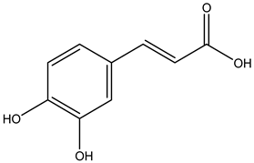
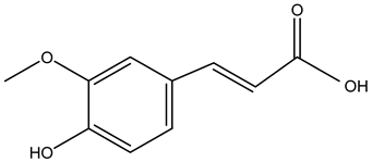
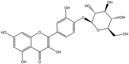
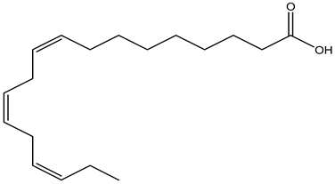
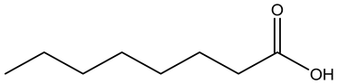
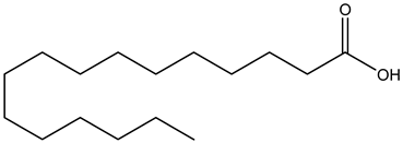
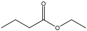
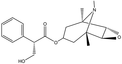
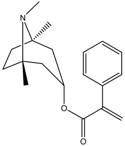
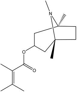
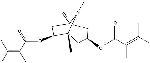
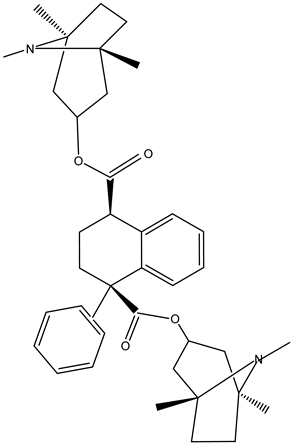
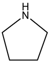
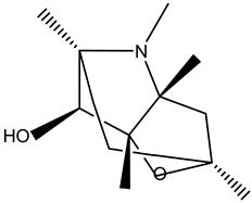
Tropane alkaloids found in abundance in M. autumnalis roots include scopolamine, and L-hyoscyamine, which, when extracted, yield the racemic combination of L- and R-hyosciamine, often referred to as atropine [27]. Additional tropane alkaloids derived from the roots include calystegines, α- and β-belladonnine, apoatropine, 3α-tigloyloxytropane, and 3,6-ditigloyloxytropane. Cuscohygrine, an alkaloid of pyrrolidine, was also produced [26], and belladonnines were absent from the fresh roots but present in the dried roots [28]. Moreover, an earlier study showed that the ethanol extract of M. autumnalis leaves had significant levels of flavonoids and phenols along with other phytochemicals such as terpenoids, anthraquinones, coumarins, phlobatannins, and tannins [13]. According to LC-MS analysis of the ethanol crude extract of M. autumnalis leaves, quercetin 3,4′-diglucoside was detected in large proportions, followed by quinic acid, chlorogenic acid, and quercetin 4′-O-glucoside [29]. Four flavonoids were extracted from the mature fruit of M. autumnalis using ethyl acetate extract, and by comparing their 1H- and 13C-NMR spectrum analyses with those published in the literature, their structures were determined to be kaempferol, luteolin, myricetin, and taxifolin [13]. Furthermore, it was discovered that the main constituents of M. autumnalis berries were ethyl esters of middle-chain acids, along with ethyl caprylate, linoleic acid, n-hexyl acetate, ethyl caprate, and ethyl caproate [30]. Table 3 lists the most important phytochemical composition of M. autumnalis roots, leaves, fruits, and essential oils extracted from M. autumnalis seeds, including the extraction solvent and the methods used to identify these compounds.
Table 3.
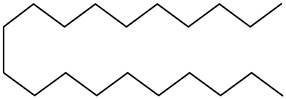
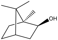
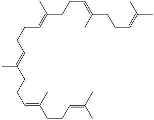
The most important bioactive metabolites from the roots, leaves, fruits, and essential oils extracted from M. autumnalis, their extraction solvent, and identification methods (all the structures were retrieved from ChemDraw Pro 8 software).
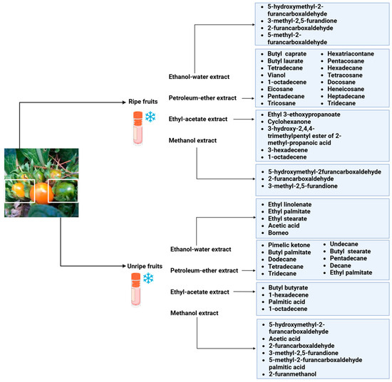
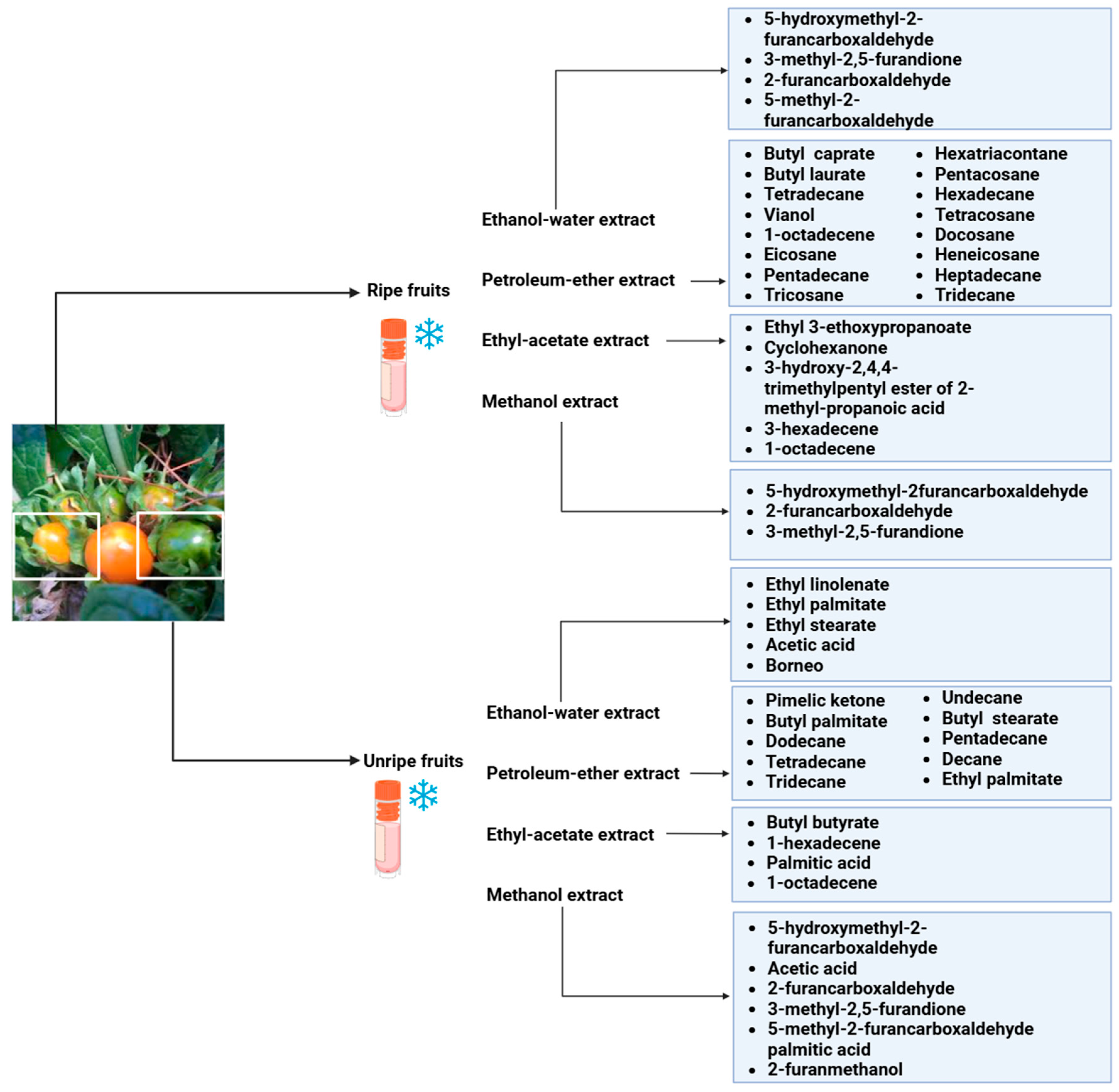
A total of 23 n-alkanes, 1 branched-chain alkane, 2 cyclohexanes, 8 alkenes, 2 branched-chain alkenes, 3 alcohols, 3 aldehydes, 6 ketones, 8 heterocyclic compounds, 4 thio compounds, 6 benzene hydrocarbons, 3 phenols, 18 carboxylic acids, and 48 carboxylic acid esters were among the 125 compounds identified in the fruit extracts of Mandragora autumnalis after the seeds were removed. The major compounds of different extracts of ripe and unripe M. autumnalis fruit are shown in Figure 4 [30,32,33,34]. Many identified compounds contribute to the fruit’s pleasant aroma and taste, albeit in different amounts, where ripe fruit contains significant “odor compounds”. Solavetivone is a representative phytoalexin that has been isolated from potato (Solanum tuberosum) tubers infected with the blight fungus Phytophthora infestans [35], air-cured tobacco (Nicotiana tabaccum) leaves [36], and several Solanaceae species. Furthermore, furan derivatives, especially 5-hydroxymethyl-2-furancarboxaldehyde (HMF), were present in higher amounts in methanolic extracts of ripe fruits (81.4%) than in unripe (57.0%) fruits, which may be explained by the fact that the higher HMF concentration in ripe fruits was proportionate to their higher sugar concentration. However, according to research by Muratore et al. on heated grapes, the HMF concentration was also highly affected by the change in temperature, pH, and sugar concentration; however, the HMF levels were higher in the methanolic extracts of unripe fruit than in those of ripe fruit, suggesting that methanol interacted in a sugar-independent manner [23]. Mandragora autumnalis compositional studies are insufficient, as the previously cited investigations demonstrated. Thus, a thorough examination of the phenolic compounds and flavonoids present in M. autumnalis extracts, especially the leaves and roots, as well as other classes of bioactive metabolites, should be the main goal of future research.
Figure 4.
The major compounds isolated from lyophilized ripe and unripe fruits of Mandragora autumnalis using different extraction solvents (the figure was designed using https://app.biorender.com/). Accessed online on 25 February 2025.
6. Pharmacological Bioactivities of Mandragora autumnalis
6.1. Antioxidant Activities
Reactive oxygen species (ROS) production and accumulation are out of balance with the body’s capacity to detoxify through antioxidant defenses [37]. This imbalance leads to oxidative stress, which causes premature aging and increases the risk of some diseases, including cancer, inflammatory disorders, diabetes, and neurodegenerative diseases [38,39]. Growing interest has been shown in the function of antioxidants in preventing oxidative damage in recent years [40]. Synthetic antioxidants have been widely used to lessen oxidative stress. These compounds are frequently used as food and consumer product preservatives and have been linked to dubious toxic and cancer-causing effects. Food-produced peroxides can interact with synthetic phenolic antioxidants, and research has demonstrated their harmful and cancer-causing impacts [41]. Propyl gallate (PG), propylated hydroxyanisole (BHA), propylated hydroxytoluene (BHT), and tertiary butylhydroquinone (TBHQ) are the most common synthetic phenol derivatives [42,43]. Yet, considering issues about these antioxidants’ safety, natural sources like plants have been used instead because they contain bioactive metabolites with potent antioxidant activity [44]. The antioxidant activities of various M. autumnalis extracts are displayed in Table 4.
Table 4.
Antioxidant activities of Mandragora autumnalis (IC: Inhibitory concentration; TE: Trolox equivalent).
The strong scavenging actions of the M. autumnalis flavonoid fruit fraction against the 1,1-diphenyl-2-picrylhydrazyl (DPPH) assay demonstrated the presence of antioxidants. The extract had an IC50 value of 5.37 ± 0.41 µg/mL, which is comparable to the standard antioxidant Trolox (IC50 = 2.23 ± 1.23 µg/mL) in terms of free radical scavenging properties [13]. Moreover, other research studied the antioxidant activity of the methanolic extract of M. autumnalis leaves (L-Met) and flowers (F-Met), as well as the acetone extract of the leaves (L-Ac) and flowers (F-Ac) using several assays. The phosphomolybdenum method was used to assess the total antioxidant capacity [47]. The extracts’ overall antioxidant capacity was greater in L-Ac (1.98 mmol TE/g extract). However, when using a DPPH assay [48] and compared to the other extracts, F-Met has the strongest free radical scavenging activity, with 73.09 mg TE/g of extract. In terms of metal-chelating activity [49], F-Met had the highest level (15.94 mg EDTA/g). Furthermore, The FRAP and CUPRAC assays are frequently used to assess the plant’s antioxidant capacity [50,51]; as a result, the methanolic extract of the flowers showed the highest antioxidant activity based on these two methods. Additionally, previous research assessed the DPPH radical scavenging activity of the aqueous root extract and synthesized silver nanoparticles of M. autumnalis; both extracts showed significant dose-dependent antioxidant activities, with an IC50 of 47.16 ± 0.41 and 51.81 ± 0.10 µg/mL [46]. Thus, M. autumnalis has strong antioxidant properties that demand more research. Green extraction techniques that produce higher concentrations of antioxidant compounds from plants have recently attracted more attention to promote longevity, reduce aging effects, and prevent disorders linked to oxidative stress. It should be noted that in vitro assays, which are frequently prone to errors because of the chemical diversity of phytochemicals, have been used to evaluate M. autumnalis’ antioxidant capacity, which should be carefully interpreted and backed up in future studies that concentrate on using in vivo techniques to measure antioxidant activity.
6.2. Antimicrobial Activities
The widespread and inappropriate use of antibiotics has resulted in antimicrobial resistance, representing a major global challenge and serious threat to human health [52]. Due to the declining effectiveness of traditional drug therapies, there has been a pressing need to develop new antimicrobial agents using novel therapies based on natural metabolites because of their chemical diversity and efficacy [53]; in fact, extracts from medicinal plants have been widely reported to exhibit antimicrobial activities, acting by both increasing the activity of antibiotics and inhibiting the growth of various pathogens, including bacteria, fungi, and viruses [54,55]. This has made it necessary to look for and create new antimicrobial agents using natural compounds. Because medicinal plants produce bioactive compounds with known therapeutic properties, they have been thoroughly investigated as possible antimicrobial agents. Several studies have assessed the antibacterial efficacy of M. autumnalis extracts, as shown in Table 5.
Table 5.
The antimicrobial activities of M. autumnalis.
A microdilution assay was employed to evaluate the antimicrobial activity of the fruit flavonoid fraction of M. autumnalis. The findings demonstrated that this fraction possesses the strongest antibacterial activity against the K. pneumoniae strain, higher than ampicillin and comparable to the reference medication ciprofloxacin. In addition, this fraction showed antibacterial inhibitory effects against S. aureus, E. coli, P. aeruginosa, and Methicillin-resistant Staphylococcus aureus (MRSA) and strong antifungal activity against C. albicans when compared with the commercial antifungal drug fluconazole [13]. Moreover, it was discovered that P. aeruginosa bacteria are more sensitive to silver nanoparticles synthesized from M. autumunalis aqueous roots extract (Ma-AgNPs) compared to M. autumnalis roots extract, which showed the highest sensitivity against B. subtilis [46]. The MIC values for M. autumnalis ethanol leaf extract and fractions were ascertained using a microdilution assay. Gram-positive bacteria (B. subtilis) and Gram-negative bacteria (P. aurignosa) were vulnerable to the antibacterial properties of ethanol extract and n-hexane fraction. Additionally, the n-hexane fraction demonstrated antifungal activity against Candida albicans, with a minimum inhibitory concentration (MIC) of 12.5 mg/mL. However, in both bacterial and fungal tests, aqueous/methanol and aqueous fractions exhibited no activity [29]. These studies support the potential use of M. autumnalis extracts as antibacterial agents. However, investigating the mechanism underlying the observed antibacterial activity is necessary.
Further research should determine how M. autumnalis extracts work against other pathogens. Together, these findings suggest that M. autumnalis may provide new antimicrobial agents that could aid in botanical screening efforts to find new medications to combat antibiotic resistance and guarantee future antibiotic-free management of microbial growth.
6.3. Anticancer Activities
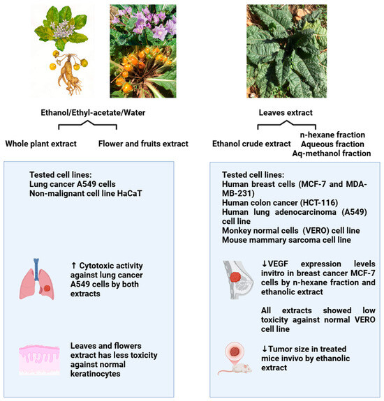
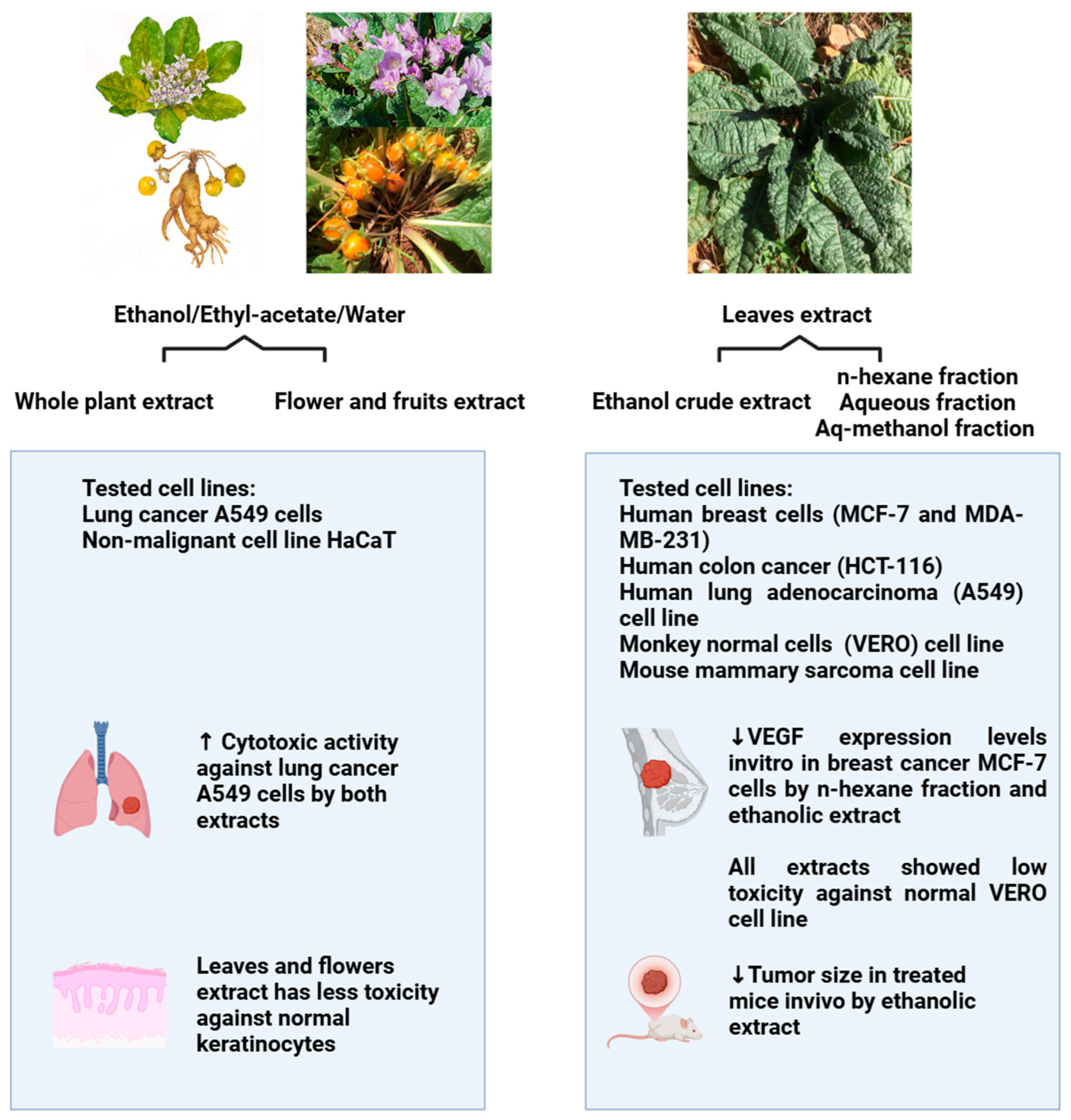
Cancer remains a major cause of death globally, even with significant progress in cancer treatment [56,57]. Globally, cancer is the leading cause of death and a serious public health concern [58]. In addition, traditional cancer treatment plans are frequently linked to negative side effects and multidrug resistance, which has raised interest in the hunt for novel bioactive compounds derived from plants [59,60]. In this context, several investigations have evaluated M. autumnalis’s anticancer capabilities. The anticancer activity of extracts of Mandagora autumnalis’s flowers, fruits, and whole plant (extracted using equal proportions of ethanol/ethyl acetate/water) was shown to be 2.7- and 3.5-fold more cytotoxic to (A549) lung cancer cells than to non-cancerous human keratinocyte (HaCat) cells, respectively [61]. Moreover, another study evaluated the anticancer activity of Mandragora autumnalis crude ethanolic leaf extract and several leaf fractions (n-hexane, aqueous, and aqueous-methanolic) [14]. To assess the anticancer effect of M. autumnalis leaves in both in vitro and in vivo models, ethanol crude extract and solvent fractions were extracted. By using the 3-(4,5-Dimethylthiazol-2-yl)-2,5-Diphenyltetrazolium Bromide (MTT) method, five cancer cell lines—human lung adenocarcinoma (A549), human colon cancer (HCT-116), human breast cancer (MCF-7 and MDA-MB-231), a mouse mammary sarcoma cell line (EMT6/p), and one normal monkey cell line (VERO)—were evaluated in vitro [62,63,64,65,66,67]. Low cytotoxicity against the normal VERO cell line, the downregulation of VEGF expression, and the strongest antitumor activity against MCF-7 breast cancer cells were demonstrated via ethanol extract and n-hexane fractions. Furthermore, mice administered M. autumnalis ethanol extract showed a significant reduction in tumor size when compared to the negative control [14]. To assess the anticancer potential of various M. autumnalis extracts, the referenced studies employed cell viability assays; these investigations demonstrated the potent anticancer properties of the plant’s extracts and the isolated compounds mentioned above against a variety of cancer types and cancer hallmarks. Figure 5 depicts M. autumnalis’s intriguing anticancer properties; Table 6 summarizes these properties. However, further research must confirm M. autumnalis’s anticancer potential and examine the underlying molecular mechanisms.
Figure 5.
The anticancer activities of M. autumnalis against various cancer cell lines (the figure was designed using https://app.biorender.com/). Accessed online in 25 February 2025.
Table 6.
The anticancer effects of Mandragora autumnalis.
6.4. Antidiabetic, Anti-Enzymatic, and Anti-Obesity Activities
Research suggests that Mandragora autumnalis may have antidiabetic effects. Studies have shown that extracts from M. autumnalis can inhibit enzymes like α-amylase and α-glucosidase [68]. These enzymes play a crucial role in the digestion of carbohydrates, and inhibiting them can help slow down the absorption of glucose into the bloodstream [69]. This can be beneficial for managing blood sugar levels in people with diabetes hyperglycemia or elevated blood glucose concentration, which is a hallmark of diabetes, a chronic metabolic disease caused by an insufficient or malfunctioning insulin supply [70]. Serious complications in many organs, such as kidney and vision loss, heart attacks, and strokes, are caused by uncontrolled diabetes [71,72]. On the other hand, the majority of patients with type 2 diabetes have dyslipidemia, which is characterized by high triglycerides, low HDL-C, and a preponderance of small-dense LDL particles. Between 60% and 70% of diabetic patients have at least one lipid abnormality, though not all patients show all symptoms [73]. Previous research showed that the majority of patients with elevated lipase also have elevated amylase [74], and since herbal medicine is inexpensive and has few adverse effects, it has been preferred over synthetic medications for the treatment of diabetes. Insulin plays a crucial role in preserving glucose homeostasis by increasing GLUT4 translocation to the muscle’s plasma membrane (PM) [75]. When muscle cells were treated with flavonoid fruit fraction both with and without insulin, a dose-dependent increase in GLUT4 translocation was observed. When no insulin treatment was administered, the fruit flavonoid fraction at concentrations of 0.125, 0.25, and 0.5 µg/mL significantly increased GLUT4 translocation by 1.33 ± 0.13, 1.33 ± 0.22, and 1.56 ± 0.21 µg/mL, respectively. Additionally, the fraction significantly increased GLUT4 translocation to the PM in the presence of insulin by 2.35 ± 0.43 and 2.35 ± 0.19 at 0.25 and 0.5 mg/mL, respectively [13]. Additionally, the results showed that the fraction exhibited dose-dependent α-amylase and α-glucosidase inhibitory activity in comparison to the commercial antidiabetic therapeutic agent acarbose. Furthermore, the fraction exhibited dose-dependent pancreatic lipase inhibitory activity in comparison to orlistat [13]. Moreover, other research showed that the acetone extract exhibited greater activity for α-amylase compared to methanol extract. According to the α-amylase assay, the leaves and flower acetone extracts correspond to the highest amylase inhibitory activity compared to the methanolic leaves and flower methanolic extracts. However, only the acetone extract of the leaves extract did not exhibit any α-glucosidase inhibitory activity, whereas the rest of the extracts exhibited mild α-glucosidase inhibitory activity. All extracts were found to have stronger inhibitory effects against α-amylase than α-glucosidase [45]. Table 7 illustrates the anti-enzymatic bioactivities of Mandragora autumnalis. Furthermore, there are serious and occasionally lethal side effects associated with some synthetic antidiabetic drugs. Therefore, over the past 20 years, scientists have concentrated their research on natural products like flavonoids as possible alternatives to acarbose and other antidiabetic drugs.
Table 7.
The anti-enzymatic potential of M. autumnalis extracts against several conditions.
6.5. Anticholinesterase and Anti-Tyrosinase Activities
Alzheimer’s disease is the most common cause of dementia and is a neurodegenerative disorder of the brain that is clinically characterized by a progressive decline in cognitive abilities [76]. Acetylcholine deficiency or loss is linked to a marked decrease in central cholinergic neurotransmission [77]. The primary treatment approach is to restore acetylcholine levels by inhibiting the enzymes butyrylcholinesterase (BChE) and acetylcholinesterase (AChE), which hydrolyze acetylcholine [78,79]. On the other hand, the term “Parkinson’s disease dementia” (PDD) refers to the growing recognition of dementia in Parkinson’s disease (PD) cases [80]. The strongest neuropathological correlate of developing dementia in Parkinson’s disease appears to be the spread of fibrillar α-synuclein (α-syn) pathology from the brainstem to limbic and neocortical structures; novel treatment approaches involve blocking brain tyrosinase levels and neuromelanin production [81]. Thus, previous research that studied the inhibition activity M. autumnalis’s cholinesterase inhibitory properties was evaluated concerning the Alzheimer’s disease-related enzymes AChE and BChE. The results showed that the acetone extract of the leaves demonstrated notable inhibitory activity on BChE, whereas the methanolic extract of the flower extract had the strongest activity on AChE. Moreover, the tyrosinase inhibitory activity of the acetone extract of the leaves was the highest, at 29.68 mgKAE/g extract, whereas the methanol extract of the flower showed no tyrosinase-inhibiting properties. The hydroxyl group of phenolic compounds in M. autumnalis may be responsible for inhibiting tyrosinase activity [82]. These findings pave the way for the future usage of M. autumnalis for treating or decreasing the symptoms of Alzheimer’s and Parkinson’s disease. The anti-enzymetic activities of different extracts of Mandragora autumnalis are summarized in Table 8.
6.6. Anti-Inflammatory Activities
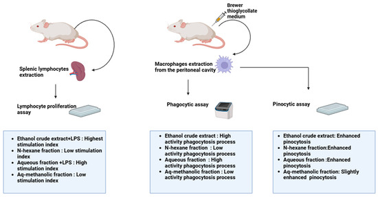
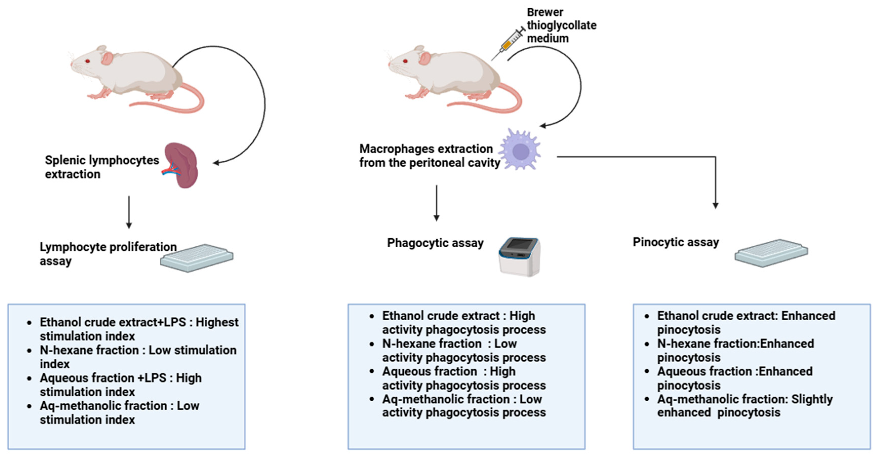
The body uses inflammation as a self-defense and homeostatic mechanism, always maintained under homeostatic control. On the other hand, chronic or excessive inflammation can lead to the development of several illnesses, including cancer, diabetes, and cardiovascular disease [38,83]. An important component of the acquired immune system, lymphocytes’ capacity to proliferate is thought to be a measure of the degree of cell immunity [84]. In the presence and absence of mitogens, including lipopolysaccharide (LPS), which stimulates B cells, and Concanavalin A (con A), which stimulates T cells, lymphocyte proliferation was induced, and the outcomes were roughly comparable. The aqueous fraction and the ethanol extract containing lipopolysaccharide (LPS) had the highest stimulation index values: 2.5 and 3.6, respectively. Conversely, the aqueous/methanol fractions and n-hexane demonstrated reduced activity at the highest concentration, with low stimulation index values [13]. Macrophage activity was increased by ethanol extract and the aqueous fraction of M. autumnalis leaves through phagocytosis. However, the n-hexane and aqueous/methanol fractions, which had phagocytic indices of 143 and 165, respectively, demonstrated less activity. Furthermore, the ethanol crude extract, aqueous, and n-hexane fractions enhanced the macrophages’ capacity for pinocytosis [13]. The immunomodulatory effects of different extracts of M. autumnalis leaves are summarized in Table 8 and illustrated in Figure 6.
Figure 6.
The immunomodulatory effects of different extracts of M. autumnalis leaves (the figure was designed using https://app.biorender.com/). Accessed online on 25 February 2025.
Table 8.
The anti-inflammatory effects of different extracts of M. autumnalis leaves.
6.7. Green Nanotechnology
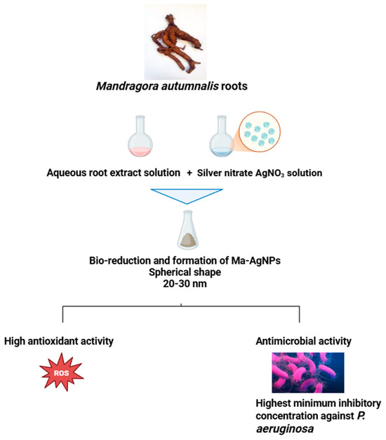
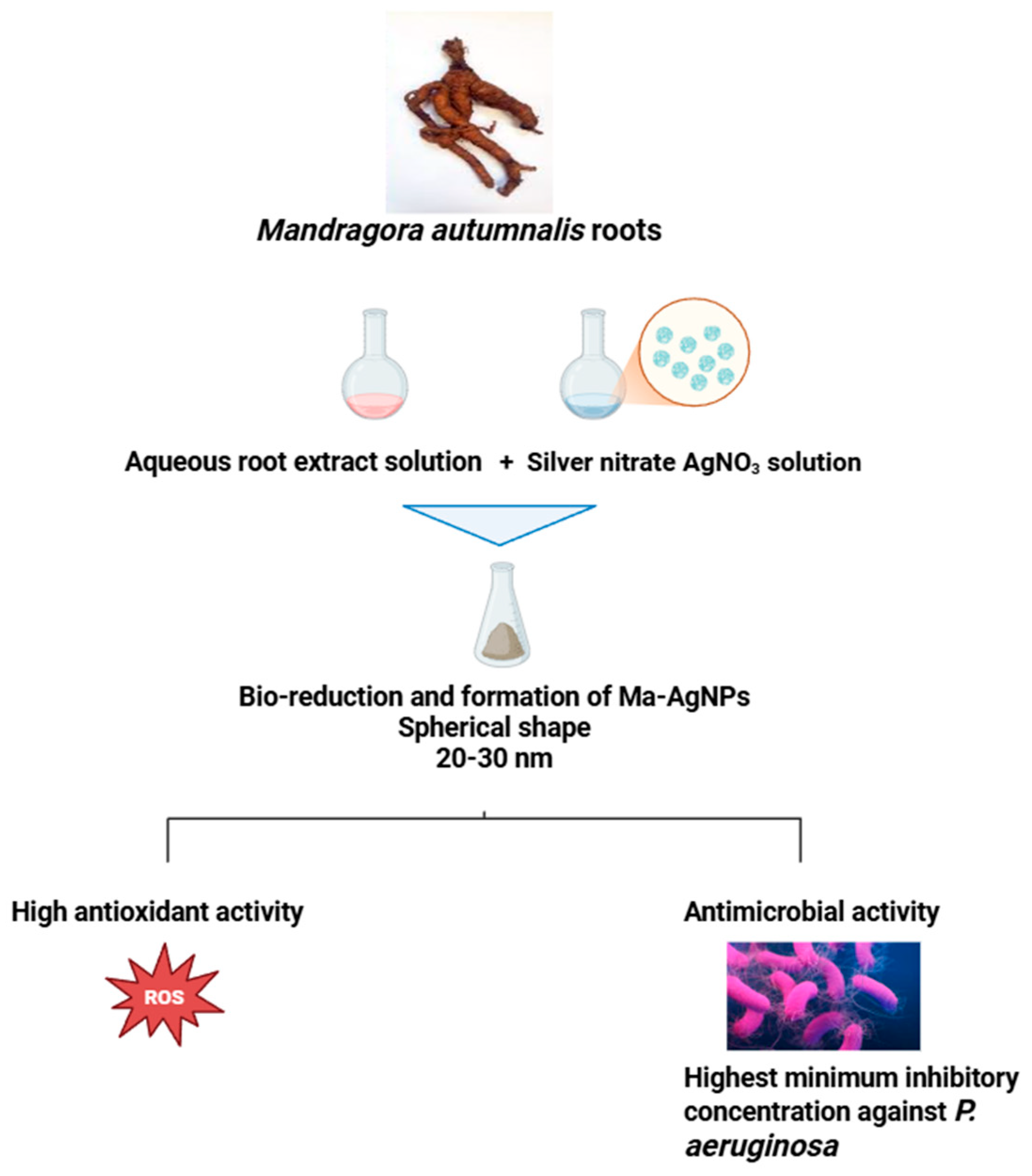
Nanotechnology is a pioneering field that has profound effects on consumer goods and is transforming several industries [85,86]. This rapidly growing field of study (nanoscience and nanotechnology) deals with systems, devices, and structures with unique characteristics and capabilities because of the way their atoms are arranged on the 1–100 nm scale [87]. Generations of nanomaterials have surfaced and are employed in interdisciplinary scientific domains; nanoscale manufacturing will soon be integrated into practically every field of science and technology, as evidenced by the quick development of nanoscience [88,89]. Silver nanoparticles (AgNPs) are tiny silver particles that are produced chemically or environmentally [90]. They are affordable and possess special physicochemical qualities. Silver nanoparticles are useful in a variety of applications, including textiles, biomedical applications, protective surface coatings, and wound healing, due to their well-known antimicrobial and antiviral properties [91]. AgNPs can be produced conventionally using physical techniques, like laser ablation, chemical processes, like reduction reactions, and green synthesis routes using biomaterials like fungi, bacteria, and plant extracts. The green synthesis of AgNPs guarantees the creation of environmentally benign and nontoxic nanoparticles with improved stability, biocompatibility, and biological activity [92,93,94,95]. The potential of M. autumnalis-mediated AgNPs for antioxidant activity was examined using an in vitro DPPH test system to assess the antioxidant activities of nanoparticles synthesized from M. autamnalis aqueous root extract (Ma-AgNPs). Ma-AgNPs were found to be potent free radical scavengers, demonstrating effective inhibitory activity in a dose-dependent fashion [46]. Moreover, the antibacterial properties of Ma-AgNPs were examined in Gram-negative (Escherichia coli and Pseudomonas aeruginosa) and Gram-positive (Bacillus subtilis and Staphylococcus aureus) bacteria. The results demonstrated that Ma-AgNPs exhibit antibacterial activity against every tested bacterial strain when present at a concentration of 25 µg/mL, and it was discovered that P. aeruginosa bacteria were more sensitive to Ma-AgNPs than any other bacteria tested [46]. The antibacterial and antioxidant properties of AgNPs produced by M. autumnalis are depicted in Figure 7. When taken together, these findings highlight the potential of M. autumnalis in environmentally friendly nanoparticle synthesis and call for more research in this field.
Figure 7.
Mandragora autumnalis roots produce silver nanoparticles (AgNPs) with antioxidant and antibacterial properties (the figure was designed using https://app.biorender.com/). Accessed online on 25 February 2025.
6.8. Toxicological Studies
There are growing concerns about the safety profile of herbal medicinal products as their use spreads throughout the world. It is not required to assess these products’ safety or toxicity before going on sale because most of them are categorized as foods or dietary supplements in most countries [2,96,97]. However, toxicological research is crucial when creating herbal medicine to guarantee efficacy, quality, and safety to prevent any potential negative health effects [98,99]. Numerous studies assessed Mandragora autumnalis’s safety and toxicological profile. For that reason, previous research studied the anesthetic properties of Mandragora autumnalis roots [100], where the commercial value of fish residues is reduced when synthetic anesthetics are used [101]. The study aimed to use Mandragora autumnalis roots rather than synthetic anesthetics that leave harmful residues. In vivo investigations on fish were conducted to examine mandrake root extracts and determine the proper experimental dosage. The Mandragora aqueous root extract obtained using a maceration method showed the best potential to be used as a fish anesthetic drug [100]. This can be related to the presence of tropane alkaloids that exhibit an anticholinergic effect by binding to either muscarinic receptors (mAChR) or nicotinic acetylcholine receptors (nAChR), which blocks the action of the neurotransmitter acetylcholine (ACh) in the central and peripheral nervous systems (CNS and PNS) [102]. This anticholinergic action affects both CNS and PNS, and symptoms include respiratory and heart rate changes, muscle contraction, hallucinations, and local anesthesia [103].
Moreover, topical exposure to M.autumnalis was believed to result in irritant contact dermatitis. According to reports, applying Mandragora root extract topically caused erythema, swelling, and burning. Erythematous, oedematous, and scaly plaque lesions were among the symptoms linked to allergic contact dermatitis brought on by Mandragora root sap [104,105]. Furthermore, recent research assessed the in vivo toxicity of the Mandragora autumnalis ethanol leaf extra by measuring liver and kidney parameters. Serum levels of aspartate aminotransferase, alanine transaminase, and creatinine were normal in the tumor-bearing mice given the M. autumnalis extract [14]. In general, additional toxicological analysis is required to confirm M. autumnalis’s safety profile for medical use.
7. Conclusions and Future Perspectives
Botanicals have intrigued humans since antiquity due to their extraordinary nutritional, cosmetic, and therapeutic properties. An increasing amount of information demonstrates that herbal medications have fewer adverse effects than most synthetic therapies, making them an essential source for creating novel pharmaceuticals, cosmetics, and nutritional supplements. Recently, there has been an increase in interest in research on plant products because plants are now, more than ever, considered as potential leading metabolite producers for drug development. However, plant metabolites should be optimized for efficacy and thoroughly evaluated for toxicity in plant-derived drug discovery. M. autumnalis has been shown in phytochemical and pharmacological studies to be a rich source of bioactive metabolites with a wide range of applications, further supporting its ethnopharmacological uses. Previous research showed that M. autumnalis has free radical scavenging activity, with an ability to inhibit the growth of tested microbial strains, decrease the viability of several cancer cell lines by increasing the cytotoxicity against various cancer cell lines in vitro, and decrease tumor size in experimental mice in vivo. Moreover, it increases the immunomodulatory activity of macrophages by increasing phagocytotic and pinocytotic activity, as well as increasing the proliferation potential of lymphocytes. Furthermore, it has a promising potential against diabetes and obesity. However, the identification of the bioactive metabolites mediating M. autumnalis’s biological activities should be another goal of future research, where drugs that target a range of illnesses could be developed using molecular mechanisms of action. The food and nutraceutical industries may also make use of these bioactive metabolites. Additionally, before being tested in clinical trials and utilized for drug development, the safety profile and effectiveness of the bioactive metabolites should be assessed using in vivo models following their identification and isolation. It is crucial to remember that the literature does not provide enough proof of M. autumnalis’s anticancer and anti-inflammatory properties. Toxicological studies should be conducted alongside additional research on the pharmacodynamic, pharmacokinetic, and mechanism of action of M. autumnalis extract to develop safe and natural pharmaceutical medications from this medicinal plant. Methods to lessen toxicity while preserving bioactivity and enhancing therapeutic effect are essential for advancing such medications to drug development and clinical trials. The elucidation of new methods should be retrieved to increase the delivery of the bioactive components of Mandragora and lessen its toxicological profile; for example, applying new fractionation systems to the different parts of Mandragora autumnalis, which will allow for the usage of effective beneficial components from this plant and eliminate the toxic related compounds. Thus, more pharmacological research is required to support the clinical efficacy of treatments, including in vitro and in vivo tests. There is currently insufficient data from clinical and even in vivo investigations to substantiate the in vitro findings. The lack of diversity in pharmacological research on Mandragora species can be attributed to issues with plant accessibility and toxicity.
Author Contributions
Conceptualization, G.A., A.H. and E.B.; investigation, G.A., A.B. and E.B.; writing—review and editing, G.A., J.E.M. and Z.A.B.; visualization, E.B., A.D. and A.H.; project administration, A.H., E.B. and M.A. All authors have read and agreed to the published version of the manuscript.
Funding
This research received no external funding.
Data Availability Statement
Not applicable.
Acknowledgments
The authors would like to thank the University Research Board (URB) of the American University of Beirut and the University of Petra for their support. Open Access funding provided by Qatar University (QU) Health.
Conflicts of Interest
The authors declare no conflicts of interest.
References
- Alamgir, A.N.M. Therapeutic Use of Medicinal Plants and Their Extracts; Progress in drug research; Springer: Cham, Switzerland, 2017; ISBN 978-3-319-63861-4. [Google Scholar]
- Ekor, M. The Growing Use of Herbal Medicines: Issues Relating to Adverse Reactions and Challenges in Monitoring Safety. Front. Pharmacol. 2014, 4, 177. [Google Scholar] [CrossRef] [Scilit]
- Alum, E.U. The Role of Indigenous Knowledge in Advancing the Therapeutic Use of Medicinal Plants: Challenges and Opportunities. Plant Signal. Behav. 2024, 19, 2439255. [Google Scholar] [CrossRef] [Scilit]
- Chaachouay, N.; Zidane, L. Plant-Derived Natural Products: A Source for Drug Discovery and Development. Drugs Drug Candidates 2024, 3, 184–207. [Google Scholar] [CrossRef] [Scilit]
- Ernest, T.B.; Elder, D.P.; Martini, L.G.; Roberts, M.; Ford, J.L. Developing Paediatric Medicines: Identifying the Needs and Recognizing the Challenges. J. Pharm. Pharmacol. 2007, 59, 1043–1055. [Google Scholar] [CrossRef] [Scilit] [PubMed]
- Chin, Y.-W.; Balunas, M.J.; Chai, H.B.; Kinghorn, A.D. Drug Discovery from Natural Sources. AAPS J. 2006, 8, E239–E253. [Google Scholar] [CrossRef] [Scilit] [PubMed]
- Chihomvu, P.; Ganesan, A.; Gibbons, S.; Woollard, K.; Hayes, M.A. Phytochemicals in Drug Discovery-A Confluence of Tradition and Innovation. Int. J. Mol. Sci. 2024, 25, 8792. [Google Scholar] [CrossRef] [Scilit]
- Alzweiri, M.; Sarhan, A.A.; Mansi, K.; Hudaib, M.; Aburjai, T. Ethnopharmacological Survey of Medicinal Herbs in Jordan, the Northern Badia Region. J. Ethnopharmacol. 2011, 137, 27–35. [Google Scholar] [CrossRef] [Scilit]
- Old Names—New Growth: Proceedings of the 2nd ASPNS Conference, University of Graz, 6–10 June 2007, and Related Essays; Anglo-Saxon Plant-Name Survey; Bierbaumer, P., Klug, H.W., Eds.; Peter Lang: Frankfurt, Germany; New York, NY, USA, 2009; ISBN 978-3-631-58316-6. [Google Scholar]
- Piccillo, G.A.; Mondati, E.G.M.; Moro, P.A. Six Clinical Cases of Mandragora autumnalis Poisoning: Diagnosis and Treatment. Eur. J. Emerg. Med. 2002, 9, 342–347. [Google Scholar] [CrossRef] [Scilit]
- Piccillo, G.A.; Miele, L.; Mondati, E.; Moro, P.A.; Musco, A.; Forgione, A.; Gasbarrini, G.; Grieco, A. Anticholinergic Syndrome Due to ‘Devil’s Herb’: When Risks Come from the Ancient Time: DEVIL’S HERB. Int. J. Clin. Pract. 2006, 60, 492–494. [Google Scholar] [CrossRef] [Scilit]
- Hanuš, L.O.; Řezanka, T.; Spížek, J.; Dembitsky, V.M. Substances Isolated from Mandragora Species. Phytochemistry 2005, 66, 2408–2417. [Google Scholar] [CrossRef] [Scilit]
- Al-Maharik, N.; Jaradat, N.; Bassalat, N.; Hawash, M.; Zaid, H. Isolation, Identification and Pharmacological Effects of Mandragora autumnalis Fruit Flavonoids Fraction. Molecules 2022, 27, 1046. [Google Scholar] [CrossRef] [Scilit] [PubMed]
- Mahmod, A.I.; Talib, W.H. Anticancer Activity of Mandragora autumnalis: An in Vitro and in Vivo Study. Pharmacia 2021, 68, 827–835. [Google Scholar] [CrossRef] [Scilit]
- Monadi, T.; Azadbakht, M.; Ahmadi, A.; Chabra, A. A Comprehensive Review on the Ethnopharmacology, Phytochemistry, Pharmacology, and Toxicology of the Mandragora Genus; from Folk Medicine to Modern Medicine. Curr. Pharm. Des. 2021, 27, 3609–3637. [Google Scholar] [CrossRef] [Scilit] [PubMed]
- Volis, S.; Fogel, K.; Tu, T.; Sun, H.; Zaretsky, M. Evolutionary History and Biogeography of Mandragora L. (Solanaceae). Mol. Phylogenet. Evol. 2018, 129, 85–95. [Google Scholar] [CrossRef] [Scilit]
- Dafni, A.; Blanché, C.; Khatib, S.A.; Petanidou, T.; Aytaç, B.; Pacini, E.; Kozuharova, E.; Geva-Kleinberger, A.; Shahvar, S.; Dajic, Z.; et al. In Search of Traces of the Mandrake Myth: The Historical, and Ethnobotanical Roots of Its Vernacular Names. J. Ethnobiol. Ethnomed. 2021, 17, 68. [Google Scholar] [CrossRef] [Scilit]
- Mandragora Autumnalis—Mandrake. Available online: https://www.magicgardenseeds.com/Autumn-Mandrake-Mandragora-autumnalis-seeds?srsltid=AfmBOorgLYodi6rr0PK5t_ohDKN9kgd3-A3yWT4uwe0riFzb0sOjWx_R (accessed on 10 February 2025).
- Azab, A. Solanaceae Plants of Israel and Palestine. Rich Source of Medicinally Active Natural Products. Eur. Chem. Bull. 2020, 9, 199. [Google Scholar] [CrossRef] [Scilit]
- Fleisher, A.; Fleisher, Z. The Fragrance of Biblical Mandrake. Econ. Bot. 1994, 48, 243–251. [Google Scholar] [CrossRef] [Scilit]
- King, H. Hippocrates’ Woman: Reading the Female Body in Ancient Greece, 1st ed.; Routledge: London, UK, 2002; ISBN 978-0-203-02599-4. [Google Scholar]
- Théophraste; Hort, A. Enquiry into Plants and Minor Works on Odours and Weather Signs; Loeb classical library; Harvard University Press: Cambridge, MA, USA; London, UK, 1990; ISBN 978-0-674-99077-7. [Google Scholar]
- Byl, S. Suzanne AMIGUES, Théophraste. Recherches Sur Les Plantes. Tome V. Livre IX. L’Antiquité Class. 2007, 76, 276–278. [Google Scholar]
- Salazar, C.F. L. Y. Beck (Tr.), Pedanius Dioscorides of Anazarbus, De Materia Medica (Altertumswissenschaftliche Texte Und Studien, Vol. 38), Hildesheim-Zurich-New York: Olms-Weidmann, 2005, 540 + Xxviii Pp., ISBN 3-487-12881-0. Ex. Class. 2007, 11, 397–400. [Google Scholar] [CrossRef] [Scilit]
- Safadi, R. The Liver in Greco-Arabic and Islamic Medicine. Clin. Liver Dis. 2024, 23, e0137. [Google Scholar] [CrossRef] [Scilit]
- Benítez, G.; Leonti, M.; Böck, B.; Vulfsons, S.; Dafni, A. The Rise and Fall of Mandrake in Medicine. J. Ethnopharmacol. 2023, 303, 115874. [Google Scholar] [CrossRef] [Scilit] [PubMed]
- Dewick, P.M. Medicinal Natural Products: A Biosynthetic Approach, 3rd ed.; John Wiley and Sons, Ltd.: Chichester, UK, 2009; ISBN 978-0-470-74168-9. [Google Scholar]
- Jackson, B.P.; Berry, M.I. Hydroxytropane Tiglates in the Roots of Mandragora Species. Phytochemistry 1973, 12, 1165–1166. [Google Scholar] [CrossRef] [Scilit]
- Mahmod, A.I.; Talib, W.H. Chemical Composition, Antioxidant, Antimicrobial, and Immunomodulatory Activity of Mandragora autumnalis Grown in Jordan. Nat. Prod. J. 2023, 13, e020622205552. [Google Scholar] [CrossRef] [Scilit]
- Baser, K.H.C.; Kürkçüoglu, M.; Demirci, B.; Erdogmus, H. Composition of the Essential Oil and the Headspace Sample of Mandragora autumnalis Bertol. Fruits. J. Essent. Oil Res. 1998, 10, 632–634. [Google Scholar] [CrossRef] [Scilit]
- Kaya, A.; Demirci, B.; Başer, K.H.C. Study of the Essential Oils from the Flowers and Fruits of Scandix iberica Bieb. Growing in Turkey. J. Essent. Oil Res. 2007, 19, 155–156. [Google Scholar] [CrossRef] [Scilit]
- Suleiman, R.K.; Zarga, M.A.; Sabri, S.S. New Withanolides from Mandragora officinarum: First Report of Withanolides from the Genus Mandragora. Fitoterapia 2010, 81, 864–868. [Google Scholar] [CrossRef] [Scilit] [PubMed]
- Comparative Study of Volatile Compounds in the Fresh Fruits of Mandragora autumnalis. Available online: https://www.researchgate.net/publication/237806241_Comparative_study_of_volatile_compounds_in_the_fresh_fruits_of_Mandragora_autumnalis (accessed on 10 February 2025).
- Cornara, L.; Smeriglio, A.; Frigerio, J.; Labra, M.; Di Gristina, E.; Denaro, M.; Mora, E.; Trombetta, D. The Problem of Misidentification between Edible and Poisonous Wild Plants: Reports from the Mediterranean Area. Food Chem. Toxicol. 2018, 119, 112–121. [Google Scholar] [CrossRef] [Scilit]
- Takemoto, Y.; Kuraoka, S.; Ohra, T.; Yonetoku, Y.; Iwata, C. Diastereoselective Total Synthesis of (−)-Solavetivone via a Copper-Catalyzed Conjugate Addition of Me3Al to a Cyclohexa-2,5-Dienone Intermediate. Tetrahedron 1997, 53, 603–616. [Google Scholar] [CrossRef] [Scilit]
- Fuchs, A.; Slobbe, W.; Mol, P.C.; Posthumus, M.A. GC/MS Analysis of Fungitoxic Terpenoids from Tobacco. Phytochemistry 1983, 22, 1197–1199. [Google Scholar] [CrossRef] [Scilit]
- Pizzino, G.; Irrera, N.; Cucinotta, M.; Pallio, G.; Mannino, F.; Arcoraci, V.; Squadrito, F.; Altavilla, D.; Bitto, A. Oxidative Stress: Harms and Benefits for Human Health. Oxid. Med. Cell. Longev. 2017, 2017, 8416763. [Google Scholar] [CrossRef] [Scilit]
- Liguori, I.; Russo, G.; Curcio, F.; Bulli, G.; Aran, L.; Della-Morte, D.; Gargiulo, G.; Testa, G.; Cacciatore, F.; Bonaduce, D.; et al. Oxidative Stress, Aging, and Diseases. Clin. Interv. Aging 2018, 13, 757–772. [Google Scholar] [CrossRef] [Scilit] [PubMed]
- Yang, J.; Luo, J.; Tian, X.; Zhao, Y.; Li, Y.; Wu, X. Progress in Understanding Oxidative Stress, Aging, and Aging-Related Diseases. Antioxidants 2024, 13, 394. [Google Scholar] [CrossRef] [Scilit] [PubMed]
- Tan, B.L.; Norhaizan, M.E.; Liew, W.-P.-P.; Sulaiman Rahman, H. Antioxidant and Oxidative Stress: A Mutual Interplay in Age-Related Diseases. Front. Pharmacol. 2018, 9, 1162. [Google Scholar] [CrossRef] [Scilit] [PubMed]
- Xu, X.; Liu, A.; Hu, S.; Ares, I.; Martínez-Larrañaga, M.-R.; Wang, X.; Martínez, M.; Anadón, A.; Martínez, M.-A. Synthetic Phenolic Antioxidants: Metabolism, Hazards and Mechanism of Action. Food Chem. 2021, 353, 129488. [Google Scholar] [CrossRef] [Scilit]
- Ni, Y.; Wang, L.; Kokot, S. Voltammetric Determination of Butylated Hydroxyanisole, Butylated Hydroxytoluene, Propyl Gallate and Tert-Butylhydroquinone by Use of Chemometric Approaches. Anal. Chim. Acta 2000, 412, 185–193. [Google Scholar] [CrossRef] [Scilit]
- Lourenço, S.C.; Moldão-Martins, M.; Alves, V.D. Antioxidants of Natural Plant Origins: From Sources to Food Industry Applications. Molecules 2019, 24, 4132. [Google Scholar] [CrossRef] [Scilit]
- Dogra, V.; Verma, D.; Fortunati, E. Biopolymers and Nanomaterials in Food Packaging and Applications. In Nanotechnology-Based Sustainable Alternatives for the Management of Plant Diseases; Elsevier: Amsterdam, The Netherlands, 2022; pp. 355–374. ISBN 978-0-12-823394-8. [Google Scholar]
- Uysal, S.; Zengin, G.; Aktümsek, A. Antioxidant Properties and Enzyme Inhibitory Effects of Extracts from Mandragora autumnalis and Its Fatty Acid Composition. Marmara Pharm. J. 2016, 20, 144. [Google Scholar] [CrossRef] [Scilit]
- Dağlioğlu, Y.; Öztürk, B. Green Synthesis of Silver Nanoparticles Using Mandragora autumnalis; Its Characterization, Antioxidant and Antimicrobial Activities. Erzincan Üniversitesi Fen Bilim. Enstitüsü Derg. 2021, 14, 1039–1054. [Google Scholar] [CrossRef] [Scilit]
- Jafri, L.; Saleem, S.; Ihsan-ul-Haq; Ullah, N.; Mirza, B. In Vitro Assessment of Antioxidant Potential and Determination of Polyphenolic Compounds of Hedera Nepalensis K. Koch. Arab. J. Chem. 2017, 10, S3699–S3706. [Google Scholar] [CrossRef] [Scilit]
- Cos, P.; Calomme, M.; Pieters, L.; Vlietinck, A.J.; Berghe, D.V. Structure-Activity Relationship of Flavonoids as Antioxidant and Pro-Oxidant Compounds. In Studies in Natural Products Chemistry; Elsevier: Amsterdam, The Netherlands, 2000; Volume 22, pp. 307–341. ISBN 978-0-444-50588-0. [Google Scholar]
- Gulcin, İ.; Alwasel, S.H. Metal Ions, Metal Chelators and Metal Chelating Assay as Antioxidant Method. Processes 2022, 10, 132. [Google Scholar] [CrossRef] [Scilit]
- Ghagane, S.C.; Puranik, S.I.; Kumbar, V.M.; Nerli, R.B.; Jalalpure, S.S.; Hiremath, M.B.; Neelagund, S.; Aladakatti, R. In Vitro Antioxidant and Anticancer Activity of Leea Indica Leaf Extracts on Human Prostate Cancer Cell Lines. Integr. Med. Res. 2017, 6, 79–87. [Google Scholar] [CrossRef] [Scilit]
- Özyürek, M.; Güçlü, K.; Tütem, E.; Başkan, K.S.; Erçağ, E.; Esin Çelik, S.; Baki, S.; Yıldız, L.; Karaman, Ş.; Apak, R. A Comprehensive Review of CUPRAC Methodology. Anal. Methods 2011, 3, 2439. [Google Scholar] [CrossRef] [Scilit]
- Ahmed, S.K.; Hussein, S.; Qurbani, K.; Ibrahim, R.H.; Fareeq, A.; Mahmood, K.A.; Mohamed, M.G. Antimicrobial Resistance: Impacts, Challenges, and Future Prospects. J. Med. Surg. Public Health 2024, 2, 100081. [Google Scholar] [CrossRef] [Scilit]
- Vaou, N.; Stavropoulou, E.; Voidarou, C.; Tsigalou, C.; Bezirtzoglou, E. Towards Advances in Medicinal Plant Antimicrobial Activity: A Review Study on Challenges and Future Perspectives. Microorganisms 2021, 9, 2041. [Google Scholar] [CrossRef] [Scilit] [PubMed]
- Cowan, M.M. Plant Products as Antimicrobial Agents. Clin. Microbiol. Rev. 1999, 12, 564–582. [Google Scholar] [CrossRef] [Scilit]
- Abdallah, E.M.; Alhatlani, B.Y.; De Paula Menezes, R.; Martins, C.H.G. Back to Nature: Medicinal Plants as Promising Sources for Antibacterial Drugs in the Post-Antibiotic Era. Plants 2023, 12, 3077. [Google Scholar] [CrossRef] [Scilit]
- Nagai, H.; Kim, Y.H. Cancer Prevention from the Perspective of Global Cancer Burden Patterns. J. Thorac. Dis. 2017, 9, 448–451. [Google Scholar] [CrossRef] [Scilit]
- Yuzhalin, A.E.; Kutikhin, A.G. Introduction. In Interleukins in Cancer Biology; Elsevier: Amsterdam, The Netherlands, 2015; pp. 1–16. ISBN 978-0-12-801121-8. [Google Scholar]
- Ma, X.; Yu, H. Global Burden of Cancer. Yale J. Biol. Med. 2006, 79, 85–94. Available online: https://pubmed.ncbi.nlm.nih.gov/17940618/ (accessed on 10 February 2025).
- Asma, S.T.; Acaroz, U.; Imre, K.; Morar, A.; Shah, S.R.A.; Hussain, S.Z.; Arslan-Acaroz, D.; Demirbas, H.; Hajrulai-Musliu, Z.; Istanbullugil, F.R.; et al. Natural Products/Bioactive Compounds as a Source of Anticancer Drugs. Cancers 2022, 14, 6203. [Google Scholar] [CrossRef] [Scilit]
- Catalano, A.; Iacopetta, D.; Ceramella, J.; Scumaci, D.; Giuzio, F.; Saturnino, C.; Aquaro, S.; Rosano, C.; Sinicropi, M.S. Multidrug Resistance (MDR): A Widespread Phenomenon in Pharmacological Therapies. Molecules 2022, 27, 616. [Google Scholar] [CrossRef] [Scilit]
- Jiménez-González, V.; Benítez, G.; Pastor, J.E.; López-Lázaro, M.; Calderón-Montaño, J.M. Evaluation of Anticancer Activity of 76 Plant Species Collected in Andalusia (Spain) against Lung Cancer Cells. Plants 2023, 12, 3275. [Google Scholar] [CrossRef] [Scilit] [PubMed]
- Lieber, M.; Todaro, G.; Smith, B.; Szakal, A.; Nelson-Rees, W. A Continuous Tumor-cell Line from a Human Lung Carcinoma with Properties of Type II Alveolar Epithelial Cells. Int. J. Cancer 1976, 17, 62–70. [Google Scholar] [CrossRef] [Scilit] [PubMed]
- Welsh, J. Animal Models for Studying Prevention and Treatment of Breast Cancer. In Animal Models for the Study of Human Disease; Elsevier: Amsterdam, The Netherlands, 2013; pp. 997–1018. ISBN 978-0-12-415894-8. [Google Scholar]
- Westergard, L.; Christensen, H.M.; Harris, D.A. The Cellular Prion Protein (PrPC): Its Physiological Function and Role in Disease. Biochim. Et Biophys. Acta (BBA) Mol. Basis Dis. 2007, 1772, 629–644. [Google Scholar] [CrossRef] [Scilit] [PubMed]
- Elemam, N.M.; Al-Jaderi, Z.; Hachim, M.Y.; Maghazachi, A.A. HCT-116 Colorectal Cancer Cells Secrete Chemokines Which Induce Chemoattraction and Intracellular Calcium Mobilization in NK92 Cells. Cancer Immunol. Immunother. 2019, 68, 883–895. [Google Scholar] [CrossRef] [Scilit] [PubMed]
- Twentyman, P.R.; Wright, K.A.; Fox, N.E. Characterisation of a Mouse Tumour Cell Line with in Vitro Derived Resistance to Verapamil. Br. J. Cancer 1990, 61, 279–284. [Google Scholar] [CrossRef] [Scilit]
- Ammerman, N.C.; Beier-Sexton, M.; Azad, A.F. Growth and Maintenance of Vero Cell Lines. Curr. Protoc. Microbiol. 2008, 11, A.4E.1–A.4E.7. [Google Scholar] [CrossRef] [Scilit]
- Kazeem, M.I.; Adamson, J.O.; Ogunwande, I.A. Modes of Inhibition of α-Amylase and α-Glucosidase by Aqueous Extract of Morinda lucida Benth Leaf. BioMed Res. Int. 2013, 2013, 527570. [Google Scholar] [CrossRef] [Scilit]
- Matsui, T.; Ogunwande, I.; Abesundara, K.; Matsumoto, K. Anti-Hyperglycemic Potential of Natural Products. Mini-Reviews Med. Chem. 2006, 6, 349–356. [Google Scholar] [CrossRef] [Scilit]
- American Diabetes Association. Diagnosis and Classification of Diabetes Mellitus. Diabetes Care 2009, 32, S62–S67. [Google Scholar] [CrossRef]
- Kooti, W.; Farokhipour, M.; Asadzadeh, Z.; Ashtary-Larky, D.; Asadi-Samani, M. The Role of Medicinal Plants in the Treatment of Diabetes: A Systematic Review. Electron. Physician 2016, 8, 1832–1842. [Google Scholar] [CrossRef] [Scilit]
- Modak, M.; Dixit, P.; Londhe, J.; Ghaskadbi, S.; Devasagayam, T.P.A. Indian Herbs and Herbal Drugs Used for the Treatment of Diabetes. J. Clin. Biochem. Nutr. 2007, 40, 163–173. [Google Scholar] [CrossRef] [Scilit] [PubMed]
- Parhofer, K.G. Interaction between Glucose and Lipid Metabolism: More than Diabetic Dyslipidemia. Diabetes Metab. J. 2015, 39, 353. [Google Scholar] [CrossRef] [Scilit] [PubMed]
- Grimmelmann, I.; Momma, M.; Zimmer, L.; Hassel, J.C.; Heinzerling, L.; Pföhler, C.; Loquai, C.; Ruini, C.; Utikal, J.; Thoms, K.-M.; et al. Lipase Elevation and Type 1 Diabetes Mellitus Related to Immune Checkpoint Inhibitor Therapy—A Multicentre Study of 90 Patients from the German Dermatooncology Group. Eur. J. Cancer 2021, 149, 1–10. [Google Scholar] [CrossRef] [Scilit]
- Wang, T.; Wang, J.; Hu, X.; Huang, X.; Chen, G.-X. Current Understanding of Glucose Transporter 4 Expression and Functional Mechanisms. World J. Biol. Chem. 2020, 11, 76–98. [Google Scholar] [CrossRef] [Scilit] [PubMed]
- DeTure, M.A.; Dickson, D.W. The Neuropathological Diagnosis of Alzheimer’s Disease. Mol. Neurodegener. 2019, 14, 32. [Google Scholar] [CrossRef] [Scilit]
- Chen, Z.-R.; Huang, J.-B.; Yang, S.-L.; Hong, F.-F. Role of Cholinergic Signaling in Alzheimer’s Disease. Molecules 2022, 27, 1816. [Google Scholar] [CrossRef] [Scilit]
- Minic, J.; Chatonnet, A.; Krejci, E.; Molgó, J. Butyrylcholinesterase and Acetylcholinesterase Activity and Quantal Transmitter Release at Normal and Acetylcholinesterase Knockout Mouse Neuromuscular Junctions. Br. J. Pharmacol. 2003, 138, 177–187. [Google Scholar] [CrossRef] [Scilit]
- Colovic, M.B.; Krstic, D.Z.; Lazarevic-Pasti, T.D.; Bondzic, A.M.; Vasic, V.M. Acetylcholinesterase Inhibitors: Pharmacology and Toxicology. Curr. Neuropharmacol. 2013, 11, 315–335. [Google Scholar] [CrossRef] [Scilit]
- Irwin, D.J.; Lee, V.M.-Y.; Trojanowski, J.Q. Parkinson’s Disease Dementia: Convergence of α-Synuclein, Tau and Amyloid-β Pathologies. Nat. Rev. Neurosci. 2013, 14, 626–636. [Google Scholar] [CrossRef] [Scilit]
- Qi, S.; Guo, L.; Liang, J.; Wang, K.; Liao, Q.; He, S.; Lyu, W.; Cheng, Z.; Wang, J.; Luo, X.; et al. A New Strategy for the Treatment of Parkinson’s Disease: Discovery and Bio-Evaluation of the First Central-Targeting Tyrosinase Inhibitor. Bioorg. Chem. 2024, 150, 107612. [Google Scholar] [CrossRef] [Scilit]
- Prasad, K.N.; Yang, B.; Shi, J.; Yu, C.; Zhao, M.; Xue, S.; Jiang, Y. Enhanced Antioxidant and Antityrosinase Activities of Longan Fruit Pericarp by Ultra-High-Pressure-Assisted Extraction. J. Pharm. Biomed. Anal. 2010, 51, 471–477. [Google Scholar] [CrossRef] [Scilit] [PubMed]
- Okin, D.; Medzhitov, R. Evolution of Inflammatory Diseases. Curr. Biol. 2012, 22, R733–R740. [Google Scholar] [CrossRef] [Scilit] [PubMed]
- Chaplin, D.D. Overview of the Immune Response. J. Allergy Clin. Immunol. 2010, 125, S3–S23. [Google Scholar] [CrossRef] [Scilit] [PubMed]
- Festus-Ikhuoria, I.C.; Obiuto, N.C.; Adebayo, R.A.; Olajiga, O.K. Nanotechnology in Consumer Products: A Review of Applications and Safety Considerations. World J. Adv. Res. Rev. 2023, 21, 2050–2059. [Google Scholar] [CrossRef] [Scilit]
- Khan, I.; Saeed, K.; Khan, I. Nanoparticles: Properties, Applications and Toxicities. Arab. J. Chem. 2019, 12, 908–931. [Google Scholar] [CrossRef] [Scilit]
- Malik, S.; Muhammad, K.; Waheed, Y. Nanotechnology: A Revolution in Modern Industry. Molecules 2023, 28, 661. [Google Scholar] [CrossRef] [Scilit]
- Darwish, M.A.; Abd-Elaziem, W.; Elsheikh, A.; Zayed, A.A. Advancements in Nanomaterials for Nanosensors: A Comprehensive Review. Nanoscale Adv. 2024, 6, 4015–4046. [Google Scholar] [CrossRef] [Scilit]
- Patel, B.; Darji, P.; Fnu, P.I.J.; Nalla, S.; Khatri, V.; Parikh, S. A Comprehensive Review and Insight into the Latest Advancements in Nanotechnology. Biosci. Biotech. Res. Asia 2024, 21, 985–1000. [Google Scholar] [CrossRef] [Scilit]
- Pachaiappan, R.; Ponce, L.C.; Manavalan, K.; Awad, F.; Rajan, V.F. Nanoparticles as an Exotic Antibacterial, Antifungal, and Antiviral Agents. In Advances in Nanotechnology for Marine Antifouling; Elsevier: Amsterdam, The Netherlands, 2023; pp. 231–270. ISBN 978-0-323-91762-9. [Google Scholar]
- Burdușel, A.-C.; Gherasim, O.; Grumezescu, A.M.; Mogoantă, L.; Ficai, A.; Andronescu, E. Biomedical Applications of Silver Nanoparticles: An Up-to-Date Overview. Nanomaterials 2018, 8, 681. [Google Scholar] [CrossRef] [Scilit]
- Gudikandula, K.; Charya Maringanti, S. Synthesis of Silver Nanoparticles by Chemical and Biological Methods and Their Antimicrobial Properties. J. Exp. Nanosci. 2016, 11, 714–721. [Google Scholar] [CrossRef] [Scilit]
- Wang, H.; Qiao, X.; Chen, J.; Ding, S. Preparation of Silver Nanoparticles by Chemical Reduction Method. Colloids Surf. A Physicochem. Eng. Asp. 2005, 256, 111–115. [Google Scholar] [CrossRef] [Scilit]
- Alhamid, M.Z.; Hadi, B.S.; Khumaeni, A. Synthesis of Silver Nanoparticles Using Laser Ablation Method Utilizing Nd:YAG Laser. AIP Conf. Proc. 2019, 2202, 020013. [Google Scholar]
- Ali, I.; Qiang, T.; Ilahi, N.; Kakakhel, M.; Sajjad, W. Green Synthesis of Silver Nanoparticles by Using Bacterial Extract and Its Antimicrobial Activity against Pathogens. Int. J. Biosci. 2018, 13, 1–15. [Google Scholar] [CrossRef] [Scilit]
- Zhang, J.; Onakpoya, I.J.; Posadzki, P.; Eddouks, M. The Safety of Herbal Medicine: From Prejudice to Evidence. Evid.-Based Complement. Altern. Med. 2015, 2015, 316706. [Google Scholar] [CrossRef] [Scilit]
- Mssusa, A.K.; Holst, L.; Kagashe, G.; Maregesi, S. Safety Profile of Herbal Medicines Submitted for Marketing Authorization in Tanzania: A Cross-Sectional Retrospective Study. J. Pharm. Policy Pract. 2023, 16, 149. [Google Scholar] [CrossRef] [Scilit]
- Wang, H.; Chen, Y.; Wang, L.; Liu, Q.; Yang, S.; Wang, C. Advancing Herbal Medicine: Enhancing Product Quality and Safety through Robust Quality Control Practices. Front. Pharmacol. 2023, 14, 1265178. [Google Scholar] [CrossRef] [Scilit]
- Van Wyk, A.S.; Prinsloo, G. Health, Safety and Quality Concerns of Plant-Based Traditional Medicines and Herbal Remedies. S. Afr. J. Bot. 2020, 133, 54–62. [Google Scholar] [CrossRef] [Scilit]
- An, I.; Ozturk, M. Phytodermatitis in East and Southeast of Turkey: A Prospective Study. Cutan. Ocul. Toxicol. 2019, 38, 176–181. [Google Scholar] [CrossRef] [Scilit]
- Purbosari, N.; Warsiki, E.; Syamsu, K.; Santoso, J. Natural versus Synthetic Anesthetic for Transport of Live Fish: A Review. Aquac. Fish. 2019, 4, 129–133. [Google Scholar] [CrossRef] [Scilit]
- Müller, J.; Wanke, K. Intoxikationspsychosen durch Atropin und Skopolamin. Fortschr. Neurol. Psychiatr. 1998, 66, 289–295. [Google Scholar] [CrossRef] [Scilit]
- Shim, K.H.; Kang, M.J.; Sharma, N.; An, S.S.A. Beauty of the Beast: Anticholinergic Tropane Alkaloids in Therapeutics. Nat. Prod. Bioprospect. 2022, 12, 33. [Google Scholar] [CrossRef] [Scilit] [PubMed]
- Katırcı, Y.; Demirel, B.; Coşkun, F. Contact Dermatitis Associated with Mandragora Plant. Acta Med. Anatol. 2014, 2, 27. [Google Scholar] [CrossRef] [Scilit]
- Baysak, S.; Gönül, M.; Atacan, D.; Ergin, C. A Case Report of Allergic Contact Dermatitis Due to Mandragora Radix. Case Rep. Immunol. 2015, 2015, 591438. [Google Scholar] [CrossRef] [Scilit]




Disclaimer/Publisher’s Note: The statements, opinions and data contained in all publications are solely those of the individual author(s) and contributor(s) and not of MDPI and/or the editor(s). MDPI and/or the editor(s) disclaim responsibility for any injury to people or property resulting from any ideas, methods, instructions or products referred to in the content. |
© 2025 by the authors. Licensee MDPI, Basel, Switzerland. This article is an open access article distributed under the terms and conditions of the Creative Commons Attribution (CC BY) license (https://creativecommons.org/licenses/by/4.0/).