


Abstract
Hydrogen (H2) detection under practical operating conditions requires sensing materials that simultaneously provide accessible reaction sites, efficient gas diffusion pathways, and fast interfacial charge transfer. Here, a hierarchical star-sphere ZnCo2O4 (ZC) architecture was integrated with graphene oxide (GO) and Pt supported on graphitized carbon (Pt/C) to develop hybrid chemiresistive sensing layers for low-temperature hydrogen detection. The synthesized ZC-based material exhibited a hierarchical morphology consisting of porous microspheres and star-shaped assemblies, providing a multiscale framework for gas access and surface reactions. By varying the GO content from 0.1 to 1 wt% at a fixed Pt/C loading, the ZC-0.5G composite achieved the most balanced structure, with well-distributed GO sheets, preserved star–sphere morphology, the highest specific surface area (53.6 m2/g), and the largest pore volume (0.09 cm3/g). The optimized sensor gave responses of 12.96%, 19.20%, 22.87%, and 26.43% for 500, 4000, 8000 and 10,000 ppm H2 concentrations, respectively, with measurable response down to 50 ppm. The highest sensing performance was achieved at 50 °C and 60% relative humidity (RH), where the hierarchical oxide framework, GO-assisted interfacial pathways, and Pt catalytic sites acted in concert. The sensor also showed repeatable cyclic behavior and preferential response to H2 compared to methanol, isopropanol, ethanol, acetone, and dimethylformamide. The improved sensing performance is attributed to the synergistic combination of the hierarchical ZC framework, GO-assisted interfacial pathways, and Pt-assisted catalytic activation, which together facilitate gas diffusion, surface reactions, and resistance modulation.
1. Introduction
Hydrogen (H2) is widely considered for future sustainable energy systems due to its high gravimetric energy density and clean combustion that produces only water as a byproduct. Hydrogen is the most abundant element in the universe and is increasingly important in chemical production, oil refining, aerospace technologies, and fuel cell systems [1,2]. Despite these advantages, H2 poses serious safety challenges due to its wide flammability range (4–75% vol. in air), low ignition energy, and small molecular size, which allows rapid diffusion and leakage through storage and transportation systems. Because H2 is colorless, odorless, and tasteless, its leakage is difficult to detect and significantly increases the risk of fire and explosion. Consequently, the development of reliable, sensitive, and rapid H2 sensors is essential for the safe implementation of H2-based technologies in industrial and domestic environments [3,4,5].
Beyond safety monitoring, H2 sensing is also important from a materials science perspective because it provides insight into hydrogen–solid interactions at the nanoscale, which is relevant to hydrogen storage materials, catalytic interfaces, and energy conversion systems [6]. To date, various sensing materials—including noble metals, metal oxide semiconductors, carbon-based nanomaterials, and conductive polymers—have been investigated for H2 detection [7]. Among the various sensing platforms, metal oxide semiconductor (MOS)-based chemiresistive sensors remain the most studied due to their simple fabrication, low cost, high sensitivity, and good long-term stability [8,9,10,11]. However, achieving high response under mild operating conditions while maintaining fast kinetics, selectivity, and environmental stability remains a major challenge.
Recent advances in H2 chemiresistive sensors have focused on hierarchical nanostructure engineering, interface modulation, two-dimensional (2D) material integration, and noble metal sensitization to improve low-temperature sensing performance. These strategies have significantly enhanced gas diffusion, catalytic activation, and interfacial charge transfer. However, practical challenges remain, including the trade-off between low-temperature performance and fast surface reaction kinetics, limited selectivity toward hydrogen in complex atmospheres, humidity-dependent response changes, and long-term operational stability. Therefore, the development of sensing materials that simultaneously provide efficient gas transport, abundant and accessible active sites, effective interfacial charge modulation, and catalytic activation of hydrogen remains an important research goal [11,12].
ZnCo2O4 (ZC) spinel has emerged as a promising metal oxide semiconductor for gas sensing due to its stable spinel framework, mixed metal redox activity, and surface reactivity. In its cubic spinel lattice, Zn2+ ions mainly occupy tetrahedral sites, while Co3+ ions occupy octahedral sites, providing a chemically active oxide framework for oxygen adsorption and gas-induced surface redox reactions [13,14]. Therefore, in the current sensing system, ZC acts as the primary semiconductor sensing matrix, while its hierarchical porous architecture enhances H2 diffusion and exposes accessible reaction sites. However, pristine ZC may still exhibit limited surface accessibility, surface charge modulation, and selectivity, which requires further morphology and interface engineering [15,16]. Hybridization with graphene oxide (GO) provides oxygen-containing functional groups and defect-rich adsorption sites, suppresses oxide aggregation, and creates extensive oxide–GO interfaces that contribute to resistance modulation during gas exposure [17,18]. However, GO loading needs to be optimized because insufficient GO provides limited surface coverage, while excessive GO can block the active sites of ZC and limit gas access.
Morphological engineering is another important factor in controlling gas-sensing performance. Hierarchical architectures can improve sensor response by increasing surface accessibility, shortening gas diffusion pathways, and creating abundant surface reaction sites [19,20]. In particular, porous microspheres can facilitate gas diffusion and provide accessible adsorption domains, while star-like structures can offer exposed edges and interparticle contact points for surface reactions and resistance modulation. The coexistence of different morphologies within one material system has been observed in several self-assembled and hydrothermally prepared materials, where synthesis conditions direct parallel growth or self-organization pathways [21,22].
In addition to the morphology and interface engineering, the Pt nanoparticles provide the catalytic sensitization of the ZC-G sensor surface. Pt enhances the dissociation of H2 molecules into active hydrogen species, which subsequently migrate to the oxide surface via a spillover pathway and react with adsorbed oxygen species. This process accelerates the surface reaction kinetics at relatively low operating temperatures. Pt can also contribute to the electronic sensitization by modulating the local surface barrier and charge transfer behavior at the Pt/ZC contact. As a result, the complementary functions of the hierarchical ZC sensor matrix, the GO-mediated surface and interfacial adsorption network, and the Pt-assisted catalytic activation collectively enhance the gas accessibility, surface reaction kinetics, and resistance modulation during H2 exposure. Pt-assisted H2 dissociation, spillover, and metal-semiconductor contact modulation are well-established mechanisms in Pt-decorated metal oxide H2 sensors [23,24].
Previous studies have demonstrated the potential of spinel-type metal oxides, ZC-based materials, and GO-modified metal oxide systems for gas sensing. Spinel ferrite/oxide semiconductors have been widely investigated as resistive gas-sensing materials, where exposure to reducing gases can modulate the sensor resistance through surface reactions with adsorbed oxygen species [25]. ZC-based nanostructures have also been explored for detecting gases such as liquefied petroleum gas, formaldehyde, triethylamine, and H2S, confirming the relevance of this spinel oxide family for gas-sensing applications. In parallel, GO-modified metal oxide sensors have shown particular promise for H2 sensing; for example, Rasch et al. reported a GO-covered ZnO microwire sensor in which the GO membrane acted as a molecular-sieving layer, enabling selective room-temperature H2 detection [26]. These reports suggest that combining a semiconducting oxide framework with GO-based interfacial/selective transport effects can be useful for gas sensing. However, studies combining hierarchical ZC star–sphere architectures, optimized GO loading, and Pt-assisted catalytic sensitization for H2 sensing remain limited.
In this work, hierarchical ZC star–sphere architectures integrated with GO and Pt nanoparticles were synthesized and systematically investigated for H2 sensing. The design goal is based on the simultaneous optimization of three key factors: dual hierarchical morphology for gas diffusion and active site exposure, GO-mediated oxide–carbon interfaces for charge transfer, and Pt-assisted catalytic activation for hydrogen dissociation. By tuning the GO content from 0.1 to 1 wt%, the relationship between the composite architecture and sensing performance was clarified. The optimized ZC-0.5G sensor showed improved H2 response under mild operating conditions, and the best performance was achieved at 50 °C and 60% relative humidity (RH). The results demonstrate that coupling the star–sphere morphology of ZC with GO surface networks and Pt catalytic sites provides an effective strategy for developing high-performance metal oxide/carbon hybrid sensors for H2 detection.
2. Experimental
2.1. Materials
Zinc nitrate hexahydrate (Zn(NO3)2·6H2O, Merck (Darmstadt, Germany), ≥99%), cobalt nitrate hexahydrate (Co(NO3)2·6H2O, Merck, ≥99%), urea (Sigma-Aldrich, St. Louis, MO, USA, ≥99%), ammonium fluoride (NH4F, Sigma-Aldrich, ≥98%), ethylene glycol (Merck, ≥99.5%), ethanol (EtOH, Merck, ≥99.8%), a commercial platinum catalyst supported on graphitized carbon (20 wt% Pt, Pt/C; Sigma-Aldrich), isopropanol (IPA, Sigma-Aldrich, ≥99.7%), polyvinylpyrrolidone (PVP, Sigma-Aldrich), and Nafion solution (5 wt%, Sigma-Aldrich) were used as received. Graphite powder (Sigma-Aldrich, ≥99.5%), concentrated sulfuric acid (H2SO4, 98%, Sigma-Aldrich), potassium permanganate (KMnO4, Merck, ≥99%), hydrogen peroxide (H2O2, 30%, Merck), and hydrochloric acid (HCl, 37%, Sigma-Aldrich) were used for the synthesis of GO by the modified Hummers method. Deionized (DI) water was used throughout all synthesis and washing steps.
2.2. Synthesis of ZC
The ZC precursor was prepared by dissolving 1.868 g of Zn(NO3)2·6H2O and 3.655 g of Co(NO3)2·6H2O in 60 mL of DI water under continuous stirring for 20 min. Subsequently, 20 mL of ethylene glycol was added dropwise to enhance solvation and control the nucleation environment [27], and the mixture was stirred for an additional 10 min. A urea solution (0.6 g in 5 mL DI water) was then added as a slow-release source of OH−, followed by the incorporation of NH4F (0.15 g), which acts as a facet-directing agent; the suspension was further stirred for 10 min to ensure homogeneous mixing. The final mixture was transferred into a Teflon-lined autoclave and subjected to hydrothermal treatment at 180 °C for 12 h. After cooling naturally to room temperature, the resulting solid was collected by centrifugation and thoroughly washed with DI water and EtOH. The product was dried at 70 °C for 12 h and subsequently calcined at 400 °C for 2 h to obtain the crystalline ZC spinel.
2.3. Synthesis of GO
GO was synthesized via a modified Hummers method [28]. Briefly, 2 g of graphite was dispersed in 60 mL of concentrated H2SO4 under ice-bath conditions to ensure controlled oxidation, and the mixture was stirred for 30 min. KMnO4 (6 g) was then added slowly to prevent excessive heat evolution, and oxidation was allowed to proceed for an additional 30 min. The reaction temperature was subsequently raised to 40 °C and maintained for 2 h to promote layer expansion and oxygen functionalization. Afterward, 250 mL of DI water was introduced dropwise, followed by continuous stirring for 30 min to dilute the reaction medium. The oxidation process was terminated by the gradual addition of 10 mL of 30% H2O2, which yielded a characteristic bright-yellow suspension. The resulting product was purified by repeated centrifugation and washing with 5% HCl and DI water to remove manganese species and acid residues [29]. Finally, the GO was dried under vacuum at 60 °C for 24 h to obtain well-exfoliated, oxidation-rich GO.
2.4. Preparation of Pt/C-Decorated Composites with Different GO Loadings
Pt/C-decorated ZC–G composites with nominal GO contents of 0.1, 0.5, and 1 wt% relative to ZC were prepared using a dispersion–coupling route. For each composition, 0.330 g of ZC powder was dispersed in 10 mL of a 1:1 (v/v) DI/EtOH mixture and ultrasonicated for 10 min. GO amounts of 0.33, 1.65, and 3.30 mg were separately dispersed in 5 mL of the same solvent mixture to prepare the ZC-0.1G, ZC-0.5G, and ZC-1G composites, respectively. Each GO dispersion was added slowly to the corresponding ZC suspension under continuous stirring, followed by 10 min of ultrasonication.
A commercial catalyst containing 20 wt% Pt supported on graphitized carbon was used as the Pt source. For each batch, 9 mg of Pt/C, containing 1.8 mg of elemental Pt and corresponding to an approximate nominal Pt loading of 0.5 wt% with respect to the total composite mass, was dispersed in 5 mL of a 1:1 (v/v) DI water/EtOH mixture. The resulting Pt/C suspension had a concentration of 1.8 mg Pt/C mL−1 and was ultrasonicated until homogeneous. It was then added dropwise to the ZC–GO suspension under continuous stirring, followed by an additional 10 min of ultrasonication. The same Pt/C amount and incorporation procedure were used for all composites. The suspensions were stirred for 30 min, washed repeatedly with DI water and EtOH, and dried at 60 °C for 12 h. Finally, the powders were thermally treated at 200 °C for 1 h. The resulting composites were denoted as ZC-0.1G, ZC-0.5G, and ZC-1G. A schematic illustration of the synthesis routes for ZC, GO, and the ZC–G composites is shown in Figure 1.
Figure 1.
Schematic illustration of the synthesis routes: Step 1, hydrothermal synthesis of hierarchical ZC; Step 2, synthesis of GO by the modified Hummers method; and Step 3, incorporation of GO and a fixed Pt/C loading into ZC to obtain the Pt/C-decorated ZC–G composites.
For the control experiments, ZC–GO containing 0.5 wt% GO was prepared without Pt/C, whereas ZC–Pt was prepared using the same Pt/C loading without GO. All other processing conditions were kept unchanged.
2.5. Fabrication of Sensing Layers and Gas-Sensing Measurements
The sensing layers were fabricated using the synthesized composites by the drop-casting method, as illustrated in Figure 2a. In a typical procedure, 5 mg of composite powder was dispersed in a mixture of 800 μL EtOH and 200 μL DI water and sonicated for 20 min to obtain a homogeneous dispersion. Subsequently, 0.5 mg of PVP and 10 μL of Nafion solution (5 wt%) were added, where PVP served as a dispersing agent to improve particle dispersion and minimize agglomeration, while Nafion acted as a film-forming binder to enhance film adhesion and mechanical stability on the substrate. These additives were employed primarily to improve particle dispersion, coating uniformity, film adhesion, mechanical stability, and fabrication reproducibility. The resulting mixture was further sonicated for 10 min to obtain a stable and homogeneous sensor ink suitable for coating.
Figure 2.
(a) Drop-casting fabrication of the sensing layer on an epoxy–glass substrate containing circular copper electrodes and (b) photograph of the gas-sensing measurement setup.
Circular copper electrodes fabricated on 3 × 3 cm2 glass–epoxy substrates were thoroughly cleaned by successive washing with DI water, EtOH, and acetone to remove surface contaminants and improve coating adhesion. For each sensor, the prepared sensing ink was deposited onto the electrode area in three successive drop-casting steps (~0.3–0.4 mL per deposition). After each deposition step, the coated substrate was dried at 60 °C for 10 min to promote solvent evaporation and improve film uniformity. The coated sensors were then dried under vacuum for 1 h to remove residual solvent. Finally, the sensing layers were thermally treated at 100 °C for 1 h in air to improve coating adhesion and stabilize the sensor layer before gas-sensing measurements. Gas sensing measurements were performed in a sealed chamber with a total volume of 2 L. The desired H2 concentrations were obtained by independently controlling the flow rates of H2 (99.99%) and N2 flow rates using separate calibrated flowmeters, with both gas streams introduced into the sensing chamber at the prescribed flow rates. Sensing measurements were carried out at operating temperatures of 40–60 °C and RH levels of 40–60%. The ambient laboratory humidity (approximately 60% RH) was used as the reference condition. Lower RH levels (40% and 50%) were achieved by placing different amounts of molecular sieve desiccant inside the sealed sensing chamber. Before each measurement, the chamber was flushed with nitrogen and allowed to equilibrate at the selected temperature and RH until a stable baseline resistance was achieved.
The electrical resistance of the sensor was monitored during the sensing test using a digital multimeter. The H2 sensing response was calculated from the relative resistance change as follows:
where is the stable baseline resistance under the nitrogen atmosphere and is the resistance measured during H2 exposure. The response time () was defined as the time required to reach 90% of the total resistance change after H2 introduction, whereas the recovery time () was defined as the time required to recover 90% of the resistance change after H2 removal and nitrogen purging. A schematic illustration of the experimental sensor setup is presented in Figure 2b.
2.6. Characterization Techniques
The phase composition and crystallographic structure of the synthesized materials were investigated by X-ray diffraction (XRD) using a Rigaku Miniflex diffractometer (Rigaku Corporation, Tokyo, Japan) equipped with Cu Kα radiation (λ = 1.5406 Å). Diffraction patterns were recorded over the 2θ range of 10–80°. The morphological features, elemental composition, and spatial distribution of elements were examined using field-emission scanning electron microscopy (FESEM, JEOL JSM-7600F, JEOL Ltd., Tokyo, Japan) operated at an accelerating voltage of 15 kV. Nanoscale structural information was obtained using transmission electron microscopy (TEM) and high-resolution transmission electron microscopy (HRTEM) with a JEOL JEM-2100F microscope (JEOL Ltd., Tokyo, Japan) operated at an accelerating voltage of 200 kV. Fourier transform infrared (FTIR) spectra were recorded using a Spectrum BX spectrometer (PerkinElmer Inc., Waltham, MA, USA) over the range of 400–4000 cm−1. Optical absorption properties were evaluated using ultraviolet–visible (UV–Vis) diffuse reflectance spectroscopy with a Lambda 25 spectrometer (PerkinElmer Inc., Waltham, MA, USA). The specific surface area and textural properties were investigated by nitrogen adsorption–desorption measurements at 77 K using a Micromeritics surface area analyzer (Micromeritics Instrument Corporation, Norcross, GA, USA). The specific surface area was calculated using the Brunauer–Emmett–Teller (BET) method, while the pore size distribution was determined from the desorption branch using the Barrett–Joyner–Halenda (BJH) method. The surface elemental composition and chemical states of the optimized ZC-0.5G composite were investigated by X-ray photoelectron spectroscopy (XPS) using a Scienta Omicron instrument (Scienta Omicron GmbH, Taunusstein, Germany). Spectral fitting was performed using CasaXPS software (version 2.3.26PR1.0; Casa Software Ltd., Teignmouth, Devon, UK).
3. Results and Discussion
3.1. Structural and Phase Analysis by X-Ray Diffraction
The crystal structure of the synthesized samples was investigated by XRD in the 2θ range from 10 to 80° (Figure 3). The ZC diffraction pattern shows well-defined peaks characteristic of the cubic spinel phase of ZC with the space group Fd-3m and is in good agreement with the standard reference pattern ICDD/PDF No. 23-1390. The diffraction peaks observed at approximately 18.9°, 31.2°, 36.8°, 38.5°, 44.8°, 55.6°, 59.3°, 65.2° and 77° are attributed to the crystallographic planes (111), (220), (311), (222), (400), (422), (511), (440) and (533) of ZC spinel, respectively [30,31]. The most intense diffraction peak in pristine ZC and all ZC–G composites is located at approximately 36.8° and is indexed to the (311) crystallographic plane. Although the relative intensities of the weaker reflections, including the (222) peak, vary slightly among the composites, the (311) reflection remains the dominant diffraction peak in all samples. Other characteristic peaks of the spinel phase appear at almost the same angles in the original ZC and composite samples, indicating that the incorporation of GO and Pt/C does not change the crystallographic structure of the ZC lattice [32]. The XRD patterns of the ZC-G composites are very similar to those of the reference ZC sample, with differences mainly observed in peak intensity and broadening. Such changes are commonly reported for composites and are associated with changes in crystallite size and lattice coherence caused by interfacial interactions with carbon phases, without the appearance of additional crystalline oxide phases [33]. A weak diffraction feature observed just above 10° in the composite samples is related to the (001) reflection of GO, which arises from the enlarged interlayer spacing caused by the oxygen-containing functional groups. Due to the low GO content (0.1–1 wt%) and the dominant diffraction intensity of the ZC crystalline phase, this reflection appears with low intensity and does not affect the overall diffraction structure of the composites [29,34,35,36].
Figure 3.
XRD patterns of ZC and ZC-G composites with different GO contents (0.1, 0.5, and 1 wt%) recorded in the 2θ range of 10–80°.
3.2. Morphological Characteristics of Dual Hierarchical Star–Sphere ZC Architectures
FESEM images reveal the specific morphological features of each component in the ZC-G system. Pure ZC exhibits two distinct hierarchical architectures, including micro stars composed of radially oriented nanorods (Figure 4a) and porous microspheres (Figure 4b). The coexistence of these two morphologies (Figure 4c) can be related to the hydrolysis of urea, which gradually supplies ions and regulates precursor precipitation and self-assembly into microspheres, while NH4F acts as a morphology-directing agent by modifying crystal growth and promoting anisotropic star-like architectures [21,37,38]. The highly porous shell of the microspheres indicates the aggregation of primary nanocrystals. Figure 4d for GO shows folded and stacked nanosheets with typical layered folding produced under mild oxidation conditions [39,40]. In the composite containing 0.1 wt% GO (Figure 4e), GO nanosheets appear associated with the oxide surface without causing significant changes in the intrinsic morphologies of ZC. These sheets appear to be partially interspersed throughout the microspheres and star-shaped units, suggesting a high affinity of GO functional groups for spinel surfaces. The retention of both morphologies suggests that GO acts primarily as a surface modifier rather than a growth directing agent at this level [41]. For ZC-0.5G (Figure 4f–h), the GO sheets appear more uniformly distributed over the preformed ZC structures, providing extended oxide–carbon interfacial contact while preserving the hierarchical dual star–sphere morphology. The moderate GO loading effectively limits secondary particle aggregation and maintains an open porous architecture, thereby increasing the accessibility of the oxide surface and facilitating gas diffusion through the sensing layer [42]. The coexistence of well-defined spheres and star-like clusters is consistent with the elemental mapping, which indicates the presence of zinc, cobalt, oxygen, carbon and platinum-containing regions across the composite. The coexistence of porous microspheres and star-like structures is expected to provide complementary functions. The microspheres offer a porous framework for gas diffusion and adsorption, while the star-like structures expose high-energy edges and facets that facilitate surface reactions and charge transfer [43,44].
Figure 4.
(a–c) FESEM micrographs of pristine ZC microspheres and star-like architectures, (d) GO, and the ZC-G nanocomposites with 0.1 wt% (e), 0.5 wt% (f–h), and 1 wt% GO (i). (j–l) Microsphere diameter distributions of the ZC-0.1G, ZC-0.5G, and ZC-1G composites, respectively, obtained from FESEM images using ImageJ software (National Institutes of Health, Bethesda, MD, USA). The solid curves represent Gaussian fits to the measured size distributions.
Morphology optimization in the present work was achieved by systematically adjusting the GO loading while maintaining the same hydrothermal synthesis conditions and Pt content for all composites. The ZC-0.5G sample showed the most homogeneous distribution of GO sheets without disturbing the hierarchical ZC star–sphere architecture. At this optimized loading, GO effectively suppressed particle aggregation, maintained open diffusion pathways, and promoted intimate oxide–GO interfacial contact, which is consistent with the highest specific surface area (53.6 m2/g) and pore volume (0.09 cm3/g). In contrast, insufficient GO (0.1 wt%) provided limited interfacial coverage, while excessive GO (1 wt%) partially covered the star-like structures and reduced surface accessibility. These morphology-dependent structural changes contribute to the superior H2 sensing performance of the optimized ZC-0.5G composite [45,46]. At the highest GO loading (1 wt%; Figure 4i), the star-like units remain visible but appear partially covered and less well defined, which may be associated with greater surface coverage and secondary aggregation or restacking of GO [41,47]. This results in broader and less defined edges and a partial loss of the characteristic radial geometry of the initial stars.
Figure 4j–l presents the microsphere diameter distributions obtained from FESEM images of ZC-0.1G, ZC-0.5G, and ZC-1G, respectively, using ImageJ analysis. The three composites exhibit unimodal, near-Gaussian distributions, with average diameters of approximately 12.24, 11.61, and 14.34 μm, respectively. Among the investigated composites, ZC-0.5G shows the smallest average microsphere diameter and the most compact distribution, consistent with the relatively uniform dispersion observed in the corresponding FESEM images. The increase in apparent diameter for ZC-1G may be associated with greater GO coverage and secondary aggregation at the higher loading. These GO-loading-dependent differences in particle organization can influence surface accessibility and gas diffusion pathways within the sensing layer [48,49].
3.3. Nanoscale Structural Analysis by Transmission Electron Microscopy
Transmission electron microscopy (TEM) was used to investigate the nanoscale architecture of the ZC-0.5G composite and the spatial association between ZC nanocrystallites and GO-derived sheets. As shown in Figure 5a,b, ZC exhibits two simultaneous morphologies–porous microspheres and star-like architectures formed by sheet/plate-shaped subunits. This resulting structure is characteristic of hierarchical ZC spinel grown under competitive kinetic nucleation/growth conditions, where anisotropic attachment and directional aggregation can create branched stars alongside spherical aggregates. This dual morphology, reported for spinel cobaltites, could be attractive for sensing because porosity, as a gas diffusion facilitator, is combined with abundant edge/defect-rich surfaces to increase available adsorption/reaction sites [44]. HRTEM images of the pristine ZC (Figure 5c) show lattice fringes of approximately 0.43, 0.25, and 0.20–0.21 nm. These values are reasonably associated with the (111), (311), and (400) planes of cubic ZC, respectively, consistent with the XRD results.
Figure 5.
TEM images of (a,b) ZC and (d,e) ZC-0.5G. HRTEM of (c,f) ZC and ZC- 0.5G, respectively. (g) FESEM image of the ZC-G nanocomposite showing selected regions for point-EDS analysis. (h–j) EDS spectra from the marked points. (k) Elemental mapping of the studied area, along with separate maps showing the spatial distribution of C, Pt, O, Co, and Zn in the spherical and star-shaped structures of the composite.
After hybridization, TEM images of the ZC-0.5G composite (Figure 5d,e) show thin, transparent, and wrinkled sheets on and among the ZC units, consistent with GO-derived layers. The two-dimensional GO sheets provide extended contact surfaces around the preformed ZC particles, which can improve their spatial dispersion, reduce secondary aggregation, and increase oxide–carbon interfacial contact relevant to charge transfer in resistive sensing [50]. Inside the composite, the star-like ZC clusters remain clearly recognizable, together with the rough contrast of the porous microspheres, indicating that the post-synthetic incorporation of GO preserves the hierarchical ZC framework while introducing GO-derived sheets around and between the preformed ZC structures [50]. In these integrated structures, the GO sheets provide an extended interfacial support network and additional charge-transport junctions, while the spinel provides the primary chemisorption/catalytic surface for gas reactions [51]. HRTEM of the ZC-0.5G composite (Figure 5f) shows multiple lattice spacings. The spacings of approximately 0.27–0.30 nm are primarily consistent with the (220) reflection of ZC, whereas the larger spacings of approximately 0.47–0.63 nm are attributed to locally expanded and heterogeneous interlayer regions of the GO-derived sheets [52]. The presence of these lattice spacings in a region is a direct result of oxide–carbon coupling. Notably, the oxide lattice fringes remain continuous up to the oxide–graphene interface, indicating close interfacial adhesion. Such close connections, in addition to maintaining abundant catalytic/chemisorption sites on the oxide surface, facilitate efficient charge transfer between the ZC semiconductor and the graphene-derived interfacial network [53]. Pt nanoparticles at low loadings are not clearly separated into discrete crystalline domains in TEM images [54,55]. Therefore, the presence and chemical state of Pt are better supported by EDS mapping and XPS. Overall, TEM analysis confirms that the optimized ZC-0.5G composite retains the hierarchical spinel morphology of ZC, while integrating graphene-derived sheets as an interfacial support network. The simultaneous presence of porous spheres, star-shaped architectures, and graphene layers, along with well-resolved lattice fringes of both components, creates a robust nanoscale framework that promotes efficient gas diffusion, rich active sites, and enhanced interfacial charge transport.
3.4. Elemental Composition and Spatial Distribution
EDS analysis of the ZC-0.5G nanocomposite reveals two distinct but compositionally related morphologies—porous microspheres and star-shaped structures—each with different elemental distributions while maintaining the Zn-Co-O-rich regions across the composite (Figure 5g). EDS of a spot taken from the star-shaped region (Figure 5h) reveals Zn and Co as the main metallic components, along with moderate Pt and a minor carbon signal, indicating a Zn-rich oxide domain with detectable Co and Pt-containing species. In contrast, the porous sphere (Figure 5i) exhibits significantly higher carbon and Pt contents along with lower Zn/Co intensities, indicating a carbon- and Pt-enriched region near the microsphere surface. The third region (Figure 5j) shows stronger Zn, Co, and O signals with moderate Pt intensity, indicating a more oxide-rich Zn–Co–O region within the composite. This element distribution pattern is consistent with the literature on ZC-G hybrid structures, where carbon sheets and noble metals are preferentially adsorbed on high-energy surfaces and in interparticle junctions [56]. Elemental mapping in Figure 5k supports this interpretation. The Zn and Co maps show a uniform spatial overlap in both morphologies, consistent with the presence of the Zn–Co–O framework across both morphological domains. The O map follows the spatial distributions of Zn and Co, showing extensive overlap among Zn, Co, and O across both morphological domains. In contrast, the C and Pt maps show greater signal intensity around the outer regions of the microspheres, indicating surface enrichment of the carbon-supported Pt component [57,58,59,60].
3.5. FTIR Analysis
Figure 6a shows the FTIR spectra of GO, ZC spinel, and ZC-G nanocomposites. The pristine GO exhibits a broad O-H stretching band at ~3440–3450 cm−1, along with a band at ~1620–1635 cm−1, which is usually attributed to C=C vibrations with a δ(H–O–H) contribution from adsorbed water. Strong C–O and C–O–C stretching vibrations are observed in the region of 1000–1250 cm−1, while the C=O band near ~1720 cm−1 appears weak, an FTIR feature often reported for GO synthesized via modified Hummers methods depending on the degree of oxidation and post-synthesis conditions [61]. Weak absorptions below 900 cm−1 have been identified in the spectrum of GO and can be attributed to C–O vibrations, which are characteristic of GO sheets [62]. Pure ZC exhibits two characteristic metal-oxygen (M–O) stretching modes of the spinel structure, a band with a higher wavenumber around 660–670 cm−1 (ν1, associated with tetrahedral sites) and a band with a lower wavenumber around 560–580 cm−1 (ν2, associated with octahedral sites), supporting the formation of a ZC spinel network. Such ν1/ν2 vibrational features have been well reported for ZC and the related cobaltite spinel [63]. All ZC-G composites retain the characteristic spinel bands ν1 (~663 cm−1) and ν2 (~563 cm−1), indicating that the ZC framework is structurally preserved after the incorporation of GO and Pt/C. With increasing GO content, the relative intensity of the C–O/C–O–C region (~1020–1130 cm−1) becomes more prominent, consistent with the increasing contribution of oxygen-containing functional groups from GO to the composite spectra [63,64,65]. The O-H stretching band at ~3440 cm−1 shows a lower intensity in the composite samples compared to the pristine GO, which is attributed to the reduced contribution of free hydroxyl groups and adsorbed water after the composite formation. A slight broadening of the band at ~1630 cm−1 is also observed after the addition of GO, a behavior that is associated in metal oxide–GO nanocomposites with interfacial interactions between the metal oxide and graphene-based sheets [66,67]. The persistence of the band at ~663 cm−1 in all composite samples, which is absent in pristine GO, serves as a clear vibrational fingerprint of the ZC spinel phase [68].
Figure 6.
(a) FTIR and (b) UV-Vis spectra of pristine GO, pure ZC, and ZC–(0.1–1)G nanocomposites. N2 adsorption−desorption isotherm plots of (c1) ZC-0.1G; (c2) ZC-0.5G; (c3) ZC-1G; Inset represents the pore size distribution results calculated from the BJH desorption pore volume data for the respective sample.
3.6. UV–Vis Optical Properties
The UV-Vis absorption spectra of GO, ZC and ZC-G composites were recorded over the wavelength range of 200–800 nm, as shown in Figure 6b. GO exhibits an absorption band at approximately 230–240 nm, assigned to the π → π* transition of C=C bonds in sp2 domains, while the weak and poorly resolved contribution near 300 nm is associated with n → π* transitions of oxygen-containing groups [69,70,71]. Pristine ZC shows strong absorption in the deep-UV region between 230 and 280 nm, followed by a broad absorption tail extending into the visible range. These features are commonly associated with O2− → Co3+ charge-transfer transitions and Co2+/Co3+ d–d transitions in the spinel structure [72].
Although the principal ZC-related absorption features are retained in all composites, their intensity, broadening, and degree of spectral overlap vary with GO loading. ZC-0.1G displays the highest overall absorbance and a broad two-feature profile, consisting of an intense UV band and a broad shoulder extending through approximately 300–450 nm. This behavior may arise from the superposition of the intrinsic ZC absorption, GO-related electronic transitions, and interfacial interactions between ZC and well-dispersed GO sheets. In ZC-0.5G, the absorption features become broader and less resolved, while a relatively extended absorption tail is maintained toward the visible region, indicating stronger overlap of the ZC- and GO-related contributions at the intermediate GO loading. By contrast, ZC-1G exhibits a spectral profile closer to that of pristine ZC and lower absorbance over part of the investigated range. This decrease may be associated with partial GO restacking or aggregation at the higher loading, which can reduce the effective interaction between incident light and the ZC particles by blocking or scattering the incident light [73,74]. Therefore, the spectra are mainly similar in the positions of the characteristic ZC absorption features,, whereas their intensity and line shape remain dependent on the GO content [75].
3.7. BET Surface Area and Pore Structure
The N2 adsorption–desorption isotherms of ZC-G composites exhibit type IV isotherms with H3/H4-type hysteresis characteristics, indicating the coexistence of gap-like pores originating from GO sheets and intrinsic interparticle porosity in spinel microspheres (Figure 6(c1–c3)). Such hybrid porosity is known to facilitate fast gas diffusion kinetics and adsorption-related surface reactions in chemiresistive sensors [76,77]. According to Table 1, among the three composites, ZC-0.5G exhibits the highest SBET (53.6 m2/g), which is significantly larger than ZC-0.1G (26 m2/g) and ZC-1G (38 m2/g). The increase in specific surface area in ZC-0.5G is attributed to the optimized interfacial interaction between ZC microspheres and GO layers, where GO sheets prevent particle aggregation and enhance the formation of mesopores [41]. The total pore volume also follows the same trend, with ZC-0.5G showing a significant increase to 0.09 cm3/g compared to 0.01 cm3/g in both ZC-0.1G and ZC-1G. This increase in pore volume indicates that the addition of GO at a concentration of 0.5 wt% creates interconnected diffusion channels and increases mesoporosity—an effect that is known to accelerate molecular transport and improve surface reaction dynamics in gas sensing [78]. Despite the variation in surface area and pore volume, all three composites exhibit comparable average pore diameters in the range of 12–14.5 nm. Such structural features can increase active site exposure, reduce diffusion resistance, and improve charge transport pathways as key factors in controlling the amplitude and kinetics of the sensor response [79].
Table 1.
BET surface area, total pore volume and average pore size of the ZC-G composites.
3.8. Surface Chemical Composition and Oxidation States
X-ray photoelectron spectroscopy was employed to examine the surface elemental composition and chemical states of the optimized ZC-0.5G composite. All binding energies were charge-corrected relative to the adventitious C 1s component at 284.8 eV [80]. The survey spectrum in Figure 7a shows signals corresponding to C, O, Zn, Co, and Pt, confirming the expected surface composition of the Pt-decorated ZC-G composite.
Figure 7.
XPS analysis of the optimized ZC-0.5G composite: (a) survey spectrum and high-resolution spectra of (b) C 1s, (c) O 1s, (d) Zn 2p, (e) Co 2p, and (f) Pt 4f. The experimental spectra, fitted components, backgrounds, and cumulative fitted envelopes are displayed.
The high-resolution C 1s spectrum in Figure 7b was deconvoluted into components centered at 284.79, 285.87, and 288.71 eV. The dominant component at 284.79 eV is assigned to C–C/C=C bonds within the carbon framework, while the components at 285.87 and 288.71 eV are attributed to C–O and O–C=O species, respectively [41]. The oxygen-containing C 1s components are primarily associated with GO, whereas part of the graphitic carbon contribution may also originate from the carbon support of the Pt/C catalyst. These functional groups can serve as anchoring and interfacial-contact sites for oxide and Pt species; however, the XPS data alone do not establish a specific chemical bond between all components. Similar C 1s assignments have been reported for oxygen-functionalized graphene-based materials [81].
The O 1s spectrum in Figure 7c contains three components at 529.61, 531.87, and 533.43 eV. The low-binding-energy component at 529.61 eV is assigned to lattice metal-oxygen (M–O) bonds in the ZC spinel framework. The component at 531.87 eV is attributed to overlapping contributions from defect-associated oxygen, surface hydroxyl groups, and adsorbed oxygen species. The higher-binding-energy component at 533.43 eV is mainly associated with C–O-containing surface functionalities of GO and adsorbed H2O. Accordingly, the O 1s spectrum demonstrates the coexistence of lattice oxygen and chemically distinct surface oxygen species relevant to adsorption and surface redox processes during gas sensing [82,83].
The high-resolution Zn 2p spectrum in Figure 7d exhibits two characteristic peaks located at 1021.63 and 1044.73 eV, corresponding to the Zn 2p3/2 and Zn 2p1/2 states, respectively. The spin–orbit splitting of approximately 23.10 eV is characteristic of Zn2+ and agrees well with previously reported values for spinel ZC. These results confirm that zinc is present in the divalent oxidation state [84,85].
The Co 2p spectrum in Figure 7e shows the characteristic Co 2p3/2 and Co 2p1/2 envelopes, together with shake-up satellite features. The components at 780.64 and 795.63 eV are assigned to Co3+ 2p3/2 and Co3+ 2p1/2, respectively, whereas the components at 782.41 and 797.56 eV are attributed to Co2+ 2p3/2 and Co2+ 2p1/2, respectively. The satellite peaks centered at approximately 789.65 and 805.04 eV further support the multiplet character of the cobalt oxide surface. The coexistence of Co2+ and Co3+ confirms the mixed-valence surface chemistry of the cobalt-containing spinel. This redox-active surface may contribute to oxygen adsorption and gas-induced surface reactions; however, a direct quantitative relationship with the sensing response cannot be established from XPS alone. Mixed Co2+/Co3+ states and associated satellite features are widely observed in ZC spinel [84,86].
The Pt 4f spectrum in Figure 7f shows a weak but resolvable Pt 4f doublet, consistent with the low Pt loading in the composite. The Pt 4f7/2 and Pt 4f5/2 components are centered at 70.90 and 74.23 eV, respectively, giving a spin–orbit separation of 3.33 eV. Their fitted areas of 36.33 and 27.25, respectively, provide a Pt 4f7/2:Pt 4f5/2 area ratio of approximately 4:3, as expected for a physically consistent Pt 4f doublet. The binding energies are characteristic of predominantly metallic Pt0, rather than oxidized Pt species. Metallic Pt 4f7/2 and Pt 4f5/2 peaks near 70.9 and 74.2 eV have been independently reported for supported Pt catalysts [87]. The detection of metallic Pt supports its proposed catalytic role in H2 dissociation; nevertheless, the low signal-to-noise ratio indicates that the Pt oxidation-state interpretation should be restricted to the resolved fitted doublet and should not be extended to detailed quantitative surface-speciation analysis [88,89].
Overall, the XPS results confirm the presence of Zn2+, mixed Co2+/Co3+ states, lattice and surface-associated oxygen species, oxygen-functionalized carbon groups derived from GO, and predominantly metallic Pt0 on the optimized composite surface. These surface components provide the chemical basis for oxygen adsorption, oxide–GO interfacial interactions, and Pt-assisted H2 activation proposed in the sensing mechanism.
4. H2 Gas Sensing Performance
4.1. Influence of GO Loading on H2 Sensing Characteristics
The H2 sensing performance of the fabricated ZC-G sensors was evaluated against H2 concentrations of 500–10,000 ppm. Three sensor layers with different GO contents, denoted as ZC-0.1G, ZC-0.5G, and ZC-1G, were investigated to identify the optimal composition. The base resistance was strongly affected by GO loading and increased from 94.1 kΩ for ZC-0.1G to 12.37 and 12.26 MΩ for ZC-0.5G and ZC-1G, respectively. The increase in resistance can be attributed to the formation of additional ZC-G heterogeneous junctions and surface depletion regions and the lower conductivity of GO compared to metal oxide. These effects alter the charge transport pathways and increase the barrier for carrier migration across the sensor network [90,91].
Figure 8a–c shows the dynamic resistance-time curves of the three sensors under successive cycles of exposure to H2 and N2. All sensors showed reversible resistance modulation upon H2 introduction and recovery upon gas removal, confirming the reproducible interaction between H2 molecules and the active sensor surface. The response of the layers increased with increasing H2 concentration, indicating concentration-dependent surface reaction kinetics. The corresponding response-time curves are presented in Figure 8d–f. The ZC-0.1G sensor showed limited responses of 4.14%, 4.99%, 5.42% and 5.66% for concentrations of 500, 4000, 8000 and 10,000 ppm H2, respectively. Increasing the GO content to 1 wt% significantly improved the response to 12.19, 16.58, 18.61 and 24.14% at the same concentrations. However, the highest response was obtained for the ZC-0.5G sensor with response values of 12.96%, 19.20%, 22.87%, and 26.43% from 500 to 10,000 ppm H2 concentrations, confirming that 0.5 wt% GO provides the most effective balance between surface accessibility, interfacial charge transfer, and gas diffusion pathways. The detailed sensing parameters, including response, tres, and trec, for all sensing layers are summarized in Table 2. In addition to the improved response, the ZC-G composites generally showed shorter response and trec than the pristine ZC, indicating faster surface reaction kinetics and more efficient charge transfer. Among the sensing samples, ZC-0.5G provided a favorable balance between sensing response and dynamic performance.
Figure 8.
Dynamic resistance transients and corresponding sensing responses of (a,d) ZC-0.1G, (b,e) ZC-0.5G, and (c,f) ZC-1G toward 500–10,000 ppm H2. The operating temperature and RH for each measurement are indicated in the corresponding panels.
Table 2.
Response, tres, and trec of ZC-G sensors with different GO loadings toward various H2 concentrations.
The superior sensing performance of ZC-0.5G is closely correlated with its structural and textural characteristics. FESEM and TEM analyses revealed that the optimized composite preserves the hierarchical star–sphere architecture of ZC while establishing a homogeneous distribution of GO sheets and intimate oxide–carbon interfacial contact. Furthermore, BET measurements demonstrated that ZC-0.5G possesses the highest specific surface area (53.6 ) and pore volume (0.09 ) among the investigated composites. The combination of a hierarchical dual-morphology structure, enlarged accessible surface area, and improved interfacial connectivity facilitates H2 diffusion, increases the density of active adsorption sites, and promotes more efficient charge transfer during gas exposure [10,92]. These structural advantages are reflected in the enhanced sensing response of ZC-0.5G compared with both pristine ZC and the other ZC-G composites.
For comparison purposes, pristine ZC was also evaluated as a sensing material and its sensing performance is compared with the ZC-G layers in Figure 9a, while the corresponding sensing parameters are included in Table 2. The pristine ZC showed no detectable response at 500 ppm H2 and only limited responses of 4.60%, 5.78% and 6.30% toward 4000, 8000 and 10,000 ppm H2, respectively. Furthermore, significantly longer tres/trec were observed compared to composite sensors. Such behavior is usually associated with the limited density of active adsorption sites, slower surface reaction kinetics and the absence of conductive interfacial pathways and catalytic activation centers, which limit efficient charge transfer and hydrogen dissociation at relatively low operating temperatures [10,93].
Figure 9.
Concentration-dependent H2 sensing performance of ZC-G sensors: (a) response comparison of different GO loadings; (b) Logarithmic fitting of the sensor response as a function of H2 concentration for ZC-0.1G, ZC-0.5G, and ZC-1G sensing layers. (c,d) resistance and response curves of ZC-0.5G at 50–250 ppm H2; (e) response over 50–10,000 ppm H2; and (f) response and tres versus H2 concentration. Error bars in panels (a,f) represent the standard deviation of three repeated measurements performed using the same sensing layer ().
Figure 9b shows the logarithmic fit of the response of the sensing layers as a function of H2 concentration. An almost linear relationship between log (Response) and log (H2 concentration, ppm) for all the sensing layers indicates that the sensing behavior follows a power-law relationship depending on the concentration. The slope (β) values obtained for ZC-0.1G, ZC-0.5G, and ZC-1G are 0.101, 0.236, and 0.194, respectively. Among the investigated samples, ZC-0.5G with the highest β value along with a high fitting coefficient (R2 = 0.973) shows the strongest dependence of the response on H2 concentration. This behavior indicates a more effective balance between active adsorption sites, interfacial charge transfer and gas diffusion pathways and is the result of the concentration sensitivity to H2.
To evaluate the low-concentration sensing capability of the optimized sensor, ZC-0.5G was further tested toward 50, 150, and 250 ppm H2, as shown in Figure 9c–f. The sensor produced clear and detectable responses of 7.38%, 8.05%, and 10.73% with tres of 33.3, 38.7, and 50.4 s, respectively. A gradual increase in tres with increasing H2 concentration was also observed, which can be attributed to the larger number of H2 molecules participating in the surface reaction. These results demonstrate the ability of the optimized ZC-0.5G layer to detect low H2 concentrations with stable and concentration-dependent behavior. Observation of the response at concentrations as low as 50 ppm shows that the sensor signal is significantly above the baseline oscillation level, demonstrating the sensor’s ability to detect H2 at low concentrations. The improved sensing performance of ZC-0.5G can be attributed to the synergistic contribution of hierarchical ZC, GO, and Pt nanoparticles. The hierarchical star–sphere architecture enhances surface accessibility and gas diffusion, while GO improves interfacial charge transfer and prevents severe particle aggregation at optimal loading. Pt nanoparticles act as catalytic sensitizers, enhancing H2 dissociation and accelerating surface reactions through chemical sensitization. The superior sensing performance of ZC-0.5G suggests that 0.5 wt% GO provides the best balance between surface accessibility, gas diffusion pathways, oxide–GO interfacial contact, Pt-assisted H2 activation, and charge transport. Low GO loading provides insufficient interfacial modulation, while excessive GO coverage can partially shield the active oxide sites and hinder gas access. As a result, the optimal composition of ZC-0.5G provides an efficient sensing interface in which gas diffusion, charge transfer and hydrogen activation are simultaneously enhanced, leading to the highest H2 response among the sensors investigated.
4.2. Effect of Operating Temperature and RH on H2 Sensing Performance
To evaluate the environmental robustness of the optimized ZC-0.5G sensor, the H2 sensing behavior was investigated under different operating temperatures and RH. The dynamic resistance and response transients obtained under these conditions are presented in Figure 10, while the corresponding sensing parameters are summarized in Table 3 and presented in Figure 11 for comparison.
Figure 10.
Dynamic resistance transients and corresponding responses of ZC-0.5G toward 500–10,000 ppm H2: measurements at different temperatures and RH levels: (a,b) 40 °C, 60% RH; (c,d) 60 °C, 60% RH; (e,f) 50 °C, 40% RH; (g,h) 50 °C, 50% RH. Panels (a,c,e,g) show resistance transients, and panels (b,d,f,h) show the corresponding responses.
Table 3.
H2 sensing parameters of the optimized ZC-0.5G sensor under different temperature and RH conditions.
Figure 11.
(a) Effect of operating temperature and (b) effect of RH on the H2 sensing response. (c) H2 sensing selectivity and (d) comparison of tres toward different gases. (e) repeatability of the optimized ZC-0.5G sensor toward 4000 ppm H2. (f) Comparison of H2 sensing responses of the ZC-0.5G before and after 90 days of storage. Error bars in panels (a,b) represent the standard deviation of three repeated measurements performed using the same sensing layer ().
The effect of operating temperature was first investigated at a constant of 60% RH. As shown in Figure 10a–d and Figure 11a, the sensor exhibited reversible and concentration-dependent responses to H2 concentrations from 500 to 10,000 ppm at all temperatures investigated. At 40 °C, the response increased from 10.86% to 23.10% as the H2 concentration increased from 500 to 10,000 ppm. Increasing the operating temperature to 50 °C further enhanced the sensing performance, resulting in the highest response values over the entire concentration range, reaching 26.43% at 10,000 ppm H2. However, further increasing the temperature to 60 °C resulted in a significant decrease in response, with the maximum value at the same gas concentration decreasing to 15.43%.
The observed temperature dependence reflects the balance between H2 adsorption and desorption, the coverage and reactivity of oxygen-containing surface species, the dissociation of H2 by Pt, and the kinetics of subsequent surface redox reactions. At 40 °C, the lower thermal energy limits the rate of H2 activation and surface reaction, resulting in lower resistance modulation. Increasing the temperature to 50 °C accelerates these processes while maintaining sufficient surface residence time and coverage of the adsorbed species, resulting in the highest response in the temperature range studied. At 60 °C, the accelerated desorption and shorter residence time of the reactive surface species reduce the effective surface coverage and thus the response. Therefore, 50 °C was considered as the optimum temperature within the experimentally investigated temperature range of 40–60 °C, rather than as an absolute operating temperature limit for the material [45]. Room-temperature operation of the optimized ternary ZC-0.5G sensor was beyond the scope of the present study. Therefore, although it remains an important objective for practical H2 sensors, no conclusion regarding room-temperature sensing performance can be drawn from the present results [12,94].
Subsequently, the effect of RH was evaluated at a constant operating temperature of 50 °C for the ZC-0.5G sensing layer. The 60% RH condition corresponded to the typical laboratory ambient humidity during the measurements and was used as the reference condition, while additional experiments were performed at 40% and 50% RH to quantify the humidity-dependent changes. As shown in Figure 10e–h and Figure 11b, the sensor maintained reversible and H2 concentration-dependent responses at all investigated RH levels. However, the response amplitude and recovery behavior varied with humidity. At a concentration of 10,000 ppm H2, the responses were 22.69%, 13.39% and 26.43% at 40%, 50% and 60% RH, respectively. This non-uniform trend suggests that humidity does not have a purely inhibitory or enhancing effect. Adsorbed water can change the surface hydroxylation and surface charge state, while the oxygen-containing functional groups of GO provide additional sites for water interaction. At the same time, water molecules may compete with H2 and oxygen-containing species for the available adsorption sites. Therefore, the measured response results from a balance between surface hydroxylation, competitive adsorption, and humidity-dependent modulation of the surface resistance. Accordingly, 60% RH represents the condition that produced the highest response among the RH levels investigated. Humidity interference in metal oxide H2 sensors is strongly dependent on the surface chemistry and morphology and therefore needs to be evaluated and calibrated for the intended operating environment [95,96,97].
Overall, the superior sensing performance observed at 50 °C and 60% RH can be directly attributed to the structural features of the optimized ZC-0.5G composite. BET analysis revealed that this sample has the highest specific surface area and pore volume among the investigated compounds, which provides abundant active adsorption sites and efficient diffusion pathways for H2 molecules. Morphological observations also revealed a well-preserved hierarchical star–sphere architecture that is uniformly integrated with GO sheets and forms numerous interfacial connections that facilitate charge transfer. In addition, the Pt nanoparticles act as catalytic enhancers by enhancing H2 dissociation and accelerating surface redox reactions. Therefore, the combined contribution of hierarchical porosity, efficient interfacial charge transfer, and catalytic surface activation enables efficient resistance modulation and improved H2 sensing under optimal environmental conditions.
4.3. Selectivity and Cycling Stability of the Optimized ZC-0.5G Sensor
The selectivity of the optimized ZC-0.5G sensor towards 500 ppm of several interfering volatile compounds, including methanol (MT), isopropanol (IPA), dimethylformamide (DMF), EtOH and acetone (AC), was evaluated under the same operating conditions (50 °C and 60% RH). The sensor responses and corresponding tres are presented in Figure 11c,d. As shown in Figure 11c, the sensor showed the highest response towards H2, reaching a value of 12.96%. Lower responses were obtained for MT (11.25%), IPA (10.90%), DMF (10.70%), EtOH (10.30%) and AC (7.45%). Although several VOCs produced measurable signals, H2 showed the highest response among all gases tested, confirming the preferential interaction of this sensing layer with H2 molecules. More importantly, a significant difference in the response kinetics was observed (Figure 11d). The tres for H2 was only 59.4 s, while significantly longer tres were measured for MT (184 s), IPA (175 s), DMF (117.9 s), and EtOH (122.4 s). Thus, H2, in addition to showing the highest response, also produced the fastest sensing dynamics. This combination of improved response and accelerated kinetics provides another criterion for reliably distinguishing H2 from other interfering gases. The preferential sensing behavior toward H2 can be attributed to several factors. First, H2 has the smallest molecular diameter (≈0.289 nm) among the gases studied, which allows for rapid diffusion through the porous architecture and efficient access to active adsorption sites. Second, Pt nanoparticles dispersed on the composite surface enhance the catalytic dissociation of H2 into highly reactive atomic hydrogen species via a spillover mechanism. These dissociated species readily react with oxygen ions chemisorbed on the ZC surface, causing significant charge transfer and resistance modulation. In contrast, larger VOC molecules require more complex surface oxidation pathways and exhibit slower diffusion and reaction kinetics, leading to longer tres and weaker effective resistance changes [12].
To further investigate, the repeatability of the optimized sensor was tested through repeated sensing cycles at a concentration of 4000 ppm H2, as shown in Figure 11e. Almost equal response values were obtained in successive cycles, without significant changes in the base resistance or response amplitude. The excellent repeatability confirms the stability of the adsorption–desorption processes and demonstrates the robustness of the ZC-0.5G sensing layer under repeated H2 exposure. Such stable cyclic behavior demonstrates the efficient recovery of active surface sites and highlights the suitability of the sensor for practical applications.
4.4. Long-Term Stability of the Optimized ZC-0.5G Sensor
To evaluate the long-term stability of the optimized sensing layer, the ZC-0.5G sensor was re-evaluated after 90 days of storage in laboratory ambient conditions toward different concentrations of H2 under the same operating conditions. As summarized in Table 4 and Figure 11f, the sensor retained approximately 91% of its initial response after the storage period, demonstrating the good long-term functional stability of the optimized sensing layer.
Table 4.
Long-term stability of the optimized ZC-0.5G sensor after 90 days of storage.
4.5. Morphological and Sensing Evaluation of the Control Composites
To distinguish the individual contributions of GO and Pt to the H2 sensing behavior, control composites ZC-G and ZC-Pt were prepared using the same synthesis and sensor layer fabrication methods as applied to the corresponding components in the ternary composites. FESEM analysis was first performed to verify whether the characteristic hierarchical morphology of ZC was preserved after the separate incorporation of GO and Pt.
The FESEM images of ZC–G at different magnifications (Figure 12a,b) reveal the coexistence of microspherical particles and irregular star-like hierarchical aggregates. The microspheres exhibit comparatively rough surfaces composed of closely assembled fine particles, while the star-like domains remain distinguishable within the surrounding agglomerated oxide matrix. Although the individual GO sheets cannot be unambiguously distinguished by FESEM, the preservation of both morphological components indicates that GO incorporation preserves the characteristic hierarchical morphology of ZC. The heterogeneous particle packing and interparticle voids also produce an open, texturally rough structure that may facilitate gas diffusion within the sensing layer.
Figure 12.
FESEM images of the control composites: (a,b) ZC–GO and (c,d) ZC–Pt; dynamic H2 sensing transients (e) ZC–GO and (f) ZC–Pt; and (g) comparison of the sensing responses of the two control composites toward different H2 concentrations. Error bars in panels (g) represent the standard deviation of three repeated measurements performed using the same sensing layer ().
The ZC–Pt control composite (Figure 12c,d) similarly retains the dual morphology, comprising rounded microspherical particles together with preserved star-like hierarchical structures. Compared with ZC–GO, several microspheres exhibit relatively smoother and more compact outer surfaces, whereas fine particulate aggregates remain distributed around and between the larger structures. Individual Pt nanoparticles cannot be resolved at the available FESEM magnification because of the low Pt loading; therefore, the images primarily confirm that the Pt incorporation process preserves the overall star–sphere framework without altering the characteristic hierarchical architecture of ZC.
The H2-sensing performances of the ZC–GO and ZC–Pt control composites were subsequently evaluated to distinguish the individual contributions of GO and Pt to the sensing behavior (Figure 12e–g). The ZC–GO and ZC–Pt control sensors were evaluated at room temperature and 50 °C, respectively, under the same RH of 60%, corresponding to the conditions at which stable and measurable sensing responses were obtained. Both control samples exhibited stable and reversible resistance changes upon repeated H2 exposure, demonstrating that each component individually contributes to the sensing process. The ZC–GO sample showed a more pronounced concentration-dependent increase in response, particularly at higher H2 concentrations, indicating that GO effectively enhances the concentration-dependent sensing response, likely through improved interfacial charge transport and enhanced surface accessibility. In contrast, the ZC–Pt sample exhibited comparatively lower response values but substantially faster response kinetics, reflecting the catalytic role of Pt in promoting H2 dissociation and accelerating the surface reaction. Conversely, although the ZC–GO control exhibited a considerably higher sensing response, its response kinetics were markedly slower than those of ZC–Pt, further demonstrating the distinct functional roles of GO and Pt during H2 sensing. Under the tested control conditions, neither control sample reproduced the combined high response and favorable kinetics observed for the ternary ZC–GO–Pt composite. These results support the superior H2-sensing characteristics originate from the synergistic combination of GO-assisted charge transport, Pt-catalyzed surface activation, and the hierarchical ZC architecture rather than from the individual contribution of either GO or Pt alone.
These control experiments provide direct experimental support for the complementary functions proposed for GO and Pt. While GO primarily enhances the sensing response through improved interfacial charge transport and gas accessibility, Pt predominantly accelerates the surface reaction kinetics via catalytic H2 dissociation. The combination of these complementary effects in the ternary composite therefore results in a synergistic enhancement that cannot be achieved by either component individually.
Table 5 compares the H2 sensing performance of the current sensor with previously reported graphene-based and spinel-based sensing materials. Although the reported studies were conducted under different operating temperatures, hydrogen concentrations, and response definitions, the ZC-0.5G sensor exhibits competitive sensing characteristics. In particular, the sensor achieves a relatively high response at a relatively low operating temperature of 50 °C while maintaining a tres comparable to many previously reported spinel-based systems. Overall, the comparison demonstrates a competitive balance between sensing response, response kinetics, and operating temperature. More importantly, the significance of the present platform lies in the integrated structural, interfacial, and catalytic design of the sensing layer, supported by systematic evaluation under different H2 concentrations and environmental conditions.
Table 5.
Comparison of the H2 sensing performance of the present ZC-G sensor with representative graphene-based and metal oxide/graphene hybrid H2 sensors reported in the literature.
5. Schematic Illustration and Discussion of the H2 Sensing Mechanism
The H2 sensing mechanism in the ZC-G sensor is controlled by the synergistic contributions of surface oxygen chemisorption, catalytic H2 dissociation on Pt nanoparticles, interfacial charge transfer, and heterojunction-induced resistance modulation within the oxide–graphene hybrid.
As schematically illustrated in Figure 13, the sensing response originates from charge carrier transport processes occurring at the surface and interfaces of the sensing layer, which dynamically regulate the depletion region thickness and consequently modulate the overall electrical conductivity of the composite [104]. Under the baseline atmosphere, oxygen species pre-adsorbed on the ZC/Pt surface participate in the sensing reaction, while N2 was used as the carrier/purge gas during recovery [97]. At the operating temperatures investigated (40–60 °C), is expected to be the dominant chemisorbed oxygen species, while a small amount of more reactive oxygen species may be present at the Pt-assisted active sites [96].
Figure 13.
Schematic illustration of the H2 sensing mechanism in the Pt-decorated ZC−G composite toward H2.
The electron withdrawal from the near-surface region leads to the formation of a surface electron depletion layer in the ZC. Simultaneously, electronic equilibration at the ZC–G interface forms an interfacial depletion region, which provides an additional resistance modulation pathway during H2 exposure.
O2 (gas) → O2 (ads)
Pt contributes through both chemical and electronic sensitization mechanisms, accelerating hydrogen oxidation kinetics and enhancing the resistance modulation at the metal-semiconductor interface. After the introduction of H2 gas, hydrogen molecules are efficiently dissociated into highly reactive atomic hydrogen species (H*) on Pt nanoparticles owing to their strong catalytic activity and low activation energy barrier [88]. The generated H* species rapidly migrate toward the oxide surface through a spillover-assisted pathway and react with the adsorbed oxygen ions on both Pt and ZC surfaces, forming H2O molecules and releasing trapped electrons back into the conduction channel [105,106]:
H2 (gas) → 2H* (ads)
Consequently, the reaction between hydrogen and the chemically adsorbed oxygen species returns electrons to the sensing layer, leading to a reduction in the thickness of the depletion layer and a corresponding decrease in the sensor resistance. The catalytic spillover of hydrogen from the Pt to the oxide surface significantly accelerates these reactions, explaining the increased sensitivity and faster response dynamics observed in the ZC-G sensor [107]. In addition to chemical sensitization, electronic sensitization plays an important role at the Pt/ZC interface. The adsorbed hydrogen species can modify the local electronic state of the supported Pt nanoparticles and the surrounding Pt/C–oxide contact regions, thereby contributing to the observed resistance modulation [108].
Furthermore, the hierarchical star–sphere dual architecture offers an additional structural advantage. The microsphere domains act as porous reservoirs for gas adsorption and diffusion, while the star-like protrusions expose abundant edge sites, corners, and high-energy facets that act as preferential reaction centers. The coexistence of these complementary morphological domains facilitates rapid gas transport, increases the density of accessible active sites, and promotes efficient charge exchange across the sensing layer [109,110]. In addition, the ZC–G heterojunction provides an additional resistance modulation pathway through dynamic variation in the interfacial depletion region during H2 exposure [111]. GO sheets create interfacial charge transfer pathways, suppress particle aggregation, and create extended oxide–carbon contact areas with ZC, thereby increasing the density of interfacial sites involved in resistance modulation.
In conclusion, the outstanding sensing behavior of the optimized ZC-0.5G composite arises from the synergistic integration of the hierarchical dual star–sphere architecture of ZC that provides abundant adsorption and reaction sites, GO-mediated interfacial coupling and efficient charge transfer pathways, and Pt-induced chemical and electronic sensitization. The simultaneous coupling of morphology engineering, GO-mediated interface modulation, and Pt-assisted catalytic sensitization creates an efficient H2 sensing platform with improved response under the investigated temperature and humidity conditions [112,113].
6. Conclusions
In this work, hierarchical ZC-G nanocomposites with different GO contents were successfully synthesized and investigated as H2 sensing materials. This study emphasizes the importance of integrating dual hierarchical morphology, optimized carbon coupling, and catalytic sensitization into a single metal oxide/carbon hybrid platform. The main findings are summarized as follows:
- XRD analysis confirmed the formation of cubic spinel phase of ZC without any detectable secondary oxide phase after addition of GO and decoration with Pt.
- FESEM and TEM images revealed a dual hierarchical architecture of ZC consisting of porous microspheres and star-shaped assemblies. This star–sphere structure provides a multi-scale morphology that can simultaneously support gas diffusion, active site exposure, and surface contact with GO sheets.
- BET analysis showed that ZC-0.5G had the highest specific surface area (53.6 m2/g) and pore volume (0.09 cm3/g), supporting the role of the optimized architecture in increasing accessible surface area and improving pore accessibility/gas diffusion pathways.
- Among the investigated sensing layers, ZC-0.5G showed the best H2 sensing performance with responses of 12.96%, 19.20%, 22.87%, and 26.43% for concentrations of 500, 4000, 8000, and 10,000 ppm H2, respectively.
- The ZC-0.5G sensor was able to detect low H2 concentrations down to 50 ppm and produced a clear response of 7.38%, demonstrating its potential for low H2 levels.
- Sensing measurements performed at temperatures of 40–60 °C and 40–60% RH showed that the highest response among the conditions investigated was obtained at 50 °C and 60% RH. The sensor maintained reversible sensing behavior throughout the investigated range, although the magnitude of the response and recovery characteristics were dependent on RH.
- Selectivity testing showed a preferential response to H2 compared to MT, IPA, EtOH, AC, and DMF. Although the interfering gases produced measurable responses, H2 showed the highest response and faster response kinetics, supporting its preferential detection.
- Repeatability tests confirmed repeatable sensing behavior over successive cycles, indicating reversible gas–surface interactions and operational stability of the optimized sensing layer.
- Long-term stability evaluation showed that the optimized ZC-0.5G sensor retained approximately 91% of its initial sensing response after 90 days of storage, indicating good durability and stability of the sensor architecture.
This study provides a systematic investigation of the H2 sensing behavior of ZC-G-based composites, including the effects of GO loading, H2 concentration, operating temperature, humidity, selectivity, reproducibility, and storage stability. The results establish a clear correlation between composite architecture, surface properties, and sensing performance, highlighting the critical role of optimal metal oxide–GO integration in achieving efficient H2 detection.
The superior sensing performance of the optimized ZC-0.5G sensor is attributed to the synergistic interaction between the hierarchical ZC architecture, GO-mediated interfacial charge transfer, and Pt-assisted catalytic activation. The ZC–GO and ZC–Pt control experiments further support the complementary contributions of GO and Pt, with GO predominantly enhancing the sensing response and Pt promoting faster H2 response kinetics. This combination enables improved response, reliable detection at low concentrations, and stable sensing behavior under operational conditions. Overall, this work demonstrates that the integration of hierarchical ZC structures with GO and Pt nanoparticles provides an effective strategy for the development of high-performance H2 sensors. These findings provide valuable guidance for the design of next-generation metal oxide/carbon hybrid sensors in which morphology engineering, interface modulation, and catalytic sensitization are simultaneously optimized.
7. Limitations and Future Perspectives
Although the proposed sensor showed promising H2 sensing performance, several aspects require further investigation. First, the sensing behavior at sub-ppm H2 concentrations has not been investigated and needs to be evaluated for early leak detection applications. Second, long-term stability beyond the current test period and performance under continuously varying humidity conditions need to be investigated. Room-temperature sensing performance also remains to be experimentally evaluated, together with RH-dependent calibration and real-time humidity compensation under dynamically changing environmental conditions. Third, in situ spectroscopic investigations and theoretical calculations may provide deeper insights into the interfacial charge transfer mechanism and catalytic role of Pt nanoparticles. Future studies may also focus on flexible sensor platforms, noble metal optimization, and advanced oxide/graphene heterostructures to further improve the sensitivity, selectivity, and response kinetics under real-world operating conditions.
Author Contributions
Conceptualization, H.A.Y., Z.A., M.A.A., R.A.H. and H.A.L.; Methodology, Z.A.S.; Investigation, Z.A.S. and Z.A.; Resources, H.A.Y., M.A.A., R.A.H. and H.A.L.; Writing—original draft, Z.A.S. and Z.A.; Writing—review & editing, H.A.Y., M.A.A., R.A.H. and H.A.L.; Visualization, Z.A.S. and Z.A.; Supervision, H.A.Y., Z.A., M.A.A., R.A.H. and H.A.L.; Project administration, R.A.H. and H.A.L.; Funding acquisition, M.A.A. and H.A.L. All authors have read and agreed to the published version of the manuscript.
Funding
This work was supported by the Deanship of Research, Sultan Qaboos University, under the Co-Funding Program, project number CL/--/DVC/NRC/26/472.
Institutional Review Board Statement
Not applicable.
Informed Consent Statement
Not applicable.
Data Availability Statement
The data that support the findings of this study are available within the article. Raw data are available from the corresponding author upon reasonable request.
Acknowledgments
The authors gratefully acknowledge the Surface Science laboratory, Physics Department, College of Science, Sultan Qaboos University for X-ray photoelectron spectroscopy measurement.
Conflicts of Interest
The authors declare no conflict of interest.
References
- Hübert, T.; Boon-Brett, L.; Black, G.; Banach, U. Hydrogen sensors—A review. Sens. Actuators B Chem. 2011, 157, 329–352. [Google Scholar] [CrossRef] [Scilit]
- Kim, S. Capacitance response characteristics of hydrogen sensor with tantalum oxide dielectric layer. Int. J. Hydrogen Energy 2018, 43, 19810–19815. [Google Scholar] [CrossRef] [Scilit]
- Huo, L.; Yang, X.; Liu, Z.; Tian, X.; Qi, T.; Wang, X.; Yu, K.; Sun, J.; Fan, M. Modulation of potential barrier heights in Co3O4/SnO2 heterojunctions for highly H2-selective sensors. Sens. Actuators B Chem. 2017, 244, 694–700. [Google Scholar] [CrossRef] [Scilit]
- Mirzaei, A.; Janghorban, K.; Hashemi, B.; Bonyani, M.; Leonardi, S.G.; Neri, G. A novel gas sensor based on Ag/Fe2O3 core-shell nanocomposites. Ceram. Int. 2016, 42, 18974–18982. [Google Scholar] [CrossRef] [Scilit]
- Yahya, N.A.M.; Hamid, M.R.Y.; Ibrahim, S.A.; Ong, B.H.; Rahman, N.A.; Zain, A.R.M.; Mahdi, M.A.; Yaacob, M.H. H2 sensor based on tapered optical fiber coated with MnO2 nanostructures. Sens. Actuators B Chem. 2017, 246, 421–427. [Google Scholar] [CrossRef] [Scilit]
- Wadell, C.; Syrenova, S.; Langhammer, C. Plasmonic hydrogen sensing with nanostructured metal hydrides. ACS Nano 2014, 8, 11925–11940. [Google Scholar] [CrossRef] [Scilit] [PubMed]
- Gu, H.; Wang, Z.; Hu, Y. Hydrogen gas sensors based on semiconductor oxide nanostructures. Sensors 2012, 12, 5517–5550. [Google Scholar] [CrossRef] [Scilit] [PubMed]
- Lu, G.; Miura, N.; Yamazoe, N. High-temperature hydrogen sensor based on stabilized zirconia and a metal oxide electrode. Sens. Actuators B Chem. 1996, 35, 130–135. [Google Scholar] [CrossRef] [Scilit]
- Gao, L.; Tian, Y.; Hussain, A.; Guan, Y.; Xu, G. Recent developments and challenges in resistance-based hydrogen gas sensors based on metal oxide semiconductors. Anal. Bioanal. Chem. 2024, 416, 3697–3715. [Google Scholar] [CrossRef] [Scilit] [PubMed]
- Zhang, Z.; Zou, X.; Xu, L.; Liao, L.; Liu, W.; Ho, J.; Xiao, X.; Jiang, C.; Li, J. Hydrogen gas sensor based on metal oxide nanoparticles decorated graphene transistor. Nanoscale 2015, 7, 10078–10084. [Google Scholar] [CrossRef] [Scilit] [PubMed]
- Luo, Y.; Zhang, C.; Zheng, B.; Geng, X.; Debliquy, M. Hydrogen sensors based on noble metal doped metal-oxide semiconductor: A review. Int. J. Hydrogen Energy 2017, 42, 20386–20397. [Google Scholar] [CrossRef] [Scilit]
- Korotcenkov, G. Metal oxides for solid-state gas sensors: What determines our choice? Mater. Sci. Eng. B 2007, 139, 1–23. [Google Scholar] [CrossRef] [Scilit]
- Kim, T.W.; Woo, M.A.; Regis, M.; Choi, K.-S. Electrochemical Synthesis of Spinel Type ZnCo2O4 Electrodes for Use as Oxygen Evolution Reaction Catalysts. J. Phys. Chem. Lett. 2014, 5, 2370–2374. [Google Scholar] [CrossRef] [Scilit] [PubMed]
- Chakraborty, B.; Indra, A.; Menezes, P.V.; Driess, M.; Menezes, P.W. Improved chemical water oxidation with Zn in the tetrahedral site of spinel-type ZnCo2O4 nanostructure. Mater. Today Chem. 2020, 15, 100226. [Google Scholar] [CrossRef] [Scilit]
- Gao, X.; Li, C.; Zhu, C.; Ouyang, Q.; Zhang, X.; Chen, Y. Synthesis and low-temperature sensing property of the porous ZnCo2O4 nanosheets. J. Mater. Sci. Mater. Electron. 2019, 30, 5357–5365. [Google Scholar] [CrossRef] [Scilit]
- Bao, F.; Wang, X.; Zhao, X.; Wang, Y.; Ji, Y.; Zhang, H.; Liu, X. Controlled growth of mesoporous ZnCo2O4 nanosheet arrays on Ni foam as high-rate electrodes for supercapacitors. RSC Adv. 2014, 4, 2393–2397. [Google Scholar] [CrossRef] [Scilit]
- Rezapour, A.; Jamnani, S.R.; Khatibani, A.B.; Tirehdast, Z.; Tochaee, E.B. Breathtaking competition in ethylene glycol sensing of ZnCo2O4 and ZnCo2O4/graphene nanoparticles. J. Sol.-Gel Sci. Technol. 2025, 115, 1677–1693. [Google Scholar] [CrossRef] [Scilit]
- Nemiwal, M.; Zhang, T.C.; Kumar, D. Recent progress in g-C3N4, TiO2 and ZnO based photocatalysts for dye degradation: Strategies to improve photocatalytic activity. Sci. Total Environ. 2021, 767, 144896. [Google Scholar] [CrossRef] [Scilit] [PubMed]
- Li, X.; Zhang, Y.; Cheng, Y.; Chen, X.; Tan, W. MOF-derived porous hierarchical ZnCo2O4 microflowers for enhanced performance gas sensor. Ceram. Int. 2021, 47, 9214–9224. [Google Scholar] [CrossRef] [Scilit]
- Liu, T.; Liu, J.; Liu, Q.; Song, D.; Zhang, H.; Zhang, H.; Wang, J. Synthesis, characterization and enhanced gas sensing performance of porous ZnCo2O4 nano/microspheres. Nanoscale 2015, 7, 19714–19721. [Google Scholar] [CrossRef] [Scilit] [PubMed]
- Lv, J.; Guo, W.; Liang, T. The effects of urea concentration on microstructures of ZnCo2O4 and its supercapacitor performance. Ceram. Int. 2017, 43, 6168–6174. [Google Scholar] [CrossRef] [Scilit]
- Wang, W. Facile hydrothermal synthesis of ZnCo2O4 nanostructures: Controlled morphology and magnetic properties. J. Mater. Sci. Mater. Electron. 2021, 32, 16662–16668. [Google Scholar] [CrossRef] [Scilit]
- Cheng, I.-K.; Lin, C.-Y.; Pan, F.-M. Gas sensing behavior of ZnO toward H2 at temperatures below 300 °C and its dependence on humidity and Pt-decoration. Appl. Surf. Sci. 2021, 541, 148551. [Google Scholar] [CrossRef] [Scilit]
- Chen, Z.; Hu, K.; Yang, P.; Fu, X.; Wang, Z.; Yang, S.; Xiong, J.; Zhang, X.; Hu, Y.; Gu, H. Hydrogen sensors based on Pt-decorated SnO2 nanorods with fast and sensitive room-temperature sensing performance. J. Alloys Compd. 2019, 811, 152086. [Google Scholar] [CrossRef] [Scilit]
- Liu, Y.-L.; Liu, Z.-M.; Yang, Y.; Yang, H.-F.; Shen, G.-L.; Yu, R.-Q. Simple synthesis of MgFe2O4 nanoparticles as gas sensing materials. Sens. Actuators B Chem. 2005, 107, 600–604. [Google Scholar] [CrossRef] [Scilit]
- Rasch, F.; Postica, V.; Schütt, F.; Mishra, Y.K.; Nia, A.S.; Lohe, M.R.; Feng, X.; Adelung, R.; Lupan, O. Highly selective and ultra-low power consumption metal oxide based hydrogen gas sensor employing graphene oxide as molecular sieve. Sens. Actuators B Chem. 2020, 320, 128363. [Google Scholar] [CrossRef] [Scilit]
- Rajesh, J.A.; Ahn, K.-S. Facile Hydrothermal Synthesis and Supercapacitor Performance of Mesoporous Necklace-Type ZnCo2O4 Nanowires. Catalysts 2021, 11, 1516. [Google Scholar] [CrossRef] [Scilit]
- Hummers, W.S.J.; Offeman, R.E. Preparation of Graphitic Oxide. J. Am. Chem. Soc. 1958, 80, 1339. [Google Scholar] [CrossRef] [Scilit]
- Marcano, D.C.; Kosynkin, D.V.; Berlin, J.M.; Sinitskii, A.; Sun, Z.; Slesarev, A.; Alemany, L.B.; Lu, W.; Tour, J.M. Improved Synthesis of Graphene Oxide. ACS Nano 2010, 4, 4806–4814. [Google Scholar] [CrossRef] [Scilit] [PubMed]
- Varalakshmi, N.; Narayana, A.L.; Rekha, D.; Hussain, O.M.; Sreedhar, N.Y. Sodium dodecyl sulphate assisted hydrothermally synthesized hexagonal prismatic nanocrystalline zinc cobaltite for high performance supercapacitors. Ionics 2019, 25, 3897–3905. [Google Scholar] [CrossRef] [Scilit]
- Xu, T.; Zhao, J.; Zhao, F.; Cong, W.; Wang, G. Synthesis of ZnO/ZnCo2O4 hollow tube clusters by a template method for high-sensitive H2S sensor. Sens. Actuators B Chem. 2023, 394, 134338. [Google Scholar] [CrossRef] [Scilit]
- Ali, A.; Henda, R.; Fagerberg, R. Effect of temperature and discharge voltage on the properties of Co-doped ZnO thin films deposited by pulsed electron beam ablation. Appl. Surf. Sci. 2017, 422, 1082–1092. [Google Scholar] [CrossRef] [Scilit]
- Chen, J.; Wang, H.; Zhang, X.; Liu, B.; Xu, L.; Zhang, Z.; Zhang, Y. 2D ultrathin nanosheet-assembled Nb2O5 microflowers for lithium ion batteries. Mater. Lett. 2018, 227, 112–115. [Google Scholar] [CrossRef] [Scilit]
- Hidayah, N.M.S.; Liu, W.-W.; Lai, C.-W.; Noriman, N.Z.; Khe, C.-S.; Hashim, U.; Lee, H.C. Comparison on graphite, graphene oxide and reduced graphene oxide: Synthesis and characterization. AIP Conf. Proc. 2017, 1892, 150002. [Google Scholar] [CrossRef] [Scilit]
- Sharma, N.; Sharma, V.; Jain, Y.; Kumari, M.; Gupta, R.; Sharma, S.K.; Sachdev, K. Synthesis and characterization of graphene oxide (GO) and reduced graphene oxide (rGO) for gas sensing application. Macromol. Symp. 2017, 376, 1700006. [Google Scholar] [CrossRef] [Scilit]
- Habte, A.T.; Ayele, D.W. Synthesis and characterization of reduced graphene oxide (rGO) started from graphene oxide (GO) using the tour method with different parameters. Adv. Mater. Sci. Eng. 2019, 2019, 5058163. [Google Scholar] [CrossRef] [Scilit]
- Cheng, L.; Xu, M.; Zhang, Q.; Li, G.; Chen, J.; Lou, Y. NH4F assisted and morphology-controlled fabrication of ZnCo2O4 nanostructures on Ni-foam for enhanced energy storage devices. J. Alloys Compd. 2019, 781, 245–254. [Google Scholar] [CrossRef] [Scilit]
- Naskar, I.; Deshagani, S.; Deepa, M. Zinc cobaltite micro-stars with a zinc oxide nano-stubs overlayer based supercapacitor colors a polyaniline//tungsten oxide electrochromic device. Electrochim. Acta 2021, 396, 139250. [Google Scholar] [CrossRef] [Scilit]
- Ning, G.; Zhang, S.; Xiao, Z.; Wang, H.; Ma, X. Efficient conducting networks constructed from ultra-low concentration carbon nanotube suspension for Li ion battery cathodes. Carbon 2018, 132, 323–328. [Google Scholar] [CrossRef] [Scilit]
- Mag-isa, A.E.; Lee, C.-K.; Kim, S.-M.; Kim, J.-H.; Oh, C.-S. Rapid determination of the number of graphene layers by using relative luminance. Carbon 2015, 94, 646–649. [Google Scholar] [CrossRef] [Scilit]
- Rai, A.K.; Thi, T.V.; Paul, B.J.; Kim, J. Synthesis of nano-sized ZnCo2O4 anchored with graphene nanosheets as an anode material for secondary lithium ion batteries. Electrochim. Acta 2014, 146, 577–584. [Google Scholar] [CrossRef] [Scilit]
- Rapulenyane, N.; Ferg, E.; Luo, H. High-performance Li1.2Mn0.6Ni0.2O2 cathode materials prepared through a facile one-pot co-precipitation process for lithium ion batteries. J. Alloys Compd. 2018, 762, 272–281. [Google Scholar] [CrossRef] [Scilit]
- Sumayli, A.; Alamoudi, J.A.; Alqahtani, A.S. A superior ethylene glycol gas sensor based on cobalt doped highly porous ZnFe2O4 microspheres. Sci. Rep. 2025, 15, 23168. [Google Scholar] [CrossRef] [Scilit] [PubMed]
- Li, Y.; Yuan, Z.; Meng, F. Spinel-type materials used for gas sensing: A review. Sensors 2020, 20, 5413. [Google Scholar] [CrossRef] [Scilit] [PubMed]
- Meng, F.-L.; Guo, Z.; Huang, X.-J. Graphene-based hybrids for chemiresistive gas sensors. TrAC Trends Anal. Chem. 2015, 68, 37–47. [Google Scholar] [CrossRef] [Scilit]
- Haddad, K.; Abokifa, A.; An, S.; Lee, J.; Raman, B.; Biswas, P.; Fortner, J.D. Crumpled graphene oxide for enhanced room temperature gas sensing: Understanding the critical roles of surface morphology and functionalization. J. Mater. Chem. A 2022, 11, 447–459. [Google Scholar] [CrossRef] [Scilit]
- Chen, C.; Liu, Y.; Zhou, J.; He, X.; Chen, C.; Xiao, G.; Tang, Y.; Chen, W. A rapid response room temperature hydrogen sensor based on a three-dimensional Pd–In2O3/rGO aerogel. New J. Chem. 2024, 48, 5866–5876. [Google Scholar] [CrossRef] [Scilit]
- Galstyan, V.; Ponzoni, A.; Kholmanov, I.; Natile, M.M.; Comini, E.; Nematov, S.; Sberveglieri, G. Reduced Graphene Oxide–TiO2 Nanotube Composite: Comprehensive Study for Gas-Sensing Applications. ACS Appl. Nano Mater. 2018, 1, 7098–7105. [Google Scholar] [CrossRef] [Scilit]
- Yang, Y.; Wang, H.; Wang, L.; Ge, Y.; Kan, K.; Shi, K.; Chen, J. A novel gas sensor based on porous α-Ni(OH)2 ultrathin nanosheet/reduced graphene oxide composites for room temperature detection of NOx. New J. Chem. 2016, 40, 4678–4686. [Google Scholar] [CrossRef] [Scilit]
- Dreyer, D.R.; Park, S.; Bielawski, C.W.; Ruoff, R.S. The chemistry of graphene oxide. Chem. Soc. Rev. 2010, 39, 228–240. [Google Scholar] [CrossRef] [Scilit] [PubMed]
- Zhou, X.; Feng, W.; Wang, C.; Hu, X.; Li, X.; Sun, P.; Shimanoe, K.; Yamazoe, N.; Lu, G. Porous ZnO/ZnCo2O4 hollow spheres: Synthesis, characterization, and applications in gas sensing. J. Mater. Chem. A 2014, 2, 17683–17690. [Google Scholar] [CrossRef] [Scilit]
- Vianelli, A.; Candini, A.; Treossi, E.; Palermo, V.; Affronte, M. Observation of different charge transport regimes and large magnetoresistance in graphene oxide layers. Carbon 2015, 89, 188–196. [Google Scholar] [CrossRef] [Scilit]
- Madhaiyan, G.; Tung, T.-W.; Zan, H.-W.; Meng, H.-F.; Lu, C.-J.; Ansari, A.; Chuang, W.-T.; Lin, H.-C. UV-enhanced room-temperature ultrasensitive NO gas sensor with vertical channel nano-porous organic diodes. Sens. Actuators B Chem. 2020, 320, 128392. [Google Scholar] [CrossRef] [Scilit]
- Tang, Z.; Wang, J.; Zhang, L.; Li, Q.; Yang, C.; Pang, Y.; Yang, Y.; Chen, J.; Meng, Q.; Jing, Q. A Highly Sensitive ppb-Level H2 Gas Sensor Based on Pt/PtO and Pd/PdOx Co-Decorated WO3 Nanofibers Prepared by Electrospinning. Sensors 2026, 26, 3079. [Google Scholar] [CrossRef] [Scilit] [PubMed]
- Šoltić, M.; Gracheva, M.; Baran, N.; Dražić, G.; Peter, R.; Lázár, K.; Štefanić, G.; Marciuš, M.; Novosel, N.; Kiss, L.F.; et al. Synthesis and microstructural properties of Pt-decorated α-Fe2O3 nanotubes for hydrogen gas sensing applications. RSC Adv. 2026, 16, 9264–9279. [Google Scholar] [CrossRef] [Scilit] [PubMed]
- Ehsanizadeh, S.A.; Ahmadi-Kashani, M.; Abdulhusain, Z.H.; Abed, M.J.; Salavati-Niasari, M. Highly efficient ZnCo2O4 nanospheres and ZnCo2O4/GO nanocomposites as visible light photocatalysts for degradation of organic pollutants. Appl. Water Sci. 2025, 15, 128. [Google Scholar] [CrossRef] [Scilit]
- Wang, X.; Chen, Q.; Zhao, P.; Wang, M. Synthesis of interconnected mesoporous ZnCo2O4 nanosheets on a 3D graphene foam as a binder-free anode for high-performance Li-ion batteries. RSC Adv. 2018, 8, 33717–33727. [Google Scholar] [CrossRef] [Scilit] [PubMed]
- Wang, Y.; Zhang, Y.; Ou, J.; Zhao, Q.; Liao, M.; Xiao, D. Facile fabrication of reduced graphene oxide covered ZnCo2O4 porous nanowire array hierarchical structure on Ni-foam as a high performance anode for a lithium-ion battery. RSC Adv. 2016, 6, 547–554. [Google Scholar] [CrossRef] [Scilit]
- Liu, Y.; Jiang, H.; Hao, J.; Liu, Y.; Shen, H.; Li, W.; Li, J. Metal–Organic Framework-Derived Reduced Graphene Oxide-Supported ZnO/ZnCo2O4/C Hollow Nanocages as Cathode Catalysts for Aluminum–O2 Batteries. ACS Appl. Mater. Interfaces 2017, 9, 31841–31852. [Google Scholar] [CrossRef] [Scilit] [PubMed]
- Pradhan, A.K.; Halder, S.; Chakraborty, C. Pt-nanoparticles on ZnO/carbon quantum dots: A trifunctional nanocomposite with superior electrocatalytic activity boosting direct methanol fuel cells and zinc–air batteries. J. Mater. Chem. A 2025, 13, 243–256. [Google Scholar] [CrossRef] [Scilit]
- Khalili, D. Graphene oxide: A promising carbocatalyst for the regioselective thiocyanation of aromatic amines, phenols, anisols and enolizable ketones by hydrogen. New J. Chem. 2016, 40, 2547–2553. [Google Scholar] [CrossRef] [Scilit]
- Liu, Z.; Nørgaard, K.; Overgaard, M.H.; Ceccato, M.; Mackenzie, D.M.A.; Stenger, N.; Stipp, S.L.S.; Hassenkam, T. Direct observation of oxygen configuration on individual graphene oxide sheets. Carbon 2018, 127, 141–148. [Google Scholar] [CrossRef] [Scilit]
- Prasad, R.; Sony; Singh, P. Low temperature complete combustion of a lean mixture of LPG emissions over cobaltite catalysts. Catal. Sci. Technol. 2013, 3, 3223–3233. [Google Scholar] [CrossRef] [Scilit]
- Asadullah; Aleena; Khan, A.; Ur Rahman, U.; Humayun, M.; Akbar, A.; Faleh Alanazi, A.; Bououdina, M. Green Synthesis of Zinc Oxide-Graphene Oxide Composite via Ex Situ and In Situ Methods for the Photoassisted Removal of Congo Red Dye: A Comparative Study. ACS Omega 2025, 10, 27112–27126. [Google Scholar] [CrossRef] [Scilit] [PubMed]
- Benhebal, H.; Wolfs, C.; Kadi, S.; Tilkin, R.G.; Allouche, B.; Belabid, R.; Collard, V.; Felten, A.; Louette, P.; Lambert, S.D.; et al. Visible Light Sensitive SnO2/ZnCo2O4 Material for the Photocatalytic Removal of Organic Pollutants in Water. Inorganics 2019, 7, 77. [Google Scholar] [CrossRef] [Scilit]
- Kaftelen-Odabaşı, H. Evaluation of morphological, structural, thermal, electrical, and chemical composition properties of graphene oxide, and reduced graphene oxide obtained by sequential reduction methods. Carbon Trends 2024, 17, 100429. [Google Scholar] [CrossRef] [Scilit]
- Stankovich, S.; Dikin, D.A.; Piner, R.D.; Kohlhaas, K.A.; Kleinhammes, A.; Jia, Y.; Wu, Y.; Nguyen, S.T.; Ruoff, R.S. Synthesis of graphene-based nanosheets via chemical reduction of exfoliated graphite oxide. Carbon 2007, 45, 1558–1565. [Google Scholar] [CrossRef] [Scilit]
- Rabchinskii, M.K.; Dideikin, A.T.; Kirilenko, D.A.; Baidakova, M.V.; Shnitov, V.V.; Roth, F.; Konyakhin, S.V.; Besedina, N.A.; Pavlov, S.I.; Kuricyn, R.A.; et al. Facile reduction of graphene oxide suspensions and films using glass wafers. Sci. Rep. 2018, 8, 14154. [Google Scholar] [CrossRef] [Scilit] [PubMed]
- Fan, K.; Liu, J.; Liu, Y.; Liu, X.; Wang, X. Phase reversal behavior on two-dimension plane of fluorinated graphene during defluorination. Carbon 2021, 183, 660–671. [Google Scholar] [CrossRef] [Scilit]
- Tene, T.; Guevara, M.; Benalcázar Palacios, F.; Morocho Barrionuevo, T.P.; Vacacela Gomez, C.; Bellucci, S. Optical properties of graphene oxide. Front. Chem. 2023, 11, 1214072. [Google Scholar] [CrossRef] [Scilit] [PubMed]
- Khan, M.; Al-Marri, A.H.; Khan, M.; Shaik, M.R.; Mohri, N.; Adil, S.F.; Kuniyil, M.; Alkhathlan, H.Z.; Al-Warthan, A.; Tremel, W. Green approach for the effective reduction of graphene oxide using Salvadora persica L. root (Miswak) extract. Nanoscale Res. Lett. 2015, 10, 281. [Google Scholar] [CrossRef] [Scilit] [PubMed]
- Helal, S.; Ebrahiem, E.E.; Khalifa, R.E.; Atrees, M.; Adel, M.; Farrag, H.; Shalaby, A.A. Hydrothermal Synthesis of Zinc Cobaltite (ZnCo2O4) Nano Particles for Green Hydrogen Production via Water Splitting by Photocatalysis. Egypt. J. Chem. 2025, 68, 447–455. [Google Scholar] [CrossRef] [Scilit]
- Ebrahimifar, M.; Kazeminezhad, I. Enhanced acetone gas sensor based on ZnCo2O4/rGO/Ag composite: Morphology and concentration effects. Mater. Sci. Eng. B 2025, 322, 118587. [Google Scholar] [CrossRef] [Scilit]
- Lai, Q.; Zhu, S.; Luo, X.; Zou, M.; Huang, S. Ultraviolet-visible spectroscopy of graphene oxides. AIP Adv. 2012, 2, 032146. [Google Scholar] [CrossRef] [Scilit]
- Tantubay, K.; Bairy, B.; Dutta, A.; Akhtar, A.J.; Pal, S.; Dam, S.; Baskey, M. Insight into the 4-nitrophenol reduction, supercapacitive behavior, and antimicrobial activity of ZnCo2O4-rGO nanocomposite fabricated by the simple reflux method. Environ. Sci. Pollut. Res. 2023, 30, 110764–110778. [Google Scholar] [CrossRef] [Scilit] [PubMed]
- Gupta Chatterjee, S.; Chatterjee, S.; Ray, A.K.; Chakraborty, A.K. Graphene–metal oxide nanohybrids for toxic gas sensor: A review. Sens. Actuators B Chem. 2015, 221, 1170–1181. [Google Scholar] [CrossRef] [Scilit]
- Kim, H.-J.; Lee, J.-H. Highly sensitive and selective gas sensors using p-type oxide semiconductors: Overview. Sens. Actuators B Chem. 2014, 192, 607–627. [Google Scholar] [CrossRef] [Scilit]
- Yuksel, N.; Kose, A.; Fellah, M.F. A Density Functional Theory study of molecular hydrogen adsorption on Mg site in OFF type zeolite cluster. Int. J. Hydrogen Energy 2020, 45, 34983–34992. [Google Scholar] [CrossRef] [Scilit]
- Thangamani, G.J.; Deshmukh, K.; Kovářík, T.; Nambiraj, N.A.; Ponnamma, D.; Sadasivuni, K.K.; Khalil, H.P.S.A.; Pasha, S.K.K. Graphene oxide nanocomposites based room temperature gas sensors: A review. Chemosphere 2021, 280, 130641. [Google Scholar] [CrossRef] [Scilit] [PubMed]
- Lin, J.; Fu, B.; Bi, L.; Fu, J.; Wen, J.; Tian, W.; Wang, C.; Wang, Y. Low temperatures and concentrations ethylene glycol sensing based on ultrathin graphene oxide-decorated CuO nanosheets. Microchem. J. 2025, 216, 114542. [Google Scholar] [CrossRef] [Scilit]
- Kong, C.; Lei, W.; Lei, B.; Pu, F.; Wang, G.; Zhang, X.; Zhou, C.; Yang, Z. CoFe nanoparticle-decorated reduced graphene oxide for the highly efficient reduction of 4-nitrophenol. Langmuir 2021, 37, 10987–10993. [Google Scholar] [CrossRef] [Scilit] [PubMed]
- Anandan, K.; Jothibas, M.; Mathivanan, A.; Emerson, P.; Manikandan, A. Fabrication of ZnCo2O4 on Reduced Graphene Sheets Driven by Tunable Molar Ratios for High-Capacitance Applications. Vacuum 2025, 245, 114941. [Google Scholar] [CrossRef] [Scilit]
- Zhao, S.; Yu, X.; Chen, H.; Tao, K.; Hu, Y.; Han, L. Zeolitic imidazolate framework derived ZnCo2O4 hollow tubular nanofibers for long-life supercapacitors. RSC Adv. 2020, 10, 13922–13928. [Google Scholar] [CrossRef] [Scilit] [PubMed]
- Zhang, D.; Jin, Y.; Chen, H.; Luo, Y.; Zhang, Y. Carbon microsphere-templated synthesis of ZnCo2O4 hollow spheres functionalized with Ag nanoparticles for sub-ppm-level acetone gas detection. Ceram. Int. 2020, 46, 15176–15182. [Google Scholar] [CrossRef] [Scilit]
- Natesan, M.; Chen, Y.-C. Temperature-driven phase engineering of spinel ZnCo2O4 catalyst for sensitive detection of fungicide in environmental sample. Appl. Surf. Sci. 2025, 714, 164368. [Google Scholar] [CrossRef] [Scilit]
- Cong, W.; Miao, R.; Tao, B.; Miao, F. MnO2/ZnCo2O4 with binder-free arrays on nickel foam loaded with graphene as a high performance electrode for advanced asymmetric supercapacitors. RSC Adv. 2019, 9, 32889. [Google Scholar] [CrossRef] [Scilit] [PubMed]
- Kon, Y.; Nakashima, T.; Yada, A.; Fujitani, T.; Onozawa, S.; Kobayashi, S.; Sato, K. Pt-Catalyzed selective oxidation of alcohols to aldehydes with hydrogen peroxide using continuous flow reactors. Org. Biomol. Chem. 2021, 19, 1115–1121. [Google Scholar] [CrossRef] [Scilit] [PubMed]
- Wang, L.; Wang, Y.; Yu, K.; Wang, S.; Zhang, Y.; Wei, C. A novel low temperature gas sensor based on Pt-decorated hierarchical 3D SnO2 nanocomposites. Sens. Actuators B Chem. 2016, 232, 91–101. [Google Scholar] [CrossRef] [Scilit]
- Han, X.; Xu, M.; Zhao, L.; Fan, Z.; Dai, Y.; Chen, Y.; Xie, Y. Effect of loading of Pt at different positions on the photocatalytic hydrogen production performance of TiO2 hollow spheres. Sep. Purif. Technol. 2025, 361, 131586. [Google Scholar] [CrossRef] [Scilit]
- Lu, Z.; Liu, Y.; Wang, M.; Zhang, C.; Li, Z.; Huo, Y.; Li, Z.; Xu, S.; Man, B.; Jiang, S. A novel natural surface-enhanced Raman spectroscopy (SERS) substrate based on graphene oxide-Ag nanoparticles-Mytilus coruscus hybrid system. Sens. Actuators B Chem. 2018, 261, 1–10. [Google Scholar] [CrossRef] [Scilit]
- Low, M.J.; Lee, H.; Lim, C.H.J.; Sandeep, C.S.S.; Murukeshan, V.M.; Kim, S.-W.; KIm, Y.-J. Laser-induced reduced-graphene-oxide micro-optics patterned by femtosecond laser direct writing. Appl. Surf. Sci. 2020, 526, 146647. [Google Scholar] [CrossRef] [Scilit]
- Park, J.Y.; Asokan, K.; Choi, S.-W.; Kim, S.S. Growth kinetics of nanograins in SnO2 fibers and size dependent sensing properties. Sens. Actuators B Chem. 2011, 152, 254–260. [Google Scholar] [CrossRef] [Scilit]
- Gawande, K.B.; Gawande, S.B.; Thakare, S.R.; Mate, V.R.; Kadam, S.R.; Kale, B.B.; Kulkarni, M. V Effect of zinc: Cobalt composition in ZnCo2O4 spinels for highly selective liquefied petroleum gas sensing at low and high temperatures. RSC Adv. 2015, 5, 40429–40436. [Google Scholar] [CrossRef] [Scilit]
- Maggioni, G.; Carturan, S.; Tonezzer, M.; Quaranta, A.; Della Mea, G. Plasma-deposited copper phthalocyanine: A single gas-sensing material with multiple responses. Sens. Actuators B Chem. 2008, 131, 496–503. [Google Scholar] [CrossRef] [Scilit]
- Vallejos, S.; Gràcia, I.; Pizúrová, N.; Figueras, E.; Čechal, J.; Hubálek, J.; Cané, C. Gas sensitive ZnO structures with reduced humidity-interference. Sens. Actuators B Chem. 2019, 301, 127054. [Google Scholar] [CrossRef] [Scilit]
- Barsan, N.; Koziej, D.; Weimar, U. Metal oxide-based gas sensor research: How to? Sens. Actuators B Chem. 2007, 121, 18–35. [Google Scholar] [CrossRef] [Scilit]
- Wang, C.; Yin, L.; Zhang, L.; Xiang, D.; Gao, R. Metal Oxide Gas Sensors: Sensitivity and Influencing Factors. Sensors 2010, 10, 2088–2106. [Google Scholar] [CrossRef] [Scilit] [PubMed]
- Mojtabazadeh, H.; Safaei-Ghomi, J. High conductivity graphite paste for radio frequency identification tag with wireless hydrogen sensor based on CeO2–Fe2O3–graphene oxide. RSC Adv. 2025, 15, 12773–12784. [Google Scholar] [CrossRef] [Scilit] [PubMed]
- Maji, B.; Barik, B.; Sahoo, S.J.; Achary, L.S.K.; Sahoo, K.K.; Kar, J.P.; Dash, P. Shape selective comprehensive gas sensing study of different morphological manganese-cobalt oxide based nanocomposite as potential room temperature hydrogen gas sensor. Sens. Actuators B Chem. 2023, 380, 133348. [Google Scholar] [CrossRef] [Scilit]
- Azmoodeh, Z.; Abedi, M.; Khatibani, A.B.; Moghaddam, H.M.; Nasirian, S. Advanced hollow ZnMn2O4–Polypyrrole nanocomposites for hydrogen gas detection: Synergistic approach with machine learning optimization. Colloids Surf. A Physicochem. Eng. Asp. 2025, 727, 138235. [Google Scholar] [CrossRef] [Scilit]
- Azmoodeh, Z.; Nasirian, S.; Moghaddam, H.M. Improving H2 gas sensing with ZnMn2O4/Polypyrrole Nanocomposite. Int. J. Hydrogen Energy 2024, 85, 854–864. [Google Scholar] [CrossRef] [Scilit]
- Arafat, M.M.; Ong, J.Y.; Haseeb, A. Selectivity shifting behavior of Pd nanoparticles loaded zinc stannate/zinc oxide (Zn2SnO4/ZnO) nanowires sensors. Appl. Surf. Sci. 2018, 435, 928–936. [Google Scholar] [CrossRef] [Scilit]
- Zhang, X.; Sun, J.; Tang, K.; Wang, H.; Chen, T.; Jiang, K.; Zhou, T.; Quan, H.; Guo, R. Ultralow detection limit and ultrafast response/recovery of the H2 gas sensor based on Pd-doped rGO/ZnO-SnO2 from hydrothermal synthesis. Microsyst. Nanoeng. 2022, 8, 67. [Google Scholar] [CrossRef] [Scilit] [PubMed]
- Yamazoe, N.; Sakai, G.; Shimanoe, K. Oxide semiconductor gas sensors. Catal. Surv. Asia 2003, 7, 63–75. [Google Scholar] [CrossRef] [Scilit]
- Ilnicka, A.; Lukaszewicz, J.P. Graphene-based hydrogen gas sensors: A review. Processes 2020, 8, 633. [Google Scholar] [CrossRef] [Scilit]
- Shi, Y.; Xu, H.; Liu, T.; Zeb, S.; Nie, Y.; Zhao, Y.; Qin, C.; Jiang, X. Advanced development of metal oxide nanomaterials for H2 gas sensing applications. Mater. Adv. 2021, 2, 1530–1569. [Google Scholar] [CrossRef] [Scilit]
- Hu, J.; Zhang, L.; Song, H.; Su, Y.; Lv, Y. Ozone-inducted ratiometric cataluminescence for aromatic compounds discrimination based on Eu,Tb co-doped MgO. Sens. Actuators B Chem. 2021, 327, 128939. [Google Scholar] [CrossRef] [Scilit]
- Muller, R.; Yue, Z.; Ahmadi, S.; Ng, W.; Grosse, W.M.; Cook, M.J.; Wallace, G.G.; Moulton, S.E. Development and validation of a seizure initiated drug delivery system for the treatment of epilepsy. Sens. Actuators B Chem. 2016, 236, 732–740. [Google Scholar] [CrossRef] [Scilit]
- Gomes dos Santos, A.; Elaadssi, Y.; Chevallier, V.; Leroux, C.; Lopes-Moriyama, A.L.; Arab, M. Insights of Nanostructured Ferberite as Photocatalyst, Growth Mechanism and Photodegradation Under H2O2-Assisted Sunlight. Molecules 2025, 30, 4026. [Google Scholar] [CrossRef] [Scilit] [PubMed]
- Khazi, M.I.; Balachandra, C.; Shin, G.; Jang, G.-H.; Govindaraju, T.; Kim, J.-M. Co-solvent polarity tuned thermochromic nanotubes of cyclic dipeptide–polydiacetylene supramolecular system. RSC Adv. 2020, 10, 35389–35396. [Google Scholar] [CrossRef] [Scilit] [PubMed]
- He, Z.; Byun, J.-H.; Zhou, G.; Park, B.-J.; Kim, T.-H.; Lee, S.-B.; Yi, J.-W.; Um, M.-K.; Chou, T.-W. Effect of MWCNT content on the mechanical and strain-sensing performance of Thermoplastic Polyurethane composite fibers. Carbon 2019, 146, 701–708. [Google Scholar] [CrossRef] [Scilit]
- Zhang, Z.; Huang, L.; Sheng, S.; Jiang, C.; Wang, Y. MIL-125(Ti)-derived COOH functionalized TiO2 grafted molecularly imprinted polymers for photoelectrochemical sensing of ofloxacin. Sens. Actuators B Chem. 2021, 343, 130119. [Google Scholar] [CrossRef] [Scilit]
- Raza, A.; Abid, R.; Murtaza, I.; Fan, T. Room temperature NH3 gas sensor based on PMMA/RGO/ZnO nanocomposite films fabricated by in-situ solution polymerization. Ceram. Int. 2023, 49, 27050–27059. [Google Scholar] [CrossRef] [Scilit]
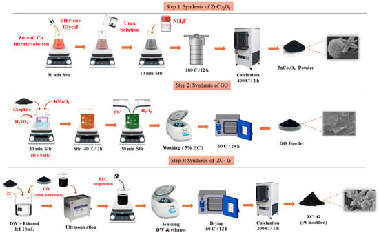
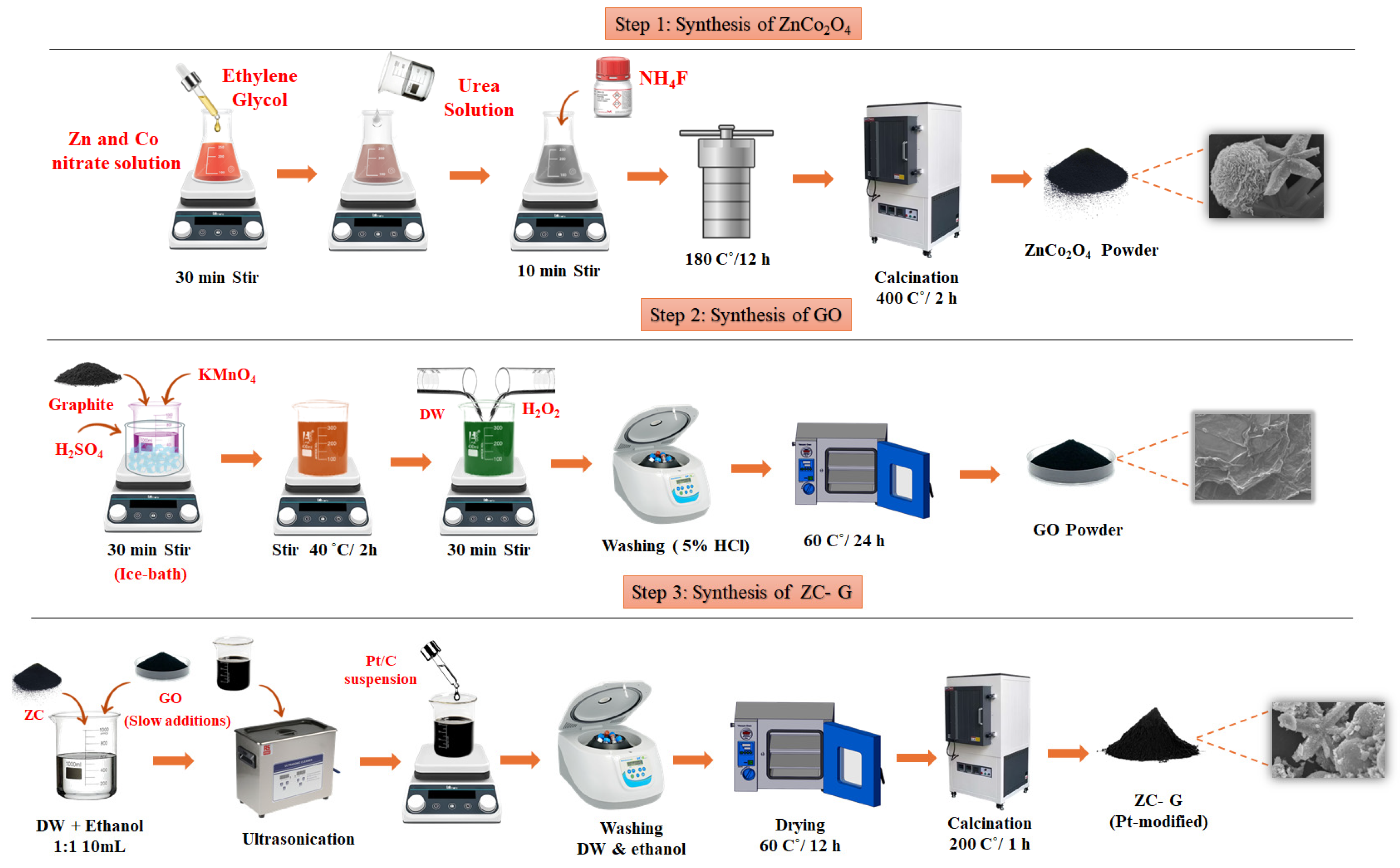
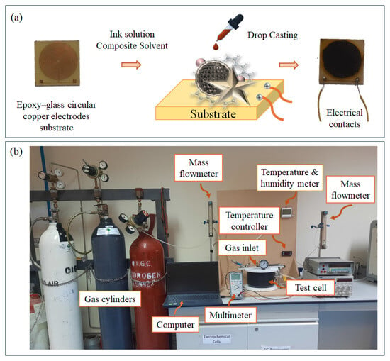
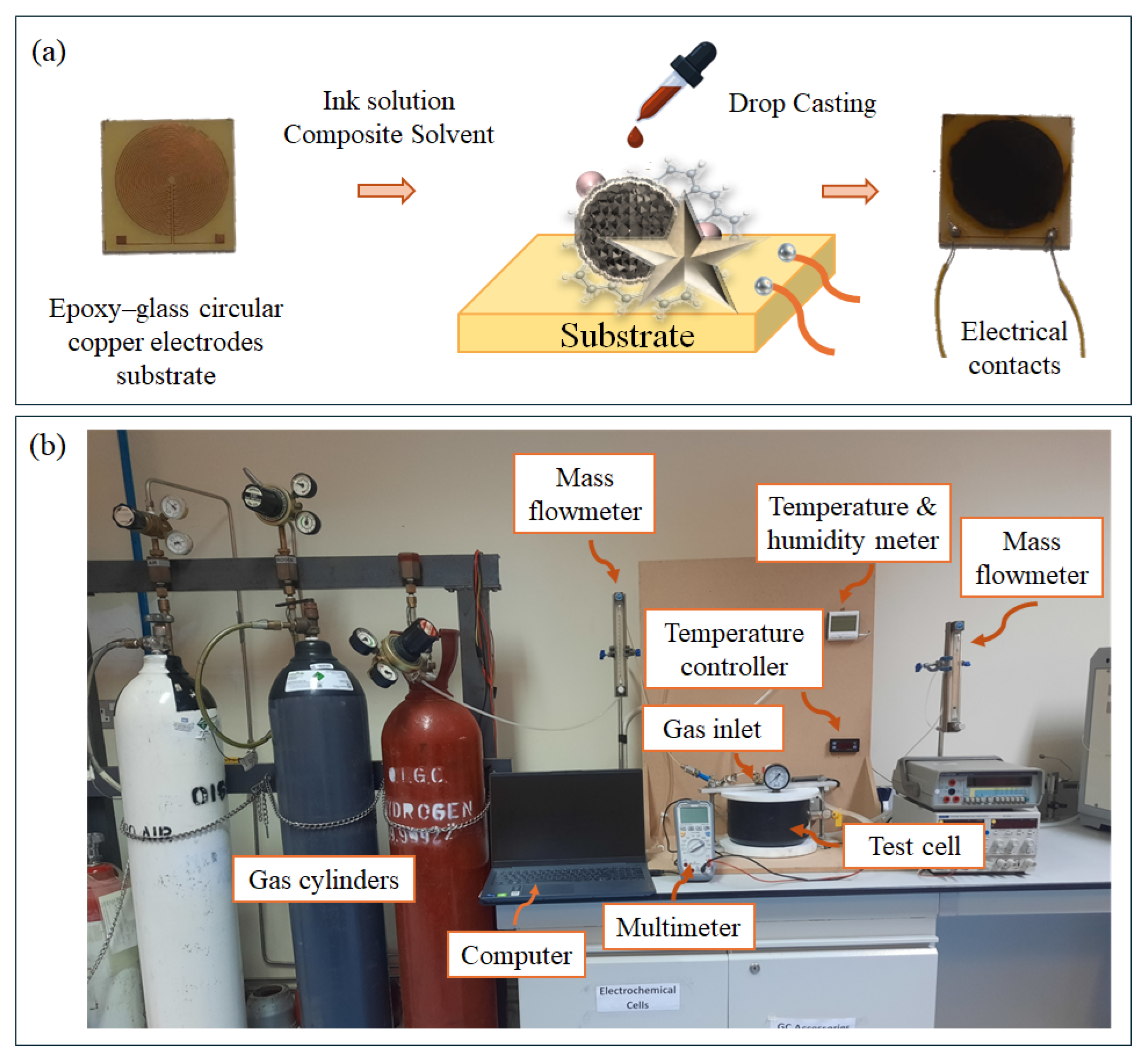
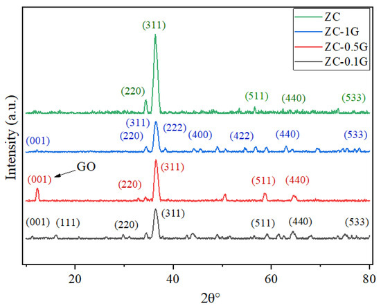
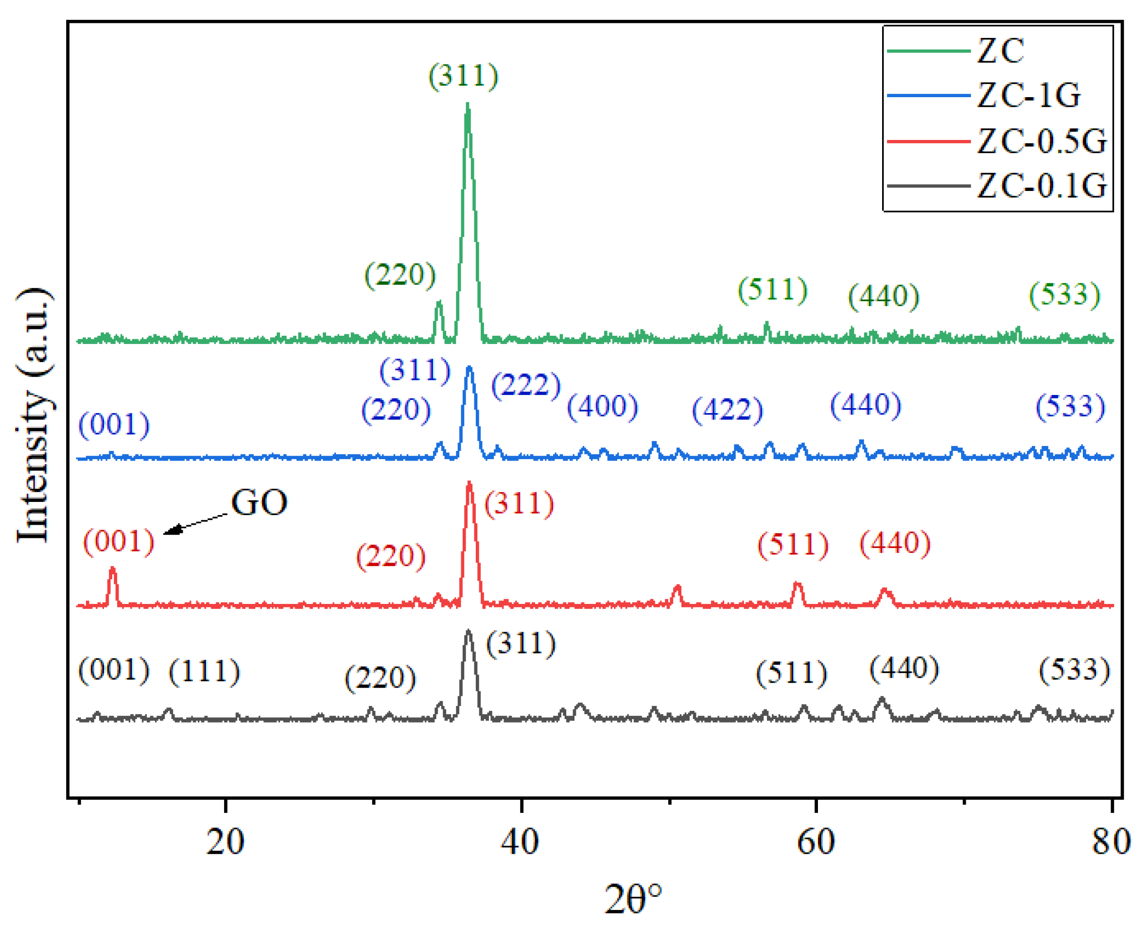
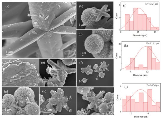
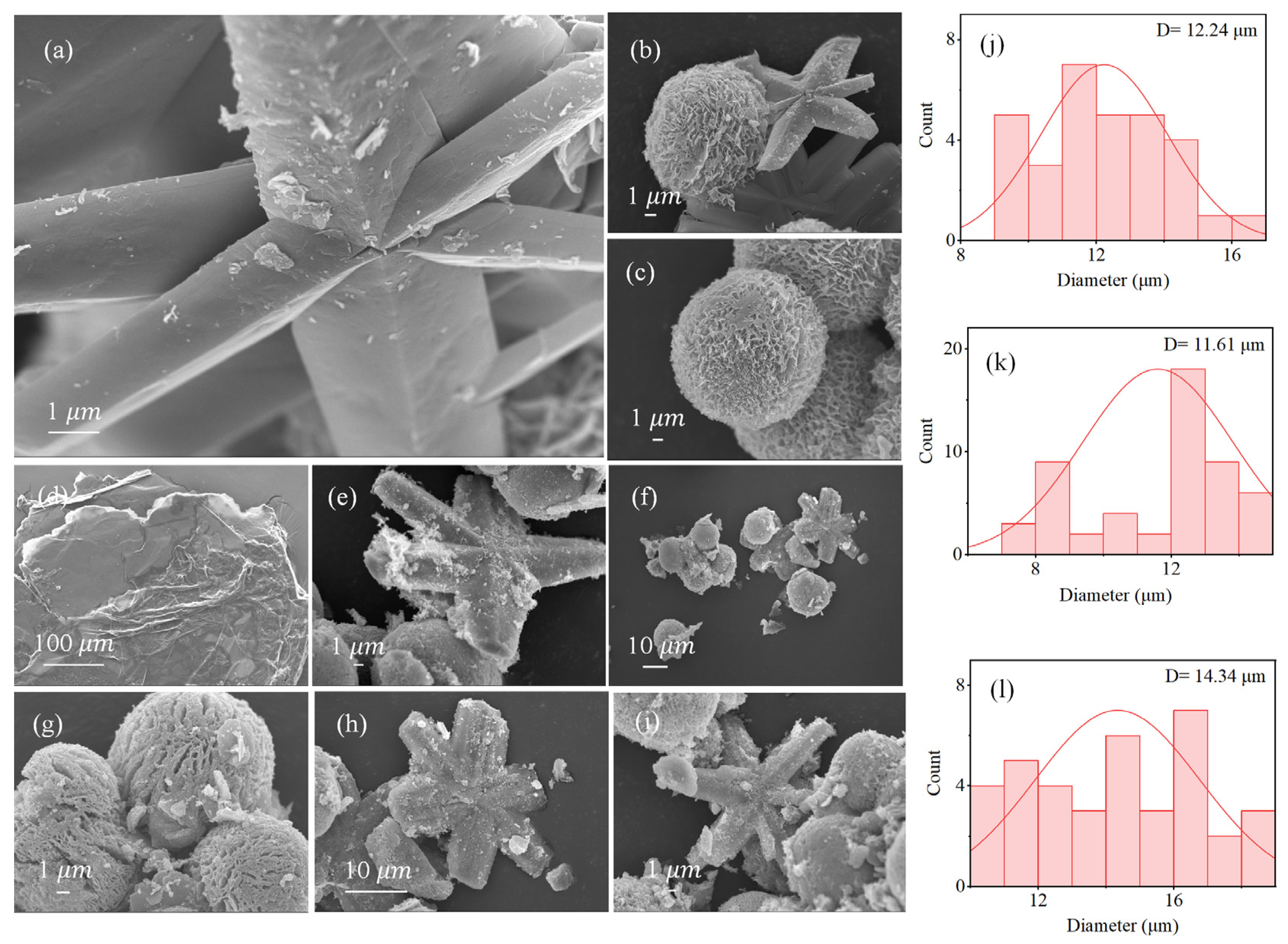
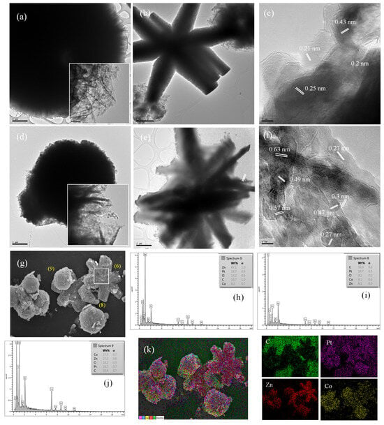
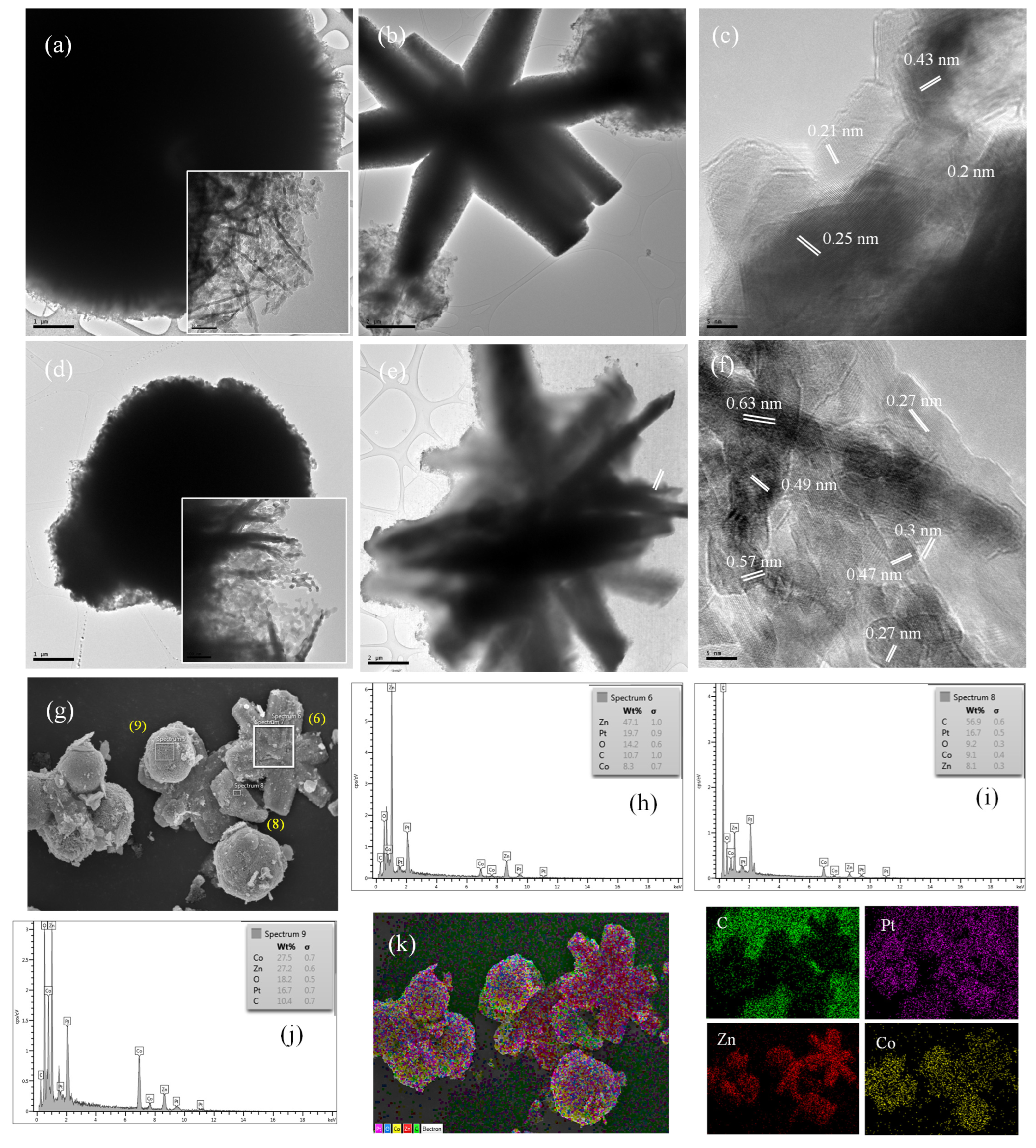
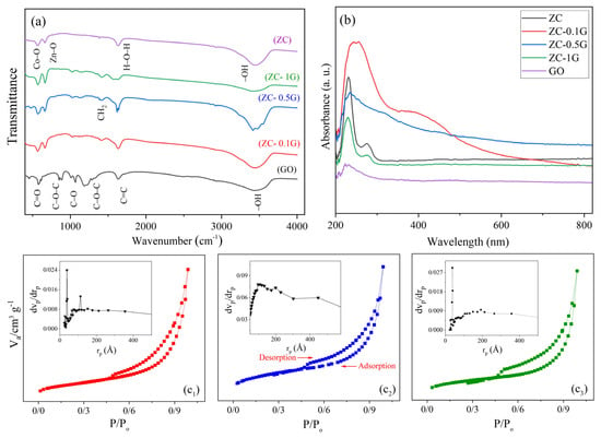
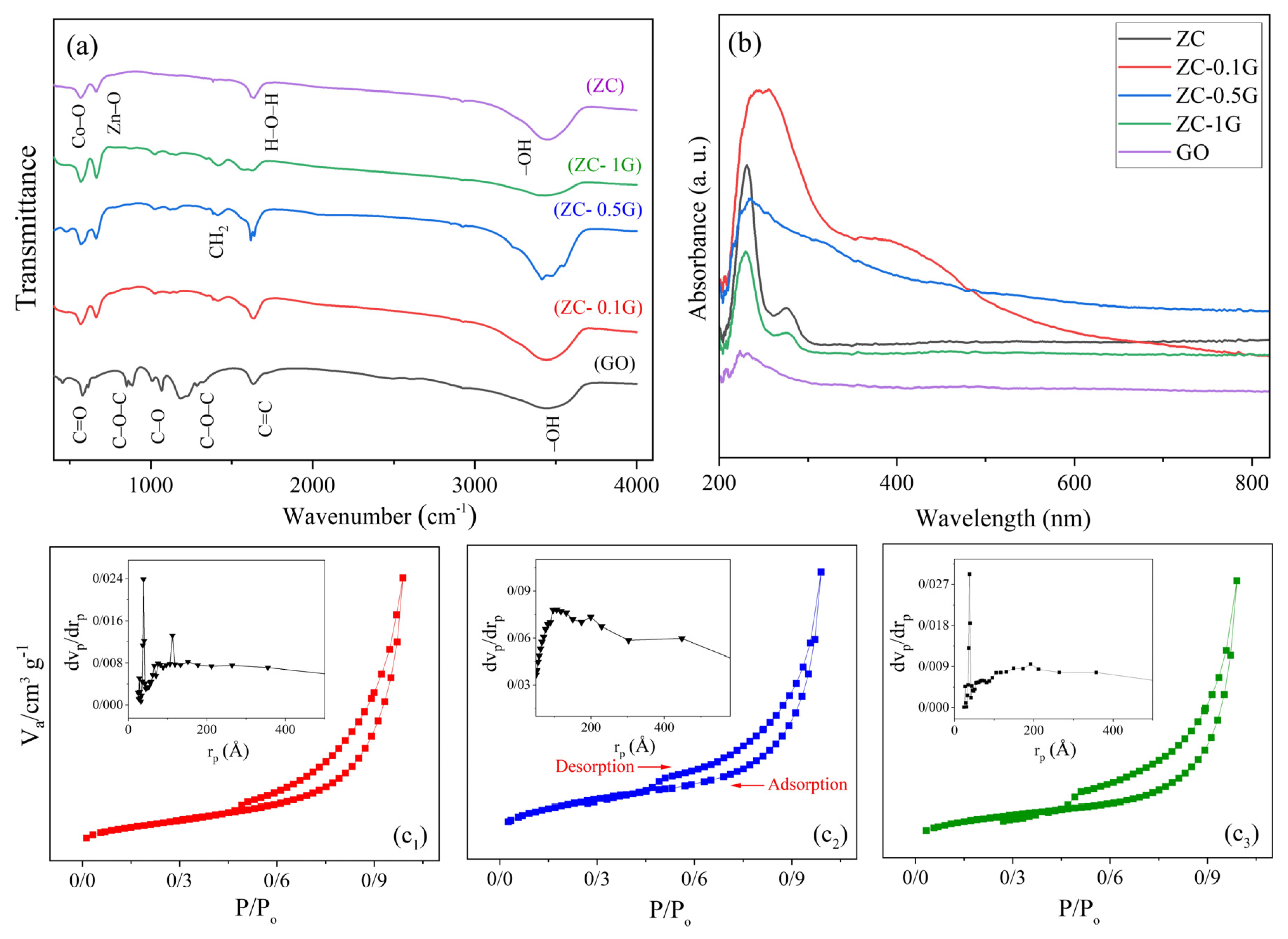
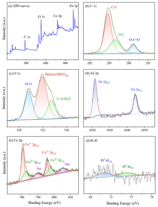
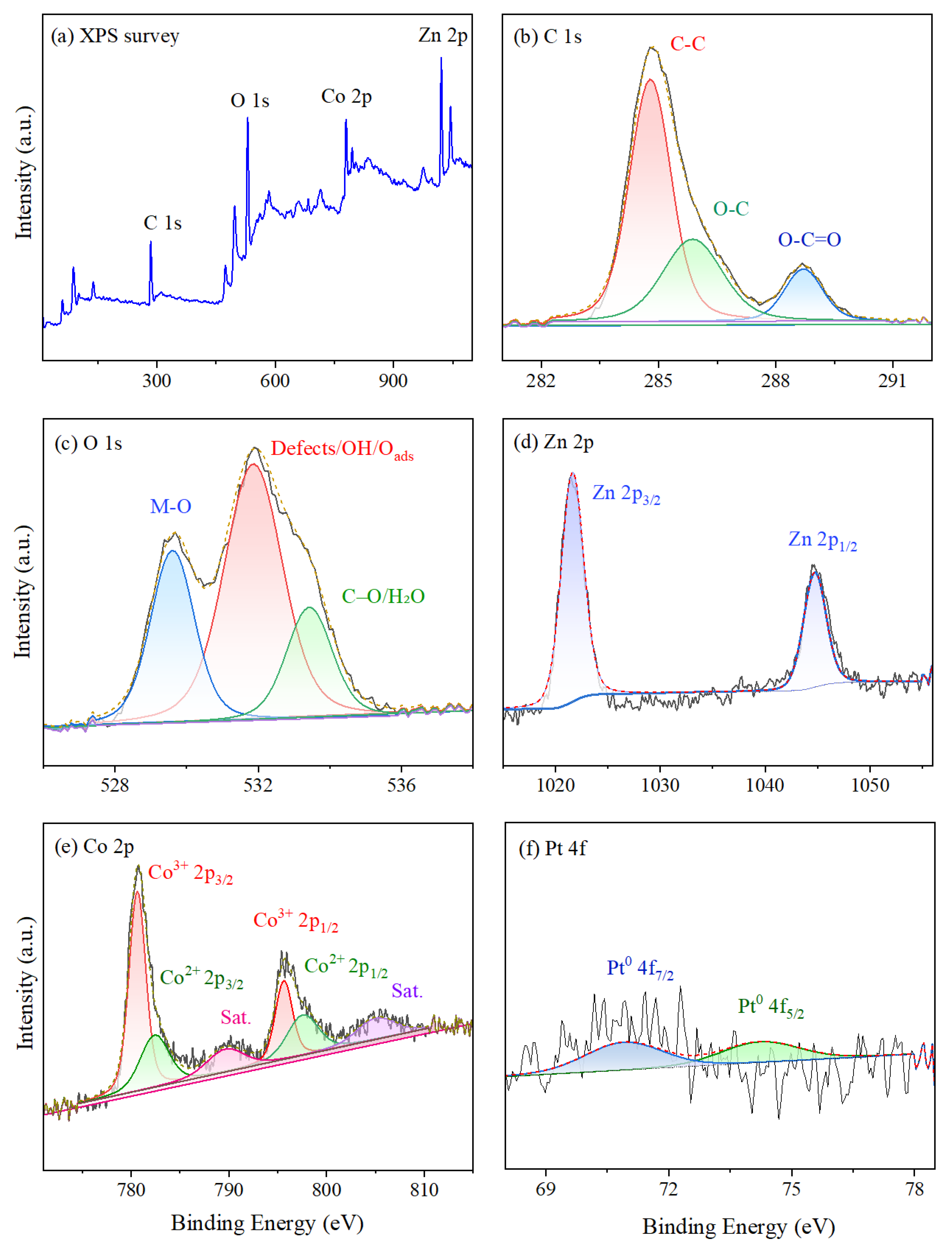
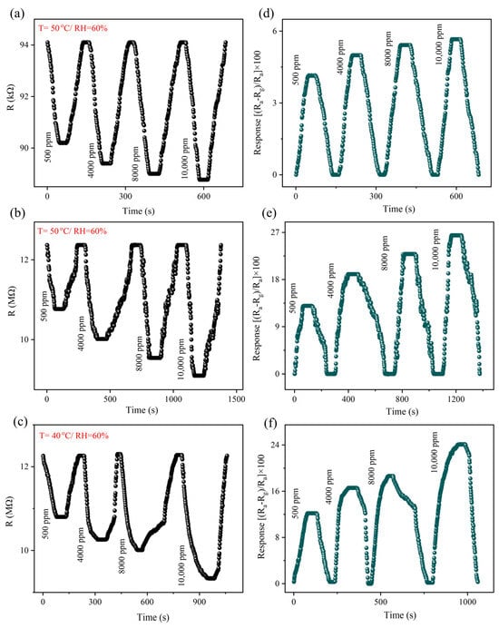
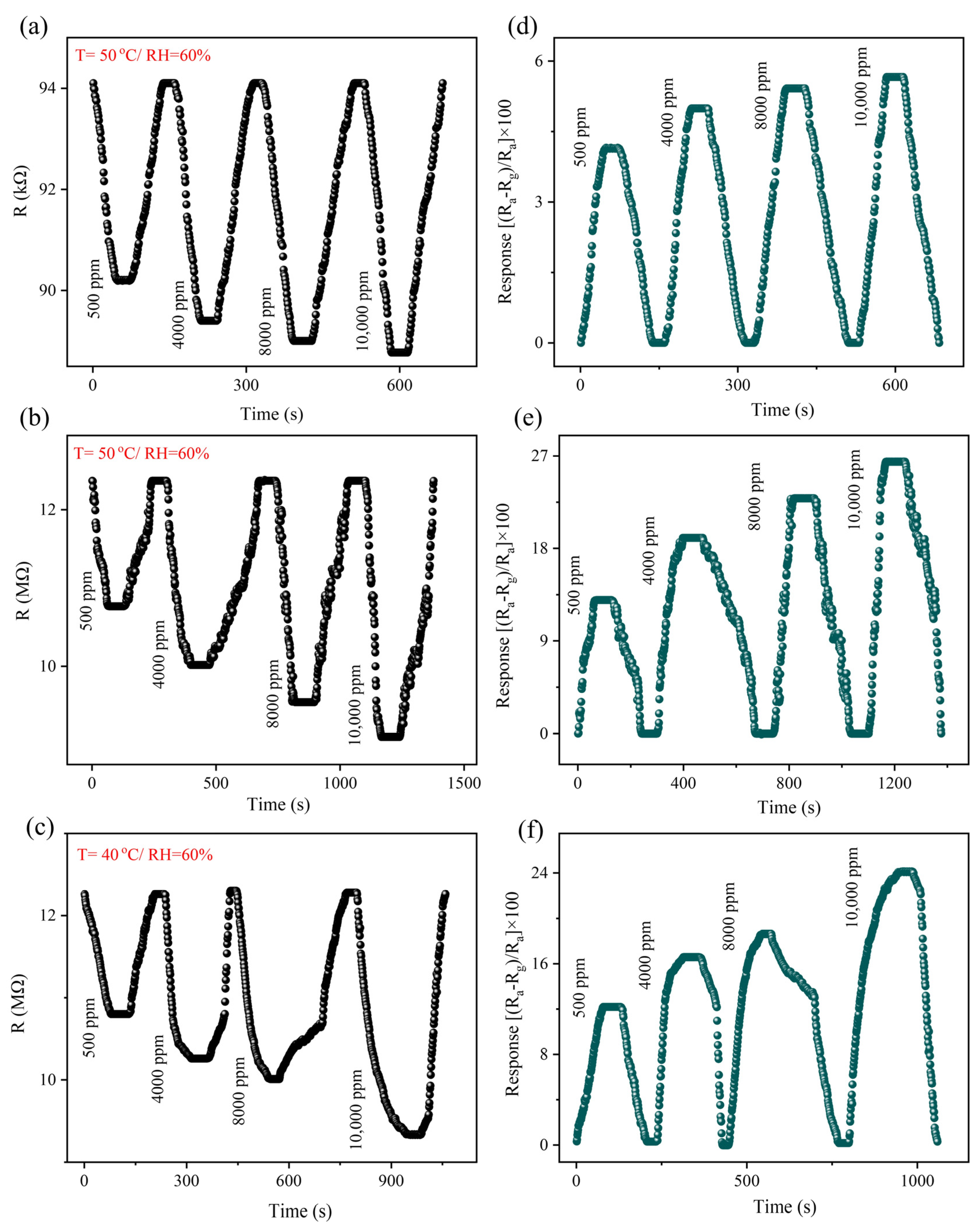
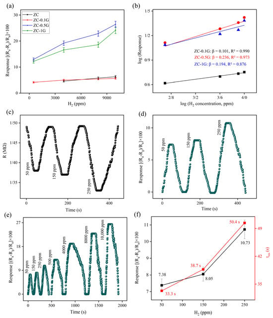
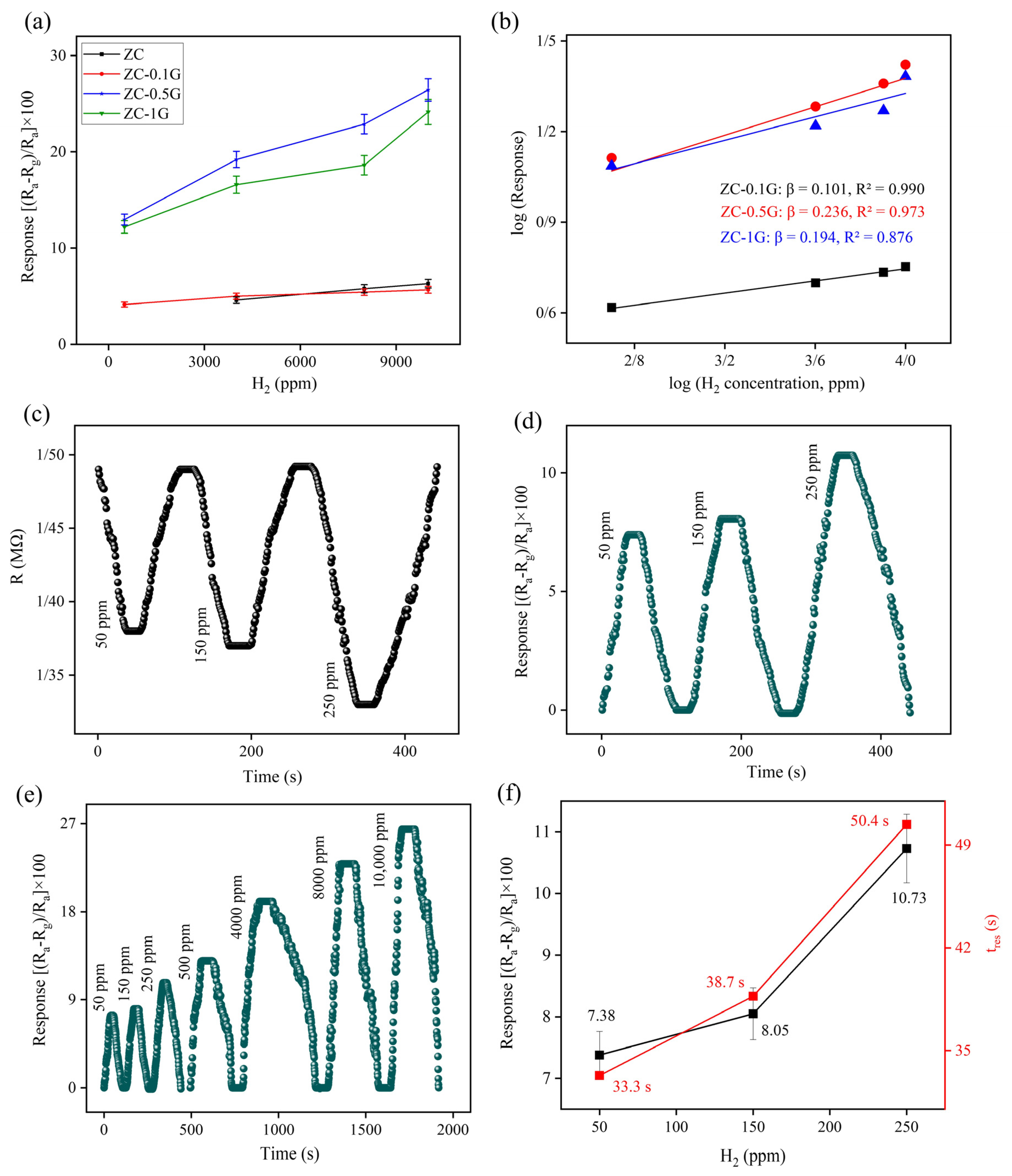
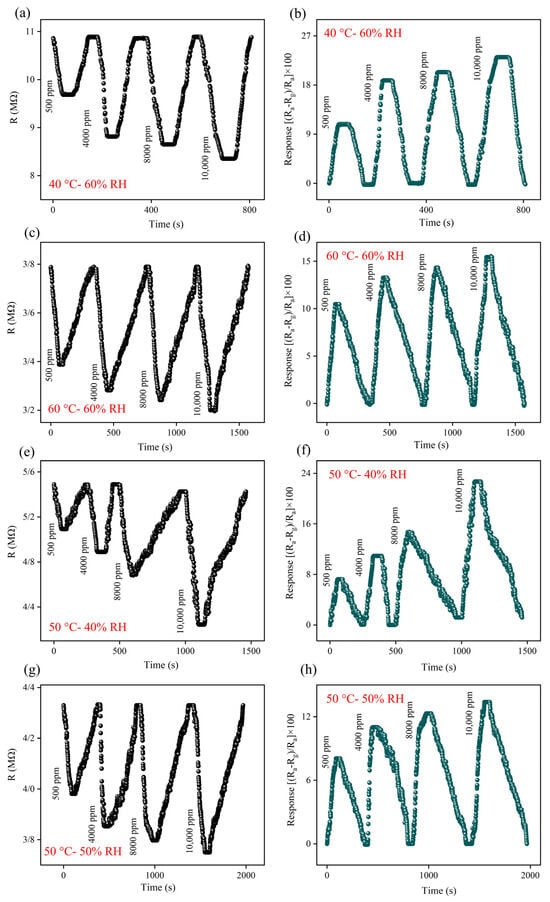
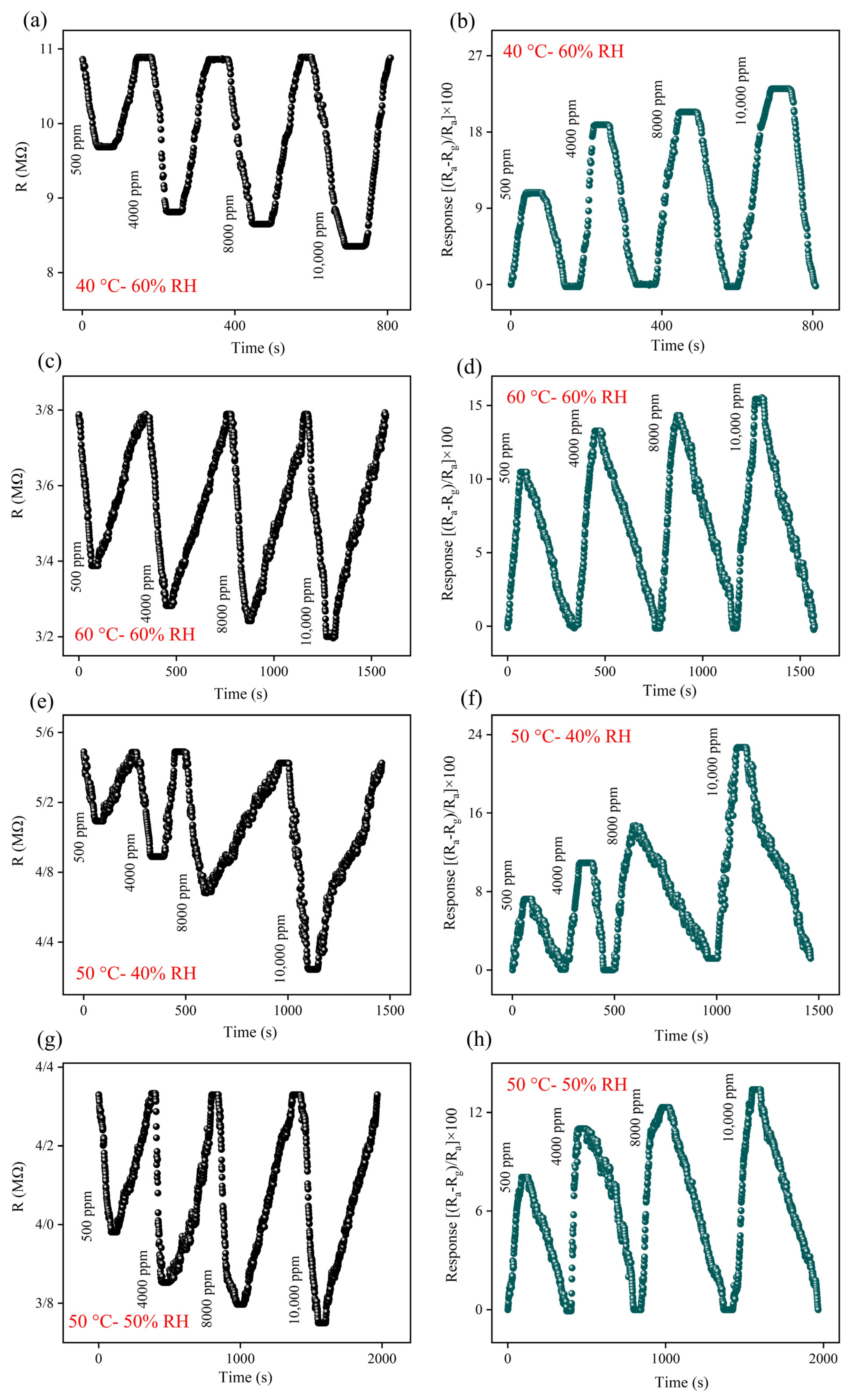
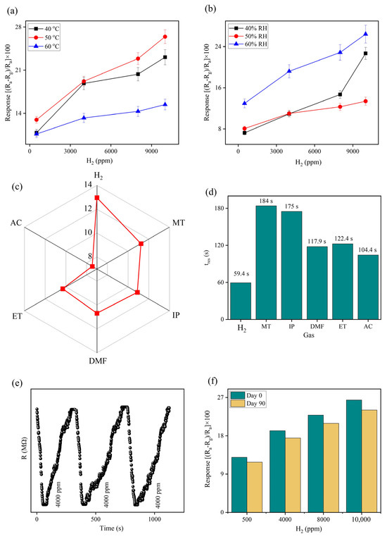
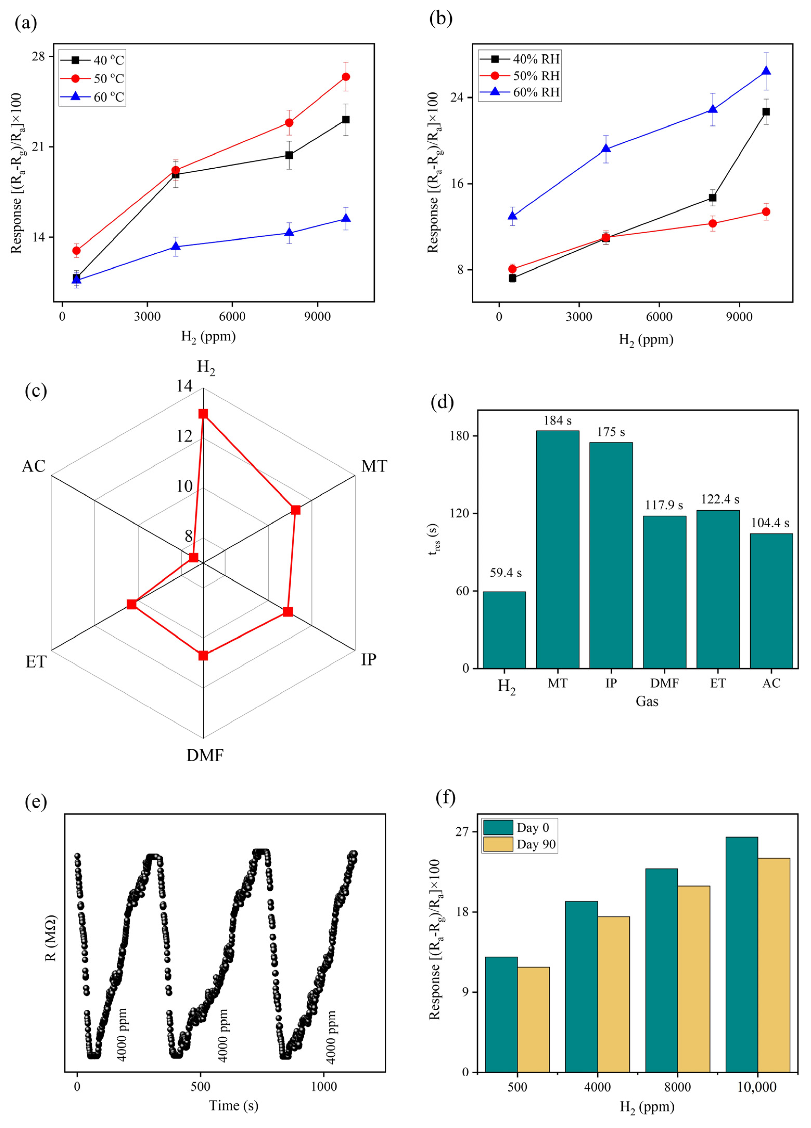
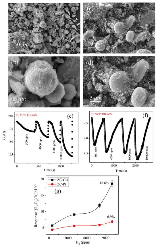
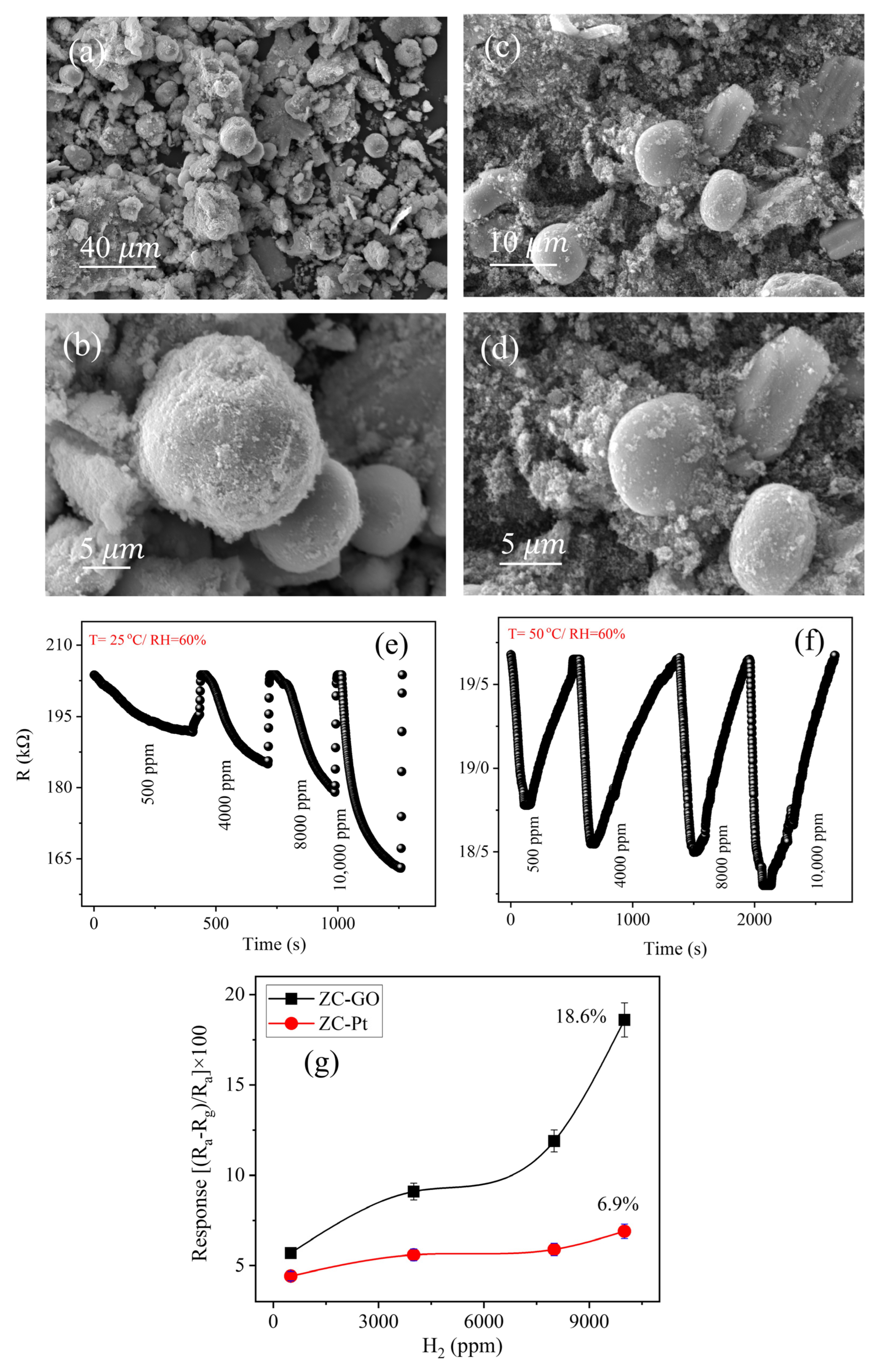
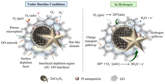

Disclaimer/Publisher’s Note: The statements, opinions and data contained in all publications are solely those of the individual author(s) and contributor(s) and not of MDPI and/or the editor(s). MDPI and/or the editor(s) disclaim responsibility for any injury to people or property resulting from any ideas, methods, instructions or products referred to in the content. |
© 2026 by the authors. Licensee MDPI, Basel, Switzerland. This article is an open access article distributed under the terms and conditions of the Creative Commons Attribution (CC BY) license.