


Abstract
Mideopsis roztoczensis Biesiadka & Kowalik, 1979 (Acari, Hydrachnidia, Mideopsidae) is a common and often abundant component of macroinvertebrate communities in Palearctic streams. Although the external morphology and genetic structure of this species are relatively well documented, knowledge of its internal anatomy remains limited, despite its importance for an integrative understanding of morphology and evolutionary diversity. The present study utilized histological techniques to investigate and describe the internal structures of M. roztoczensis based on newly collected material from Serbia. Histological analyses revealed the organization of several internal organs, including an extensive blind sac-like excretory organ that was morphologically distinct from the midgut, while a hindgut was not identified. Additionally, other structures, including dermal glands, the midgut, salivary glands, and the synganglion, were characterized. The observed variation in internal organization, particularly the morphology of the excretory organ and the number of paired dermal glands, provides additional characteristics in comparative morphological studies of water mite. This study contributes to our current knowledge of the internal anatomy of Hydrachnidia and highlights the value of histological approaches for future integrative studies of water mites.
1. Introduction
Freshwater water mites (Hydrachnidia) comprise more than 7500 described species grouped into 550 genera [1]. They inhabit a diverse range of aquatic environments, including lotic, lentic, temporary, and interstitial waters [2]. As a highly diverse group of aquatic invertebrates, Hydrachnidia represents a crucial component of freshwater ecosystems [3]. Their complex life cycle is divided into several developmental stages: egg, prelarva, larva, protonymph, deutonymph, tritonymph and adult. These stages are primarily defined by their state of activity [4], where larvae, deutonymphs, and adults are active, while protonymphs and tritonymphs remain inactive. These inactive stages offer a distinct evolutionary advantage by allowing mites to endure unfavorable conditions in stressful environments, thereby underpinning their wide diversity and broad distribution [3].
Water mites are highly sensitive to contaminants and physical or chemical alterations in water conditions, making them powerful bioindicators for assessing ecological status and habitat quality [3,5]. Because of their sensitivity to habitat degradation and water pollution, the conservation of freshwater mite diversity is closely linked to the preservation of freshwater ecosystems. This bioindicating value is further reinforced by their high abundance, a life cycle entirely dependent on aquatic ecosystems, and diverse ecological interactions with other biocenosis members across different developmental stages [6]. Consequently, numerous studies have demonstrated the effectiveness of water mites as indicators of water quality in both lotic and lentic systems [5,6,7,8].
Mideopsis roztoczensis Biesiadka & Kowalik, 1979 is a species within the water mite family Mideopsidae. It is widely distributed across running waters in Western and Central Europe, the Balkans, and Russia, with additional records from Turkey and Iran [9,10]. Recently, Pešić et al. [9] investigated the species-level phylogeny of the genus Mideopsis in Europe and confirmed M. roztoczensis as a single species exhibiting a pronounced phylogeographic structure.
Despite its wide distribution, the internal anatomy of M. roztoczensis has not been specifically investigated. The importance of describing internal structures in water mites has been emphasized by several authors, who highlight the necessity of integrative morphological approaches in acarology [11,12,13,14,15]. To address this gap, the present study aims to characterize the internal anatomy of M. roztoczensis using histological sections.
2. Materials and Methods
2.1. Water Mite Sampling and Morphological Analysis
Water mites were collected by hand-netting from the Kutinska River near Niš, Serbia (43°13′17.69″ N 22°01′40.62″ E). The sampling location is shown on Supplementary Figure S1.
Immediately after collection, specimens were preserved in 96% ethanol for subsequent molecular analysis, and 70% ethanol for histological analysis. The taxonomic status of Mideopsis roztoczensis was confirmed using a combination of DNA barcoding, based on the cytochrome c oxidase subunit I (COI) gene, and morphological analysis. DNA amplification and sequencing protocols for the COI gene followed Pešić et al. [16]. Morphological nomenclature follows Gerecke et al. [17]. All studied material is deposited in the research collection of the final author (VP) at the Department of Biology, University of Montenegro, Podgorica.
The species used in this study is not covered by institutional or national regulations governing the use of animals in research; therefore, no specific ethical approval was required for its use in laboratory experiments.
2.2. Histological Analyses
For internal morphology observation, five female water mite adults were subjected to standard protocol for paraffin tissue processing. Samples were dehydrated in series of ethanol with rising concentrations (80%, 90%, 96%, and 100%) and then transferred to toluene for 10 min. The samples were rinsed in tissue-embedding paraffin and left over night in an incubator at 58 °C. The next day, paraffin was changed, and the samples were embedded and cooled down for sectioning. Sections 5 µm thick were made on Leica® RM 2125RT microtome (Leica Microsystems, Wetzlar, Germany) and stained using standard Hematoxylin & Eosin method (H&E). Slides were analysed and photographed using light photomicroscope Leica® DM 2500.
3. Results
3.1. Internal Anatomy of Mideopsis roztoczensis
3.1.1. Digestive System
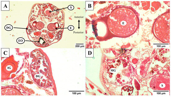
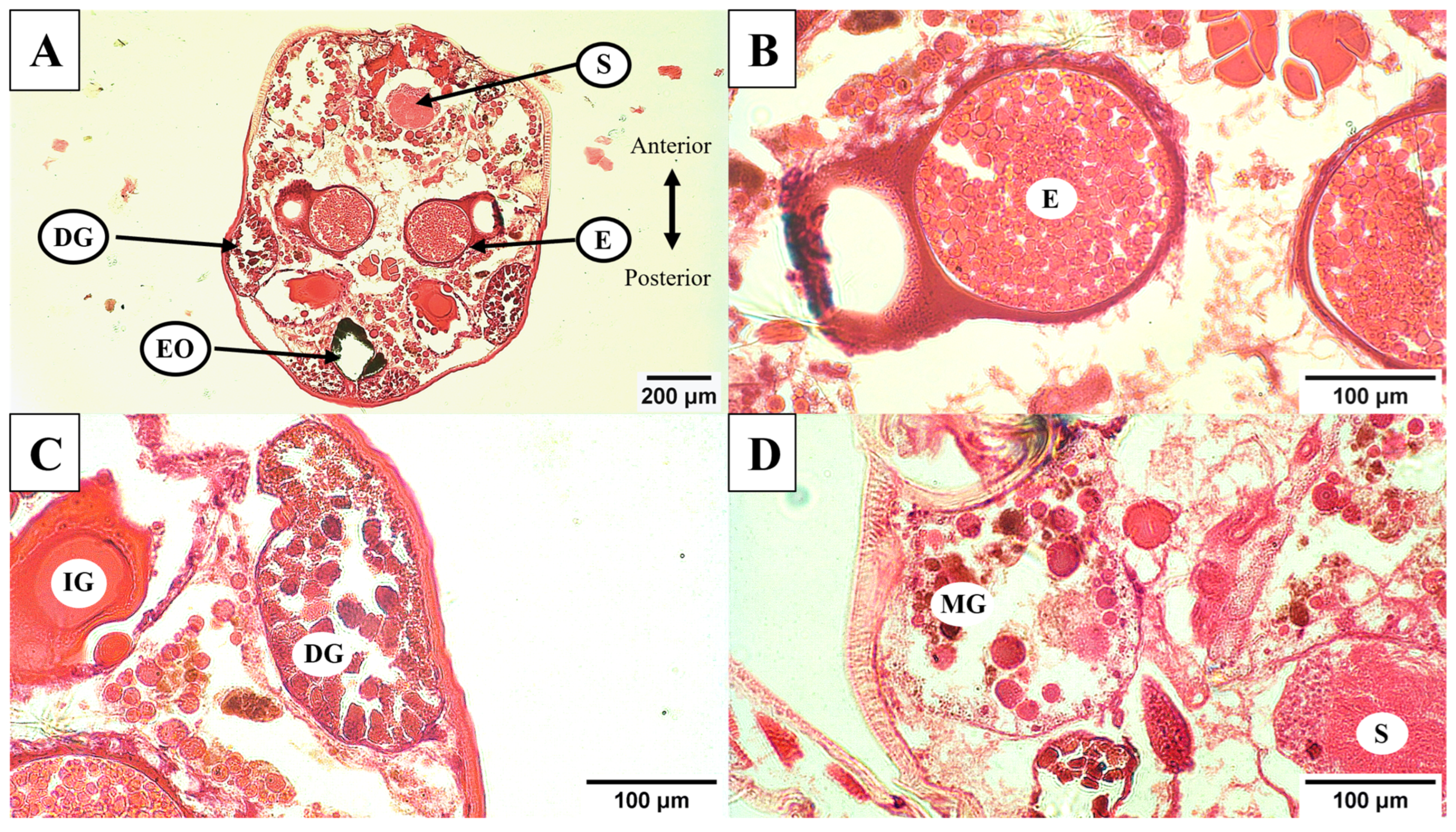
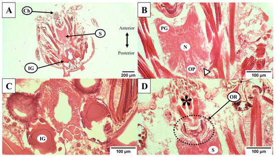
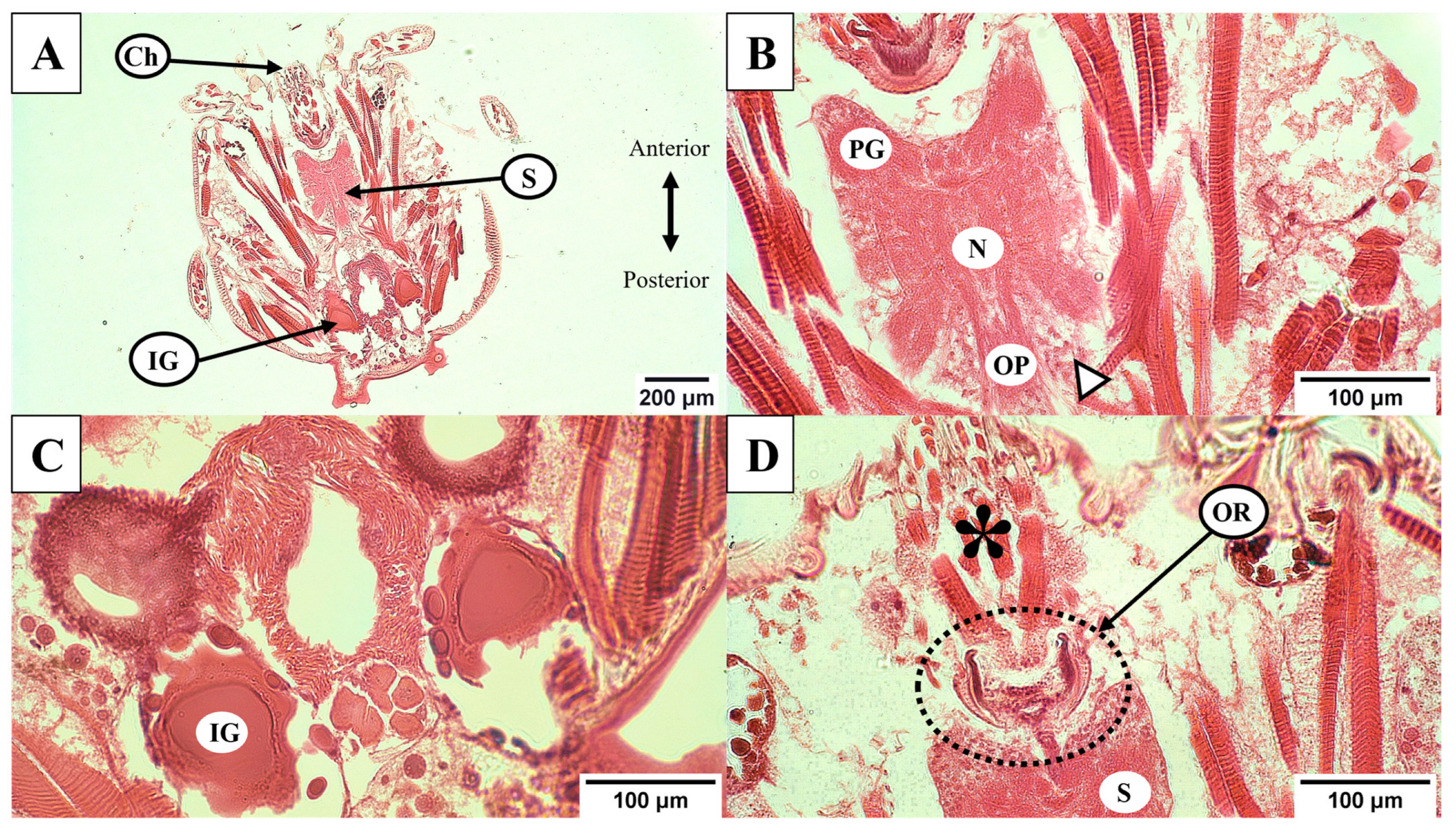
The digestive system began in the oral region, located ventrally at the base of the chelicerae. The midgut occupied a substantial portion of the water mite body cavity and represented the largest component of the digestive system observed in the examined sections. It was characterized by heterogeneous contents, including numerous vacuole-like structures and abundant inclusions differing in size, shape, and staining intensity (Figure 1D). A pair of muscular chelicerae extended anteriorly from this region (Figure 2A,D). No morphologically distinct hindgut was identified in the histological sections examined. Distinct microvilli were not observed.
Figure 1.
Longitudinal section of adult M. roztoczensis passing through the dorsal region of the body, showing the general organization of the internal organs. (A) In the anterior part of the body synganglion is clearly visible (S). Rounded structures interpreted as eggs (E) occupy the central region, whereas the excretory organ (EO) containing dark material is situated posteriorly. Common dermal glands (DG) are associated beneath the integument. (B) Round structure, interpreted as egg (E) filled with eosinophilic material. (C) Common dermal gland (DG) with granular material. (D) Midgut (MG) showing numerous inclusions.
Figure 2.
Longitudinal section of adult M. roztoczensis passing through the ventral region of the body, showing the general organization of the internal organs. (A) Section showing the synganglion (S), an idiosomal gland (IG), and prominent longitudinal muscle bundles surrounding the synganglion. (B) Higher magnification of the synganglion showing the neuropile (N), four pairs of pedal ganglia (PG), the cortical layer of the synganglion (arrowhead), and the opisthosomal nerve. (C) Pair of idiosomal glands (IG). (D) Anterior body region showing the synganglion (S) and oral region (OR) at the base of the chelicerae (dotted outline). Asterisk (*) indicates the muscles of the chelicerae.
3.1.2. Excretory System
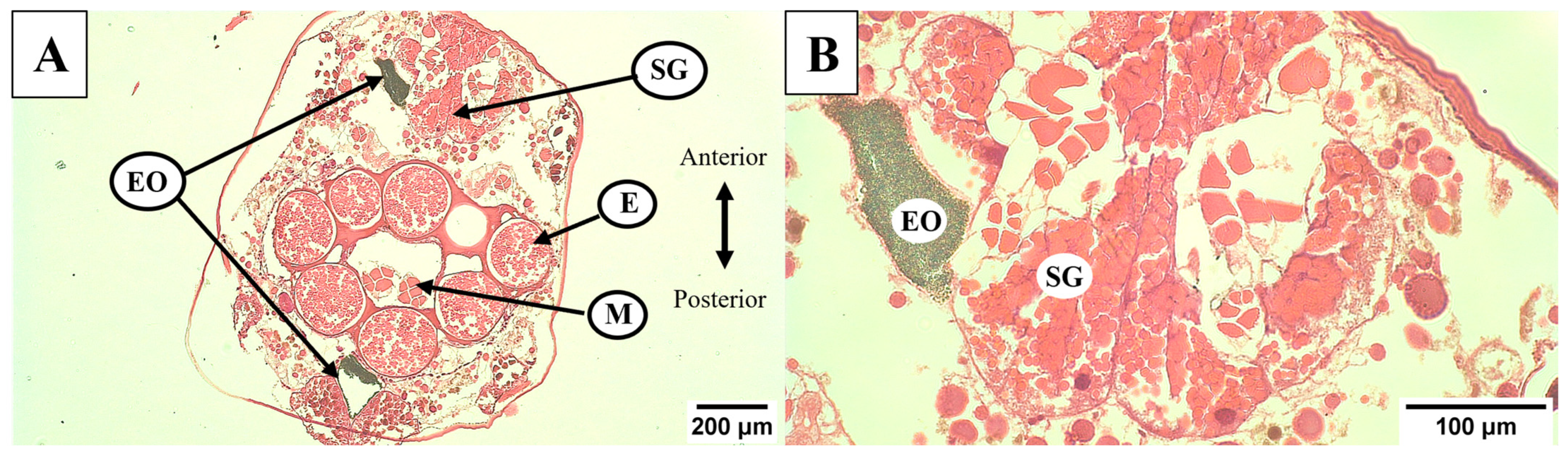
The excretory system of the M. roztoczensis was represented by a tubular structure located in the posterior region of the body (Figure 1A). The lumen appeared relatively spacious and contained numerous darkly stained inclusions, most of which were located adjacent to the organ wall (Figure 3A). From the posterior region, the structure extended anteriorly and branched into two blind extensions terminating near the synganglion (Figure 4C). The wall of the excretory organ was thin and clearly delineated from the surrounding tissue, forming a well-defined boundary.
Figure 3.
Histological sections of adult M. roztoczensis. (A) Longitudinal section through the dorsal region of the body, showing rounded structures interpreted as eggs (E) that are arranged around a central cavity, where two muscle bundles were observed (M). Excretory organ (EO), containing dark material, is situated posteriorly. Salivary glands (SG) are located in the anterior body region. (B) Higher magnification of the anterior body region showing the salivary gland with densely granular eosinophilic cytoplasm. Part of the excretory organ (EO) is also visible adjacent to the salivary gland, demonstrating that the organ extends anteriorly.
Figure 4.
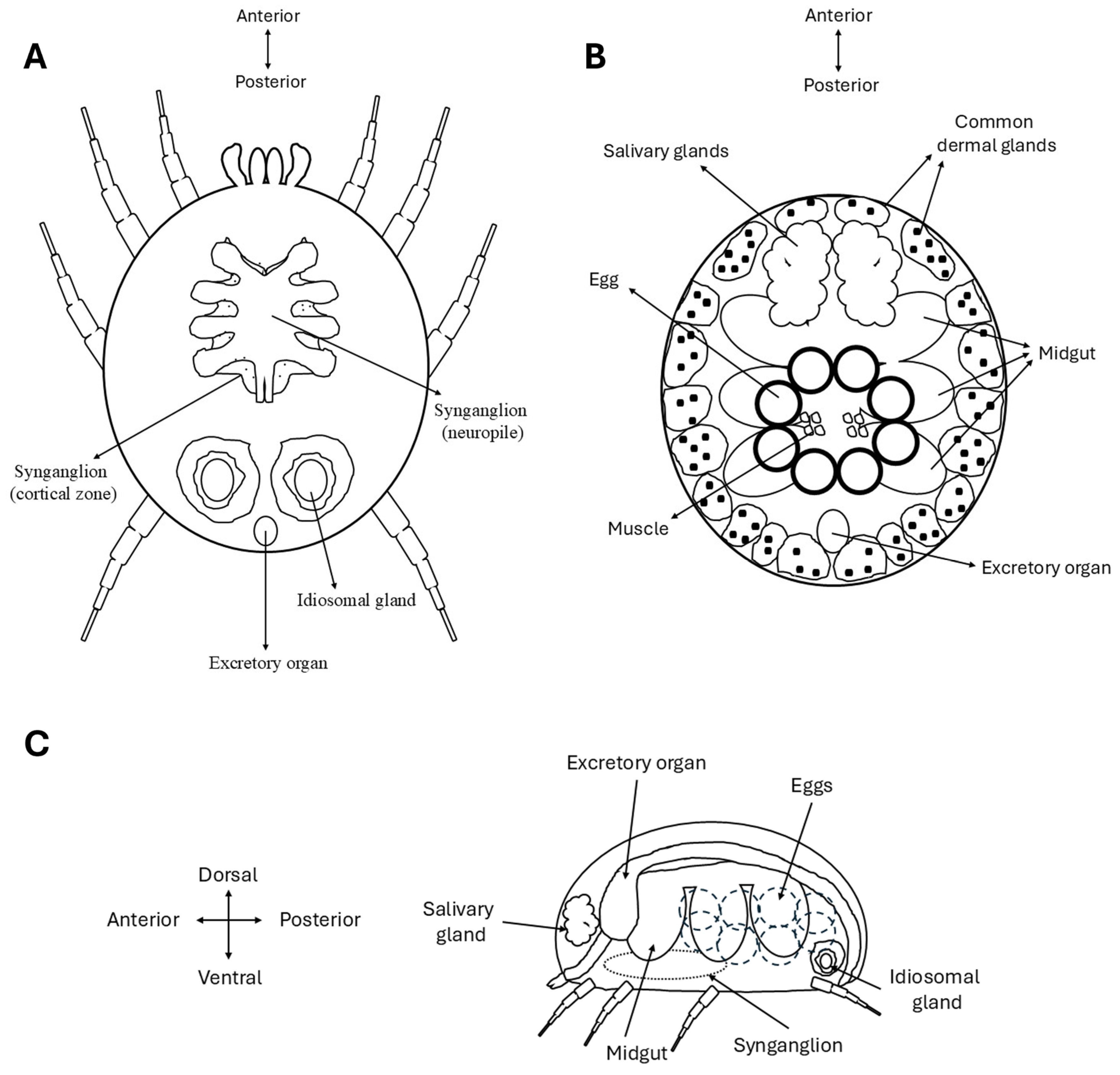
Schematic representation of the internal organization of adult M. roztoczensis based on serial histological sections. (A) Longitudinal section passing through the ventral region of the body. (B) Longitudinal section passing through the dorsal region of the body. (C) Lateral view illustrating the approximate spatial arrangement of the principal organ systems. The diagram is based on histological observations and is intended to illustrate the relative positions of the major anatomical structures; it is not drawn to scale.
3.1.3. The Nervous System
The main component of the nervous system was the central nervous mass, synganglion, located anteroventrally within the body cavity (Figure 2A,B and Figure 4A). Histological observations revealed two morphologically distinct layers that were recognizable within the central nerve mass. The external layer consisted of rounded cortical neuronal cell bodies that exhibited stronger basophilic staining affinity. In contrast, the inner neuropile was predominantly fibrous in appearance. From the central nervous mass, four pairs of pedal ganglia were observed extending distally, while a posteriorly directed nerve originating from the synganglion was interpreted as the opisthosomal nerve (Figure 2B).
3.1.4. Salivary Glands
A pair of well-developed salivary glands was observed in the anterolateral region of the body cavity (Figure 3A and Figure 4C). The glands were composed of closely arranged large glandular cells. The cytoplasm of these cells was strongly eosinophilic and densely packed with fine granular material. The characteristic morphology and staining properties of the salivary glands made them readily distinguishable from the surrounding tissue (Figure 3B).
3.1.5. Dermal Glands
Longitudinal histological sections of adult M. roztoczensis revealed 10 pairs of common dermal glands distributed along the body wall, as well as one pair of idiosomal glands (Figure 4B). The common dermal glands appeared as large structures lined by a thin epithelial layer. Their lumina contained abundant granular material that was mostly concentrated adjacent to the gland wall (Figure 1C).
In addition, a pair of large idiosomal glands was observed ventrally in the posterior region of the body. These glands exhibited a sac-like morphology and were lined by an epithelium surrounding a central lumen filled with homogeneous eosinophilic material. The eosinophilic material occupied more than a half of the lumen forming a large central mass with two clearly observed regions. The central region was composed of homogeneous material, whereas the peripheral region formed a distinct layer surrounding the central material. Large eosinophilic vesicle-like structures were also observed in the lumen, with some of them closely associated with the epithelial lining (Figure 2C).
3.1.6. Other Internal Structures
Histological analysis revealed eight rounded structures arranged radially around the central cavity (Figure 3A). Each structure was surrounded by homogeneous material and contained numerous eosinophilic granules of variable size (Figure 1B). Together, the eight structures formed a cluster that was enclosed by a well-defined basophilic boundary, separating them from other tissues (Figure 3A).
The central cavity was irregular in shape and remained visible through consecutive sections. No distinct epithelial or muscular lining of the central cavity could be recognized. Two transversely sectioned muscle bundles were observed in the same central region (Figure 3A).
4. Discussion
This study provides the first histological characterization of the internal anatomy of Mideopsis roztoczensis. The general organization of the major organ systems was largely consistent with previous descriptions of other water mites, including the digestive, nervous, excretory and reproductive systems [11,12,13,14,15,18,19,20,21,22]. However, several features observed in M. roztoczensis show differences or similarities of comparative interest and are discussed below in relation to previous morphological studies.
The organization of the oral region observed in the present study is consistent with previous descriptions of Hydrachnidia, where the mouth opening is situated at the base of chelicerae within the gnathosomal region and represents the beginning of the foregut [23]. In the examined specimens, the oral region was located immediately above the synganglion and at the base of the well-developed muscular chelicerae. Although the individual components of the foregut could not be unequivocally identified using the routine H&E histology, this topographical arrangement closely matches the organization of the anterior digestive tract previously described in water mites, where the pharynx continues into the oesophagus, which passes posteriorly through the synganglion [15].
The midgut occupied a substantial portion of the body cavity and represented the most prominent component of the digestive system in M. roztoczensis. Ultrastructural investigations of Teutonia cometes demonstrated that the digestive epithelium contains numerous intracellular structures associated with digestion and nutrient storage, while the gut lumen may contain heterogeneous digestive contents depending on the feeding conditions [12]. Similarly, histological studies of Unionicola aegyptiaca describe heterogenous luminal contents, including food material and free digestive cells [15]. The numerous vacuole-like structures and rounded inclusions observed in the midgut of M. roztoczensis may therefore represent either luminal digestive content or structures associated with intracellular digestion. Their precise nature, however, cannot be resolved by routine H&E histology. In contrast to these similarities in midgut appearance, the organization of the posterior digestive tract appears to vary considerably among water mite taxa.
Histological analyses did not reveal a morphologically distinct hindgut, as previously reported for some water mites [15]. Its apparent absence in M. roztoczensis may be functionally related to the feeding strategy of adult water mites. Predatory Hydrachnidia feed by piercing the prey, injecting digestive enzymes, and subsequently ingesting the liquefied tissues, while the empty integument is usually discarded [24]. Such a feeding strategy would generate relatively little undigested solid material, potentially reducing the functional importance of a well-developed hindgut. This interpretation is consistent with the predatory lifestyle of adult M. roztoczensis recently demonstrated in a predator-prey system with Chironomus riparius [25].
Instead, a dorsomedially positioned structure was observed in the posterior region of the body, corresponding to the location where the hindgut would typically be expected. The organ extended anteriorly and divided into two blind branches terminating near the synganglion, closely resembling the organization of the excretory organ described in Parasitengona. In several families within this cohort, the posterior region of the midgut has been reported to transform into dorsomedian excretory organ, which is considered homologous to the post-colon [18]. As reported in previous histological studies, the lumen contained dark granular waste material whose precise composition remains uncertain [11]. The development of the excretory organ may also be considered in relation to the feeding biology of adult M. roztoczensis. As already described, adult Hydrachnidia feed on liquefied tissues of animal prey following extraoral digestion, resulting in a protein-rich diet [24]. The metabolism of protein rich food generates considerable amounts of nitrogenic waste, which in mites is excreted predominantly as guanine [11]. Therefore, the distribution of the excretory organ throughout the body of M. roztoczensis may represent a functional adaptation that provides capacity for the accumulation and elimination of nitrogenous waste products. The lack of an observable continuity between the midgut and the excretory organ further supports the interpretation that, in adult M. roztoczensis, these structures are anatomically independent.
The synganglion of M. roztoczensis exhibited two histologically distinct regions corresponding to the cortical zone and neuropile. In addition, four pairs of pedal ganglia and a posterior opisthosomal nerve were recognized, indicating that the principal structural organization of the central nervous system can be reliably identified using routine H&E staining, without the need for ultrastructural analyses. The arrangement of these structures corresponds well with previous anatomical and ultrastructural descriptions of the acariform synganglion [14,15], suggesting a relatively conserved organization of the central nervous system among the water mite taxa examined so far.
The histological organization of the salivary glands in M. roztoczensis was generally consistent with that described for other water mites [15]. Their organization may be related to their functional importance during feeding. Salivary secretions play an important role in extraoral digestion, enabling enzymatic breakdown and liquefaction of prey tissues before ingestion [24]. Shatrov (2012) suggested that the considerable development of the salivary glands in Piona carnea reflects their important role in this process [26]. A similar functional association may apply to the salivary glands of M. roztoczensis, considering the predatory feeding strategy of adult water mites.
Ten pairs of common dermal glands were identified in adult M. roztoczensis, corresponding to the number reported for T. cometes, whereas 14 pairs have been described in Limnesia maculata [12,13]. This variation among water mite taxa supports previous observations that the number of common dermal glands is not uniform across Hydrachnidida. Shatrov (2013) proposed that a reduction in their number may represent an evolutionary trend in some lineages, suggesting its potential value as comparative morphological character within Hydrachnidia [12].
In addition to the common dermal glands, a morphologically distinct pair of idiosomal glands was identified in the posterior body region. The two gland types differed not only in their general morphology and position but also in the histological appearance of their luminal contents. While the common dermal glands contained predominantly granular material, the idiosomal glands were characterized by a large homogenous eosinophilic mass with distinct central and peripheral regions. These differences suggest that the two gland types produce secretory materials of different characteristics, consistent with previous ultrastructural observations in water mites [20]. However, their biological roles remain poorly understood. Several functions have been proposed for dermal glands, including chemical defense against predators, pheromonal communication, and protection against adverse environmental conditions. Secretion release has also been associated with temporary moistening of the body surface during exposure to desiccation [20].
Shatrov (2023) [20] emphasized the considerable adaptive plasticity of water mites and proposed that dermal glands represent an important and potentially multifunctional organ system contributing to their adaptation to diverse aquatic environments. However, understanding the diversity, functional specialization and evolutionary patterns of this system requires comparative investigation of the internal gland morphology in a substantially larger number of water mite species [20]. In this context, the present histological characterization of the common dermal and idiosomal glands of M. roztoczensis, including their number, distribution, morphology, and distinct luminal contents, provides additional data for such comparative analyses.
The interpretation of the eight rounded structures as developing eggs is supported by their morphology and internal contents. Histological observations in other mites have shown that developing eggs contain abundant yolk and lipid-rich material, producing a granular appearance in routine sections [27]. Female gonads in mites show considerable structural diversity [28]. More recently, Derdak et al. (2026) provided new insights into ovary organization specifically in Hydrachnidia, challenging earlier reports of a panoistic ovary in this group and demonstrating a meroistic ovary in all water mite species examined [28]. In this context, the identity of the central cavity around which the presumed eggs were arranged remains uncertain. Although its close spatial association with eggs suggests a possible relationship with the female reproductive system, its boundaries and continuity with other reproductive structures could not be unequivocally established.
The two transversely sectioned structures observed in the central region were identified as muscle bundles based on their compact eosinophilic appearance and comparison with muscle bundles identified in H&E-stained cross sections of other mites [29]. However, their three-dimensional course and spatial relationship to the female reproductive structures could not be reconstructed from available sections. Consequently, their specific function or association with the reproductive system cannot be determined from the present material.
The present study provides the first histological characterization of the internal organization of M. roztoczensis and shows that its major organ systems generally correspond to the anatomical organization previously described in other Hydrachnidia. The apparent absence of a morphologically distinct hindgut, together with the extensive distribution and anatomical separation of excretory organ from the midgut, may reflect functional specialization associated with the predatory feeding strategy of adult water mites. Common dermal and idiosomal glands showed pronounced differences in morphology and luminal contents, indicating distinct characteristics of their secretory material, although their specific functions cannot be determined from histological observations alone. Overall, the present findings add morphological data for M. roztoczensis to the still limited comparative information available on the internal anatomy of water mites.
Supplementary Materials
The following supporting information can be downloaded at https://www.mdpi.com/article/10.3390/d18090533/s1, Figure S1: Collection site of Mideopsis roztoczensis in the Kutinska reka near Niš, Serbia (43°13′17.69″ N, 22°01′40.62″ E, elevation: 269.4 m a.s.l.). Source: Google Earth (https://earth.google.com/web/).
Author Contributions
A.M.—Investigation, Methodology, Writing—original draft. J.S.—Methodology, Validation, Formal analysis, Writing—original draft. Đ.M.—Conceptualization, Project administration, Supervision, Validation, Writing—review and editing. D.S.-Z.—Conceptualization, Investigation, Validation, Writing—review and editing. V.P.—Supervision, Validation, Writing—review and editing. All authors have read and agreed to the published version of the manuscript.
Funding
This study was supported by the Serbian Ministry of Science, Technological Development and Innovation, Republic of Serbia, Grant no. 451-03-33/2026-03/200124, Grant no. 451-03-34/2026 03/200124, and by the Ministry of Science and Technological Development of Montenegro through the National Scientific Research Project: Advanced Monitoring of Microplastics in Karst Springs and Groundwater of Montenegro.
Data Availability Statement
No new data were created or analyzed in this study. Data sharing is not applicable to this article.
Acknowledgments
We would like to thank students of the Faculty of Sciences and Mathematics, Department of Biology and Ecology, in Niš for their help during different stages of the experiment. During the preparation of this work the authors used Chat GPT (GPT-5.6 Sol, OpenAI) in order to review the English language structure, tone and narrative flow. The authors have reviewed and edited the output and take full responsibility for the content of this publication.
Conflicts of Interest
The authors declare no conflicts of interest.
References
- Smit, H. Water mites of the world, with keys to the families, subfamilies, genera and subgenera (Acari: Hydrachnidia). Monogr. Ned. Entomol. Ver. 2020, 12, 1–774. [Google Scholar]
- Davids, C.; Di Sabatino, A.; Gerecke, R.; Gledhill, T.; Smit, H.; Van der Hammen, H. Acari: Hydrachnidia I. In Süßwasserfauna von Mitteleuropa; Gerecke, R., Ed.; Elsevier GmbH, Spektrum Akademischer Verlag: München, Germany, 2007; Volume 7, pp. 241–376. [Google Scholar] [CrossRef] [Scilit]
- Di Sabatino, A.; Gerecke, R.; Goldschmidt, T.; Matsumoto, N.; Cicolani, B. Global diversity of water mites (Acari, Hydrachnidia; Arachnida) in freshwater. In Freshwater Animal Diversity Assessment; Balian, E.V., Lévêque, C., Segers, H., Martens, K., Eds.; Springer: Dordrecht, The Netherlands, 2007; Volume 198. [Google Scholar] [CrossRef] [Scilit]
- Böttger, K. The general life cycle of freshwater mites (Hydrachnelliae, Acari). Acarologia 1976, 18, 496–502. [Google Scholar]
- Goldschmidt, T. Water mites (Acari, Hydrachnidia): Powerful but widely neglected bioindicators—A review. Neotrop. Biodivers. 2016, 2, 12–25. [Google Scholar] [CrossRef] [Scilit]
- Stenger, L.R.; Ferguson, F.M.; Skvarla, M. Water mites and their use as bioindicators of water quality conditions: A Pennsylvania case study. Acarologia 2024, 64, 146–163. [Google Scholar] [CrossRef] [Scilit]
- Miccoli, F.P.; Lombardo, P.; Cicolani, B. Indicator value of lotic water mites (Acari: Hydrachnidia) and their use in macroinvertebrate-based indices for water quality assessment purposes. Knowl. Manag. Aquat. Ecosyst. 2013, 411, 08. [Google Scholar] [CrossRef] [Scilit]
- Więcek, M.; Martin, P.; Lipiński, A. Water mites as potential long-term bioindicators in formerly drained and rewetted raised bogs. Ecol. Indic. 2013, 34, 332–335. [Google Scholar] [CrossRef] [Scilit]
- Pešić, V.; Bańkowska, A.; Zawal, A.; Rewicz, T.; Smit, H. Molecular diversity and species delimitation in the genus Mideopsis Neuman, 1880 in Europe (Acari, Hydrachnidia, Mideopsidae). Ecol. Montenegr. 2023, 70, 101–115. [Google Scholar] [CrossRef] [Scilit]
- Pešić, V.; Bankowska, A.; Goldschmidt, T.; Hårsaker, K.; Jovanović, M.; Kaitetzidou, E.; Krakowiak, M.; Kozłowska, A.; Michaloudi, E.; Michoński, G.; et al. Checklist of water mites from the Balkan Peninsula: Second supplement, new DNA barcodes and description of a new species. Zootaxa 2025, 5676, 1–74. [Google Scholar] [CrossRef] [Scilit]
- Shatrov, A.B. Comparative morphology, ultrastructure and functions of the excretory organ (postventricular midgut) in Parasitengona (Acariformes). Acarologia 2010, 50, 93–112. [Google Scholar] [CrossRef] [Scilit][Green Version]
- Shatrov, A.B. Anatomy and ultrastructure of dermal glands in an adult water mite, Teutonia cometes (Koch, 1837) (Acariformes: Hydrachnidia: Teutoniidae). Arthropod Struct. Dev. 2013, 42, 115–125. [Google Scholar] [CrossRef] [Scilit]
- Shatrov, A.B.; Soldatenko, E.V. Organization of dermal glands and characteristics of secretion in the freshwater mite Limnesia maculata (O. F. Müller, 1776) (Acariformes, Limnesiidae). J. Morphol. 2021, 263, 346–362. [Google Scholar] [CrossRef] [Scilit]
- Sonenshine, D.E.; Posada-Florez, F.; Laudier, D.; Gulbronson, C.J.; Ramsey, S.; Cook, S.C. Histological atlas of the internal anatomy of female Varroa destructor (Mesostigmata: Varroidae) mites in relation to feeding and reproduction. Ann. Entomol. Soc. Am. 2022, 115, 163–193. [Google Scholar] [CrossRef] [Scilit]
- Ramadan, S.A.; Ismai, T.G.; Mustafa, A.N. Histological studies of some internal organs from the freshwater mite, Unionicola aegyptiaca collected from the mussel, Caelatura aegyptiaca, Sohag, Egypt. Sohag J. Sci. 2021, 7, 15–23. [Google Scholar] [CrossRef] [Scilit]
- Pešić, V.; Zawal, A.; Bańkowska, A.; Araujo, R.; Sługocki, Ł.; Rewicz, T.; Krakowiak, M.; Michoński, G.; Girão, D.; da Silva, L.P.; et al. Exploring the water mite fauna (Acari, Hydrachnidia) of the Madeira archipelago: DNA barcoding reveals a remarkable species endemicity. Zootaxa 2025, 5621, 501–513. [Google Scholar] [CrossRef] [Scilit]
- Gerecke, R.; Gledhill, T.; Pešić, V.; Smit, H. Chelicerata: Acari III. In Süßwasserfauna von Mitteleuropa; Gerecke, R., Ed.; Springer-Verlag: Berlin/Heidelberg, Germany, 2016; Volume 7/2-3, pp. 1–429. [Google Scholar] [CrossRef] [Scilit]
- Filimonova, S. Fine structure of the posterior midgut in the mite Anystis baccarum (L.). Arthropod Struct. Dev. 2023, 72, 101218. [Google Scholar] [CrossRef] [Scilit]
- Sabelis, M.W.; Bruin, J. The water mite family Pontarachnidae, with new data on its peculiar morphological structures (Acari: Hydrachnidia). In Trends in Acarology; Springer: Dordrecht, The Netherlands, 2010; pp. 71–79. [Google Scholar] [CrossRef] [Scilit]
- Shatrov, A.B. Secretion of dermal glands in freshwater mites (Acariformes, Parasitengonina, Hydrachnidia) and its structural characteristics. Parazitologiya 2023, 57, 319–336. [Google Scholar] [CrossRef] [Scilit]
- Polilov, A.A.; Makarova, A.A. Constant neuropilar ratio in the insect brain. Sci. Rep. 2020, 10, 21424. [Google Scholar] [CrossRef] [Scilit]
- Huang, B.; Li, M.; Jiang, X.; Zhang, B.; Huang, Y.; Xu, X. Reproductive structures of female Phytoseiulus persimilis (Acari: Phytoseiidae) and the development of egg and embryo in the body. Agriculture 2024, 14, 1647. [Google Scholar] [CrossRef] [Scilit]
- Shatrov, A.B.; Soldatenko, E.L.; Petrov, A.A. Functional morphology and ultrastructure of the mouth apparatus in the freshwater mite larvae Limnochares aquatica (L., 1758) (Acariformes, Limnocharidae). Arthropod Struct. Dev. 2024, 78, 101331. [Google Scholar] [CrossRef] [Scilit]
- Proctor, H.C. Hydrachnida (Water Mites). In Encyclopedia of Inland Waters; Likens, G.E., Ed.; Elsevier: Oxford, UK, 2009; Volume 2, pp. 335–345. [Google Scholar] [CrossRef] [Scilit]
- Zdravković-Savić, D.; Pešić, V.; Manović, A.; Samardžić, A.; Stojanović, J.; Nikolić, M.; Onjia, A.; Milošević, D. Biotic interactions regulate the fate and trophic transfer of particulate contaminants in sediment–water systems: An overlooked role of water mite–chironomid interactions. Aquat. Toxicol. 2026, 299, 107964. [Google Scholar] [CrossRef] [Scilit]
- Shatrov, A. Anatomy and ultrastructure of the salivary (prosomal) glands in unfed water mite larvae Piona carnea (C.L. Koch, 1836) (Acariformes: Pionidae). Zool. Anz. A J. Comp. Zool. 2012, 251, 279–287. [Google Scholar] [CrossRef] [Scilit]
- Toyoshima, S.; Nakamura, M.; Nagahama, Y.; Amano, H. Process of egg formation in the female body cavity and fertilization in male eggs of Phytoseiulus persimilis (Acari: Phytoseiidae). Exp. Appl. Acarol. 2000, 24, 441–451. [Google Scholar] [CrossRef] [Scilit]
- Derdak, A.; Mąkol, J.; Małota, K.; Jędrzejowska, I. The ovary structure and oogenesis in Hydrachnidia (Acariformes: Parasitengona). J. Anat. 2026. [Google Scholar] [CrossRef] [Scilit]
- Gisder, S.; Genersch, E. Direct Evidence for Infection of Varroa destructor Mites with the Bee-Pathogenic Deformed Wing Virus Variant B—But Not Variant A—Via Fluorescence-in situ-Hybridization Analysis. J. Virol. 2021, 95, 10–1128. [Google Scholar] [CrossRef] [Scilit]
Disclaimer/Publisher’s Note: The statements, opinions and data contained in all publications are solely those of the individual author(s) and contributor(s) and not of MDPI and/or the editor(s). MDPI and/or the editor(s) disclaim responsibility for any injury to people or property resulting from any ideas, methods, instructions or products referred to in the content. |
© 2026 by the authors. Licensee MDPI, Basel, Switzerland. This article is an open access article distributed under the terms and conditions of the Creative Commons Attribution (CC BY) license.