


Abstract
The genus Trypanosoma includes several protozoans that cause infections of both medical and veterinary importance. Human African trypanosomiasis (sleeping sickness), caused by Trypanosoma brucei gambiense and Trypanosoma brucei rhodesiense, and human American trypanosomiasis (Chagas disease), caused by Trypanosoma cruzi, are neglected tropical diseases that affect millions of residents of Africa and Latin America as well as of other parts of the world including West Asia. A multitude of trypanosomes, such as Trypanosoma evansi, Trypanosoma vivax, and Trypanosoma congolense, are pathogenic for various classes of vertebrates, from fish to mammals. The aim of this systematic review is to provide information on the epidemiology and distribution of Trypanosoma spp. in 18 countries of West Asia whereby published information was retrieved from 75 scientific papers published in the period 1971–2024. We found 25 Trypanosoma spp. of both known and yet unknown pathogenicity reported from 13 countries in the region: Azerbaijan, Bahrain, Iraq, Israel, Jordan, Kuwait, Oman, Qatar, Saudi Arabia, Palestine, Syria, Türkiye, and the United Arab Emirates. No case of human trypanosomiasis has been published thus far from these countries. Nearly 80% (61 out of 75) of all the available publications were dedicated to the investigation of Trypanosoma spp. in mammals, primarily livestock (camels and cattle in particular) and pets. Trypanosoma evansi can be considered the most widely studied pathogenic microorganism within the genus Trypanosoma in the region, with 46 papers devoted to this pathogen. Notably, 60% of the screened publications emanated from just two countries, namely Iraq and Saudi Arabia. The reported diversity of trypanosomes highlights the complexity of their transmission and host interactions in the region, with important implications for animal and human health and food security, underscoring the need for coordinated One Health surveillance and interventions.
1. Introduction
Neglected tropical diseases (NTD) are diseases caused by a variety of pathogens that have major human health implications primarily in tropical, impoverished countries [1]. Neglected tropical diseases caused by protozoans are of great importance since they affect over a billion people throughout the world. Neglected tropical diseases impose an enormously heavy burden on people living in endemic areas, causing substantial morbidity, and in some cases, significant mortality [2]. Climate change and socio-political instabilities can drive emergence and re-emergence in places where causative pathogens have never been present or where they had previously been eliminated [3]. The complex etiology of NTDs necessitates interdisciplinary investigations within a One Health framework that integrates biological, medical, veterinary, social, and political sciences [3,4,5].
Parasites from the genus Trypanosoma are well-documented pathogens of medical and veterinary importance. They have complex life cycles involving an array of invertebrate and vertebrate hosts that can carry trypanosomes of unknown pathogenicity [6,7]. This genus was first discovered in 1843 by Gruby in the blood of a dying rabbit [8]. Several African Trypanosoma species are transmitted by tsetse flies (Glossina spp.), which serve as the principal biological vectors of African trypanosomiasis [9,10]. Trypanosomes are unicellular, flagellated organisms having simple and distinctive “V”-shaped body structure, featuring a single nucleus and a kinetoplast, a specialized organelle containing mitochondrial DNA. The kinetoplast is involved in several metabolic functions [11]. The life cycle of trypanosomes involves different stages in the vertebrate host and vector. In the bloodstream of the host, they assume a slender shape and reproduce by binary fission. They can also attack the host immune system by changing their surface antigens. When trypanosomes are ingested by the vector, they transform into a procyclic form in the midgut, where they start multiplying and differentiate into epimastigotes and metacyclic trypomastigotes. The latter stages are infective and may be transmitted when the vector bites other hosts. Subsequently, the metacyclic trypomastigotes enter host cells and transform into amastigotes and start replicating inside the cell. As a result, these cells burst and release more trypomastigotes into the bloodstream of the host [8,12,13,14]. Pathogenic trypanosomes have been primarily found in Africa, Asia, the Middle East, and the Americas; however, occasional cases and outbreaks have been reported from Europe and Oceania [7,9]. Trypanosoma spp. causing diseases in humans and animals are mainly transmitted by arthropod vectors, most of which are flying insects (tsetse flies, triatomine bugs, stable flies, horseflies, etc.); however, ticks are also suspected to carry and transmit them [10,15,16]. In a recent study conducted in the United Arab Emirates, Trypanosoma spp. DNA was not detected in any of the tick samples analyzed. This finding is consistent with the current understanding that ticks are unlikely to play a significant role in the transmission of trypanosomes in the UAE, where tsetse flies—the primary vectors of many Trypanosoma species—are absent [17]. The major diseases of medical importance are sleeping sickness (also known as human African trypanosomiasis), which is caused by subspecies of Trypanosoma brucei, and Chagas disease, or American trypanosomiasis, caused by Trypanosoma cruzi [18,19]. In terms of animal health, three infectious diseases have been linked with trypanosomal agents, namely, nagana, which can be caused by a variety of trypanosomes that will be described in detail below; surra, which is only related to T. evansi; and dourine, caused by Trypanosoma equiperdum [20].
West Asia is a fast growing and highly populated region with more than 300 million residents that includes Armenia, Azerbaijan, Bahrain, Cyprus, Georgia, Iraq, Israel, Jordan, Kuwait, Lebanon, Oman, State of Palestine, Qatar, Saudi Arabia, Syrian Arab Republic, Türkiye, United Arab Emirates, and Yemen (https://unstats.un.org/unsd/methodology/m49, accessed on 12 January 2025). The importance of the region arises from its geographical position, a plethora of natural resources, and current economic advancements with unprecedented global involvement [21]. Numerous studies have been conducted to understand the burden, establish control measures, and utilize monitoring tools for emerging viral zoonoses of public health concern such as Middle East respiratory syndrome (MERS) and Crimean–Congo hemorrhagic fever (CCHF) [22,23]. Although protozoan infections have been studied by scientists working in the region, the burden of vector-borne protozoan pathogens remains an enigma when compared to strictly zoonotic or food-borne protozoal diseases [24,25,26]. To address this gap in understanding the burden and distribution of vector-borne protozoan pathogens in West Asia, we conducted a systematic review of the occurrence of Trypanosoma spp. among diverse animal hosts—including invertebrates such as insects and ticks, as well as fish, amphibians, reptiles, birds, and mammals—across 18 West Asian countries (Figure 1) between 1971 and 2024. We summarize the biological characteristics of trypanosomes and briefly describe the infections they cause, focusing on the principal modes of transmission and clinical manifestations of species of medical and veterinary importance. We also provide an overview of available treatment and prevention strategies and synthesize descriptive epidemiological data obtained from publicly accessible sources. This evidence may help identify knowledge gaps and inform future research, surveillance, and policy decisions based on a One Health approach. Ultimately, these efforts could strengthen regional responses to the emerging threat of trypanosomiasis and contribute to relevant United Nations Sustainable Development Goals, particularly SDG 3 (Good Health and Well-Being) [27].
Figure 1.
West Asian countries (this map is created for the purpose of review following the list of West Asian countries according to the UN Department of Economic and Social Affairs (Statistics Divisions—(https://unstats.un.org/unsd/methodology/m49/, accessed on 12 January 2025)).
2. Materials and Methods
2.1. Data Retrieving Strategy
For data retrieving, we searched the relevant literature published on Trypanosoma in animals in 18 of the West Asian countries using the Preferred Reporting Items for Systematic Reviews and Meta-Analyses (PRISMA) statement protocol (Figure 2) [28]. We used search engines such as Google Scholar, PubMed, Science Direct, Scopus, and Web of Science to retrieve relevant articles in West Asia from 1971 to 2024. Search terms, “Trypanosoma” “trypanosomiasis”, “animals”, “ticks”, “livestock,” “wildlife,” “small mammals,” and the name of the concerned countries were used for retrieving the data. The filters were used such as timeline (1971–2024) and West Asia/West Asia countries.
Figure 2.
A PRISMA flowchart detailing the search strategies and selection criteria of eligible articles for this study.
2.2. Data Extraction Strategy
We systematically reviewed and analyzed studies on Trypanosoma detection, prevalence, and distribution in animals and ticks, alongside associated case reports. We did not use an automation tool. Data extraction details are provided in the PRISMA flow chart diagram (Figure 2). A total of 517 potentially relevant records were identified through the database search. After removing 112 duplicate records and 67 records for other predefined reasons, including records that did not pertain to Western Asian countries, 338 records remained for title and abstract screening. Of these, 220 records were excluded because they did not contain relevant epidemiological data, leaving 118 reports for retrieval. Eleven reports could not be retrieved; therefore, 107 full-text articles were assessed for eligibility. Following full-text assessment, 32 articles were excluded because they did not meet the predefined eligibility criteria, including letters to the editor, commentaries, conference proceedings, and non-English publications. Ultimately, 75 studies met all eligibility criteria and were included in the final review. All articles with relevant data according to our searches are summarized in (Supplementary Table S1).
2.3. Data Synthesis and Analysis
Data from the eligible studies were systematically extracted and organized according to country, year of sampling/publication, host species or host group, Trypanosoma species/taxon, diagnostic method, and reported infection/exposure or prevalence estimates. Diagnostic methods were further grouped into classical parasitological, serological, and molecular approaches. Studies employing more than one diagnostic approach were included in each applicable category; therefore, diagnostic categories were not mutually exclusive. All extracted data were compiled and analyzed using R statistical software (version 4.6.1). Descriptive analyses included frequencies, proportions, percentages, and ranges, as appropriate. Graphical summaries were generated using the ggplot2 package (R version 4.6.1) to illustrate geographical distribution, host patterns, Trypanosoma species diversity, and temporal trends in diagnostic approaches.
3. Results and Discussion
We documented the presence of Trypanosoma spp. diversity, their prevalence, diagnostic techniques, and animal hosts in different counties from 1971 to 2024 (Supplementary Table S1). Trypanosoma species have been recorded from 13 Arab countries including Azerbaijan, Bahrain, Iraq, Israel, Jordan, Kuwait, Oman, Qatar, Saudi Arabia, Palestine, Syria, Türkiye, and the United Arab Emirates. No data could be obtained regarding Armenia, Cyprus, Georgia, Lebanon, and Yemen.
3.1. Epidemiology
To the best of our knowledge, no human cases of trypanosomal infection, including human African trypanosomiasis and Chagas disease, have been reported in the scientific literature from West Asia to date. This may, in part, reflect the absence of the principal biological vectors such as tsetse flies (Glossinidae) for human African trypanosomiasis and competent triatomine bugs (Reduviidae: Triatominae) for Chagas disease from the region. Although these vectors have broad distributions within their respective endemic areas, their geographical ranges are largely restricted to specific regions. In sub-Saharan Africa, for example, the distribution of tsetse flies defines the so-called “tsetse belt” and is a major determinant of the geographical distribution of human African trypanosomiasis [29]. American trypanosomiasis was historically geographically restricted primarily to Latin America, largely because of the distribution of its principal vectors, blood-feeding triatomine bugs [30]. However, several reports indicate that Chagas disease is now recognized beyond its traditionally endemic regions in the Americas, with cases reported on other continents, largely due to international migration and travel [31,32]. In non-endemic regions, transmission may occur through non-vectorial routes, including congenital transmission, blood transfusion, and organ transplantation. These findings highlight the importance of considering appropriate screening and diagnostic evaluation of individuals with compatible clinical manifestations and relevant epidemiological risk factors, particularly those originating from or having traveled to Chagas disease-endemic areas [33,34] In contrast, animal trypanosomiasis has been reported across 13 countries in West Asia. The absence of published evidence should not be interpreted as evidence of the absence of Trypanosoma infections in these countries. Rather, it highlights important knowledge and surveillance gaps and underscores the need to investigate potential barriers to disease detection, reporting, and epidemiological surveillance. A wide range of mammals, predominantly domestic camels, as well as other vertebrate hosts, have been reported with Trypanosoma infections in West Asia. Among the identified trypanosomes, T. evansi appears to be the most widely reported species, with records from Iraq, Israel, Jordan, Kuwait, Oman, Palestine, Saudi Arabia, Syria, and the United Arab Emirates. This parasite is the causative agent of surra and is of veterinary importance because of its potential for transmission within camel herds, resulting in substantial animal health and economic impacts [35]. Trypanosoma spp. of known and unknown pathogenicity have been detected in a variety of hosts including Trypanosoma arabica, Trypanosoma avium, T. brucei, Trypanosoma carassii, Trypanosoma chattoni, Trypanosoma cf. cervi, T. congolense, T. equiperdum, T. evansi, Trypanosoma gallinarum, Trypanosoma hannai, Trypanosoma lewisi, Trypanosoma longiflagellum, Trypanosoma loricatum, Trypanosoma luciopercae, Trypanosoma magnusi, Trypanosoma markewitschi, Trypanosoma melophagium, Trypanosoma neveulemairei, Trypanosoma pseudomiyagii, Trypanosoma rotatorium, Trypanosoma theileri, Trypanosoma thomasbancrofti, Trypanosoma uniforme, and T. vivax (Supplementary Table S1).
The most extensive research dedicated to animal trypanosomiasis has been carried out, according to the number of available publications, in Iraq (24 publications) and Saudi Arabia (21 publications) with broad epidemiological coverage in terms of geographic extent and number of animal species tested (Figure 3). However, in most of the investigated places, no exhaustive monitoring has been established (or published) including nation-wide surveys and reports from state authorities. In countries such as Saudi Arabia and Iraq, research has largely been limited to local studies focusing on specific animal species. Countries, such as the United Arab Emirates and Oman, are showing an increasing interest in investigating the presence of T. evansi and its patterns of circulation among local camelids. In regions where camels play a limited role in livestock production, research has focused on other livestock, commercially important species, companion animals, and wildlife as potential hosts of Trypanosoma spp. Avian and aquatic trypanosomes were also reported in several study areas although their potential implications for human health were not addressed and therefore could not be evaluated in this review. Notably, limited information was available on the vectors involved in the transmission of trypanosomes in West Asia. This is a significant knowledge gap in the understanding of transmission dynamics and could hinder the development of appropriately targeted surveillance and control measures.
Figure 3.
Publications reported Trypanosoma spp. in West Asia from 1971–2024.
Temporal trends were quantified in diagnostic approaches. Classical parasitological methods were used in 100% of studies published during 1975–1999 and 2000–2009, whereas molecular methods were absent during 1975–1999 and used in 12.5% of studies during 2000–2009. Molecular method use subsequently increased to 33.3% in 2010–2014, 58.8% in 2015–2019, and 58.3% in 2020–2024 (Figure 4). Thus, molecular diagnostics have increased steadily over the last two decades (Figure 4).
Figure 4.
Temporal trends in diagnostic approaches used for detection of Trypanosoma spp. in West Asia, 1975–2024. Diagnostic categories were not mutually exclusive because some studies used multiple methods. Classical parasitological methods included microscopy, buffy-coat/hematocrit techniques, inoculation, and culture; serological methods included CATT, ELISA, Passive Hemagglutination (PHA), and complement fixation; molecular methods included PCR-based assays (Supplementary Table S1).
The geographical distribution of reported Trypanosoma taxa varied considerably across West Asia. Iraq showed the greatest diversity of taxa, followed by Saudi Arabia, where T. evansi predominated. Trypanosoma evansi was also reported across several other countries, including Oman, the UAE, Israel, Kuwait, Jordan, Syria, and Palestine, demonstrating its broad regional distribution. In contrast, other taxa showed more restricted geographical patterns. Overall, the figure highlights both the dominance and widespread occurrence of T. evansi and the comparatively greater diversity of Trypanosoma taxa reported from Iraq (Figure 5).
Figure 5.
Distribution of reported Trypanosoma taxa by country. The number represents the frequency of study reports in which each Trypanosoma taxon was detected or investigated.
Trypanosomes were reported from a broad range of vertebrate hosts, including mammals, birds, fish, amphibians, and reptiles. Among mammals, infections/exposure were reported in camels, cattle, buffaloes, sheep, goats, horses, donkeys, mules, dogs, cats, rodents, and bats (Figure 6).
Figure 6.
Distribution of host groups associated with Trypanosoma detection across the included studies. Camels represented the most frequently investigated host group, reflecting the predominance of T. evansi in the region (Figure 5).
The geographical distribution of Trypanosoma species showed considerable variation across West Asia between 1971 and 2024. Iraq exhibited the greatest reported species diversity, with 15 species, followed by Türkiye with six species and Saudi Arabia with four species. Azerbaijan and Kuwait each recorded three species, while Israel reported two. In contrast, Bahrain, Jordan, Oman, Palestine, Qatar, Syria, and the United Arab Emirates each had one reported species. These findings highlight marked geographical differences in Trypanosoma diversity in the region, which may reflect variations in host diversity, sampling intensity, surveillance activities, and diagnostic approaches among countries (Figure 7).
Figure 7.
Geographical distribution of reported Trypanosoma species diversity in West Asia, 1971–2024.
Camels were the most frequently investigated host and showed consistently high prevalence estimates across several studies, reflecting the regional importance of T. evansi infection (Table 1). High prevalence was also reported in buffaloes, cattle, sheep, goats, fish, and other hosts. However, these estimates should be interpreted cautiously because prevalence varied considerably according to the country, study population, sampling strategy, and diagnostic method used. Molecular and serological methods often yielded higher detection rates than conventional microscopy, limiting direct comparison of prevalence across studies and host groups.
Table 1.
Highest reported prevalence of Trypanosoma infection among different host groups in West Asia.
3.1.1. Azerbaijan
The trypanosomes of fish, T. carassii as well as T. markewitschi and T. luciopercae, were found in different fish species such as pike (Esox lucius L.), vobla (Rutilus rutilus caspicus (Jakowlew), murzo (Barbus mursa), bream (Abramis brama orientalis Berg), sazan, or common carp (Cyprinus carpio L), catfish (Silurus glanis L), and zander (Schizostedion luciopercae L.) in the country’s natural and artificial bodies of water [36,37].
3.1.2. Bahrain
Hematological parameters of 301 dromedary camels were assessed between September and December 1990 and no evidence of hemoparasites was found [38].
3.1.3. Iraq
The earliest evidence of trypanosomatid infections from Iraq emerged in 1977 when T. longiflagellum was described in the tomb bats Taphozous nudiventris from the city of Numaniya [39], and T. magnusi in bats (Pipistrellus kuhli and Taphozous nudiventris) collected from several caves across the country [40]. Later, Trypanosoma spp. were found in invertebrates, such as stable fly [41], and multiple kinds of vertebrate animals, including fish [42,43] and amphibians [44]. Parasites microscopically identified as T. avium complex were observed in 2.4% of birds (Western Marsh Harrier (Circus aeruginosus), Common Kestrel (Falco tinnunculus), Masked Shrike (Lanius nubicus), and Red-backed Shrike (Lanius collurio) screened for hematozoa [45]. The same pathogen was also found in falconiform Milvus migrans [46]. Studies on cattle in Mosul and Wasit province revealed the following Trypanosoma spp. in their blood: T. vivax, T. brucei, T. congolense, T. theileri, T. evansi, T. uniforme, and T. cf. cervi [47,48,49,50,51]. Furthermore, other livestock like sheep, goats, buffalos, and horses are also known to be affected by the pathogens including T. evansi, T. congolense, T. vivax, T. bruci, T. simiae, and T. Godfrey [49,52,53]. The distribution of T. evansi in Iraqi camels has been investigated extensively across several provinces [54,55,56,57,58,59]. In Wasit Province, qPCR analysis detected the parasite in 26 of 96 randomly selected camels, resulting in a positivity rate of 27.1% [59]. In comparison, parasitological examination of 150 camel blood samples in each of the Karbala and Al-Najaf governorates revealed positivity rates of 22% and 14.3%, respectively [56]. Additionally, T. evansi has been found in stray dogs of Baghdad [60] and domestic dogs of Mosul [61]. In a recent study, an epidemiological survey of cats of different ages in the city of Mosul showed that Trypanosoma spp., including T. evansi, was detected in a third of blood samples using microscopic examination and molecular techniques [62].
3.1.4. Israel
In 1996, canine trypanosomiasis caused by T. congolense in two dogs imported from Kenya was diagnosed based on morphological and kinetic features of the parasites [63]. Later studies utilizing a variety of techniques (blood smear, card agglutination test, PCR) detected T. evansi in local livestock, including camels, donkeys, and horses [64,65]. The nationwide serological screening of equids concluded that the seroprevalence of T. evansi was 4.6% among 614 horses [66].
3.1.5. Jordan
Trypanosoma evansi in various livestock has been detected in the country. In 1993–1994, a study investigating surra in four zones with different climatic conditions examined 437 camels, 83 horses, 40 donkeys, 35 goats, and 32 cattle. After performing thick blood smears followed by rodent inoculation, 8.2% of camels were found positive as well as 9.6% of horses. Interestingly, all of them were living in the warm desert climate near the Dead Sea [67]. Two additional studies demonstrated that the occurrence of T. evansi in camel blood was as high as 33%, which was determined by microscopic examination with subsequent mouse inoculation [68,69].
3.1.6. Kuwait
From 1971 to 1973, a general survey on avian blood parasites in different areas of Kuwait revealed the presence of Trypanosoma spp., among other hematozoa, in birds from the order Passeriformes (namely, Luscinia megarhynchos, Phoenicurus phoenfeurus, and Phylloscopus sibilatrix) with an overall prevalence of 0.43% [70]. Another study in the country was conducted in 2003–2004 on falcons, which are known to be of great importance in the region. Blood smears of 921 ill and 56 healthy falcons were inspected, and twelve sick birds were found positive for T. avium showing clinical signs of infections such as weakness, poor appetite, weight loss, and inability to fly high [71]. In addition, Trypanosoma evansi was reported from camels in Shuwaikh region in a 1980–1982 study whereby two (1.7%) camels of a local herd comprising 115 heads were infected. The diagnosis was confirmed by using isoenzyme electrophoresis after screening wet films, stained thick drops, and thin smears [72].
3.1.7. Oman
To investigate the existence and distribution of T. evansi in the Sultanate of Oman, research was conducted in three areas (willayat) of North Ash-Sharqiya Governorate between July 2014 and November 2015. Serum samples from 4364 camels were examined with card agglutination test for T. evansi and the seroprevalence was found to be 38%. Particularly, in one area, half of the samples were found positive for antibodies against the pathogen [73]. Additional studies conducted in the same region after 2020 indicated that seroprevalence of T. evansi measured by the card agglutination test was 19.5–26.3% [74,75]. Further studies on camels have demonstrated that T. evansi is present in other parts of the country, which is established by both parasitological [76] and molecular methods [76,77].
3.1.8. Palestine
In Palestine, T. evansi was detected using both microscopic and molecular methods in three districts in Gaza and five in the West Bank with an overall prevalence of 18%. The parasite was identified in all the examined animal species with the following prevalence estimates: 50% in mules, 30% in camels, 17% in horses, 13% in goats, 11% in donkeys, and 4% in sheep [78]. In total, 259 animals were examined, and the overall prevalence was 18%. The blood samples were assessed by both microscopic and molecular techniques [78].
3.1.9. Qatar
The earliest available data on the presence of Trypanosoma spp. in Qatar comes from a study on the parasites of reptilians and amphibians. Trypanosoma sp. that could not be morphologically identified to the species was detected in the lizard Scinus muscatensis captured in an area near Doha [79]. A survey of rodents (Rattus norvegicus, Rattus rattus, Mus musculus) was conducted in the city of Doha in 2019–2020 to understand the risk of rodent-borne parasitic infections in this urban setting. Trypanosoma lewisi was detected in both rat species examined, R. norvegicus and R. rattus [80].
3.1.10. Saudi Arabia
Trypanosoma evansi was first recorded in the Kingdom of Saudi Arabia during a study (from 1981 to 1987) on blood parasites in indigenous livestock with a prevalence of 13.2% of camels [81]. In 1990–1991, a similar study indicated that up to 40% of camels examined were positive for T. evansi in certain regions [82]. Later, immunological methods were introduced for the detection of T. evansi [83,84] along with molecular ones [85,86]. During 2010–2011, blood samples of 242 camels from four provinces suspected for surra were examined by real-time PCR assay. In total, almost half (42.15%) of the animals were declared infected with T. evansi [86]. Thus, multiple studies have been conducted on T. evansi in camels [84,87,88,89,90,91,92,93,94,95,96,97]. Recently, Al Malki and Hussein have reported T. vivax and T. congolense in camels from Taif governorate [98]. Apart from camels, Trypanosoma spp. was found in other animals’ species such as horses, donkeys, rodents, and dogs in the country. The prevalence of trypanosomiasis caused by T. evansi in horses and donkeys of the Riyadh province was 3.3% and 2.8%, respectively [99]. Trypanosomes resembling T. lewisi were detected in rodent blood smears (18.6%) during a study on the parasites of veterinary importance in the city of Jeddah [100]. In addition, trypanosomes were also detected from dogs in Riyadh Province, Saudi Arabia [101].
3.1.11. Syria
In 2010, blood samples were collected from 207 cattle showing no symptoms in central Syria. The molecular survey showed the presence of the T. evansi using internal transcribed spacer 1 gene in 13% of the tested samples. It is relatively high considering that all sampled animals were clinically healthy [102].
3.1.12. Türkiye
The detection of Trypanosoma spp. was performed in an array of animal species in Türkiye, including mammals [103,104], birds [105,106], and fish [107]. Mainly exploiting blood smear techniques, microorganisms resembling T. hannai and T. melophagium were found in the blood samples of pigeons [105] and sheep [103], respectively, as well as unidentified Trypanosoma sp. in African sharp tooth catfish [107]. Molecular biology methods were used to screen the presence of T. equiperdum; however, the results showed the absence of this pathogen among tested horses [108]. In 2015, 400 geese were sampled in Kars province. Almost half of them were positive for the Trypanosoma 18S rRNA gene. Subsequently, the phylogenetic analysis of the positive blood samples showed that those isolates were 99.7–100% identical to T. thomasbancrofti, T. gallinarium, and T. avium from Xanthomyza phrygia and Gallus gallus [106].
3.1.13. United Arab Emirates
Unidentified Trypanosoma sp. was found in the blood of the lizard Scinus muscatensis in the 1990–1993 and 1996–1998 surveys aimed at understanding the abundance and diversity of amphibian and reptilian parasites in the UAE and Qatar [79]. Other studies from the country have been centered around camel health and welfare. Immunosuppression due to acute T. evansi infection in breeding camels was thought to be related to the development of Clostridium perfringens type A-caused enterotoxaemia after finding that 71% of camels tested positive for trypanosomes either microscopically or serologically during clostridial outbreaks [109]. In 2019, researchers from Abu Dhabi Agriculture and Food Safety Authority (ADAFSA) compared three diagnostic methods in terms of their efficiency for T. evansi detection. Positivity rates were 18%, 22%, and 60% for microscopy, card agglutination test, and real-time PCR, respectively [110]. These data correspond with a general pattern: molecular methods if available are more sensitive and accurate for detecting T. evansi than others [111]. A recently published study conducted between 2017 and 2019 demonstrated that among camel blood samples collected in eastern, central, and northern parts of the country, the presence of T. evansi was detected in 4% (18 out of 425) of tested animals [112].
3.2. Trypanosomiasis in Animals
Several members of the genus Trypanosoma are known to be causative agents of diseases in vertebrates [113]. The most common of them are animal trypanosomiasis of African origin (ATAO) that include nagana, surra, and dourine [114]. Trypanosoma cruzi, largely addressed as a human pathogen, can also cause clinical symptoms in certain animal species and is classified as a zoonotic agent [115].
3.2.1. Nagana
Nagana can be caused by several species of trypanosomes, with T. vivax being the most widespread causative agent [116]. Other relevant pathogenic species include T. congolense, T. brucei sensu lato (mainly T. brucei brucei, but also T. brucei gambiense and T. brucei rhodesiense to a lesser extent), T. simiae, T. godfreyi, and T. uniforme. The occurrence of nagana has been confirmed in the countries of Africa, Latin America, and the Middle East [116,117,118,119,120,121]. This disease is associated with tsetse flies (Glossina spp.) that transmit the protozoans both cyclically (where there is no development of the protozoan in the vector) and mechanically (where the protozoan undergoes development into the infective stage in the vector). Other blood-sucking insects, such as stable flies and horseflies, have been proven to mechanically transmit the disease as well [113]. The adaptation of vectors other than tsetse flies is epidemiologically important, allowing the extensive spread of the disease beyond the tsetse fly belt of Africa [113,114,116,122]. Trpanosomes can cause nagana in a wide range of hosts including wild mammals as well as cattle, sheep, goats, camels, horses, and dogs. Trypanosoma simiae, unlike other nagana-causing trypanosomes, is primarily harmful for pigs. Infected animals serve as reservoirs for trypanosomes, including T. b. rhodesiense and T. b. gambiense, which are primarily human parasites [119,120,123,124]. The main clinical manifestation of Nagana is anemia that makes this illness frequently misdiagnosed or underdiagnosed. Other signs are weakness, fever, abortion, cachexia, and lymph node swelling, in addition to the involvement of the skin and nervous system. The debilitating nature of the condition will eventually lead to death, if not treated. Consequently, circulating trypanosomes in the animal’s production systems could detrimentally affect local economies in these regions [125,126].
3.2.2. Surra
Surra is a notorious, hard-to-control disease caused by T. evansi that has been described in Africa, Asia, the Middle East, and Central and South America [127]. Occasional outbreaks were reported from some European countries as well [128,129]. The wide distribution of T. evansi is likely linked to the abundance of suitable, competent hematophagous Diptera, such as stable flies and horseflies. Additionally, T. evansi can be transmitted horizontally, vertically, sexually, iatrogenically (inadvertently through medical or veterinary procedures), by vampire bats, and through eating contaminated meat [127,130]. Trypanosoma evansi could also be carried and transmitted by Hyalomma ticks [131], which must be considered due to the high abundance of these acari in West Asia, especially in the MENA region. Both domestic and wild animals are prone to contract the infection. Most cases can be attributed to livestock, in particular camels, cattle, buffalos, and horses [132,133,134]. The clinical symptoms of surra vary considerably and are nonspecific including anemia, fever, loss of weight, appetite and productivity, hemorrhages of different localizations, abortion, and diverse neurological damage [135,136]. The infection can be acute as well as chronic. The severity depends on a variety of factors from both host and pathogen. Immunosuppression caused by T. evansi contributes to the pathogenesis of the disease [132].
3.2.3. Dourine
Trypanosoma equiperdum causes the disease called dourine. The pathogen has been found in Africa, Asia, Latin America, the Middle East, and parts of Europe [137,138]. Dourine is a venereal disease that has few other uncommon ways of transmission such as vertical transmission. Transmission via arthropod vectors does not occur. There are no data that any other animals except infected equids can be reservoirs for the pathogen [139]. Trypanosoma equiperdum, unlike most of the other pathogenic trypanosomes, is a tissue parasite that can hardly be found in the blood. Equids like horses, donkeys, and mules are susceptible to the infection. The most prominent signs of dourine are associated with genitalia, skin, and nervous system and include the edematous swelling of the external reproductive organs, multiple skin lesions, and neurological impairment that are typical at the late stage of the disease [140].
3.3. Animal Trypanosoma cruzi Infection
Chagas disease poses a significant threat to human health throughout the Americas and has increasingly become a focus of research due to its implications for animal health. [141]. The most common way of T. cruzi transmission is via triatomine bug feces, but other ways such as vertical and iatrogenic (blood transfusion and organ donation), have been documented [142,143]. In addition, contaminated food is an important source of transmission [144]. Both wild and domestic animals can be reservoirs for the pathogen. However, the growing importance of synanthropic species (human-associated species) has attracted scientific attention [115,145,146,147]. Notably, T. cruzi can actually cause clinical symptoms in some animals (cats, dogs, pigs, rodents) which include fever, lymphadenopathy, tachycardia and other cardiac malfunctions, weakness and lethargy, as well as other neurological disorders. These clinical presentations are typical for acute forms while chronic disease may be completely asymptomatic, raising concerns about the potential role of these species as reservoirs [148,149].
3.4. Trypanosomiasis in Humans
Trypanosomiasis in humans is caused by T. brucei and T. cruzi. This disorder is more prevalent in Sub-Saharan Africa and South and Central America that affects millions of people every year. Trypanosomiasis can cause several nervous and systemic disorders if not diagnosed or treated properly [150,151,152]. Two subspecies of T. brucei, T. b. gambiense, and T. b. rhodesiense cause different forms of trypanosomiasis in humans. T. b. rhodesiense causes the acute form of the disease whereas T. b. gambiense causes the chronic form of trypanosomiasis that takes several years to progress [153,154]. The early symptoms of trypanosomiasis are usually non-specific and mild, often leading people to not seek medical care in the initial stages of the disease [155]. The disease may eventually result in the following symptoms: muscle pains, headache, itching, fever, and joint pains. Over time, the disease progresses to the second stage with symptoms that are specific for trypanosomiasis including disrupted sleep patterns such as daytime sleepiness and insomnia, increased lethargy, emotional instability and slurred speech. Ultimately, the disease begins to affect the central nervous system (CNS) that progresses to serious neurological disorders such as weakness, tremors, seizures, and paralysis. The disease can also lead to inflammation of the brain and heart and swelling of lymph nodes which can cause serious life-threatening complications [156,157,158,159,160].
3.4.1. Human African Trypanosomiasis (HAT)
Human African Trypanosomiasis, also known as sleeping sickness, is caused by T. brucei that lives and multiplies intracellularly in tissue fluids and the blood of their mammalian host and is transmitted by tsetse flies. This infection is classified as a neglected tropical disease, affecting the poorest populations of the World, for which there is no satisfactory treatment [161]. The African Trypanosome is a popular organism for biological study as it is responsible for causing sleeping sickness in more than 36 of the poorest countries of sub-Saharan Africa. Much is known about the biochemistry of T. brucei as its genome was sequenced and published in 2005 [162]. Human African trypanosomiasis is a re-emerging infectious disease of public health concern in endemic populations in various parts of rural Africa that had caused devastating epidemics during the 20th century. By adopting coordinated and sustained efforts and research over the last 15 years, the overall number of reported cases has been significantly reduced. The disease is targeted for elimination by World Health Organization, and less than 300 cases were reported in 2015 [163]. Irregular distribution of the numerous vector species restricts the disease in the region of sub-Saharan Africa [164]. African trypanosomiasis has badly repressed the cultural and economic development of central Africa. Owing to the reported patients from endemic countries, Human African trypanosomiasis must be considered in differential diagnosis for migrants, tourists, travelers, or for those people who have lived or visited endemic regions [163].
3.4.2. Human American Trypanosomiasis
American trypanosomiasis was discovered in 1909 by Carlos Chagas, a Brazilian physician who, soon after discovering the parasite in the blood insects, discovered the parasite in the blood of infected mammals (cats, dogs, etc.) and that of a feverish child. This discovery represents one of the most remarkable events from a historical perspective of medicine [165,166]. Chagas disease is endemic in Latin America including South America, Central America, and Mexico with an increasing global presence, infecting an estimated 7 million people and causing over 10,000 deaths annually. Chagas disease is zoonotic and is caused by T. cruzi. DNA of T. cruzi has been discovered in tissue samples of nearly 9000-year-old mummies found in pre-Colombian Andean countries [167,168]. The infection can be transmitted to humans through several routes, including blood transfusion, congenital (transplacental) transmission, and vectorial transmission by blood-feeding triatomine bugs (Hemiptera: Reduviidae: Triatominae). Numerous triatomine species are recognized as vectors of T. cruzi, including, but not limited to, Triatoma infestans, T. brasiliensis, T. sordida, T. dimidiata, Rhodnius prolixus, and Panstrongylus megistus [169]. The prevalence of T. cruzi ranges between 23–81% in rural areas of Latin America and 2–51% in urban areas [170]. Chagas disease consists of two successive phases: acute and chronic. The acute stage develops shortly after infection, while the chronic phase may last several years. After many years of asymptomatic infection, almost 25% of infected individuals suffer from chronic heart failure and sudden death, digestive lesions, and peripheral nervous disorders [171,172].
3.5. Common Routes of Human Trypanosomiasis Transmission
The transmission of trypanosomiasis is complex, involving various environmental and biological factors. Some of the common transmission routes are described next.
3.5.1. Vectorial Transmission
One of the most common vectors of transmission of T. brucei parasites that causes African trypanosomiasis are the tsetse flies. These insect vectors are found in certain areas of sub-Saharan Africa that transmit trypanosomes when they bite animals or humans. The tsetse flies transmit trypanosomes through their saliva. When the insects bite humans or animals, they transmit the parasite into the bloodstream, where they start multiplying and eventually cause disease [173]. For T. cruzi, the classic well-described vectors are Triatominae, universally known as kissing bugs since they tend to feed around people’s mouths. Trypanosomes are transmitted from the feces or urine of the triatomines entering the human host through cutaneous lesions or mucous membranes [174].
3.5.2. Blood Transfusions
Another way of transmission of Trypanosoma is through blood transfusions. Although this mode of transmission is very rare, it can occur when infected blood is transfused into healthy individuals. One way to overcome the risk of parasite transmission is the screening of blood donations in endemic regions. After transfusion of 1 unit of blood from an infected individual, the risk of Chagas disease is as high as 10–20% [175,176].
3.5.3. Congenital Transmission
Trypanosomes can also transmit from mother to child during pregnancy or delivery. This mode of transmission is also rare but can occur when the mother is infected with the parasite during pregnancy. Congenital transmission can cause serious complications including premature or stillborn babies. The rate of transmission may be 5% or more of pregnancies in those women suffering from chronic infection of the parasite in some areas of Paraguay, Chile, and Bolivia and 1–2% in most other endemic other countries [175].
3.5.4. Consumption of Contaminated Meat
Consumption of contaminated meat from infected animals is another way of transmission of trypanosomes. This mode of transmission is common in Central and South America, where the trypanosomes causing Chagas disease are more prevalent in certain animals such as armadillos. Eating raw or undercooked meat from infected animals can lead to infection [177,178].
3.5.5. Use of Contaminated Needles
Another mode of trypanosomes’ transmission is the use of contaminated syringes or needles. This transmission route is common in hospitals and other healthcare settings where medical equipment is not properly sterilized. Also, using recreational injectable drugs via contaminated syringes can lead to transmission of trypanosomes [179,180].
3.6. Clinical Manifestations of Sleeping Sickness and Chagas Disease
Trypanosoma has two clinical stages: the acute phase is usually asymptomatic while about 70% of those chronically infected remain asymptomatic with only 10–30% developing symptoms. The clinical analysis of Human African trypanosomiasis symptoms is often similar to other infections in the region resulting in delayed diagnosis [175]. The invasion of host by trypanosomes develops local lesions at the first stage and then progresses to systemic disorders [181]. At the inoculation site of the trypanosomes by the tsetse fly, a lesion develops that overcomes the first cutaneous line of defense and enters the lymphatic system. The parasite escapes the immune system of the host by exchanging their surface coats that have a variable surface of glycoprotein. Progression and multiplication continue until the pathogen enters the bloodstream and invades several organ systems, some of which are more prone to infecting the central nervous system and heart [182,183]. Three major types of clinical manifestations of the illness due to trypanosomes are: general, cardiac and neuropsychiatric [184]. Parasites may migrate to the CNS in the latter stage of disease with neuroinflammatory response that include neuroinflammatory cell infiltration and astrocyte activation [185]. The rate at which the disease develops varies depending on the strain of trypanosomes. However, involvement of the nervous system is faster in the trypanosomiasis caused by the T. b. rhodesiense and much slower in the case of T. b. gambiense.
In the case of T. cruzi, the acute phase (up to 8 weeks) can be either asymptomatic or symptomatic. The latter usually looks like an acute nonspecific febrile illness with possible skin manifestations (such as inoculation chagomas). Nevertheless, there is an important pathognomonic feature (a diagnostic characteristic) of Chagas disease called Romana’s sign which follows the penetration of trypanosomes into the mucosa of ocular tissues. Characterized by unilateral bipalpebral edema, conjunctivitis, and preauricular adenopathy, this is a relatively rare presentation that can be very helpful for clinical diagnosis and resource-limited settings [186]. If not adequately treated, the acute infection could lead to a chronic form. Only around 40% of infected people develop symptoms that can be related to cardiovascular or digestive system or both [187,188]. Factors predisposing the development of a certain disease type are not well understood and can be attributed to the bio-ecological characteristics of parasites along with the host immune response. Chagas heart disease is believed to be the most severe form and can manifest as a variety of syndromes, including sudden cardiac death. Digestive forms of American trypanosomiasis are megacolon, which can cause serious consequences such as mesenteric ischemia and intestinal obstruction, and esophagopathy usually presented as megaoesophagus and can lead to bronchoaspiration among other severe complications [189].
3.7. Atypical Human Infections Caused by Animal Trypanosomes
A few cases of unusual infections due to typical animal parasites have been reported from several Asian, African, and Arab countries [190,191,192,193]. These occurrences are rare since there are known innate immune mechanisms underlying an inability of African animal trypanosomes to flourish in humans. Mutations in certain genes, such as coding apolipoprotein L-1 (apoL-1), can lead to the loss of these limiting factors and, consequently, this organism becomes susceptible to T. evansi and other infective agents. The role of immunosuppression or immaturity of the immune system itself in the acquisition of animal trypanosomiasis in humans is not yet clear [194].
The growing importance of T. lewisi, a widespread parasite of rodents, as a possible human pathogen, has captured the attention of researchers recently. Several reports stating the molecular detection of T. lewisi and T. lewisi-like trypanosomes in ill patients has been already published [195,196]. Recognizing a zoonotic potential of this yet poorly studied parasite from a biological and ecological point of view, the monitoring of T. lewisi in different sorts of settlements is being established in the African and Asian continents as well as in the Middle East [80,197,198].
3.8. Treatment and Prevention of Trypanosomiasis
Even though there are a great number of challenges related to the eradication of pathogenic trypanosomes in endemic areas, humans or animals that contracted one of the infections can be successfully treated in most cases [199,200]. A prominent feature of Human African Trypanosomiasis is a necessity to use two different types of drugs depending on the stage of the disease. Suramin, pentamidine, and fexinidazole are advised to be used before pathogens undergo migration to the central nervous system. Once the pathogens migrate into the CNS, other drugs, such as melarsoprol, eflornithine, and nifurtimox, or their combination (also known as nifurtimox–eflornithine combination therapy, NECT) that can cross the blood–brain barrier is typically administered. Fexinidazole, a relatively new drug, can be used for both stages if the infection is caused by T. b. gambiense. Other treatment options are being discovered since the current medications have multiple drawbacks, related to the toxicity of the therapy, means of drug administration, and treatment costs [199,200,201,202]. There are two available drugs of choice for Chagas disease treatment, namely benznidazole and nifurtimox. They are prescribed for acute and chronic forms of disease in adults and children. However, as both drugs have side effects that can lead to discontinuation of the treatment, efforts are underway to find new targets and find alternate medications [203].
In veterinary medicine, the management of African animal trypanosomiasis poses a great challenge due to several reasons, among which, poorly developed strategies for prescription surveillance are especially notable as the development of resistance in the absence of alternative drugs can lead to uncontrollable consequences. Both prophylaxis and treatment options are currently available for different animal species, including camels, cattle, sheep, goats, horses, and dogs. Widely used drugs against T. brucei include ethidium bromide, isomethamidium chloride, and diminazine aceturate, while quinapyramine is mainly used for the prevention of T. evansi infection in horses, camels, and cattle [200].
No effective vaccine has been developed against either human African or American trypanosomiasis [204]. Personal protective measures mainly related to hindering insect bites are advised for people traveling or residing in endemic areas. In addition, there are multiple initiatives aiming to control insect vector populations in both Africa and South America. Nevertheless, there is an urgent need to discover more effective ways to manage the spread of diseases in both human and animal populations [205,206].
3.9. A One Health Approach to Trypanosoma spp.
Collaborative endeavors to tame certain infectious diseases that can be harmful for both human and animal populations and those having prominent zoonotic potential are being undertaken under the One Health initiatives worldwide [207,208]. The necessity to combine surveillance tools for humans, animals, and environmental factors has been proposed for pathogenic trypanosomes, especially causative agents of sleeping sickness and Chagas disease as they are known to circulate between different mammalian groups. For example, in a study conducted across the United Arab Emirates, blood samples from 425 dromedary camels were examined using PCR, DNA sequencing, phylogenetic analysis, and statistical assessment of potential risk factors. Trypanosoma brucei evansi was detected in 4% of the examined camels. By establishing the prevalence and molecular identity of the parasite in camels from different locations, the study provided epidemiological evidence that could support continuous surveillance and targeted measures to reduce the risk of future disease outbreaks in the UAE [112]. The main outcome of using the One Health framework is to establish policies and governing regulations needed to control the spread of diseases [5]. There are several successful implementations after thorough One Health-driven investigations in Africa and South America [209,210,211]. In line with the current advancements, similar approaches may be undertaken in West Asia.
4. Limitations
Several limitations in our study can be highlighted. Firstly, we did not include studies published in languages other than English which can critically narrow the actual understanding of Trypanosoma epidemiology. For some countries, complete epidemiological data were not available, preventing us from developing a more detailed understanding of Trypanosomiasis in the region. Another limitation of this review is that we did not use broader search terms such as mammal and vector which may have reduced search sensitivity.
5. Conclusion
Parasites from the genus Trypanosoma detrimentally affect millions of living species, both humans and animals, in the most vulnerable areas of the world, with the diseases that they cause being classified as neglected tropical diseases. In this review, we attempted to gain an understanding of the state of epidemiological knowledge about Trypanosoma spp. in West Asia by providing available descriptive data from 13 counties in the region. Substantial knowledge gaps remain regarding the true distribution and burden of trypanosomiasis in West Asia, as records from several countries are scarce or entirely absent. These disparities may reflect limited funding for research and surveillance of infectious diseases of public health and veterinary importance, as well as the effects of political instability and conflict in parts of the region. Thus, research capabilities are not equal and there is a need for increased research in this area. We can conclude that human trypanosomiasis is currently not a major concern in West Asia. However, it does not mean that efforts to control these infections in West Asia should be neglected. At the same time, trypanosomiasis of animals poses a challenge to regional communities as livestock trading and movements can increase the chances of local outbreaks perpetuated by vector abundance. Additional surveillance based on the One Health concept must be implemented to prevent the expansion of the disease, especially since cases of atypical human trypanosomiasis are on the rise. Policies to monitor, diagnose, and control diseases need to be formulated to successfully combat diseases.
Supplementary Materials
The following supporting information can be downloaded at https://www.mdpi.com/article/10.3390/d18100608/s1: Table S1. Chronological reporting of Trypanosoma pathogens, hosts, detection method, and prevalence in West Asia from 1971–2024. Table S2: PRISMA Checklist. Reference [212] is cited in the Supplementary Materials.
Author Contributions
Conceptualization; N.P. and A.L.W.; methodology, N.P., and S.B.M.; formal analysis, N.P., O.A.S., and Y.S.; investigations: D.I.; visualization: N.P. and T.Z.; resources, N.P., A.L.W., S.B.M., and T.Z.; writing—original draft preparation: D.I. and N.P. writing—review and editing: N.P., S.B.M., T.Z., Y.S., O.A.S., and A.L.W.; supervision, A.L.W.; project administration, A.L.W. All authors have read and agreed to the published version of the manuscript.
Funding
This research received no external funding.
Institutional Review Board Statement
Not applicable.
Data Availability Statement
All data are available in the manuscript.
Acknowledgments
We thank United Arab Emirates University.
Conflicts of Interest
The authors declare no conflicts of interest.
Abbreviations
The following abbreviations are used in this manuscript:
| ADAFSA | Abu Dhabi Agriculture and Food Safety Authority |
| ATAO | Animal trypanosomiasis of African origin |
| CATT | Card Agglutination Trypanosomiasis Test |
| CCHF | Crimean–Congo hemorrhagic fever |
| CNS | Central nervous system |
| DNA | Deoxyribonucleic acid |
| ELISA | Enzyme-Linked Immunosorbent Assay |
| HAT | Human African Trypanosomiasis |
| MERS | Middle East respiratory syndrome |
| NECT | Nifurtimox-eflornithine combination therapy |
| NTDs | Neglected tropical diseases |
| PHA | Passive Hemagglutination |
| PCR | Polymerase chain reaction |
| PRISMA | Preferred Reporting Items for Systematic Reviews and Meta-Analyses |
| WHO | World Health Organization |
| UAE | United Arab Emirates |
References
- World Health Organization. Global Report on Neglected Tropical Diseases 2024: Executive Summary; WHO: Geneva, Switzerland, 2024; Available online: https://www.who.int/publications/i/item/B09040 (accessed on 26 June 2025).
- Varikuti, S.; Jha, B.K.; Volpedo, G.; Ryan, N.M.; Halsey, G.; Hamza, O.M.; McGwire, B.S.; Satoskar, A.R. Host-Directed Drug Therapies for Neglected Tropical Diseases Caused by Protozoan Parasites. Front. Microbiol. 2018, 9, 2655. [Google Scholar] [CrossRef] [Scilit] [PubMed]
- Feng, X.; Wang, S.; Cheng, G.; Guo, X.; Zhou, X. Editorial: Needs and Potential Application of One Health Approach in the Control of Vector-Borne and Zoonotic Infectious Disease. Front. Microbiol. 2022, 13, 1089174. [Google Scholar] [CrossRef] [Scilit] [PubMed]
- Peterson, J.K.; Bakuza, J.; Standley, C.J. One health and neglected tropical diseases—multisectoral solutions to endemic challenges. Trop. Med. Infect. Dis. 2021, 6, 4. [Google Scholar] [CrossRef] [Scilit] [PubMed]
- Perveen, N.; Sparagano, O.; Gharbi, M.; Khan, G.; Qablan, M.; Iliashevich, D.; Kishore, U.; Willingham, A.L. Tick Threats in the Context of Climate Change: One Health Response Strategies in the Middle East and North Africa Region. Sci. One Health 2026, 5, 100148. [Google Scholar] [CrossRef] [Scilit] [PubMed]
- Hamilton, P.B.; Stevens, J.R.; Gidley, J.; Holz, P.; Gibson, W.C. A New Lineage of Trypanosomes from Australian Vertebrates and Terrestrial Bloodsucking Leeches (Haemadipsidae). Int. J. Parasitol. 2005, 35, 431–443. [Google Scholar] [CrossRef] [Scilit] [PubMed]
- Magri, A.; Galuppi, R.; Fioravanti, M. Autochthonous Trypanosoma Spp. In European Mammals: A Brief Journey amongst the Neglected Trypanosomes. Pathogens 2021, 10, 334. [Google Scholar] [CrossRef] [Scilit] [PubMed]
- Garcia, E.S.; Azambuja, P. Development and Interactions of Trypanosoma cruzi within the Insect Vector. Parasitol. Today 1991, 7, 240–244. [Google Scholar] [CrossRef] [Scilit] [PubMed]
- Thompson, C.K.; Godfrey, S.S.; Thompson, R.C.A. Trypanosomes of Australian Mammals: A Review. Int. J. Parasitol. Parasites Wildl. 2014, 3, 57–66. [Google Scholar] [CrossRef] [Scilit] [PubMed]
- Schofield, C.J.; Kabayo, J.P. Trypanosomiasis Vector Control in Africa and Latin America. Parasites Vectors 2008, 1, 24. [Google Scholar] [CrossRef] [Scilit] [PubMed]
- Batista, J.S.; Rodrigues, C.M.F.; García, H.A.; Bezerra, F.S.B.; Olinda, R.G.; Teixeira, M.M.G.; Soto-Blanco, B. Association of Trypanosoma vivax in Extracellular Sites with Central Nervous System Lesions and Changes in Cerebrospinal Fluid in Experimentally Infected Goats. Vet. Res. 2011, 42, 63. [Google Scholar] [CrossRef] [Scilit] [PubMed]
- Schaub, G.A.; Lösch, P. Trypanosoma Cruzi: Origin of Metacyclic Trypomastigotes in the Urine of the Vector Triatoma Infestans. Exp. Parasitol. 1988, 65, 174–186. [Google Scholar] [CrossRef] [Scilit] [PubMed]
- Kollien, A.; Schaub, G. The Development of Trypanosoma cruzi in Triatominae. Parasitol. Today 2000, 16, 381–387. [Google Scholar] [CrossRef] [Scilit] [PubMed]
- Barker, D.C. The Ultrastructure of Kinetoplast DNA with Particular Reference to the Interpretation of Dark Field Electron Microscopy Images of Isolated, Purified Networks. Micron 1980, 11, 21–62. [Google Scholar] [CrossRef] [Scilit]
- Wamwiri, F.N.; Changasi, R.E. Tsetse Flies (Glossina) as Vectors of Human African Trypanosomiasis: A Review. BioMed Res. Int. 2016, 2016, 6201350. [Google Scholar] [CrossRef] [Scilit] [PubMed]
- Luu, L.; Bown, K.J.; Palomar, A.M.; Kazimírová, M.; Bell-Sakyi, L. Isolation and Partial Characterisation of a Novel Trypanosoma from the Tick Ixodes Ricinus. Ticks Tick Borne Dis. 2020, 11, 101501. [Google Scholar] [CrossRef] [Scilit] [PubMed]
- Perveen, N.; Iliashevich, D.; Muhammad, K.; Ben Said, M.; Sudalaimuthuasari, N.; Mohteshamuddin, K.; Aldarwich, A.; Sparagano, O.A.; Kishore, U.; Willingham, A.L. Molecular Detection and Phylogenetic Characterization of Pathogenic and Endosymbiont Microorganisms in Hyalomma Ticks Collected from Livestock. Parasites Vectors 2026, 19, 171. [Google Scholar] [CrossRef] [Scilit] [PubMed]
- Welburn, S.C.; Maudlin, I.; Simarro, P.P. Controlling Sleeping Sickness—A Review. Parasitology 2009, 136, 1943–1949. [Google Scholar] [CrossRef] [Scilit] [PubMed][Green Version]
- Blum, J.A.; Zellweger, M.J.; Burri, C.; Hatz, C. Cardiac Involvement in African and American Trypanosomiasis. Lancet Infect. Dis. 2008, 8, 631–641. [Google Scholar] [CrossRef] [Scilit] [PubMed]
- Ikenna, E. Animal Trypanosomiasis in Africa: Aetiology and Epidemiology. Anim. Res. Int. 2008, 5, 811–815. [Google Scholar] [CrossRef] [Scilit]
- Karim, M.S.; Ambrosetti, E.; Ouadah-Bedidi, Z. Demographic Features in West Asian and North African Countries: The Impact of Population Policies. In International Handbook of Population Policies; Springer: Cham, Switzerland, 2022. [Google Scholar]
- Al-Jasser, F.S.; Nouh, R.M.; Youssef, R.M. Epidemiology and Predictors of Survival of MERS-CoV Infections in Riyadh Region, 2014–2015. J. Infect. Public Health 2019, 12, 171–177. [Google Scholar] [CrossRef] [Scilit] [PubMed]
- Perveen, N.; Khan, G. Crimean—Congo Hemorrhagic Fever in the Arab World: A Systematic Review. Front. Vet. Sci. 2022, 9, 938601. [Google Scholar] [CrossRef] [Scilit] [PubMed]
- Dagci, H.; Kurt, Ö.; Demirel, M.; Östan, I.; Azizi, N.R.; Mandiracioglu, A.; Yurdagül, C.; Tanyüksel, M.; Eroglu, E.; Ak, M. The Prevalence of Intestinal Parasites in the Province of Izmir, Turkey. Parasitol. Res. 2008, 103, 839–845. [Google Scholar] [CrossRef] [Scilit] [PubMed]
- Mosa, I.S.; Mahdi, S.G.; Abdul-Karim, M.I.; Khistawi, A.N.; Saheb, E.J. An Epidemiology Study of Some Protozoan Parasitic Diseases in Iraq from 2011 Till 2015. J. Al-Nahrain Univ. Sci. 2017, 20, 115–120. [Google Scholar] [CrossRef] [Scilit]
- Abdelbaky, H.H.; Umeda, K.; Nguyen, T.T.; Mohamed, A.E.A.; Fereig, R.M. A review on current knowledge of major zoonotic protozoan diseases affecting farm and pet animals. Ger. J. Vet. Res. 2021, 1, 61–76. [Google Scholar] [CrossRef] [Scilit]
- WHO. Ending the Neglect to Attain the Sustainable Development Goals: A Road Map for Neglected Tropical Diseases 2021–2030; WHO: Geneva, Switzerland, 2021. [Google Scholar]
- Page, M.J.; McKenzie, J.E.; Bossuyt, P.M.; Boutron, I.; Hoffmann, T.C.; Mulrow, C.D.; Shamseer, L.; Tetzlaff, J.M.; Akl, E.A.; Brennan, S.E.; et al. The PRISMA 2020 Statement: An Updated Guideline for Reporting Systematic Reviews. BMJ 2021, 372, n71. [Google Scholar] [CrossRef] [Scilit] [PubMed]
- Robinson, T.; Rogers, D.; Williams, B. Mapping Tsetse Habitat Suitability in the Common Fly Belt of Southern Africa Using Multivariate Analysis of Climate and Remotely Sensed Vegetation Data. Med. Vet. Entomol. 1997, 11, 235–245. [Google Scholar] [CrossRef] [Scilit] [PubMed]
- Liu, Q.; Zhou, X.-N. Preventing the transmission of American trypanosomiasis and its spread into non-endemic countries. Infect. Dis. Poverty 2015, 4, 60. [Google Scholar] [CrossRef] [Scilit] [PubMed]
- Alarcón de Noya, B.; Díaz-Bello, Z.; Ruiz-Guevara, R.; Noya, O. Chagas Disease Expands Its Epidemiological Frontiers from Rural to Urban Areas. Front. Trop. Dis. 2022, 3, 799009. [Google Scholar] [CrossRef] [Scilit]
- Lidani, K.C.F.; Andrade, F.A.; Bavia, L.; Damasceno, F.S.; Beltrame, M.H.; Messias-Reason, I.J.; Sandri, T.L. Chagas Disease: From Discovery to a Worldwide Health Problem. J. Phys. Oceanogr. 2019, 49, 166. [Google Scholar] [CrossRef] [Scilit] [PubMed]
- Abras, A.; Ballart, C.; Fernández-Arévalo, A.; Pinazo, M.J.; Gascón, J.; Muñoz, C.; Gállego, M. Worldwide Control and Management of Chagas Disease in a New Era of Globalization: A Close Look at Congenital Trypanosoma cruzi Infection. Clin. Microbiol. Rev. 2022, 35, e00152-21. [Google Scholar] [CrossRef] [Scilit] [PubMed]
- Gómez-Ochoa, S.A.; Rojas, L.Z.; Echeverría, L.E.; Muka, T.; Franco, O.H. Global, Regional, and National Trends of Chagas Disease from 1990 to 2019: Comprehensive Analysis of the Global Burden of Disease Study. Glob. Heart 2022, 17, 59. [Google Scholar] [CrossRef] [Scilit] [PubMed]
- Eyob, E.; Matios, L. Review on Camel Trypanosomosis (Surra) Due to Trypanosoma evansi: Epidemiology and Host Response. J. Vet. Med. Anim. Health 2013, 5, 334–343. [Google Scholar]
- Ali, H.M.; Huseynov, C.; Ali, M.; Mm, S.-R. Fish Blood Parasites from Middle Kura Basin of Azerbaijan. J. Entomol. Zool. Stud. 2016, 4, 726–729. [Google Scholar]
- Suleymanova, A.V. Distribution and Control of Parasitic Diseases in the Fish Farm Pools of Azerbaijan Republic. Egypt. J. Vet. Sci. 2023, 54, 13–22. [Google Scholar] [CrossRef] [Scilit]
- Ibrahim, A.B.M.; Gaffar, A.A.A.; Gameel, A.A.; Nayel, N.M.; Gaffar, A.A.; El Gailani, M. A Note on the Haemogram of the Dromedary Camel in Bahrain. Rev. D’elevage Med. Vet. Pays Trop. 1992, 45, 318–320. [Google Scholar] [CrossRef] [Scilit]
- Marinkelle, C.J. Trypanosoma (Herpetosoma) longiflagellum Sp.n. from the Tomb Rat, Taphozous nudiventris, from Iraq. J. Wildl. Dis. 1977, 13, 262–264. [Google Scholar] [CrossRef] [Scilit]
- Shamsuddin, M.; Mohammad, M.K. Observations on the Large Bat Trypanosomes of Iraq. Bull. Nat. Hist. Res. Cent. Univ. Baghdad 1978, 7, 35–47. [Google Scholar]
- Hadi, A.M.; Al-Amery, A.M.A. Isolation and Identification of Some Blood Parasites from Midgut of Stable Fly (Stomoxys calcitrans). Al Qadisiyah J. Vet. Med. Sci. 2012, 11, 28–33. [Google Scholar]
- Al-Salim, N.K.; Al-Daraji, S.A.M. Trypanosoma basrensis n. sp. from the Freshwater Fish, Aspius vorax Heckel, 1843 in Iraq. Basrah J. Agric. Sci. 1992, 5, 151–155. [Google Scholar]
- Bashě, S.K.R.; Abdullah, S.M.A. Parasitic Fauna of Spiny Eel Mastacembelus mastacembelus from Greater Zab River in Iraq. Iran. J. Vet. Res. 2010, 11, 18–27. [Google Scholar] [CrossRef] [Scilit]
- Molan, A.L.; Saeed, I.S.; Miyata, A. Haemoprotozoa Detected in Rana ridibunda in Iraq. Proc. Jpn. Soc. Syst. Zool. 1989, 40, 3–iii. [Google Scholar]
- Shamsuddin, M.; Mohammad, M.K. Haematozoa of Some Iraqi Birds with Description of Two New Species, Haemoproteus pteroclis and Leucocytozoon nycticoraxi (Protozoa: Haemosporina). Bull. Nat. Hist. Res. Cent. 1981, 7, 111–155. [Google Scholar]
- Jasim, M.K. Haematozoa of Some Falconiformes (Aves) of Iraq. Iraq J. Sci. 2006, 47, 50–53. [Google Scholar]
- Rhaymah, M.S.H.; Al-Badrani, B.A. Clinical and Diagnostic Study of Trypanosomiasis of Cattle in Mosul, Iraq. Res. Opin. Anim. Vet. Sci. 2012, 2, 23–26. [Google Scholar]
- Al-Badrani, B.A. Clinical and Hematological Study of Trypanosoma brucei and Trypanosoma congolense in Cattle in Mosul City, Iraq. Res. Opin. Anim. Vet. Sci. 2012, 2, 92–97. [Google Scholar]
- Alimam, H.M.S.; Moosa, D.A.; Ajaj, E.A.; Dahl, M.O.; Al-Robaiee, I.A.; Hasab Allah, S.F.; Al-Jumaa, Z.M.; Hadi, E.D. Proportion and Seasonality of Blood Parasites in Animals in Mosul Using the Veterinary Teaching Hospital Lab Data. PLoS ONE 2022, 17, e0264121. [Google Scholar] [CrossRef] [Scilit] [PubMed]
- Albadrani, B. Cattle Blood Analyses for Parasitic Infestation in Mosul, Iraq. Res. Opin. Anim. Vet. Sci. 2012, 2, 535–543. [Google Scholar]
- Shaeel, A.; Hussein, Z.; Al-Sadoon, Z. Microscopic Investigation and Molecular Phylogeny of Trypanosomiasis in Cattle of Wasit Province, Iraq. Plant Arch. 2020, 20, 963–971. [Google Scholar]
- Mahmood, M.S.; Alobaidii, W.A. Molecular Detection of Trypanosoma Species in Sheep and Goats in Mosul City. Iraqi J. Vet. Sci. 2022, 36, 445–449. [Google Scholar] [CrossRef] [Scilit]
- Alsaad, K.M.; Jarad, A.; Lafta, M.H. Trypanosomiasis evansi of Buffalo at Basrah, Iraq—Clinico-Hematobiochemical and Diagnostic Studies. Biochem. Cell. Arch. 2021, 21, 2035–2042. [Google Scholar]
- Amery, A.M.; Faraj, A.A.; Majeed, S.A. Detection of haemoprotozoa in camel in Al-Najaf Province, Iraq. Int. J. Adv. Res. Biol. Sci. 2017, 4, 238–241. [Google Scholar]
- Al-Abedi, G.J.; Sray, A.H.; Hussein, A.J.; Gharban, H.A. Detection and Bloody Profiles Evaluation of Naturally Infected Camels with Subclinical Trypanosoma evansi, Iraq. Ann. Trop. Med. Public Health 2020, 23, 232–243. [Google Scholar] [CrossRef] [Scilit]
- Al-Hakak, Z.M. A Comparative Field Survey of Iraqi Camels with Trypanosomiasis between Karbala Governorate and Najaf Governorate. Indian J. Forensic Med. Toxicol. 2021, 15, 1174–1181. [Google Scholar] [CrossRef] [Scilit]
- Al-Naily, Z.H.C.; Jasim, G.A. Molecular Study of Trypanosoma spp in Camels of Al-Diwaniyah Province in Iraq. Euphrates J. Agric. Sci. 2018, 10, 62–70. [Google Scholar]
- Aboed, J.T.; Farah, A.A. Comparative Studies on Diagnosis of Trypanosoma evansi in Camels in Al Najaf Province, Iraq. Int. J. Sci. Nat. 2017, 8, 553–556. [Google Scholar]
- Asal, S.N.; Al-Samarai, F.R. The Effect of Infection with Trypanosoma evansi on Some Hematological Parameters of Iraqi Local Camels in the Wasit Province: Part I. Biochem. Cell. Arch. 2020, 20, 5033. [Google Scholar]
- Faraj, A.A.; Hadi, A.M.; Al-Amery, A.M. Prevalence of Trypanosomiasis of Stray Dog in Baghdad City, Iraq. Int. J. Rec. Sci. Res. 2015, 6, 7206–7208. [Google Scholar]
- Altaliby, M.A.S. Detection of Trypanosoma spp. in Dogs Using Different Laboratory Techniques in Mosul City, Iraq. Basrah J. Vet. Res. 1970, 21, 38–48. [Google Scholar] [CrossRef] [Scilit]
- Mohammed, N.H.; Moosa, D.A.; Altaliby, M.A.-M. Diagnostic Study of Trypanosomiasis of Cats in Mosul, Iraq. Open Vet. J. 2022, 12, 688–692. [Google Scholar] [CrossRef] [Scilit] [PubMed]
- Harrus, S.; Harmelin, A.; Presenty, B.; Bark, H. Trypanosoma congolense Infection in Two Dogs. J. Small Anim. Pract. 1995, 36, 83–86. [Google Scholar] [CrossRef] [Scilit] [PubMed]
- Berlin, D.; Loeb, E.; Baneth, G. Disseminated Central Nervous System Disease Caused by Trypanosoma evansi in a Horse. Vet. Parasitol. 2009, 161, 316–319. [Google Scholar] [CrossRef] [Scilit] [PubMed]
- Berlin, D.; Nasereddin, A.; Azmi, K.; Ereqat, S.; Abdeen, Z.; Baneth, G. Longitudinal Study of an Outbreak of Trypanosoma evansi Infection in Equids and Dromedary Camels in Israel. Vet. Parasitol. 2010, 174, 317–322. [Google Scholar] [CrossRef] [Scilit] [PubMed]
- Berlin, D.; Nasereddin, A.; Azmi, K.; Ereqat, S.; Abdeen, Z.; Eyal, O.; Baneth, G. Prevalence of Trypanosoma evansi in Horses in Israel Evaluated by Serology and Reverse Dot Blot. Res. Vet. Sci. 2012, 93, 1225–1230. [Google Scholar] [CrossRef] [Scilit] [PubMed]
- Abo-Shehada, M.N.; Anshassi, H.; Mustafa, G.; Amr, Z. Prevalence of Surra among Camels and Horses in Jordan. Prev. Vet. Med. 1999, 38, 289–293. [Google Scholar] [CrossRef] [Scilit] [PubMed]
- Al-Rawashdeh, O.F.; Sharif, L.A.; Al-Qudah, K.; Al-Ani, F.K. Trypanosoma evansi Infection in Camels in Jordan. Rev. Elev. Med. Vet. Pays Trop. 1999, 52, 233–238. [Google Scholar]
- Al-Rawashdeh, O.F.; Al-Ani, F.K.; Sharrif, L.A.; Al-Qudah, K.M.; Al-Hami, Y.; Frank, N. A Survey of Camel (Camelus dromedarius) Diseases in Jordan. J. Zoo Wildl. Med. 2000, 31, 335–338. [Google Scholar] [CrossRef] [Scilit]
- Mohammed, A.H.H.; Al Taqi, M.M.S. A General Survey of Blood Parasites of Birds from Kuwait. J. Univ. Kuwait Sci. 1975, 2, 167–177. [Google Scholar]
- Tarello, W. Trypanosoma Avium Incidence, Pathogenicity and Response to Melarsomine in Falcons from Kuwait. Parasite 2005, 12, 85–87. [Google Scholar] [CrossRef] [Scilit] [PubMed][Green Version]
- Al-Taqi, M.M. Characterization of Trypanosoma (Trypanozoon) evansi from Dromedary Camels in Kuwait by Isoenzyme Electrophoresis. Vet. Parasitol. 1989, 32, 247–253. [Google Scholar] [CrossRef] [Scilit] [PubMed]
- Al-Kharusi, A.H.; Elshafie, E.I.; Ali, K.E.M.; Al-Sinadi, R.; Baniuraba, N.; Al-Saifi, F. Seroprevalence of Trypanosoma evansi Infections among Dromedary Camels (Camelus dromedaries) in North Al-Sharqiya Governorate, Sultanate of Oman. J. Agric. Mar. Sci. JAMS 2021, 26, 51–55. [Google Scholar]
- Al-Harrasi, M.; Al-Habsi, K.; Ali, H.; Al-Marzooqi, W.; Al-Kharousi, K.; Al-Hamrashdi, A.; El-Tahir, H.A.; Elshafie, E.I. Circulation of Trypanosoma evansi Antibodies and Risk Variables among Dromedary Camels in Al Batinah Governorates, Sultanate of Oman. Vet. Parasitol. Reg. Stud. Rep. 2023, 40, 100863. [Google Scholar] [CrossRef] [Scilit] [PubMed]
- Al-Kharusi, A.H.; Elshafie, E.I.; Mahgoub, O.; Baqir, S.; Al-Sheriyani, F.M.; Al-Yaaqoubi, M.; Al-Dughaishi, K.N.; Al-Hinai, J.S.; Al-Musharfi, J.; Roberts, D. Seroprevalence, Risk Factors and Haematology of Trypanosoma evansi in Dromedary Camels in Northern Oman. J. Camelid Sci. 2023, 16, 30–41. [Google Scholar]
- Al-Kharusi, A.; Elshafie, E.I.; Baqir, S.; Faraz, A.; Al-Ansari, A.; Burger, P.; Mahgoub, O.; Al-Kharousi, K.; Al-Duhli, H.; Al-Sinani, M. Detection of Trypanosoma Infection in Dromedary Camels by Using Different Diagnostic Techniques in Northern Oman. Animals 2022, 12, 1348. [Google Scholar] [CrossRef] [Scilit] [PubMed]
- Khalafalla, R.E.; Al Mawly, J.H. Biometrical and Morphological Description of Trypanosoma evansi among One-Humped Camel (Camelus Dromedarius) in Oman. J. Saudi Soc. Agric. Sci. 2020, 19, 326–331. [Google Scholar] [CrossRef] [Scilit]
- Ereqat, S.; Nasereddin, A.; Al-Jawabreh, A.; Al-Jawabreh, H.; Al-Laham, N.; Abdeen, Z. Prevalence of Trypanosoma evansi in Livestock in Palestine. Parasit. Vectors 2020, 13, 21. [Google Scholar] [CrossRef] [Scilit] [PubMed]
- Kardousha, M.M. Preliminary Survey on Parasites Infecting Some Amphibian and Reptilian Species from United Arab Emirates and Qatar. Qatar Univ. Sci. J. 2002, 22, 137–157. [Google Scholar]
- Islam, M.M.; Farag, E.; Hassan, M.M.; Enan, K.A.; Mohammadi, A.; Aldiqs, A.K.; Alhussain, H.; Al Musalmani, E.; Al-Zeyara, A.A.; Al-Romaihi, H.; et al. Rodent-Borne Parasites in Qatar: A Possible Risk at the Human-Animal-Ecosystem Interface. One Health 2024, 18, 100708. [Google Scholar] [CrossRef] [Scilit] [PubMed]
- Hussein, H.S.; Al-Asgah, N.A.; Al-Khalifa, M.S.; Diab, F.M. The Blood Parasites of Indigenous Livestock in Saudi Arabia. Arab. Gulf J. Sci. Res. 1991, 9, 143–160. [Google Scholar]
- Al-Khalifa, M.S.; Hussein, H.S.; Diab, F.M.; Khalil, G.M. Blood Parasites of Livestock in Certain Regions in Saudi Arabia. Saudi J. Biol. Sci. 2009, 16, 63–67. [Google Scholar] [CrossRef] [Scilit] [PubMed]
- Omer, O.H.; Magzoub, M.; Haroun, E.M.; Mahmoud, O.M.; Abdel Hamid, Y.M. Diagnosis of Trypanosoma evansi in Saudi Arabian Camels (Camelus dromedarius) by the Passive Haemagglutination Test and Ag-ELISA. J. Vet. Med. Ser. B 1998, 45, 627–633. [Google Scholar] [CrossRef] [Scilit] [PubMed]
- Al-Qarawi, A.A.; Omar, H.M.; Abdel-Rahman, H.A.; El-Mougy, S.A.; El-Belely, M.S. Trypanosomiasis-Induced Infertility in Dromedary (Camelus dromedarius) Bulls: Changes in Plasma Steroids Concentration and Semen Characteristics. Anim. Reprod. Sci. 2004, 84, 73–82. [Google Scholar] [CrossRef] [Scilit] [PubMed]
- Yousef, M.R.; Al-Khatib, R.M.; Mazloum, K.S.; Al-Sukayran, A.; Al Nakhli, H.M. Detection of Trypanosoma evansi in Camels Using Syber Green1 Real Time PCR Assay. Assiut Vet. Med. J. 2010, 56, 1–9. [Google Scholar] [CrossRef] [Scilit]
- Al-Khatib, R.M.; Yousef, M.R.; AL Nakhli, H.M. Epidemiological Studies of T. evansi in Camels in Saudi Arabia Using Real Time Polymerase Chain Reaction Technique. Assiut Vet. Med. J. 2011, 57, 1–10. [Google Scholar] [CrossRef] [Scilit]
- Shalaby, I.M.; Banaja, A.A.; Jamoom, M.B. A Comparative Study on the Prevalence of Some Parasites in Animals Slaughtered at New Taif Abattoir. Glob. Vet. 2011, 6, 295–299. [Google Scholar]
- Tharwat, M. Ultrasonographic Findings in Camels (Camelus Dromedarius) with Trypanosomiasis. J. Camel Pract. Res. 2013, 20, 283–287. [Google Scholar]
- Mohammed, A.A.; Sharma, A.; Saied, M.A.M.; Osman, O.H.; Al-Balowi, M.H.; Salih, D.A.; Singla, L.D. Lack of Evidence for Infection of Camels with Tick-Borne Diseases in Riyadh Region, Saudi Arabia. Sudan J. Vet. Res. 2017, 32, 39–40. [Google Scholar]
- Al-Afaleq, A.I.; Elamin, E.A.; Fatani, A.; Homeida, A.G.M. Epidemiological Aspects of Camel Trypanosomosis in Saudi Arabia. J. Camel Pract. Res. 2015, 22, 231–234. [Google Scholar] [CrossRef] [Scilit]
- El Wathig, M.; Faye, B. Surveillance of Camel Trypanosomosis in Al-Jouf Región, Saudi Arabia. Camel 2013, 1, 65. [Google Scholar]
- Elwathig, M.; Faye, B.; Thevenon, S.; Ravel, S.; Bosssard, G. Epidemiological Surveys of Camel Trypanosomosis in Al-Jouf, Saudi Arabia Based on PCR and ELISA. Emir. J. Food Agric. 2016, 28, 212–216. [Google Scholar] [CrossRef] [Scilit]
- Al-Salameen, M.; Babiker, I.; Housawi, F.; El-Hassan, E. The Effect of Camel (Camels dromedarius) Sex and Age on Susceptibility to Blood Parasites Infection in AL-Ahsa Province of Saudi Arabia. J. Vet. Sci. Anim. Husb. 2016, 4, 306. [Google Scholar]
- Elobaid, N.I.; Daffalla, O.M.; Noureldin, E.M.; Abdalla, M.A. Phylogenetic Analysis of Trypanosoma evansi Isolates in Naturally Infected Camels from Kingdom of Saudi Arabia. Int. J. Curr. Microbiol. App. Sci. 2021, 10, 532–543. [Google Scholar] [CrossRef] [Scilit]
- Alarabi, M.E.M.; Mohamed, Y.O.; Elshafie, E.I.; Alharbi, Y.J.A.; Al-Mekhlafi, H.M. Molecular Detection of Trypanosoma evansi in Camels (Camelus dromedarius) in Southwestern Saudi Arabia. Thai J. Vet. Med. 2019, 49, 93–100. [Google Scholar] [CrossRef] [Scilit]
- Metwally, D.M.; Al-Turaiki, I.M.; Altwaijry, N.; Alghamdi, S.Q.; Alanazi, A.D. Molecular Identification of Trypanosoma evansi Isolated from Arabian Camels (Camelus dromedarius) in Riyadh and Al-Qassim, Saudi Arabia. Animals 2021, 11, 1149. [Google Scholar] [CrossRef] [Scilit] [PubMed]
- Alanazi, A.D.; Puschendorf, R.; Alyousif, M.S.; Al-Khalifa, M.S.; Alharbi, S.A.; Al Shehri, Z.S.; Said, A.E.; Alanazi, I.O.; Al-Mohammed, H.I. Molecular Epidemiological Studies on Trypanosoma evansi Type A and Type B in Camels (Camelus dromedaries) from Five Different Regions of Saudi Arabia Using the ITS1 RDNA and Rotat 1.2 Vsg Gene. J. Egypt. Soc. Parasitol. 2018, 48, 241–250. [Google Scholar] [CrossRef] [Scilit]
- Al Malki, J.S.; Hussien, N.A. Molecular Characterization of Trypanosoma evansi, T. vivax and T. congolense in Camels (Camelus dromedarius) of KSA. BMC Vet. Res. 2022, 18, 45. [Google Scholar] [CrossRef] [Scilit] [PubMed]
- Alanazi, A.D.; Puschendorf, R.; Salim, B.; Alyousif, M.S.; Alanazi, I.O.; Al-Shehri, H.R. Molecular Detection of Equine Trypanosomiasis in the Riyadh Province of Saudi Arabia. J. Vet. Diagn. Investig. 2018, 30, 942–945. [Google Scholar] [CrossRef] [Scilit] [PubMed]
- Allam, A.; Ibrahim, E.S.; Kazienga, A.; Al Zahrani, F. Detection of Parasites of Animal Health Importance Carried by Wild Rats Collected from Jeddah City, Saudi Arabia. Adv. Anim. Vet. Sci. 2019, 7, 921–928. [Google Scholar] [CrossRef] [Scilit]
- Alanazi, A.D. Parasitological and Molecular Detection of Canine Trypanosomiasis from Riyadh Province, Saudi Arabia. J. Parasitol. 2018, 104, 539–543. [Google Scholar] [CrossRef] [Scilit] [PubMed]
- Megasari, M.; Kyoko, H.; Alaa, T.; Xuenan, X.; Chihiro, S.; Junya, Y. First Molecular Identification of Trypanosoma evansi from Cattle in Syria. Jpn. J. Vet. Res. 2020, 68, 117–127. [Google Scholar] [CrossRef] [Scilit] [PubMed]
- Nalbantoğlu, S.; Karaer, Z. Trypanosoma melophagium in blood cell culture. Ank. Üniv. Vet. Fak. Derg. 2008, 55, 173–176. [Google Scholar]
- İnci, A. Isolation and cultivation of Trypanosoma theileri from cattle in Turkey. Ank. Üniv. Vet. Fak. Derg. 1995, 42, 97–103. [Google Scholar] [CrossRef] [Scilit]
- Gülanber, A.; Tüzer, E.; Efil, I. Case of Trypanosomosis in a Pigeon in Istanbul, Turkey. İstanb. Üniv. Vet. Fak.Derg. 2002, 28, 235–237. [Google Scholar]
- Taşçi, G.T.; Vatansever, Z.; Parmaksizoğlu Aydin, N.; Ölmez, N.; Akça, A.; Arslan, M.Ö.; Mor, N.; Sari, B. Prevalence and Molecular Characterization of Trypanosoma spp. In Domestic Geese (Anser anser domesticus) from the North-East Anatolia Region of Turkey. Kafkas Univ. Vet. Fak. Derg. 2018, 24, 539–543. [Google Scholar] [CrossRef] [Scilit]
- Konas, E.; Genc, E.; Kaya, G.; Erol, C. Occurrence of Trypanosoma sp in Wild African Sharptooth Catfish (Clarias gariepinus Burchell, 1822) in the River Asi (North-Eastern Mediterranean), Turkey. Turk. J. Zool. 2010, 34, 271–273. [Google Scholar] [CrossRef] [Scilit]
- Marenzoni, M.L.; Cuteri, V.; Parri, F.D.E.; Danzetta, M.L.; Yilmaz, Z.; Yaramis, C.P.; Kennerman, E.; Or, M.E.; Marchi, S.; Casciari, C. A Pilot Study on the Epidemiological Status of Equine Infectious Anaemia, Equine Viral Arteritis, Glanders, and Dourine in Turkey. Turk. J. Vet. Anim. Sci. 2013, 37, 76–80. [Google Scholar] [CrossRef] [Scilit]
- Wernery, U.; Seifert, H.S.; Billah, A.M.; Ali, M. Predisposing Factors in Enterotoxemias of Camels (Camelus dromedarius) Caused by Clostridium perfringens Type A. Rev. Elev. Med. Vet. Pays Trop. 1991, 44, 147–152. [Google Scholar] [PubMed]
- Habeeba, S.; Khan, R.A.; Zackaria, H.; Yammahi, S.; Mohamed, Z.; Sobhi, W.; Abdelkader, A.; Alhosani, M.A.; Muhairi, S. Al Comparison of Microscopy, Card Agglutination Test for Trypanosoma Evansi, and Real-Time PCR in the Diagnosis of Trypanosomosis in Dromedary Camels of the Abu Dhabi Emirate, UAE. J. Vet. Res. 2022, 66, 125–129. [Google Scholar] [CrossRef] [Scilit] [PubMed]
- Ramírez-Iglesias, J.R.; Eleizalde, M.C.; Gómez-Piñeres, E.; Mendoza, M. Trypanosoma evansi: A Comparative Study of Four Diagnostic Techniques for Trypanosomosis Using Rabbit as an Experimental Model. Exp. Parasitol. 2011, 128, 91–96. [Google Scholar] [CrossRef] [Scilit] [PubMed]
- Pardinilla, L.-M.; Aljaberi, S.; Procter, M.; Hamdan, L.; Pasha, S.K.; Al Aiyan, A.; Qablan, M.A. The Prevalence of Selected Vector-Borne Diseases in Dromedary Camels (Camelus dromedarius) in the United Arab Emirates. Vet. Parasitol. Reg. Stud. Rep. 2024, 50, 101006. [Google Scholar] [CrossRef] [Scilit] [PubMed]
- Nantulya, V.M. Trypanosomiasis in Domestic animals: The Problems of Diagnosis. Rev. Sci. Et Tech. Off. Int. Des Epizoot. 1990, 9, 357–367. [Google Scholar] [CrossRef] [Scilit] [PubMed]
- Desquesnes, M.; Sazmand, A.; Gonzatti, M.; Boulangé, A.; Bossard, G.; Thévenon, S.; Gimonneau, G.; Truc, P.; Herder, S.; Ravel, S.; et al. Diagnosis of animal trypanosomoses: Proper use of current tools and future prospects. Parasites Vectors 2022, 15, 235. [Google Scholar] [CrossRef] [Scilit] [PubMed]
- Jansen, A.M.; das Chagas Xavier, S.C.; Roque, A.L.R. Trypanosoma cruzi Transmission in the Wild and Its Most Important Reservoir Hosts in Brazil. Parasit. Vectors 2018, 11, 502. [Google Scholar] [CrossRef] [Scilit] [PubMed]
- Cortez, A.P.; Ventura, R.M.; Rodrigues, A.C.; Batista, J.S.; Paiva, F.; Añez, N.; Machado, R.Z.; Gibson, W.C.; Teixeira, M.M.G. The Taxonomic and Phylogenetic Relationships of Trypanosoma vivax from South America and Africa. Parasitology 2006, 133, 159–169. [Google Scholar] [CrossRef] [Scilit] [PubMed]
- Balmer, O.; Beadell, J.S.; Gibson, W.; Caccone, A. Phylogeography and Taxonomy of Trypanosoma brucei. PLoS Negl. Trop. Dis. 2011, 5, e961. [Google Scholar] [CrossRef] [Scilit] [PubMed]
- Gashumba, J.K.; Baker, R.D.; Godfrey, D.G. Trypanosoma congolense: The Distribution of Enzymic Variants in East and West Africa. Parasitology 1988, 96, 475–486. [Google Scholar] [CrossRef] [Scilit] [PubMed]
- Njiokou, F.; Simo, G.; Nkinin, S.W.; Laveissière, C.; Herder, S. Infection rate of Trypanosoma brucei s.l., T. vivax, T. congolense “forest type”, and T. simiae in small wild vertebrates in south Cameroon. Acta Trop. 2004, 92, 139–146. [Google Scholar] [CrossRef] [Scilit] [PubMed]
- Hassan-Kadle, A.A.; Ibrahim, A.M.; Nyingilili, H.S.; Yusuf, A.A.; Vieira, R.F.C. Parasitological and Molecular Detection of Trypanosoma spp. in Cattle, Goats and Sheep in Somalia. Parasitology 2020, 147, 1786–1791. [Google Scholar] [CrossRef] [Scilit] [PubMed]
- Asghari, M.M.; Rassouli, M. First Identification of Trypanosoma vivax among Camels (Camelus dromedarius) in Yazd, Central Iran, Jointly with Trypanosoma Evansi. Parasitol. Int. 2022, 86, 102450. [Google Scholar] [CrossRef] [Scilit] [PubMed]
- Desquesnes, M.; Dia, M.L. Trypanosoma Vivax: Mechanical Transmission in Cattle by One of the Most Common African Tabanids, Atylotus agrestis. Exp. Parasitol. 2003, 103, 35–43. [Google Scholar] [CrossRef] [Scilit] [PubMed]
- Fetene, E.; Leta, S.; Regassa, F.; Büscher, P. Global Distribution, Host Range and Prevalence of Trypanosoma vivax: A Systematic Review and Meta-Analysis. Parasit. Vectors 2021, 14, 80. [Google Scholar] [CrossRef] [Scilit] [PubMed]
- Hamill, L.C.; Kaare, M.T.; Welburn, S.C.; Picozzi, K. Domestic Pigs as Potential Reservoirs of Human and Animal Trypanosomiasis in Northern Tanzania. Parasit. Vectors 2013, 6, 322. [Google Scholar] [CrossRef] [Scilit] [PubMed]
- Giordani, F.; Morrison, L.J.; Rowan, T.G.; De Koning, H.P.; Barrett, M.P. The Animal Trypanosomiases and Their Chemotherapy: A Review. Parasitology 2016, 143, 1862–1889. [Google Scholar] [CrossRef] [Scilit] [PubMed]
- Oluwafemi, R.A.; Laseinde, E.A.O. The Impact of African Animal Trypanosomosis and Tsetse on the Livelihood and Well-Being of Cattle and Their Owners in the BICOT Study Area of Nigeria. Sci. Res. Essay 2007, 2, 380–383. [Google Scholar]
- Desquesnes, M.; Dargantes, A.; Lai, D.-H.; Lun, Z.-R.; Holzmuller, P.; Jittapalapong, S. Trypanosoma evansi and surra: A review and perspectives on transmission, epidemiology and control, impact, and zoonotic aspects. BioMed Res. Int. 2013, 2013, 321237. [Google Scholar] [CrossRef] [Scilit] [PubMed]
- Gutierrez, C.; Desquesnes, M.; Touratier, L.; Büscher, P. Trypanosoma evansi: Recent Outbreaks in Europe. Vet. Parasitol. 2010, 174, 26–29. [Google Scholar] [CrossRef] [Scilit] [PubMed]
- Tamarit, A.; Gutierrez, C.; Arroyo, R.; Jimenez, V.; Zagalá, G.; Bosch, I.; Sirvent, J.; Alberola, J.; Alonso, I.; Caballero, C. Trypanosoma evansi Infection in Mainland Spain. Vet. Parasitol. 2010, 167, 74–76. [Google Scholar] [CrossRef] [Scilit] [PubMed]
- Austen, J.M.; Barbosa, A.D. Diversity and Epidemiology of Bat Trypanosomes: A One Health Perspective. Pathogens 2021, 10, 1148. [Google Scholar] [CrossRef] [Scilit] [PubMed]
- Barghash, S.M.; Hafez, A.A.; Darwish, A.M.; El-Naga, T.R.A. Molecular Detection of Pathogens in Ticks Infesting Camels in Matrouh Governorate, Egypt. J. Bacteriol. Parasitol. 2016, 7, 259–262. [Google Scholar]
- Aregawi, W.G.; Agga, G.E.; Abdi, R.D.; Büscher, P. Systematic review and meta-analysis on the global distribution, host range, and prevalence of Trypanosoma evansi. Parasites Vectors 2019, 12, 67. [Google Scholar] [CrossRef] [Scilit] [PubMed]
- Herrera, H.M.; Norek, A.; Freitas, T.P.T.; Rademaker, V.; Fernandes, O.; Jansen, A.M. Domestic and Wild Mammals Infection by Trypanosoma evansi in a Pristine Area of the Brazilian Pantanal Region. Parasitol. Res. 2005, 96, 121–126. [Google Scholar] [CrossRef] [Scilit] [PubMed]
- Verloo, D.; Holland, W.; My, L.N.; Thanh, N.G.; Tam, P.T.; Goddeeris, B.; Vercruysse, J.; Büscher, P. Comparison of Serological Tests for Trypanosoma evansi Natural Infections in Water Buffaloes from North Vietnam. Vet. Parasitol. 2000, 92, 87–96. [Google Scholar] [CrossRef] [Scilit] [PubMed]
- Habila, N.; Inuwa, M.H.; Aimola, I.A.; Udeh, M.U.; Haruna, E. Pathogenic Mechanisms of Trypanosoma evansi Infections. Res. Vet. Sci. 2012, 93, 13–17. [Google Scholar] [CrossRef] [Scilit] [PubMed]
- Silva, R.A.M.S.; Herrera, H.M.; da Silveira Domingos, L.B.; Ximenes, F.A.; Dávila, A.M.R. Pathogenesis of Trypanosoma evansi Infection in Dogs and Horses: Hematological and Clinical Aspects. Ciência Rural 1995, 25, 233–238. [Google Scholar] [CrossRef] [Scilit]
- Gizaw, Y.; Megersa, M.; Fayera, T. Dourine: A Neglected Disease of Equids. Trop. Anim. Health Prod. 2017, 49, 887–897. [Google Scholar] [CrossRef] [Scilit] [PubMed]
- Vulpiani, M.P.; Carvelli, A.; Giansante, D.; Iannino, F.; Paganico, D.; Ferri, N. Reemergence of Dourine in Italy: Clinical Cases in Some Positive Horses. J. Equine Vet. Sci. 2013, 33, 468–474. [Google Scholar] [CrossRef] [Scilit]
- Yune, N.; Biratu, G.; Asefa, G. Dourine (Trypanosoma equiperdium Infection): A Review with Special Attention to Ethiopia. Eur. J. Biol. Sci. 2017, 9, 93–100. [Google Scholar]
- Yasine, A.; Ashenafi, H.; Geldhof, P.; Van Brantegem, L.; Vercauteren, G.; Bekana, M.; Tola, A.; Van Soom, A.; Duchateau, L.; Goddeeris, B. Histopathological Lesions in Reproductive Organs, Distal Spinal Cord and Peripheral Nerves of Horses Naturally Infected with Trypanosoma equiperdum. BMC Vet. Res. 2019, 15, 175. [Google Scholar] [CrossRef] [Scilit] [PubMed]
- Busselman, R.E.; Hamer, S.A. Chagas Disease Ecology in the United States: Recent Advances in Understanding Trypanosoma cruzi Transmission among Triatomines, Wildlife, and Domestic Animals and a Quantitative Synthesis of Vector–Host Interactions. Annu. Rev. Anim. Biosci. 2022, 10, 325–348. [Google Scholar] [CrossRef] [Scilit] [PubMed]
- Shikanai Yasuda, M.A. Emerging and Reemerging Forms of Trypanosoma cruzi Transmission. Mem. Inst. Oswaldo Cruz 2022, 117, e210033. [Google Scholar] [CrossRef] [Scilit] [PubMed]
- Avalos-Borges, E.E.; Rios, L.E.; Jiménez-Coello, M.; Ortega-Pacheco, A.; Garg, N.J. Animal Models of Trypanosoma cruzi Congenital Transmission. Pathogens 2022, 11, 1172. [Google Scholar] [CrossRef] [Scilit] [PubMed]
- Robertson, L.J.; Devleesschauwer, B.; Alarcón de Noya, B.; Noya Gonzalez, O.; Torgerson, P.R. Trypanosoma cruzi: Time for International Recognition as a Foodborne Parasite. PLoS Negl. Trop. Dis. 2016, 10, e0004656. [Google Scholar] [CrossRef] [Scilit] [PubMed]
- Wisnivesky-Colli, C.; Gurtler, R.E.; Solarz, N.D.; Lauricella, M.A.; Segura, E.L. Epidemiological Role of Humans, Dogs and Cats in the Transmission of Trypanosoma cruzi in a Central Area of Argentina. Rev. Inst. Med. Trop. Sao Paulo 1985, 27, 346–352. [Google Scholar] [CrossRef] [Scilit] [PubMed]
- Ghersi, B.M.; Peterson, A.C.; Gibson, N.L.; Dash, A.; Elmayan, A.; Schwartzenburg, H.; Tu, W.; Riegel, C.; Herrera, C.; Blum, M.J. In the Heart of the City: Trypanosoma cruzi Infection Prevalence in Rodents across New Orleans. Parasit. Vectors 2020, 13, 577. [Google Scholar] [CrossRef] [Scilit] [PubMed]
- Peterson, J.K.; Bartsch, S.M.; Lee, B.Y.; Dobson, A.P. Broad Patterns in Domestic Vector-Borne Trypanosoma cruzi Transmission Dynamics: Synanthropic Animals and Vector Control. Parasit. Vectors 2015, 8, 537. [Google Scholar] [CrossRef] [Scilit] [PubMed]
- Gunter, S.M.; Brown, E.L.; Gorchakov, R.; Murray, K.O.; Garcia, M.N. Sylvatic Transmission of Trypanosoma cruzi among Domestic and Wildlife Reservoirs in Texas, USA: A Review of the Historical Literature. Zoonoses Public Health 2017, 64, 313–327. [Google Scholar] [CrossRef] [Scilit] [PubMed]
- Noireau, F.; Diosque, P.; Jansen, A.M. Trypanosoma cruzi: Adaptation to Its Vectors and Its Hosts. Vet. Res. 2009, 40, 26. [Google Scholar] [CrossRef] [Scilit] [PubMed]
- Pépin, J.; Milord, F.; Khonde, A.; Niyonsenga, T.; Loko, L.; Mpia, B. Gambiense Trypanosomiasis: Frequency of, and Risk Factors for, Failure of Melarsoprol Therapy. Trans. R. Soc. Trop. Med. Hyg. 1994, 88, 447–452. [Google Scholar] [CrossRef] [Scilit] [PubMed]
- Bisser, S.; Ayed, Z.; Bouteille, B.; Stanghellini, A.; Breton, J.C.; Dumas, M.; Jauberteau, M.O. Central Nervous System Involvement in African Trypanosomiasis: Presence of Anti-Galactocerebroside Antibodies in Patients’ Cerebrospinal Fluid. Trans. R Soc. Trop. Med. Hyg. 2000, 94, 225–226. [Google Scholar] [CrossRef] [Scilit] [PubMed]
- Lejon, V.; Büscher, P.; Magnus, E.; Moons, A.; Wouters, I.; Van Meirvenne, N. A Semi-Quantitative ELISA for Detection of Trypanosoma brucei gambiense Specific Antibodies in Serum and Cerebrospinal Fluid of Sleeping Sickness Patients. Acta Trop. 1998, 69, 151–164. [Google Scholar] [CrossRef] [Scilit] [PubMed]
- Giordano, C.; Clerc, M.; Doutriaux, C.; Doucet, J.; Nozais, J.P.; Bureau, J.P.; Piquemal, M. Le Diagnostic Neurologique Au Cours Des Différentes Phases de La Trypanosomiase Humaine Africaine. Ann. Soc. Belge Med. Trop. 1977, 57, 213–225. [Google Scholar]
- Klingbeil, M.M.; Motyka, S.A.; Englund, P.T. Multiple Mitochondrial DNA Polymerases in Trypanosoma Brucei. Mol. Cell 2002, 10, 175–186. [Google Scholar] [CrossRef] [Scilit] [PubMed]
- Kohl, L.; Sherwin, T.; Gull, K. Assembly of the Paraflagellar Rod and the Flagellum Attachment Zone Complex during the Trypanosoma brucei Cell Cycle. J. Eukaryot. Microbiol. 1999, 46, 105–109. [Google Scholar] [CrossRef] [Scilit] [PubMed]
- Leal, S.; Acosta-Serrano, A.; Morris, J.; Cross, G.A.M. Transposon Mutagenesis of Trypanosoma brucei Identifies Glycosylation Mutants Resistant to Concanavalin A. J. Biol. Chem. 2004, 279, 28979–28988. [Google Scholar] [CrossRef] [Scilit] [PubMed]
- Li, Z.; Wang, C.C. A PHO80-like Cyclin and a B-Type Cyclin Control the Cell Cycle of the Procyclic Form of Trypanosoma brucei. J. Biol. Chem. 2003, 278, 20652–20658. [Google Scholar] [CrossRef] [Scilit] [PubMed]
- Madison-Antenucci, S.; Grams, J.; Hajduk, S.L. Editing Machines: The Complexities of Trypanosome RNA Editing. Cell 2002, 108, 435–438. [Google Scholar] [PubMed]
- Mair, G.; Shi, H.; Li, H.; Djikeng, A.; Aviles, H.O.; Bishop, J.R.; Falcone, F.H.; Gavrilescu, C.; Montgomery, J.L.; Santori, M.I. A New Twist in Trypanosome RNA Metabolism: Cis-Splicing of Pre-MRNA. RNA 2000, 6, 163–169. [Google Scholar] [CrossRef] [Scilit] [PubMed][Green Version]
- Kohl, L.; Robinson, D.; Bastin, P. Novel Roles for the Flagellum in Cell Morphogenesis and Cytokinesis of Trypanosomes. EMBO J. 2003, 22, 5336–5346. [Google Scholar] [CrossRef] [Scilit] [PubMed]
- Barrett, M.P. Drug Resistance in Sleeping Sickness; WHO Expert Committee on African Trypanosomiasis (Sleeping Sickness): Geneva, Switzerland, 2003; pp. 96–111. [Google Scholar]
- Berriman, M.; Ghedin, E.; Hertz-Fowler, C.; Blandin, G.; Renauld, H.; Bartholomeu, D.C.; Lennard, N.J.; Caler, E.; Hamlin, N.E.; Haas, B. The Genome of the African Trypanosome Trypanosoma brucei. Science 2005, 309, 416–422. [Google Scholar] [CrossRef] [Scilit] [PubMed]
- Büscher, P.; Cecchi, G.; Jamonneau, V.; Priotto, G. Human African Trypanosomiasis. Lancet 2017, 390, 2397–2409. [Google Scholar] [CrossRef] [Scilit] [PubMed]
- Stich, A.; Abel, P.M.; Krishna, S. Human African Trypanosomiasis. BMJ 2002, 325, 203–206. [Google Scholar] [CrossRef] [Scilit] [PubMed]
- Köberle, F. Chagas’ Disease and Chagas’ Syndromes: The Pathology of American Trypanosomiasis. Adv. Parasitol. 1968, 6, 63–116. [Google Scholar] [CrossRef] [Scilit] [PubMed]
- Chagas, C. Nova Tripanozomiaze Humana: Estudos Sobre a Morfolojia e o Ciclo Evolutivo Do Schizotrypanum cruzi n. Gen., n. sp., Ajente Etiolojico de Nova Entidade Morbida Do Homem. Mem. Inst. Oswaldo Cruz 1909, 1, 159–218. [Google Scholar] [CrossRef] [Scilit]
- Aufderheide, A.C.; Salo, W.; Madden, M.; Streitz, J.; Buikstra, J.; Guhl, F.; Arriaza, B.; Renier, C.; Wittmers, L.E., Jr.; Fornaciari, G. A 9,000-Year Record of Chagas’ Disease. Proc. Natl. Acad. Sci. USA 2004, 101, 2034–2039. [Google Scholar] [CrossRef] [Scilit] [PubMed]
- Coura, J.R.; de Abreu, L.L.; Pereira, J.B.; Willcox, H.P. Morbidade Da Doença de Chagas: IV. Estudo Longitudinal de Dez Anos Em Pains e Iguatama, Minas Gerais, Brasil. Mem. Inst. Oswaldo Cruz 1985, 80, 73–80. [Google Scholar] [CrossRef] [Scilit] [PubMed][Green Version]
- Justi, S.A.; Galvão, C. The Evolutionary Origin of Diversity in Chagas Disease Vectors. Trends Parasitol. 2017, 33, 42–52. [Google Scholar] [CrossRef] [Scilit] [PubMed]
- Freilij, H.; Altcheh, J. Congenital Chagas’ Disease: Diagnostic and Clinical Aspects. Clin. Infect. Dis. 1995, 21, 551–555. [Google Scholar] [CrossRef] [Scilit] [PubMed]
- Pereira, J.B.; Willcox, H.P.; Coura, J.R. Morbidade Da Doença de Chagas: III. Estudo Longitudinal, de Seis Anos, Em Virgem Da Lapa, MG, Brasil. Mem. Inst. Oswaldo Cruz 1985, 80, 63–71. [Google Scholar] [CrossRef] [Scilit] [PubMed][Green Version]
- Kirchhoff, L. V American Trypanosomiasis (Chagas’ Disease)—A Tropical Disease Now in the United States. New Engl. J. Med. 1993, 329, 639–644. [Google Scholar] [CrossRef] [Scilit] [PubMed]
- Roditi, I.; Lehane, M.J. Interactions between Trypanosomes and Tsetse Flies. Curr. Opin. Microbiol. 2008, 11, 345–351. [Google Scholar] [CrossRef] [Scilit] [PubMed]
- Gaunt, M.; Miles, M. The Ecotopes and Evolution of Triatomine Bugs (Triatominae) and Their Associated Trypanosomes. Mem. Inst. Oswaldo Cruz 2000, 95, 557–565. [Google Scholar] [CrossRef] [Scilit] [PubMed]
- Rassi, A.; Marin-Neto, J.A. Chagas Disease. Lancet 2010, 375, 1388–1402. [Google Scholar] [CrossRef] [Scilit] [PubMed]
- Echeverria, L.E.; Morillo, C.A. American Trypanosomiasis (Chagas Disease). Infect. Dis. Clin. North Am. 2019, 33, 119–134. [Google Scholar] [CrossRef] [Scilit] [PubMed]
- Pereira, K.S.; Schmidt, F.L.; Barbosa, R.L.; Guaraldo, A.M.A.; Franco, R.M.B.; Dias, V.L.; Passos, L.A.C. Transmission of Chagas Disease (American Trypanosomiasis) by Food. Adv. Food Nutr. Res. 2010, 59, 63–85. [Google Scholar] [CrossRef] [Scilit] [PubMed]
- Velásquez-Ortiz, N.; Ramírez, J.D. Understanding the Oral Transmission of Trypanosoma cruzi as a Veterinary and Medical Foodborne Zoonosis. Res. Vet. Sci. 2020, 132, 448–461. [Google Scholar] [CrossRef] [Scilit] [PubMed]
- Herwaldt, B.L. Laboratory-Acquired Parasitic Infections from Accidental Exposures. Clin. Microbiol. Rev. 2001, 14, 659–688. [Google Scholar] [CrossRef] [Scilit] [PubMed]
- Mihok, S.; Maramba, O.; Munyoki, E.; Kagoiya, J. Mechanical Transmission of Trypanosoma spp. by African Stomoxynae (Diptera: Muscidae). Trop. Med. Parasitol. 1995, 46, 103–105. [Google Scholar] [PubMed]
- Rifkin, M.R. Identification of the Trypanocidal Factor in Normal Human Serum: High Density Lipoprotein. Proc. Natl. Acad. Sci. USA 1978, 75, 3450–3454. [Google Scholar] [CrossRef] [Scilit] [PubMed]
- Smith, A.B.; Esko, J.D.; Hajduk, S.L. Killing of Trypanosomes by the Human Haptoglobin-Related Protein. Science 1995, 268, 284–286. [Google Scholar] [CrossRef] [Scilit] [PubMed]
- Martin, G.; Leboeuf, P.N.A.; Roubaud, É. Rapport de La Mission d’Études de La Maladie Du Sommeil Au Congo Français, 1906–1908; Masson: Paris, France, 1909. [Google Scholar]
- Fairlamb, A. Biochemistry of Trypanosomiasis and Rational Approaches to Chemotherapy. Trends Biochem. Sci. 1982, 7, 249–253. [Google Scholar] [CrossRef] [Scilit]
- Kennedy, P.G.E.; Rodgers, J. Clinical and Neuropathogenetic Aspects of Human African Trypanosomiasis. Front. Immunol. 2019, 10, 428862. [Google Scholar] [CrossRef] [Scilit] [PubMed]
- Anez, N.; Carrasco, H.; Parada, H.; Crisante, G.; Rojas, A.; Gonzalez, N.; Ramirez, J.L.; Guevara, P.; Rivero, C.; Borges, R. Acute Chagas’ Disease in Western Venezuela: A Clinical, Seroparasitologic, and Epidemiologic Study. Am. J. Trop. Med. Hyg. 1999, 60, 215–222. [Google Scholar] [CrossRef] [Scilit] [PubMed]
- Martinez, F.; Perna, E.; Perrone, S.V.; Liprandi, A.S. Chagas Disease and Heart Failure: An Expanding Issue Worldwide. Eur. Cardiol. Rev. 2019, 14, 82. [Google Scholar] [CrossRef] [Scilit] [PubMed]
- Pinto, J.-J.; Pinazo, M.-J.; Saravia, J.; Gainsborg, I.; Magne, H.-R.; Cuatrecasas, M.; Cortes-Serra, N.; Lozano, D.-F.; Gascon, J.; Torrico, F. Characterization of Digestive Disorders of Patients with Chronic Chagas Disease in Cochabamba, Bolivia. Heliyon 2019, 5, e01206. [Google Scholar] [CrossRef] [Scilit] [PubMed]
- Pérez-Molina, J.A.; Molina, I. Chagas Disease. Lancet 2018, 391, 82–94. [Google Scholar] [CrossRef] [Scilit] [PubMed]
- Van Vinh Chau, N.; Buu Chau, L.; Desquesnes, M.; Herder, S.; Phu Huong Lan, N.; Campbell, J.I.; Van Cuong, N.; Yimming, B.; Chalermwong, P.; Jittapalapong, S. A Clinical and Epidemiological Investigation of the First Reported Human Infection with the Zoonotic Parasite Trypanosoma evansi in Southeast Asia. Clin. Infect. Dis. 2016, 62, 1002–1008. [Google Scholar] [CrossRef] [Scilit] [PubMed]
- Deborggraeve, S.; Koffi, M.; Jamonneau, V.; Bonsu, F.A.; Queyson, R.; Simarro, P.P.; Herdewijn, P.; Büscher, P. Molecular Analysis of Archived Blood Slides Reveals an Atypical Human Trypanosoma Infection. Diagn. Microbiol. Infect. Dis. 2008, 61, 428–433. [Google Scholar] [CrossRef] [Scilit] [PubMed]
- Haridy, F.M.; El-Metwally, M.T.; Khalil, H.H.; Morsy, T.A. Trypanosoma evansi in Dromedary Camel: With a Case Report of Zoonosis in Greater Cairo, Egypt. J. Egypt. Soc. Parasitol. 2011, 41, 65–76. [Google Scholar] [PubMed]
- Sawitri, D.H.; Wardhana, A.H.; Sadikin, M.; Wibowo, H. Detection of Surra (Trypanosomiasis) Positivity in Humans in an Outbreak Area of Indonesia. Med. J. Indones. 2019, 28, 196–202. [Google Scholar] [CrossRef] [Scilit]
- Truc, P.; Büscher, P.; Cuny, G.; Gonzatti, M.I.; Jannin, J.; Joshi, P.; Juyal, P.; Lun, Z.-R.; Mattioli, R.; Pays, E. Atypical Human Infections by Animal Trypanosomes. PLoS Negl. Trop. Dis. 2013, 7, e2256. [Google Scholar] [CrossRef] [Scilit] [PubMed]
- Sarataphan, N.; Vongpakorn, M.; Nuansrichay, B.; Autarkool, N.; Keowkarnkah, T.; Rodtian, P.; Stich, R.W.; Jittapalapong, S. Diagnosis of a Trypanosoma lewisi-like (Herpetosoma) Infection in a Sick Infant from Thailand. J. Med. Microbiol. 2007, 56, 1118–1121. [Google Scholar] [CrossRef] [Scilit] [PubMed]
- Verma, A.; Manchanda, S.; Kumar, N.; Sharma, A.; Goel, M.; Banerjee, P.S.; Garg, R.; Singh, B.P.; Balharbi, F.; Lejon, V. Case Report: Trypanosoma lewisi or T. lewisi-like Infection in a 37-Day-Old Indian Infant. Am. J. Trop. Med. Hyg. 2011, 85, 221. [Google Scholar] [CrossRef] [Scilit] [PubMed]
- Dobigny, G.; Gauthier, P.; Houéménou, G.; Dossou, H.J.; Badou, S.; Etougbétché, J.; Tatard, C.; Truc, P. Spatio-Temporal Survey of Small Mammal-Borne Trypanosoma lewisi in Cotonou, Benin, and the Potential Risk of Human Infection. Infect. Genet. Evol. 2019, 75, 103967. [Google Scholar] [CrossRef] [Scilit] [PubMed]
- Pumhom, P.; Pognon, D.; Yangtara, S.; Thaprathorn, N.; Milocco, C.; Douangboupha, B.; Herder, S.; Chaval, Y.; Morand, S.; Jittapalapong, S. Molecular Prevalence of Trypanosoma spp. in Wild Rodents of Southeast Asia: Influence of Human Settlement Habitat. Epidemiol. Infect. 2014, 142, 1221–1230. [Google Scholar] [CrossRef] [Scilit] [PubMed]
- Pepin, J.; Milord, F. The Treatment of Human African Trypanosomiasis. Adv. Parasitol. 1994, 33, 60410–60418. [Google Scholar] [CrossRef] [Scilit] [PubMed]
- Venturelli, A.; Tagliazucchi, L.; Lima, C.; Venuti, F.; Malpezzi, G.; Magoulas, G.E.; Santarem, N.; Calogeropoulou, T.; Cordeiro-da-Silva, A.; Costi, M.P. Current Treatments to Control African Trypanosomiasis and One Health Perspective. Microorganisms 2022, 10, 1298. [Google Scholar] [CrossRef] [Scilit] [PubMed]
- Bouteille, B.; Oukem, O.; Bisser, S.; Dumas, M. Treatment Perspectives for Human African Trypanosomiasis. Fundam. Clin. Pharmacol. 2003, 17, 171–181. [Google Scholar] [CrossRef] [Scilit] [PubMed]
- Lindner, A.K.; Lejon, V.; Chappuis, F.; Seixas, J.; Kazumba, L.; Barrett, M.P.; Mwamba, E.; Erphas, O.; Akl, E.A.; Villanueva, G. New WHO Guidelines for Treatment of Gambiense Human African Trypanosomiasis Including Fexinidazole: Substantial Changes for Clinical Practice. Lancet Infect. Dis. 2020, 20, e38–e46. [Google Scholar] [CrossRef] [Scilit] [PubMed]
- Meymandi, S.; Hernandez, S.; Park, S.; Sanchez, D.R.; Forsyth, C. Treatment of Chagas Disease in the United States. Curr. Treat. Options Infect. Dis. 2018, 10, 373–388. [Google Scholar] [CrossRef] [Scilit] [PubMed]
- Black, S.J.; Mansfield, J.M. Prospects for Vaccination against Pathogenic African Trypanosomes. Parasite Immunol. 2016, 38, 735–743. [Google Scholar] [CrossRef] [Scilit] [PubMed]
- Rios, L.E.; Vázquez-Chagoyán, J.C.; Pacheco, A.O.; Zago, M.P.; Garg, N.J. Immunity and Vaccine Development Efforts against Trypanosoma Cruzi. Acta Trop. 2019, 200, 105168. [Google Scholar] [CrossRef] [Scilit] [PubMed]
- Kayode, O.T.; Lele, C.K.; Kayode, A.A.A. Trypanosomiasis: Recent Advances in Strategies for Control. Glob. J. Infect. Dis. Clin. Res. 2020, 6, 37–41. [Google Scholar] [CrossRef] [Scilit]
- World Health Organization. Ending the Neglect to Attain the Sustainable Development Goals: One Health: Approach for Action against Neglected Tropical Diseases 2021–2030; World Health Organization: Geneva, Switzerland, 2022. [Google Scholar]
- Banović, P.; Díaz-Sánchez, A.A.; Simin, V.; Foucault-Simonin, A.; Galon, C.; Wu-Chuang, A.; Mijatović, D.; Obregón, D.; Moutailler, S.; Cabezas-Cruz, A. Clinical Aspects and Detection of Emerging Rickettsial Pathogens: A “One Health” Approach Study in Serbia, 2020. Front. Microbiol. 2022, 12, 797399. [Google Scholar] [CrossRef] [Scilit] [PubMed]
- Waiswa, C.; Azuba, R.; Makeba, J.; Waiswa, I.C.; Wangoola, R.M. Experiences of the One-Health Approach by the Uganda Trypanosomiasis Control Council and Its Secretariat in the Control of Zoonotic Sleeping Sickness in Uganda. Parasite Epidemiol. Control 2020, 11, e00185. [Google Scholar] [CrossRef] [Scilit] [PubMed]
- Velázquez-Ramírez, D.D.; Pérez de Léon, A.A.; Ochoa-Díaz-López, H. Review of American Trypanosomiasis in Southern Mexico Highlights Opportunity for Surveillance Research to Advance Control through the One Health Approach. Front. Public Health 2022, 10, 838949. [Google Scholar] [CrossRef] [Scilit] [PubMed]
- Mulenga, G.M.; Namangala, B.; Chilongo, K.; Henning, L.; Gummow, B. Policy and Linkages in the Application of a One Health System for Reporting and Controlling African Trypanosomiasis and Other Zoonotic Diseases in Zambia. Pathogens 2021, 11, 30. [Google Scholar] [CrossRef] [Scilit] [PubMed]
- Al-Salim, N.K.; Al-Daraji, A.M.S. Trypanosoma arabica Sp. n. from the Freshwater Fish Silurus triostegus Heckel in Iraq. Acta Ichthyol. Piscat. 1990, 20, 85–89. [Google Scholar]
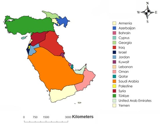
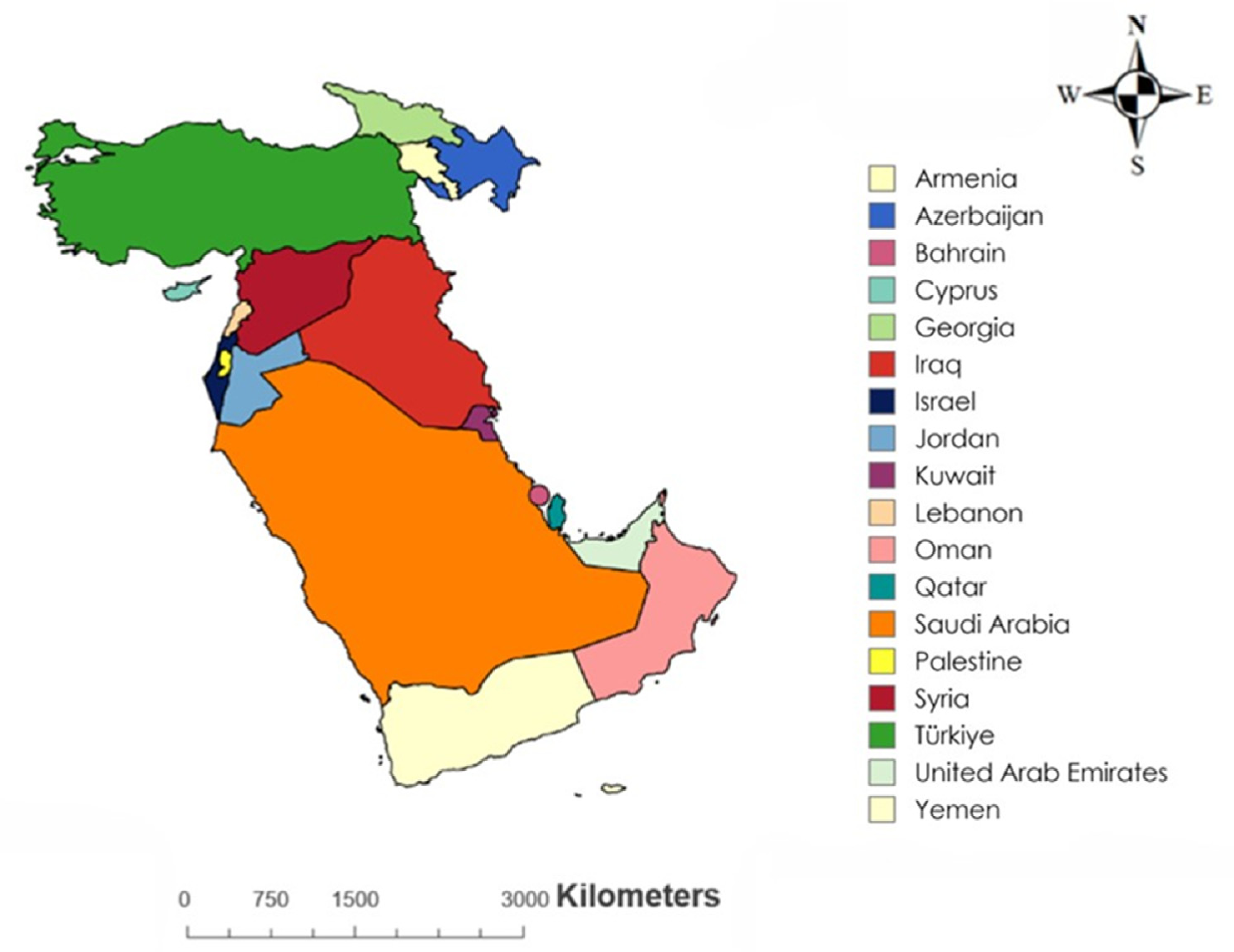
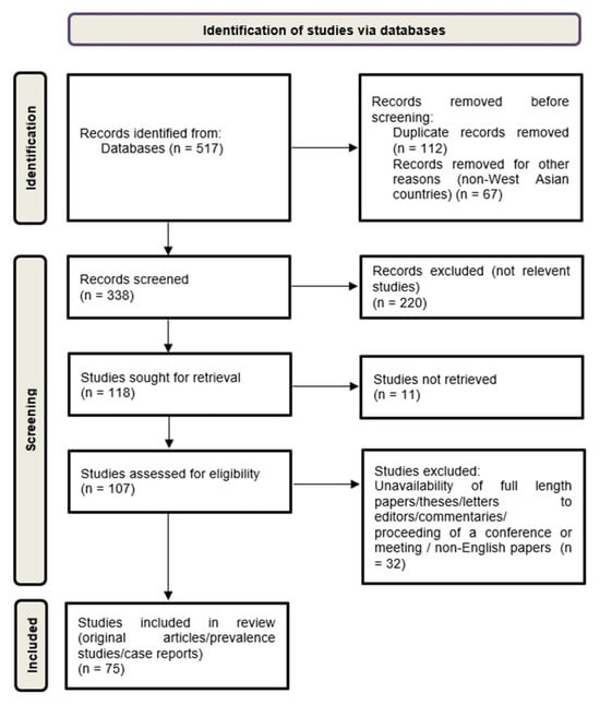
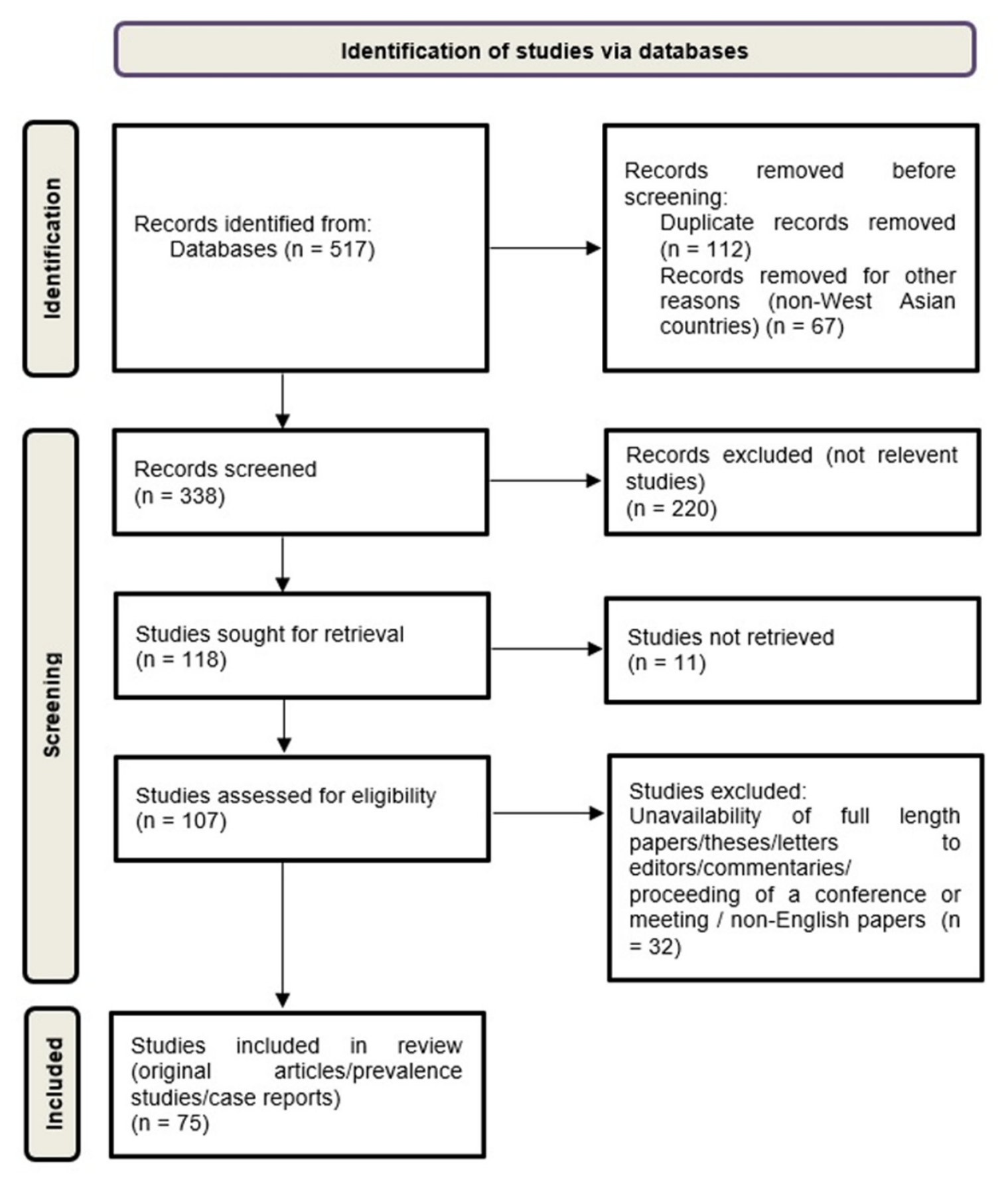
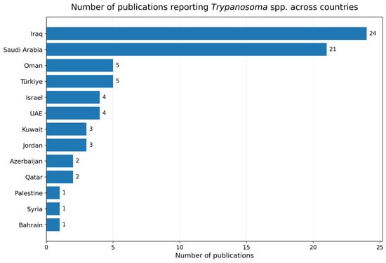
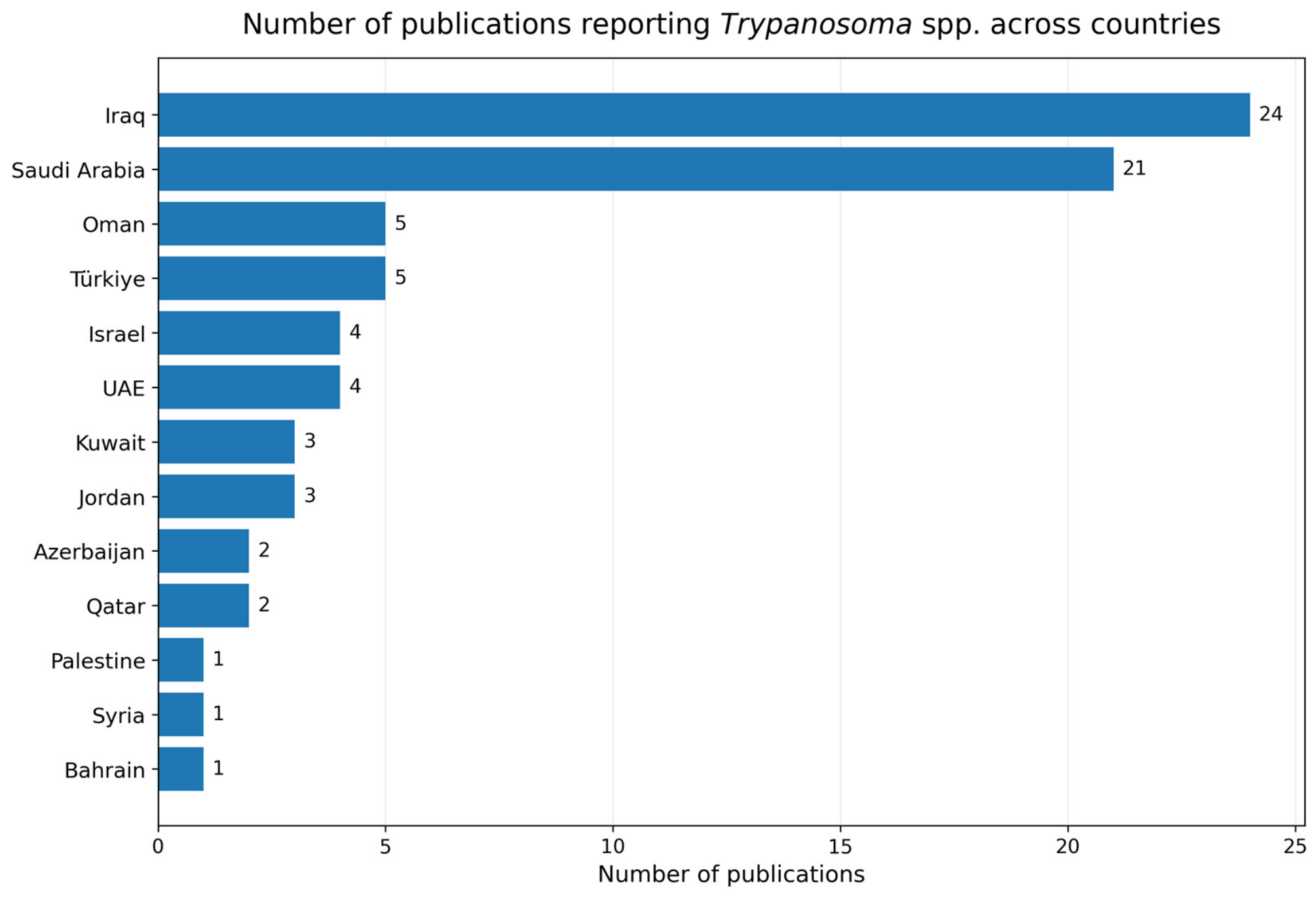
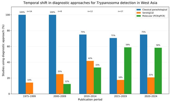
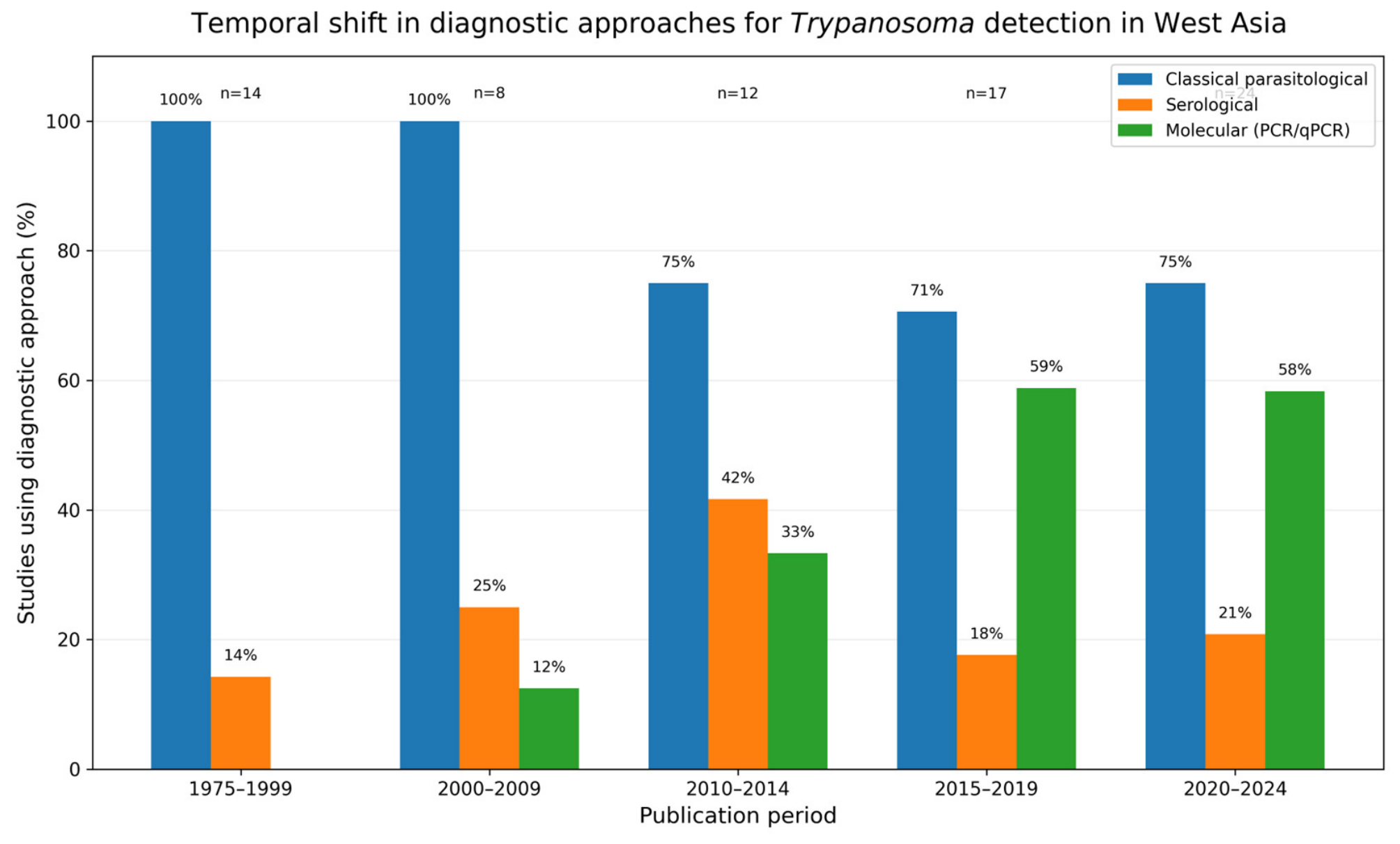
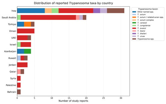
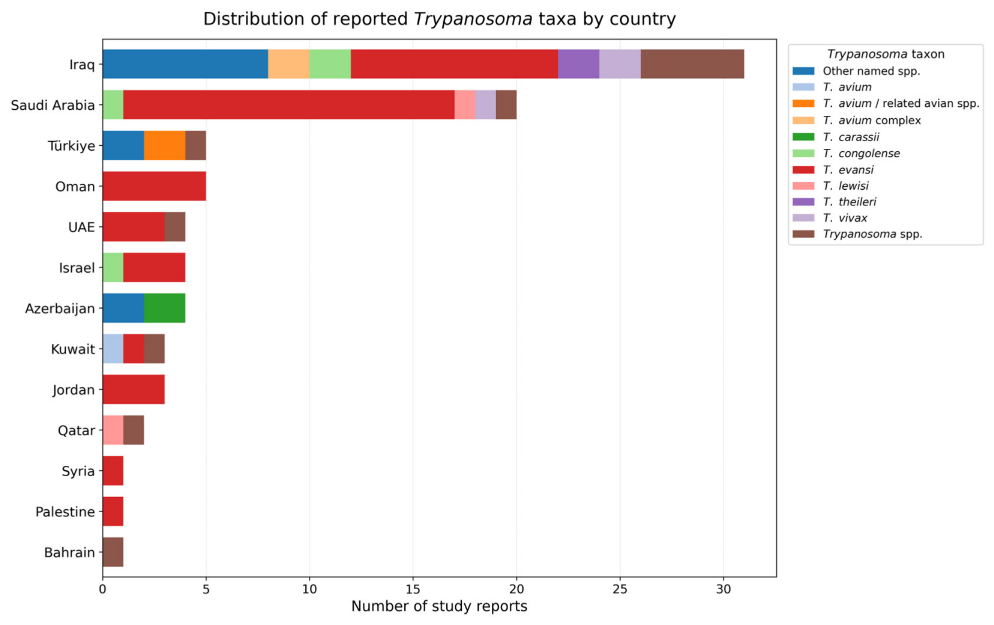
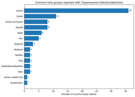
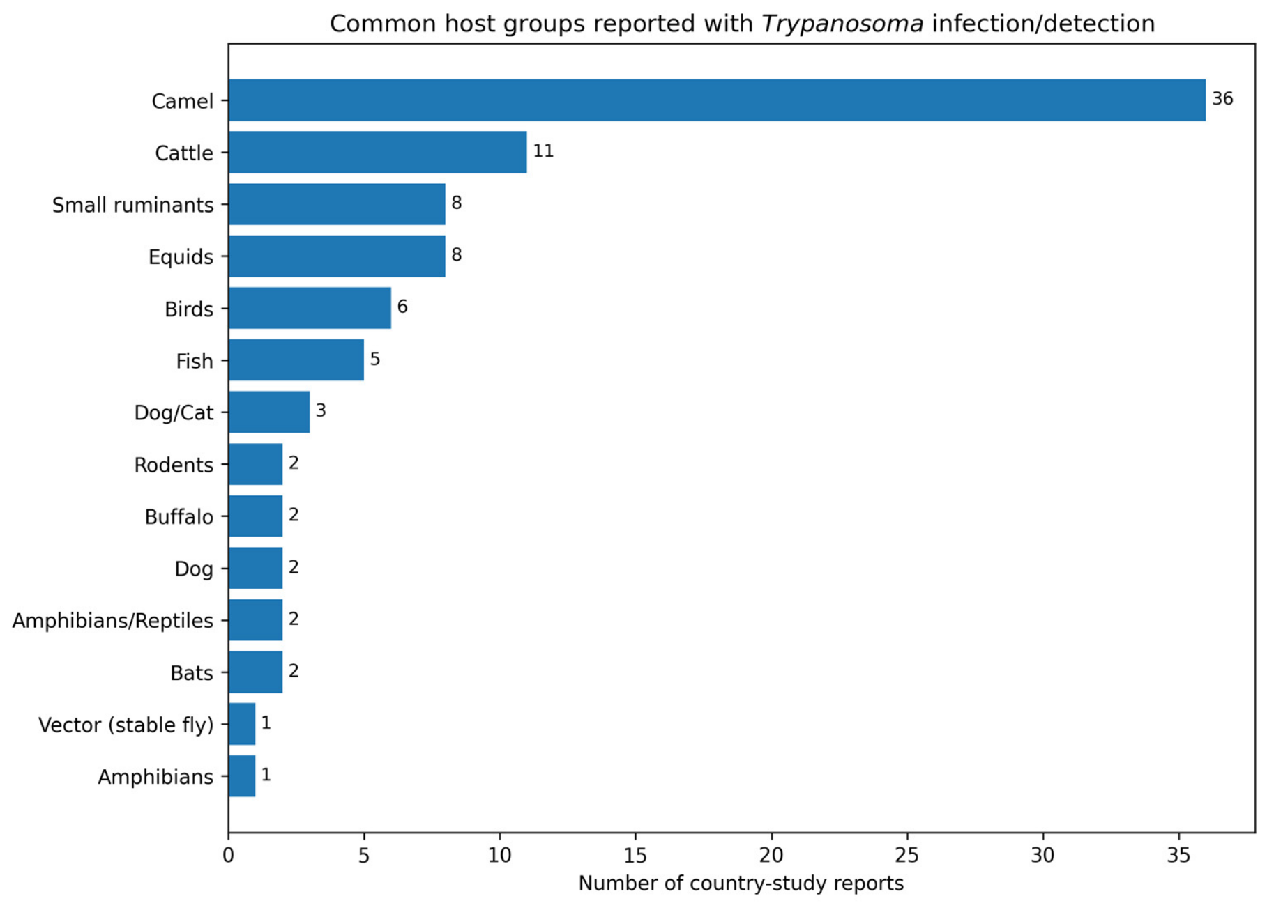
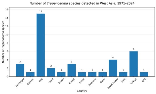
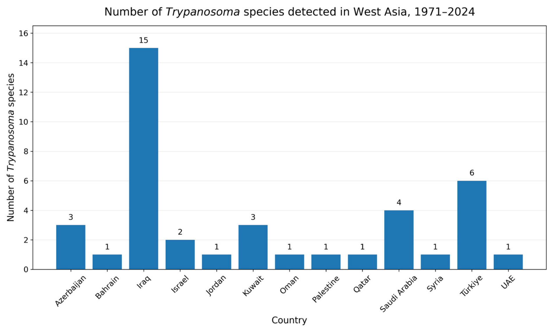

Disclaimer/Publisher’s Note: The statements, opinions and data contained in all publications are solely those of the individual author(s) and contributor(s) and not of MDPI and/or the editor(s). MDPI and/or the editor(s) disclaim responsibility for any injury to people or property resulting from any ideas, methods, instructions or products referred to in the content. |
© 2026 by the authors. Licensee MDPI, Basel, Switzerland. This article is an open access article distributed under the terms and conditions of the Creative Commons Attribution (CC BY) license.