


Abstract
The retina is a complex sensory neural tissue composed of six major types of neurons and one type of glial cell. The cell fate specification of retinal cells is tightly governed by intrinsic factors and extrinsic microenvironmental cues. Among the key regulators directing retinal cell fate differentiation is a group of bHLH family transcription factors (TFs). Our previous work demonstrated that the bHLH TF Ngn3 exhibits robust potential to induce retinogenesis in both distantly related fibroblasts in vitro and late retinal progenitor cells (RPCs) in vivo. However, the underlying molecular mechanisms remain largely elusive. In this study, we combined immunohistological examination and RNA-seq and ATAC-seq analyses to investigate the cellular and molecular mechanisms governing Ngn3-driven retinogenesis in late RPCs. Our results revealed that Ngn3 overexpression promotes premature cell cycle exit in late RPCs and remodels their transcriptomic and epigenomic landscape towards a state favoring rod photoreceptor and RGC differentiation. Furthermore, cross-comparison with Ngn3-overexpressing fibroblasts in vitro revealed cell-type-specific mechanisms underlying Ngn3-mediated neuronal fate reprogramming. These findings advance our understanding of Ngn family-mediated retinal cell fate regulation and provide a mechanistic framework for optimizing Ngn3-based retinal regeneration strategies for the treatment of retinal degeneration diseases.
1. Introduction
The retina is a complex sensory neural tissue composed of six types of neurons—retinal ganglion cells (RGCs), amacrine cells, bipolar cells, horizontal cells, rod photoreceptors, and cone photoreceptors—and one type of glial cell, Müller cells [1,2]. All these retinal cell lineages are generated by multipotent retinal progenitor cells (RPCs) during development in a sequential yet overlapping manner. Throughout this process, RPCs progressively modulate their differentiation capacity, the so-called ‘competent states’, to generate distinct retinal cell types at different developmental stages. For instance, in mice, early RPCs in embryonic retinas exclusively give rise to early-born retinal cell types, such as RGCs and cones, whereas late RPCs in postnatal retinas only produce late-born retinal lineages, including rod photoreceptors, bipolar cells, and Müller glial cells [3,4,5].
The competent states of RPCs, as well as the fate commitment and differentiation of all retinal cell types, are tightly controlled by intrinsic regulators and extrinsic microenvironmental cues. Among these regulatory factors, transcription factors (TFs)—particularly those belonging to the basic helix–loop–helix (bHLH) family and homeodomain family—play pivotal roles [6,7,8,9]. For example, the homeodomain TF Pax6 is highly expressed in RPCs and governs their proliferation and multipotency [10]; the bHLH family TF Atoh7 is expressed in early RPCs and directs their commitment to the RGC differentiation pathway [11,12]; downstream of Atoh7, Pou family of homeodomain TFs promote the terminational differentiation and maturation of RGCs [13]; while rod photoreceptor fate specification is strictly controlled by Otx2 and Crx [14,15]. A comprehensive elucidation of the molecular mechanisms by which TFs drive retinal cell fate determination is not only a central question in developmental biology but also a fundamental prerequisite for developing innovative strategies to regenerate retinal neurons for the treatment of retinal degeneration diseases. The Neurogenin (Ngn) family members are potent neurogenic bHLH-domain TFs consisting of three members: Ngn1, Ngn2, and Ngn3 [16]. These TFs are widely expressed in the central and peripheral nervous system from the early embryonic stage [17], and play critical roles in directing the fate differentiation of specific neuronal subtypes [16,18,19,20]. In the mammalian retina, only Ngn2 is endogenously expressed [17], with its expression initiating in RPCs at the earliest stages of retinogenesis and controlling the leading edge of the retinogenesis [21]. In contrast, Ngn1 and Ngn3 are not detected in the developing retina [17]. Beyond development, recent studies have demonstrated that overexpression of all three Ngn family members, in combination with other TFs, efficiently induces the transdifferentiation of distantly related fibroblasts into functional neurons [22,23,24]. This highlights the robust neurogenic capacity of Ngn family TFs in direct somatic cell reprogramming and identifies them as ideal candidates for neural fate reprogramming in therapeutic applications.
Inspired by the studies described above, our previous work investigated the ability of Ngn family TFs to reprogram fibroblasts into retinal neurons, revealing that all three members alone can induce RGC-like neuron fate in fibroblasts [25]. Notably, although only Ngn2 is endogenously expressed and functional in the retina, Ngn1 and Ngn3 exhibit stronger RGC fate-inducing activity than Ngn2 in fibroblast reprogramming. More intriguingly, we found that ectopic expression of Ngn1 and Ngn3 in late RPCs alters their differentiation competence: it promotes rod photoreceptor and RGC fates while repressing Müller glial cell and bipolar and amacrine interneuron fates [25]. These observations suggest that Ngn family TFs may serve as in vivo reprogramming tools to regenerate retinal neurons for the treatment of retinal degenerative diseases. Despite these well-established phenotypic observations from our prior study, the underlying genome-wide transcriptional and epigenomic mechanisms whereby Ngns remodel the intrinsic fate potential of late RPCs remain largely uncharacterized. In this study, we integrate immunohistological examination and RNA-seq transcriptomic and ATAC-seq epigenomic analyses to investigate the cellular and molecular mechanisms governing Ngn-driven reprogramming of the differentiation competence states in late RPCs. Given the relatively stronger capacity of Ngn3 in directing retinal neuron fate reprogramming in both in vitro and in vivo systems compared with Ngn1 and Ngn2 [25], and to minimize confounding effects from endogenously expressed retinal factors, we focus on Ngn3 in the present study. The findings of this study will advance our understanding of Ngn family-mediated retinal cell fate regulation and provide a mechanistic framework for optimizing Ngn3-based retinal regeneration strategies.
2. Results
2.1. Ngn3 Drives RPCs to Exit the Cell Cycle
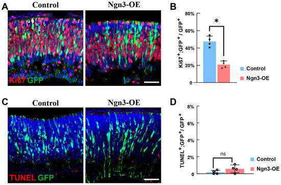
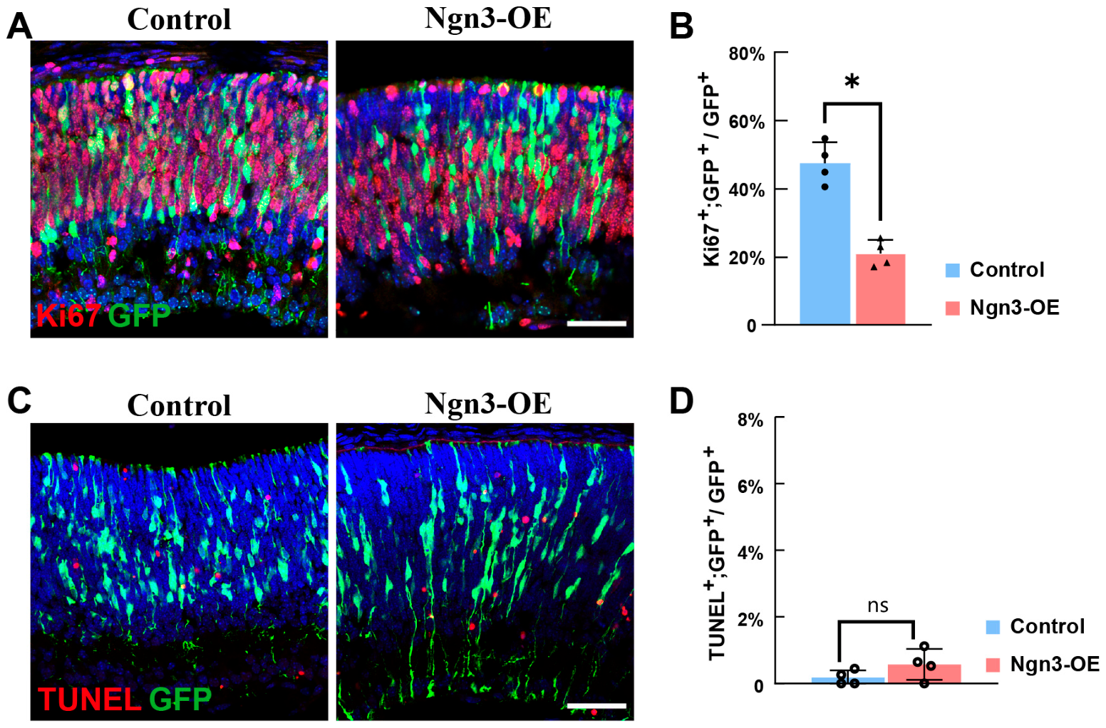
Our previous work demonstrated that Ngn3 overexpression (Ngn3-OE) in late RPCs promotes rod photoreceptor fate differentiation while inhibiting Müller glial cell and bipolar and amacrine interneuron differentiation [25]. To explore the mechanisms by which Ngn3 modulates retinal cell fate differentiation in late RPCs, we first assessed the proliferation activity of Ngn3-OE late RPCs, as progenitor cell differentiation is tightly coupled to proliferation. For this purpose, we performed in vivo electroporation on newborn mouse pups to introduce Ngn3-OE plasmids into late RPCs, and collected the retinas at day 2 after electroporation (DAE 2). We then performed Ki67 immunofluorescent staining on the retinal cryosections to evaluate the proliferation status of the retinal cells. The results showed that, in control retinas, 49.73 ± 4.94% of GFP+ electroporated RPCs exhibited Ki67 signals, indicating active proliferation (Figure 1A(left),B). In contrast, in Ngn3-OE retinas, the proportion of Ki67+ actively proliferating cells was significantly reduced to 20.95 ± 3.95% (Figure 1A(right),B). These results demonstrated that Ngn3-OE induces premature cell cycle exit in late RPCs.
Figure 1.
Ngn3 drives RPCs to exit the cell cycle. (A) Confocal immunofluorescent staining images of Ki67 and GFP in retinal cryosections from Control and Ngn3-OE groups at DAE 2, GFP labels electroporated late RPCs (B) Quantification of the percentage of Ki67+; GFP+ cells among total GFP+ electroporated RPCs in Control and Ngn3-OE groups. (C) Confocal immunofluorescent staining images of TUNEL assay of retinal cryosections from Control and Ngn3-OE groups at DAE 2, GFP labels electroporated late RPCs. (D) Quantification of the percentage of TUNEL+; GFP+ cells among total GFP+ electroporated RPCs in Control and Ngn3-OE groups. Data are presented as mean ± standard deviation (SD). Statistical significance was determined by an unpaired two-tailed t-test. ns, not significant; * p < 0.05. n ≥ 3 independent experiments. Scale bar: 50 μm.
We next examined whether Ngn3-OE affects the survival of late RPCs. To this end, we performed Terminal deoxynucleotidyl transferase dUTP nick end labeling (TUNEL) assay on DAE 2 retinas. Sporadic apoptotic events were observed in the developing retina at this stage; however, GFP-labeled electroporated RPCs rarely underwent apoptosis in the control group (Figure 1C(left),D), and Ngn3-OE did not exert a significant effect on the survival of late RPCs (Figure 1C(right),D). These results demonstrated that Ngn3-OE does not affect the survival of late RPCs.
2.2. Ngn3-OE Profoundly Remodels the Transcriptome of Late RPCs
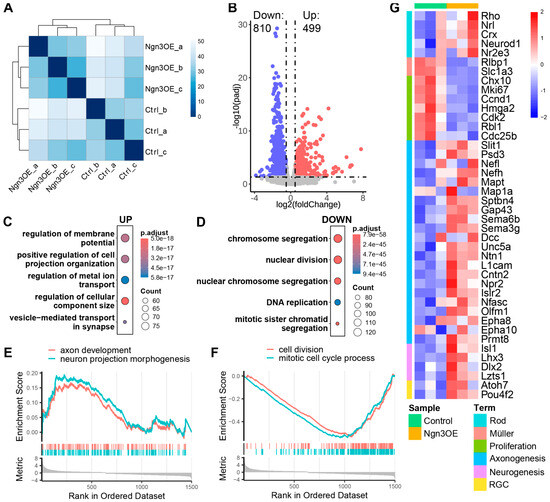
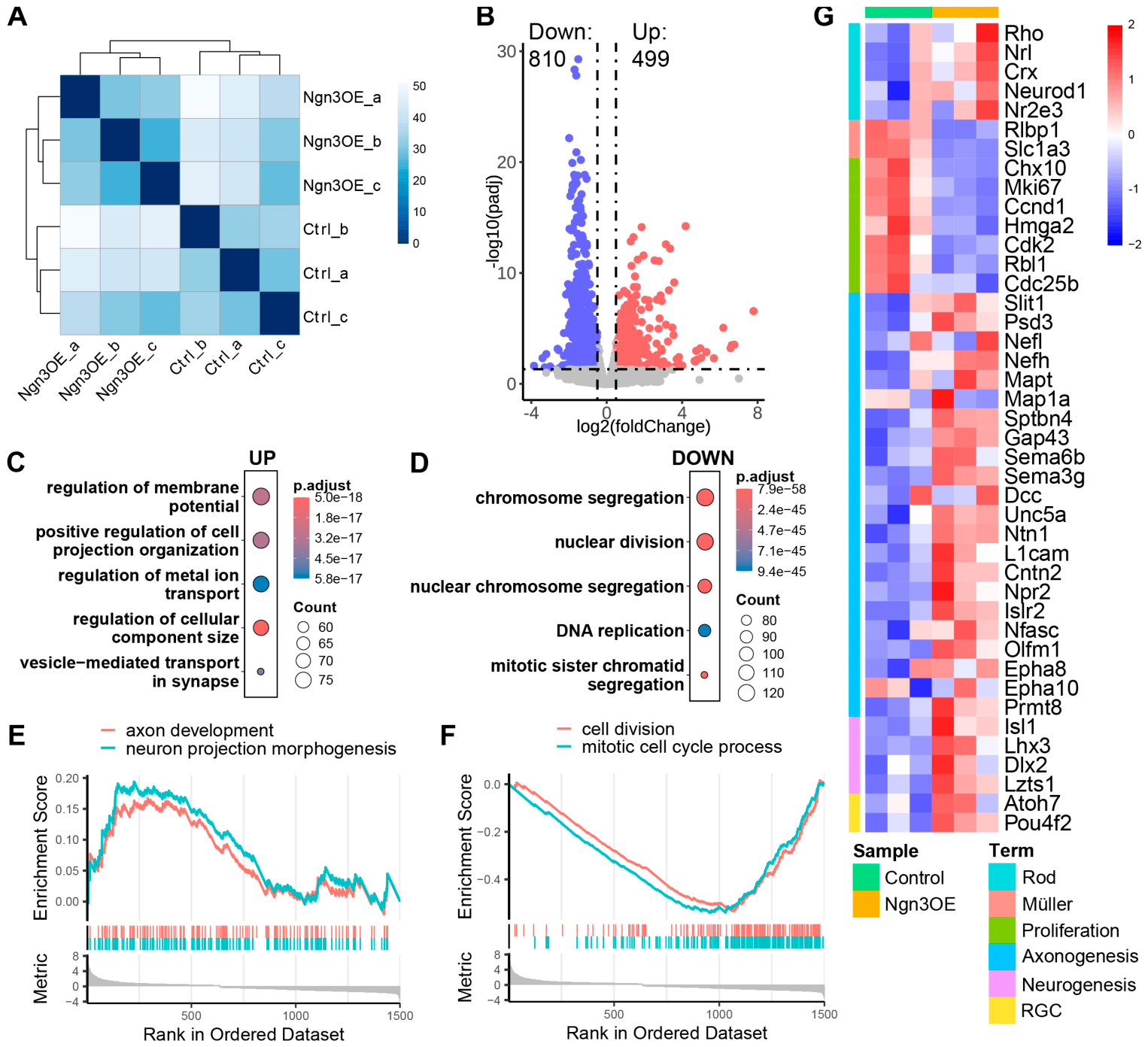
As a TF, Ngn3 exerts its cellular functions primarily by regulating the transcription of target genes. To elucidate the molecular mechanisms underlying Ngn3-OE-mediated reprogramming of late RPC differentiation competence, we performed RNA-seq on electroporated late RPCs. To this end, we collected GFP-labeled control and Ngn3-OE electroporated late RPCs at DAE 2 via fluorescence-activated cell sorting (FACS), and performed RNA-seq analyses. Sample correlation distance analysis revealed close clustering of biological replicates within each experimental group, validating the quality of the RNA-seq data, while control and Ngn3-OE samples formed distinct clusters, suggesting that Ngn3-OE profoundly altered the transcriptome of late RPCs (Figure 2A). We then performed Differentially Expressed Gene (DEG) analyses to search for genes whose expression changed upon Ngn3-OE in late RPCs. The results showed that, at the threshold set at a log2 (fold change) ≥ 0.5/≤ −0.5 and an adjusted p value ≤ 0.05, 499 genes were upregulated and 810 genes were downregulated in Ngn3-OE late RPCs compared with control RPCs (Figure 2B, and Supplementary Material S1). To examine what cellular processes were most significantly affected by Ngn3-OE in late RPCs, we performed gene ontology (GO) term enrichment analyses and gene set enrichment analysis (GSEA) analyses. The results showed that the upregulated gene set was significantly enriched for cellular processes related to neuronal function, such as ‘regulation of membrane potential’, ‘positive regulation of cell projection organization’, and ‘vesicle-mediated transport in synapse’ (Figure 2C,E). In addition, inspecting specific genes key to the cell fate development of retinal cells showed that several key rod photoreceptor fate determination TFs and functional genes, such as Rho, Nrl, Crx, and Nr2e3, and several key TFs involved in RGC fate development TFs, such as Atoh7 and Pou4f2 were upregulated in Ngn3-OE late RPCs (Figure 2G). These findings were consistent with the promotion of rod photoreceptor and RGC lineages observed at DAE 14 in our previous study [25], indicating that the early transcriptomic changes at DAE 2 are associated with subsequent cell fate commitment. On the other hand, the downregulated gene set was highly enriched for cell cycle progression processes, such as ‘chromosome segregation’, ‘nuclear division’, and ‘DNA replication’ (Figure 2D,F). This enrichment directly reinforces the premature cell cycle exit phenotype observed above (Figure 1). Additionally, key Müller glial cell functional marker genes, such as Rlbp1 and Slc1a3, were downregulated in Ngn3-OE late RPCs, which is consistent with the repressed Müller fate observed at DAE 14 in our previous study [25].
Figure 2.
Transcriptomic profiling of Ngn3-OE RPCs via RNA-seq. (A) Heatmap showing sample correlation scores derived from RNA-seq data of control and Ngn3-OE late RPCs at DAE 2. (B) Volcano plot showing differentially expressed genes (DEGs) between Ngn3-OE and control late RPCs. Red dots represent upregulated genes, and blue dots represent downregulated genes. DEGs are defined as genes with log2(fold change) ≥ 0.5 or ≤−0.5 and adjusted p-value ≤ 0.05. (C,D) Bubble plots showing the top five significantly enriched Gene Ontology (GO) terms for upregulated (C) and downregulated (D) DEGs in Ngn3-OE late RPCs compared to control late RPCs. (E,F) Gene Set Enrichment Analysis (GSEA) plots showing pathways enriched among upregulated (E) and downregulated (F) DEGs. (E) neuron projection development (NES = 0.939, Padj = 0.950) and axon development (NES = 0.921, Padj = 0.963). (F) cell division (NES = −3.058, Padj = 0.085) and mitotic cell cycle process (NES = −3.208, Padj = 0.085). (G) Heatmap showing the expression levels of representative genes in control and Ngn3-OE samples.
Collectively, RNA-seq analyses demonstrated that Ngn3-OE induces extensive transcriptomic remodeling in late RPCs, which is correlated with the observed premature cell cycle exit and altered retinal cell differentiation competence.
2.3. Ngn3 Reprograms Distinct Somatic Cell Types via Divergent Molecular Pathways with a Conserved Neurogenic Core
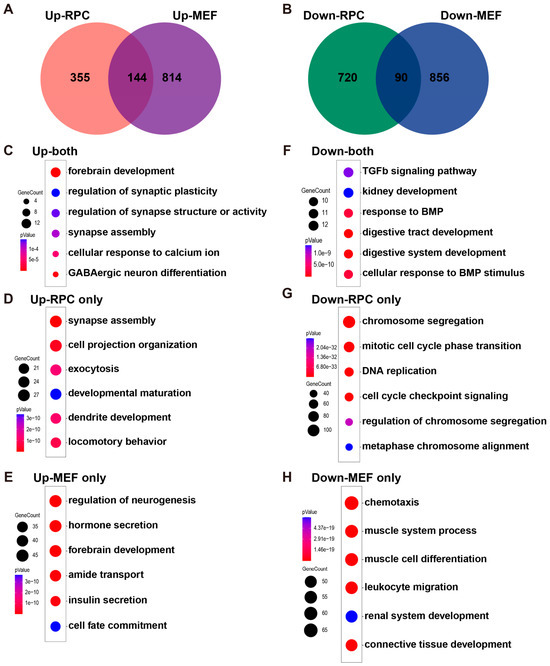
Our previous work showed that Ngn3 potently reprograms distantly related mouse embryonic fibroblasts (MEFs) into RGC-like neurons in vitro, with RNA-seq performed on Ngn3-OE MEFs at 1 day post-overexpression to elucidate the underlying mechanisms [25]. This provided a unique opportunity to compare Ngn3 function in in vivo late RPC reprogramming and in vitro fibroblast reprogramming. Cross-comparison of DEGs between the two cell types revealed a striking cell-type specificity of Ngn3-mediated reprogramming pathways, alongside a conserved core neurogenic program. Specifically, among the upregulated genes, only 144 genes were commonly upregulated in both late RPCs and MEFs upon Ngn3-OE, whereas 355 genes were uniquely upregulated in late RPCs, and 814 genes were uniquely upregulated in MEFs upon Ngn3-OE (Figure 3A). For downregulated genes, merely 90 genes were commonly downregulated in both late RPCs and MEFs upon Ngn3-OE, whereas 720 genes were uniquely downregulated in late RPCs and 856 genes uniquely downregulated in MEFs upon Ngn3-OE (Figure 3B). These results suggest that Ngn3 regulates largely distinct gene sets depending on the cellular context in which it is overexpressed. GO term enrichment analyses showed that: the commonly upregulated genes were enriched for cellular processes such as ‘forebrain development’ and ‘synapse assembly’, consistent with the potent neurogenic activity of Ngn3 across different cell types (Figure 3C); genes uniquely upregulated in late RPCs upon Ngn3-OE were further significantly enriched for processes related to neuronal function (Figure 3D), suggesting that the cellular environment in late RPCs was more permissive for the neurogenic function of Ngn3; in contrast, genes uniquely upregulated in MEFs upon Ngn3-OE were enriched for non-neurogenic pathways such as ‘hormone secretion’ and ‘insulin secretion’, unrelated to neurogenesis and neuronal function (Figure 3E), suggesting that MEFs exhibited multiple potential cell fate options in response to Ngn3-OE and required more extensive reprogramming to adopt a neuronal fate. For downregulated genes, the commonly repressed gene set was significantly enriched for cellular process related to TGF-β and BMP signaling pathways (Figure 3F), suggesting that Ngn3 universally repressed TGF-β superfamily signaling across cell types; genes uniquely downregulated in late RPCs upon Ngn3-OE were specifically enriched for processes related to cell cycle progression (Figure 3G), consistent with the strong premature cell cycle exit phenotype observed for Ngn3-OE late RPCs (Figure 1), whereas genes uniquely downregulated in MEFs upon Ngn3-OE were significantly enriched for cellular processes associated with the mesoderm identity of MEFs, such as ‘muscle system process’ and ‘connective tissue development’ (Figure 3H), suggesting that Ngn3-OE significantly repressed the mesoderm identity of MEFs to initiate neuronal reprogramming.
Figure 3.
Comparative transcriptome analyses reveal cell-type-specific and conserved gene programs induced by Ngn3 in late RPCs and MEFs. (A,B) Venn diagrams showing the overlap of upregulated (A) and downregulated (B) DEGs between Ngn3-OE late RPCs (DAE 2) and Ngn3-OE MEFs. (C–H) Bubble plots showing the top six significantly enriched GO terms for DEGs that are commonly regulated or cell-type-specifically regulated in late RPCs or MEFs: (C) GO terms for commonly upregulated genes between late RPCs and MEFs. (D) GO terms for late RPC-specifically upregulated genes. (E) GO terms for MEF-specifically upregulated genes. (F) GO terms for commonly downregulated genes between late RPCs and MEFs. (G) GO terms for late RPC-specifically downregulated genes. (H) GO terms for MEF-specifically downregulated genes.
Together, these comparative RNA-seq analyses revealed both conserved and cell-type-specific molecular mechanisms underlying Ngn3-driven neuronal fate reprogramming, highlighting the critical impact of the endogenous cellular context on the efficiency and outcome of TF-mediated reprogramming.
2.4. Ngn3-OE Alters the Chromatin Accessibility Landscape of Late RPCs
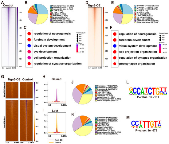
TFs often cooperate with epigenetic regulatory factors to modulate gene expression via epigenetic remodeling. To investigate how Ngn3-OE reshaped the epigenetic landscape of late RPCs, we performed ATAC-seq on control and Ngn3-OE late RPCs at DAE 2. To this end, we collected GFP-labeled control and Ngn3-OE electroporated late RPCs at DAE 2 via FACS and performed ATAC-seq analyses. Calculation of the Fraction of Reads in Peaks (FRiP) showed FRiP scores of 40.73% and 44.02% for the control and Ngn3-OE samples, respectively, indicating the high quality of our ATAC-seq dataset. Peak calling identified 128,805 accessible chromatin peaks in control late RPCs (Figure 4A, and Supplementary Material S2). Annotation of the peaks revealed that accessible genomic regions in control late RPCs were predominantly localized to promoter regions and putative distal intergenic and intronic enhancer regions (Figure 4B). GO term enrichment analyses of genes associated with these accessible regions showed significant enrichment for retinal development and function-related processes, such as ‘regulation of neurogenesis’, ‘eye development’, and ‘regulation of synapse organization’ (Figure 4C), consistent with the neural progenitor identity and endogenous developmental potential of late RPCs. For Ngn3-OE late RPCs, we identified 136,708 accessible chromatin peaks (Figure 4D, and Supplementary Material S2). Accessible genomic regions in Ngn3-OE late RPCs were also predominantly localized to promoter regions and putative distal intergenic and intronic enhancer regions (Figure 4E), and were also enriched for genes involved in retinal development and function (Figure 4F).
Figure 4.
Ngn3 reshapes the chromatin accessibility landscape of late RPCs. (A,D) Heatmaps showing accessible genomic regions in control (A) and Ngn3-OE (D) late RPCs. Regions are ordered by signal intensity. (B,E) Pie charts showing the genomic distribution of accessible chromatin peaks in control (B) and Ngn3-OE (E) late RPCs. (C,F) Bubble plots showing the top six significantly enriched GO terms for genes associated with open chromatin regions in control (C) and Ngn3-OE (F) late RPCs. (G) Heatmaps showing ATAC-seq reads density of DARs induced by Ngn3-OE in Ngn3-OE and control late RPCs. Regions are ordered by signal intensity. (H,I) Aggregate profile plots of ATAC-seq signal intensity at gained (H) and lost (I) DARs in Control vs. Ngn3-OE late RPCs. Purple represents the Ngn3-OE group, while yellow represents the Control group. (J,K) Pie charts showing the genomic distribution of gained (J) and lost (K) accessible chromatin peaks. (L,M) The most significantly enriched TF binding motif in gained (L) and lost (M) DARs.
We next compared the chromatin accessibility between Ngn3-OE and control RPCs to identify differential accessible regions (DARs) induced by Ngn3-OE. This analysis identified 2333 genomic regions that gained accessibility and 5842 genomic regions that lost accessibility upon Ngn3-OE in late RPCs (Figure 4G–I, and Supplementary Material S2). Annotation of gained and lost genomic regions in Ngn3-OE late RPCs revealed that these regions were significantly enriched in putative distal intergenic and intronic enhancer regions, with much smaller proportion localized to promoter regions (Figure 4J,K). This finding suggests that Ngn3 primarily exerts its epigenetic regulatory effects on enhancers. Next, we searched for consensus DNA sequence motif among DARs upon Ngn3-OE in late RPCs. The results revealed that the most prominently enriched consensus DNA motif in genomic regions gained accessibility upon Ngn3-OE were 5′-RCCATCTGBY-3′ E-box type motif (Figure 4L), which was identical to the canonical NGN3-binding motif identified by CUT&RUN profiling in pancreatic endocrine progenitor cells [26]. This finding suggests that NGN3 binds directly to its cognate E-box motifs to open chromatin in late RPCs to establish a neurogenically permissive epigenetic landscape. In contrast, the most enriched motifs in genomic regions lost accessibility upon Ngn3-OE were those recognized by SOX family homeodomain TFs (Figure 4M), implying that NGN3 indirectly represses chromatin accessibility at these loci via modulation of SOX family TF activity or expression.
2.5. Ngn3 Regulates Gene Expression via a Bifurcated Epigenetic Strategy in Late RPCs
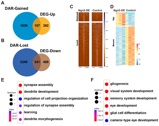
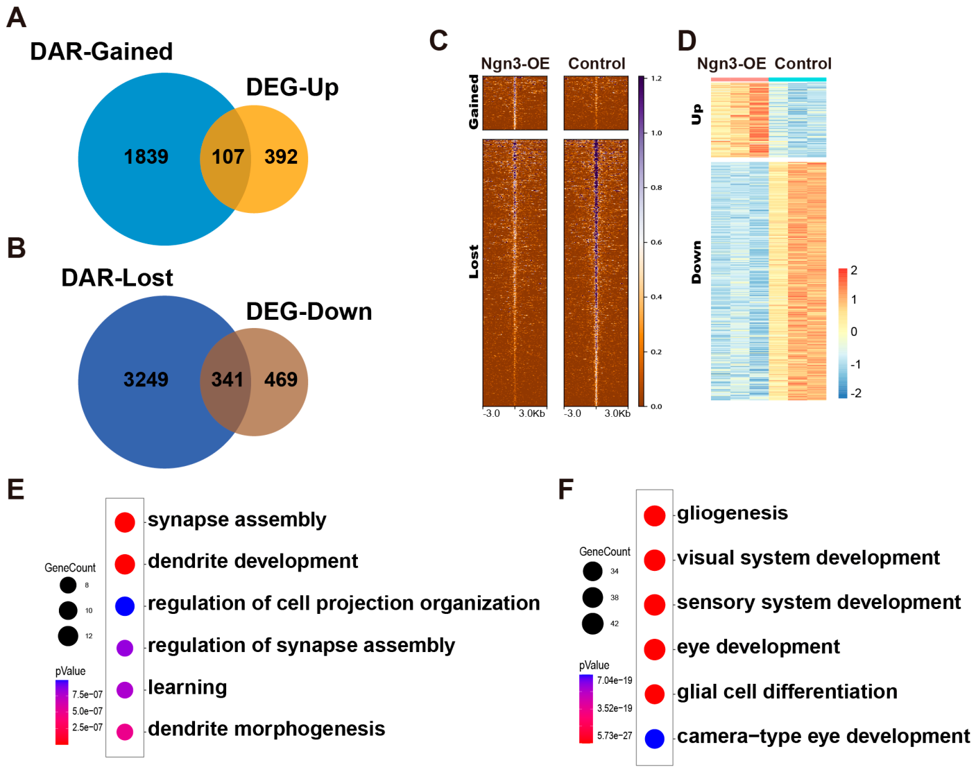
Finally, we investigated the link between Ngn3-induced epigenetic remodeling and transcriptomic changes to elucidate the epigenetic regulation of gene transcription upon Ngn3-OE in late RPCs. Given that chromatin accessibility is typically positively correlated with transcriptional upregulation, we intersected the list of upregulated genes from RNA-seq with genes associated with gained accessible genomic regions from ATAC-seq. Notably, only a small subset of upregulated genes was associated with increased chromatin accessibility (Figure 5A,C,D), suggesting that Ngn3 promotes gene transcription in late RPCs without altering chromatin accessibility. By contrast, a much larger proportion of downregulated genes was associated with reduced chromatin accessibility (Figure 5B–D), suggesting that chromatin remodeling is a major mechanism underlying Ngn3-mediated gene repression in late RPCs. These results revealed a bifurcated epigenetic regulatory strategy employed by Ngn3 in promoting and repressing gene expression in late RPCs. GO term enrichment analyses of those genes that were upregulated in Ngn3-OE late RPCs and exhibited increased chromatin accessibility were enriched for cellular processes related to neuronal function (Figure 5E), consistent with neurogenic TF character of Ngn family TFs. The downregulated genes that were associated with decreased chromatin accessibility were prominently associated with gliogenesis (Figure 5F), which aligns with the robust repression of Müller glial fate in Ngn3-OE late RPCs observed in our previous study [25]. This finding highlights that Ngn3-mediated epigenetic repression via chromatin closure is a key and stable mechanism for inhibiting glial fate in late RPCs. Collectively, these analyses establish a clear epigenetic basis for Ngn3-mediated transcriptomic remodeling and differentiation competence reprogramming in late RPCs.
Figure 5.
Link between Ngn3-induced epigenetic remodeling and transcription changes in late RPCs. (A) Venn diagrams showing the overlap between upregulated DEGs and genes associated with gained DARs. (B) Venn diagrams showing the overlap between downregulated DEGs and genes associated with lost DARs. (C) Heatmaps showing ATAC-seq reads density of DARs associated with the 107 common genes in panel (A) (upper, gained) and 341 common genes in panel (B) (lower, lost). (D) Heatmap showing the RNA-seq expression levels of the same gene set as in panel (C,E,F). Bubble plots showing significantly enriched GO term for the 107 common genes in panel (A,E) and the 341 common genes in panel (B,F).
3. Discussion
Thorough understanding of the molecular mechanisms governing retinal cell fate development is not only a central question in theoretical biology, but also the foundation for developing innovative biomedical strategies to address the therapeutic challenges faced by ophthalmologists treating retinal degenerative diseases. Accumulating evidence has highlighted the key roles of bHLH family TFs in driving retinal neuronal fate commitment [6]. Among these, Ngn2 is one of the earliest expressed neurogenic bHLH TFs and orchestrates the leading edge of retinal neuron differentiation [21]. Beyond development and retinas, Ngn family TFs have been shown to possess potent neurogenic capacity in inducing neuronal fate conversion of distantly related somatic cells via direct somatic cell reprogramming [23]. To explore the retinogenic potential of Ngn family TFs, we previously tested the effects of Ngn1/2/3 overexpression in in vitro cultured MEFs and in vivo electroporated RPCs. This study demonstrated that Ngn family TFs exert robust retinal neuron fate-inducing effects on both lineage-related RPCs and distantly related MEFs. However, the underlying molecular mechanisms, particularly in the in vivo retinal context, remain largely unexplored. In this study, we combined immunohistological examination, RNA-seq and ATAC-seq to dissect the cellular and molecular mechanisms underlying Ngn3-mediated reprogramming of the retinogenic competence of late RPCs. Our immunohistological analyses of DAE 2 retinas revealed that Ngn3-OE drove late RPCs to undergo premature cell cycle exit. Transcriptomic profiling by RNA-seq demonstrated that Ngn3-OE significantly altered the transcriptome of late RPCs, and identified the key molecular mediators underlying the neurogenic and cell cycle exit effects of Ngn3-OE in these cells. ATAC-seq analyses uncovered that Ngn3 regulates gene expression through both epigenetic remodeling-dependent and -independent strategies. Collectively, these findings fill the knowledge gap in Ngn family-mediated retinal cell fate regulation and provide mechanistic insights to optimize Ngn-based in vivo retinal regeneration strategies.
During neurogenesis, cell differentiation and cell cycle progression are tightly coupled, in that a neural progenitor cell must first exit the cell cycle before its terminal differentiation. Consistent with the potent neurogenic activity of Ngn family TFs, in the retina, Ngn2 expression in early RPCs drives these progenitors to exit the cell cycle and differentiate into RGCs—a process that shapes the formation and movement of the leading front of retinogenesis in the embryonic retina [21]. In this study, we found that Ngn3-OE in late RPCs similarly promoted cell cycle exit, despite Ngn3 not being an endogenously expressed retinogenic TF in the retina. This Ngn3-induced premature cell cycle exit likely disrupts the endogenous developmental clock that links proliferation status to lineage bias, which might contribute to the elevated rod-genesis phenotype observed at DAE 14 since postnatal day 3 (DAE 2) represents the peak time of rod neurogenesis. Furthermore, although RNA-seq was performed at DAE 2—well before overt cell fate differentiation phenotypes are detectable—key regulatory and marker genes for the promoted retinal cell fates (e.g., rods and RGCs) and repressed fates (e.g., Müller glia, bipolar cells, and amacrine cells) already exhibited significant transcriptional alterations. This observation suggests that Ngn3-induced transcriptomic remodeling is a causal driver of retinal fate reprogramming, rather than a downstream consequence of cell fate commitment. This positions Ngn3 as a potent regulator that can shift the balance of late RPC fate specification toward therapeutically relevant rod and RGC lineages—two cell types most commonly lost in retinal degenerative diseases such as age-related macular degeneration and glaucoma.
Motif enrichment analyses of chromatin regions with gained accessibility in Ngn3-OE late RPCs identified the canonical NGN3-binding E-box motif (5′-RCCATCTGBY-3′) as the most significantly enriched sequence, suggesting that NGN3 may function as a pioneer TF in late RPCs: it directly binds to its cognate motifs to open closed chromatin and establish a pro-retinogenic epigenetic landscape. In contrast, chromatin regions with lost accessibility in Ngn3-OE late RPCs were enriched for SOX family TF-binding motifs, pointing to an indirect epigenetic mechanism: Ngn3 likely represses the expression or activity of Sox family TFs, leading to chromatin closure at their target loci. Sox TFs (e.g., Sox2, Sox9) are core regulators of RPC proliferation and Müller glial fate development; thus, their indirect repression by Ngn3 likely contributes to both the premature cell cycle exit phenotype and repression of Müller glial differentiation in late RPCs. On the other hand, integrated analysis of RNA-seq and ATAC-seq data revealed that only a small subset (~12.6%) of upregulated genes is associated with gained chromatin accessibility, whereas a much larger proportion (~38.9%) of downregulated genes links to lost accessibility. This finding is consistent with the neurogenically permissive state of late RPCs, eliminating the need for Ngn3 to induce de novo chromatin opening. For gene repression, however, Ngn3 relies more on chromatin closure to achieve stable and heritable silencing of non-target fate programs—including cell cycle progression and gliogenesis. Future studies employing techniques such as CUT&RUN or ChIP-seq to characterize NGN3-binding sites in late RPCs would provide further mechanistic insights into how Ngn3-OE rewires the differentiation competence of these cells.
A key mechanistic insight from this study is the cell-type specificity of Ngn3-mediated reprogramming revealed by cross-comparison of DEGs induced by Ngn3-OE in late RPCs and MEFs. While only 144 upregulated and 90 downregulated genes were shared between the two cell types, the conserved core program points to a universal neurogenic mechanism of Ngn3: shared upregulated genes were enriched for forebrain development and synapse assembly, and shared downregulated genes targeted the TGF-β/BMP signaling pathways. TGF-β/BMP signaling is a well-characterized negative regulator of neurogenesis, and its universal repression by Ngn3 across both neural progenitors and somatic cells identifies this pathway as a potential core mediator of Ngn3’s neurogenic activity. In contrast, the cell-type-specific DEGs reflect the intrinsic differences between late RPCs and MEFs. Late RPCs exhibited unique downregulation of cell cycle progression genes—supporting the premature cell cycle exit phenotype—and unique upregulation of neuronal function-related genes, reflecting a chromatin and transcriptomic landscape that is inherently conducive to Ngn3’s retinogenic activity. MEFs, by contrast, displayed unique downregulation of mesodermal identity genes and unique upregulation of non-neurogenic genes, reflecting the need for Ngn3 to first erase somatic cell identity and the presence of alternative fate potentials in these cells.
In conclusion, this study elucidated the in vivo cellular and molecular mechanisms underlying Ngn3-mediated reprogramming of the retinogenic competence of late RPCs. These findings advance our understanding of Ngn family-mediated retinal cell fate regulation and provide a mechanistic framework for optimizing Ngn3-based in vivo retinal regeneration strategies for the treatment of retinal degenerative diseases.
4. Materials and Methods
4.1. Retinal In Vivo Electroporation (IVE)
All animal studies were performed in accordance with the protocol approved by the Institutional Animal Care and Use Committee of Zhongshan Ophthalmic Center (Z2023035). Retinal injection and electroporation were performed on postnatal day 1 (P1) mouse pups following a published protocol [27]. Briefly, P1 pups were anesthetized via hypothermia by placement on ice for 3 min. The palpebral fissure was gently opened using a 1 mL syringe needle (Winner Medical, Hunan, China), and a small incision was made in the sclera near the corneal-scleral limbus using an insulin needle. Under a Leica M620 F20 stereomicroscope (Leica Biosystems, Nussloch, Germany), 5 μL of purified plasmid DNA (2–5 μg/μL in 1 × PBS) was delivered into the subretinal space using a Hamilton syringe equipped with a 32-gauge blunt-ended needle (Hamilton, Bonaduz, Switzerland). Immediately following injection, tweezer-type electrodes (dipped in 1 × PBS) were placed to strictly encompass the head, with the anode positioned over the injected eye. Five square-wave pulses (80 V; 50 ms duration; 950 ms interval) were applied using a Gemini X2 pulse generator (BTX, Holliston, MA, USA). After the procedure, pups were placed on a warming pad for rewarming and were returned to their mothers once they regained steady physical activity.
4.2. Immunohistochemistry and Image Acquisition
Mouse pups were anesthetized via hypothermia on ice at day 2 after electroporation (DAE 2), and the eyes were carefully collected. The harvested eyes were fixed in 4% paraformaldehyde (PFA) at room temperature for 30 min. For cryoprotection, the samples were dehydrated in a graded sucrose series (15% and 30% w/v in 1 × PBS) until fully equilibrated. Tissues were then embedded in Tissue-Tek O.C.T. compound (Sakura Finetek, Torrance, CA, USA) and cut into 10 μm sections using a Leica CM1950 cryostat microtome (Leica Biosystems, Nussloch, Germany). For immunofluorescence analysis, heat antigen retrieval was performed by incubating the sections in citrate buffer (pH 6.0) at 95 °C for 30 min. After cooling to room temperature, sections were blocked and incubated with primary antibodies diluted in PBST (1 × PBS with 0.1% Triton X-100(Sigma-Aldrich, St. Louis, MO, USA)) supplemented with 5% fetal bovine serum (FBS) overnight at 4 °C. To identify proliferating late retinal progenitor cells (RPCs), a rabbit anti-Ki67 primary antibody (Abcam, Cambridge, UK; ab15580) was employed. Following three washes with PBST, sections were incubated with appropriate fluorophore-conjugated secondary antibodies for 1 h at room temperature. Nuclei were counterstained with 4′,6-diamidino-2-phenylindole (DAPI). Finally, the slides were mounted with cover glasses using VECTASHIELD mounting medium (Vector Labs, Newark, CA, USA). Immunofluorescence images were acquired using a confocal microscope (Zeiss LSM 980; Carl Zeiss Microscopy GmbH, Jena, Germany) and analyzed via imageJ woftware (version 1.53f) [28].
4.3. TUNEL Assay
To detect apoptotic cells, Terminal deoxynucleotidyl transferase dUTP nick end labeling (TUNEL) staining was performed on cryosections. Briefly, the TUNEL reaction was prepared by mixing TdT enzyme and fluorescence labeling solution at a 1:9 ratio (v/v). After washing, cryosections were incubated with the TUNEL reaction mixture for 1 h at 37 °C in the dark. After washing slides were mounted using an anti-fluorescence quenching medium and stored at −20 °C. TUNEL images were acquired using a confocal microscope (Zeiss LSM 980; Carl Zeiss Microscopy GmbH, Jena, Germany) and analyzed via imageJ software (version 1.53f) [28].
4.4. Statistical Analysis for Immunofluorescence and TUNEL Analyses
The data of immunofluorescence and TUNEL analyses were performed in at least three independent biological replicates. Data are expressed as mean ± SD, and statistical significance was determined using two-tailed Student’s t-tests. A p-value threshold of less than 0.05 was defined as statistically significant.
4.5. Fluorescence-Activated Cell Sorting (FACS)
For fluorescence-activated cell sorting (FACS), retinas were harvested from pups at DAE 2. Tissues were dissociated into single-cell suspensions using Papain (Worthington Biochemical Corp., Lakewood, NJ, USA) and resuspended in 1 × PBS supplemented with 0.04% bovine serum albumin (BSA). GFP+ populations were subsequently isolated using a BD FACSAria Fusioncell sorter (Becton, Dickinson and Company, Franklin Lakes, NJ, USA).
4.6. RNA-Seq
Sorted GFP+ cells (100,000 cells per replicate) were harvested by centrifugation and resuspended in 200 μL of TRIzol reagent (Beyotime, Shanghai, China), with the volume adjusted according to the retinal cell input. Total RNA was extracted following the manufacturer’s instructions. Sequencing libraries were constructed using the NEBNext Ultra RNA Library Prep Kit for Illumina (New England Biolabs, Ipswich, MA, USA) according to the recommended protocol. The resulting libraries were sequenced on the Illumina NovaSeq X platform to generate 150 bp paired-end reads. The raw sequencing data were processed using Trimmomatic (version 0.39)to remove adapter sequences and filter out low-quality bases, yielding high-quality clean reads. These reads were then mapped to the mouse reference genome (mm10) using HISAT2 (version 2.2.0). Gene-level quantification was performed using Subread (version 2.0.1) to generate raw read counts. Differential expression analysis was conducted using the DESeq2 R package, using a negative binomial model and Wald test for two-group comparisons. Multiple testing correction was conducted using the default Benjamini–Hochberg procedure to control the false discovery rate (FDR). Differentially expressed genes (DEGs) were identified based on a significance threshold of adjusted p-value ≤ 0.05 and a log2 (fold change) ≥ 0.5/≤ −0.5. Functional annotation and Gene Ontology (GO) enrichment analysis were performed using the clusterProfiler R package. For RNA-seq analysis, three independent biological replicates were performed for both Ngn3-OE and control groups.
4.7. Assay for Transposases-Accessible Chromatin Using Sequencing (ATAC-Seq)
ATAC-seq libraries were constructed from 5 × 10⁴ FACS-isolated cells per sample using the Hyperactive ATAC-Seq Library Prep Kit for Illumina (TD711; Vazyme, Nanjing, China), following the manufacturer’s instructions. Briefly, nuclei were isolated using a chilled lysis buffer, subjected to Tn5-mediated transposition at 37 °C for 30 min, and purified using VAHTS DNA clean beads. After PCR amplification and indexing, libraries were sequenced on an Illumina NovaSeq X platform (150 bp paired-end). For data analysis, raw reads were trimmed using Trimmomatic and aligned to the mm10 genome via Bowtie2. After removing duplicates (Picard, version 2.20.4), mitochondrial reads (SAMtools, version 1.6), and ENCODE blacklisted regions (bedtools, version 2.30.0), fragments ≤120 bp were extracted. Reads were shifted (+4/−5 bp) using deepTools (version 3.1.3) to account for Tn5 duplication prior to peak calling with MACS2 (version 2.2.4) (q < 0.05). Differentially accessible regions (DARs) were identified using DiffBind (version 3.18.0) combined with DESeq2 with a threshold of log2(fold change) > 1 and p-value < 0.05. Motif enrichment analysis was performed using HOMER (version 4.11). Fraction of Reads in Peaks(FRiP) was calculated as the ratio of reads mapped within called ATAC-seq peaks to total mapped reads, using featureCounts. A single biological sample was performed for ATAC-seq library construction and sequencing for each experimental group.
Supplementary Materials
The following supporting information can be downloaded at: https://www.mdpi.com/article/10.3390/ijms27093845/s1.
Author Contributions
S.C. conceived and designed the research. C.C., H.L., and Q.H. performed the experiments. S.C. and C.C. analyzed the data. S.C. wrote the manuscript. All authors have read and agreed to the published version of the manuscript.
Funding
This research was funded by Department of Science and Technology of Guangdong Province (2024A1515012938), the National Natural Science Foundation of China (32470848), and Research Funds of the State Key Laboratory of Ophthalmology (2025QZSPT28).
Institutional Review Board Statement
All animal studies were performed in accordance with the protocol approved by the Institutional Animal Care and Use Committee of Zhongshan Ophthalmic Center (protocol number Z2023035, approved on 15 September 2023).
Informed Consent Statement
Not Applicable.
Data Availability Statement
The original data presented in the study are openly available in GEO under at https://www.ncbi.nlm.nih.gov/geo/ (accessed on 30 March 2026) (accession number GSE326387 and GSE326388).
Acknowledgments
We thank the staff of the Laboratory Animal Center at the State Key Laboratory of Ophthalmology, Zhongshan Ophthalmic Center, for technical support.
Conflicts of Interest
The authors declare no conflicts of interest.
References
- Hoon, M.; Okawa, H.; Della Santina, L.; Wong, R.O. Functional architecture of the retina: Development and disease. Prog. Retin. Eye Res. 2014, 42, 44–84. [Google Scholar] [CrossRef] [Scilit] [PubMed]
- Masland, R.H. The neuronal organization of the retina. Neuron 2012, 76, 266–280. [Google Scholar] [CrossRef] [Scilit]
- Cepko, C.L.; Austin, C.P.; Yang, X.; Alexiades, M.; Ezzeddine, D. Cell fate determination in the vertebrate retina. Proc. Natl. Acad. Sci. USA 1996, 93, 589–595. [Google Scholar] [CrossRef] [Scilit]
- Agathocleous, M.; Harris, W.A. From progenitors to differentiated cells in the vertebrate retina. Annu. Rev. Cell Dev. Biol. 2009, 25, 45–69. [Google Scholar] [CrossRef] [Scilit]
- Cayouette, M.; Poggi, L.; Harris, W.A. Lineage in the vertebrate retina. Trends Neurosci. 2006, 29, 563–570. [Google Scholar] [CrossRef] [Scilit]
- Hatakeyama, J.; Kageyama, R. Retinal cell fate determination and bHLH factors. Semin. Cell Dev. Biol. 2004, 15, 83–89. [Google Scholar] [CrossRef] [Scilit]
- Harada, T.; Harada, C.; Parada, L.F. Molecular regulation of visual system development: More than meets the eye. Genes Dev. 2007, 21, 367–378. [Google Scholar] [CrossRef] [Scilit] [PubMed]
- Cepko, C. Intrinsically different retinal progenitor cells produce specific types of progeny. Nat. Rev. Neurosci. 2014, 15, 615–627. [Google Scholar] [CrossRef] [Scilit] [PubMed]
- Heavner, W.; Pevny, L. Eye development and retinogenesis. Cold Spring Harb. Perspect. Biol. 2012, 4, a008391. [Google Scholar] [CrossRef] [Scilit]
- Marquardt, T.; Ashery-Padan, R.; Andrejewski, N.; Scardigli, R.; Guillemot, F.; Gruss, P. Pax6 is required for the multipotent state of retinal progenitor cells. Cell 2001, 105, 43–55. [Google Scholar] [CrossRef] [Scilit]
- Wang, S.W.; Kim, B.S.; Ding, K.; Wang, H.; Sun, D.; Johnson, R.L.; Klein, W.H.; Gan, L. Requirement for math5 in the development of retinal ganglion cells. Genes Dev. 2001, 15, 24–29. [Google Scholar] [CrossRef] [Scilit] [PubMed]
- Yang, Z.; Ding, K.; Pan, L.; Deng, M.; Gan, L. Math5 determines the competence state of retinal ganglion cell progenitors. Dev. Biol. 2003, 264, 240–254. [Google Scholar] [CrossRef] [Scilit] [PubMed]
- Liu, W.; Mo, Z.; Xiang, M. The Ath5 proneural genes function upstream of Brn3 POU domain transcription factor genes to promote retinal ganglion cell development. Proc. Natl. Acad. Sci. USA 2001, 98, 1649–1654. [Google Scholar] [CrossRef] [Scilit] [PubMed]
- Furukawa, T.; Morrow, E.M.; Li, T.; Davis, F.C.; Cepko, C.L. Retinopathy and attenuated circadian entrainment in Crx-deficient mice. Nat. Genet. 1999, 23, 466–470. [Google Scholar] [CrossRef] [Scilit]
- Nishida, A.; Furukawa, A.; Koike, C.; Tano, Y.; Aizawa, S.; Matsuo, I.; Furukawa, T. Otx2 homeobox gene controls retinal photoreceptor cell fate and pineal gland development. Nat. Neurosci. 2003, 6, 1255–1263. [Google Scholar] [CrossRef] [Scilit]
- Bertrand, N.; Castro, D.S.; Guillemot, F. Proneural genes and the specification of neural cell types. Nat. Rev. Neurosci. 2002, 3, 517–530. [Google Scholar] [CrossRef] [Scilit]
- Sommer, L.; Ma, Q.; Anderson, D.J. neurogenins, a novel family of atonal-related bHLH transcription factors, are putative mammalian neuronal determination genes that reveal progenitor cell heterogeneity in the developing CNS and PNS. Mol. Cell. Neurosci. 1996, 8, 221–241. [Google Scholar] [CrossRef] [Scilit]
- Ma, Q.; Fode, C.; Guillemot, F.; Anderson, D.J. NEUROGENIN1 and NEUROGENIN2 control two distinct waves of neurogenesis in developing dorsal root ganglia. Genes Dev. 1999, 13, 1717–1728. [Google Scholar] [CrossRef] [Scilit]
- Ma, Y.-C.; Song, M.-R.; Park, J.P.; Ho, H.-Y.H.; Hu, L.; Kurtev, M.V.; Zieg, J.; Ma, Q.; Pfaff, S.L.; Greenberg, M.E. Regulation of motor neuron specification by phosphorylation of neurogenin 2. Neuron 2008, 58, 65–77. [Google Scholar] [CrossRef] [Scilit]
- Carcagno, A.L.; Di Bella, D.J.; Goulding, M.; Guillemot, F.; Lanuza, G.M. Neurogenin3 restricts serotonergic neuron differentiation to the hindbrain. J. Neurosci. 2014, 34, 15223–15233. [Google Scholar] [CrossRef] [Scilit]
- Hufnagel, R.B.; Le, T.T.; Riesenberg, A.L.; Brown, N.L. Neurog2 controls the leading edge of neurogenesis in the mammalian retina. Dev. Biol. 2010, 340, 490–503. [Google Scholar] [CrossRef] [Scilit]
- Blanchard, J.W.; Eade, K.T.; Szűcs, A.; Lo Sardo, V.; Tsunemoto, R.K.; Williams, D.; Sanna, P.P.; Baldwin, K.K. Selective conversion of fibroblasts into peripheral sensory neurons. Nat. Neurosci. 2015, 18, 25–35. [Google Scholar] [CrossRef] [Scilit]
- Tsunemoto, R.; Lee, S.; Szűcs, A.; Chubukov, P.; Sokolova, I.; Blanchard, J.W.; Eade, K.T.; Bruggemann, J.; Wu, C.; Torkamani, A.; et al. Diverse reprogramming codes for neuronal identity. Nature 2018, 557, 375–380. [Google Scholar] [CrossRef] [Scilit]
- Liu, M.-L.; Zang, T.; Zou, Y.; Chang, J.C.; Gibson, J.R.; Huber, K.M.; Zhang, C.-L. Small molecules enable neurogenin 2 to efficiently convert human fibroblasts into cholinergic neurons. Nat. Commun. 2013, 4, 2183. [Google Scholar] [CrossRef] [Scilit]
- Zhang, K.; Cai, W.; Xin, Y.; He, Q.; Chen, C.; Zeng, M.; Chen, S. Retinal Ganglion Cell Fate Induction by Ngn-Family Transcription Factors. Investig. Opthalmology Vis. Sci. 2023, 64, 32. [Google Scholar] [CrossRef] [Scilit] [PubMed]
- Schreiber, V.; Mercier, R.; Jiménez, S.; Ye, T.; García-Sánchez, E.; Klein, A.; Meunier, A.; Ghimire, S.; Birck, C.; Jost, B.; et al. Extensive NEUROG3 occupancy in the human pancreatic endocrine gene regulatory network. Mol. Metab. 2021, 53, 101313. [Google Scholar] [CrossRef] [Scilit] [PubMed]
- Matsuda, T.; Cepko, C.L. Electroporation and RNA interference in the rodent retina in vivo and in vitro. Proc. Natl. Acad. Sci. USA 2004, 101, 16–22. [Google Scholar] [CrossRef] [Scilit]
- Schneider, C.A.; Rasband, W.S.; Eliceiri, K.W. NIH Image to ImageJ: 25 years of image analysis. Nat. Methods 2012, 9, 671–675. [Google Scholar] [CrossRef] [Scilit] [PubMed]
Disclaimer/Publisher’s Note: The statements, opinions and data contained in all publications are solely those of the individual author(s) and contributor(s) and not of MDPI and/or the editor(s). MDPI and/or the editor(s) disclaim responsibility for any injury to people or property resulting from any ideas, methods, instructions or products referred to in the content. |
© 2026 by the authors. Licensee MDPI, Basel, Switzerland. This article is an open access article distributed under the terms and conditions of the Creative Commons Attribution (CC BY) license.