


Abstract
Delayed cerebral ischemia (DCI) is a major complication of aneurysmal subarachnoid hemorrhage (aSAH), strongly associated with neurological deterioration and poor outcomes. Its pathophysiology remains incompletely understood and involves multiple interacting processes. Increasing evidence highlights the role of redox imbalance triggered by hemoglobin breakdown and the subsequent generation of reactive species, leading to vascular dysfunction, impaired nitric oxide signaling, and inflammatory activation This review aims to summarize current knowledge on redox-related mechanisms involved in DCI and to explore the potential role of the peroxiredoxin (PRDX) family in this setting. A narrative review of experimental and preclinical studies was performed, focusing on molecular pathways associated with vascular regulation, cellular injury, and antioxidant defense. Particular attention was given to the distribution and biological functions of PRDX isoforms within the central nervous system. This work addresses a topic not previously systematically discussed, the potential involvement of PRDX proteins in aSAH-related complications. By integrating available data, it provides a conceptual framework linking PRDX to mechanisms relevant for DCI. The manuscript serves as a starting point for future research, particularly translational and clinical studies in humans, which are necessary to verify the relevance of these findings and to better understand their potential clinical implications.
1. Introduction
Delayed cerebral ischemia (DCI) is the most common complication of aneurysmal subarachnoid hemorrhage (aSAH) [1]. Its occurrence is defined as the development of a new neurological deficit or a decrease in the level of consciousness by at least 2 points on the Glasgow Coma Scale (GCS) for at least 2 h [2]. When DCI occurs, it significantly worsens the patient’s prognosis, impairing neurological function and even leading to death. Due to its frequent occurrence and poor outcomes, methods for the effective diagnosis and prevention of DCI are being sought [3,4]. This task, however, is very difficult, as DCI is most likely multifactorial, and its pathogenesis is still debated.
Currently, much research focuses on oxidative stress (OS) as a potential overarching cause of DCI. It is directly or indirectly linked to other proposed pathomechanisms of DCI, and increasing evidence in the literature suggests that failure of antioxidant mechanisms predisposes patients to the development of DCI [5,6,7]. OS represents a cascade of reactions after the aSAH, the exact course of which has never been fully elucidated. Available data are largely limited to analyses of selected pathways. Peroxiredoxins (PRDX) are enzymes belonging to the peroxidase family, whose primary role in the organism is the reduction of hydrogen peroxide (H2O2) [8,9]. To date, six subtypes have been identified in humans. Studies conducted in animal models indicate that they play a key role in reducing H2O2, which belongs to reactive oxygen species (ROS). However, their role is not limited solely to functioning as antioxidant enzymes; they are also believed to participate in signaling processes regulating H2O2. All PRDX subtypes have been linked in some way to neurological diseases, including both ischemic and hemorrhagic stroke. However, to date, no detailed studies have been conducted on their activity in patients with aSAH. Given the complex pathomechanism of DCI, it is unlikely that PRDX play a primary role in its development. Nevertheless, in a broader context, as one of the antioxidant defense mechanisms, their dysfunction may predispose patients to its occurrence [10].
This article presents the current state of knowledge regarding PRDX and discusses their potential role in patients with aSAH based on studies conducted in animal models. We believe that to better understand the mechanisms leading to DCI, it is necessary to analyze and investigate all antioxidant mechanisms. However, little is known about PRDX and their role in aSAH, and therefore their disfunction or deficiency may be considered potential contributors to the pathogenesis of both DCI and OS.
2. Oxidative Stress as Delayed Cerebral Ischemia Contributor
Inflammation and oxidative stress are among the earliest mechanisms implicated in the development of DCI after aSAH. Numerous studies indicate that an intensified inflammatory response accompanied by increased oxidative stress is associated with a higher incidence of DCI and poorer clinical outcomes [11,12,13]. Following hemorrhage, erythrocytes entering the subarachnoid space undergo lysis, leading to the release of hemoglobin and its degradation products. Hemoglobin is scavenged by haptoglobin (HP), which occurs in two major isoforms, HP1-1 and HP2-2. The HP2-2 phenotype has been associated with a stronger proinflammatory response, including cytokine release, leukocyte activation, and increased expression of cell adhesion molecules (CAMs), which facilitate leukocyte infiltration into the vascular wall [14,15,16]. These processes contribute to vascular dysfunction and amplify oxidative injury within the cerebral microenvironment. Several vasoactive mediators influenced by oxidative stress are believed to play an important role in the pathogenesis of DCI. Among them, endothelin-1 (ET-1), ROS, and dysregulated nitric oxide (NO) signaling are particularly important regulators of vascular tone [17,18]. ET-1 levels typically peak between the third and fourth day after SAH and contribute to vasoconstriction partly through feedback inhibition of endothelial nitric oxide synthase (eNOS), thereby reducing NO-mediated vasodilation [19,20]. Oxidative stress can disrupt NO signaling through several mechanisms. One of the key processes is the uncoupling of eNOS, which results in reduced NO production and increased generation of superoxide. This phenomenon may occur because of ROS-mediated phosphorylation of eNOS via protein kinase C (PKC) and proline-rich tyrosine kinase 2 (PYK-2), S-glutathionylation, depletion of tetrahydrobiopterin, and oxidation of essential cofactors required for eNOS activity. Additionally, increased levels of asymmetric dimethylarginine (ADMA), an endogenous inhibitor of nitric oxide synthase, have been detected in the cerebrospinal fluid of patients after SAH and correlate with both the severity and resolution of vasospasm [21,22]. Another redox-sensitive pathway involves soluble guanylyl cyclase (sGC), a key mediator of NO-dependent vasorelaxation. Under physiological conditions, binding of NO to sGC stimulates the production of cyclic guanosine monophosphate (cGMP), leading to vascular smooth muscle relaxation. Oxidative stress may impair this signaling cascade through oxidation of critical cysteine residues and loss of the heme moiety within sGC, which decreases its sensitivity to NO and promotes vasoconstriction [23,24]. Interactions between endothelin-1 signaling and NADPH oxidase (NOX) represent an additional mechanism linking OS with vascular dysfunction. ROS generated through NOX activation enhance ET-1 expression and potentiate its vasoconstrictive effects, creating a self-amplifying cycle that further increases oxidative stress and contributes to cerebral vasospasm [25,26]. OS also affects prostanoid metabolism by shifting prostacyclin synthesis toward the formation of vasoconstrictive isoprostanes, which may additionally exacerbate vascular constriction [27,28]. Beyond luminal vascular mechanisms, OS may also act through “outside-in” signaling pathways. Perivascular adipose tissue produces a variety of vasoactive substances, including NO, ROS, and adipokines, which can modulate endothelial function from the adventitial side. This mechanism has been proposed as one potential explanation for endothelial dysfunction observed after aSAH. Experimental data also suggest that oxidative stress–induced endothelial apoptosis may contribute to the development of DCI, while preservation of endothelial integrity appears to mitigate its severity [22,23]. A further consequence of excessive ROS generation is the interaction between superoxide and NO, leading to the formation of peroxynitrite (ONOO−), a highly cytotoxic molecule capable of initiating lipid peroxidation and damaging both endothelial and neuronal cells [29,30]. At the same time, OS impairs endogenous antioxidant defense systems, including superoxide dismutase (SOD), glutathione-dependent pathways, and the thioredoxin system [31,32]. This imbalance between ROS production and antioxidant capacity promotes a sustained pro-oxidative environment within the injured brain. The brain relies heavily on NADPH-dependent antioxidant mechanisms, including glutathione reductase, catalase (CAT), and the thioredoxin system. These systems depend on the activity of glucose-6-phosphate dehydrogenase (G6PD), which provides the reducing equivalents required for NADPH generation. Impaired G6PD activity may therefore compromise cellular redox homeostasis, affecting lipid metabolism, nitric oxide signaling, and the detoxification of ROS [33,34]. Clinical observations support the involvement of impaired antioxidant defenses in aSAH. Reduced activity of key enzymes, such as Zn/Cu–superoxide dismutase, has been reported in both cerebrospinal fluid and plasma after aSAH, with lower levels associated with unfavorable outcomes and increased risk of DCI [35,36]. In addition, alterations in the ratio between superoxide dismutase and glutathione peroxidase (GSH-Px) have been observed, which may promote the formation of highly reactive hydroxyl radicals [37,38]. The thioredoxin system has also been implicated, as elevated plasma thioredoxin levels positively correlate with disease severity and poorer prognosis following SAH or intracerebral hemorrhage. Despite extensive experimental evidence supporting the role of OS in DCI, therapeutic strategies targeting oxidative pathways have so far produced inconsistent clinical results. Several antioxidant agents, including tirilazad mesylate, ebselen, and Nicaraven, have shown promising neuroprotective effects in experimental models but failed to demonstrate clear benefits in clinical trials. This discrepancy likely reflects the multifactorial nature of DCI, heterogeneous patient populations, and the complex dynamics of redox signaling following aSAH [37,38,39,40]. Overall, the inflammatory and oxidative cascades triggered by the initial hemorrhage are highly complex and interconnected. Rather than representing a single pathway, OS in aSAH involves a network of interacting redox reactions that affect vascular tone, cellular metabolism, and inflammatory signaling. Most available studies have focused on individual components of these pathways, while a comprehensive understanding of their interactions remains limited. One of the key reactive species generated in this context is hydrogen peroxide (H2O2). As previously mentioned, erythrocytes released from the vascular bed undergo breakdown, leading to the conversion of oxyhemoglobin to methemoglobin with the release of iron, the superoxide anion (O2−), and H2O2. Their formation is further intensified by mitochondrial dysfunction occurring after hemorrhagic injury. Free Fe3+ reacts with H2O2 in Fenton-type reactions, leading to the formation of highly reactive hydroxyl radicals (OH•), which induce lipid peroxidation, DNA damage, and cellular apoptosis. Moreover, H2O2 promotes the formation of ferrylhemoglobin (Fe4+), which initiates an autocatalytic cycle of lipid oxidation. Several therapeutic approaches aimed at limiting H2O2-induced oxidative injury have been investigated in the context of DCI. These include the use of iron chelators and attempts to introduce hydrogen-containing respiratory gas mixtures to reduce H2O2-induced OS. However, the results of these strategies have so far remained unsatisfactory. Hydrogen peroxide can be detoxified through multiple antioxidant mechanisms involving catalase, glutathione-dependent enzymes, and the thioredoxin system. Among these, peroxiredoxins constitute a major class of thiol-dependent peroxidases responsible for the reduction in H2O2 and organic peroxides in mammalian cells. Despite their central role in cellular redox regulation, the potential contribution of peroxiredoxins to the pathophysiology of aSAH and the development of delayed cerebral ischemia remains largely unexplored (Table 1).
Table 1.
Represents data describing PRDX enzymes.
3. Peroxiredoxin-1
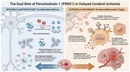
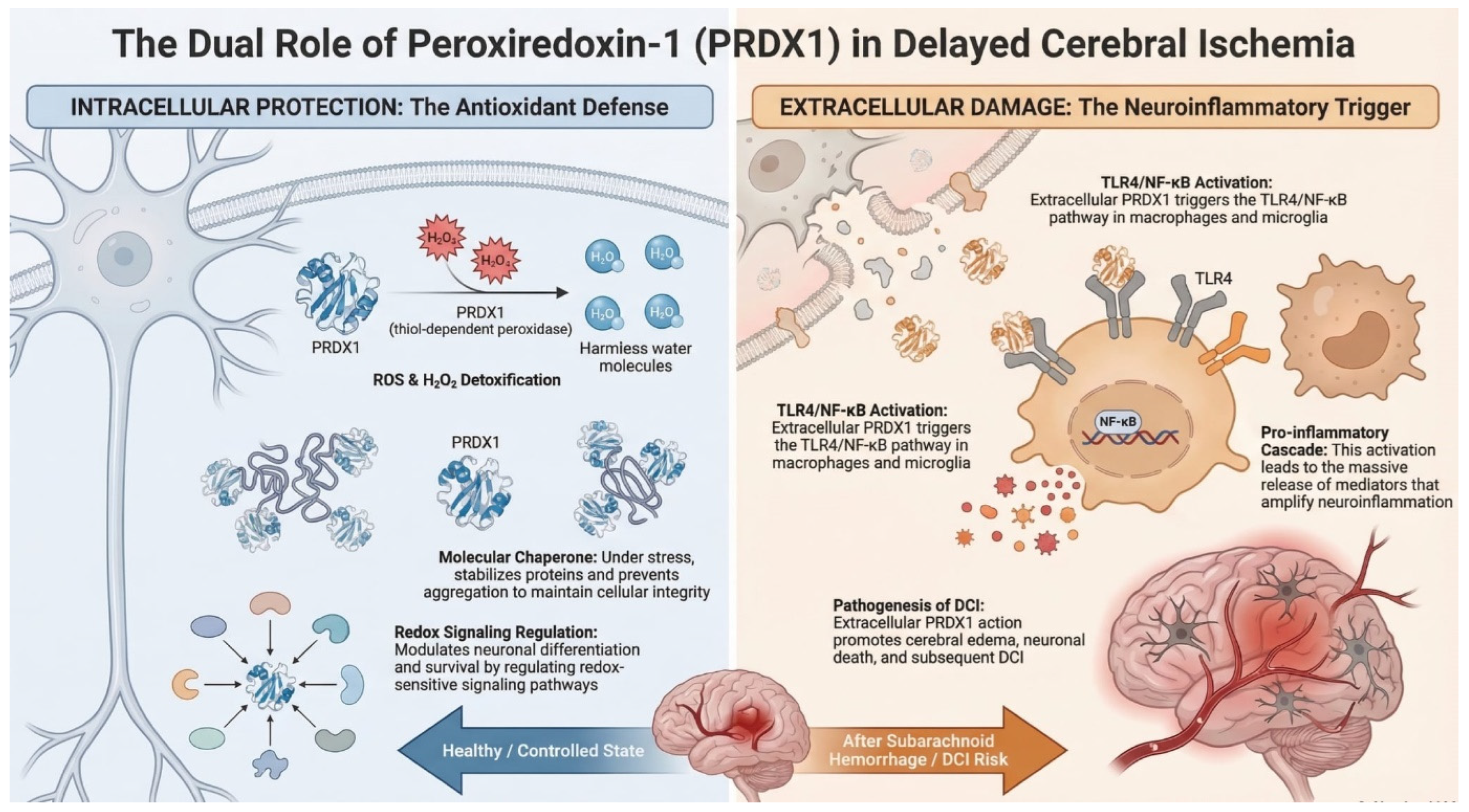
PRDX1 belongs to the family of thiol-dependent antioxidant enzymes that play an important role in cellular protection against oxidative stress. The protein was initially identified in studies comparing normal and RAS-transformed human mammary epithelial cells [41]. Subsequent analyses showed that PRDX1 is constitutively expressed in many human tissues, with particularly high expression observed in organs characterized by intensive cellular proliferation. Further investigations also identified PRDX1-related proteins in erythrocytes as factors capable of enhancing natural killer cell activity, suggesting that members of the peroxiredoxin family may participate both in immune regulation and in cellular responses to OS [42]. The induction of these proteins under conditions of increased oxidative burden supported their role in protecting cells from reactive oxygen species. PRDX1 primarily functions as a thiol-dependent peroxidase that reduces hydrogen peroxide and other peroxides, thereby contributing to the maintenance of intracellular redox balance. In addition to its enzymatic antioxidant activity, PRDX1 may also act as a molecular chaperone under stress conditions, helping to stabilize proteins and prevent their aggregation. Experimental studies have demonstrated that PRDX1 is expressed in differentiating motor neuron cells during spinal cord development [43]. In this context, PRDX1 regulates the activity of specific signaling proteins through redox-dependent reduction in disulfide bonds, highlighting its role not only in detoxification of ROS but also in the modulation of redox-sensitive signaling pathways. Through its interactions with the thioredoxin system, PRDX1 forms part of a broader antioxidant network responsible for controlling hydrogen peroxide levels and maintaining cellular redox homeostasis. By regulating both OS and redox signaling, PRDX1 contributes to the protection of cells from oxidative damage and supports normal cellular function. The role of peroxiredoxins as brain-protective factors has also been supported by experimental animal models, in which increased expression of PRDX1 was observed in astrocytes [44]. Experimental studies also suggest that PRDX1 may participate in inflammatory signaling after hemorrhagic brain injury. In a collagenase-induced mouse model of intracerebral hemorrhage, extracellular PRDX1 was shown to activate the TLR4/NF-κB pathway in macrophages, promoting the production of proinflammatory mediators and contributing to neuroinflammatory injury [45]. Interestingly, in the study by Richard et al., PRDX1 was identified as a promising biomarker for the onset of cerebral infarction, which may suggest that PRDX1 could also have prognostic potential in predicting DCI (Figure 1) [46]. Moreover, it has been demonstrated that induced overexpression of peroxiredoxin-1 in astrocytes and peroxiredoxin-2 in neurons attenuated OS and inhibited neuronal apoptosis following aSAH [47].
Figure 1.
The figure illustrates the mechanisms of action of the PRDX-1 enzyme.
4. Peroxiredoxin-2
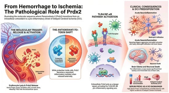
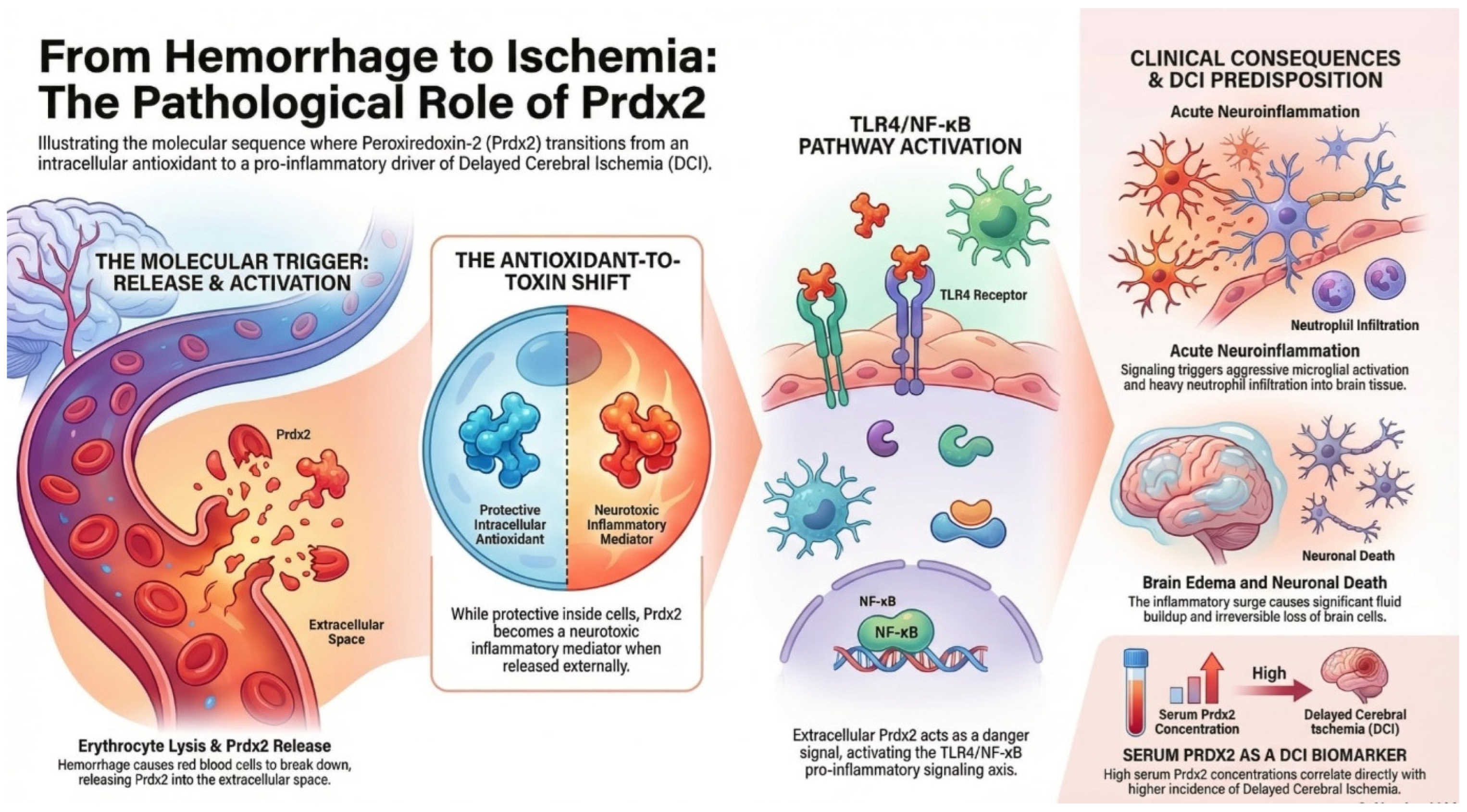
PRDX2 is another member of the PRDX family primarily involved in the regulation of cellular redox balance. The protein was originally identified as a natural killer cell–enhancing factor (NKEFB) during studies investigating erythrocyte-derived factors that modulate immune cell activity [42]. Subsequent research demonstrated that PRDX2 belongs to the group of thioredoxin-dependent peroxidases capable of reducing hydrogen peroxide and other ROS, thereby protecting cellular macromolecules from oxidative damage [48]. PRDX2 is widely expressed in many cell types but is particularly abundant in erythrocytes, where it constitutes one of the major antioxidant proteins responsible for maintaining redox stability. Experimental animal models have demonstrated the physiological importance of this enzyme: mice lacking PRDX2 develop hemolytic anemia associated with elevated reactive oxygen species, oxidative damage to erythrocyte proteins, and compensatory hematologic responses. These findings indicate that PRDX2 plays a crucial role in protecting red blood cells from OS. Beyond its antioxidant activity, PRDX2 has also been implicated in the regulation of intracellular signaling pathways. Studies have shown that PRDX2 can modulate platelet-derived growth factor (PDGF) signaling by limiting local peroxide accumulation, thereby preventing excessive activation of PDGF receptors and downstream proliferative signaling pathways [49]. Through such redox-dependent regulation, PRDX2 contributes to the control of cell proliferation and vascular remodeling. Interestingly, peroxiredoxins, including PRDX2, have also been shown to undergo cyclic redox oscillations with a periodicity of approximately 24 h in human erythrocytes, suggesting a potential role as conserved markers of cellular circadian rhythms [50]. These oscillations occur independently of transcriptional activity and may represent an evolutionarily conserved mechanism linking redox homeostasis with biological timekeeping. Recent studies further suggest that PRDX2 may function as a sensor of OS during DNA replication [51]. Under conditions of increased ROS, PRDX2 undergoes structural changes that influence the activity of proteins involved in replication fork progression, thereby slowing DNA synthesis and helping to prevent replication-associated genomic damage. Taken together, these observations indicate that PRDX2 plays multiple roles in cellular physiology. In addition to its classical antioxidant function in detoxifying ROS, PRDX2 participates in redox signaling, regulation of cell proliferation, and cellular stress responses, highlighting its importance in maintaining redox homeostasis in both physiological and pathological conditions. In a mouse model of intracerebral hemorrhage, PRDX2 appeared to act through the TLR4 signaling pathway, enhancing the inflammatory response and consequently leading to brain edema, microglial activation, neutrophil infiltration, neuronal death, and neurological deficits [52]. There are also reports indicating that PRDX2 may contribute to inflammation within the choroid plexus and damage to the ventricular wall, which in turn promotes the development of hydrocephalus [53]. Moreover, higher serum concentrations of PRDX2 in patients with aSAH have been associated with poorer clinical outcomes and a higher incidence of DCI (Figure 2) [54].
Figure 2.
The figure illustrates the mechanisms of action of the PRDX-2 enzyme.
5. Peroxiredoxin-3
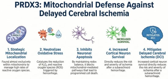
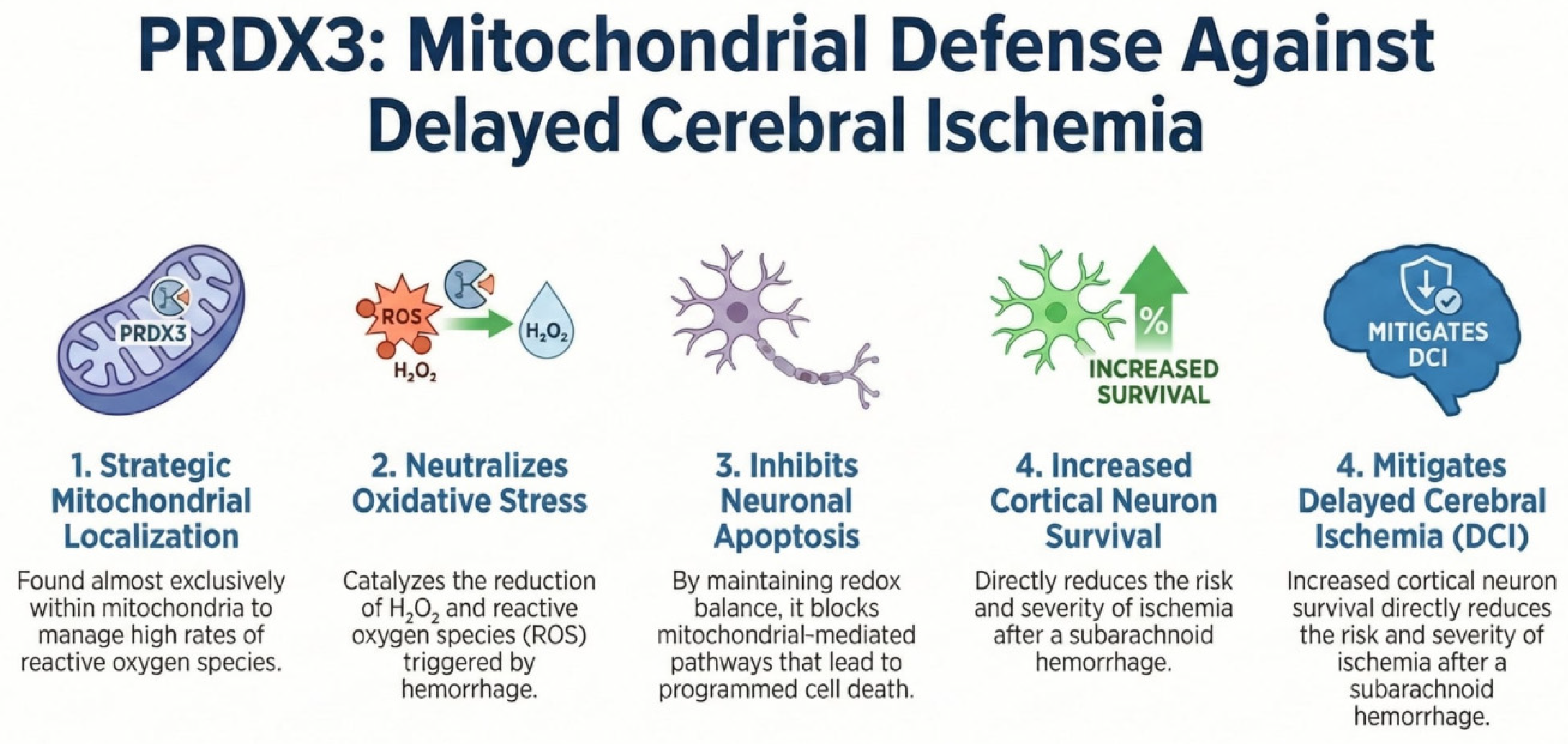
PRDX3 is an antioxidant enzyme that catalyzes the reduction of hydrogen peroxide and other peroxides. Its catalytic mechanism involves the oxidation of a conserved cysteine residue, which is subsequently reduced by the thioredoxin system. Thioredoxin, in turn, is regenerated by thioredoxin reductase using electrons derived from NADPH. Through this redox cycle, peroxiredoxins help maintain intracellular redox balance and protect cells against oxidative stress. Unlike several other PRDXs family members, PRDX3 is localized almost exclusively within mitochondria, where it plays a central role in detoxifying hydrogen peroxide and maintaining mitochondrial redox homeostasis due to the high rate of ROS generation during oxidative phosphorylation [55]. The protein was initially identified in studies on murine erythroleukemia cells, where it was suggested to participate in early stages of cellular differentiation [56]. Subsequent research demonstrated high sequence conservation between mammalian PRDX3 and other thiol-specific antioxidant proteins, highlighting its evolutionary and functional importance. Functional studies indicate that PRDX3 contributes not only to antioxidant defense but also to the regulation of mitochondrial integrity and cell survival [57]. It can inhibit mitochondrial cytochrome c release and attenuate apoptosis under oxidative stress. PRDX3 expression is regulated by transcription factors involved in metabolism and stress responses, including MYC and FOXO3A, underscoring its role in cellular adaptation to metabolic and oxidative challenges [58]. Genetic studies have demonstrated the clinical relevance of PRDX3. Mutations in the PRDX3 gene are associated with autosomal recessive spinocerebellar ataxia type 32 (SCAR32), a disorder marked by progressive cerebellar dysfunction. Patient-derived cells show reduced PRDX3 expression, mitochondrial dysfunction, elevated reactive oxygen species, and increased susceptibility to oxidative stress. Experimental models further confirmed that PRDX3 deficiency impairs neuronal viability and promotes neurodegeneration. In animal models, PRDX3 has been shown to exert a neuroprotective role in early brain injury after aSAH, enhancing the survival of cortical neurons and improving both short- and long-term functional outcomes [59]. Its protective mechanism appears to involve inhibition of mitochondrial-mediated neuronal death pathways. Additionally, increased sensitivity to OS has been observed in patients with defective mitochondrial PRDX3 (Figure 3) [60].
Figure 3.
The figure illustrates the mechanisms of action of the PRDX-3 enzyme.
6. Peroxiredoxin-4
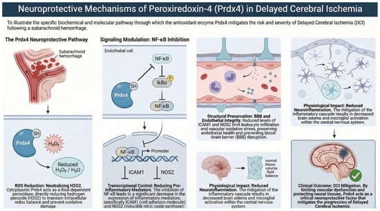
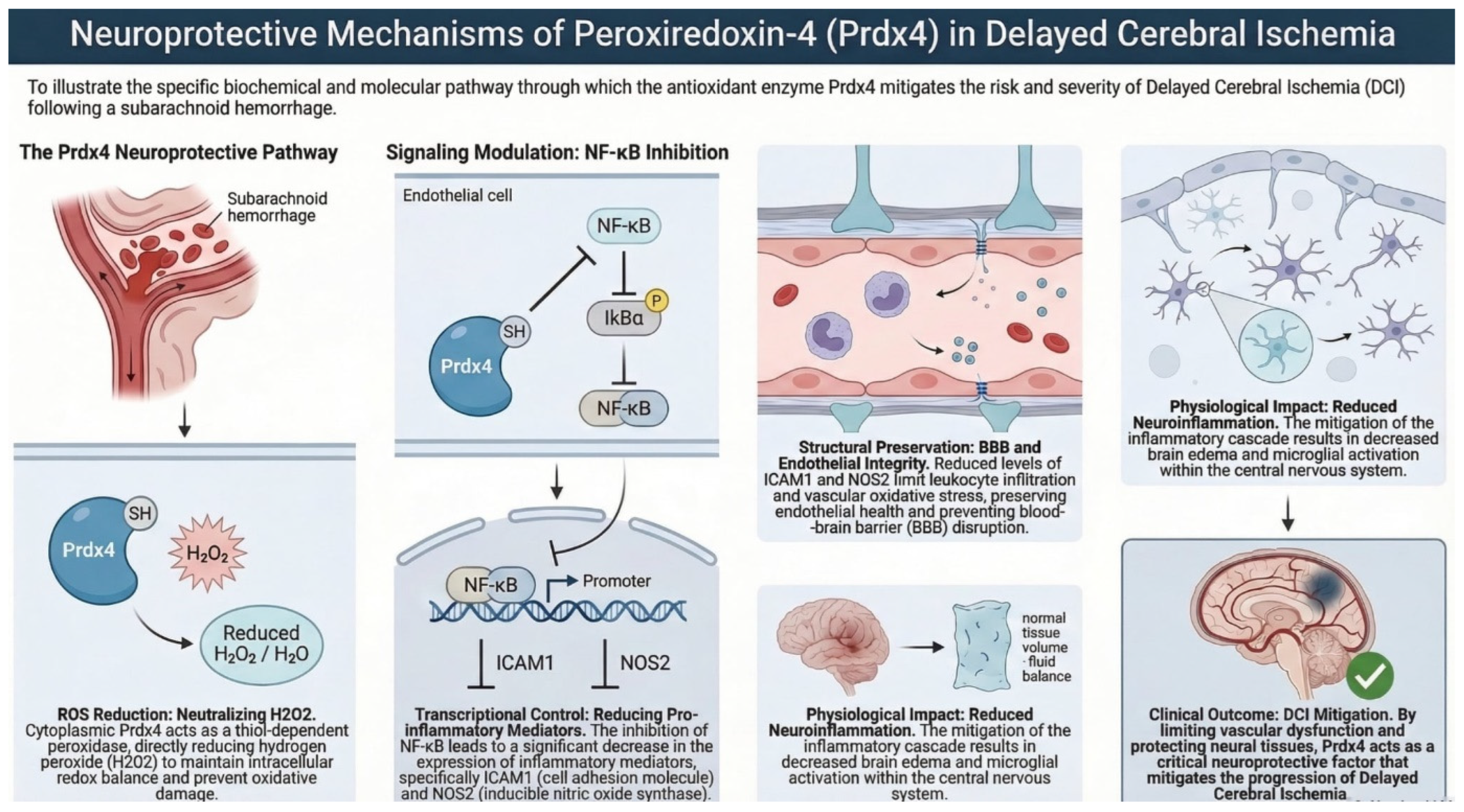
PRDX4 regulates intracellular redox balance and modulates redox-sensitive signaling pathways, including NF-κB activation [61]. Its catalytic activity involves reduction of hydrogen peroxide, which contributes to protection against OS and regulation of inflammatory responses. PRDX4 was originally identified in human cell lines using yeast two-hybrid screening and 5′-RACE, and was shown to contain conserved cysteine motifs characteristic of peroxiredoxins, along with an N-terminal signal sequence enabling secretion. The protein is predominantly localized in the cytoplasm but can also be secreted, and it is widely expressed in human tissues, with particularly high levels in the pancreas, liver, heart, spleen, thymus, and reproductive organs. Its expression is inducible under OS, such as exposure to H2O2, supporting its role in cellular protection. Functionally, PRDX4 has been shown to form both homo- and heterodimers with other cytoplasmic peroxiredoxins (e.g., PRDX1), modulating redox-sensitive signaling. It can influence NF-κB activation, IκBα phosphorylation, and proinflammatory mediator expression, including ICAM1 and NOS2, highlighting its role in the regulation of inflammatory pathways. In addition, PRDX4 protects key enzymes such as glutamine synthetase from oxidative inactivation and promotes cell survival under stress conditions. Animal studies have demonstrated tissue-specific roles of PRDX4 in the ovary [62], PRDX4 is highly expressed in granulosa cells, with levels increasing during follicle development and declining with age. OS induces PRDX4 expression, which correlates with protection from ROS-mediated damage and maintenance of cell proliferation. The human PRDX4 gene is located on chromosome Xp22.11. Taken together, these observations indicate that PRDX4 functions as both a cytoplasmic and secreted antioxidant, participating in redox regulation, inflammatory signaling, and protection from oxidative injury in multiple tissues. PRDX4 seems to play a critical neuroprotective role in the central nervous system by preserving endothelial integrity, limiting blood–brain barrier disruption, and reducing neuroinflammation after ischemia/reperfusion injury (Figure 4) [63].
Figure 4.
The figure illustrates the mechanisms of action of the PRDX-4 enzyme.
7. Peroxiredoxin-5
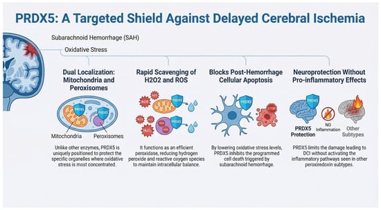
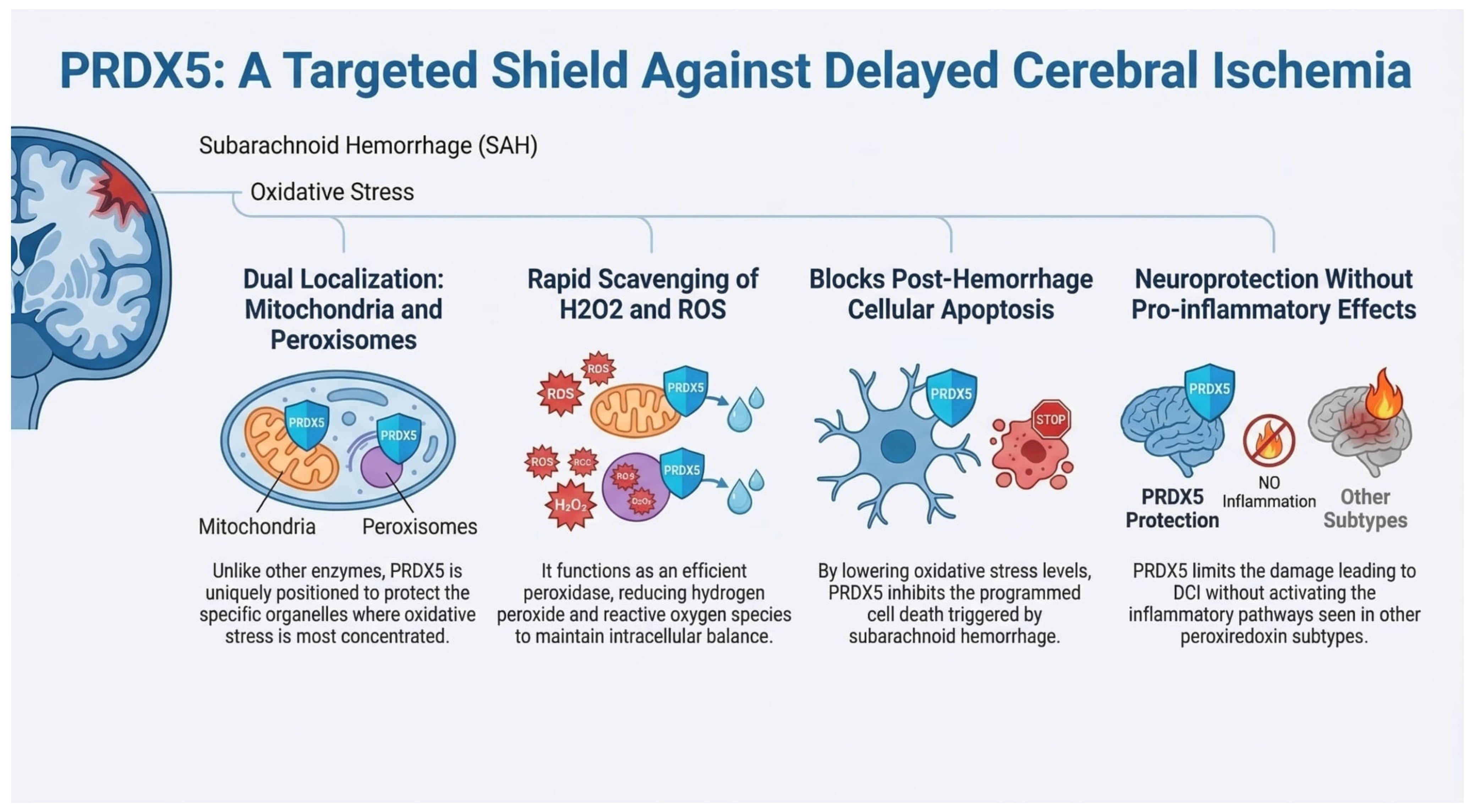
PRDX5 reduces hydrogen peroxide and other ROS, contributing to the maintenance of intracellular redox balance. Unlike other PRDXs, PRDX5 is uniquely localized to both mitochondria and peroxisomes, positioning it to protect against OS generated in these organelles [64]. PRDX5 was initially identified in bronchoalveolar lavage fluid and lung cDNA as a 17 kDa protein, later shown to contain three cysteine residues critical for antioxidant activity and a C-terminal peroxisomal targeting sequence [65]. Its expression is ubiquitous, with highest levels in thyroid, lung, kidney, adrenal gland, heart, and colon, and is inducible under oxidative or inflammatory stress, such as exposure to lipopolysaccharide or proinflammatory cytokines. Functionally, PRDX5 acts as an efficient scavenger of hydrogen peroxide, with antioxidant activity comparable to catalase. Studies in human fibroblasts, cartilage, and endothelial cells demonstrate that PRDX5 upregulation correlates with reduced peroxide accumulation, highlighting its protective role under both physiological and pathological conditions. Structural analyses revealed a thioredoxin-like fold, supporting its function within the thioredoxin-dependent antioxidant system. PRDX5 can be regenerated after overoxidation through sestrin-dependent mechanisms, maintaining the cellular antioxidant defense even under sustained OS. Genetically, the PRDX5 gene is located on chromosome 11q13, a region linked to atopic hypersensitivity. Overall, PRDX5 is a versatile antioxidant enzyme that protects multiple tissues from oxidative damage, supports mitochondrial and peroxisomal redox homeostasis, and participates in cellular stress responses. In animal studies, upregulation of PRDX5 was shown to reduce OS and apoptosis in rats after aSAH [66]. Interestingly, in studies conducted by Kunze et al., decreased plasma levels of PRDX5 were observed in patients with severe stroke, which may suggest that, unlike other PRDXs, it does not exert a proinflammatory effect. Moreover, the reason for the reduced PRDX5 levels in patients with severe stroke was not clarified; it is therefore unclear whether this reduction reflects impaired synthesis or a deficiency in individuals with extensive strokes, and whether it could serve as a prognostic factor (Figure 5) [67].
Figure 5.
The figure illustrates the mechanisms of action of the PRDX-5 enzyme.
8. Peroxiredoxin-6
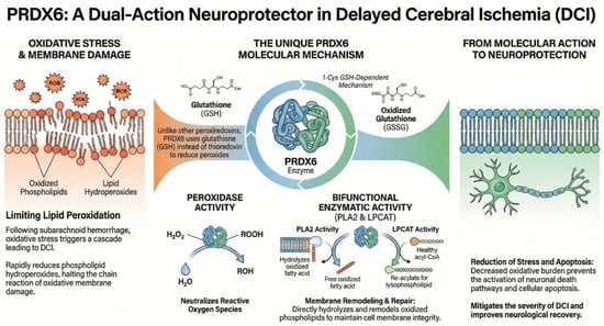
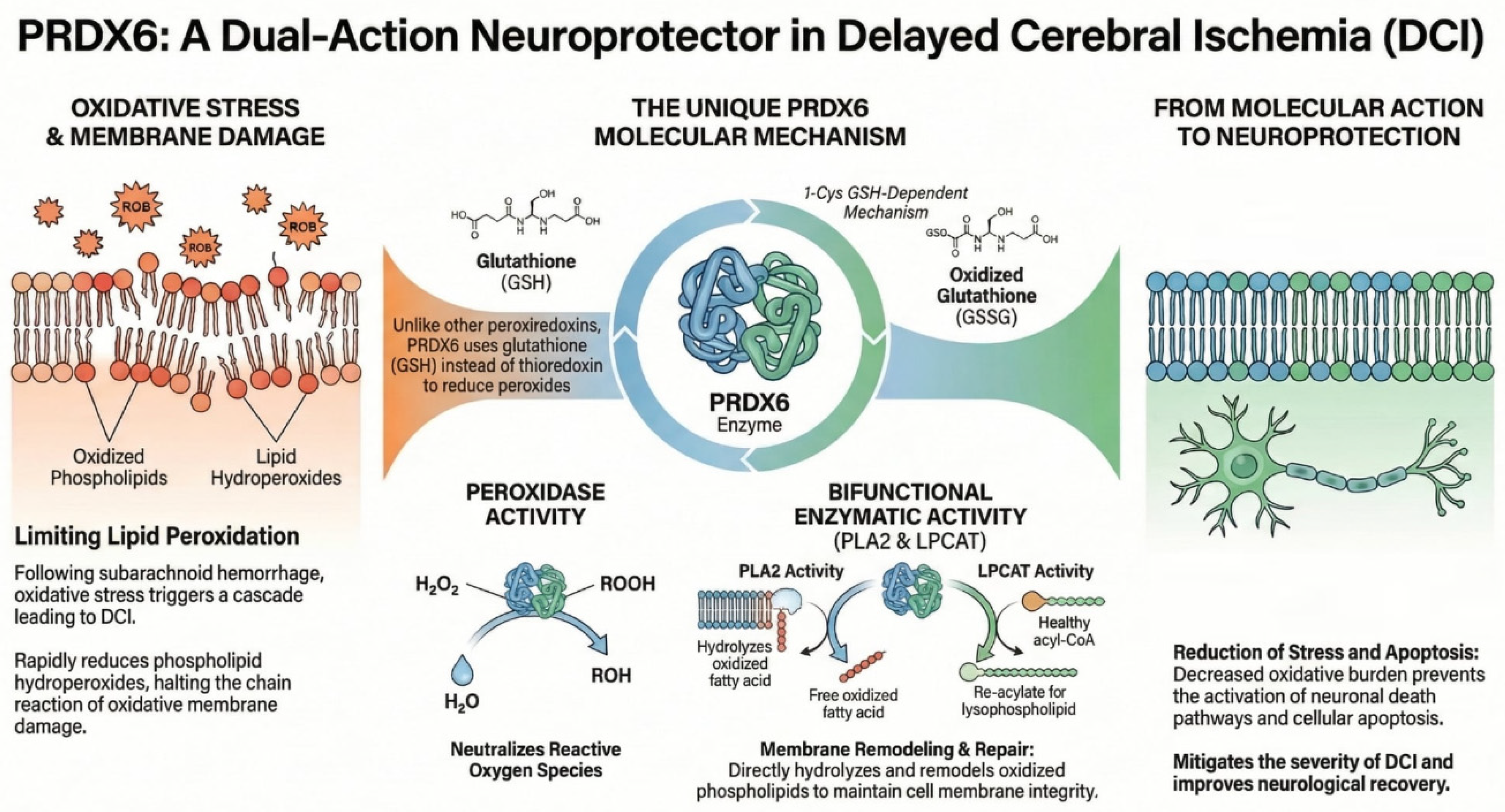
PRDX6 is a distinctive member of the PRDXs family, unique among mammalian peroxiredoxins in possessing only a single conserved cysteine residue and functioning through glutathione (GSH)-dependent mechanisms rather than the thioredoxin system used by PRDX1–5 [68]. PRDX6 participates in the reduction of hydrogen peroxide, short-chain hydroperoxides, and phospholipid hydroperoxides, contributing to cellular protection against oxidative stress and maintenance of redox homeostasis [69]. Unlike other peroxiredoxins, PRDX6 exhibits bifunctional enzymatic activity. In addition to its peroxidase activity, which reduces oxidized substrates, PRDX6 possesses calcium-independent phospholipase A2 (PLA2) activity and acyltransferase (LPCAT) activity, allowing it to hydrolyze and remodel oxidized phospholipids in cell membranes [70]. These multifunctional activities position PRDX6 not only as an antioxidant enzyme but also as a regulator of membrane lipid metabolism and repair following oxidative damage. PRDX6 is expressed in nearly all tissues, with particularly high levels observed in lung epithelial and endothelial cells, hepatocytes, leukocytes, and other cell types exposed to high oxidative loads. Its subcellular localization is primarily cytosolic, but PRDX6 can also be found in acidic organelles such as lysosomes and lamellar bodies, reflecting its roles in both intracellular redox defense and organelle-specific lipid processing. The peroxidase activity of PRDX6 is centered on a conserved cysteine (Cys47), which, during catalysis, is reduced by GSH bound to glutathione S-transferase, completing the enzymatic cycle. The PLA2 activity depends on a catalytic triad (including Ser32, His26, and Asp140) and supports hydrolysis of phospholipid sn-2 ester bonds, which facilitates membrane turnover and surfactant metabolism in the lung [71]. Through its PLA2 activity, PRDX6 can also influence NADPH oxidase assembly and ROS production, illustrating its dual role as both an antioxidant and a modulator of redox signaling. Functionally, PRDX6 protects cells from oxidative injury by reducing peroxidized lipids and repairing oxidative membrane damage, as evidenced in models of H2O2-induced stress where PRDX6 deficiency increases susceptibility to lipid peroxidation and cell death [72]. It also serves regulatory and signaling roles, interacting with factors involved in inflammation, cellular proliferation, and apoptosis, and its expression and activity are modulated by redox-sensitive transcriptional regulators. Thus, PRDX6 is a multifunctional antioxidant enzyme that not only detoxifies peroxides but also participates in lipid metabolism and redox signaling, enabling cells to adapt to and recover from oxidative challenges (Figure 6).
Figure 6.
The figure illustrates the mechanisms of action of the PRDX-6 enzyme.
9. Interplay Between PRDX’s Family
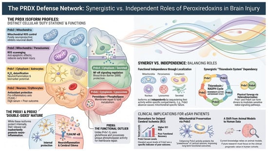
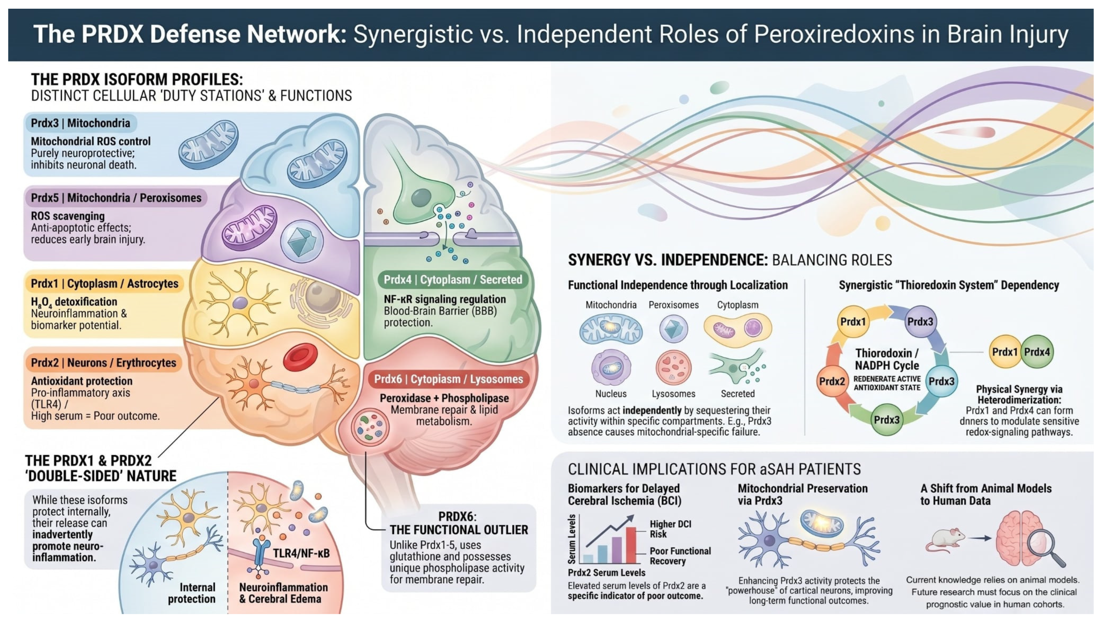
The potential efficacy of the PRDX antioxidant network following aSAH relies on a strategic compartmentalization and sophisticated functional crosstalk between its members. While PRDX1 and PRDX2 provide the primary defense in the cytosol, their roles extend into the extracellular space where PRDX2, specifically released from lysed erythrocytes, acts as a Damage-Associated Molecular Pattern (DAMP) to modulate microglial inflammatory responses, thereby linking antioxidant depletion to neuroinflammation [73]. This cytosolic activity is inherently synchronized with PRDX3 and PRDX5 within the mitochondria; these isoforms form a coordinated front against metabolic oxidative stress, where PRDX3 focuses on the reduction of hydrogen peroxide while PRDX5 provides a specialized defense against peroxynitrite, preventing nitrative stress-induced neuronal apoptosis. These relationships are further characterized by functional redundancy and compensatory mechanisms that become critical during the early brain injury phase. When the classic 2-Cys peroxiredoxins (PRDX1–4) undergo hyperoxidation and subsequent inactivation due to massive ROS surges, the atypical PRDX5 and 1-Cys PRDX6, which are more resistant to such modifications, maintain residual antioxidant capacity to preserve cellular integrity [74]. Furthermore, a “floodgate” mechanism exists between PRDX1 and PRDX2, which regulate local H2O2 concentrations to facilitate the activation of redox-sensitive signaling pathways, such as the Nrf2/HO-1 axis. This activation subsequently triggers the upregulation of other family members, particularly PRDX3, creating a reinforced defensive loop against heme-mediated toxicity. A vital collaborative interaction is also observed in the protection of neuronal membranes, where the sequential action of the family is paramount; while PRDX1-5 neutralize aqueous reactive species, PRDX6 uniquely utilizes its phospholipase A2 activity to repair peroxidized phospholipids. This synergy between the prevention of damage by 2-Cys isoforms and active repair by PRDX6 is essential for preserving the blood–brain barrier and limiting the progression of secondary injury, suggesting that the clinical development of DCI is governed by the collective balance of the entire PRDX family [75]. Figure 7 presents the integrated defense network of the PRDX family, highlighting the strategic compartmentalization of isoforms within specific cellular “duty stations” to maintain redox homeostasis after aSAH. The diagram illustrates the “double-edged sword” nature of the system, where the intracellular neuroprotective and membrane-repair activities of isoforms like PRDX3 and PRDX6 are contrasted against the pro-inflammatory DAMP signaling of extracellularly released PRDX1 and PRDX2. Ultimately, the figure underscores the clinical significance of PRDX levels as biomarkers for vascular dysfunction and the development of DCI in patients.
Figure 7.
The potential role of Peroxiredoxins PRDX in the pathophysiological cascade following aneurysmal subarachnoid hemorrhage (aSAH).
10. Limitations
The primary limitation of the current state of knowledge, as discussed in this review, is the significant reliance on experimental animal models. While these models have provided invaluable insights into the basic mechanisms of PRDX and their potential role in neuroprotection or neuroinflammation after aSAH, they do not fully replicate the complex physiological and pathological environment of the human brain. Consequently, there is a distinct lack of comprehensive clinical data evaluating the specific activity and prognostic value of various PRDX isoforms directly in human patients following aSAH. Furthermore, most available human studies focus on a limited number of biomarkers or isolated redox pathways, which may not capture the multifactorial and interconnected nature of DCI development. Recognizing these gaps, the authors of this article plan to initiate fundamental research aimed at systematically expanding clinical data in human cohorts. These future studies will focus on bridging the transition from animal models to clinical practice, with the goal of better understanding the role of peroxiredoxins in patients after hemorrhage and identifying more effective, translationally relevant therapeutic targets.
11. Conclusions
The PRDX family serves as a critical yet dual-natured defense network in aSAH patophysiology, balancing mitochondrial neuroprotection (PRDX3, 5, 6) against pro-inflammatory DAMP signaling (PRDX1, 2). By regulating hydrogen peroxide through the thioredoxin system, these enzymes maintain blood–brain barrier integrity and modulate the oxidative-inflammatory cascade. Elevated serum levels of specific isoforms, particularly PRDX2, function as significant prognostic biomarkers for delayed cerebral ischemia (DCI) and poor clinical outcomes. Furthermore, unique mechanisms like the membrane-repair activity of PRDX6 present novel but at this point, only potential—therapeutic targets for mitigating secondary brain injury. Moving forward, large-scale human clinical studies are essential to translate these experimental insights into improved management strategies for aSAH patients.
Author Contributions
Conceptualization: K.Z., K.W. and R.W.; Methodology: K.Z., A.D. and M.P.; Software: E.J.B., A.D. and N.K.; Formal analysis: K.Z., N.K., E.J.B. and B.S.; Investigation: K.Z., B.S. and R.W.; Resources: K.Z., M.P. and R.W.; Data curation: K.Z. and M.P.; Writing—original draft preparation: K.Z. and B.S.; Writing—review and editing: K.Z., D.J.J. and K.W.; Visualization: A.D. and N.K.; Supervision: D.J.J. and K.W.; Project administration: K.Z. and K.W.; Funding acquisition: no founding. All authors have read and agreed to the published version of the manuscript.
Funding
This research received no external funding.
Data Availability Statement
No new data were created or analyzed in this study. Data sharing is not applicable.
Acknowledgments
The authors would like to express their special thanks to Paweł Szczepański for his invaluable assistance and expertise in the preparation and development of the figures for this manuscript.
Conflicts of Interest
The authors declare no conflicts of interest.
References
- Ikram, A.; Javaid, M.A.; Ortega-Gutierrez, S.; Selim, M.; Kelangi, S.; Anwar, S.M.H.; Torbey, M.T.; Divani, A.A. Delayed Cerebral Ischemia after Subarachnoid Hemorrhage. J. Stroke Cerebrovasc. Dis. 2021, 30, 106064. [Google Scholar] [CrossRef] [Scilit] [PubMed]
- Vergouwen, M.D.; Vermeulen, M.; van Gijn, J.; Rinkel, G.J.; Wijdicks, E.F.; Muizelaar, J.P.; Mendelow, A.D.; Juvela, S.; Yonas, H.; Terbrugge, K.G.; et al. Definition of delayed cerebral ischemia after aneurysmal subarachnoid hemorrhage as an outcome event in clinical trials and observational studies: Proposal of a multidisciplinary research group. Stroke 2010, 41, 2391–2395. [Google Scholar] [CrossRef] [Scilit] [PubMed]
- Abdulazim, A.; Heilig, M.; Rinkel, G.; Etminan, N. Diagnosis of Delayed Cerebral Ischemia in Patients with Aneurysmal Subarachnoid Hemorrhage and Triggers for Intervention. Neurocrit. Care 2023, 39, 311–319. [Google Scholar] [CrossRef] [Scilit] [PubMed] [PubMed Central]
- Rigante, L.; van Lieshout, J.H.; Vergouwen, M.D.I.; van Griensven, C.H.S.; Vart, P.; van der Loo, L.; de Vries, J.; Vinke, R.S.; Etminan, N.; Aquarius, R.; et al. Time trends in the risk of delayed cerebral ischemia after subarachnoid hemorrhage: A meta-analysis of randomized controlled trials. Neurosurg. Focus 2022, 52, E2. [Google Scholar] [CrossRef] [Scilit] [PubMed]
- Love, S. Oxidative stress in brain ischemia. Brain Pathol. 1999, 9, 119–131. [Google Scholar] [CrossRef] [Scilit] [PubMed] [PubMed Central]
- Hao, G.; Eser, P.; Mo, J. Oxidative Stress and Intracranial Hypertension after Aneurysmal Subarachnoid Hemorrhage. Antioxidants 2022, 11, 2423. [Google Scholar] [CrossRef] [Scilit] [PubMed] [PubMed Central]
- Gaetani, P.; Pasqualin, A.; Rodriguez y Baena, R.; Borasio, E.; Marzatico, F. Oxidative stress in the human brain after subarachnoid hemorrhage. J. Neurosurg. 1998, 89, 748–754. [Google Scholar] [CrossRef] [Scilit] [PubMed]
- Brown, J.D.; Day, A.M.; Taylor, S.R.; Tomalin, L.E.; Morgan, B.A.; Veal, E.A. A peroxiredoxin promotes H2O2 signaling and oxidative stress resistance by oxidizing a thioredoxin family protein. Cell Rep. 2013, 5, 1425–1435. [Google Scholar] [CrossRef] [Scilit] [PubMed] [PubMed Central]
- Rhee, S.G.; Woo, H.A.; Kang, D. The Role of Peroxiredoxins in the Transduction of H2O2 Signals. Antioxid. Redox Signal. 2018, 28, 537–557. [Google Scholar] [CrossRef] [Scilit] [PubMed]
- Yao, Z.; Bai, Q.; Wang, G. Mechanisms of Oxidative Stress and Therapeutic Targets following Intracerebral Hemorrhage. Oxidative Med. Cell. Longev. 2021, 2021, 8815441. [Google Scholar] [CrossRef] [Scilit] [PubMed] [PubMed Central]
- Anan, M.; Nagai, Y.; Fudaba, H.; Fujiki, M. Lactate and Lactate Dehydrogenase in Cistern as Biomarkers of Early Brain Injury and Delayed Cerebral Ischemia of Subarachnoid Hemorrhage. J. Stroke Cerebrovasc. Dis. 2020, 29, 104765. [Google Scholar] [CrossRef] [Scilit] [PubMed]
- Rasmussen, R.; Bache, S.; Stavngaard, T.; Møller, K. Plasma Levels of IL-6, IL-8, IL-10, ICAM-1, VCAM-1, IFNγ, and TNFα are not Associated with Delayed Cerebral Ischemia, Cerebral Vasospasm, or Clinical Outcome in Patients with Subarachnoid Hemorrhage. World Neurosurg. 2019, 128, e1131–e1136. [Google Scholar] [CrossRef] [Scilit] [PubMed]
- Badjatia, N.; Monahan, A.; Carpenter, A.; Zimmerman, J.; Schmidt, J.M.; Claassen, J.; Connolly, E.S.; Mayer, S.A.; Karmally, W.; Seres, D. Inflammation, negative nitrogen balance, and outcome after aneurysmal subarachnoid hemorrhage. Neurology 2015, 84, 680–687. [Google Scholar] [CrossRef] [Scilit] [PubMed] [PubMed Central]
- Chaichana, K.L.; Pradilla, G.; Huang, J.; Tamargo, R.J. Role of inflammation (leukocyte-endothelial cell interactions) in vasospasm after subarachnoid hemorrhage. World Neurosurg. 2010, 73, 22–41. [Google Scholar] [CrossRef] [Scilit] [PubMed]
- Momin, E.N.; Schwab, K.E.; Chaichana, K.L.; Miller-Lotan, R.; Levy, A.P.; Tamargo, R.J. Controlled delivery of nitric oxide inhibits leukocyte migration and prevents vasospasm in haptoglobin 2-2 mice after subarachnoid hemorrhage. Neurosurgery 2009, 65, 937–945. [Google Scholar] [CrossRef] [Scilit] [PubMed]
- Pluta, R.M. Delayed cerebral vasospasm and nitric oxide: Review, new hypothesis, and proposed treatment. Pharmacol. Ther. 2005, 105, 23–56. [Google Scholar] [CrossRef] [Scilit] [PubMed]
- Biondi, A.; Ricciardi, G.K.; Puybasset, L.; Abdennour, L.; Longo, M.; Chiras, J.; Van Effenterre, R. Intra-arterial nimodipine for the treatment of symptomatic cerebral vasospasm after aneurysmal subarachnoid hemorrhage: Preliminary results. AJNR Am. J. Neuroradiol. 2004, 25, 1067–1076. [Google Scholar] [PubMed] [PubMed Central]
- Fathi, A.R.; Pluta, R.M.; Bakhtian, K.D.; Qi, M.; Lonser, R.R. Reversal of cerebral vasospasm via intravenous sodium nitrite after subarachnoid hemorrhage in primates. J. Neurosurg. 2011, 115, 1213–1220. [Google Scholar] [CrossRef] [Scilit] [PubMed] [PubMed Central]
- Thampatty, B.P.; Sherwood, P.R.; Gallek, M.J.; Crago, E.A.; Ren, D.; Hricik, A.J.; Kuo, C.-W.J.; Klamerus, M.M.; Alexander, S.A.; Bender, C.M.; et al. Role of endothelin-1 in human aneurysmal subarachnoid hemorrhage: Associations with vasospasm and delayed cerebral ischemia. Neurocrit. Care 2011, 15, 19–27. [Google Scholar] [CrossRef] [Scilit] [PubMed] [PubMed Central]
- Bian, K.; Murad, F. Nitric oxide (NO)—Biogeneration, regulation, and relevance to human diseases. Front. Biosci. 2003, 8, d264–d278. [Google Scholar] [CrossRef] [Scilit] [PubMed]
- Schulz, E.; Wenzel, P.; Münzel, T.; Daiber, A. Mitochondrial redox signaling: Interaction of mitochondrial reactive oxygen species with other sources of oxidative stress. Antioxid. Redox Signal. 2014, 20, 308–324. [Google Scholar] [CrossRef] [Scilit] [PubMed] [PubMed Central]
- Förstermann, U.; Münzel, T. Endothelial nitric oxide synthase in vascular disease: From marvel to menace. Circulation 2006, 113, 1708–1714. [Google Scholar] [CrossRef] [Scilit] [PubMed]
- Brüne, B.; Schmidt, K.U.; Ullrich, V. Activation of soluble guanylate cyclase by carbon monoxide and inhibition by superoxide anion. Eur. J. Biochem. 1990, 192, 683–688. [Google Scholar] [CrossRef] [Scilit] [PubMed]
- Cerrato, R.; Cunnington, C.; Crabtree, M.J.; Antoniades, C.; Pernow, J.; Channon, K.M.; Böhm, F. Endothelin-1 increases superoxide production in human coronary artery bypass grafts. Life Sci. 2012, 91, 723–728. [Google Scholar] [CrossRef] [Scilit] [PubMed]
- Li, L.; Watts, S.W.; Banes, A.K.; Galligan, J.J.; Fink, G.D.; Chen, A.F. NADPH oxidase-derived superoxide augments endothelin-1-induced venoconstriction in mineralocorticoid hypertension. Hypertension 2003, 42, 316–321. [Google Scholar] [CrossRef] [Scilit] [PubMed]
- Li, L.; Fink, G.D.; Watts, S.W.; Northcott, C.A.; Galligan, J.J.; Pagano, P.J.; Chen, A.F. Endothelin-1 increases vascular superoxide via endothelin(A)-NADPH oxidase pathway in low-renin hypertension. Circulation 2003, 107, 1053–1058. [Google Scholar] [CrossRef] [Scilit] [PubMed]
- Hernanz, R.; Briones, A.M.; Salaices, M.; Alonso, M.J. New roles for old pathways? A circuitous relationship between reactive oxygen species and cyclo-oxygenase in hypertension. Clin. Sci. 2014, 126, 111–121. [Google Scholar] [CrossRef] [Scilit] [PubMed]
- White, R.P.; Robertson, J.T. Comparison of piroxicam, meclofenamate, ibuprofen, aspirin, and prostacyclin efficacy in a chronic model of cerebral vasospasm. Neurosurgery 1983, 12, 40–46. [Google Scholar] [CrossRef] [Scilit] [PubMed]
- Hall, E.D.; Wang, J.A.; Bosken, J.M.; Singh, I.N. Lipid peroxidation in brain or spinal cord mitochondria after injury. J. Bioenerg. Biomembr. 2016, 48, 169–174. [Google Scholar] [CrossRef] [Scilit] [PubMed] [PubMed Central]
- Hall, E.D.; Vaishnav, R.A.; Mustafa, A.G. Antioxidant therapies for traumatic brain injury. Neurotherapeutics 2010, 7, 51–61. [Google Scholar] [CrossRef] [Scilit] [PubMed] [PubMed Central]
- Aladag, M.A.; Turkoz, Y.; Sahna, E.; Parlakpinar, H.; Gul, M. The attenuation of vasospasm by using a SOD mimetic after experimental subarachnoidal haemorrhage in rats. Acta Neurochir. 2003, 145, 673–677. [Google Scholar] [CrossRef] [Scilit] [PubMed]
- Aladag, M.A.; Turkoz, Y.; Parlakpinar, H.; Ozen, H.; Egri, M.; Unal, S.C. Melatonin ameliorates cerebral vasospasm after experimental subarachnoidal haemorrhage correcting imbalance of nitric oxide levels in rats. Neurochem. Res. 2009, 34, 1935–1944. [Google Scholar] [CrossRef] [Scilit] [PubMed]
- Marklund, S.L.; Westman, N.G.; Lundgren, E.; Roos, G. Copper- and zinc-containing superoxide dismutase, manganese-containing superoxide dismutase, catalase, and glutathione peroxidase in normal and neoplastic human cell lines and normal human tissues. Cancer Res. 1982, 42, 1955–1961. [Google Scholar] [PubMed]
- Kalinina, E.V.; Chernov, N.N.; Saprin, A.N. Involvement of thio-, peroxi-, and glutaredoxins in cellular redox-dependent processes. Biochemistry 2008, 73, 1493–1510. [Google Scholar] [CrossRef] [Scilit] [PubMed]
- Tian, W.N.; Braunstein, L.D.; Pang, J.; Stuhlmeier, K.M.; Xi, Q.C.; Tian, X.; Stanton, R.C. Importance of glucose-6-phosphate dehydrogenase activity for cell growth. J. Biol. Chem. 1998, 273, 10609–10617. [Google Scholar] [CrossRef] [Scilit] [PubMed]
- Gaetani, P.; Cafe, C.; Rodriguez y Baena, R.; Tancioni, F.; Torri, C.; Tartara, F.; Marzatico, F. Superoxide dismutase activity in cisternal cerebrospinal fluid after aneurysmal subarachnoid haemorrhage. Acta Neurochir. 1997, 139, 1033–1037. [Google Scholar] [CrossRef] [Scilit] [PubMed]
- Watanabe, T.; Sasaki, T.; Asano, T.; Takakura, K.; Sano, K.; Fuchinoue, T.; Watanabe, K.; Yoshimura, S.; Abe, K. Changes in glutathione peroxidase and lipid peroxides in cerebrospinal fluid and serum after subarachnoid hemorrhage—With special reference to the occurrence of cerebral vasospasm. Neurol. Med.-Chir. 1988, 28, 645–649. [Google Scholar] [CrossRef] [Scilit] [PubMed]
- Facchinetti, F.; Dawson, V.L.; Dawson, T.M. Free radicals as mediators of neuronal injury. Cell. Mol. Neurobiol. 1998, 18, 667–682. [Google Scholar] [CrossRef] [Scilit] [PubMed]
- Han, Y.; Su, J.; Liu, X.; Zhao, Y.; Wang, C.; Li, X. Naringin alleviates early brain injury after experimental subarachnoid hemorrhage by reducing oxidative stress and inhibiting apoptosis. Brain Res. Bull. 2017, 133, 42–50. [Google Scholar] [CrossRef] [Scilit]
- Handa, Y.; Kaneko, M.; Takeuchi, H.; Tsuchida, A.; Kobayashi, H.; Kubota, T. Effect of an antioxidant, ebselen, on development of chronic cerebral vasospasm after subarachnoid hemorrhage in primates. Surg. Neurol. 2000, 53, 323–329. [Google Scholar] [CrossRef] [Scilit] [PubMed]
- Prospéri, M.T.; Ferbus, D.; Karczinski, I.; Goubin, G. A human cDNA corresponding to a gene overexpressed during cell proliferation encodes a product sharing homology with amoebic and bacterial proteins. J. Biol. Chem. 1993, 268, 11050–11056. [Google Scholar] [CrossRef] [Scilit] [PubMed]
- Shau, H.; Butterfield, L.H.; Chiu, R.; Kim, A. Cloning and sequence analysis of candidate human natural killer-enhancing factor genes. Immunogenetics 1994, 40, 129–134. [Google Scholar] [CrossRef] [Scilit] [PubMed]
- Yan, Y.; Sabharwal, P.; Rao, M.; Sockanathan, S. The antioxidant enzyme Prdx1 controls neuronal differentiation by thiol-redox-dependent activation of GDE2. Cell 2009, 138, 1209–1221. [Google Scholar] [CrossRef] [Scilit] [PubMed] [PubMed Central]
- Wang, S.; Yu, L.; Sun, G.; Liu, Y.; Hu, W.; Liu, Y.; Peng, T.; Wang, X.; Cheng, J.; Sr, A.; et al. Danhong Injection protects hemorrhagic brain by increasing peroxiredoxin 1 in aged rats. Front. Pharmacol. 2020, 11, 346. [Google Scholar] [CrossRef] [Scilit] [PubMed] [PubMed Central]
- Liu, D.L.; Zhao, L.X.; Zhang, S.; Du, J.R. Peroxiredoxin 1-mediated activation of TLR4/NF-κB pathway contributes to neuroinflammatory injury in intracerebral hemorrhage. Int. Immunopharmacol. 2016, 41, 82–89. [Google Scholar] [CrossRef] [Scilit] [PubMed]
- Richard, S.; Lapierre, V.; Girerd, N.; Bonnerot, M.; Burkhard, P.R.; Lagerstedt, L.; Bracard, S.; Debouverie, M.; Turck, N.; Sanchez, J.-C. Diagnostic performance of peroxiredoxin 1 to determine time-of-onset of acute cerebral infarction. Sci. Rep. 2016, 6, 38300. [Google Scholar] [CrossRef] [Scilit] [PubMed] [PubMed Central]
- Lu, Y.; Zhang, X.S.; Zhou, X.M.; Gao, Y.Y.; Chen, C.L.; Liu, J.P.; Ye, Z.-N.; Zhang, Z.-H.; Wu, L.-Y.; Li, W.; et al. Peroxiredoxin 1/2 protects brain against H2O2-induced apoptosis after subarachnoid hemorrhage. FASEB J. 2019, 33, 3051–3062. [Google Scholar] [CrossRef] [Scilit] [PubMed]
- Pahl, P.; Berger, R.; Hart, I.; Chae, H.Z.; Rhee, S.G.; Patterson, D. Localization of TDPX1, a human homologue of the yeast thioredoxin-dependent peroxide reductase gene (TPX), to chromosome 13q12. Genomics 1995, 26, 602–606. [Google Scholar] [CrossRef] [Scilit] [PubMed]
- Choi, M.H.; Lee, I.K.; Kim, G.W.; Kim, B.U.; Han, Y.H.; Yu, D.Y.; Park, H.S.; Kim, K.Y.; Lee, J.S.; Choi, C.; et al. Regulation of PDGF signalling and vascular remodelling by peroxiredoxin II. Nature 2005, 435, 347–353. [Google Scholar] [CrossRef] [Scilit] [PubMed]
- O’Neill, J.S.; Reddy, A.B. Circadian clocks in human red blood cells. Nature 2011, 469, 498–503. [Google Scholar] [CrossRef] [Scilit] [PubMed] [PubMed Central]
- Somyajit, K.; Gupta, R.; Sedlackova, H.; Neelsen, K.J.; Ochs, F.; Rask, M.B.; Choudhary, C.; Lukas, J. Redox-sensitive alteration of replisome architecture safeguards genome integrity. Science 2017, 358, 797–802. [Google Scholar] [CrossRef] [Scilit] [PubMed]
- Du, Y.; Wang, J.; Zhang, J.; Li, N.; Li, G.; Liu, X.; Lin, Y.; Wang, D.; Kang, K.; Bian, L.; et al. Intracerebral hemorrhage-induced brain injury in mice: The role of peroxiredoxin 2-Toll-like receptor 4 inflammatory axis. CNS Neurosci. Ther. 2024, 30, e14681. [Google Scholar] [CrossRef] [Scilit] [PubMed] [PubMed Central]
- Tan, X.; Chen, J.; Keep, R.F.; Xi, G.; Hua, Y. Prx2 (Peroxiredoxin 2) as a cause of hydrocephalus after intraventricular hemorrhage. Stroke 2020, 51, 1578–1586. [Google Scholar] [CrossRef] [Scilit] [PubMed] [PubMed Central]
- Fu, Z.; Dai, J.; Lin, Q.; Ye, L.; Chen, X.; Li, Z.; Lu, C.; Chen, M.; Ba, H.; Sun, J.; et al. Serum peroxiredoxin 2 emerges as a predictive biomarker of poor neurological outcome and delayed cerebral ischemia after aneurysmal subarachnoid hemorrhage: A prospective cohort study. Neurosurg. Rev. 2025, 48, 412. [Google Scholar] [CrossRef] [Scilit] [PubMed]
- Chiribau, C.B.; Cheng, L.; Cucoranu, I.C.; Yu, Y.S.; Clempus, R.E.; Sorescu, D. FOXO3A regulates peroxiredoxin III expression in human cardiac fibroblasts. J. Biol. Chem. 2008, 283, 8211–8217. [Google Scholar] [CrossRef] [Scilit] [PubMed] [PubMed Central]
- Nemoto, Y.; Yamamoto, T.; Takada, S.; Matsui, Y.; Obinata, M. Antisense RNA of the latent period gene (MER5) inhibits the differentiation of murine erythroleukemia cells. Gene 1990, 91, 261–265. [Google Scholar] [CrossRef] [Scilit] [PubMed]
- Wonsey, D.R.; Zeller, K.I.; Dang, C.V. The c-Myc target gene PRDX3 is required for mitochondrial homeostasis and neoplastic transformation. Proc. Natl. Acad. Sci. USA 2002, 99, 6649–6654. [Google Scholar] [CrossRef] [Scilit] [PubMed] [PubMed Central]
- Park, J.W.; Sonn, S.K.; Lee, B.H.; Oh, G.T.; Chang, T.S. Peroxiredoxin III mitigates mitochondrial H2O2-mediated damage and supports quality control in cardiomyocytes under hypoxia-reoxygenation stress. Redox Biol. 2025, 86, 103799. [Google Scholar] [CrossRef] [Scilit] [PubMed] [PubMed Central]
- Li, H.; Wang, Z.; Xie, X.; Luo, M.; Shen, H.; Li, X.; Li, H.; Wang, Z.; Li, X.; Chen, G. Peroxiredoxin-3 plays a neuroprotective role in early brain injury after experimental subarachnoid hemorrhage in rats. Brain Res. Bull. 2023, 193, 95–105. [Google Scholar] [CrossRef] [Scilit] [PubMed]
- Mukhopadhyay, S.S.; Leung, K.S.; Hicks, M.J.; Hastings, P.J.; Youssoufian, H.; Plon, S.E. Defective mitochondrial peroxiredoxin-3 results in sensitivity to oxidative stress in Fanconi anemia. J. Cell Biol. 2006, 175, 225–235. [Google Scholar] [CrossRef] [Scilit] [PubMed] [PubMed Central]
- Jin, D.Y.; Chae, H.Z.; Rhee, S.G.; Jeang, K.T. Regulatory role for a novel human thioredoxin peroxidase in NF-kappaB activation. J. Biol. Chem. 1997, 272, 30952–30961. [Google Scholar] [CrossRef] [Scilit] [PubMed]
- Qian, Y.; Shao, L.; Yuan, C.; Jiang, C.-Y.; Liu, J.; Gao, C.; Gao, L.; Cui, Y.-G.; Jiang, S.-W.; Liu, J.-Y.; et al. Implication of differential peroxiredoxin 4 expression with age in ovaries of mouse and human for ovarian aging. Curr. Mol. Med. 2016, 16, 243–251. [Google Scholar] [CrossRef] [Scilit] [PubMed]
- Xu, N.; Jiang, X.; Zhang, W.; Shi, Y.; Leak, R.K.; Keep, R.F.; Ye, Q.; Yang, T.; Li, S.; Hu, X.; et al. Endothelial peroxiredoxin-4 is indispensable for blood-brain barrier integrity and long-term functional recovery after ischemic stroke. Proc. Natl. Acad. Sci. USA 2024, 121, e2400272121. [Google Scholar] [CrossRef] [Scilit] [PubMed] [PubMed Central]
- Knoops, B.; Clippe, A.; Bogard, C.; Arsalane, K.; Wattiez, R.; Hermans, C.; Duconseille, E.; Falmagne, P.; Bernard, A. Cloning and characterization of AOEB166, a novel mammalian antioxidant enzyme of the peroxiredoxin family. J. Biol. Chem. 1999, 274, 30451–30458. [Google Scholar] [CrossRef] [Scilit] [PubMed]
- Wattiez, R.; Hermans, C.; Bernard, A.; Lesur, O.; Falmagne, P. Human bronchoalveolar lavage fluid: Two-dimensional gel electrophoresis, amino acid microsequencing and identification of major proteins. Electrophoresis 1999, 20, 1634–1645. [Google Scholar] [CrossRef] [Scilit] [PubMed]
- Yang, J.; Wu, Q.; Lan, S.; Yuan, K.; Sun, B.; Meng, Y.; Xu, S.; Shi, H. Peroxiredoxin-5 alleviates early brain injury after subarachnoid hemorrhage by reducing oxidative stress. Brain Res. Bull. 2024, 217, 111087. [Google Scholar] [CrossRef] [Scilit] [PubMed]
- Kunze, A.; Zierath, D.; Tanzi, P.; Cain, K.; Becker, K. Peroxiredoxin 5 (PRX5) is correlated inversely to systemic markers of inflammation in acute stroke. Stroke 2014, 45, 608–610. [Google Scholar] [CrossRef] [Scilit] [PubMed] [PubMed Central]
- Rahaman, H.; Herojit, K.; Singh, L.R.; Haobam, R.; Fisher, A.B. Structural and functional diversity of the peroxiredoxin 6 enzyme family. Antioxid. Redox Signal. 2024, 40, 759–775. [Google Scholar] [CrossRef] [Scilit] [PubMed]
- Pacifici, F.; Arriga, R.; Sorice, G.P.; Capuani, B.; Scioli, M.G.; Pastore, D.; Donadel, G.; Bellia, A.; Caratelli, S.; Coppola, A.; et al. Peroxiredoxin 6, a novel player in the pathogenesis of diabetes. Diabetes 2014, 63, 3210–3220. [Google Scholar] [CrossRef] [Scilit] [PubMed]
- Fisher, A.B. The phospholipase A2 activity of peroxiredoxin 6. J. Lipid Res. 2018, 59, 1132–1147. [Google Scholar] [CrossRef] [Scilit] [PubMed] [PubMed Central]
- Chatterjee, S.; Feinstein, S.I.; Dodia, C.; Sorokina, E.; Lien, Y.C.; Nguyen, S.; Debolt, K.; Speicher, D.; Fisher, A.B. Peroxiredoxin 6 phosphorylation and subsequent phospholipase A2 activity are required for agonist-mediated activation of NADPH oxidase in mouse pulmonary microvascular endothelium and alveolar macrophages. J. Biol. Chem. 2011, 286, 11696–11706. [Google Scholar] [CrossRef] [Scilit] [PubMed] [PubMed Central]
- Xue, M.; Huang, X.; Zhu, T.; Zhang, L.; Yang, H.; Shen, Y.; Feng, L. Unveiling the significance of peroxiredoxin 6 in central nervous system disorders. Antioxidants 2024, 13, 449. [Google Scholar] [CrossRef] [Scilit] [PubMed] [PubMed Central]
- Radyuk, S.N.; Orr, W.C. The multifaceted impact of peroxiredoxins on aging and disease. Antioxid. Redox Signal. 2018, 29, 1293–1311. [Google Scholar] [CrossRef] [Scilit] [PubMed] [PubMed Central]
- Forshaw, T.E.; Holmila, R.; Nelson, K.J.; Lewis, J.E.; Kemp, M.L.; Tsang, A.W.; Poole, L.B.; Lowther, W.T.; Furdui, C.M. Peroxiredoxins in cancer and response to radiation therapies. Antioxidants 2019, 8, 11. [Google Scholar] [CrossRef] [Scilit] [PubMed] [PubMed Central]
- Shao, L.; Chen, S.; Ma, L. Secondary brain injury by oxidative stress after cerebral hemorrhage: Recent advances. Front. Cell. Neurosci. 2022, 16, 853589. [Google Scholar] [CrossRef] [Scilit] [PubMed] [PubMed Central]

Disclaimer/Publisher’s Note: The statements, opinions and data contained in all publications are solely those of the individual author(s) and contributor(s) and not of MDPI and/or the editor(s). MDPI and/or the editor(s) disclaim responsibility for any injury to people or property resulting from any ideas, methods, instructions or products referred to in the content. |
© 2026 by the authors. Licensee MDPI, Basel, Switzerland. This article is an open access article distributed under the terms and conditions of the Creative Commons Attribution (CC BY) license.