


Abstract
Cellular survival and adaptability depend on the dynamic regulation of proteins—the central actors of biological systems. Through mechanisms such as post-translational modifications, protein turnover, and the formation of membraneless organelles, cells can sense and respond to a variety of stressors. Recent advances in artificial intelligence and chemical biology have provided powerful tools to study and manipulate these processes, paving the way for novel therapeutic strategies in cancer. This review explores how cells “tame” their proteome in response to stress by coordinating protein synthesis, modification, degradation, and structural organization to maintain functional resilience.
1. Introduction
Cancer cells maintain their survival through remarkable phenotypic and molecular plasticity, allowing them to adapt to therapeutic, metabolic, and microenvironmental stressors. These adaptive mechanisms operate within tightly regulated spatiotemporal boundaries and are central to treatment outcomes. In oncology, discrepancies between transcript levels and protein abundance are particularly pronounced due to selective protein stabilization, accelerated degradation, and context-dependent translational control [1,2]. As such, the proteome provides the most accurate readout of cancer cell behavior under drug pressure.
Proteins responsible for transport, signalling, DNA repair, and apoptosis are critical determinants of therapeutic responses. Multidrug resistance proteins (e.g., ATP-binding cassette transporters), stress-responsive chaperones, and regulators of cell-cycle checkpoints illustrate how structural and functional protein dynamics shape drug sensitivity [3,4,5]. These dynamics are influenced by post-translational modifications, spatial compartmentalization, and degradation pathways, enabling cancer cells to quickly recalibrate their proteome in response to chemotherapy or targeted inhibitors.
Recent breakthroughs in structural prediction—highlighted by the 2024 Nobel Prize in Chemistry, which recognized artificial intelligence-driven advances in protein structure inference—have transformed our ability to analyze resistance-associated proteins at an unprecedented resolution [6]. Integrating such predictive tools with chemical biology allows for a deeper understanding of conformational shifts, mutational impacts, and allosteric regulation that contribute to drug tolerance.
Against this backdrop, this review reframes the concept of taming the proteome within an oncological context: cancer cells strategically rewire protein synthesis, modification, turnover, and spatial organization to survive therapeutic stress. Understanding these mechanisms provides a foundation for developing drugs that destabilize resistant phenotypes or selectively target the proteins that drive them.
2. Protein Turnover: Balancing Synthesis and Degradation in Drug Resistance
2.1. Protein Synthesis in Drug Resistance
Protein turnover begins with synthesis (Figure 1), a finely tuned process that allows cells to remodel their proteome in response to therapeutic pressure. In the context of drug resistance, translational reprogramming enables malignant cells to upregulate efflux pumps, anti-apoptotic proteins, metabolic enzymes, and stress-adaptive factors that counteract drug activity. Because translation initiation is the rate-limiting step, it is the dominant point at which resistant cells modulate protein output [7].
Figure 1.
Protein translation mechanisms and the aberrant molecular and cellular phenotypes resulting from their dysregulation [7,8].
Initiation ensures proper ribosome positioning at the start codon, recruitment of initiator tRNA, and engagement of the elongation cycle. Elongation and termination then drive the stepwise assembly and release of resistance-associated polypeptides. Foundational discoveries defining the genetic code and tRNA structure [9,10], together with advances in ribosome structural biology and mass spectrometry-based proteomics [11,12], have revealed how translational machinery is dynamically rewired under stress, including during chemotherapy, targeted therapy, hypoxia, or nutrient limitation (Figure 1).
Clinically, the translation apparatus represents both a vulnerability and an adaptive tool. Because core elongation and termination functions are essential in all cells, therapeutic strategies largely focus on translation initiation, particularly on eukaryotic initiation factors (eIFs) whose activity governs the expression of survival and resistance genes [7,13]. Pharmacological inhibition of mTOR—one of the central regulators of cap-dependent translation—reduces eIF4E availability through 4E-BP activation, dampening the synthesis of oncogenic drivers recruited during resistance. Clinical evaluation of mTOR pathway inhibition [14], including everolimus in the adjuvant setting for renal cell carcinoma, further illustrates the translational relevance of targeting this axis (Table 1). Experimental modulators such as ribavirin further illustrate how altering eIF activity can disrupt translational programs that support therapeutic escape (Table 1).
Table 1.
Targeting translation-initiation factors: small-molecule inhibitors and resistance mechanisms (clinical use *).
Unexpectedly, several antibiotic-derived compounds have revealed additional translational dependencies in drug-resistant cancer cells. Microbial metabolites and their derivatives have emerged as important scaffolds in anticancer drug discovery [42,43]. While classical oxazolidinones selectively inhibit bacterial ribosomes, novel derivatives exhibit anticancer activity in vitro by disrupting stress-responsive translation pathways utilized by rapidly dividing tumour cells. Similarly, macrolide antibiotics such as rapamycin (sirolimus) and its semisynthetic derivatives—temsirolimus and everolimus, collectively known as rapalogs (Table 1)—originate from Streptomyces species and exert anticancer effects by inhibiting mTORC1, a central regulator of protein synthesis and cell growth. Hybrid fluoroquinolone–oxazolidinone scaffolds and related compounds reveal how resistant cancer cells often exploit translational mechanisms similar to microbial stress survival strategies [42,43]. In response to pharmacological stress using microbe-derived compounds, cancer cells adopt molecular adaptations that enhance specific protein functions—such as stronger protein interaction affinities or more efficient DNA repair—contributing to clinically observed drug resistance (Table 2).
Table 2.
Antibiotic-derived compounds used in cancer therapy and mechanisms of therapeutic resistance.
When cap-dependent translation is suppressed—whether by therapy-induced stress, hypoxia, or metabolic strain—cells shift to non-canonical initiation [71,72]. Internal ribosome entry site (IRES)-dependent translation allows for continued synthesis of proteins that promote drug resistance [73,74,75], including those regulating the unfolded protein response, angiogenesis, inflammation, apoptosis evasion, and cell-cycle progression. Viral IRES elements have long served as experimental models of this switch [76], revealing mechanisms that resistant cancer cells co-opt (Table 3). IRES-mediated translation is also sensitive to modulation by stress-adaptive drugs (Table 1 and Table 3).
Table 3.
Selected human genes containing internal ribosome entry site (IRES) elements within their untranslated regions (UTRs), enabling cap-independent translation initiation under certain stress conditions.
Emerging therapeutics directly target these alternative translation modes. Zotatifin (eFT226), an eIF4A inhibitor in clinical trials (Table 1), suppresses IRES-driven translation of stress-adaptive transcripts required for resistance. Other agents such as JQ1 [IC50 = 0.2–10 µM] and CX-5461 [IC50 = 0.1–2 µM] indirectly reduce IRES-mediated synthesis of oncogenic drivers like c-Myc [94,95,96], limiting the adaptive proteomic shifts characteristic of resistant tumours (Figure 1, Table 3).
Together, these insights position translational control as a central mechanism through which cells remodel protein abundance during drug exposure. Accordingly, selective targeting of stress-adaptive translation offers a promising route to disrupt resistance-maintaining proteomes (Figure 1, Table 1, Table 2 and Table 3).
2.2. Protein Degradation in Drug Resistance
Protein degradation constitutes the second major pillar of protein turnover (Figure 2) and plays a decisive role in shaping resistance phenotypes. By eliminating damaged or misfolded proteins and regulating the steady-state abundance of signalling intermediates, the ubiquitin–proteasome system (UPS) and autophagy–lysosome pathway (Figure 2) remodel the proteome during therapeutic stress.
Figure 2.
Main pillars of protein turnover in ① normal physiology and ② acute stress conditions, mechanisms that are exploited by cancer cells [97].
The UPS mediates selective degradation of short-lived or regulatory proteins [98], many of which govern drug sensitivity, cell-cycle control, apoptosis, and DNA damage responses. Through stepwise ubiquitination, substrates are directed to the 26S proteasome for rapid proteolysis, enabling resistant cells to remove pro-apoptotic proteins, suppress negative regulators of survival pathways, or stabilize oncogenic drivers via altered ubiquitin ligase activity [99,100] (Figure 2). Disruption of substrate recognition or proteasome capacity can therefore promote resistance by altering signalling network dynamics. Therapeutic approaches, including proteasome inhibitors and E3 ligase modulators (Table 4), aim to exploit these vulnerabilities by restoring proteostatic imbalance or inducing lethal proteotoxic stress.
Table 4.
Protein degradation pathway inhibitors in cancer therapy: clinical use and resistance mechanisms.
In parallel, the autophagy–lysosome system provides a bulk degradation pathway that supports drug-resistant cell survival under metabolic or oxidative stress [141,142]. By clearing damaged organelles and protein aggregates, autophagy preserves mitochondrial function, limits ROS accumulation, and recycles nutrients required for continued proliferation. While impaired autophagic flux can sensitize tumours to therapy, excessive or compensatory autophagy frequently enhances resistance (Figure 2) [141,142], motivating investigation of autophagy modulators as therapeutic adjuncts [99,100].
Targeted Protein Degradation (TPD) technologies further extend these principles by enabling selective elimination of resistance-associated proteins previously considered undruggable [143] (Figure 3). PROTACs and molecular glues recruit endogenous E3 ligases to degrade oncogenic drivers, overcoming limitations of occupancy-based inhibition [99,100,143]. Clinically advanced agents such as ARV-110 and ARV-471 illustrate the therapeutic potential of enforced degradation [99,100].
Figure 3.
Therapeutic strategies using targeted protein degradation (TPD) [143,144,145].
Importantly, TPD approaches can target oncogenic fusion proteins (e.g., BCR-ABL, PML-RARα, EWS-FLI1) that confer resistance to conventional therapies [99,145,146]. Agents including cereblon-binding molecular glues, arsenic trioxide, ATRA, and HSP90 inhibitors (Table 5) destabilize these aberrant proteins and promote their clearance. By targeting structurally complex or scaffolding-dependent oncoproteins, TPD expands druggability beyond traditional active-site inhibition.
Table 5.
Therapeutic agents promoting degradation of fusion proteins in clinical practice.
Ultimately, protein degradation provides the rapid adaptive capacity required for resistance evolution. While synthesis gradually builds a resistant proteome (Figure 1), degradative systems enable immediate recalibration of signalling components (Figure 2). However, emerging evidence shows that tumours can also acquire resistance to TPD strategies [148,150,156], highlighting the continued evolutionary plasticity of proteostasis networks.
2.3. Clearance and Metabolic Recycling in Drug Resistance
Clearance and metabolic recycling of protein degradation products are key modulators of cancer drug resistance as they regulate intracellular nutrient availability and detoxification capacity. Anticancer therapies induce proteotoxic stress, increasing proteasomal and lysosomal degradation and generating amino acids and metabolic intermediates that must be efficiently reused or eliminated (Figure 2) [157,158]. As shown in Figure 4, targeted clearance depends on specific amino acid transporters and organelle-associated export systems that channel metabolites toward biosynthesis, signalling, or controlled efflux, whereas non-targeted routes include bulk autophagic turnover and systemic hepatobiliary or renal elimination [159,160].
Figure 4.
Clearance mechanisms of protein breakdown products through targeted and non-targeted routes.
Lysosomal proteolysis is particularly critical in resistant cancer cells, supplying amino acids that sustain protein synthesis (Figure 1), preserve redox balance, and support mitochondrial function [157]. This recycling limits proteotoxic damage and attenuates therapy-induced apoptosis. Recovered amino acids further activate nutrient-sensing pathways such as mTOR, reinforcing anabolic metabolism under therapeutic stress [158]. Concurrently, resistant cells upregulate amino acid transporters and metabolic enzymes to optimize metabolite uptake and redistribution (Figure 4) [159,161].
Systemic clearance mechanisms additionally shape therapeutic outcomes by regulating xenobiotic and metabolite elimination, thereby influencing intracellular drug accumulation and efficacy [160,161]. Table 6 summarizes clinically used small-molecule inhibitors that target metabolic enzymes, transporters, or clearance pathways. By disrupting amino acid production, utilization, or export, these agents impair metabolic adaptation and enhance treatment sensitivity.
Table 6.
Small-molecule inhibitors targeting cancer metabolism in clinical oncology: mechanisms of action and resistance.
Collectively, intracellular recycling and systemic clearance integrate nutrient recovery with detoxification, enabling cancer cells to maintain bioenergetic stability during therapeutic stress. Targeting these interconnected pathways therefore represents a promising strategy to overcome metabolic resistance. However, tumour cells frequently develop secondary adaptations—including target mutation or amplification, metabolic pathway rewiring, and altered drug transport or efflux (Table 6)—that continue to limit the long-term efficacy of metabolic and antimetabolite therapies used in clinical oncology [180], such as methotrexate, 5-fluorouracil, L-asparaginase, IDH inhibitors, and pentostatin.
3. Genotoxic Stressors and the DNA Damage Response in Cancer Therapy
3.1. Characteristics of Therapeutically Relevant Genotoxic Stressors
In cancer therapy, genotoxic stressors are intentionally applied to damage tumour DNA and induce cytotoxicity. However, the diversity of DNA lesions they generate—and the tumour cell’s capacity to repair them—creates major challenges in predicting treatment responses [181]. Radiotherapy, chemotherapy, and endogenous sources of DNA damage all contribute to complex mixtures of lesions that activate the DNA damage response (DDR) [182], allowing cancer cells to survive therapy.
Genotoxic agents used in oncology can be broadly categorized into direct and indirect inducers of DNA damage. Direct genotoxicity arises when a therapeutic agent interacts with DNA itself [183], generating chemical or physical lesions that stall replication and transcription. Indirect genotoxicity results from disruptions to cellular metabolism or oxidative balance—such as ROS production (Table 7)—that subsequently injure nuclear DNA [184]. Parameters including chemical reactivity, pharmacokinetics, exposure duration, and intracellular accumulation strongly shape the type and extent of DNA damage generated during treatment [185].
Physical genotoxic agents, particularly those used in radiotherapy [186], generate some of the most cytotoxic lesions in oncology. Ionizing radiation (X-rays, γ-rays, particle beams) produces a spectrum of base damage [187], single-strand breaks (SSBs), and highly lethal double-strand breaks (DSBs), triggering ATM- and ATR-dependent DDR signalling cascades [188]. Non-ionizing UV radiation induces pyrimidine dimers [189] and bulky helix distortions, lesions typically repaired through nucleotide excision repair pathways. Although mechanical or thermal stress contributes less directly to therapeutic DNA damage, they can promote secondary ROS generation [189], amplifying oxidative DNA injury. In clinical practice, radiation exposure in imaging and radiotherapy represents a controlled but potent form of genotoxic stress designed to overwhelm tumour repair capacity [190].
Chemical genotoxic agents (Table 7), including multiple classes of chemotherapeutics, interact covalently or noncovalently with DNA [191]. Alkylating agents (Table 7) introduce alkyl groups onto nucleotide bases, causing mispairing, depurination, helix distortion, and replication fork collapse. Cross-linking agents—such as platinum compounds (Table 7) and nitrogen mustards—form intra- and inter-strand bridges that block both DNA replication and transcription. Attempts by cancer cells to resolve these structures can generate secondary DSBs, making their repair heavily dependent on homologous recombination, Fanconi anemia pathways, and translesion synthesis [192]. These mechanisms are frequently upregulated in therapy-resistant tumours [193].
Table 7.
Well-characterized DNA-damaging chemicals and the cellular pathways ensuring survival.
Genotoxic stress in human cells can arise not only from therapeutic agents but also from biological sources. Oncoviruses such as Human papillomavirus (HPV), Epstein–Barr virus (EBV), and Hepatitis B virus (HBV) integrate into the host genome or express proteins that inhibit DDR components, including p53 and ATM, thereby promoting mutagenesis and altering therapeutic sensitivity [208]. The bacterial pathogen Helicobacter pylori—also classified as a type 1 carcinogen (the highest level) [209]—induces chronic inflammation and ROS-driven DNA damage [210] while fungal metabolites like aflatoxins generate bulky adducts that require nucleotide excision repair [211]. These endogenous and exogenous stressors contribute to replication stress, a driver of genomic instability, cellular senescence, and aging [212]. Chemotherapy itself can exacerbate genotoxic stress in normal tissues, as exemplified by cisplatin-induced DNA lesions and nephrotoxicity [213].
Successfully overcoming drug resistance in cancer demands a multidisciplinary approach, leveraging insights from biology, medicine, physical sciences, mathematics, and data analytics to model DDR dynamics and predict treatment outcomes [214,215]. Long-term chemotherapy also impacts normal hematopoietic cells, highlighting the need to balance tumour targeting with preservation of tissue integrity [216]. Collectively, these biological and therapeutic genotoxic stressors underscore the critical importance of robust, adaptable DDR systems in human cells (Table 7).
3.2. DNA Damage, Repair Capacity, and the Biology of Tumour Survival
DNA damage is both a source of oncogenic transformation and a persistent threat to tumour survival. To cope with endogenous lesions and therapy-induced insults, cells rely on the DDR, a coordinated network that integrates DNA repair with cell-cycle checkpoints and global stress responses (Table 7) [217]. DDR genes are frequently mutated in cancer, fostering genomic instability—an enabling hallmark that promotes tumour evolution, metastasis and therapeutic adaptation [218]. Yet these same defects create exploitable vulnerabilities.
A paradigm is provided by BRCA1/2-mutant tumours, which are defective in homologous recombination and therefore hypersensitive to PARP inhibition through synthetic lethality [217,219]. The clinical success of PARP inhibitors validated DDR targeting but also revealed challenges, including resistance mechanisms and the need for predictive biomarkers [219]. Notably, our previous study demonstrated that cisplatin-resistant cancers frequently develop an addiction to PARP1 activity and rely on ATM-mediated DDR signalling by suppressing tumour suppressors [220,221]. Targeting these acquired dependencies establishes a new synthetic lethal framework beyond BRCA mutations, potentially expanding PARP-based therapies to platinum-refractory cancers. Current strategies expand beyond first-generation PARP inhibitors to include next-generation PARP1-selective agents, checkpoint kinase inhibitors and compounds targeting additional “caretaker” repair pathways [217,218]. Simultaneous targeting of multiple DDR nodes or rational combinations with chemo- and radiotherapy are under active investigation [219].
Tumour repair capacity is further shaped by replication stress and transcription-associated DNA damage. Transcription–replication conflicts generate double-strand breaks (DSBs) and genomic rearrangements (Table 7) [222]. Pharmacological targeting of proliferating cell nuclear antigen (PCNA) with AOH1996 selectively exacerbates these conflicts, inducing transcription-dependent DSBs and suppressing tumour growth, thereby exploiting a cancer-selective vulnerability (Table 7) [223].
Beyond enzymatic repair pathways, genome stability depends on nuclear architecture and chromatin dynamics. DSB repair involves chromatin remodelling, repair condensates and large-scale genome reorganization (Figure 2) [224]. Irradiation-induced strengthening of topologically associating domain boundaries in an ATM-dependent manner suggests that 3D genome organization actively contributes to repair fidelity [225]. Moreover, chromatin-modifying complexes such as genetic suppressor element 1 (GSE1), which interacts with a deacetylase/demethylase co-repressor complex, regulate ATR signalling and histone ubiquitination during DDR [226].
Finally, oncogenic mutations rewire protein–protein interaction networks that govern DDR signalling hubs, altering repair pathway choice and stress adaptation (Figure 3) [227,228]. Therapeutic innovation now also leverages platinum–protein interactions to overcome drug efflux and chemoresistance (Table 7) [229]. Collectively, tumour survival reflects a dynamic equilibrium between DNA damage burden, adaptive repair capacity and evolving interaction networks—an equilibrium increasingly amenable to precise therapeutic disruption (Table 7).
4. Post-Translational Modifications: A Regulatory Layer in Resistance Acquisition
4.1. Ubiquitination in Therapy-Induced Adaptation
Ubiquitination, a pivotal post-translational modification, plays a central role in dynamically regulating protein stability and cellular adaptation under therapeutic stress [230]. This process involves the covalent attachment of the 76-amino-acid protein ubiquitin to substrates, either as monoubiquitin or polyubiquitin chains (Figure 4 and Figure 5), orchestrated by the sequential activity of E1, E2, and over 600 E3 ligases [231,232]. Polyubiquitin chains generally signal proteasomal degradation (Figure 2), facilitating rapid clearance of damaged or drug-bound proteins, whereas monoubiquitination modulates non-degradative processes such as protein trafficking, DNA repair, and activation of bypass signalling pathways [233]. The reversibility of ubiquitination, mediated by about 90 human deubiquitinating enzymes (DUBs), allows for precise remodelling of the proteome in response to therapy.
Figure 5.
Critical regulatory steps of autophagy and ubiquitin–proteasome system (UPS) targeted by clinically approved small-molecule inhibitors (as mentioned in Table 4).
Several components of the ubiquitination machinery, including E1 activating enzymes, E3 ligases, DUBs, and the proteasome itself (Figure 5), have been successfully targeted by small-molecule inhibitors in clinical development or practice (Table 4).
The ubiquitin-proteasome system (UPS) is responsible for the majority of intracellular protein degradation in mammalian cells, yet recent studies indicate that proteasome activity itself, not just rates of ubiquitination, critically determines substrate fate [234]. The 26S proteasome employs a multistep, ATP-dependent mechanism, with structural and biochemical features that ensure selective degradation, efficient ubiquitin recycling, and prevention of non-specific proteolysis (Figure 4). Proteasome function is further regulated by interacting proteins, subunit modifications such as phosphorylation, and coordinated activity with ubiquitinating and deubiquitinating enzymes.
In cancer, dysregulation of ubiquitination or proteasome activity underlies adaptive resistance (Table 4). Altered E3 ligase or DUB expression selectively eliminates pro-apoptotic proteins while stabilizing survival-promoting factors [230,235]. Small-molecule inhibitors, PROTACs, and molecular glues can manipulate this system to degrade oncogenic proteins and overcome resistance, as demonstrated in colorectal cancer and HPV-driven malignancies [230,232]. Collectively, ubiquitination and proteasome regulation constitute a versatile and finely tunable layer of therapy-induced adaptation, offering compelling targets for improving treatment outcomes.
4.2. Phosphorylation as a Driver of Adaptive Signalling and Drug Resistance
Post-translational modifications (PTMs) constitute a dynamic regulatory layer that enables cancer cells to rapidly adapt to therapeutic stress. Among these, phosphorylation is particularly central to resistance acquisition because it directly rewires signalling networks without requiring genetic alteration. A global structural analysis of phosphoproteins demonstrated that phosphorylation often induces subtle but stabilizing conformational changes, modulates residue fluctuations, and, in some cases, mechanically couples distal regulatory and functional sites, consistent with allosteric “domino” models of signal propagation [236]. Such structural plasticity provides a mechanistic basis for adaptive signalling in drug-treated cells.
The clinical relevance of phosphorylation as a therapeutic target is reflected in the development of multiple classes of protein kinase inhibitors designed to suppress aberrant oncogenic signalling. The three main classes of protein kinase inhibitors currently used in clinical oncology are summarized in Table 8, highlighting their mechanisms of action.
Table 8.
Three main classes of protein kinase inhibitors used in clinics to target overactive kinases in cancer.
At the systems level, phosphorylation responses are not uniformly proportional to pathway activation. Quantitative phosphoproteomics of the ERK cascade revealed variable phosphorylation thresholds among ERK substrates, with some sites saturating at low ERK activity and others responding only at high activation levels [242]. Low-threshold targets included transcriptional repressors that promote proliferation when inactivated, whereas high-threshold targets were enriched in DNA damage response proteins. These findings suggest that graded ERK activity can selectively engage proliferative or stress-response programs, thereby shaping cell fate decisions under therapeutic pressure.
Clinically, such adaptive rewiring underlies resistance to kinase inhibitors. Resistance to protein tyrosine kinase inhibitors frequently involves compensatory activation of parallel pathways, microenvironmental cues, and metabolic reprogramming [243]. In TP53-mutated ovarian cancer, a transmembrane receptor-tyrosine-kinase-like orphan receptor 1 (ROR1)–PI3K/AKT signalling node toggles between proliferative bypass and DNA repair states in response to cell cycle blockade [244]. Previously, we showed that cells adapt to Epstein–Barr virus (EBV) infection not only by tolerating translational disruption, but by converting it into a growth-stimulatory signal through selective activation of a PI3K isoform predominantly expressed in B cells [245]. EBV thereby exploits an intrinsic translation–growth feedback loop to promote survival and proliferation of infected cells while simultaneously facilitating immune evasion. Key phosphorylation-dependent resistance mechanisms and associated kinase or signalling inhibitors are detailed in Table 9.
Table 9.
Targeting phosphorylation in cancer therapy: common kinase and signalling inhibitors in drug resistance.
Together, these studies highlight phosphorylation as a tunable, structurally encoded, and threshold-sensitive mechanism that enables dynamic resistance to targeted therapies.
4.3. PTMs as Regulators of the DNA Damage Response in Resistant Tumours
PTMs are central regulators of the DDR, particularly in therapy-resistant tumours where proteome plasticity (Figure 2) enables survival under genotoxic stress. PTMs expand protein functional diversity by altering conformation, localization, stability, charge, and protein–protein interactions, thereby rewiring signalling networks that govern DNA repair and cell fate [257]. More than 400–650 PTM types have been described, including phosphorylation, ubiquitination, SUMOylation, acetylation, and redox modifications, many of which dynamically coordinate DDR signalling cascades [257,258].
A key mechanism through which PTMs regulate DDR is through control of protein stability (Figure 2). PTM-regulated degrons determine whether repair factors are stabilized or targeted for proteasomal degradation, fine-tuning the amplitude and duration of repair signalling [259]. In resistant tumours, aberrant phosphorylation or ubiquitination can stabilize oncogenic DDR mediators or inactivate negative regulators, promoting tolerance to chemotherapy or radiotherapy. Large-scale proteogenomic analyses across cancers reveal shared PTM patterns linked to dysregulated DNA repair, particularly phosphorylation-driven DDR subtypes that transcend tissue origin [260].
Beyond histones, non-histone PTMs of checkpoint kinases, repair enzymes, and replication factors critically drive cancer progression and therapeutic resistance [261]. These modifications often occur within multiprotein complexes whose architecture and interaction networks must be carefully represented in mechanistic schematics to capture PTM-dependent regulation [262]. Moreover, molecular chaperones such as HSP90 and HSP70 modulate the stability and PTM state of DDR client proteins, integrating proteostasis (Figure 2) with repair signalling and contributing to treatment resistance when overexpressed [263]. Due to their key role in stabilizing DDR mediators, molecular chaperones have become therapeutic targets (Table 10); however, despite extensive clinical investigation of small-molecule inhibitors, few have achieved regulatory approval as chaperone-specific therapies.
Table 10.
Targeting the human chaperome: common inhibitors and resistance pathways.
Overall, PTMs create a multilayered regulatory system—through cellular signalling pathways, epigenetic modifications, and/or genetic alterations—that allows cancer cells to dynamically integrate genotoxic cues, metabolic state, and therapeutic pressure. Understanding these modification networks (Figure 6) is essential for predicting resistance trajectories and developing next-generation therapies that disrupt the adaptive signalling landscape.
Figure 6.
Architectures of proteome remodelling under anthropogenic stress—a perspective inspired by the King’s Garden of Versailles.
5. New Perspectives on Functional Reprogramming: Biomolecular Condensates and Structural Plasticity in Therapy Resistance
5.1. Membraneless Organelles and Liquid–Liquid Phase Separation in Treatment Adaptation
Membraneless organelles (MLOs) formed through liquid–liquid phase separation (LLPS) have emerged as central regulators of cellular adaptation to stress and therapy. LLPS enables proteins and RNAs to demix from the surrounding cytoplasm or nucleoplasm into dynamic biomolecular condensates, enriching specific factors while excluding others [272]. These condensates, including stress granules, P-bodies, and signalling hubs, provide rapid and reversible compartmentalization without membrane boundaries, thereby facilitating adaptive responses to environmental and pharmacological stress [273]. In cancer, such plasticity supports treatment adaptation by reorganizing signalling networks, modulating DNA repair foci, and buffering proteotoxic stress (Figure 2 and Figure 6).
LLPS is governed by multivalent weak interactions influenced by protein concentration, PTMs, pH, ionic strength, and the presence of RNA or partner proteins [274]. Intrinsically disordered regions (IDRs), which lack stable tertiary structures, are particularly important drivers of LLPS. Their high solvent accessibility and conformational flexibility render them sensitive to cellular physicochemistry, enabling them to function as sensors and actuators of stress signals [275]. PTMs within IDRs can shift interaction valency and phase behavior, dynamically tuning condensate assembly during therapeutic challenge [275], shaping the formation of reversible liquid-like condensates or more rigid assemblies (Figure 7).
Figure 7.
Dynamic condensates formed by ① liquid–liquid phase separation (LLPS) and ② the structural landscape of intrinsically disordered proteins (IDPs) [276,277].
RNA is not merely a scaffold but an active regulator of condensate dynamics. The updated RPS 2.0 database catalogs over 170,000 LLPS-associated RNAs across 24 condensate types, highlighting the extensive post-transcriptional regulation of phase behavior [278]. Dysregulated LLPS can drive pathological solidification of condensates, contributing to disease states including cancer and neurodegeneration [273,274].
Emerging strategies now explore pharmacological targeting of LLPS, either by modulating PTMs, altering physicochemical conditions, or engineering reversible supramolecular assemblies. Notably, molecular motor-driven systems demonstrate externally controllable LLPS, offering conceptual frameworks for adaptive therapeutic materials (Table 9 and Table 10) [279].
5.2. Intrinsically Disordered Proteins and AI-Driven Design of Targeted Therapies
Intrinsically disordered proteins (IDPs) and intrinsically disordered regions (IDRs) challenge the classical structure–function paradigm by lacking a single stable three-dimensional fold while remaining highly functional (Figure 7). Instead, they exist as dynamic ensembles of interconverting conformations whose structural biases are encoded by sequence-dependent interactions [280]. Their high solvent accessibility and conformational plasticity render IDRs exquisitely sensitive to physicochemical changes, positioning them as molecular sensors and actuators of cellular state (Figure 2 and Figure 6) [275]. This responsiveness underlies key roles in signalling, transcription, DNA repair, and stress adaptation—processes frequently dysregulated in cancer and other diseases.
Historically, the structural heterogeneity of IDPs limited their therapeutic tractability. However, advances in computational modelling and artificial intelligence (AI) are transforming this landscape. A general algorithm for designing IDP variants with tailored conformational properties has demonstrated that sequence features can be engineered to modulate compaction, long-range contacts, and phase separation propensity [281]. Integration of machine learning models accelerates prediction of disorder-driven structural behavior, enabling rational tuning of functional properties (Figure 1 and Figure 2).
More recently, generative AI approaches have enabled the design of high-affinity binders that target disordered proteins directly. Using RFdiffusion, researchers generated protein binders against diverse IDPs and IDRs across multiple conformational states, achieving nanomolar affinities and functional activity in cells [282]. Notably, designed binders disrupted stress granule formation via G3BP1 targeting and inhibited amyloid fibril formation, illustrating the therapeutic potential of targeting dynamic disorder rather than fixed structures (Figure 7).
Together, these advances establish a new framework in which IDRs are not obstacles but programmable targets. By coupling biophysical insights into disorder sensitivity [275] with AI-driven protein design, it is now possible to develop precision therapeutics that modulate dynamic conformational ensembles, opening up innovative avenues for targeting previously “undruggable” proteins.
6. Conclusions
Across cancer biology, protein regulation emerges not as a static hierarchy, but as a dynamic craft: the art of domestication in which cells continuously sculpt, restrain, and redeploy their proteome to survive stress. Drug resistance is not merely the failure of inhibition but the success of proteostatic adaptation—where synthesis, modification, degradation, spatial organization, and recycling are orchestrated with remarkable precision. Cancer cells do not passively endure therapeutic pressure; they actively tame their proteins.
Small-molecule inhibitors have become the most deliberate tools in this contest. Beyond simple enzyme blockade, they function as proteome sculptors—altering conformational states, exposing or masking degrons, redirecting post-translational modification networks, and reshaping phase-separated environments. Translation inhibitors, kinase blockers, degraders, and DDR modulators reveal that effective therapy increasingly depends on manipulating protein fate rather than single activities. In this sense, modern pharmacology converges with chemical biology: drugs are no longer just antagonists, but agents that reprogram protein lifecycles.
Strikingly, this domestication is not exclusive to designed therapeutics. Environmental anthropic pollutants—heavy metals, endocrine disruptors, particulates, and industrial chemicals—act as unintended small molecules that chronically perturb proteostasis. By inducing oxidative stress, misfolding, aberrant PTMs, and persistent DNA damage, these exposures precondition cells toward adaptive proteome states, shaping resistance long before therapy begins. Cancer evolution thus unfolds at the intersection of clinical intervention and environmental chemistry, where both curated drugs and accidental pollutants apply selective pressure on protein networks.
Advances in AI-driven structural prediction, ensemble modelling, and disorder-aware design now allow us to see this proteomic choreography with unprecedented clarity. As intrinsically disordered proteins, membraneless organelles, and PTM crosstalk come into focus, the challenge shifts from identifying targets to mastering context.
Ultimately, controlling cancer will depend on our ability to domesticate proteins more skillfully than the tumour itself—anticipating adaptive routes, exploiting proteostatic dependencies, and transforming chemical pressure into durable therapeutic control.
Funding
This research received no external funding.
Data Availability Statement
No new data were created or analyzed in this study. Data sharing is not applicable to this article.
Conflicts of Interest
The author declares no conflicts of interest.
Abbreviations
4E-BP—eIF4E-binding protein; ABC—ATP-binding cassette (transporters); AKT—Protein kinase B; ALL—Acute lymphoblastic leukemia; AML—Acute myeloid leukemia; AP-1—Activator protein 1; ATF4—Activating transcription factor 4; ATM—Ataxia telangiectasia mutated; ATR—Ataxia telangiectasia and Rad3-related; BCR-ABL—Breakpoint Cluster Region gene–Abelson murine leukemia viral oncogene homolog 1 fusion; BCR-ABL T315I—Substitution of isoleucine for threonine at codon 315 of BCR-ABL; BER—Base excision repair; BET—Bromodomain and extraterminal domain family proteins; BRCA1/2—Breast cancer susceptibility genes 1 and 2; BRD4 domain—Bromodomain-containing protein 4; CDK—Cyclin-dependent kinase; CH2O—Formaldehyde; c-Myc—Proto-oncogene; CYP26—Cytochrome P450 family 26; CYP2E1—Cytochrome P450 family 2 subfamily E member 1; DDR—DNA damage response; DHFR—Dihydrofolate reductase; DHODH—Dihydroorotate dehydrogenase; DNMT—DNA methyltransferases; DSB—Double-strand break; dTMP—Deoxythymidine monophosphate; EBV—Epstein–Barr virus; EGFR T790M—Substitution of methionine for threonine at codon 790 of epidermal growth factor receptor; eIF—Eukaryotic initiation factor; eIF2α—Eukaryotic initiation factor 2 alpha; eIF2B—Eukaryotic initiation factor 2B; eIF4A—RNA helicase component of eIF4F; eIF4E—Cap-binding protein; eIF4F—Translation initiation complex; eIF4G—Scaffold protein of eIF4F; EMT—Epithelial-to-mesenchymal transition; ERK—Extracellular signal-regulated kinase; EWS-FLTI1—Chimeric protein formed by a tumour-specific 11/22 translocation found in both Ewing’s sarcoma and primitive neuroectodermal tumour; FGF2—Fibroblast growth factor 2; GSH—Glutathione; GST—Glutathione-S-transferase; H2O2—Hydrogen peroxide; HBV—Hepatitis B virus; HDACs—Histone deacetylases; HHT—Homoharringtonine; HIF1A—Hypoxia-inducible factor 1 alpha; HPV—Human papillomavirus; HSP—Heat shock protein; IDH1—Isocitrate dehydrogenase 1; IDH2—Isocitrate dehydrogenase 2; IDP—Intrinsically disordered protein; IDR—Intrinsically disordered region; IKZF1/3—Ikaros family zinc finger proteins 1 and 3; IMPDH—Inosine monophosphate dehydrogenase; IMiDs—Immunomodulatory imide drugs; IRE1—Inositol-requiring transmembrane kinase/endonuclease; IRES—Internal ribosome entry site; ISR—Integrated stress response; ISRIB—Integrated stress response inhibitor; LLPS—Liquid–liquid phase separation; MAPK—Mitogen-activated protein kinase; MDC1—Mediator of DNA damage checkpoint 1; MDR-1/P-gp—Multidrug resistance protein 1/P-glycoprotein; MET—Mesenchymal–epithelial transition factor; MGMT—O6-methylguanine-DNA methyltransferase; MNK1/2—MAP kinase-interacting kinases 1 and 2; mTOR—Mechanistic target of rapamycin; mTORC1/2—mTOR complex 1/2; NAE—NEDD8-activating enzyme; NEDD8—Neural precursor cell expressed, developmentally downregulated 8; NETs—Neuroendocrine tumours; NQO1—NAD(P)H dehydrogenase 1; NRF2—Nuclear factor erythroid 2-related factor 2; O2−—Superoxide; PCNA—Proliferating cell nuclear antigen; PERK—PKR-like ER kinase; PFASs—Per- and polyfluoroalkyl substances; PI3K—Phosphoinositide 3-kinase; PML-RARα—Promyelocytic leukemia–retinoic acid receptor alpha fusion; PROTAC—Proteolysis-targeting chimera; PSMB5—Proteasome subunit beta type-5; POI—Protein of interest; PTM—Post-translational modification; PVC—Polyvinyl chloride; RCC—Renal cell carcinoma; RAS—Rat sarcoma virus (proto-oncogene); RB1—Retinoblastoma protein 1; RNF8—Ring finger protein 8; ROR1—Receptor tyrosine kinase-like orphan receptor 1; ROS—Reactive oxygen species; RTKs—Receptor tyrosine kinases; S6K1—Ribosomal protein S6 kinase 1; SOD—Superoxide dismutase; SSB—Single-strand break; SUMO—Small ubiquitin-like modifier; SUMOylation—Post-translational modification by covalent attachment of small ubiquitin-like modifier proteins; TF—Transcription factor; TKIs—Tyrosine kinase inhibitors; TNBC—Triple-negative breast cancer; TPD—Targeted protein degradation; TS—Thymidylate synthase; UBA1/UAE1—Ubiquitin-like modifier activating enzyme 1/Ubiquitin activating enzyme 1; UPR—Unfolded protein response; UPS—Ubiquitin–proteasome system; VEGF—Vascular endothelial growth factor; VHL—Von Hippel–Lindau tumour suppressor; XR—X-rays/γ-rays/particle beams.
References
- Schubert, U.; Antón, L.C.; Gibbs, J.; Norbury, C.C.; Yewdell, J.W.; Bennink, J.R. Rapid degradation of a large fraction of newly synthesized proteins by proteasomes. Nature 2000, 404, 770–774. [Google Scholar] [CrossRef] [Scilit] [PubMed]
- Diamond, P.D.; Sauer, P.V.; Holm, M.; Swanson-Swett, C.J.; Ferguson, L.; Bratset, N.M.; Wienker, G.W.; Sim, J.S.; Adams, H.K.; Kenner, L.; et al. Context-dependent translation inhibition as a cancer therapeutic modality. Nat. Commun. 2026, 17, 1963. [Google Scholar] [CrossRef] [Scilit] [PubMed] [PubMed Central]
- Long, J.E.; Wongchenko, M.J.; Nickles, D.; Chung, W.J.; Wang, B.E.; Riegler, J.; Li, J.; Li, Q.; Sandoval, W.; Eastham-Anderson, J.; et al. Therapeutic resistance and susceptibility is shaped by cooperative multi-compartment tumor adaptation. Cell Death Differ. 2019, 26, 2416–2429. [Google Scholar] [CrossRef] [Scilit] [PubMed] [PubMed Central]
- Senatore, E.; Rinaldi, L.; Chiuso, F.; Bianco, A.G.; Feliciello, A. Reshaping cancer cell plasticity by P-body dynamics and protein translation. Commun. Biol. 2026, 9, 160. [Google Scholar] [CrossRef] [Scilit] [PubMed] [PubMed Central]
- Pytel, D.; Fromm Longo, J. The Proteostasis Network in Proteinopathies: Mechanisms and Interconnections. Am. J. Pathol. 2025, 195, 1998–2014. [Google Scholar] [CrossRef] [Scilit] [PubMed] [PubMed Central]
- Jumper, J.; Evans, R.; Pritzel, A.; Green, T.; Figurnov, M.; Ronneberger, O.; Tunyasuvunakool, K.; Bates, R.; Žídek, A.; Potapenko, A.; et al. Highly accurate protein structure prediction with AlphaFold. Nature 2021, 596, 583–589. [Google Scholar] [CrossRef] [Scilit] [PubMed] [PubMed Central]
- Song, P.; Yang, F.; Jin, H.; Wang, X. The regulation of protein translation and its implications for cancer. Signal Transduct. Target. Ther. 2021, 6, 68. [Google Scholar] [CrossRef] [Scilit] [PubMed] [PubMed Central]
- Jia, X.; He, X.; Huang, C.; Li, J.; Dong, Z.; Liu, K. Protein translation: Biological processes and therapeutic strategies for human diseases. Signal Transduct. Target. Ther. 2024, 9, 44. [Google Scholar] [CrossRef] [Scilit] [PubMed] [PubMed Central]
- Nirenberg, M.W.; Matthaei, J.H. The dependence of cell-free protein synthesis in E. coli upon naturally occurring or synthetic polyribonucleotides. Proc. Natl. Acad. Sci. USA 1961, 47, 1588–1602. [Google Scholar] [CrossRef] [Scilit] [PubMed] [PubMed Central]
- Holley, R.W.; Apgar, J.; Everett, G.A.; Madison, J.T.; Marquisee, M.; Merrill, S.H.; Penswick, J.R.; Zamir, A. STRUCTURE OF A RIBONUCLEIC ACID. Science 1965, 147, 1462–1465. [Google Scholar] [CrossRef] [Scilit] [PubMed]
- Fenn, J.B.; Mann, M.; Meng, C.K.; Wong, S.F.; Whitehouse, C.M. Electrospray ionization for mass spectrometry of large biomolecules. Science 1989, 246, 64–71. [Google Scholar] [CrossRef] [Scilit] [PubMed]
- Wimberly, B.T.; Brodersen, D.E.; Clemons, W.M., Jr.; Morgan-Warren, R.J.; Carter, A.P.; Vonrhein, C.; Hartsch, T.; Ramakrishnan, V. Structure of the 30S ribosomal subunit. Nature 2000, 407, 327–339. [Google Scholar] [CrossRef] [Scilit] [PubMed]
- Sonenberg, N.; Hinnebusch, A.G. Regulation of translation initiation in eukaryotes: Mechanisms and biological targets. Cell 2009, 136, 731–745. [Google Scholar] [CrossRef] [Scilit] [PubMed] [PubMed Central]
- Kentsis, A.; Topisirovic, I.; Culjkovic, B.; Shao, L.; Borden, K.L. Ribavirin suppresses eIF4E-mediated oncogenic transformation by physical mimicry of the 7-methyl guanosine mRNA cap. Proc. Natl. Acad. Sci. USA 2004, 101, 18105–18110. [Google Scholar] [CrossRef] [Scilit] [PubMed] [PubMed Central]
- Jin, J.; Jiang, D.Z.; Mai, W.Y.; Meng, H.T.; Qian, W.B.; Tong, H.Y.; Huang, J.; Mao, L.P.; Tong, Y.; Wang, L.; et al. Homoharringtonine in combination with cytarabine and aclarubicin resulted in high complete remission rate after the first induction therapy in patients with de novo acute myeloid leukemia. Leukemia 2006, 20, 1361–1367. [Google Scholar] [CrossRef] [Scilit] [PubMed]
- Beaucourt, S.; Vignuzzi, M. Ribavirin: A drug active against many viruses with multiple effects on virus replication and propagation: Molecular basis of ribavirin resistance. Curr. Opin. Virol. 2014, 8, 10–15. [Google Scholar] [CrossRef] [Scilit] [PubMed] [PubMed Central]
- Li, F.; Ling, Q.; Hu, C.; Wang, H.; Ye, W.; Li, X.; Zhang, X.; Lin, X.; Wei, W.; Huang, X.; et al. Characterization of the Newly Established Homoharringtonine-(HHT-) Resistant Cell Lines and Mechanisms of Resistance. J. Oncol. 2022, 2022, 2813938. [Google Scholar] [CrossRef] [Scilit] [PubMed] [PubMed Central]
- Descamps, G.; Gomez-Bougie, P.; Tamburini, J.; Green, A.; Bouscary, D.; Maïga, S.; Moreau, P.; Le Gouill, S.; Pellat-Deceunynck, C.; Amiot, M. The cap-translation inhibitor 4EGI-1 induces apoptosis in multiple myeloma through Noxa induction. Br. J. Cancer. 2012, 106, 1660–1667. [Google Scholar] [CrossRef] [Scilit] [PubMed] [PubMed Central]
- Weidemann, H.; Sarsour, A.D.; Brodie, C. Ouabain—A double-edged sword in tumor development and progression? a review of half a century. Front. Physiol. 2025, 16, 1685871. [Google Scholar] [CrossRef] [Scilit] [PubMed] [PubMed Central]
- Ernst, J.T.; Thompson, P.A.; Nilewski, C.; Sprengeler, P.A.; Sperry, S.; Packard, G.; Michels, T.; Xiang, A.; Tran, C.; Wegerski, C.J.; et al. Design of Development Candidate eFT226, a First in Class Inhibitor of Eukaryotic Initiation Factor 4A RNA Helicase. J. Med. Chem. 2020, 63, 5879–5955. [Google Scholar] [CrossRef] [Scilit] [PubMed]
- Zhang, W.; Gong, P.; Tian, Q.; Han, S.; Wang, J.; He, P.; Guo, Y.; Wang, G.; Chen, Q.; Huang, J.; et al. The eIF4A Inhibitor Silvestrol Blocks the Growth of Human Glioblastoma Cells by Inhibiting AKT/mTOR and ERK1/2 Signaling Pathway. J. Oncol. 2022, 2022, 4396316. [Google Scholar] [CrossRef] [Scilit] [PubMed] [PubMed Central]
- Barranco, C. Anticancer effects of zotatifin are mediated by RNA structure remodelling. Nat. Rev. Mol. Cell Biol. 2025, 26, 336. [Google Scholar] [CrossRef] [Scilit] [PubMed]
- Cencic, R.; Im, Y.K.; Naineni, S.K.; Moustafa-Kamal, M.; Jovanovic, P.; Sabourin, V.; Annis, M.G.; Robert, F.; Schmeing, T.M.; Koromilas, A.; et al. A second-generation eIF4A RNA helicase inhibitor exploits translational reprogramming as a vulnerability in triple-negative breast cancer. Proc. Natl. Acad. Sci. USA 2024, 121, e2318093121. [Google Scholar] [CrossRef] [Scilit] [PubMed] [PubMed Central]
- Steinberger, J.; Shen, L.; JKiniry, S.; Naineni, S.K.; Cencic, R.; Amiri, M.; Aboushawareb, S.A.E.; Chu, J.; Maïga, R.I.; Yachnin, B.J.; et al. Identification and characterization of hippuristanol-resistant mutants reveals eIF4A1 dependencies within mRNA 5’ leader regions. Nucleic Acids Res. 2020, 48, 9521–9537. [Google Scholar] [CrossRef] [Scilit] [PubMed] [PubMed Central]
- Luo, J.; Ng, W.; Liu, Y.; Wang, L.; Gong, C.; Zhou, Y.; Fang, C.; Zhu, S.; Yao, C. Rocaglamide promotes infiltration and differentiation of T cells and coordinates with PD-1 inhibitor to overcome checkpoint resistance in multiple tumor models. Cancer Immunol. Immunother. 2024, 73, 137. [Google Scholar] [CrossRef] [Scilit] [PubMed] [PubMed Central]
- He, Q.; Schuessler, P.J.; Srinivasan, A.; Caprino, J.; Barbi, J.; Walker, S.E. Translation Inhibition by Rocaglamide A Enhances Susceptibility of Yeasts to Caspofungin. bioRxiv 2025. bioRxiv:2025.09.21.677667. [Google Scholar] [CrossRef] [Scilit] [PubMed] [PubMed Central]
- Bryant, K.F.; Macari, E.R.; Malik, N.; Boyce, M.; Yuan, J.; Coen, D.M. ICP34.5-dependent and -independent activities of salubrinal in herpes simplex virus-1 infected cells. Virology 2008, 379, 197–204. [Google Scholar] [CrossRef] [Scilit] [PubMed] [PubMed Central]
- Chen, M.C.; Hsu, L.L.; Wang, S.F.; Pan, Y.L.; Lo, J.F.; Yeh, T.S.; Tseng, L.M.; Lee, H.C. Salubrinal Enhances Cancer Cell Death during Glucose Deprivation through the Upregulation of xCT and Mitochondrial Oxidative Stress. Biomedicines 2021, 9, 1101. [Google Scholar] [CrossRef] [Scilit] [PubMed] [PubMed Central]
- Sekine, Y.; Zyryanova, A.; Crespillo-Casado, A.; Fischer, P.M.; Harding, H.P.; Ron, D. Stress responses: Mutations in a translation initiation factor identify the target of a memory-enhancing compound. Science 2015, 348, 1027–1030. [Google Scholar] [CrossRef] [Scilit] [PubMed] [PubMed Central]
- Sidrauski, C.; McGeachy, A.M.; Ingolia, N.T.; Walter, P. The small molecule ISRIB reverses the effects of eIF2α phosphorylation on translation and stress granule assembly. elife 2015, 4, e05033. [Google Scholar] [CrossRef] [Scilit] [PubMed] [PubMed Central]
- Zyryanova, A.F.; Weis, F.; Faille, A.; Alard, A.A.; Crespillo-Casado, A.; Sekine, Y.; Harding, H.P.; Allen, F.; Parts, L.; Fromont, C.; et al. Binding of ISRIB reveals a regulatory site in the nucleotide exchange factor eIF2B. Science 2018, 359, 1533–1536. [Google Scholar] [CrossRef] [Scilit] [PubMed] [PubMed Central]
- Hu, R.; Chen, X.; Su, Q.; Wang, Z.; Wang, X.; Gong, M.; Xu, M.; Le, R.; Gao, Y.; Dai, P.; et al. ISR inhibition reverses pancreatic β-cell failure in Wolfram syndrome models. Cell Death Differ. 2024, 31, 322–334. [Google Scholar] [CrossRef] [Scilit] [PubMed] [PubMed Central]
- Santag, S.; Siegel, F.; Wengner, A.M.; Lange, C.; Bömer, U.; Eis, K.; Pühler, F.; Lienau, P.; Bergemann, L.; Michels, M.; et al. BAY 1143269, a novel MNK1 inhibitor, targets oncogenic protein expression and shows potent anti-tumor activity. Cancer Lett. 2017, 390, 21–29. [Google Scholar] [CrossRef] [Scilit] [PubMed]
- Hsieh, A.C.; Liu, Y.; Edlind, M.P.; Ingolia, N.T.; Janes, M.R.; Sher, A.; Shi, E.Y.; Stumpf, C.R.; Christensen, C.; Bonham, M.J.; et al. The translational landscape of mTOR signalling steers cancer initiation and metastasis. Nature 2012, 485, 55–61. [Google Scholar] [CrossRef] [Scilit] [PubMed] [PubMed Central]
- Huang, X.B.; Yang, C.M.; Han, Q.M.; Ye, X.J.; Lei, W.; Qian, W.B. MNK1 inhibitor CGP57380 overcomes mTOR inhibitor-induced activation of eIF4E: The mechanism of synergic killing of human T-ALL cells. Acta Pharmacol. Sin. 2018, 39, 1894–1901. [Google Scholar] [CrossRef] [Scilit] [PubMed] [PubMed Central]
- Wan, W.; Zhang, X.; Huang, C.; Chen, L.; Yang, X.; Bao, K.; Peng, T. Preclinical anti-angiogenic and anti-cancer activities of BAY1143269 in glioblastoma via targeting oncogenic protein expression. Pharmacol. Res. Perspect. 2022, 10, e00981. [Google Scholar] [CrossRef] [Scilit] [PubMed] [PubMed Central]
- Ferrario, C.; Mackey, J.; Gelmon, K.A.; Levasseur, N.; Sorensen, P.H.; Oo, H.Z.; Negri, G.L.; Tse, V.W.L.; Spencer, S.E.; Cheng, G.; et al. Phase Ib Pharmacodynamic Study of the MNK Inhibitor Tomivosertib (eFT508) Combined with Paclitaxel in Patients with Refractory Metastatic Breast Cancer. Clin. Cancer Res. 2025, 31, 491–502. [Google Scholar] [CrossRef] [Scilit] [PubMed]
- Ryan, C.W.; Tangen, C.M.; Heath, E.I.; Stein, M.N.; Meng, M.V.; Alva, A.S.; Pal, S.K.; Puzanov, I.; Clark, J.I.; Choueiri, T.K.; et al. Adjuvant everolimus after surgery for renal cell carcinoma (EVEREST): A double-blind, placebo-controlled, randomised, phase 3 trial. Lancet 2023, 402, 1043–1051. [Google Scholar] [CrossRef] [Scilit] [PubMed] [PubMed Central]
- Lamming, D.W. Inhibition of the Mechanistic Target of Rapamycin (mTOR)-Rapamycin and Beyond. Cold Spring Harb. Perspect. Med. 2016, 6, a025924. [Google Scholar] [CrossRef] [Scilit] [PubMed] [PubMed Central]
- Rodrik-Outmezguine, V.S.; Okaniwa, M.; Yao, Z.; Novotny, C.J.; McWhirter, C.; Banaji, A.; Won, H.; Wong, W.; Berger, M.; de Stanchina, E.; et al. Overcoming mTOR resistance mutations with a new-generation mTOR inhibitor. Nature 2016, 534, 272–276. [Google Scholar] [CrossRef] [Scilit] [PubMed] [PubMed Central]
- Nakayama, Y.; Enomoto, D.; Yamamoto, K.; Takara, K. Molecular Characteristics of Everolimus-resistant Renal Cell Carcinoma Cells Generated by Continuous Exposure to Everolimus. Anticancer Res. 2023, 43, 4349–4357. [Google Scholar] [CrossRef] [Scilit] [PubMed]
- Kapoor, R.; Saini, A.; Sharma, D. Indispensable role of microbes in anticancer drugs and discovery trends. Appl. Microbiol. Biotechnol. 2022, 106, 4885–4906. [Google Scholar] [CrossRef] [Scilit] [PubMed]
- Giurini, E.F.; Godla, A.; Gupta, K.H. Redefining bioactive small molecules from microbial metabolites as revolutionary anticancer agents. Cancer Gene Ther. 2024, 31, 187–206. [Google Scholar] [CrossRef] [Scilit] [PubMed] [PubMed Central]
- Mattioli, R.; Ilari, A.; Colotti, B.; Mosca, L.; Fazi, F.; Colotti, G. Doxorubicin and other anthracyclines in cancers: Activity, chemoresistance and its overcoming. Mol. Aspects Med. 2023, 93, 101205. [Google Scholar] [CrossRef] [Scilit] [PubMed]
- Chen, Y.; Yang, W.; Cui, X.; Zhang, H.; Li, L.; Fu, J.; Guo, H. Research Progress on the Mechanism, Monitoring, and Prevention of Cardiac Injury Caused by Antineoplastic Drugs-Anthracyclines. Biology 2024, 13, 689. [Google Scholar] [CrossRef] [Scilit] [PubMed] [PubMed Central]
- Giddings, E.L.; Champagne, D.P.; Wu, M.H.; Laffin, J.M.; Thornton, T.M.; Valenca-Pereira, F.; Culp-Hill, R.; Fortner, K.A.; Romero, N.; East, J.; et al. Mitochondrial ATP fuels ABC transporter-mediated drug efflux in cancer chemoresistance. Nat. Commun. 2021, 12, 2804. [Google Scholar] [CrossRef] [Scilit] [PubMed] [PubMed Central]
- Wei, L.; Kim, S.H.; Armaly, A.M.; Aubé, J.; Xu, L.; Wu, X. HuR inhibition overcomes cFLIP-mediated doxorubicin resistance in triple-negative breast cancer. npj Precis. Oncol. 2024, 8, 286. [Google Scholar] [CrossRef] [Scilit] [PubMed] [PubMed Central]
- Alimohammadi, M.; Kahkesh, S.; Khoshnazar, S.M.; Khajehpour, H.; Farahani, N.; Alaei, E.; Mafi, A.; Hashemi, M.; Hedayati, N.; Taheriazam, A.; et al. Circular RNAs and doxorubicin resistance in cancer: Molecular mechanisms and potential treatment targets. Gene 2025, 964, 149636. [Google Scholar] [CrossRef] [Scilit] [PubMed]
- Codini, M.; Conte, C.; Cataldi, S.; Arcuri, C.; Lazzarini, A.; Ceccarini, M.R.; Patria, F.; Floridi, A.; Mecca, C.; Ambesi-Impiombato, F.S.; et al. Nuclear Lipid Microdomains Regulate Daunorubicin Resistance in Hepatoma Cells. Int. J. Mol. Sci. 2018, 19, 3424. [Google Scholar] [CrossRef] [Scilit] [PubMed] [PubMed Central]
- Kaczorowska, A.; Sareło, P.; Gąsior-Głogowska, M.; Zioło, E.; Karpiński, P.; Łaczmański, Ł.; Sobas, M.; Wróbel, T.; Podbielska, H.; Kopaczyńska, M.; et al. Flow cytometry-based targeted diagnostics for rapid assessment of daunorubicin resistance in acute myeloid leukemia. Sci. Rep. 2025, 16, 1214. [Google Scholar] [CrossRef] [Scilit] [PubMed] [PubMed Central]
- Sayal, K.; Gounaris, I.; Basu, B.; Freeman, S.; Moyle, P.; Hosking, K.; Iddawela, M.; Jimenez-Linan, M.; Abraham, J.; Brenton, J.; et al. Epirubicin, Cisplatin, and Capecitabine for Primary Platinum-Resistant or Platinum-Refractory Epithelial Ovarian Cancer: Results of a Retrospective, Single-Institution Study. Int. J. Gynecol. Cancer 2015, 25, 977–984. [Google Scholar] [CrossRef] [Scilit] [PubMed] [PubMed Central]
- Felipe, A.V.; Oliveira, J.; Moraes, A.A.; França, J.P.; Silva, T.D.; Forones, N.M. Reversal of Multidrug Resistance in an Epirubicin-Resistant Gastric Cancer Cell Subline. Asian Pac. J. Cancer Prev. 2018, 19, 1237–1242. [Google Scholar] [CrossRef] [Scilit] [PubMed] [PubMed Central]
- Wang, F.; Yang, S.; Lv, M.; Chen, F.; Yin, H.; Gao, S.; Tang, J.; Yu, J. Novel Long Noncoding RNA 005620 Induces Epirubicin Resistance in Triple-Negative Breast Cancer by Regulating ITGB1 Expression. Front. Oncol. 2021, 11, 592215. [Google Scholar] [CrossRef] [Scilit] [PubMed] [PubMed Central]
- Komiyama, T.; Ogura, A.; Kajiwara, T.; Okada, Y.; Kobayashi, H. Analysis of Candidate Idarubicin Drug Resistance Genes in MOLT-3 Cells Using Exome Nuclear DNA. Genes. 2018, 9, 390. [Google Scholar] [CrossRef] [Scilit] [PubMed] [PubMed Central]
- Zhang, Y.; Wei, W.; Li, C.; Yan, S.; Wang, S.; Xiao, S.; He, C.; Li, J.; Qi, Z.; Li, B.; et al. Idarubicin combats abiraterone and enzalutamide resistance in prostate cells via targeting XPA protein. Cell Death Dis. 2022, 13, 1034. [Google Scholar] [CrossRef] [Scilit] [PubMed] [PubMed Central]
- van Gelder, M.A.; Li, Y.; Wander, D.P.A.; Berlin, I.; Overkleeft, H.S.; van der Zanden, S.Y.; Neefjes, J.J.C. Novel N,N-Dimethyl-idarubicin Analogues Are Effective Cytotoxic Agents for ABCB1-Overexpressing, Doxorubicin-Resistant Cells. J. Med. Chem. 2024, 67, 13802–13812. [Google Scholar] [CrossRef] [Scilit] [PubMed] [PubMed Central]
- Dortet, L.; Girlich, D.; Virlouvet, A.L.; Poirel, L.; Nordmann, P.; Iorga, B.I.; Naas, T. Characterization of BRPMBL, the Bleomycin Resistance Protein Associated with the Carbapenemase NDM. Antimicrob. Agents Chemother. 2017, 61, e02413-16. [Google Scholar] [CrossRef] [Scilit] [PubMed] [PubMed Central]
- Belinfante, A.; Kolar, J.; Shore, S.M.; Nicholson, T.L. The Streptococcus suis ble gene does not encode a functional bleomycin resistance protein. Vet. Microbiol. 2026, 312, 110769. [Google Scholar] [CrossRef] [Scilit] [PubMed]
- Deng, T.; Liu, B.; Duan, X.; Zhang, T.; Cai, C.; Zeng, G. Systematic Review and Cumulative Analysis of the Combination of Mitomycin C plus Bacillus Calmette-Guérin (BCG) for Non-Muscle-Invasive Bladder Cancer. Sci. Rep. 2017, 7, 3172. [Google Scholar] [CrossRef] [Scilit] [PubMed] [PubMed Central]
- Shannon, N.B.; Tan, J.W.; Tan, H.L.; Wang, W.; Chen, Y.; Lim, H.J.; Tan, Q.X.; Hendrikson, J.; Ng, W.H.; Loo, L.Y.; et al. A set of molecular markers predicts chemosensitivity to Mitomycin-C following cytoreductive surgery and hyperthermic intraperitoneal chemotherapy for colorectal peritoneal metastasis. Sci. Rep. 2019, 9, 10572. [Google Scholar] [CrossRef] [Scilit] [PubMed] [PubMed Central]
- Petersen, M.E.; Khamas, A.B.; Østergaard, L.J.; Jørgensen, N.P.; Meyer, R.L. Combination therapy delays antimicrobial resistance after adaptive laboratory evolution of Staphylococcus aureus. Antimicrob. Agents Chemother. 2025, 69, e0148324. [Google Scholar] [CrossRef] [Scilit] [PubMed] [PubMed Central]
- Tiburcio, P.D.B.; Chen, K.; Xu, L.; Chen, K.S. Actinomycin D and bortezomib disrupt protein homeostasis in Wilms tumor. bioRxiv 2024. bioRxiv:2024.06.11.598518. [Google Scholar] [CrossRef] [Scilit] [PubMed] [PubMed Central]
- Willson, J.K.; Long, B.H.; Marks, M.E.; Brattain, D.E.; Wiley, J.E.; Brattain, M.G. Mitomycin C resistance in a human colon carcinoma cell line associated with cell surface protein alterations. Cancer Res. 1984, 44, 5880–5885. [Google Scholar] [PubMed]
- Tajer, L.; Paillart, J.C.; Dib, H.; Sabatier, J.M.; Fajloun, Z.; Abi Khattar, Z. Molecular Mechanisms of Bacterial Resistance to Antimicrobial Peptides in the Modern Era: An Updated Review. Microorganisms 2024, 12, 1259. [Google Scholar] [CrossRef] [Scilit] [PubMed] [PubMed Central]
- Katayama, E.; Usui, H.; Nakamura, N.; Sato, A.; Sakai, N.; Otsuka, S.; Okuya, R.; Habu, Y.; Matsuoka, A.; Nishikimi, K.; et al. Comparison of the efficacy and safety of five-day methotrexate versus pulse actinomycin D for low-risk gestational trophoblastic neoplasia: A single-center historical cohort study☆. Int. J. Gynecol. Cancer 2025, 35, 102657. [Google Scholar] [CrossRef] [Scilit] [PubMed]
- Choi, E.S.; Nam, J.S.; Jung, J.Y.; Cho, N.P.; Cho, S.D. Modulation of specificity protein 1 by mithramycin A as a novel therapeutic strategy for cervical cancer. Sci. Rep. 2014, 4, 7162. [Google Scholar] [CrossRef] [Scilit] [PubMed] [PubMed Central]
- Nivina, A.; Grieb, M.S.; Loot, C.; Bikard, D.; Cury, J.; Shehata, L.; Bernardes, J.; Mazel, D. Structure-specific DNA recombination sites: Design, validation, and machine learning-based refinement. Sci. Adv. 2020, 6, eaay2922. [Google Scholar] [CrossRef] [Scilit] [PubMed] [PubMed Central]
- Deng, J.; Tian, A.L.; Pan, H.; Sauvat, A.; Leduc, M.; Liu, P.; Zhao, L.; Zhang, S.; Chen, H.; Taly, V.; et al. Everolimus and plicamycin specifically target chemoresistant colorectal cancer cells of the CMS4 subtype. Cell Death Dis. 2021, 12, 978. [Google Scholar] [CrossRef] [Scilit] [PubMed] [PubMed Central]
- Georgievski, A.; Bellaye, P.S.; Tournier, B.; Choubley, H.; Pais de Barros, J.P.; Herbst, M.; Béduneau, A.; Callier, P.; Collin, B.; Végran, F.; et al. Valrubicin-loaded immunoliposomes for specific vesicle-mediated cell death in the treatment of hematological cancers. Cell Death Dis. 2024, 15, 328. [Google Scholar] [CrossRef] [Scilit] [PubMed] [PubMed Central]
- Wani, M.K.; Koseki, Y.; Yarber, R.H.; Sweatman, T.W.; Ahmed, A.; Samant, S.; Hengesteg, A.; Israel, M.; Robbins, K.T. Rationale for intralesional valrubicin in chemoradiation of squamous cell carcinoma of the head and neck. Laryngoscope 2000, 110, 2026–2032. [Google Scholar] [CrossRef] [Scilit] [PubMed]
- Solodushko, V.; Fouty, B. Terminal hairpins improve protein expression in IRES-initiated mRNA in the absence of a cap and polyadenylated tail. Gene Ther. 2023, 30, 620–627. [Google Scholar] [CrossRef] [Scilit] [PubMed] [PubMed Central]
- Mahé, M.; Rios-Fuller, T.; Katsara, O.; Schneider, R.J. Non-canonical mRNA translation initiation in cell stress and cancer. NAR Cancer 2024, 6, zcae026. [Google Scholar] [CrossRef] [Scilit] [PubMed] [PubMed Central]
- Walters, B.; Thompson, S.R. Cap-Independent Translational Control of Carcinogenesis. Front. Oncol. 2016, 6, 128. [Google Scholar] [CrossRef] [Scilit] [PubMed] [PubMed Central]
- Badawi, A.; Biyanee, A.; Nasrullah, U.; Winslow, S.; Schmid, T.; Pfeilschifter, J.; Eberhardt, W. Inhibition of IRES-dependent translation of caspase-2 by HuR confers chemotherapeutic drug resistance in colon carcinoma cells. Oncotarget 2018, 9, 18367–18385. [Google Scholar] [CrossRef] [Scilit] [PubMed] [PubMed Central]
- Damiano, F.; Di Chiara Stanca, B.; Giannotti, L.; Stanca, E.; Dinoi, A.F.; Siculella, L. Targeting Cancer Translational Plasticity: IRES-Driven Metabolism and Survival Within the Tumor Microenvironment. Cancers 2025, 17, 2731. [Google Scholar] [CrossRef] [Scilit] [PubMed] [PubMed Central]
- Lee, K.M.; Chen, C.J.; Shih, S.R. Regulation Mechanisms of Viral IRES-Driven Translation. Trends Microbiol. 2017, 25, 546–561. [Google Scholar] [CrossRef] [Scilit] [PubMed]
- Labbé, K.; LeBon, L.; King, B.; Vu, N.; Stoops, E.H.; Ly, N.; Lefebvre, A.E.Y.T.; Seitzer, P.; Krishnan, S.; Heo, J.M.; et al. Specific activation of the integrated stress response uncovers regulation of central carbon metabolism and lipid droplet biogenesis. Nat. Commun. 2024, 15, 8301. [Google Scholar] [CrossRef] [Scilit] [PubMed] [PubMed Central]
- Adjibade, P.; Mazroui, R. Regulation of Translation of ATF4 mRNA: A Focus on Translation Initiation Factors and RNA-Binding Proteins. Cells 2026, 15, 188. [Google Scholar] [CrossRef] [Scilit] [PubMed] [PubMed Central]
- Cammas, A.; Dubrac, A.; Morel, B.; Lamaa, A.; Touriol, C.; Teulade-Fichou, M.P.; Prats, H.; Millevoi, S. Stabilization of the G-quadruplex at the VEGF IRES represses cap-independent translation. RNA Biol. 2015, 12, 320–329. [Google Scholar] [CrossRef] [Scilit] [PubMed] [PubMed Central]
- LaFargue, C.J.; Amero, P.; Noh, K.; Mangala, L.S.; Wen, Y.; Bayraktar, E.; Umamaheswaran, S.; Stur, E.; Dasari, S.K.; Ivan, C.; et al. Overcoming adaptive resistance to anti-VEGF therapy by targeting CD5L. Nat. Commun. 2023, 14, 2407. [Google Scholar] [CrossRef] [Scilit] [PubMed] [PubMed Central]
- Lee, C.; Kim, M.J.; Kumar, A.; Lee, H.W.; Yang, Y.; Kim, Y. Vascular endothelial growth factor signaling in health and disease: From molecular mechanisms to therapeutic perspectives. Signal Transduct. Target. Ther. 2025, 10, 170. [Google Scholar] [CrossRef] [Scilit] [PubMed] [PubMed Central]
- Tétu, P.; Delyon, J.; André, J.; Reger de Moura, C.; Sabbah, M.; Ghanem, G.E.; Battistella, M.; Mourah, S.; Lebbé, C.; Dumaz, N. FGF2 Induces Resistance to Nilotinib through MAPK Pathway Activation in KIT Mutated Melanoma. Cancers 2020, 12, 1062. [Google Scholar] [CrossRef] [Scilit] [PubMed] [PubMed Central]
- Yi, T.; Papadopoulos, E.; Hagner, P.R.; Wagner, G. Hypoxia-inducible factor-1α (HIF-1α) promotes cap-dependent translation of selective mRNAs through up-regulating initiation factor eIF4E1 in breast cancer cells under hypoxia conditions. J. Biol. Chem. 2013, 288, 18732–18742. [Google Scholar] [CrossRef] [Scilit] [PubMed] [PubMed Central]
- Yang, Z.; Su, W.; Wei, X.; Pan, Y.; Xing, M.; Niu, L.; Feng, B.; Kong, W.; Ren, X.; Huang, F.; et al. Hypoxia inducible factor-1α drives cancer resistance to cuproptosis. Cancer Cell 2025, 43, 937–954.e9. [Google Scholar] [CrossRef] [Scilit] [PubMed]
- Li, H.; Chen, Y.; Zhang, J.; Lin, Y.; Yang, Z.; Tan, J.; Qiao, W. Identification of the internal ribosome entry sites in the 5’-untranslated region of the c-fos gene. Int. J. Mol. Med. 2021, 47, 56. [Google Scholar] [CrossRef] [Scilit] [PubMed]
- Kuonen, F.; Li, N.Y.; Haensel, D.; Patel, T.; Gaddam, S.; Yerly, L.; Rieger, K.; Aasi, S.; Oro, A.E. c-FOS drives reversible basal to squamous cell carcinoma transition. Cell Rep. 2021, 37, 109774. [Google Scholar] [CrossRef] [Scilit] [PubMed] [PubMed Central]
- Stoneley, M.; Subkhankulova, T.; Le Quesne, J.P.; Coldwell, M.J.; Jopling, C.L.; Belsham, G.J.; Willis, A.E. Analysis of the c-myc IRES; a potential role for cell-type specific trans-acting factors and the nuclear compartment. Nucleic Acids Res. 2000, 28, 687–694. [Google Scholar] [CrossRef] [Scilit] [PubMed] [PubMed Central]
- Yeh, D.W.; Zhao, X.; Siddique, H.R.; Zheng, M.; Choi, H.Y.; Machida, T.; Narayanan, P.; Kou, Y.; Punj, V.; Tahara, S.M.; et al. MSI2 promotes translation of multiple IRES-containing oncogenes and virus to induce self-renewal of tumor initiating stem-like cells. Cell Death Discov. 2023, 9, 141. [Google Scholar] [CrossRef] [Scilit] [PubMed] [PubMed Central]
- Casacuberta-Serra, S.; González-Larreategui, Í.; Capitán-Leo, D.; Soucek, L. MYC and KRAS cooperation: From historical challenges to therapeutic opportunities in cancer. Signal Transduct. Target. Ther. 2024, 9, 205. [Google Scholar] [CrossRef] [Scilit] [PubMed] [PubMed Central]
- Amiri, M.; Mahmood, N.; Tahmasebi, S.; Sonenberg, N. eIF4F-mediated dysregulation of mRNA translation in cancer. RNA 2025, 31, 416–428. [Google Scholar] [CrossRef] [Scilit] [PubMed] [PubMed Central]
- Ji, B.; Harris, B.R.; Liu, Y.; Deng, Y.; Gradilone, S.A.; Cleary, M.P.; Liu, J.; Yang, D.Q. Targeting IRES-Mediated p53 Synthesis for Cancer Diagnosis and Therapeutics. Int. J. Mol. Sci. 2017, 18, 93. [Google Scholar] [CrossRef] [Scilit] [PubMed] [PubMed Central]
- Marcel, V.; Nguyen Van Long, F.; Diaz, J.J. 40 Years of Research Put p53 in Translation. Cancers 2018, 10, 152. [Google Scholar] [CrossRef] [Scilit]
- Fusée, L.T.S.; Marín, M.; Fåhraeus, R.; López, I. Alternative Mechanisms of p53 Action During the Unfolded Protein Response. Cancers 2020, 12, 401. [Google Scholar] [CrossRef] [Scilit] [PubMed] [PubMed Central]
- Deng, X.; Yu, Y.V.; Jin, Y.N. Non-canonical translation in cancer: Significance and therapeutic potential of non-canonical ORFs, m6A-modification, and circular RNAs. Cell Death Discov. 2024, 10, 412. [Google Scholar] [CrossRef] [Scilit] [PubMed] [PubMed Central]
- Kang, J.; Brajanovski, N.; Chan, K.T.; Xuan, J.; Pearson, R.B.; Sanij, E. Ribosomal proteins and human diseases: Molecular mechanisms and targeted therapy. Signal Transduct. Target. Ther. 2021, 6, 323. [Google Scholar] [CrossRef] [Scilit] [PubMed] [PubMed Central]
- Lee, H.C.; Wang, H.; Baladandayuthapani, V.; Lin, H.; He, J.; Jones, R.J.; Kuiatse, I.; Gu, D.; Wang, Z.; Ma, W.; et al. RNA Polymerase I Inhibition with CX-5461 as a Novel Therapeutic Strategy to Target MYC in Multiple Myeloma. Br. J. Haematol. 2017, 177, 80–94. [Google Scholar] [CrossRef] [Scilit] [PubMed] [PubMed Central]
- Klaips, C.L.; Jayaraj, G.G.; Hartl, F.U. Pathways of cellular proteostasis in aging and disease. J. Cell Biol. 2018, 217, 51–63. [Google Scholar] [CrossRef] [Scilit] [PubMed] [PubMed Central]
- Müller, L.; Hoppe, T. UPS-dependent strategies of protein quality control degradation. Trends Biochem. Sci. 2024, 49, 859–874. [Google Scholar] [CrossRef] [Scilit] [PubMed]
- Tsai, J.M.; Nowak, R.P.; Ebert, B.L.; Fischer, E.S. Targeted protein degradation: From mechanisms to clinic. Nat. Rev. Mol. Cell Biol. 2024, 25, 740–757. [Google Scholar] [CrossRef] [Scilit] [PubMed]
- Zhong, G.; Chang, X.; Xie, W.; Zhou, X. Targeted protein degradation: Advances in drug discovery and clinical practice. Signal Transduct. Target. Ther. 2024, 9, 308. [Google Scholar] [CrossRef] [Scilit] [PubMed] [PubMed Central]
- Sogbein, O.; Paul, P.; Umar, M.; Chaari, A.; Batuman, V.; Upadhyay, R. Bortezomib in cancer therapy: Mechanisms, side effects, and future proteasome inhibitors. Life Sci. 2024, 358, 123125. [Google Scholar] [CrossRef] [Scilit] [PubMed]
- Li, F.; Liu, J.; Fu, Y. Acquired Bortezomib Resistance in Multiple Myeloma: From Mechanisms to Strategy. Curr. Treat. Options Oncol. 2024, 25, 1354–1365. [Google Scholar] [CrossRef] [Scilit] [PubMed]
- Kozalak, G.; Bütün, İ.; Toyran, E.; Koşar, A. Review on Bortezomib Resistance in Multiple Myeloma and Potential Role of Emerging Technologies. Pharmaceuticals 2023, 16, 111. [Google Scholar] [CrossRef] [Scilit] [PubMed] [PubMed Central]
- Landgren, O.; Sonneveld, P.; Jakubowiak, A.; Mohty, M.; Iskander, K.S.; Mezzi, K.; Siegel, D.S. Carfilzomib with immunomodulatory drugs for the treatment of newly diagnosed multiple myeloma. Leukemia 2019, 33, 2127–2143. [Google Scholar] [CrossRef] [Scilit] [PubMed] [PubMed Central]
- Besse, A.; Stolze, S.C.; Rasche, L.; Weinhold, N.; Morgan, G.J.; Kraus, M.; Bader, J.; Overkleeft, H.S.; Besse, L.; Driessen, C. Carfilzomib resistance due to ABCB1/MDR1 overexpression is overcome by nelfinavir and lopinavir in multiple myeloma. Leukemia 2018, 32, 391–401. [Google Scholar] [CrossRef] [Scilit] [PubMed] [PubMed Central]
- Żyłka, K.; Łuczak, M.; Kostrzewska-Poczekaj, M.; Bednarek, K.; Bolomsky, A.; Kubicki, T.; Jarmuż-Szymczak, M.; Ludwig, H.; Dytfeld, D. Carfilzomib resistance in multiple myeloma: A comparative metabolomic analysis. J. Appl. Genet. 2025. [Google Scholar] [CrossRef] [Scilit] [PubMed]
- Moreau, P.; Masszi, T.; Grzasko, N.; Bahlis, N.J.; Hansson, M.; Pour, L.; Sandhu, I.; Ganly, P.; Baker, B.W.; Jackson, S.R.; et al. Oral Ixazomib, Lenalidomide, and Dexamethasone for Multiple Myeloma. N. Engl. J. Med. 2016, 374, 1621–1634. [Google Scholar] [CrossRef] [Scilit] [PubMed]
- Cook, G.; Ashcroft, A.J.; Senior, E.; Olivier, C.; Hockaday, A.; Richards, J.; Cavenagh, J.D.; Snowden, J.A.; Drayson, M.T.; de Tute, R.; et al. Ixazomib as consolidation and maintenance versus observation in patients with relapsed multiple myeloma eligible for salvage autologous stem-cell transplantation (Myeloma XII [ACCoRD]): Interim analysis of a multicentre, open-label, randomised, phase 3 trial. Lancet Haematol. 2024, 11, e816–e829. [Google Scholar] [CrossRef] [Scilit] [PubMed]
- Kumar, S.; Moreau, P.; Hari, P.; Mateos, M.V.; Ludwig, H.; Shustik, C.; Masszi, T.; Spencer, A.; Hájek, R.; Romeril, K.; et al. Management of adverse events associated with ixazomib plus lenalidomide/dexamethasone in relapsed/refractory multiple myeloma. Br. J. Haematol. 2017, 178, 571–582. [Google Scholar] [CrossRef] [Scilit] [PubMed] [PubMed Central]
- Bobin, A.; Manier, S.; De Keizer, J.; Srimani, J.K.; Hulin, C.; Karlin, L.; Caillot, D.; Lafon, I.; Mariette, C.; Araujo, C.; et al. Ixazomib, pomalidomide and dexamethasone in relapsed or refractory multiple myeloma characterized with highrisk cytogenetics: The IFM 2014-01 study. Haematologica 2025, 110, 758–763. [Google Scholar] [CrossRef] [Scilit] [PubMed] [PubMed Central]
- Rajan, A.M.; Kumar, S. New investigational drugs with single-agent activity in multiple myeloma. Blood Cancer J. 2016, 6, e451. [Google Scholar] [CrossRef] [Scilit] [PubMed] [PubMed Central]
- Roth, P.; Gorlia, T.; Reijneveld, J.C.; de Vos, F.; Idbaih, A.; Frenel, J.S.; Le Rhun, E.; Sepulveda, J.M.; Perry, J.; Masucci, G.L.; et al. Marizomib for patients with newly diagnosed glioblastoma: A randomized phase 3 trial. Neuro Oncol. 2024, 26, 1670–1682. [Google Scholar] [CrossRef] [Scilit] [PubMed] [PubMed Central]
- Niewerth, D.; Jansen, G.; Riethoff, L.F.; van Meerloo, J.; Kale, A.J.; Moore, B.S.; Assaraf, Y.G.; Anderl, J.L.; Zweegman, S.; Kaspers, G.J.; et al. Antileukemic activity and mechanism of drug resistance to the marine Salinispora tropica proteasome inhibitor salinosporamide A (Marizomib). Mol. Pharmacol. 2014, 86, 12–19. [Google Scholar] [CrossRef] [Scilit] [PubMed] [PubMed Central]
- Piskorz, W.M.; Krętowski, R.; Cechowska-Pasko, M. Marizomib Promotes Senescence or Long-Term Apoptosis in Melanoma Cancer Cells. Molecules 2024, 29, 5652. [Google Scholar] [CrossRef] [Scilit] [PubMed] [PubMed Central]
- Ghobrial, I.M.; Vij, R.; Siegel, D.; Badros, A.; Kaufman, J.; Raje, N.; Jakubowiak, A.; Savona, M.R.; Obreja, M.; Berdeja, J.G. A Phase Ib/II Study of Oprozomib in Patients with Advanced Multiple Myeloma and Waldenström Macroglobulinemia. Clin. Cancer Res. 2019, 25, 4907–4916. [Google Scholar] [CrossRef] [Scilit] [PubMed]
- Bo Kim, K. Proteasomal adaptations to FDA-approved proteasome inhibitors: A potential mechanism for drug resistance? Cancer Drug Resist. 2021, 4, 634–645. [Google Scholar] [CrossRef] [Scilit] [PubMed] [PubMed Central]
- Tong, S.; Si, Y.; Yu, H.; Zhang, L.; Xie, P.; Jiang, W. MLN4924 (Pevonedistat), a protein neddylation inhibitor, suppresses proliferation and migration of human clear cell renal cell carcinoma. Sci. Rep. 2017, 7, 5599. [Google Scholar] [CrossRef] [Scilit] [PubMed] [PubMed Central]
- Ferris, J.; Espona-Fiedler, M.; Hamilton, C.; Holohan, C.; Crawford, N.; McIntyre, A.J.; Roberts, J.Z.; Wappett, M.; McDade, S.S.; Longley, D.B.; et al. Pevonedistat (MLN4924): Mechanism of cell death induction and therapeutic potential in colorectal cancer. Cell Death Discov. 2020, 6, 61. [Google Scholar] [CrossRef] [Scilit] [PubMed] [PubMed Central]
- Zheng, S.; Leclerc, G.M.; Li, B.; Swords, R.T.; Barredo, J.C. Inhibition of the NEDD8 conjugation pathway induces calcium-dependent compensatory activation of the pro-survival MEK/ERK pathway in acute lymphoblastic leukemia. Oncotarget 2017, 9, 5529–5544. [Google Scholar] [CrossRef] [Scilit] [PubMed] [PubMed Central]
- Wei, L.Y.; Wu, Z.X.; Yang, Y.; Zhao, M.; Ma, X.Y.; Li, J.S.; Yang, D.H.; Chen, Z.S.; Fan, Y.F. Overexpression of ABCG2 confers resistance to pevonedistat, an NAE inhibitor. Exp. Cell Res. 2020, 388, 111858. [Google Scholar] [CrossRef] [Scilit] [PubMed]
- Shoji, H.; Takahari, D.; Ooki, A.; Hirano, H.; Okita, N.; Nagashima, K.; Adachi, J.; Yamaguchi, K.; Kato, K.; Boku, N. Phase I study of pevonedistat combined with capecitabine and oxaliplatin in patients with platinum-refractory advanced gastric cancer. Sci. Rep. 2025, 16, 692. [Google Scholar] [CrossRef] [Scilit] [PubMed] [PubMed Central]
- Hyer, M.L.; Milhollen, M.A.; Ciavarri, J.; Fleming, P.; Traore, T.; Sappal, D.; Huck, J.; Shi, J.; Gavin, J.; Brownell, J.; et al. A small-molecule inhibitor of the ubiquitin activating enzyme for cancer treatment. Nat. Med. 2018, 24, 186–193. [Google Scholar] [CrossRef] [Scilit] [PubMed]
- Boer, D.R.; Bijlmakers, M.J. Differential Inhibition of Human and Trypanosome Ubiquitin E1S by TAK-243 Offers Possibilities for Parasite Selective Inhibitors. Sci. Rep. 2019, 9, 16195. [Google Scholar] [CrossRef] [Scilit] [PubMed] [PubMed Central]
- Zhang, X.; Wu, R.; Tian, C.; Wang, W.; Zhou, L.; Guo, T.; Yu, J.; Wu, C.; Shen, Y.; Liu, X.; et al. GRP78 blockade overcomes intrinsic resistance to UBA1 inhibitor TAK-243 in glioblastoma. Cell Death Discov. 2022, 8, 133. [Google Scholar] [CrossRef] [Scilit] [PubMed] [PubMed Central]
- Vaena, S.G.; Romeo, M.J.; Mina-Abouda, M.; Funk, E.C.; Fullbright, G.; Long, D.T.; Delaney, J.R. Autophagy unrelated transcriptional mechanisms of hydroxychloroquine resistance revealed by integrated multi-omics of evolved cancer cells. Cell Cycle 2024, 23, 796–816. [Google Scholar] [CrossRef] [Scilit] [PubMed] [PubMed Central]
- Jiang, Y.; Shen, X.; Zhi, F.; Wen, Z.; Gao, Y.; Xu, J.; Yang, B.; Bai, Y. An overview of arsenic trioxide-involved combined treatment algorithms for leukemia: Basic concepts and clinical implications. Cell Death Discov. 2023, 9, 266. [Google Scholar] [CrossRef] [Scilit] [PubMed] [PubMed Central]
- Gill, H.; Raghupathy, R.; Hou, H.A.; Cheng-Hong Tsai, X.; Tantiworawit, A.; Ooi, M.G.; Gan, G.G.; Wong, C.L.; Yim, R.; Chin, L.; et al. Acute Promyelocytic Leukemia Asian Consortium study of arsenic trioxide in newly diagnosed patients: Impact and outcome. Blood Adv. 2025, 9, 862–876. [Google Scholar] [CrossRef] [Scilit] [PubMed] [PubMed Central]
- Liu, X.; Chen, J.; Yu, S.; Yan, L.; Guo, H.; Dai, J.; Zhang, W.; Zhu, J. All-trans retinoic acid and arsenic trioxide fail to derepress the monocytic differentiation driver Irf8 in acute promyelocytic leukemia cells. Cell Death Dis. 2017, 8, e2782. [Google Scholar] [CrossRef] [Scilit] [PubMed] [PubMed Central]
- Balasundaram, N.; Ganesan, S.; Chendamarai, E.; Palani, H.K.; Venkatraman, A.; Alex, A.A.; David, S.; Kumar, S.P.; Radhakrishnan, N.R.; Yasar, M.; et al. Metabolic adaptation drives arsenic trioxide resistance in acute promyelocytic leukemia. Blood Adv. 2022, 6, 652–663. [Google Scholar] [CrossRef] [Scilit] [PubMed] [PubMed Central]
- Yu, P.H.; Zhu, C.Y.; Kang, Y.Y.; Naranmandura, H.; Yang, C. Mutation in the Unrearranged PML Allele Confers Resistance to Arsenic Trioxide in Acute Promyelocytic Leukemia. Research 2025, 8, 0696. [Google Scholar] [CrossRef] [Scilit] [PubMed] [PubMed Central]
- Chari, A.; Vogl, D.T.; Gavriatopoulou, M.; Nooka, A.K.; Yee, A.J.; Huff, C.A.; Moreau, P.; Dingli, D.; Cole, C.; Lonial, S.; et al. Oral Selinexor-Dexamethasone for Triple-Class Refractory Multiple Myeloma. N. Engl. J. Med. 2019, 381, 727–738. [Google Scholar] [CrossRef] [Scilit] [PubMed]
- Kalakonda, N.; Maerevoet, M.; Cavallo, F.; Follows, G.; Goy, A.; Vermaat, J.S.P.; Casasnovas, O.; Hamad, N.; Zijlstra, J.M.; Bakhshi, S.; et al. Selinexor in patients with relapsed or refractory diffuse large B-cell lymphoma (SADAL): A single-arm, multinational, multicentre, open-label, phase 2 trial. Lancet Haematol. 2020, 7, e511–e522. [Google Scholar] [CrossRef] [Scilit] [PubMed]
- Lin, K.H.; Rutter, J.C.; Xie, A.; Killarney, S.T.; Vaganay, C.; Benaksas, C.; Ling, F.; Sodaro, G.; Meslin, P.A.; Bassil, C.F.; et al. P2RY2-AKT activation is a therapeutically actionable consequence of XPO1 inhibition in acute myeloid leukemia. Nat. Cancer 2022, 3, 837–851. [Google Scholar] [CrossRef] [Scilit] [PubMed] [PubMed Central]
- Zheng, X.; Wang, C.; Chen, F.; Li, S.; Zhang, H.; Dong, G.; Yang, S.; Kang, X.; Kang, Z.; Han, C.; et al. Zanubrutinib delays selinexor resistance evolution in biopsy sample-derived primary central nervous system lymphoma models. iScience 2024, 27, 109799. [Google Scholar] [CrossRef] [Scilit] [PubMed] [PubMed Central]
- Kulig, P.; Milczarek, S.; Bakinowska, E.; Szalewska, L.; Baumert, B.; Machaliński, B. Lenalidomide in Multiple Myeloma: Review of Resistance Mechanisms, Current Treatment Strategies and Future Perspectives. Cancers 2023, 15, 963. [Google Scholar] [CrossRef] [Scilit] [PubMed] [PubMed Central]
- Zhu, Y.X.; Shi, C.X.; Bruins, L.A.; Wang, X.; Riggs, D.L.; Porter, B.; Ahmann, J.M.; de Campos, C.B.; Braggio, E.; Bergsagel, P.L.; et al. Identification of lenalidomide resistance pathways in myeloma and targeted resensitization using cereblon replacement, inhibition of STAT3 or targeting of IRF4. Blood Cancer J. 2019, 9, 19. [Google Scholar] [CrossRef] [Scilit] [PubMed] [PubMed Central]
- Ng, Y.L.D.; Ramberger, E.; Bohl, S.R.; Dolnik, A.; Steinebach, C.; Conrad, T.; Müller, S.; Popp, O.; Kull, M.; Haji, M.; et al. Proteomic profiling reveals CDK6 upregulation as a targetable resistance mechanism for lenalidomide in multiple myeloma. Nat. Commun. 2022, 13, 1009. [Google Scholar] [CrossRef] [Scilit] [PubMed] [PubMed Central]
- Zhuang, Y.; Li, C.; Jiang, H.; Li, L.; Zhang, Y.; Yu, W.; Fu, W. Multi-omics investigation of the resistance mechanisms of pomalidomide in multiple myeloma. Front. Oncol. 2023, 13, 1264422. [Google Scholar] [CrossRef] [Scilit] [PubMed] [PubMed Central]
- Bjorklund, C.C.; Kang, J.; Amatangelo, M.; Polonskaia, A.; Katz, M.; Chiu, H.; Couto, S.; Wang, M.; Ren, Y.; Ortiz, M.; et al. Iberdomide (CC-220) is a potent cereblon E3 ligase modulator with antitumor and immunostimulatory activities in lenalidomide- and pomalidomide-resistant multiple myeloma cells with dysregulated CRBN. Leukemia 2020, 34, 1197–1201. [Google Scholar] [CrossRef] [Scilit] [PubMed] [PubMed Central]
- Słabicki, M.; Park, J.; Nowak, R.P.; Roy Burman, S.S.; Pellman, J.; Zou, C.; Razumkov, H.; Carreiro, J.; Rastogi, S.; Goldstein, A.; et al. Expanding the druggable zinc-finger proteome defines properties of drug-induced degradation. Mol. Cell 2025, 85, 3184–3201.e14. [Google Scholar] [CrossRef] [Scilit] [PubMed] [PubMed Central]
- White, E.; Lattime, E.C.; Guo, J.Y. Autophagy Regulates Stress Responses, Metabolism, and Anticancer Immunity. Trends Cancer 2021, 7, 778–789. [Google Scholar] [CrossRef] [Scilit] [PubMed] [PubMed Central]
- Liu, S.; Yao, S.; Yang, H.; Liu, S.; Wang, Y. Autophagy: Regulator of cell death. Cell Death Dis. 2023, 14, 648. [Google Scholar] [CrossRef] [Scilit] [PubMed] [PubMed Central]
- Yoon, H.; Rutter, J.C.; Li, Y.D.; Ebert, B.L. Induced protein degradation for therapeutics: Past, present, and future. J. Clin. Investig. 2024, 134, e175265. [Google Scholar] [CrossRef] [Scilit] [PubMed] [PubMed Central]
- Zhao, L.; Zhao, J.; Zhong, K.; Tong, A.; Jia, D. Targeted protein degradation: Mechanisms, strategies and application. Signal Transduct. Target. Ther. 2022, 7, 113. [Google Scholar] [CrossRef] [Scilit] [PubMed] [PubMed Central]
- Liu, J.; Tokheim, C.; Lee, J.D.; Gan, W.; North, B.J.; Liu, X.S.; Pandolfi, P.P.; Wei, W. Genetic fusions favor tumorigenesis through degron loss in oncogenes. Nat. Commun. 2021, 12, 6704, Erratum in Nat. Commun. 2025, 16, 7014. https://doi.org/10.1038/s41467-025-62490-7. [Google Scholar] [CrossRef] [Scilit] [PubMed] [PubMed Central]
- Cai, H.; Zhang, T.; Hu, Y. Global landscape of PROTAC: Perspectives from patents, drug pipelines, clinical trials, and licensing transactions. Eur. J. Med. Chem. 2025, 299, 118055. [Google Scholar] [CrossRef] [Scilit] [PubMed]
- Békés, M.; Langley, D.R.; Crews, C.M. PROTAC targeted protein degraders: The past is prologue. Nat. Rev. Drug Discov. 2022, 21, 181–200. [Google Scholar] [CrossRef] [Scilit] [PubMed] [PubMed Central]
- Kurimchak, A.M.; Herrera-Montávez, C.; Montserrat-Sangrà, S.; Araiza-Olivera, D.; Hu, J.; Neumann-Domer, R.; Kuruvilla, M.; Bellacosa, A.; Testa, J.R.; Jin, J.; et al. The drug efflux pump MDR1 promotes intrinsic and acquired resistance to PROTACs in cancer cells. Sci. Signal. 2022, 15, eabn2707. [Google Scholar] [CrossRef] [Scilit] [PubMed] [PubMed Central]
- Chin, L.; Kumana, C.R.; Kwong, Y.L.; Gill, H. The Development and Clinical Applications of Oral Arsenic Trioxide for Acute Promyelocytic Leukaemia and Other Diseases. Pharmaceutics 2022, 14, 1945. [Google Scholar] [CrossRef] [Scilit] [PubMed] [PubMed Central]
- Pearson, S.A.; Cowan, J.A. Glutathione-coordinated metal complexes as substrates for cellular transporters. Metallomics 2021, 13, mfab015. [Google Scholar] [CrossRef] [Scilit] [PubMed] [PubMed Central]
- Giuli, M.V.; Hanieh, P.N.; Giuliani, E.; Rinaldi, F.; Marianecci, C.; Screpanti, I.; Checquolo, S.; Carafa, M. Current Trends in ATRA Delivery for Cancer Therapy. Pharmaceutics 2020, 12, 707. [Google Scholar] [CrossRef] [Scilit] [PubMed] [PubMed Central]
- Rehman, U.U.; Lübbert, M. All-trans retinoic acid beyond acute promyelocytic leukemia. Cancer Cell 2025, 43, 998–1000. [Google Scholar] [CrossRef] [Scilit] [PubMed]
- Isoherranen, N.; Zhong, G. Biochemical and physiological importance of the CYP26 retinoic acid hydroxylases. Pharmacol. Ther. 2019, 204, 107400. [Google Scholar] [CrossRef] [Scilit] [PubMed] [PubMed Central]
- Li, Z.N.; Luo, Y. HSP90 inhibitors and cancer: Prospects for use in targeted therapies. Oncol. Rep. 2023, 49, 6. [Google Scholar] [CrossRef] [Scilit] [PubMed] [PubMed Central]
- Kim, Y.; Lim, S.Y.; Kim, H.O.; Ha, S.J.; Park, J.A.; Won, Y.W.; Chae, S.; Lim, K.S. Combination Strategies with HSP90 Inhibitors in Cancer Therapy: Mechanisms, Challenges, and Future Perspectives. Pharmaceuticals 2025, 18, 1083. [Google Scholar] [CrossRef] [Scilit] [PubMed] [PubMed Central]
- Vogt, M.; Dienstbier, N.; Schliehe-Diecks, J.; Scharov, K.; Tu, J.W.; Gebing, P.; Hogenkamp, J.; Bilen, B.S.; Furlan, S.; Picard, D.; et al. Co-targeting HSP90 alpha and CDK7 overcomes resistance against HSP90 inhibitors in BCR-ABL1+ leukemia cells. Cell Death Dis. 2023, 14, 799. [Google Scholar] [CrossRef] [Scilit] [PubMed] [PubMed Central]
- Higuchi-Sanabria, R.; Shen, K.; Kelet, N.; Frankino, P.A.; Durieux, J.; Bar-Ziv, R.; Sing, C.N.; Garcia, E.J.; Homentcovschi, S.; Sanchez, M.; et al. Lysosomal recycling of amino acids affects ER quality control. Sci. Adv. 2020, 6, eaaz9805. [Google Scholar] [CrossRef] [Scilit] [PubMed] [PubMed Central]
- Zhu, Y.; Yan, W.; Tong, L.; Yang, J.; Ge, S.; Fan, J.; Jia, R.; Wen, X. Metabolic Reprogramming: A Crucial Contributor to Anticancer Drug Resistance. MedComm 2025, 6, e70358. [Google Scholar] [CrossRef] [Scilit] [PubMed] [PubMed Central]
- Paulusma, C.C.; Lamers, W.H.; Broer, S.; van de Graaf, S.F.J. Amino acid metabolism, transport and signalling in the liver revisited. Biochem. Pharmacol. 2022, 201, 115074. [Google Scholar] [CrossRef] [Scilit] [PubMed]
- Hur, W.; Seong, G.H.; Choi, H.S. Next generation drug clearance insights: Real-time tracking in hepatobiliary and renal systems. Light Sci. Appl. 2025, 14, 98. [Google Scholar] [CrossRef] [Scilit] [PubMed] [PubMed Central]
- Knol, M.G.E.; Wulfmeyer, V.C.; Müller, R.U.; Rinschen, M.M. Amino acid metabolism in kidney health and disease. Nat. Rev. Nephrol. 2024, 20, 771–788. [Google Scholar] [CrossRef] [Scilit] [PubMed]
- Andritsos, L.A.; Dunavin, N.; Lozanski, G.; Jones, J.A.; Blachly, J.S.; Lucas, D.M.; Byrd, J.C.; Kraut, E.; Grever, M.R. Reduced dose pentostatin for initial management of hairy cell leukemia patients who have active infection or risk of hemorrhage is safe and effective. Haematologica 2015, 100, e18–e20. [Google Scholar] [CrossRef] [Scilit] [PubMed] [PubMed Central]
- Sanders, T.J.; Nabel, C.S.; Brouwer, M.; Hermant, A.L.; Chaible, L.; Deglasse, J.P.; Rosewick, N.; Pabois, A.; Cathou, W.; Smets, A.; et al. Inhibition of ENT1 relieves intracellular adenosine-mediated T cell suppression in cancer. Nat. Immunol. 2025, 26, 854–865. [Google Scholar] [CrossRef] [Scilit] [PubMed] [PubMed Central]
- Krawczyk, A.; Kravčenia, B.; Maślanka, T. Mycophenolate mofetil: An update on its mechanism of action and effect on lymphoid tissue. Front. Immunol. 2025, 15, 1463429. [Google Scholar] [CrossRef] [Scilit] [PubMed] [PubMed Central]
- Songwisit, S.; Kosiyakul, P.; Jitprapaikulsan, J.; Prayoonwiwat, N.; Ungprasert, P.; Siritho, S. Efficacy and safety of mycophenolate mofetil therapy in neuromyelitis optica spectrum disorders: A systematic review and meta-analysis. Sci. Rep. 2020, 10, 16727. [Google Scholar] [CrossRef] [Scilit] [PubMed] [PubMed Central]
- Zhang, C.; Chu, M. Leflunomide: A promising drug with good antitumor potential. Biochem. Biophys. Res. Commun. 2018, 496, 726–730. [Google Scholar] [CrossRef] [Scilit] [PubMed]
- Mullen, N.J.; Shukla, S.K.; Thakur, R.; Kollala, S.S.; Wang, D.; Chaika, N.; Santana, J.F.; Miklavcic, W.R.; LaBreck, D.A.; Mallareddy, J.R.; et al. DHODH inhibition enhances the efficacy of immune checkpoint blockade by increasing cancer cell antigen presentation. eLife 2024, 12, RP87292. [Google Scholar] [CrossRef] [PubMed] [PubMed Central]
- Kakkadath, M.; Naidu, D.; Kanthlal, S.K.; Sharun, K. Combating Methotrexate Resistance in Cancer Treatment: A Review on Navigating Pathways and Enhancing Its Efficacy with Fat-Soluble Vitamins. Scientifica 2025, 2025, 8259470. [Google Scholar] [CrossRef] [Scilit] [PubMed] [PubMed Central]
- Yang, R.; Wang, B.; Su, Z.; Song, Y.; Zhang, Y.; Liu, Y.; Zhang, Y.; He, C.; Yang, X.; Zhong, F.; et al. Methotrexate exerts antitumor immune activity and improves the clinical efficacy of immunotherapy in patients with solid tumors. Sci. Transl. Med. 2025, 17, eadn6921. [Google Scholar] [CrossRef] [Scilit] [PubMed]
- Meng, X.N.; Ma, J.F.; Liu, Y.H.; Li, S.Q.; Wang, X.; Zhu, J.; Cai, M.D.; Zhang, H.S.; Song, T.; Xing, S.; et al. Dynamic genomic changes in methotrexate-resistant human cancer cell lines beyond DHFR amplification suggest potential new targets for preventing drug resistance. Br. J. Cancer. 2024, 130, 1819–1827. [Google Scholar] [CrossRef] [Scilit] [PubMed] [PubMed Central]
- Zhong, C.; Wang, S.; Jiang, W.J.; Li, Z.; Wang, X.; Fan, S.; Huang, J.; Wu, H.J.; Sheng, R.; Fei, T. Chemoresistance mechanisms to 5-Fluorouracil and reversal strategies in lung and breast cancer. Sci. Rep. 2025, 15, 6074, Erratum in Sci. Rep. 2025, 15, 32404. https://doi.org/10.1038/s41598-025-12487-5. [Google Scholar] [CrossRef] [Scilit] [PubMed] [PubMed Central]
- Biswas, M.; Sengupta, S.; Gandhi, K.A.; Gupta, S.K.; Gera, P.B.; Nayak, B.S.; Jagadeb, M.; Gota, V.; Sonawane, A. Engineered L-asparaginase variants with enhanced therapeutic properties to improve treatment of childhood acute lymphatic leukemia. Cancer Gene Ther. 2025, 32, 1062–1075. [Google Scholar] [CrossRef] [Scilit] [PubMed]
- Wang, X.; Gong, W.; Xiong, X.; Jia, X.; Xu, J. Asparagine: A key metabolic junction in targeted tumor therapy. Pharmacol. Res. 2024, 206, 107292. [Google Scholar] [CrossRef] [Scilit] [PubMed]
- Montesinos, P.; Recher, C.; Vives, S.; Zarzycka, E.; Wang, J.; Bertani, G.; Heuser, M.; Calado, R.T.; Schuh, A.C.; Yeh, S.P.; et al. Ivosidenib and Azacitidine in IDH1-Mutated Acute Myeloid Leukemia. N. Engl. J. Med. 2022, 386, 1519–1531. [Google Scholar] [CrossRef] [Scilit] [PubMed]
- Reinbold, R.; Hvinden, I.C.; Rabe, P.; Herold, R.A.; Finch, A.; Wood, J.; Morgan, M.; Staudt, M.; Clifton, I.J.; Armstrong, F.A.; et al. Resistance to the isocitrate dehydrogenase 1 mutant inhibitor ivosidenib can be overcome by alternative dimer-interface binding inhibitors. Nat. Commun. 2022, 13, 4785. [Google Scholar] [CrossRef] [Scilit] [PubMed] [PubMed Central]
- Venugopal, S.; Takahashi, K.; Daver, N.; Maiti, A.; Borthakur, G.; Loghavi, S.; Short, N.J.; Ohanian, M.; Masarova, L.; Issa, G.; et al. Efficacy and safety of enasidenib and azacitidine combination in patients with IDH2 mutated acute myeloid leukemia and not eligible for intensive chemotherapy. Blood Cancer J. 2022, 12, 10. [Google Scholar] [CrossRef] [Scilit] [PubMed] [PubMed Central]
- Richard-Carpentier, G.; Gupta, G.; Koraksic, C.; Cathelin, S.; Wang, L.; Bankar, A.; Davidson, M.; Gupta, V.; Maze, D.; Minden, M.D.; et al. Enasidenib plus venetoclax in patients with IDH2-mutated relapsed or refractory acute myeloid leukaemia or myelodysplastic syndrome (ENAVEN-AML): A multicentre, single-arm, phase 1b/2 trial. Lancet Haematol. 2025, 12, e887–e897. [Google Scholar] [CrossRef] [Scilit] [PubMed]
- Yao, K.; Liu, H.; Yu, S.; Zhu, H.; Pan, J. Resistance to mutant IDH inhibitors in acute myeloid leukemia: Molecular mechanisms and therapeutic strategies. Cancer Lett. 2022, 533, 215603. [Google Scholar] [CrossRef] [Scilit] [PubMed]
- Gouda, M.A.; Voss, M.H.; Tawbi, H.; Gordon, M.; Tykodi, S.S.; Lam, E.T.; Vaishampayan, U.; Tannir, N.M.; Chaves, J.; Nikolinakos, P.; et al. A phase I/II study of the safety and efficacy of telaglenastat (CB-839) in combination with nivolumab in patients with metastatic melanoma, renal cell carcinoma, and non-small-cell lung cancer. ESMO Open. 2025, 10, 104536. [Google Scholar] [CrossRef] [Scilit] [PubMed] [PubMed Central]
- Gonçalves, A.C.; Richiardone, E.; Jorge, J.; Polónia, B.; Xavier, C.P.R.; Salaroglio, I.C.; Riganti, C.; Vasconcelos, M.H.; Corbet, C.; Sarmento-Ribeiro, A.B. Impact of cancer metabolism on therapy resistance—Clinical implications. Drug Resist. Updat. 2021, 59, 100797. [Google Scholar] [CrossRef] [Scilit] [PubMed]
- Rouault, C.D.; Charafe-Jauffret, E.; Ginestier, C. The interplay of DNA damage, epigenetics and tumour heterogeneity in driving cancer cell fitness. Nat. Commun. 2025, 16, 8733. [Google Scholar] [CrossRef] [Scilit] [PubMed] [PubMed Central]
- Fielden, J.; Siegner, S.M.; Gallagher, D.N.; Schröder, M.S.; Dello Stritto, M.R.; Lam, S.; Kobel, L.; Schlapansky, M.F.; Jackson, S.P.; Cejka, P.; et al. Comprehensive interrogation of synthetic lethality in the DNA damage response. Nature 2025, 640, 1093–1102. [Google Scholar] [CrossRef] [Scilit] [PubMed] [PubMed Central]
- Olaizola, I.; Odriozola-Gimeno, M.; Olaizola, P.; Caballero-Camino, F.J.; Pastor-Toyos, N.; Tena-Garitaonaindia, M.; Lapitz, A.; Val, B.; Guimaraes, A.R.; Asensio, M.; et al. New platinum derivatives selectively cause double-strand DNA breaks and death in naïve and cisplatin-resistant cholangiocarcinomas. J. Hepatol. 2025, 83, 1077–1091. [Google Scholar] [CrossRef] [Scilit] [PubMed] [PubMed Central]
- Panieri, E.; Santoro, M.M. ROS homeostasis and metabolism: A dangerous liason in cancer cells. Cell Death Dis. 2016, 7, e2253. [Google Scholar] [CrossRef] [Scilit] [PubMed] [PubMed Central]
- Wilsker, D.F.; Barrett, A.M.; Dull, A.B.; Lawrence, S.M.; Hollingshead, M.G.; Chen, A.; Kummar, S.; Parchment, R.E.; Doroshow, J.H.; Kinders, R.J. Evaluation of Pharmacodynamic Responses to Cancer Therapeutic Agents Using DNA Damage Markers. Clin. Cancer Res. 2019, 25, 3084–3095. [Google Scholar] [CrossRef] [Scilit] [PubMed] [PubMed Central]
- Verginadis, I.I.; Citrin, D.E.; Ky, B.; Feigenberg, S.J.; Georgakilas, A.G.; Hill-Kayser, C.E.; Koumenis, C.; Maity, A.; Bradley, J.D.; Lin, A. Radiotherapy toxicities: Mechanisms, management, and future directions. Lancet 2025, 405, 338–352. [Google Scholar] [CrossRef] [Scilit] [PubMed] [PubMed Central]
- Wei, W.; Ren, Y.; Lan, J.; Yi, J.; Wang, M.; Zhang, Y.; Wang, S.; Xu, Y.; Han, G.; Fu, Y.; et al. Ionizing radiation: Molecular mechanisms, biological effects, and therapeutic targets. Mol. Biomed. 2026, 7, 3. [Google Scholar] [CrossRef] [Scilit] [PubMed] [PubMed Central]
- Huang, R.X.; Zhou, P.K. DNA damage response signaling pathways and targets for radiotherapy sensitization in cancer. Signal Transduct. Target. Ther. 2020, 5, 60. [Google Scholar] [CrossRef] [Scilit] [PubMed] [PubMed Central]
- Yu, Z.W.; Zheng, M.; Fan, H.Y.; Liang, X.H.; Tang, Y.L. Ultraviolet (UV) radiation: A double-edged sword in cancer development and therapy. Mol. Biomed. 2024, 5, 49. [Google Scholar] [CrossRef] [Scilit] [PubMed] [PubMed Central]
- Roos, W.P.; Thomas, A.D.; Kaina, B. DNA damage and the balance between survival and death in cancer biology. Nat. Rev. Cancer 2016, 16, 20–33. [Google Scholar] [CrossRef] [Scilit] [PubMed]
- Andrezálová, L.; Országhová, Z. Covalent and noncovalent interactions of coordination compounds with DNA: An overview. J. Inorg. Biochem. 2021, 225, 111624. [Google Scholar] [CrossRef] [Scilit] [PubMed]
- van de Kooij, B.; van der Wal, F.J.; Rother, M.B.; Creixell, P.; Stout, M.; Wiegant, W.; Joughin, B.A.; Vornberger, J.; van Vugt, M.A.T.M.; Altmeyer, M.; et al. The Fanconi anemia core complex promotes CtIP-dependent end-resection to drive homologous recombination at DNA double-strand breaks. Nat. Commun. 2024, 15, 7076. [Google Scholar] [CrossRef] [Scilit]
- Su, W.P.; Ho, Y.C.; Wu, C.K.; Hsu, S.H.; Shiu, J.L.; Huang, J.C.; Chang, S.B.; Chiu, W.T.; Hung, J.J.; Liu, T.L.; et al. Chronic treatment with cisplatin induces chemoresistance through the TIP60-mediated Fanconi anemia and homologous recombination repair pathways. Sci. Rep. 2017, 7, 3879. [Google Scholar] [CrossRef] [Scilit] [PubMed] [PubMed Central]
- Gutierrez, R.; O’Connor, T.R. DNA direct reversal repair and alkylating agent drug resistance. Cancer Drug Resist. 2021, 4, 414–423. [Google Scholar] [CrossRef] [Scilit] [PubMed] [PubMed Central]
- Fang, Q. The Versatile Attributes of MGMT: Its Repair Mechanism, Crosstalk with Other DNA Repair Pathways, and Its Role in Cancer. Cancers 2024, 16, 331. [Google Scholar] [CrossRef] [Scilit] [PubMed] [PubMed Central]
- Venugopal, S.; Sharma, V.; Mehra, A.; Singh, I.; Singh, G. DNA intercalators as anticancer agents. Chem. Biol. Drug Des. 2022, 100, 580–598. [Google Scholar] [CrossRef] [Scilit] [PubMed]
- Sharma, N.K.; Bahot, A.; Sekar, G.; Bansode, M.; Khunteta, K.; Sonar, P.V.; Hebale, A.; Salokhe, V.; Sinha, B.K. Understanding Cancer’s Defense against Topoisomerase-Active Drugs: A Comprehensive Review. Cancers 2024, 16, 680. [Google Scholar] [CrossRef] [Scilit] [PubMed] [PubMed Central]
- Liu, T.; Cai, T.; Huo, J.; Liu, H.; Li, A.; Yin, M.; Mei, Y.; Zhou, Y.; Fan, S.; Lu, Y.; et al. Force-enhanced sensitive and specific detection of DNA-intercalative agents directly from microorganisms at single-molecule level. Nucleic Acids Res. 2024, 52, e86. [Google Scholar] [CrossRef] [Scilit] [PubMed] [PubMed Central]
- Chaudhary, P.; Janmeda, P.; Docea, A.O.; Yeskaliyeva, B.; Abdull Razis, A.F.; Modu, B.; Calina, D.; Sharifi-Rad, J. Oxidative stress, free radicals and antioxidants: Potential crosstalk in the pathophysiology of human diseases. Front. Chem. 2023, 11, 1158198. [Google Scholar] [CrossRef] [Scilit] [PubMed] [PubMed Central]
- Hong, Y.; Boiti, A.; Vallone, D.; Foulkes, N.S. Reactive Oxygen Species Signaling and Oxidative Stress: Transcriptional Regulation and Evolution. Antioxidants 2024, 13, 312. [Google Scholar] [CrossRef] [Scilit] [PubMed] [PubMed Central]
- Chandimali, N.; Bak, S.G.; Park, E.H.; Lim, H.J.; Won, Y.S.; Kim, E.K.; Park, S.I.; Lee, S.J. Free radicals and their impact on health and antioxidant defenses: A review. Cell Death Discov. 2025, 11, 19. [Google Scholar] [CrossRef] [Scilit] [PubMed] [PubMed Central]
- Suh, M.; Proctor, D.; Chappell, G.; Rager, J.; Thompson, C.; Borghoff, S.; Finch, L.; Ellis-Hutchings, R.; Wiench, K. A review of the genotoxic, mutagenic, and carcinogenic potentials of several lower acrylates. Toxicology 2018, 402–403, 50–67. [Google Scholar] [CrossRef] [Scilit] [PubMed]
- Gollapudi, B.B.; Williams, A.L.; Bus, J.S. A review of the genotoxicity of the industrial chemical cumene. Mutat. Res. Rev. Mutat. Res. 2021, 787, 108364. [Google Scholar] [CrossRef] [Scilit] [PubMed]
- Audebert, M.; Assmann, A.S.; Azqueta, A.; Babica, P.; Benfenati, E.; Bortoli, S.; Bouwman, P.; Braeuning, A.; Burgdorf, T.; Coumoul, X.; et al. New approach methodologies to facilitate and improve the hazard assessment of non-genotoxic carcinogens-a PARC project. Front. Toxicol. 2023, 5, 1220998. [Google Scholar] [CrossRef] [Scilit] [PubMed] [PubMed Central]
- Strupp, C.; Corvaro, M.; Cohen, S.M.; Corton, J.C.; Ogawa, K.; Richert, L.; Jacobs, M.N. Increased Cell Proliferation as a Key Event in Chemical Carcinogenesis: Application in an Integrated Approach for the Testing and Assessment of Non-Genotoxic Carcinogenesis. Int. J. Mol. Sci. 2023, 24, 13246. [Google Scholar] [CrossRef] [Scilit] [PubMed] [PubMed Central]
- Gillespie, L.; Martin, J.H.; Anderson, A.L.; Bernstein, I.R.; Stanger, S.J.; Trigg, N.A.; Schjenken, J.E.; Gannon, A.L.; Parameswaran, S.; Smyth, S.P.; et al. Exposure of mice to environmentally relevant per- and polyfluoroalkyl substances (PFAS) alters the sperm epigenome. Commun. Biol. 2025, 8, 1487. [Google Scholar] [CrossRef] [Scilit] [PubMed] [PubMed Central]
- Hasenböhler, A.; Javaux, G.; Payen de la Garanderie, M.; de Edelenyi, F.S.; Yvroud-Hoyos, P.; Agaësse, C.; De Sa, A.; Huybrechts, I.; Pierre, F.; Audebert, M.; et al. Intake of food additive preservatives and incidence of cancer: Results from the NutriNet-Santé prospective cohort. BMJ 2026, 392, e084917. [Google Scholar] [CrossRef] [Scilit] [PubMed] [PubMed Central]
- Szewczyk-Roszczenko, O.; Roszczenko, P.; Vassetzky, Y.; Sjakste, N. Genotoxic consequences of viral infections. Npj Viruses 2025, 3, 5. [Google Scholar] [CrossRef] [Scilit] [PubMed] [PubMed Central]
- de Martel, C.; Georges, D.; Bray, F.; Ferlay, J.; Clifford, G.M. Global burden of cancer attributable to infections in 2018: A worldwide incidence analysis. Lancet Glob Health 2020, 8, e180–e190. [Google Scholar] [CrossRef] [Scilit] [PubMed]
- Jain, U.; Saxena, K.; Chauhan, N. Helicobacter pylori induced reactive oxygen Species: A new and developing platform for detection. Helicobacter 2021, 26, e12796. [Google Scholar] [CrossRef] [Scilit] [PubMed]
- Caceres, I.; Khoury, A.A.; Khoury, R.E.; Lorber, S.; Oswald, I.P.; Khoury, A.E.; Atoui, A.; Puel, O.; Bailly, J.D. Aflatoxin Biosynthesis and Genetic Regulation: A Review. Toxins 2020, 12, 150. [Google Scholar] [CrossRef] [Scilit] [PubMed] [PubMed Central]
- Herr, L.M.; Schaffer, E.D.; Fuchs, K.F.; Datta, A.; Brosh, R.M., Jr. Replication stress as a driver of cellular senescence and aging. Commun. Biol. 2024, 7, 616. [Google Scholar] [CrossRef] [Scilit] [PubMed] [PubMed Central]
- Tang, C.; Livingston, M.J.; Safirstein, R.; Dong, Z. Cisplatin nephrotoxicity: New insights and therapeutic implications. Nat. Rev. Nephrol. 2023, 19, 53–72. [Google Scholar] [CrossRef] [Scilit] [PubMed]
- Patel, H.; Wu, Z.X.; Chen, Y.; Bo, L.; Chen, Z.S. Drug resistance: From bacteria to cancer. Mol. Biomed. 2021, 2, 27. [Google Scholar] [CrossRef] [Scilit] [PubMed] [PubMed Central]
- Ingham, J.; Ruan, J.L.; Coelho, M.A. Breaking barriers: We need a multidisciplinary approach to tackle cancer drug resistance. BJC Rep. 2025, 3, 11. [Google Scholar] [CrossRef] [Scilit] [PubMed] [PubMed Central]
- Mitchell, E.; Pham, M.H.; Clay, A.; Sanghvi, R.; Williams, N.; Pietsch, S.; Hsu, J.I.; Øbro, N.F.; Jung, H.; Vedi, A.; et al. The long-term effects of chemotherapy on normal blood cells. Nat. Genet. 2025, 57, 1684–1694, Erratum in Nat. Genet. 2025, 57, 2075. https://doi.org/10.1038/s41588-025-02315-x. [Google Scholar] [CrossRef] [Scilit] [PubMed] [PubMed Central]
- Drew, Y.; Zenke, F.T.; Curtin, N.J. DNA damage response inhibitors in cancer therapy: Lessons from the past, current status and future implications. Nat. Rev. Drug Discov. 2025, 24, 19–39. [Google Scholar] [CrossRef] [Scilit] [PubMed]
- Groelly, F.J.; Fawkes, M.; Dagg, R.A.; Blackford, A.N.; Tarsounas, M. Targeting DNA damage response pathways in cancer. Nat. Rev. Cancer 2023, 23, 78–94. [Google Scholar] [CrossRef] [Scilit] [PubMed]
- Li, Q.; Qian, W.; Zhang, Y.; Hu, L.; Chen, S.; Xia, Y. A new wave of innovations within the DNA damage response. Signal Transduct. Target. Ther. 2023, 8, 338. [Google Scholar] [CrossRef] [Scilit] [PubMed] [PubMed Central]
- Pyndiah, S.; Tanida, S.; Ahmed, K.M.; Cassimere, E.K.; Choe, C.; Sakamuro, D. c-MYC suppresses BIN1 to release poly(ADP-ribose) polymerase 1: A mechanism by which cancer cells acquire cisplatin resistance. Sci. Signal. 2011, 4, ra19. [Google Scholar] [CrossRef] [Scilit] [PubMed]
- Folk, W.P.; Kumari, A.; Iwasaki, T.; Cassimere, E.K.; Pyndiah, S.; Martin, E.; Homlar, K.; Sakamuro, D. New Synthetic Lethality Re-Sensitizing Platinum-Refractory Cancer Cells to Cisplatin In Vitro: The Rationale to Co-Use PARP and ATM Inhibitors. Int. J. Mol. Sci. 2021, 22, 13324. [Google Scholar] [CrossRef] [Scilit] [PubMed] [PubMed Central]
- Milano, L.; Gautam, A.; Caldecott, K.W. DNA damage and transcription stress. Mol. Cell 2024, 84, 70–79. [Google Scholar] [CrossRef] [Scilit] [PubMed]
- Gu, L.; Li, M.; Li, C.M.; Haratipour, P.; Lingeman, R.; Jossart, J.; Gutova, M.; Flores, L.; Hyde, C.; Kenjić, N.; et al. Small molecule targeting of transcription-replication conflict for selective chemotherapy. Cell Chem. Biol. 2023, 30, 1235–1247.e6. [Google Scholar] [CrossRef] [Scilit] [PubMed] [PubMed Central]
- Chiolo, I.; Altmeyer, M.; Legube, G.; Mekhail, K. Nuclear and genome dynamics underlying DNA double-strand break repair. Nat. Rev. Mol. Cell Biol. 2025, 26, 538–557. [Google Scholar] [CrossRef] [Scilit] [PubMed] [PubMed Central]
- Sanders, J.T.; Freeman, T.F.; Xu, Y.; Golloshi, R.; Stallard, M.A.; Hill, A.M.; San Martin, R.; Balajee, A.S.; McCord, R.P. Radiation-induced DNA damage and repair effects on 3D genome organization. Nat. Commun. 2020, 11, 6178. [Google Scholar] [CrossRef] [Scilit] [PubMed] [PubMed Central]
- Vcelkova, T.; Reiter, W.; Zylka, M.; Hollenstein, D.M.; Schuckert, S.; Hartl, M.; Seiser, C. GSE1 links the HDAC1/CoREST co-repressor complex to DNA damage. Nucleic Acids Res. 2023, 51, 11748–11769. [Google Scholar] [CrossRef] [Scilit] [PubMed] [PubMed Central]
- Greenblatt, J.F.; Alberts, B.M.; Krogan, N.J. Discovery and significance of protein-protein interactions in health and disease. Cell 2024, 187, 6501–6517. [Google Scholar] [CrossRef] [Scilit] [PubMed] [PubMed Central]
- Fu, H.; Mo, X.; Ivanov, A.A. Decoding the functional impact of the cancer genome through protein-protein interactions. Nat. Rev. Cancer 2025, 25, 189–208. [Google Scholar] [CrossRef] [Scilit] [PubMed] [PubMed Central]
- Wang, F.; Braverman, J.; Eng, G.; Leylek, O.; Petrone, N.L.; Honeycutt, D.S.; Imada, S.; Pallares, B.; Zhang, D.; Mrosla, J.M.; et al. Leveraging platinum-protein interactions to overcome chemoresistance. Nat. Commun. 2025, 16, 9263. [Google Scholar] [CrossRef] [Scilit] [PubMed] [PubMed Central]
- Yang, X.; Lan, T.; Zhang, B.; Tao, X.; Qi, W.; Xie, K.; Cai, Y.; Liu, C.; Han, J.; Wu, H. Targeting ubiquitination in disease and therapy. Signal Transduct. Target. Ther. 2025, 10, 424. [Google Scholar] [CrossRef] [Scilit] [PubMed] [PubMed Central]
- Magits, W.; Sablina, A.A. The regulation of the protein interaction network by monoubiquitination. Curr. Opin. Struct. Biol. 2022, 73, 102333. [Google Scholar] [CrossRef] [Scilit] [PubMed]
- Wootton, L.M.; Morgan, E.L. Ubiquitin and ubiquitin-like proteins in HPV-driven carcinogenesis. Oncogene 2025, 44, 713–723. [Google Scholar] [CrossRef] [Scilit] [PubMed] [PubMed Central]
- Chen, X.; Htet, Z.M.; López-Alfonzo, E.; Martin, A.; Walters, K.J. Proteasome interaction with ubiquitinated substrates: From mechanisms to therapies. FEBS J. 2021, 288, 5231–5251. [Google Scholar] [CrossRef] [Scilit] [PubMed] [PubMed Central]
- Collins, G.A.; Goldberg, A.L. The Logic of the 26S Proteasome. Cell 2017, 169, 792–806. [Google Scholar] [CrossRef] [Scilit] [PubMed] [PubMed Central]
- Su, R.; Shao, Y.; Wang, Q.; Liu, D.; Wang, Y.; Kong, D.; Qiu, Y. Targeting the ubiquitin-proteasome system and drug therapy in colorectal cancer. J. Mol. Cell Biol. 2026, 17, mjaf027. [Google Scholar] [CrossRef] [Scilit] [PubMed] [PubMed Central]
- Correa Marrero, M.; Mello, V.H.; Sartori, P.; Beltrao, P. Global comparative structural analysis of responses to protein phosphorylation. Nat. Commun. 2025, 16, 9407. [Google Scholar] [CrossRef] [Scilit] [PubMed] [PubMed Central]
- Shyam Sunder, S.; Sharma, U.C.; Pokharel, S. Adverse effects of tyrosine kinase inhibitors in cancer therapy: Pathophysiology, mechanisms and clinical management. Signal Transduct. Target. Ther. 2023, 8, 262. [Google Scholar] [CrossRef] [Scilit] [PubMed] [PubMed Central]
- Kumar, R.; Goel, H.; Solanki, R.; Rawat, L.; Tabasum, S.; Tanwar, P.; Pal, S.; Sabarwal, A. Recent developments in receptor tyrosine kinase inhibitors: A promising mainstay in targeted cancer therapy. Med. Drug Discov. 2024, 23, 100195. [Google Scholar] [CrossRef] [Scilit] [PubMed] [PubMed Central]
- Latosińska, M.; Latosińska, J.N. Serine/Threonine Protein Kinases as Attractive Targets for Anti-Cancer Drugs-An Innovative Approach to Ligand Tuning Using Combined Quantum Chemical Calculations, Molecular Docking, Molecular Dynamic Simulations, and Network-like Similarity Graphs. Molecules 2024, 29, 3199. [Google Scholar] [CrossRef] [Scilit] [PubMed] [PubMed Central]
- Gild, M.L.; Tsang, V.H.M.; Clifton-Bligh, R.J.; Robinson, B.G. Multikinase inhibitors in thyroid cancer: Timing of targeted therapy. Nat. Rev. Endocrinol. 2021, 17, 225–234. [Google Scholar] [CrossRef] [Scilit] [PubMed]
- Jing, R.; Wu, N.; Wu, Y.; Zhang, Q.; Liang, Q.; Huang, P.; Yi, S. Efficacy and Safety of Multikinase Inhibitors for Patients with Refractory Thyroid Cancer: Systematic Review and Network Meta-Analysis. J. Clin. Endocrinol. Metab. 2024, 109, 2658–2672. [Google Scholar] [CrossRef] [Scilit] [PubMed]
- Hayashi, K.; Sekaran, S.; Simpson, P.; Ebmeier, C.C.; Michel, C.R.; Ahn, N.G. Variable thresholds for phosphorylation targets of the ERK signaling pathway. Proc. Natl. Acad. Sci. USA 2026, 123, e2517889123. [Google Scholar] [CrossRef] [Scilit] [PubMed] [PubMed Central]
- Yang, Y.; Li, S.; Wang, Y.; Zhao, Y.; Li, Q. Protein tyrosine kinase inhibitor resistance in malignant tumors: Molecular mechanisms and future perspective. Signal Transduct. Target. Ther. 2022, 7, 329. [Google Scholar] [CrossRef] [Scilit] [PubMed] [PubMed Central]
- Raivola, J.; Rantanen, F.; Dini, A.; Piki, E.; Barker, H.; Multamäki, E.; Bentz, F.; Sunkel, P.; Kiiski, A.M.; Paavolainen, L.; et al. ROR1-PI3K/AKT signaling drives adaptive resistance to cell cycle blockade in TP53 mutated ovarian cancer. Cell Death Dis. 2026. [Google Scholar] [CrossRef] [Scilit] [PubMed]
- Gnanasundram, S.V.; Pyndiah, S.; Daskalogianni, C.; Armfield, K.; Nylander, K.; Wilson, J.B.; Fåhraeus, R. PI3Kδ activates E2F1 synthesis in response to mRNA translation stress. Nat. Commun. 2017, 8, 2103. [Google Scholar] [CrossRef] [Scilit] [PubMed] [PubMed Central]
- Cooper, A.J.; Sequist, L.V.; Lin, J.J. Third-generation EGFR and ALK inhibitors: Mechanisms of resistance and management. Nat. Rev. Clin. Oncol. 2022, 19, 499–514, Erratum in Nat. Rev. Clin. Oncol. 2022, 19, 744. https://doi.org/10.1038/s41571-022-00680-8. [Google Scholar] [CrossRef] [Scilit] [PubMed] [PubMed Central]
- Asciolla, J.J.; Wu, X.; Adamopoulos, C.; Gavathiotis, E.; Poulikakos, P.I. Resistance mechanisms and therapeutic strategies of CDK4 and CDK6 kinase targeting in cancer. Nat. Cancer 2025, 6, 24–40. [Google Scholar] [CrossRef] [Scilit] [PubMed] [PubMed Central]
- Pandey, K.; An, H.J.; Kim, S.K.; Lee, S.A.; Kim, S.; Lim, S.M.; Kim, G.M.; Sohn, J.; Moon, Y.W. Molecular mechanisms of resistance to CDK4/6 inhibitors in breast cancer: A review. Int. J. Cancer 2019, 145, 1179–1188. [Google Scholar] [CrossRef] [Scilit] [PubMed] [PubMed Central]
- Wright, S.C.E.; Vasilevski, N.; Serra, V.; Rodon, J.; Eichhorn, P.J.A. Mechanisms of Resistance to PI3K Inhibitors in Cancer: Adaptive Responses, Drug Tolerance and Cellular Plasticity. Cancers 2021, 13, 1538. [Google Scholar] [CrossRef] [Scilit] [PubMed] [PubMed Central]
- Hermansen, J.U.; Athanasiadis, P.; Yin, Y.; Rise, A.F.; Arribas, A.J.; Cascione, L.; Russnes, H.G.; Helland, Å.; Mato, A.R.; Bertoni, F.; et al. Proteasome inhibition overcomes resistance to targeted therapies in B-cell malignancy models and in an index patient. Cell Death Dis. 2025, 16, 555. [Google Scholar] [CrossRef] [Scilit] [PubMed] [PubMed Central]
- Bi, J.; Zhang, Y.; Malmrose, P.K.; Losh, H.A.; Newtson, A.M.; Devor, E.J.; Thiel, K.W.; Leslie, K.K. Blocking autophagy overcomes resistance to dual histone deacetylase and proteasome inhibition in gynecologic cancer. Cell Death Dis. 2022, 13, 59. [Google Scholar] [CrossRef] [Scilit] [PubMed] [PubMed Central]
- Lü, S.; Wang, J. The resistance mechanisms of proteasome inhibitor bortezomib. Biomark. Res. 2013, 1, 13. [Google Scholar] [CrossRef] [Scilit] [PubMed] [PubMed Central]
- Zuo, W.F.; Pang, Q.; Zhu, X.; Yang, Q.Q.; Zhao, Q.; He, G.; Han, B.; Huang, W. Heat shock proteins as hallmarks of cancer: Insights from molecular mechanisms to therapeutic strategies. J. Hematol. Oncol. 2024, 17, 81. [Google Scholar] [CrossRef] [Scilit] [PubMed] [PubMed Central]
- Zhang, C.; Li, J.; Tang, Q.; Li, L.; Cao, D. Targeting proteostasis for cancer therapy: Current advances, challenges, and future perspectives. Mol. Cancer 2025, 24, 265. [Google Scholar] [CrossRef] [Scilit] [PubMed] [PubMed Central]
- Mweempwa, A.; Wilson, M.K. Mechanisms of resistance to PARP inhibitors—An evolving challenge in oncology. Cancer Drug Resist. 2019, 2, 608–617. [Google Scholar] [CrossRef] [Scilit] [PubMed] [PubMed Central]
- Jackson, L.M.; Moldovan, G.L. Mechanisms of PARP1 inhibitor resistance and their implications for cancer treatment. NAR Cancer 2022, 4, zcac042. [Google Scholar] [CrossRef] [Scilit] [PubMed] [PubMed Central]
- Zhong, Q.; Xiao, X.; Qiu, Y.; Xu, Z.; Chen, C.; Chong, B.; Zhao, X.; Hai, S.; Li, S.; An, Z.; et al. Protein posttranslational modifications in health and diseases: Functions, regulatory mechanisms, and therapeutic implications. MedComm 2023, 4, e261. [Google Scholar] [CrossRef] [Scilit] [PubMed] [PubMed Central]
- Ramazi, S.; Zahiri, J. Posttranslational modifications in proteins: Resources, tools and prediction methods. Database 2021, 2021, baab012. [Google Scholar] [CrossRef] [Scilit] [PubMed] [PubMed Central]
- Lee, J.M.; Hammarén, H.M.; Savitski, M.M.; Baek, S.H. Control of protein stability by post-translational modifications. Nat. Commun. 2023, 14, 201. [Google Scholar] [CrossRef] [Scilit] [PubMed] [PubMed Central]
- Geffen, Y.; Anand, S.; Akiyama, Y.; Yaron, T.M.; Song, Y.; Johnson, J.L.; Govindan, A.; Babur, Ö.; Li, Y.; Huntsman, E.; et al. Pan-cancer analysis of post-translational modifications reveals shared patterns of protein regulation. Cell 2023, 186, 3945–3967.e26. [Google Scholar] [CrossRef] [Scilit] [PubMed] [PubMed Central]
- Li, Z.; Zhu, T.; Wu, Y.; Yu, Y.; Zang, Y.; Yu, L.; Zhang, Z. Functions and mechanisms of non-histone post-translational modifications in cancer progression. Cell Death Discov. 2025, 11, 125. [Google Scholar] [CrossRef] [Scilit] [PubMed] [PubMed Central]
- Pandey, N.; Franklin, K.A.; Haynes, K.A.; Rapé, M.; Cristea, I.M. Adding post-translational modifications and protein-protein interactions to protein schematics. Trends Biochem. Sci. 2023, 48, 407–409. [Google Scholar] [CrossRef] [Scilit] [PubMed]
- Gu, J.; He, Y.; He, C.; Zhang, Q.; Huang, Q.; Bai, S.; Wang, R.; You, Q.; Wang, L. Advances in the structures, mechanisms and targeting of molecular chaperones. Signal Transduct. Target. Ther. 2025, 10, 84, Erratum in Signal. Transduct. Target. Ther. 2025, 10, 117. https://doi.org/10.1038/s41392-025-02215-w. [Google Scholar] [CrossRef] [Scilit] [PubMed] [PubMed Central]
- Cai, R.D.; Lin, M.J.; Ye, Q.M. Overcoming cancer drug resistance through small-molecule targeting of HSP90 and HSP70. Cancer Drug Resist. 2025, 8, 60. [Google Scholar] [CrossRef] [Scilit] [PubMed] [PubMed Central]
- Cho, W.C.; Wong, C.F. Potential benefits of combined treatment with Hsp90 inhibitor AUY922 and cisplatin for overcoming drug resistance in nasopharyngeal carcinoma. Am. J. Cancer Res. 2025, 15, 533–545. [Google Scholar] [CrossRef] [Scilit] [PubMed] [PubMed Central]
- Fog, C.K.; Zago, P.; Malini, E.; Solanko, L.M.; Peruzzo, P.; Bornaes, C.; Magnoni, R.; Mehmedbasic, A.; Petersen, N.H.T.; Bembi, B.; et al. The heat shock protein amplifier arimoclomol improves refolding, maturation and lysosomal activity of glucocerebrosidase. EBioMedicine 2018, 38, 142–153. [Google Scholar] [CrossRef] [Scilit] [PubMed] [PubMed Central]
- Kunachowicz, D.; Król-Kulikowska, M.; Raczycka, W.; Sleziak, J.; Błażejewska, M.; Kulbacka, J. Heat Shock Proteins, a Double-Edged Sword: Significance in Cancer Progression, Chemotherapy Resistance and Novel Therapeutic Perspectives. Cancers 2024, 16, 1500. [Google Scholar] [CrossRef] [Scilit] [PubMed] [PubMed Central]
- Alderson, T.R.; Roche, J.; Gastall, H.Y.; Dias, D.M.; Pritišanac, I.; Ying, J.; Bax, A.; Benesch, J.L.P.; Baldwin, A.J. Local unfolding of the HSP27 monomer regulates chaperone activity. Nat. Commun. 2019, 10, 1068. [Google Scholar] [CrossRef] [Scilit] [PubMed] [PubMed Central]
- Selig, E.E.; Zlatic, C.O.; Cox, D.; Mok, Y.F.; Gooley, P.R.; Ecroyd, H.; Griffin, M.D.W. N- and C-terminal regions of αB-crystallin and Hsp27 mediate inhibition of amyloid nucleation, fibril binding, and fibril disaggregation. J. Biol. Chem. 2020, 295, 9838–9854. [Google Scholar] [CrossRef] [Scilit] [PubMed] [PubMed Central]
- Nada, H.; Choi, Y.; Kim, S.; Jeong, K.S.; Meanwell, N.A.; Lee, K. New insights into protein-protein interaction modulators in drug discovery and therapeutic advance. Signal Transduct. Target. Ther. 2024, 9, 341. [Google Scholar] [CrossRef] [Scilit] [PubMed] [PubMed Central]
- Fang, Y.; Cheng, L.; Huang, M.; Cao, Y.; Zou, Q.; Cai, J.; Zhang, Y.; Xia, Y.; Huang, H.; Chen, X.; et al. Heat shock factor 1 promotes proliferation and chemoresistance in diffuse large B-cell lymphoma by enhancing the cell cycle and DNA repair. Cell Death Dis. 2025, 16, 533. [Google Scholar] [CrossRef] [Scilit] [PubMed] [PubMed Central]
- Wang, B.; Zhang, L.; Dai, T.; Qin, Z.; Lu, H.; Zhang, L.; Zhou, F. Liquid-liquid phase separation in human health and diseases. Signal Transduct. Target. Ther. 2021, 6, 290. [Google Scholar] [CrossRef] [Scilit] [PubMed] [PubMed Central]
- Li, Y.; Liu, Y.; Yu, X.Y.; Xu, Y.; Pan, X.; Sun, Y.; Wang, Y.; Song, Y.H.; Shen, Z. Membraneless organelles in health and disease: Exploring the molecular basis, physiological roles and pathological implications. Signal Transduct. Target. Ther. 2024, 9, 305. [Google Scholar] [CrossRef] [Scilit] [PubMed] [PubMed Central]
- Wang, F.; Zhang, Y. Physiology and pharmacological targeting of phase separation. J. Biomed. Sci. 2024, 31, 11. [Google Scholar] [CrossRef] [Scilit] [PubMed] [PubMed Central]
- Moses, D.; Ginell, G.M.; Holehouse, A.S.; Sukenik, S. Intrinsically disordered regions are poised to act as sensors of cellular chemistry. Trends Biochem. Sci. 2023, 48, 1019–1034. [Google Scholar] [CrossRef] [Scilit] [PubMed] [PubMed Central]
- Wang, Y.L.; Zhao, W.W.; Shi, J.; Wan, X.B.; Zheng, J.; Fan, X.J. Liquid-liquid phase separation in DNA double-strand breaks repair. Cell Death Dis. 2023, 14, 746. [Google Scholar] [CrossRef] [Scilit] [PubMed] [PubMed Central]
- Hsiao, A.S. Protein Disorder in Plant Stress Adaptation: From Late Embryogenesis Abundant to Other Intrinsically Disordered Proteins. Int. J. Mol. Sci. 2024, 25, 1178. [Google Scholar] [CrossRef] [Scilit] [PubMed] [PubMed Central]
- He, Y.; Bao, X.; Chen, T.; Jiang, Q.; Zhang, L.; He, L.N.; Zheng, J.; Zhao, A.; Ren, J.; Zuo, Z. RPS 2.0: An updated database of RNAs involved in liquid-liquid phase separation. Nucleic Acids Res. 2025, 53, D299–D309. [Google Scholar] [CrossRef] [Scilit] [PubMed] [PubMed Central]
- Xu, F.; Ovalle, M.; Fu, Y.; Stuart, M.A.C.; Feringa, B.L. Molecular motor-driven reversible liquid-liquid phase separation of supramolecular assemblies. Nat. Commun. 2025, 16, 10017. [Google Scholar] [CrossRef] [Scilit] [PubMed] [PubMed Central]
- Trivedi, R.; Nagarajaram, H.A. Intrinsically Disordered Proteins: An Overview. Int. J. Mol. Sci. 2022, 23, 14050. [Google Scholar] [CrossRef] [Scilit] [PubMed] [PubMed Central]
- Pesce, F.; Bremer, A.; Tesei, G.; Hopkins, J.B.; Grace, C.R.; Mittag, T.; Lindorff-Larsen, K. Design of intrinsically disordered protein variants with diverse structural properties. Sci. Adv. 2024, 10, eadm9926. [Google Scholar] [CrossRef] [Scilit] [PubMed] [PubMed Central]
- Liu, C.; Wu, K.; Choi, H.; Han, H.L.; Zhang, X.; Watson, J.L.; Ahn, G.; Zhang, J.Z.; Shijo, S.; Good, L.L.; et al. Diffusing protein binders to intrinsically disordered proteins. Nature 2025, 644, 809–817. [Google Scholar] [CrossRef] [Scilit] [PubMed] [PubMed Central]
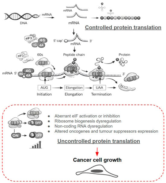
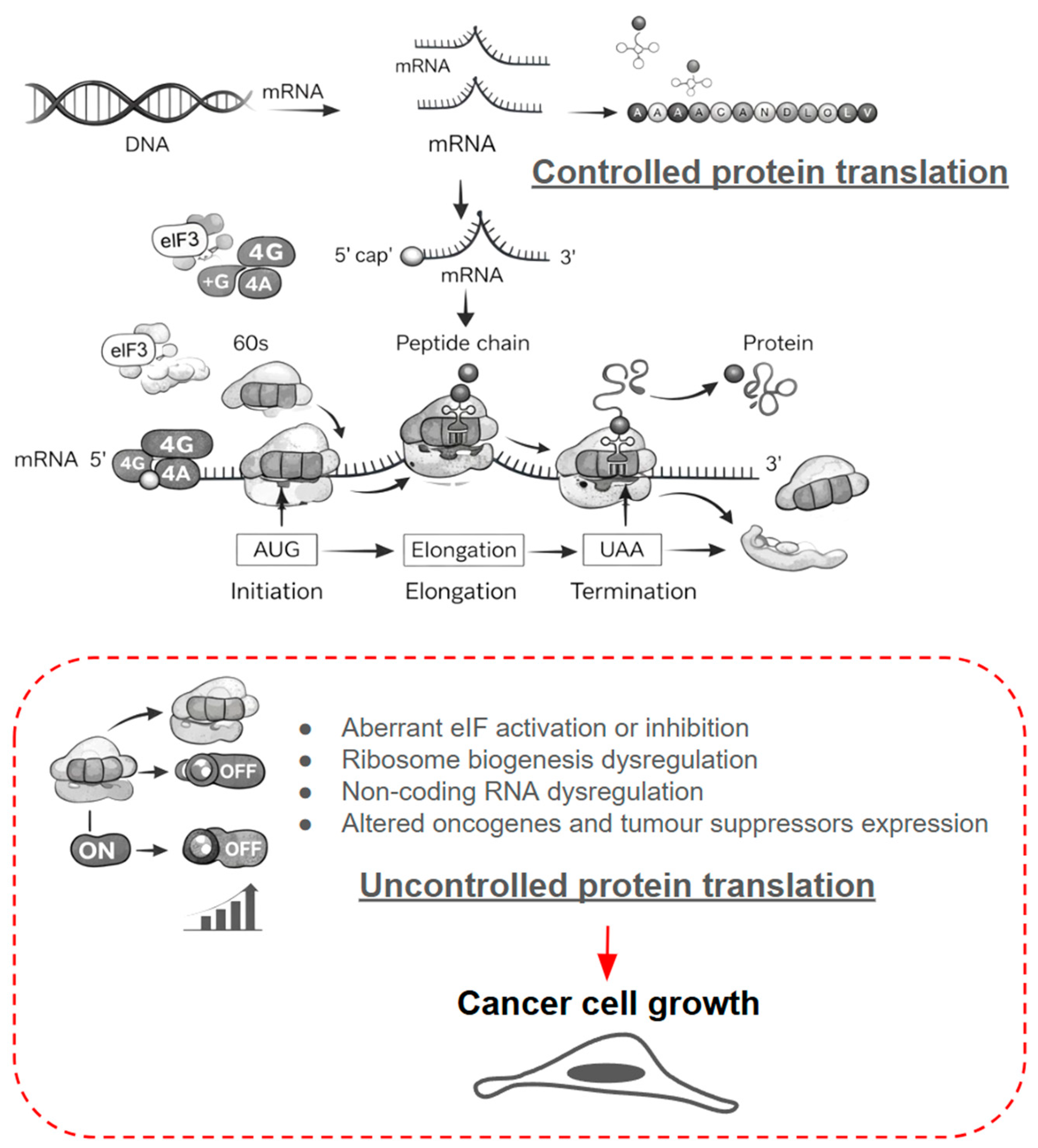
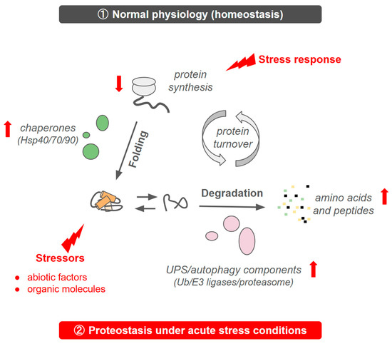
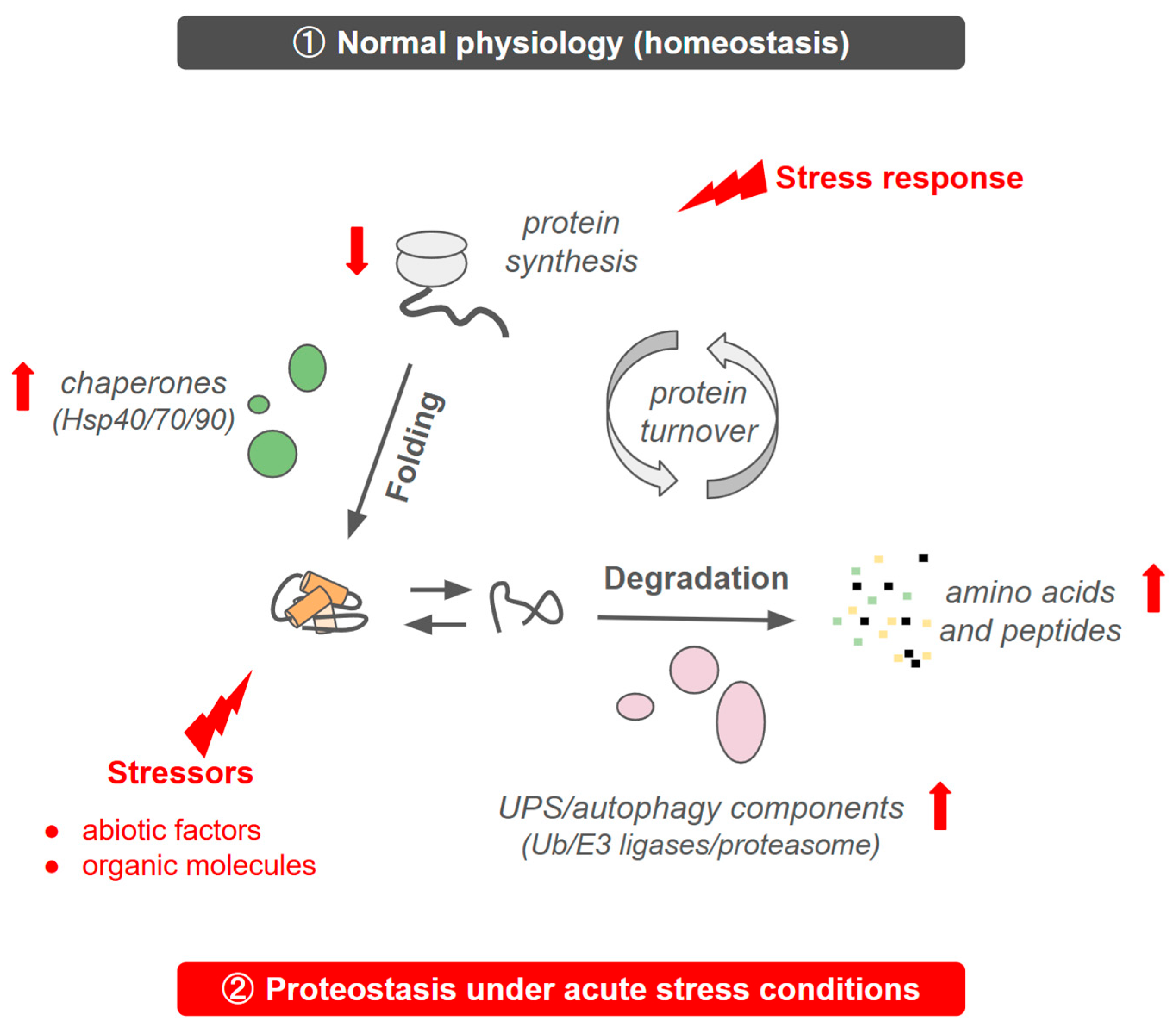
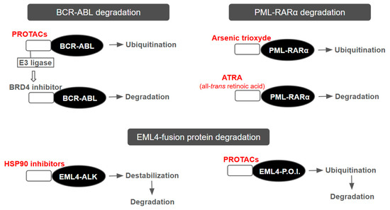
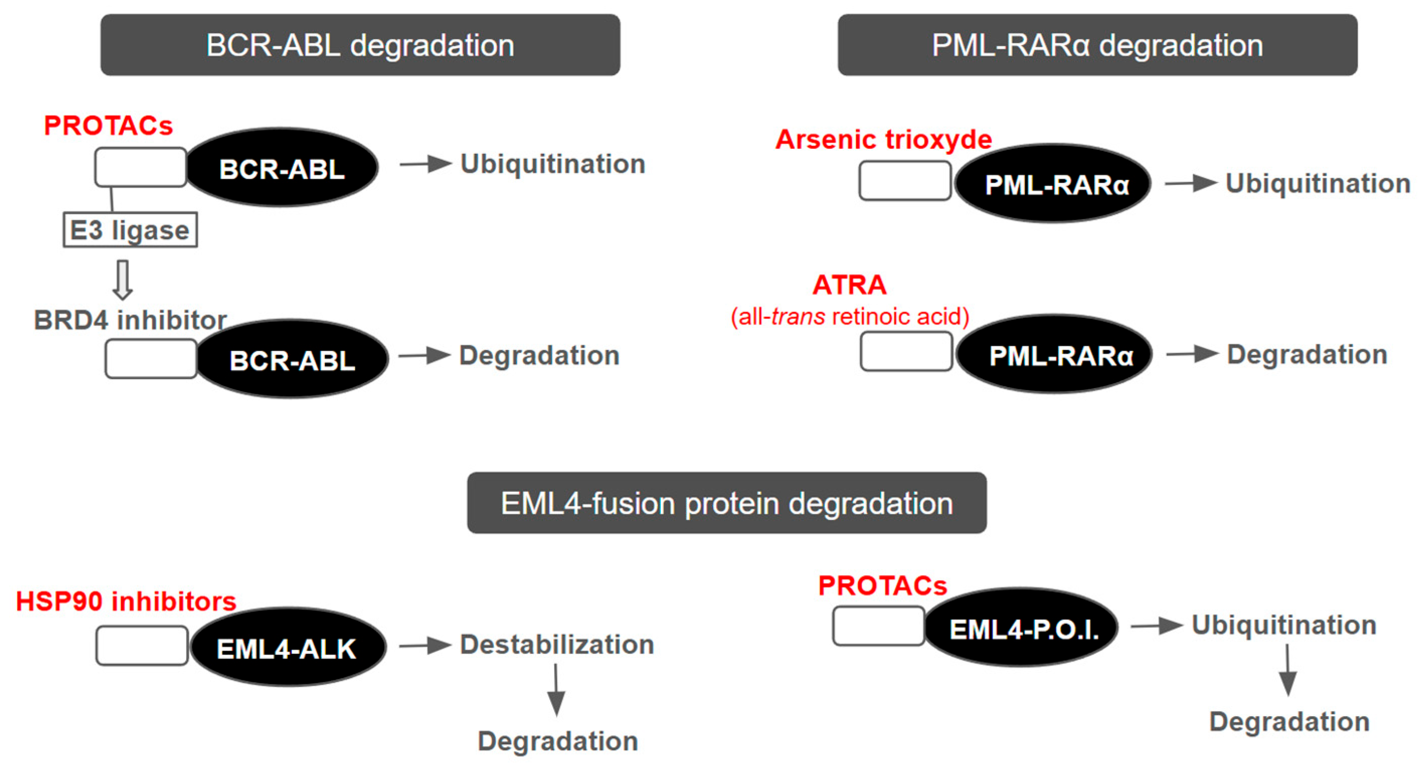
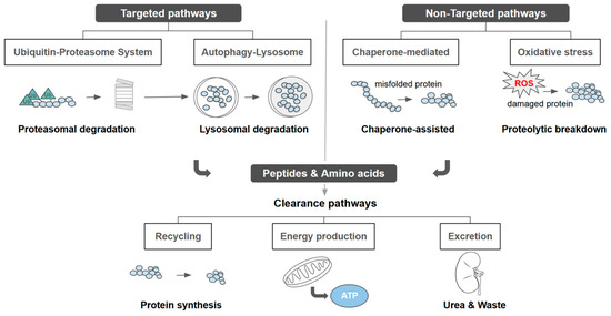
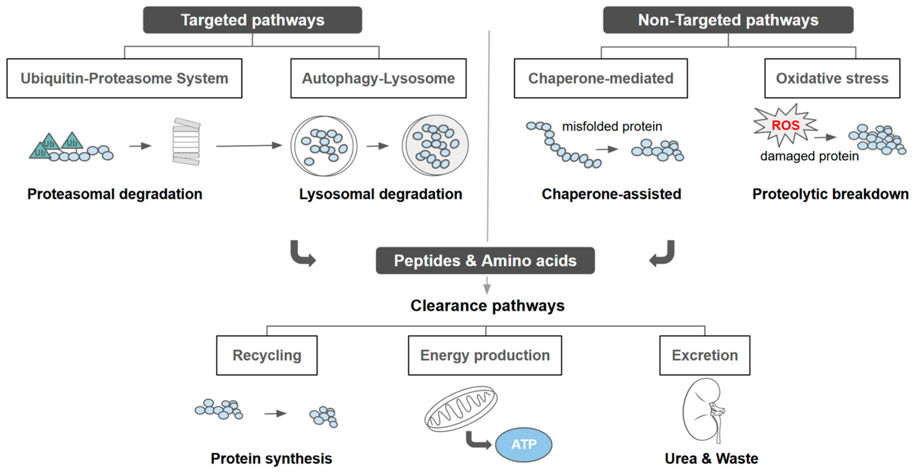
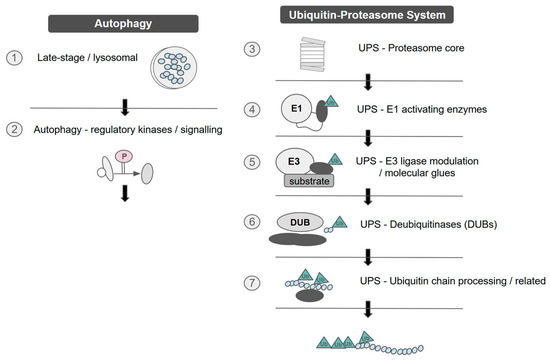
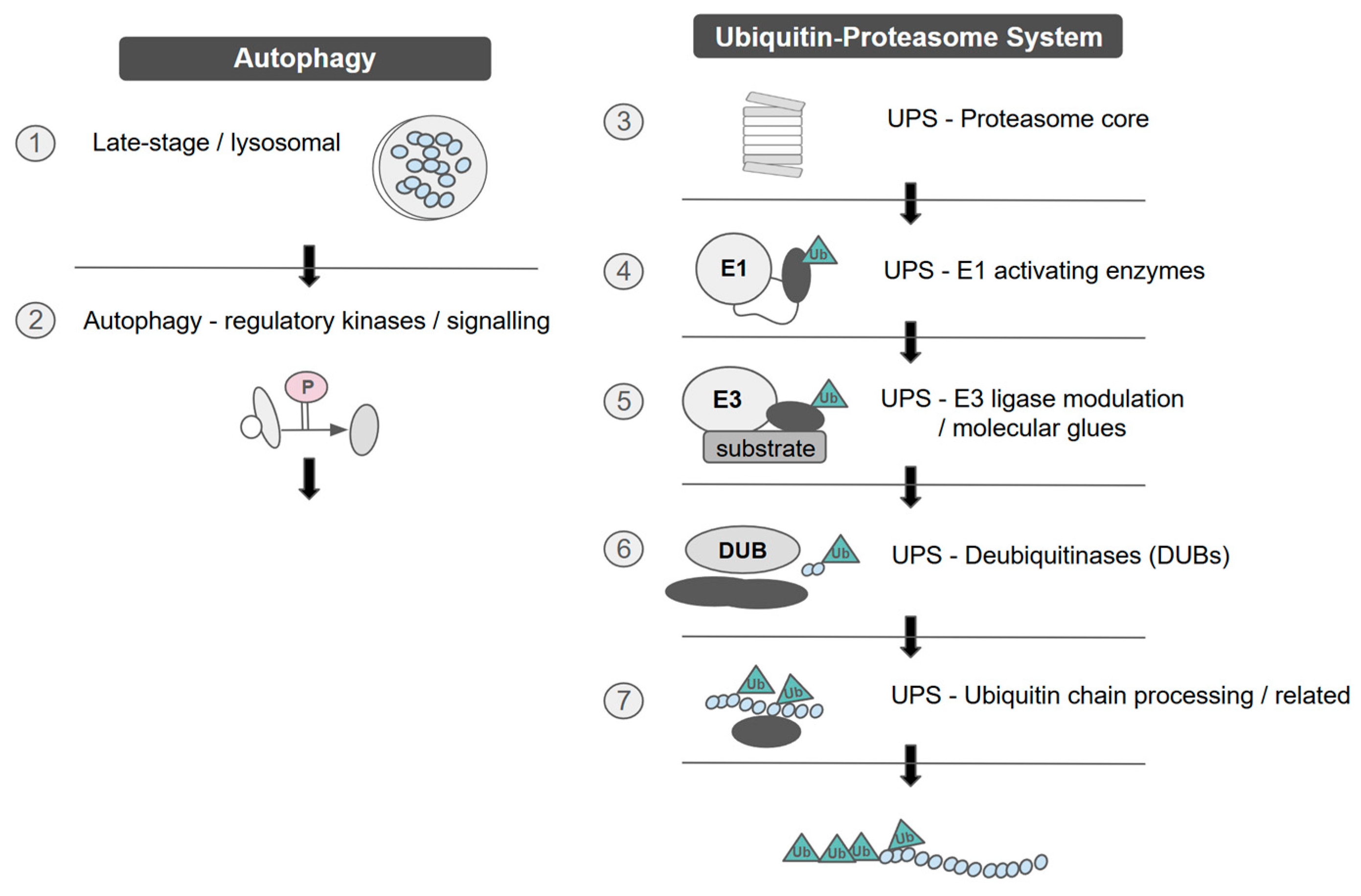
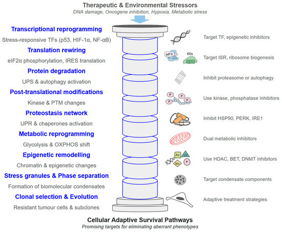
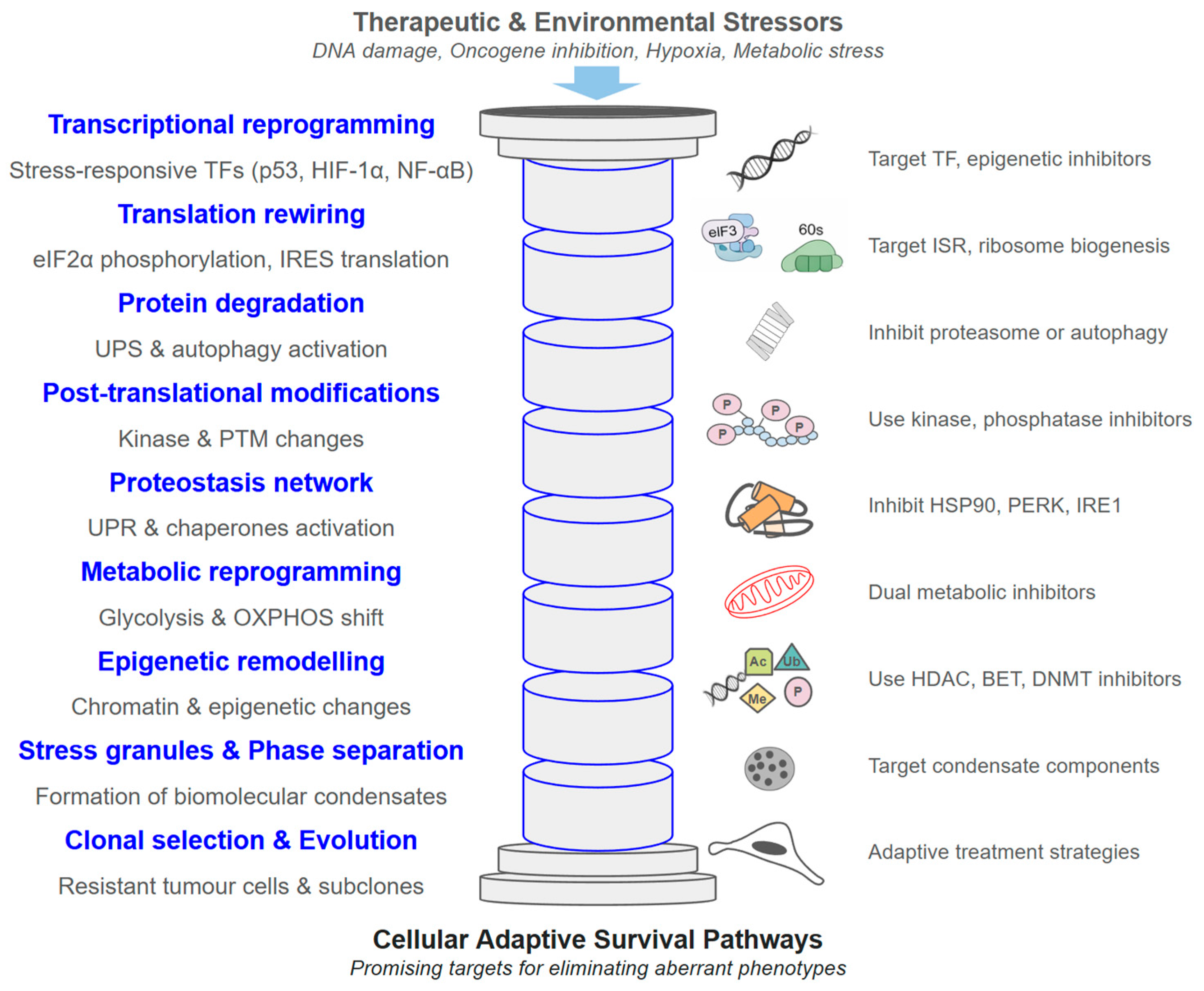
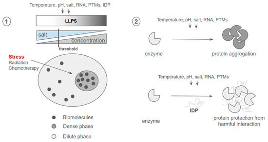
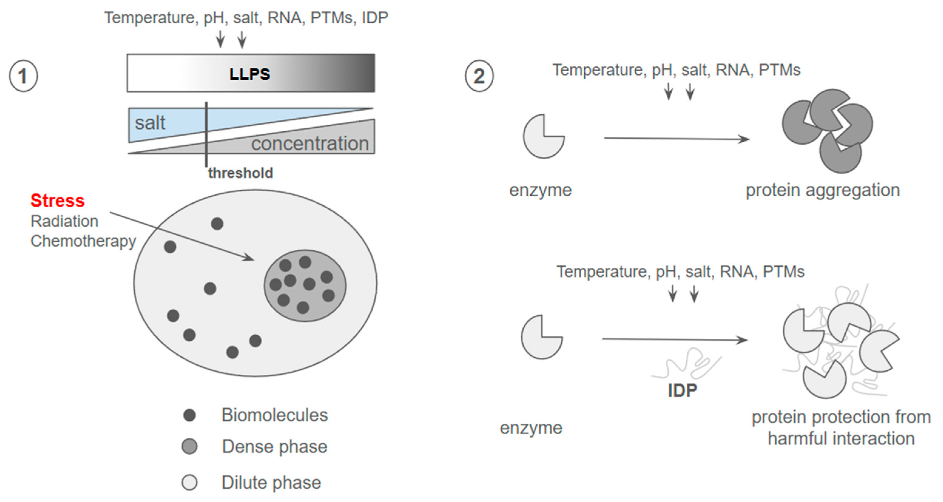

Disclaimer/Publisher’s Note: The statements, opinions and data contained in all publications are solely those of the individual author(s) and contributor(s) and not of MDPI and/or the editor(s). MDPI and/or the editor(s) disclaim responsibility for any injury to people or property resulting from any ideas, methods, instructions or products referred to in the content. |
© 2026 by the author. Licensee MDPI, Basel, Switzerland. This article is an open access article distributed under the terms and conditions of the Creative Commons Attribution (CC BY) license.