


Abstract
Oxidative stress contributes to retinal pigment epithelial (RPE) cell dysfunction and is implicated in the pathogenesis of retinal diseases, including age-related macular degeneration (AMD). Lutein, a naturally occurring xanthophyll carotenoid that accumulates in the macula, plays an important role in antioxidant defence and light filtration. This study investigated the antioxidant and cytoprotective effects of lutein-rich gac (Momordica cochinchinensis) peel extract (GPE) in human RPE cells subjected to hydrogen peroxide (H2O2)-induced oxidative stress. Cells were pre-treated with GPE (0.1–200 µg/mL), purified lutein (10 µg/mL), or the reference antioxidant Trolox (50 µg/mL) for 12–72 h before oxidative challenge. Cell viability, intracellular reactive oxygen species (ROS) accumulation, and the expression of genes associated with antioxidant defence, angiogenesis, and inflammation were evaluated. GPE at 10 and 100 µg/mL exhibited greater cytoprotective effects than purified lutein and Trolox under the tested conditions. GPE treatment significantly reduced intracellular ROS accumulation and protected RPE cells against H2O2-induced cell death. Furthermore, GPE increased the expression of antioxidant response regulators Nrf2 and HO-1, while reducing the expression of vascular endothelial growth factor A (VEGF-A) and interleukin-6 (IL-6). These findings demonstrate the antioxidant and cytoprotective activity of GPE and suggest that modulation of oxidative stress-associated molecular responses may contribute to its protective effects. This study highlights GPE as a promising source of natural bioactive compounds for further investigation in retinal protection and oxidative stress-related ocular disorders.
1. Introduction
Age-related macular degeneration (AMD) is the most prevalent eye disease in the elderly, especially among those over 65. It is one of the leading causes of irreversible blindness worldwide with ~193 million people affected in 2019 and is predicted to exceed 243 million by 2030 [1]. AMD is a chronic degenerative retinal disease that occurs when photoreceptors and retinal pigment epithelium (RPE) cells in the macula are degraded, resulting in a loss of visual acuity and central vision [2]. The RPE is a monolayer of cells located below the photoreceptors of the retina and superficial to Bruch’s membrane (BM) of the choroid [3]. These cells play essential roles in maintenance of the outer blood–retinal barrier, retinal adhesion, nutrient supply to photoreceptors, absorption of scattered light, and the production and recycling of photopigments [2,3]. As a result of their high metabolic activity and chronic exposure to light, RPE cells generate excessive levels of reactive oxygen species (ROS) which leads to accumulated oxidative stress. Cellular oxidative stress modifies mitochondrial DNA (mtDNA) [4,5] and leads to the secretion of pro-inflammatory cytokines such as IL-6 and IL-8 [5]. The damage of RPE cells cascades as degeneration of photoreceptors and accumulation of drusen, which are clinical indicators of early AMD [4]. In addition, oxidative stress in RPE cells triggers the secretion of vascular endothelial growth factor (VEGF), which promotes the growth of abnormal leaky blood vessels leading to late-stage AMD and irreversible blindness [6]. Therefore, reducing the level of oxidative stress in the macula, in particular RPE cells, is likely to be an effective strategy to prevent and reduce AMD disease progression.
Cellular ROS levels are controlled by antioxidant enzymes such as glutathione S-transferase (GST), superoxide dismutase (SODs), catalase (CAT), and heme oxygenase 1 (HO-1) [4]. These antioxidant enzymes are regulated by nuclear factor-erythroid 2-related factor 2 (Nrf2) [7]. Along with ageing, the expression of Nrf2 is impaired while ROS accumulation increases, resulting in oxidative damage to these cells [8]. Therefore, dietary antioxidants and supplements play a vital role in the neutralization of ROS and reducing cellular damage from oxidative stress, and novel treatments for eye health are needed.
Lutein, zeaxanthin, and its isomer meso-zeaxanthin are the three carotenoids that accumulate in the macula of the eye. However, humans are unable to synthesise lutein and zeaxanthin [9], which are sourced mainly from plant material. Lutein is more abundant in nature, comprising numerous geometrical stereoisomers [10,11], but they are often bound and not bioavailable [12,13]. Studies have shown that lutein supplementation from marigold either alone or in combination with zeaxanthin and meso-zeaxanthin prevents and slows down the progression of AMD [14,15,16,17,18,19,20,21,22,23,24]. Gac fruit is one of the richest natural sources of carotenoids, especially lycopene and β-carotene from the aril [25,26,27]. However, the peel, which accounts for ~16% of the gac fruit, is often discarded [26] and contains high lutein [28,29]. Potential eye health benefits of lutein from gac peel have not been studied yet. Therefore, this study aimed to (1) evaluate the cytoprotective and antioxidant effects of lutein-rich gac peel extract relative to a lutein standard and the reference antioxidant Trolox in an oxidative stress model; (2) investigate its influence on intracellular ROS levels and antioxidant defence pathways, with a focus on Nrf2 and HO-1 expression; and (3) assess modulation of the inflammatory marker IL-6 and the angiogenic marker vascular endothelial growth factor (VEGF). This study will evaluate the eye health benefits of lutein from gac fruit and provide evidence for the utilisation of GPE as a novel source of lutein for eye health supplementation.
2. Results
2.1. Chemical Characterisation of Gac Peel Extract
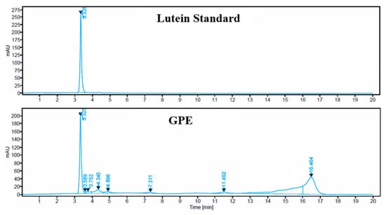
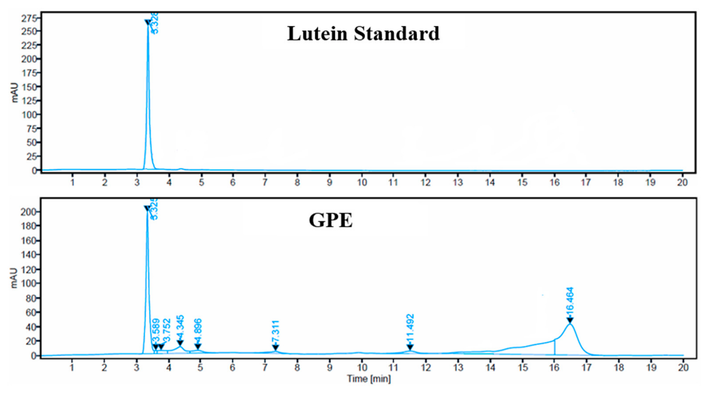
The chemical profile of lutein-rich gac peel extract (GPE) was characterised using high-performance liquid chromatography (HPLC) and Fourier-transform infrared (FTIR) spectroscopy. HPLC analysis revealed a prominent peak at a retention time of approximately 3.32 min in both the lutein reference standard and GPE chromatograms (Figure 1), supporting the presence of lutein in the extract. Quantitative HPLC analysis using an external calibration curve constructed from the lutein reference standard revealed a lutein content of ~76.05% (w/w) in GPE. In addition to the lutein peak, GPE exhibited several other chromatographic peaks at retention times of approximately 3.59, 3.75, 4.35, 4.90, 7.31, 11.49, and 16.46 min, suggesting the presence of additional constituents that were not further identified.
Figure 1.
HPLC spectra of the lutein standard and lutein-rich gac peel extract (GPE).
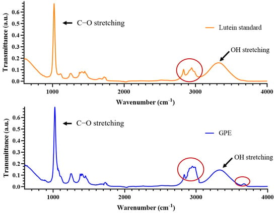
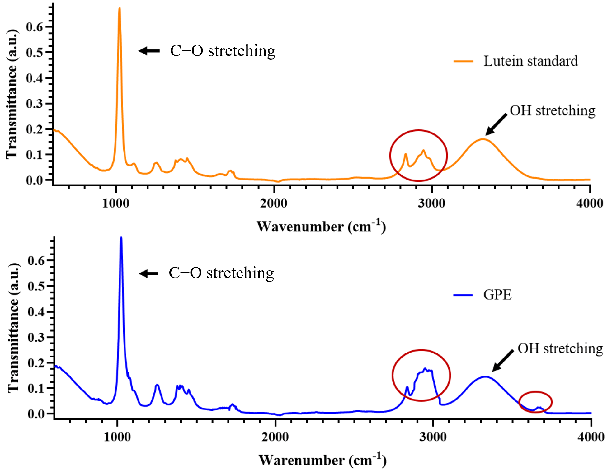
FTIR spectroscopy revealed a broad absorption band in the region of 3180–3520 cm−1 in both the lutein standard and GPE spectra (Figure 2), consistent with O–H stretching vibrations associated with hydroxyl groups. However, differences in absorption intensities and spectral patterns were observed between the two samples, particularly in the highlighted regions, indicating differences in their spectral characteristics (Figure 2). However, these spectral differences alone do not permit the identification of specific additional compounds in GPE.
Figure 2.
Fourier-transform infrared (FTIR) spectra of the lutein standard (orange) and lutein-rich gac peel extract (GPE; blue). Red circles highlight regions exhibiting differences in absorption features between the two samples, suggesting variations in their chemical composition.
2.2. Time Course Comparison of the Protective Effects of GPE, Lutein, and Trolox Against H2O2-Induced Stress
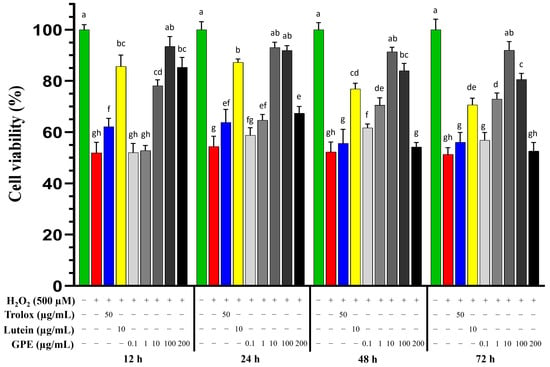
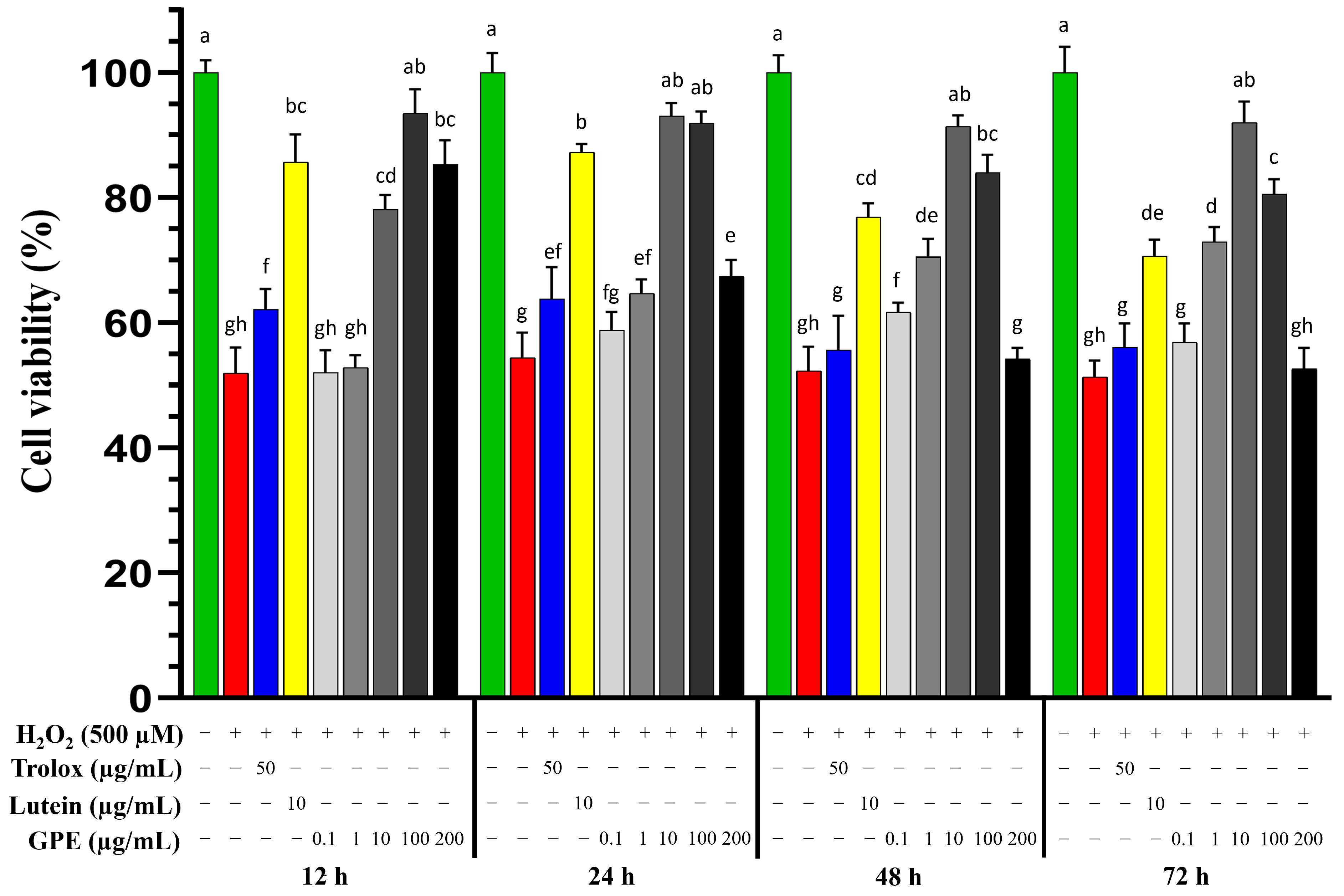
The challenge condition (oxidative stress at 500 µM H2O2 for 24 h) for the RPE cells (ARPE-19) was selected based on a preliminary concentration-response experiment where cell viability fell to ~50% of that observed in untreated controls (Supplementary File S1). Pre-treatment with the lutein standard (10 µg/mL) and GPE (10 and 100 µg/mL) significantly reduced H2O2-induced cytotoxicity resulting in >80% viability, at all treatment times between 12 and 72 h (p ≤ 0.05), whereas Trolox was only effective for the first 24 h (p ≤ 0.05) (Figure 3). The lutein standard pre-treatment conferred maximal protection to the cells (~90% viability) for the first 24 h before it became less effective. This was different to that of GPE, which exerted sustained protective effects over 72 h exceeding 70% viability.
Figure 3.
Effect of different GPE pre-treatment times and doses on RPE cells exposed to H2O2 stress. RPE cells were pre-treated with either Trolox (50 µg/mL), the lutein standard (10 µg/mL), or GPE (0–200 µg/mL) for 12, 24, 48, or 72 h prior to stress with 500 µM H2O2 for 24 h. Cell viability is expressed as a percentage of the untreated controls and is shown as the mean + SEM of three replicates performed in triplicate. Statistical analysis was performed using one-way ANOVA with Tukey’s honestly significant difference (HSD) test. Treatments with different letters indicate statistical differences at p ≤ 0.05, while treatments sharing the same letters indicate no statistical differences between treatments at p > 0.05.
The greatest protective effects (~90% viability (p ≤ 0.05)) occurred in cells pre-treated with 10 µg/mL GPE for at least 24 h (Figure 3). The 12 h pre-treatment with 100 µg/mL GPE conferred greater protective effects against H2O2-induced cytotoxicity, ~95% viability. However, the efficacy of 100 µg/mL GPE reduced over time and was less effective than 10 µg/mL GPE after 24 h, suggesting time-dependent effects. These results demonstrated that GPE (10–100 µg/mL) conferred sustained protection to RPE cells from H2O2-induced cell death with higher effectiveness than that of either Trolox or the purified lutein standard.
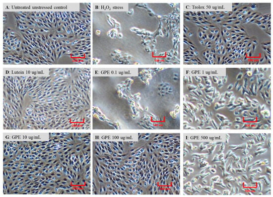
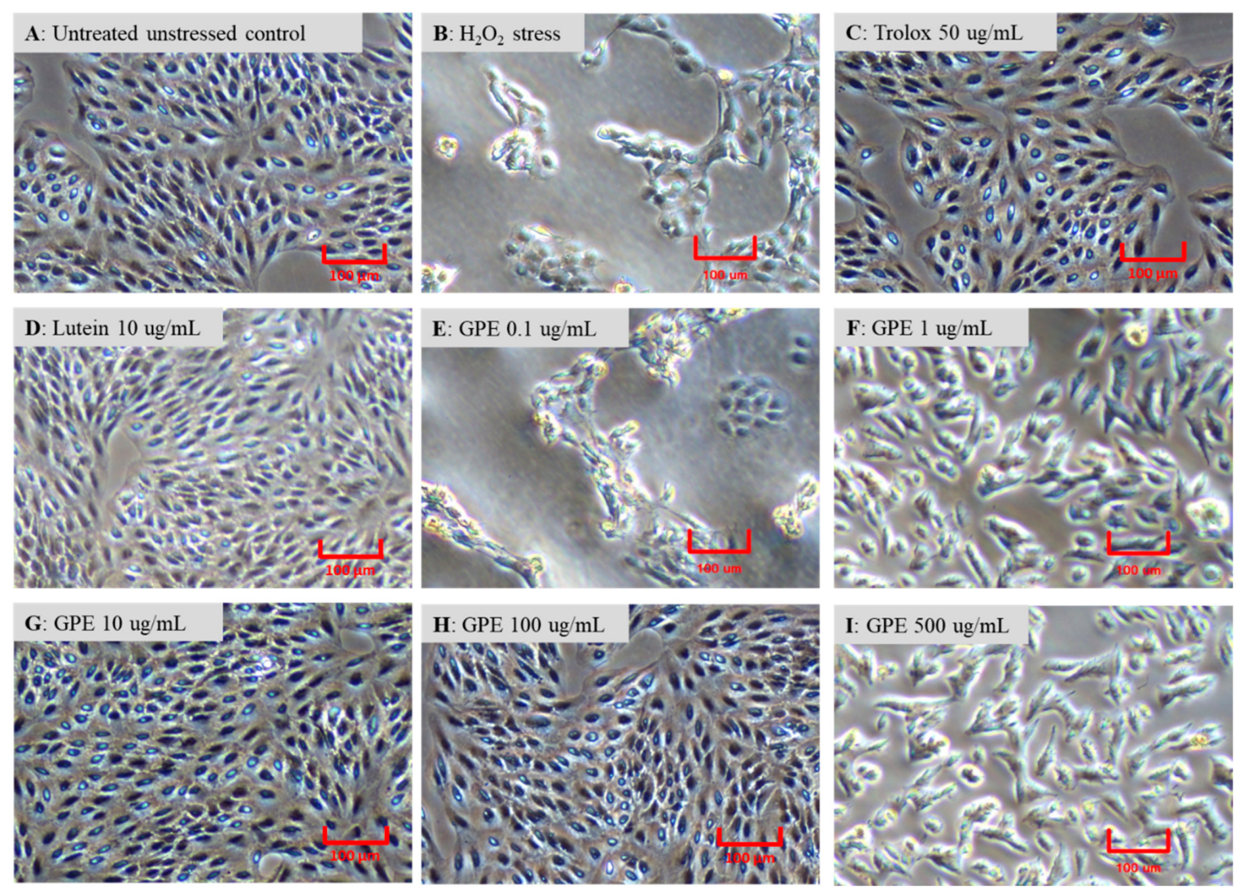
Untreated control RPE cells displayed advanced elongations, and were firmly attached and maintained a high growth rate resulting in ~80% confluency after 24 h (Figure 4A). Cells stressed with H2O2 appeared swollen and deformed with significantly reduced numbers (~10% confluency) (Figure 4B). Trolox pre-treatment increased the number of H2O2-stressed cells to 80% confluency, akin to high baseline control numbers and morphology (Figure 4C). The lutein standard conferred cellular protection and reversed the H2O2 stress to baseline untreated unstressed cells (~80% confluency) (Figure 4D). GPE pre-treatment at 10 or 100 µg/mL also conferred protection to the cells, similar to untreated unstressed cells (~80% confluency) (Figure 4G,H). At other GPE pre-treatment doses (0.1, 1, and 200 µg/mL), there was cellular swelling (Figure 4F,I) and reduced viability (Figure 4E) akin to the H2O2-stressed untreated cells, indicating dose dependency.
Figure 4.
Morphology of pre-treated RPE cells stressed with H2O2. RPE cells were pre-treated with the antioxidant Trolox (50 µg/mL), the lutein standard (10 µg/mL), or GPE (0–200 µg/mL) for 24 h prior to stress with 500 µM H2O2 for 24 h. (A) = untreated unstressed control, (B) = H2O2 stressed, (C) = Trolox pre-treated, (D) = lutein pre-treated, (E) = 0.1 µg/mL GPE pre-treated, (F) = 1 µg/mL GPE pre-treated, (G) = 10 µg/mL GPE pre-treated, (H) = 100 µg/mL GPE pre-treated, and (I) = 200 µg/mL GPE pre-treated. Scale bar = 100 µm.
2.3. GPE Suppressed ROS Induced by H2O2 Stress in RPE Cells
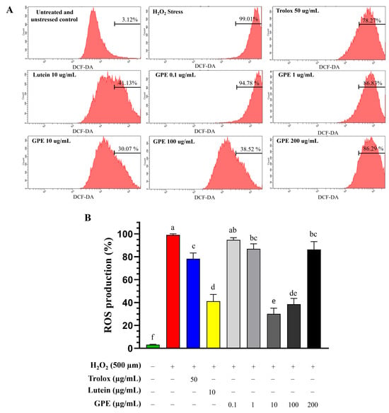
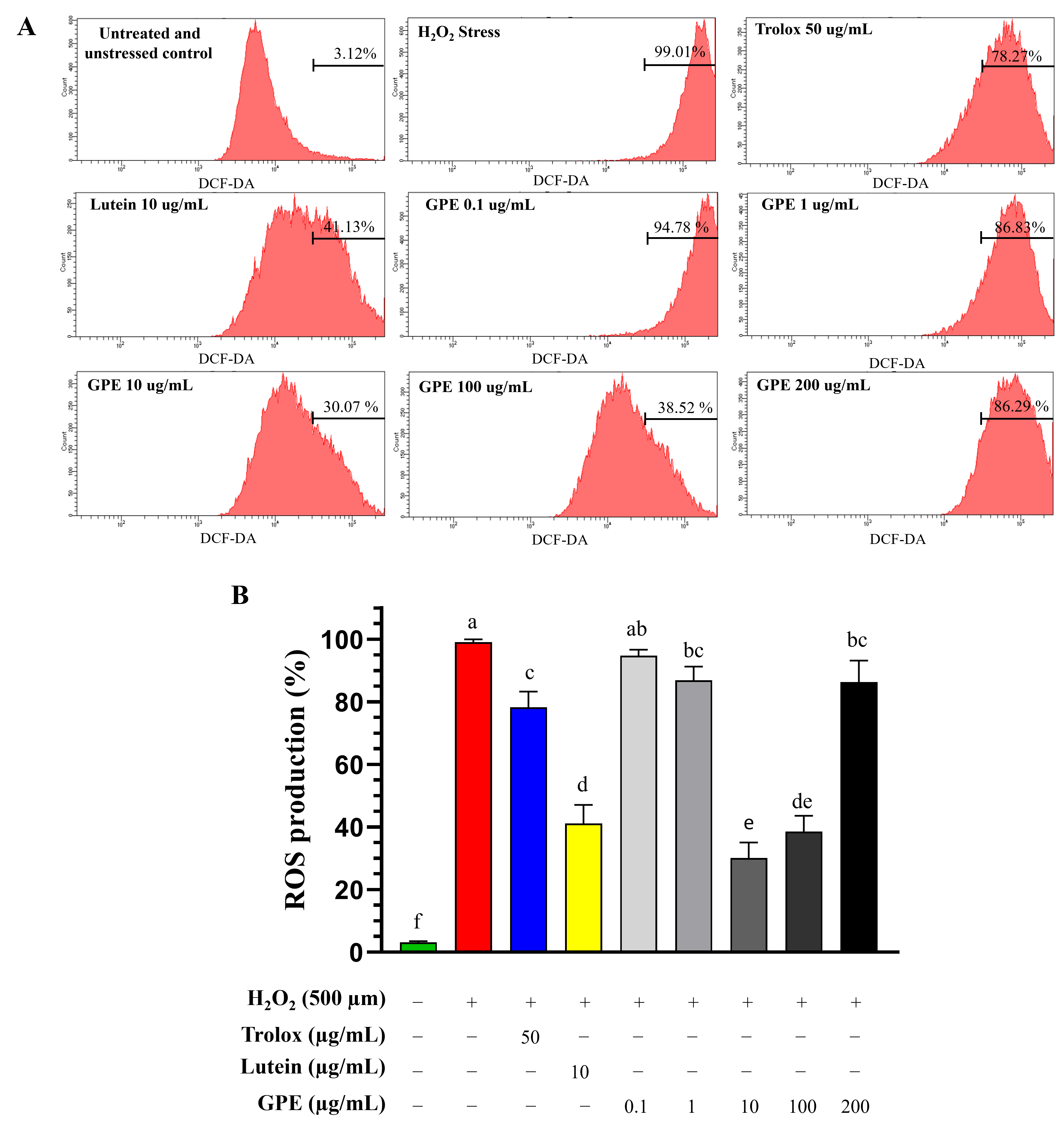
GPE reduced ROS production induced by H2O2 stress; however, the level of reduction was dependent on pre-treatment concentrations. Unstressed RPE cells generated low ROS (3%) but, upon H2O2 exposure, almost all the cells produced ROS (99%) (Figure 5). The high level of ROS due to H2O2 exposure inversely correlated with the low viability of these stressed cells. When the H2O2-stressed cells were pre-treated with the pure lutein standard or GPE (10 or 100 µg/mL) for 24 h, ROS were optimally lowered to 41, 30, and 39%, respectively, even more than pre-treatments with Trolox or suboptimal doses of GPE (0.1, 1, and 200 µg/mL) that remained above 78% (Figure 5).
Figure 5.
Effect of GPE pre-treatment on ROS levels in H2O2-stressed RPE cells. (A) Flow cytometry profiles of RPE cells treated with either Trolox (50 µg/mL), lutein (10 µg/mL), or GPE (0–200 µg/mL) for 24 h prior to 500 µM H2O2 exposure for 24 h. ROS were detected using the DCFH-DA stain and each profile represents 20,000 events. (B) Graphical representation of the effect of pre-treatment on cellular ROS levels. ROS levels are expressed as percentage mean + SEM of three replicate values. Statistical analysis was performed using one-way ANOVA with Tukey’s honestly significant difference (HSD) test. Treatments with different letters indicate statistical differences at p ≤ 0.05, while treatments sharing the same letters indicate no statistical differences between treatments at p > 0.05.
2.4. GPE Enhanced the Expression of Nrf2 and HO-1
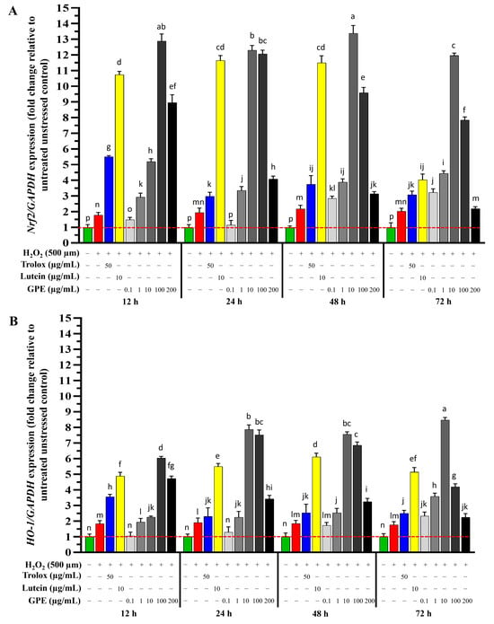
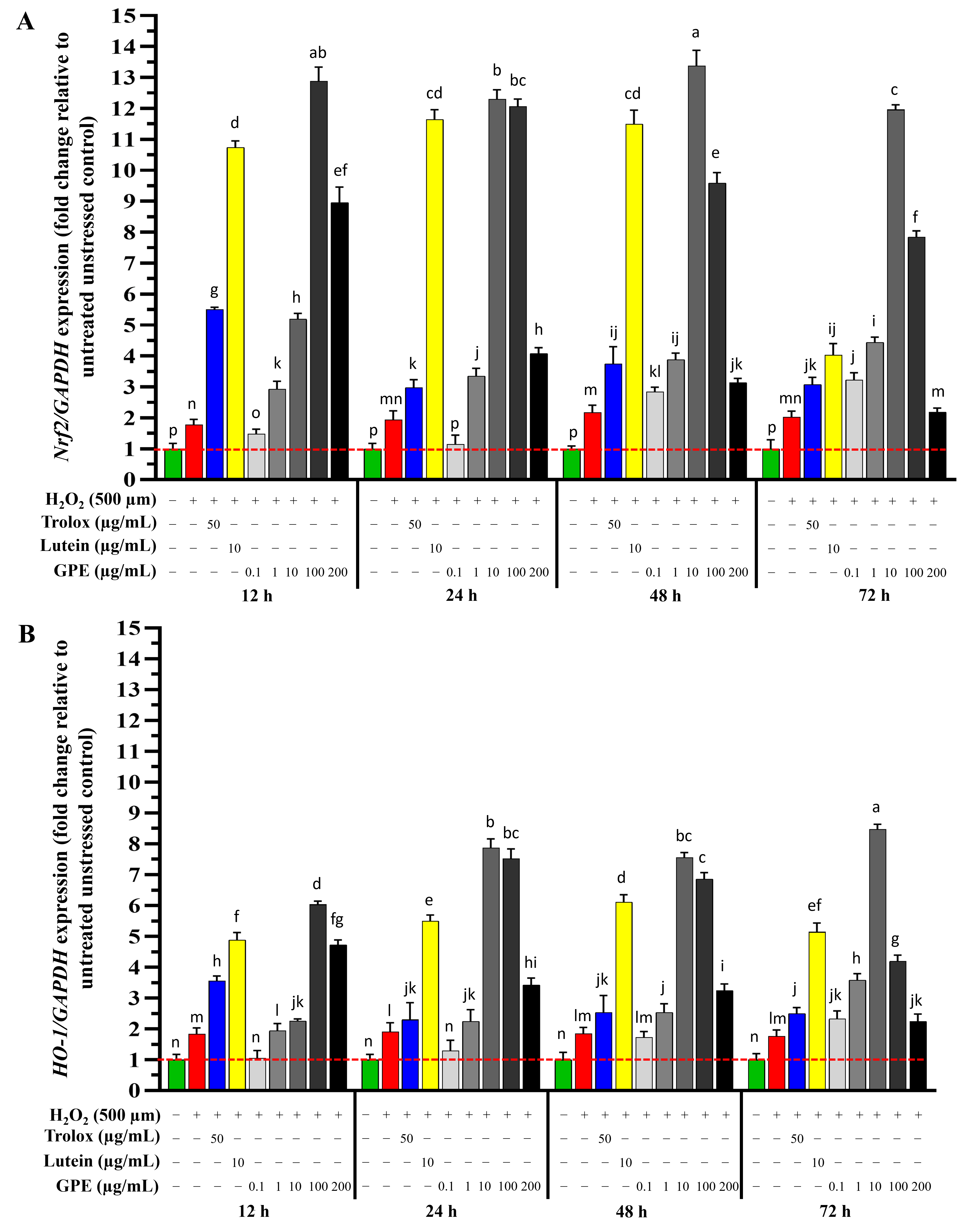
Nrf2 is a transcription factor that plays a key role in the cellular response to oxidative stress [7]. It is activated under oxidative stress leading to the upregulation of genes encoding for antioxidant enzymes. However, under excessive oxidative stress conditions, Nrf2 expression is suppressed [30]. Pre-treatment with either Trolox, the lutein standard, or GPE significantly upregulated Nrf2 expression (p ≤ 0.05) due to H2O2 stress with different intensities depending on treatment time and doses. When the RPE cells were stressed with H2O2, Nrf2 expression marginally increased by ~2-fold compared to the unstressed controls (p ≤ 0.05) (Figure 6A). Pre-treatment with Trolox increased Nrf2 expression in the H2O2-stressed cells, with a maximal increase observed at 12 h (5-fold, p ≤ 0.05) that declined over time compared to the H2O2-stressed cells. Lutein pre-treatments had a greater stimulatory effect on Nrf2 expression than Trolox at all time points (Figure 6A). Nrf2 expression was maximal for a longer time (up to 48 h), which increased ~12-fold but lost its intensity by a third at 72 h (4-fold increase).
Figure 6.
Effect of pre-treatments on the expression of Nrf2 (A) and HO-1 (B) genes in H2O2-stressed RPE cells. RPE cells were treated with either Trolox (50 µg/mL), the pure lutein standard (10 µg/mL), or GPE (0–200 µg/mL) for 12, 24, 48, or 72 h prior to 500 µM H2O2 stress for 24 h. Results were presented as relative fold change compared to untreated control cells (value of 1), expressed as the mean ± SEM of three replicates performed in triplicate. Statistical analysis was performed using one-way ANOVA with Tukey’s honestly significant difference (HSD) test. Treatments with different letters indicate statistical differences at p ≤ 0.05, while treatments sharing the same letters indicate no statistical differences between treatments at p > 0.05.
GPE pre-treatment also upregulated Nrf2 expression, even more than both the Trolox and lutein controls. Maximal activation of Nrf2 was ~13-fold with GPE pre-treatment at 100 µg/mL at 12 h pre-treatment, and 10 µg/mL at 48 h pre-treatment. The greatest sustained effect on Nrf2 expression was 10 µg/mL GPE, which started lower at 12 h (five-fold) but peaked afterwards until 72 h (~13-fold).
Nrf2 enhances the expression of the heme oxygenase-1 (HO-1) enzyme, which is responsible for oxidative cleavage [31]. Pre-treatment with GPE significantly increased HO-1 expression at different levels of upregulation depending on treatment dose and time (p ≤ 0.05). The expression patterns of HO-1 following pre-treatments with Trolox, lutein, and GPE were similar to those of Nrf2, but levels of upregulation were more than halved (Figure 6B). The highest upregulation was with 10 µg/mL GPE at 72 h, which increased HO-1 expression by ~9-fold, which was also sustained beyond 72 h. The increase in Nrf2 and HO-1 expression was associated with decreased ROS accumulation in the RPE cells and reduced cell death caused by H2O2 stress.
2.5. GPE Suppressed the Expression of IL-6 and Vascular Endothelial Growth Factor-A (VEGF-A) Genes
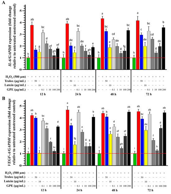
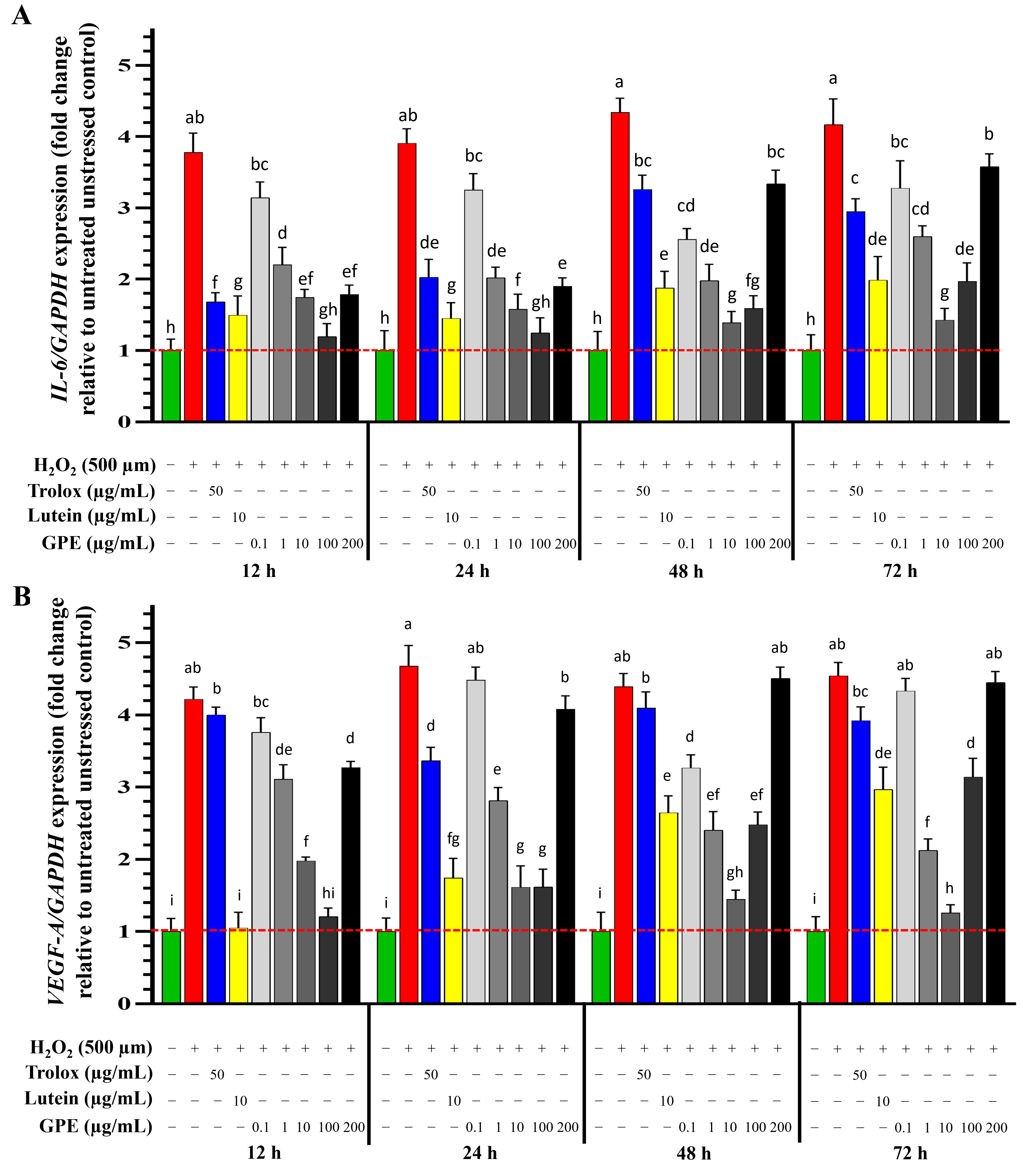
The accumulation of proinflammatory factors such as interleukin-6 and -8 (IL-6 and IL-8) contribute to inflammation, leading to the degeneration of RPE cells [32]. H2O2 stress increased IL-6 expression by ~3.5-fold at all time points (Figure 7A). Trolox pre-treatment reduced the upregulation of IL-6 (~2-fold) at 12 h, but the reduction was decreased as treatment time increased (~3-fold at 72 h). Lutein pre-treatment displayed higher efficacy in reducing the upregulation of IL-6 than Trolox (~1.5 fold), almost reaching unstressed baselines. The reduction was longer lasting but still decreased as treatment time increased after 48 h (p ≤ 0.05). Pre-treatment with GPE reduced the upregulation of IL-6 induced by H2O2 stress, with the effectiveness impacted by treatment dose (p ≤ 0.05) and time (p ≤ 0.05). The maximal reduction in IL-6 expression was observed after pre-treatment with 100 µg/mL GPE at 12–24 h, which was similar to that seen in unstressed cells (p ≤ 0.05); however, at other suboptimal doses, IL-6 expression was variable, and some were similar to untreated stressed cells (Figure 5A). With 10 µg/mL GPE, IL-6 expression significantly decreased and the reduction was more sustained over a longer time (p ≤ 0.05).
Figure 7.
Effect of pre-treatments on the expression of IL-6 (A) and VEGF-A (B) genes in H2O2-stressed RPE cells. RPE cells were treated with Trolox antioxidant control (50 µg/mL), the pure lutein standard (10 µg/mL), or GPE (0–200 µg/mL) for 12, 24, 48, or 72 h prior to 500 µM H2O2 stress for 24 h. Results were presented as relative fold change compared to untreated control cells which were given the value of unity (1), expressed as the mean ± SEM of three replicates performed in triplicate. Statistical analysis was performed using one-way ANOVA with Tukey’s honestly significant difference (HSD) test. Treatments with different letters indicate statistical differences at p ≤ 0.05, while treatments sharing the same letters indicate no statistical differences between treatments at p > 0.05.
Vascular endothelial growth factor (VEGF)-A is an angiogenic protein that enhances the formulation of new blood vessels in the retina. Activation of VEGF-A promotes neovascularisation that can increase vascular permeability, causing neovascular forms of AMD [6]. Therefore, discovering new VEGF-A inhibitors is a key therapeutic strategy used in the current management of wet AMD [33]. H2O2 stress increased VEGF-A expression ~4-fold compared to unstressed controls (p ≤ 0.05) at all time points (Figure 7B). Trolox pre-treatment was minimal and caused 3–4-fold upregulation of VEGF-A expression, akin to stressed baselines. Lutein pre-treatment acutely reduced VEGF-A expression at 12 h to the unstressed control baseline, but the reductions were not maintained over time. Optimal GPE pre-treatment also decreased VEGF-A expression, with maximal reductions with 10 µg/mL at 72 h and 100 µg/mL at 12 h, paralleled with unstressed baselines. Other suboptimal GPE concentrations were not as effective. Overall, pre-treatment with GPE significantly decreased VEGF-A expression induced by H2O2 stress, with the level of reduction dependent on treatment time and dose (p ≤ 0.05).
3. Discussion
The results of this study demonstrate that lutein-rich gac peel extract (GPE) exerts cytoprotective effects in retinal pigment epithelial (RPE) cells exposed to H2O2-induced oxidative stress. These protective effects were associated with reduced intracellular oxidative activity and changes in the expression of genes involved in antioxidant defence, inflammation, and angiogenic signalling. Oxidative stress plays a central role in retinal cellular dysfunction, particularly in RPE cells, which are exposed to chronic light stimulation, high metabolic demands, phagocytosis of photoreceptor outer segments, and photosensitising compounds [34]. Exposure to oxidants such as hydrogen peroxide can further exacerbate oxidative damage through the generation of highly reactive oxygen species [35]. Therefore, identifying natural bioactive compounds capable of protecting RPE cells against oxidative stress may provide a valuable foundation for further investigation into potential retinal protective strategies.
Pre-treatment with GPE exhibited superior acute and sustained antioxidant activity compared with both the reference antioxidant Trolox and a purified lutein standard. Trolox exerts its antioxidant effects primarily through reducing intracellular ROS generation [36] and activating endogenous antioxidant defences via the Nrf2 pathway [37]. However, the relatively modest protective efficacy of Trolox observed in this model may be attributed to limited cellular uptake or degradation in aqueous culture conditions [38], which underscores the importance of matrix stability and bioavailability when evaluating antioxidant ingredients.
Previous studies have identified 10 µg/mL as an optimal concentration for purified lutein in protecting RPE cells against oxidative stress [26,39]. In the present study, GPE and purified lutein were compared at the same nominal treatment concentration (10 µg/mL); however, these treatments were not matched for lutein content. Based on the measured lutein content of GPE (~76% w/w), 10 µg/mL GPE contained ~7.6 µg/mL lutein, compared with 10 µg/mL in the purified lutein treatment. Despite this lower lutein content, GPE produced greater and more sustained cytoprotective effects under the conditions tested, including at 72 h. This observation suggests that constituents of GPE other than lutein may contribute to its biological activity. Gac peel is known to contain additional carotenoids and other phytochemicals [25,40,41]. Additional constituents in GPE may contribute to its enhanced cytoprotective activity compared with purified lutein under the tested conditions, although their individual contributions and potential synergistic interactions remain to be established [42]. However, because the treatments were not matched for lutein content and the additional constituents of GPE were not comprehensively characterised, the present findings cannot establish synergistic interactions between lutein and other components of the extract. Future studies using lutein-matched treatment conditions together with detailed phytochemical profiling will be required to determine the relative contributions and potential interactions of individual GPE constituents.
This observation supports a growing body of evidence that whole food extracts can outperform isolated compounds due to compositional complexity and matrix-driven interactions.
At the cellular level, the protective effects of GPE were associated with a marked reduction in intracellular ROS accumulation, thereby limiting downstream oxidative damage and secondary responses such as inflammation and angiogenic signalling. Exposure of RPE cells to H2O2 resulted in a significant reduction in cell viability, concurrent with elevated ROS levels. Excessive ROS are known to damage cellular macromolecules, including DNA, proteins, and lipid membranes, ultimately leading to loss of cellular function and cell death [43]. ROS homeostasis is maintained by endogenous antioxidant enzymes such as HO-1, GST, SOD, and CAT [4], the expression of which is regulated by the transcription factor Nrf2 [44]. Activation of the Nrf2 pathway promotes upregulation of antioxidant defences and suppression of oxidative stress [45].
In the present study, pre-treatment with both lutein and GPE significantly reduced intracellular ROS levels in stressed RPE cells, accompanied by increased expression of Nrf2 and HO-1. Lutein itself has a well-established capacity to directly quench singlet oxygen due to its extensive conjugated double-bond system [46]. Accordingly, the observed antioxidant effects likely arise from a combination of direct radical scavenging and indirect activation of endogenous cellular defence pathways. Comparable studies have reported modest increases in Nrf2 and HO-1 expression (1.5- and 1.8-fold, respectively) following lutein treatment of unstressed RPE-19 cells [47]. In contrast, the substantially greater induction observed in this study (10-fold for Nrf2 and 4-fold for HO-1) under oxidative challenge suggests that stress context, extract composition, and formulation may significantly influence bioactivity outcomes.
Beyond oxidative damage, elevated ROS levels can modify lipids, lipoproteins, and mitochondrial DNA [48], triggering inflammatory responses through increased production of interleukins such as IL-6, which contribute to cellular dysfunction in retinal tissues [32]. Previous studies using purified lutein have reported minimal or non-significant changes in IL-6 under oxidative stress conditions [42]. In contrast, the present findings indicate that GPE produced a more pronounced and sustained reduction in IL-6 expression compared with lutein, particularly at longer treatment durations. This further supports the superior functional performance of the extract format over the isolated compound.
Oxidative stress can also stimulate the production of vascular endothelial growth factor (VEGF), promoting aberrant angiogenic signalling [48]. In this study, lutein reduced VEGF-A upregulation induced by H2O2, consistent with previous observations in retinal models [49]. However, this effect was transient and diminished with prolonged incubation, likely reflecting lutein’s susceptibility to degradation by light, oxygen, and temperature [10]. In contrast, GPE maintained anti-angiogenic activity for up to 72 h, indicating improved stability and sustained bioavailability despite lower free lutein content. Notably, the optimal GPE dose shifted with exposure time, with higher concentrations required for short-term exposure (≤24 h) and lower concentrations effective over longer periods (>24 h), highlighting the importance of dose–time relationships in functional ingredient evaluation.
Although extrapolation from in vitro systems to dietary intake requires caution, these findings have potential translational relevance. In vivo studies have shown that high-dose lutein supplementation (100 mg/kg body weight/day) confers rapid protection against light-induced oxidative stress in animal models [50], whereas lower doses (1–2.2 mg/kg body weight/day) require extended intake periods to achieve comparable effects [51]. While differences exist between in vitro and in vivo systems, accumulating evidence supports the role of dietary lutein in daily consumption, and further positions gac peel extract as a promising candidate for development as a food-based antioxidant ingredient.
4. Materials and Methods
4.1. Materials
GPE (76% lutein) was prepared in-house using the extraction and saponification procedures described previously [52]. HPLC-grade solvents (methanol, ethyl acetate, acetonitrile, and ethanol, KOH, Na2SO4, HCl), the lutein standard (Batch BCCC1688), ROS assay using 2′,7′-dichlorofluorescein diacetate (DCFH-DA, ab287839; Abcam, Cambridge, UK), Cell Counting Kit-8 (CCK-8) viability assay, TRIzol regent, Trolox, and 2,6-di-tert-butyl-4-methylphenol (BHT) were purchased from Sigma-Aldrich (Melbourne, VIC, Australia). Dulbecco’s modified Eagle’s Medium (DMEM) low-glucose (plus L-glutamine and sodium pyruvate), foetal bovine serum (FBS), cell culture antibiotics (5000 U penicillin and 5 mg streptomycin/mL), dimethyl sulfoxide (DMSO), hydrogen peroxide (H2O2) (30% v/v), and trypLE were obtained from Thermo Fisher Scientific (Melbourne, Vic, Australia). Primers Nrf2, HO-1, IL-6, and VEGF-A were purchased from Bioneer Pacific (Daejeon, Republic of Korea). SensiFAST SYBR No-ROX qPCR and superscript III reverse transcriptase kits were purchased from Bioline (Melbourne, VIC, Australia).
4.2. HPLC Analysis
The concentration of lutein in the GPE was determined using a C18 column (4.6 mm × 250 mm, 5 µm), kept at constant temperature (40 °C) in an Agilent 1260 HPLC system with Openlab v2.7 software equipped with a diode array detector (DAD) set at 450 nm. The mobile phase was composed of methanol:acetonitrile:ethyl acetate (80:19:1 v/v/v), operated at a flow rate of 0.8 mL/min and injection volume of 20 µL. Compound identification was confirmed using the retention time and UV spectra (450 nm) of the lutein standard. For quantification, the lutein standard was solubilized in ethyl acetate containing 0.1% BHT at a concentration of 500 µg/mL. Then, a series of dilutions (2.5, 5, 10, 20, 40, 80, and 160 µg/mL) were prepared with the mobile phase (methanol:acetonitrile:ethyl acetate (80:19:1 v/v/v)) to generate calibration curves. The quantification method was validated for accuracy by calculating the limit of detection (LOD) and limit of quantification (LOQ) using Equations (1) and (2). The calibration curve calculation is provided in Supplementary File S3. The validation of HPLC quantification is shown in Table 1, where σ is the standard deviation of the y-intercept of the regression line and S is the slope of the calibration line.
Table 1.
Linear calibration equation, coefficient of determination (R2), limit of detection (LOD), and limit of quantification (LOQ) for the lutein reference standard. In the calibration equation (y = ax + b), x represents the lutein concentration (µg/mL) and y represents the integrated HPLC peak area.
For lutein quantification, GPE was dissolved in the mobile phase (methanol:acetonitrile: ethyl acetate, 80:19:1, v/v/v) containing 0.1% BHT to obtain a stock concentration of 500 µg/mL. The stock solution was subsequently diluted to 50 µg/mL with the mobile phase and filtered through a 0.22 µm membrane before HPLC analysis. Each sample was analysed in triplicate. The lutein concentration in the injected GPE solution was determined from the integrated lutein peak area using the external calibration equation. The lutein content was then calculated as a percentage of the total GPE mass (% w/w). The calibration data and detailed calculations are provided in Supplementary File S3.
4.3. Fourier-Transform Infrared Spectroscopy (FTIR)
A JASCO-FT/IR 4600 instrument (Jasco instruments, Easton, MD, USA) using the attenuated total reflection (ATR) technique was used to record the FTIR spectra of the lutein standard, unsaponified GPE, and saponified GPE. The sample compartment was flushed with argon prior to each run. Each sample was dissolved in ethyl acetate at a concentration of 1 mg/mL, and then diluted to 100 µg/mL in the same solvent. Then, 1 µL of each sample was spotted onto the FTIR instrument with a scanning range of 600–4000 cm−1. The ethyl acetated peaks were subtracted from the original spectrum using PerkinElmer FT-IR Spectrum Version 10.4.2 to obtain the final IR value and analysed.
4.4. In Vitro Cell Culture Studies
Human retinal pigment epithelial cells (ARPE-19) were provided by the School of Health and Biomedical Science, RMIT University, Australia. The cells between passages 8–15 were cultivated in 25 cm2 cell culture flasks and maintained at 37 °C at 5% CO2. DMEM low-glucose culture medium supplemented with 10% (v/v) foetal bovine serum (FBS) and 100 U/mL penicillin and streptomycin was used to culture the cells. The cells were passaged every 3–4 days and trypsinised using TrypLE™ Express Enzyme (Gibco, Thermo Fisher Scientific, Waltham, MA, USA).
4.5. Comparisons Between GPE with Lutein and Trolox over Time
GPE cytotoxicity was examined at 0–400 µg/mL GPE for 48 h. The highest saponified GPE dose (400 µg/mL) modestly reduced cell viability, and doses up to 200 µg/mL were selected for subsequent assays (Supplementary File S2). RPE cells were seeded at a density of 4 × 10³ cells per well in 96-well plates containing 100 µL DMEM plus 10% FBS. Cells were incubated for 24 h at 37 °C in 5% CO2, and then serum-starved (DMEM with 2% FBS) for 12 h before treatment with either GPE containing 76.05% lutein (0.1, 1, 10, 100, and 200 µg/mL), the lutein standard (10 µg/mL), Trolox (50 µg/mL), or 0.1% DMSO for 12, 24, 48, and 72 h at 37 °C. GPE, lutein, and Trolox were dissolved in DMSO, and the final concentration of this solvent in the tissue culture media was <0.1%. Following treatment, the cells were washed with fresh DMEM media and then exposed to 500 µM H2O2 in DMEM plus 10% FBS for 24 h at 37 °C. The media were removed and the cells were washed with PBS and incubated for 2 h at 37 °C in 100 µL DMEM containing 10 µL of CCK-8. The absorbance was read at 450 nm using a CLARIOstar® High Performance Monochromator Multimode Microplate Reader (BMG LABTECH, Ortenberg, Germany). Cell-free blank wells containing CCK-8 reagent in culture medium were included for background absorbance correction, while additional cell-free wells containing GPE/lutein at the corresponding treatment concentrations were included to assess potential interference with absorbance measurements. Each experiment was performed independently three times, with three technical replicate wells per treatment condition. Cell viability was expressed as a percentage of untreated control cells using Equation (3), where At represents the absorbance of treated cells, Ab represents the absorbance of cell-free blanks, and Ac represents the absorbance of untreated control cells.
4.6. Measurement of Reactive Oxygen Species (ROS) Generation
Intracellular ROS levels were measured using a DCFH-DA assay, following the manufacturer’s protocol (ROS detection kit Abcam 287839, Cambridge, UK). Briefly, 2 × 10⁵ cells/well were seeded in 6well plates containing 2 mL DMEM plus 10% FBS and incubated for 24 h at 37 °C in 5% CO2. RPE cells were serum-deprived (DMEM with 2% FBS) for 12 h before being treated with either GPE at various concentrations (0.1, 1, 10, 100, and 200 µg/mL), lutein (10 µg/mL), Trolox (50 µg/mL), or 0.1% DMSO (solvent control) for 24 h at 37 °C. Cells were then washed with fresh DMEM media and stressed with 500 µM H2O2 for 24 h at 37 °C. After which, the cells were again washed with fresh DMEM media, trypsinised, and incubated with 2 mL of DMEM media containing 20 µM DCFH-DA for 30 min at 37 °C in the dark. The media containing DCFH-DA were removed and the cells washed once with fresh DMEM media, and twice with PBS before being transferred to flow cytometry tubes for ROS measurements. ROS levels were measured using a BD LSR Fortesa X-20 flow cytometer (BD Biosciences, San Jose, CA, USA), where 20,000 events were recorded at an excitation wavelength of 488 nm and an emission wavelength of 530 nm. DCFH-DA fluorescence was assessed by flow cytometry, and results were presented as the percentage of cells exceeding the fluorescence threshold compared to untreated controls. Data were presented as the mean and standard error of 3 independent experiments.
4.7. QPCR Gene Expression
RPE cells (3 × 10⁵ cells/well) were seeded in 6-well plates containing 2 mL DMEM with 10% FBS and incubated at 37 °C in 5% CO2 until they reached ~75% confluency. The cells were serum-deprived (DMEM with 2% FBS) for 12 h before being treated with GPE (0.1, 1, 10, 100, and 200 µg/mL), lutein (10 µg/mL), Trolox (50 µg/mL), or 0.1% DMSO (solvent control) for 12, 24, 48, and 72 h at 37 °C. The cells were then washed with fresh PBS and incubated in DMEM with 10% FBS containing 500 µM H2O2 for 24 h at 37 °C. Next, the cells were washed with fresh DMEM and the RNA was extracted using TRIzol™ Reagent (Invitrogen, Thermo Fisher Scientific, Waltham, MA, USA), precipitated using isopropyl alcohol, and washed with 75% ethanol. The RNA was dissolved in DEPC-treated water (Thermo Fisher Scientific, Life Technology, Australia) and quantified spectrophotometrically at 260 nm (Nanodrop ND1000, Thermo Fisher Scientific, Waltham, MA, USA). The RNA’s purity was checked using the ratios of absorbance at 260/280 nm and 260/230 nm. cDNA was synthesised from 1 μg total RNA using SuperScript III reverse transcriptase (Thermo Fisher Scientific, Waltham, MA, USA) with oligo (dT)20 primers according to the manufacturer’s instructions. QPCR was performed with 4 µL (10 ng/µL) cDNA, 10 µL 2x SensiFAST SYBR No-ROX Mix (Bioline, Melbourne, Vic, Australia), 0.8 µL (10 µM) forward primer, 0.8 µL (10 µM) reverse primer, and 4.4 µL ddH2O. The QPCR was performed on a Rotor-Gene Q (Qiagen, Hilden, Germany) using software version 2.3.1.49. The thermal cyclic conditions were initial activation at 95 °C for 2 min, 39 cycles of 95 °C 5 s, 60 °C for 10 s, 72 °C for 15 s, and melting curve analysis from 65 °C to 95 °C. Expression levels of Nrf2, HO-1, IL-6, and VEGF-A mRNA were analysed using the Pfaffl actual calculation method [53], with normalization to the housekeeping gene, glyceraldehyde-3-phosphate dehydrogenase (GAPDH). The expression was calculated as x-fold gene of interest in treated cells relative to that of untreated control cells. The amplification efficiency (E) for each primer pair was determined using a cDNA dilution series and the calculation of gene expression (Table 2). Gene expression data were submitted to GenBank (GEO accession: GSE237892).
Table 2.
Primers used in this study and their amplification efficiencies (E).
4.8. Statistical Analysis
All experiments were performed using three independent biological replicates, each with three technical replicates per treatment condition. Data were expressed as the mean ± standard error of the mean (SEM). Data normality was assessed using the Shapiro–Wilk test. For normally distributed data, differences among treatment groups were analysed using one-way analysis of variance (ANOVA), followed by Tukey’s honestly significant difference (HSD) post hoc test for multiple comparisons. Statistical analyses were performed using Minitab statistical software (version 21), with statistical significance set at p ≤ 0.05. In bar graphs, different letters indicate statistically significant differences between treatment groups (p ≤ 0.05), whereas groups sharing at least one letter were not significantly different (p > 0.05).
5. Conclusions
This study provides preliminary in vitro evidence of the cytoprotective effects of lutein-rich gac peel extract (GPE) in H2O2-challenged ARPE-19 cells, as demonstrated by improved cell viability, reduced intracellular oxidative activity, and changes in the expression of genes associated with antioxidant defence, inflammation, and angiogenesis. However, these findings are limited to an in vitro cell model and do not establish efficacy in preventing or treating retinal diseases, including age-related macular degeneration. Furthermore, changes in Nrf2, HO-1, IL-6, and VEGF-A gene expression do not directly demonstrate pathway activation, changes in protein abundance, or functional anti-inflammatory and anti-angiogenic effects. Although purified lutein and Trolox were included as reference compounds, further investigations are warranted to comprehensively characterise the bioactive constituents of GPE, validate the observed molecular responses at the protein and functional levels, and evaluate its biological effects in more physiologically relevant retinal and in vivo models using appropriate reference treatments. Beyond its potential biomedical applications, the utilisation of gac peel also presents opportunities for the sustainable valorisation of agricultural byproducts into high-value natural bioactive ingredients.
Supplementary Materials
The following supporting information can be downloaded at: https://www.mdpi.com/article/10.3390/ijms272210335/s1.
Author Contributions
D.N.: conceptualisation, methodology, investigation, data curation, visualization, and writing–original draft. T.H.: conceptualisation, methodology, supervision, and writing–review and editing. T.T.: supervision and writing–review and editing. T.P.: supervision and writing–review and editing. All authors have read and agreed to the published version of the manuscript.
Funding
This research received no external funding.
Institutional Review Board Statement
Not applicable.
Informed Consent Statement
Not applicable.
Data Availability Statement
Gene expression data analysed in this study were deposited in Gene Expression Omnibus with GEO accession GSE237892. Other datasets generated and/or analysed during the current study are available from the corresponding author on reasonable request. Voucher specimens of gac fruits were deposited at the national Herbarium of Victoria, Melbourne, Australia (accession: MEL2472087).
Conflicts of Interest
The authors declare no conflicts of interest or relationship, financial or otherwise, that might be perceived as influencing the research or objectivity.
References
- World Health Organization (WHO). World Vision Infographic Final. 2019. Available online: https://iris.who.int/server/api/core/bitstreams/a1406549-82eb-468b-a5a7-1c30a233eb2f/content (accessed on 18 January 2026).
- Ash, J.D.; Anderson, R.E.; LaVail, M.M.; Bowes Rickman, C.; Hollyfield, J.G.; Grimm, C. (Eds.) Retinal Degenerative Diseases: Mechanisms and Experimental Therapy. In Advances in Experimental Medicine and Biology; Springer: Cham, Switzerland, 2018; Volume 1074. [Google Scholar] [CrossRef] [Scilit]
- Boulton, M.; Dayhaw-Barker, P. The role of the retinal pigment epithelium: Topographical variation and ageing changes. Eye 2001, 15, 384–389. [Google Scholar] [CrossRef] [Scilit] [PubMed]
- Abokyi, S.; To, C.H.; Lam, T.T.; Tse, D.Y. Central role of oxidative stress in age-related macular degeneration: Evidence from a review of the molecular mechanisms and animal models. Oxid. Med. Cell. Longev. 2020, 2020, 7901270. [Google Scholar] [CrossRef] [Scilit] [PubMed]
- Bernard, D.; Lin, H.; Maidana, D.E.; Tian, B.; Miller, J.B.; Bouzika, J.; Miller, J.W.; Vavvas, D.G. Mitochondrial DNA has a pro-inflammatory role in AMD. Biochim. Biophys. Acta 2015, 1853, 2897–2906, Erratum in Biochim. Biophys. Acta Mol. Cell Res. 2024, 1871, 119790. https://doi.org/10.1016/j.bbamcr.2024.119790. PMID: 26305120. [Google Scholar] [CrossRef] [Scilit] [PubMed]
- Marazita, M.C.; Dugour, A.; Marquioni-Ramella, M.D.; Figueroa, J.M.; Suburo, A.M. Oxidative stress-induced premature senescence dysregulates VEGF and CFH expression in retinal pigment epithelial cells: Implications for age-related macular degeneration. Redox Biol. 2016, 7, 78–87. [Google Scholar] [CrossRef] [Scilit] [PubMed]
- Cuadrado, A.; Manda, G.; Hassan, A.; Alcaraz, M.J.; Barbas, C.; Daiber, A.; Ghezzi, P.; León, R.; López, M.G.; Oliva, B.; et al. Transcription factor NRF2 as a therapeutic target for chronic diseases: A systems medicine approach. Pharmacol. Rev. 2018, 70, 348–383. [Google Scholar] [CrossRef] [Scilit] [PubMed]
- Felszeghy, S.; Viiri, J.; Paterno, J.J.; Hyttinen, J.M.; Koskela, A.; Chen, M.; Leinonen, H.; Tanila, H.; Kivinen, N.; Koistinen, A.; et al. Loss of NRF-2 and PGC-1alpha genes leads to retinal pigment epithelium damage resembling dry age-related macular degeneration. Redox Biol. 2019, 20, 1–12. [Google Scholar] [CrossRef] [Scilit] [PubMed]
- Bone, R.A.; Landrum, J.T.; Dixon, Z.; Chen, Y.; Llerena, C.M. Lutein and zeaxanthin in the eyes, serum and diet of human subjects. Exp. Eye Res. 2000, 71, 239–245. [Google Scholar] [CrossRef] [Scilit]
- Arunkumar, R.; Gorusupudi, A.; Bernstein, P.S. The macular carotenoids: A biochemical overview. Biochim. Biophys. Acta Mol. Cell Biol. Lipids 2020, 1865, 158617. [Google Scholar] [CrossRef] [Scilit] [PubMed]
- Khachik, F.; Chang, A.-N. Total synthesis of (3R,3′R,6′R)-lutein and its stereoisomers. J. Org. Chem. 2009, 74, 3875–3885. [Google Scholar] [CrossRef] [Scilit] [PubMed]
- Etzbach, L.; Pfeiffer, A.; Weber, F.; Schieber, A. Characterization of carotenoid profiles in goldenberry (Physalis peruviana L.) fruits at various ripening stages and in different plant tissues by HPLC-DAD-APCI-MS(n). Food Chem. 2018, 245, 508–517. [Google Scholar] [CrossRef] [Scilit] [PubMed]
- Mariutti, L.R.B.; Mercadante, A.Z. Carotenoid esters analysis and occurrence: What do we know so far? Arch. Biochem. Biophys. 2018, 648, 36–43. [Google Scholar] [CrossRef] [Scilit] [PubMed]
- AREDS2 Research Group. Secondary analyses of the effects of lutein/zeaxanthin on age-related macular degeneration progression: AREDS2 report No. 3. JAMA Ophthalmol. 2014, 132, 142–149. [Google Scholar] [CrossRef] [Scilit] [PubMed]
- Akuffo, K.O.; Beatty, S.; Peto, T.; Stack, J.; Stringham, J.; Kelly, D.; Leung, I.; Corcoran, L.; Nolan, J.M. The impact of supplemental antioxidants on visual function in nonadvanced age-related macular degeneration: A head-to-head randomized clinical trial. Investig. Ophthalmol. Vis. Sci. 2017, 58, 5347–5360. [Google Scholar] [CrossRef] [Scilit] [PubMed]
- Bone, R.A.; Davey, P.G.; Roman, B.O.; Evans, D.W. Efficacy of commercially available nutritional supplements: Analysis of serum uptake, macular pigment optical density and visual functional response. Nutrients 2020, 12, 1321. [Google Scholar] [CrossRef] [Scilit] [PubMed]
- Bone, R.A.; Landrum, J.T.; Cao, Y.; Howard, A.N.; Alvarez-Calderon, F. Macular pigment response to a supplement containing meso-zeaxanthin, lutein and zeaxanthin. Nutr. Metab. 2007, 4, 12. [Google Scholar] [CrossRef] [Scilit] [PubMed]
- Bone, R.A.; Landrum, J.T.; Mukherjee, A. Efficacy of diacetate esters of macular carotenoids: Effect of supplementation on macular pigment. J. Nutr. Metab. 2018, 2018, 4632081. [Google Scholar] [CrossRef] [Scilit] [PubMed]
- Landrum, J.; Bone, R.; Mendez, V.; Valenciaga, A.; Babino, D. Comparison of dietary supplementation with lutein diacetate and lutein: A pilot study of the effects on serum and macular pigment. Acta Biochim. Pol. 2012, 591, 167–169. [Google Scholar] [CrossRef] [Scilit] [PubMed]
- Korobelnik, J.-F.; Rougier, M.-B.; Delyfer, M.-N.; Bron, A.; Merle, B.M.J.; Savel, H.; Chêne, G.; Delcourt, C.; Creuzot-Garcher, C. Effect of dietary supplementation with lutein, zeaxanthin, and omega-3 on macular pigment: A randomized clinical trial. JAMA Ophthalmol. 2017, 135, 1259–1266. [Google Scholar] [CrossRef] [Scilit] [PubMed]
- Loughman, J.; Nolan, J.M.; Howard, A.N.; Connolly, E.; Meagher, K.; Beatty, S. The impact of macular pigment augmentation on visual performance using different carotenoid formulations. Investig. Ophthalmol. Vis. Sci. 2012, 53, 7871–7880. [Google Scholar] [CrossRef] [Scilit] [PubMed]
- Murray, I.J.; Makridaki, M.; van der Veen, R.L.P.; Carden, D.; Parry, N.R.A.; Berendschot, T.T.J.M. Lutein supplementation over a one-year period in early AMD might have a mild beneficial effect on visual acuity: The CLEAR study. Investig. Ophthalmol. Vis. Sci. 2013, 54, 1781–1788. [Google Scholar] [CrossRef] [Scilit] [PubMed]
- Sawa, M.; Shunto, T.; Nishiyama, I.; Yokoyama, A.; Shigeta, R.; Miura, S.; Kawasaki, R. Effects of lutein supplementation in Japanese patients with unilateral age-related macular degeneration: The Sakai lutein study. Sci. Rep. 2020, 10, 5958. [Google Scholar] [CrossRef] [Scilit] [PubMed]
- Schalch, W.; Cohn, W.; Barker, F.M.; Köpcke, W.; Mellerio, J.; Bird, A.C.; Robson, A.G.; Fitzke, F.F.; van Kuijk, F.J. Xanthophyll accumulation in the human retina during supplementation with lutein or zeaxanthin: The LUXEA (lutein xanthophyll eye accumulation) study. Arch. Biochem. Biophys. 2007, 458, 128–135. [Google Scholar] [CrossRef] [Scilit] [PubMed]
- Wimalasiri, D.; Piva, T.; Urban, S.; Huynh, T. Morphological and genetic diversity of Momordica cochinchinensis (Cucurbitaceae) in Vietnam and Thailand. Genet. Resour. Crop Evol. 2016, 63, 19–33. [Google Scholar] [CrossRef] [Scilit]
- Nguyen, D.; Huynh, T. Diversity in nutrition and bioactivity of Gac fruit. In Gac Fruit: Advances in Cultivation, Utilization, Health Benefits and Processing Technologies; CABI: Wallingford, UK, 2022; pp. 15–24. [Google Scholar] [CrossRef] [Scilit]
- Huynh, T.; Nguyen, D.; Nguyen, M. Biomedical importance of Momordica cochinchinensis (Gac) fruit and future applications. Biomed. J. Sci. Tech. Res. 2018, 8, 6656–6658. [Google Scholar] [CrossRef] [Scilit]
- Chuyen, H.V.; Roach, P.D.; Golding, J.B.; Parks, S.E.; Nguyen, M.H. Optimisation of extraction conditions for recovering carotenoids and antioxidant capacity from Gac peel using response surface methodology. Int. J. Food Sci. Technol. 2017, 52, 972–980. [Google Scholar] [CrossRef] [Scilit]
- Kubola, J.; Siriamornpun, S. Phytochemicals and antioxidant activity of different fruit fractions (peel, pulp, aril and seed) of Thai gac (Momordica cochinchinensis Spreng). Food Chem. 2011, 127, 1138–1145. [Google Scholar] [CrossRef] [Scilit] [PubMed]
- Liu, Y.; Ren, Y.; Wang, X.; Liu, X.; Xu, Y.; He, Y. Down regulation of UCP2 expression in retinal pigment epithelium cells under oxidative stress: An in vitro study. Int. J. Ophthalmol. 2019, 12, 1089–1094. [Google Scholar] [CrossRef] [Scilit] [PubMed]
- Kansanen, E.; Kuosmanen, S.M.; Leinonen, H.; Levonen, A.-L. The Keap1-Nrf2 pathway: Mechanisms of activation and dysregulation in cancer. Redox Biol. 2013, 1, 45–49. [Google Scholar] [CrossRef] [Scilit] [PubMed]
- Kim, S.Y.; Kambhampati, S.P.; Bhutto, I.A.; McLeod, D.S.; Lutty, G.A.; Kannan, R.M. Evolution of oxidative stress, inflammation and neovascularization in the choroid and retina in a subretinal lipid induced age-related macular degeneration model. Exp. Eye Res. 2021, 203, 108391. [Google Scholar] [CrossRef] [Scilit] [PubMed]
- Campochiaro, P.A.; Akhlaq, A. Sustained suppression of VEGF for treatment of retinal/choroidal vascular diseases. Prog. Retin. Eye Res. 2021, 83, 100921. [Google Scholar] [CrossRef] [Scilit] [PubMed]
- Jabbehdari, S.; Handa, J.T. Oxidative stress as a therapeutic target for the prevention and treatment of early age-related macular degeneration. Surv. Ophthalmol. 2021, 66, 423–440. [Google Scholar] [CrossRef] [Scilit] [PubMed]
- Ransy, C.; Vaz, C.; Lombès, A.; Bouillaud, F. Use of H2O2 to cause oxidative stress, the catalase issue. Int. J. Mol. Sci. 2020, 21, 9149. [Google Scholar] [CrossRef] [Scilit] [PubMed]
- Boulebd, H. Comparative study of the radical scavenging behavior of ascorbic acid, BHT, BHA and Trolox: Experimental and theoretical study. J. Mol. Struct. 2020, 1201, 127210. [Google Scholar] [CrossRef] [Scilit]
- Sotolongo, K.; Ghiso, J.; Rostagno, A. Nrf2 activation through the PI3K/GSK-3 axis protects neuronal cells from Aβ-mediated oxidative and metabolic damage. Alzheimers Res. Ther. 2020, 12, 13, Erratum in Alzheimers Res Ther. 2020, 12, 32. https://doi.org/10.1186/s13195-020-00605-6. [Google Scholar] [CrossRef] [Scilit] [PubMed]
- Lúcio, M.; Nunes, C.; Gaspar, D.; Ferreira, H.; Lima, J.L.F.C.; Reis, S. Antioxidant activity of vitamin E and Trolox: Understanding of the factors that govern lipid peroxidation studies in vitro. Food Biophys. 2009, 4, 312–320. [Google Scholar] [CrossRef] [Scilit]
- Nguyen, D.; Sarkar, S.; Thrimawithana, T.; Piva, T.; Conn, C.E.; Luu, C.; Huynh, T. Optimizing lutein formulations for targeted ocular drug delivery: In vitro and in vivo insights. ACS Appl. Mater. Interfaces 2025, 17, 53327–53342. [Google Scholar] [CrossRef] [Scilit] [PubMed]
- Olowosoke, C.B.; Thrimawithana, T.; Huynh, T. Non-invasive pharmacological advances in early retinopathy treatment: Bioactive herbal compounds, polymer delivery systems, and computational bioprospecting of functional targets. Pharmacol. Rep. 2026, 78, 123–172. [Google Scholar] [CrossRef] [Scilit] [PubMed]
- Nguyen, D.; Holien, J.; Dekiwadia, C.; Thrimawithana, T.; Piva, T.; Huynh, T. Momordica cochinchinensis (Gấc) seed extracts induce apoptosis and necrosis in melanoma cells. Pharmaceuticals 2023, 16, 100. [Google Scholar] [CrossRef] [Scilit] [PubMed]
- Leung, H.H.; Galano, J.-M.; Crauste, C.; Durand, T.; Lee, J.C.-Y. Combination of lutein and zeaxanthin, and DHA regulated polyunsaturated fatty acid oxidation in H2O2-stressed retinal cells. Neurochem. Res. 2020, 45, 1007–1019. [Google Scholar] [CrossRef] [Scilit] [PubMed]
- Auten, R.L.; Davis, J.M. Oxygen toxicity and reactive oxygen species: The devil is in the details. Pediatr. Res. 2009, 66, 121–127. [Google Scholar] [CrossRef] [Scilit] [PubMed]
- Cano, M.; Thimmalappula, R.; Fujihara, M.; Nagai, N.; Sporn, M.; Wang, A.L.; Neufeld, A.H.; Biswal, S.; Handa, J.T. Cigarette smoking, oxidative stress, the anti-oxidant response through Nrf2 signaling, and age-related macular degeneration. Vis. Res. 2010, 50, 652–664. [Google Scholar] [CrossRef] [Scilit] [PubMed]
- Golestaneh, N.; Chu, Y.; Xiao, Y.Y.; Stoleru, G.L.; Theos, A.C. Dysfunctional autophagy in RPE, a contributing factor in age-related macular degeneration. Cell Death Dis. 2017, 8, e2537. [Google Scholar] [CrossRef] [Scilit] [PubMed]
- Kalariya, N.M.; Ramana, K.V.; van Kuijk, F.J. Focus on molecules: Lutein. Exp. Eye Res. 2012, 102, 107–108. [Google Scholar] [CrossRef] [Scilit] [PubMed]
- Frede, K.; Ebert, F.; Kipp, A.P.; Schwerdtle, T.; Baldermann, S. Lutein activates the transcription factor Nrf2 in human retinal pigment epithelial cells. J. Agric. Food Chem. 2017, 65, 5944–5952. [Google Scholar] [CrossRef] [Scilit] [PubMed]
- Shaw, P.X.; Stiles, T.; Douglas, C.; Ho, D.; Fan, W.; Du, H.; Xiao, X. Oxidative stress, innate immunity, and age-related macular degeneration. AIMS Mol. Sci. 2016, 3, 196–221. [Google Scholar] [CrossRef] [Scilit] [PubMed]
- Izumi-Nagai, K.; Nagai, N.; Ohgami, K.; Satofuka, S.; Ozawa, Y.; Tsubota, K.; Umezawa, K.; Ohno, S.; Oike, Y.; Ishida, S. Macular pigment lutein is antiinflammatory in preventing choroidal neovascularization. Arterioscler. Thromb. Vasc. Biol. 2007, 27, 2555–2562. [Google Scholar] [CrossRef] [Scilit] [PubMed]
- Nguyen, D.; Thrimawithana, T.; Piva, T.J.; Grando, D.; Huynh, T. Benefits of plant carotenoids against age-related macular degeneration. J. Funct. Foods 2023, 106, 105597. [Google Scholar] [CrossRef] [Scilit]
- Madhavan, J.; Chandrasekharan, S.; Priya, M.K.; Godavarthi, A. Modulatory effect of carotenoid supplement constituting lutein and zeaxanthin (10:1) on anti-oxidant enzymes and macular pigments level in rats. Pharmacogn. Mag. 2018, 14, 268–274. [Google Scholar] [CrossRef] [Scilit] [PubMed]
- Nguyen, D.; Thrimawithana, T.; Piva, T.; Huynh, T. Optimisation of lutein extraction from Gac (Momordica cochinchinensis) fruit peel and evaluation of in vitro eye protective effects of the extracts. J. Food Process. Preserv. 2024, 2024, 3594099. [Google Scholar] [CrossRef] [Scilit]
- Pfaffl, M.W. A new mathematical model for relative quantification in real-time RT-PCR. Nucleic Acids Res. 2001, 29, e45. [Google Scholar] [CrossRef] [Scilit] [PubMed]

Disclaimer/Publisher’s Note: The statements, opinions and data contained in all publications are solely those of the individual author(s) and contributor(s) and not of MDPI and/or the editor(s). MDPI and/or the editor(s) disclaim responsibility for any injury to people or property resulting from any ideas, methods, instructions or products referred to in the content. |
© 2026 by the authors. Licensee MDPI, Basel, Switzerland. This article is an open access article distributed under the terms and conditions of the Creative Commons Attribution (CC BY) license.