


Abstract
Drug resistance limits cancer therapy, and metabolic reprogramming helps tumor cells adapt to therapeutic stress. This review examines how altered lipid uptake, synthesis, storage, and oxidation support survival by changing signaling, membrane composition, energy production, redox balance, and tumor-microenvironment interactions. The SCAP/SREBP axis is a key transcriptional regulator; oncogenic and nutrient signaling (EGFR, PI3K/AKT/mTOR, mutant p53) controls SREBP abundance and activation, and sustained SREBP activity during treatment distinguishes resistant from sensitive cells in several models, including gefitinib-resistant lung cancer and sorafenib/lenvatinib-resistant hepatocellular carcinoma (HCC). MicroRNAs regulate these pathways post-transcriptionally, including ferroptosis-related targets (ACSL4, SLC7A11, GPX4). Inhibitors of lipogenesis, SCAP/SREBP, cholesterol metabolism, fatty-acid oxidation, lipid uptake and storage, phospholipid and sphingolipid signaling, and ferroptosis have improved drug sensitivity in preclinical models. However, many studies show only an association with resistance rather than that modulating a pathway restores sensitivity; findings vary between tumor types; and compensatory rewiring, toxicity, and limited tumor selectivity constrain single-target approaches, while miRNA-based therapeutics remain early-stage. Rational combinations guided by lipidomic and transcriptomic profiling are a priority.
1. Introduction
Cancer is a complex and highly heterogeneous disease characterized by the progressive acquisition of molecular and cellular alterations that enable malignant cells to proliferate uncontrollably, evade cell death, invade surrounding tissues, and adapt to changing environmental conditions. The hallmarks of cancer provide an important conceptual framework for understanding the fundamental biological capabilities acquired during malignant transformation. Weinberg and Hanahan first outlined these traits as common features shared by many types of cancer at the cellular level [1,2]. Cancer can differ substantially in its tissue of origin, genetic alterations, molecular characteristics, and clinical behavior; these hallmarks describe biological capabilities that are broadly observed across many human malignancies. The updated hallmarks of cancer from 2026 include nine acquired functional capabilities: sustaining proliferative signaling, inactivating growth suppressors, resisting programmed cell death, establishing replicative immortality, inducing or accessing vasculature, deregulating cellular metabolism, unlocking phenotypic plasticity, evading immune destruction and activating invasion and metastasis [3]. These are acquired at different stages and not necessarily in a fixed sequence. Their importance can differ among tumor types. More recent updates have emphasized the importance of altered metabolism and immune evasion as central components of malignant progression [3]. Importantly, these processes do not function independently. Instead, they form an interconnected network in which genetic alterations, signaling pathways, metabolic adaptations, and interactions with the tumor microenvironment (TME) collectively determine tumor growth and therapeutic response. This interconnected nature of cancer biology is particularly relevant to treatment resistance, because malignant cells can modify several biological processes simultaneously to survive therapeutic stress. The medical management of cancer involves several therapeutic approaches aimed at eliminating malignant cells, suppressing tumor growth, or controlling disease progression [4,5,6,7,8,9]. Together, these approaches form the major therapeutic strategies used in modern cancer management.
Despite major advances, therapeutic efficacy is limited by intrinsic or acquired multidrug resistance, driven by drug efflux (e.g., P-glycoprotein), metabolic alterations, DNA repair, and changes in gene expression [10,11]. Tumor-cell plasticity, including epithelial-mesenchymal transition (EMT), stem-like traits, and stress-induced epigenetic reprogramming, further generates heterogeneity and drug-tolerant states [3]. A central driver of this adaptability is the reprogramming of lipid metabolism (synthesis, uptake, storage, oxidation, cholesterol metabolism, membrane remodeling, and lipid signaling), which supports membrane integrity, proliferation, survival signaling, drug sequestration, and stress responses, and intersects with oncogenic pathways, mitochondria, and the TME [12]. Lipid metabolic reprogramming therefore represents a coordinated adaptive network, rather than a series of independent changes, that allows cancer cells to survive therapeutic pressure. At the transcriptional level, this network is coordinated in part by the sterol regulatory element-binding protein (SREBP) family and its escort protein, SREBP cleavage-activating protein (SCAP), which link oncogenic and nutrient signaling to lipid synthesis, uptake, storage, and membrane remodeling [13,14,15,16].
Despite extensive work on individual lipid pathways, several issues remain unresolved and limit how this literature can be applied to drug resistance. First, there is no integrated summary. Lipid uptake, oxidation, synthesis and storage, phospholipid and sphingolipid remodeling, and cholesterol metabolism, together with their transcriptional control by SCAP/SREBP, their post-transcriptional control by microRNAs, and their pharmacological targeting, are largely reported separately, which makes it difficult to see how these layers relate within a single resistance program. Second, the evidence is of uneven strength. Many studies show that lipid pathways support migration, invasion, EMT, or metastasis, whereas fewer show directly that modulating a pathway changes drug sensitivity, and the two are often treated as equivalent. Third, findings conflict or depend on context. Lipogenesis is generally thought to restrain fatty-acid oxidation (FAO), yet some resistant cells maintain or increase oxidation; the protective role of stearoyl-CoA desaturase 1 (SCD1) against ferroptosis varies between tumor types [17]. Cholesterol depletion reduced P-glycoprotein {ATP-binding cassette subfamily B member 1 (ABCB1)} activity and increased intracellular drug accumulation in multidrug-resistant (MDR) cells, whereas cholesterol replenishment restored transporter function and excess cholesterol further enhanced drug efflux [18]. In lung adenocarcinoma, cholesterol reduced platinum sensitivity by upregulating ATP-binding cassette subfamily G member 2 (ABCG2) [19]. However, cholesterol-associated regulation of ABCB1 is context-dependent: its expression varies with lipoprotein type and concentration in uterine sarcoma cells, while 7-ketocholesterol-induced P-gp expression differs between MCF-7 and MDA-MB-231 breast cancer cells [20]. Fourth, clinical translation remains challenging because lipid-targeting agents may cause toxicity, lack tumor selectivity, and trigger compensatory metabolic rewiring. Furthermore, biomarkers to identify lipid-dependent tumors remain limited.
This review addresses these gaps by linking lipid metabolic reprogramming to drug resistance through metabolic, transcriptional (SCAP/SREBP axis), post-transcriptional (microRNA-mediated regulation of target mRNAs), microenvironmental, and pharmacological mechanisms (inhibition of lipid-metabolic enzymes, transporters, and signaling proteins). Throughout, we distinguish direct evidence linking lipid metabolic pathways to altered drug sensitivity from indirect or context-dependent associations. Finally, we discuss key knowledge gaps and priorities for future research and clinical translation.
2. Metabolic Rewiring of Lipid Pathways in Cancer Drug Resistance
Reprogramming of lipid metabolism is a hallmark of cancer, enabling tumor cells to rewire fatty acid synthesis, uptake, oxidation, and storage to sustain proliferation, survive metabolic stress, and develop therapy resistance [12,21,22]. This metabolic plasticity allows cancer cells to maintain biosynthetic capacity and energy production under nutrient limitation, hypoxia, and therapeutic stress by reorganizing interconnected pathways rather than relying on a single route, thereby compensating for metabolic perturbations and adapting to treatment pressure. Dissecting these changes is therefore essential for identifying drug-resistance mechanisms and developing combinatorial strategies to overcome therapy adaptation [12,17,22].
These adaptations involve several interconnected lipid-metabolic pathways operating across distinct subcellular compartments. De novo fatty-acid synthesis occurs primarily in the cytosol, whereas fatty-acid elongation and desaturation occur predominantly in the endoplasmic reticulum (ER). Many glycerophospholipids are synthesized in the ER, while some, such as phosphatidylserine (PS), are also synthesized in mitochondria-associated membranes. Phosphatidylcholine (PC) can also be produced in the ER via the cytidine diphosphate-Choline (CDP-choline) pathway. Cholesterol biosynthesis involves both the cytosol and ER, while sphingolipid synthesis begins in the ER and includes further processing in the Golgi apparatus [23,24]. Fatty-acid oxidation (FAO) occurs mainly in mitochondria, with peroxisomes contributing to the breakdown of very-long-chain fatty acids [24]. Lipid droplets, which originate from the ER and reside in the cytoplasm, store neutral lipids and provide a readily available fatty-acid reservoir through lipolysis [25]. Together, these spatially organized processes coordinate lipid biosynthesis, membrane remodeling, energy production and lipid storage, and their dysregulation can contribute to cancer-cell adaptation and drug resistance [24,25].
2.1. Lipid Reprogramming and Altered Metabolism in Cancer
Lipid metabolic reprogramming is a recurrent feature of cancer. Transcriptomic and proteomic studies consistently show upregulation of lipogenic enzymes such as acetyl-CoA carboxylase (ACC), fatty acid synthase (FASN), and ATP-citrate lyase (ACLY), which supply fatty acids and cholesterol for membranes, energy, and lipid-derived signaling. FASN overexpression, for instance, correlates with poor outcomes from early lesions to metastatic stages in breast and prostate cancers [17]. In parallel, tumor cells enhance FAO through carnitine palmitoyltransferase-1 (CPT1) isoforms, supporting survival under nutrient- and oxygen-limited conditions, and CPT1 inhibition restores sensitivity to chemotherapy and radiotherapy [26]. Cholesterol handling is similarly altered; pancreatic tumors depend heavily on LDL receptor (LDLR)-mediated cholesterol uptake, whereas high-relapse colorectal cancers show increased ATP-binding cassette subfamily A member 1 (ABCA1), acyl-CoA synthetase long-chain family member 1 (ACSL1), a glycerol-3-phosphate O-acyltransferase 1 (AGPAT1), and Stearoyl-CoA desaturase (SCD)expression, which are emerging as stage-independent prognostic candidates [27,28]. Lipidomic and imaging technologies (mass spectrometry, Raman spectroscopy, electrospray ionization) have further shown that these changes extend to lipid composition and spatial organization, for example, saturated phosphatidylcholine (PC) species in breast cancer, a polyunsaturated phosphatidylinositol (PI) shift in invasive versus in situ lesions [29], elevated phosphatidylglycerol (PG) in Myc-driven lymphomas [30], and altered cardiolipin in brain tumors [31].
These concepts frame the sections that follow. First, lipid reprogramming is not only a metabolic hallmark but an adaptive response under therapeutic pressure; tumor cells can rewire lipid synthesis, uptake, oxidation, storage, and membrane composition to maintain survival, signaling, and plasticity, favoring drug-tolerant states [3,12,17,20,22]. Second, the lipids driving this adaptation are not exclusively tumor cell-autonomous. Adipocytes, tumor-associated macrophages (TAMs), and neutrophils supply fatty acids, triglycerides, and cholesterol together with metabolic signals, so lipid-dependent resistance emerges from a TME network [3,12,20]. Third, the available evidence is of different strengths. Some studies directly show that modulating a lipid pathway changes drug sensitivity (direct evidence of resistance), whereas others show that lipid pathways support migration, invasion, EMT, metastasis, or progression. The latter are important indicators of plasticity but do not by themselves demonstrate resistance, and we distinguish the two throughout [12,17,20,22]. Section 2.2, Section 2.3, Section 2.4, Section 2.5, Section 2.6, Section 2.7 and Section 2.8 discuss lipid uptake, FAO, lipid synthesis and storage, phospholipid and sphingolipid remodeling, and cholesterol metabolism, respectively, alongside the regulatory role of SREBPs, master transcriptional regulators coordinating several lipid metabolic pathways. The relationship between lipid metabolic reprogramming and ferroptosis, including the regulation of ferroptosis through lipid metabolic pathways, is discussed in Section 2.8, Section 3.8 and Section 3.9. Together, these lipidomic and metabolic signatures expose vulnerabilities that could be exploited by precision approaches and combination strategies aimed at overcoming treatment resistance [12,20].
2.2. Lipid Uptake and Tumor Cell Plasticity
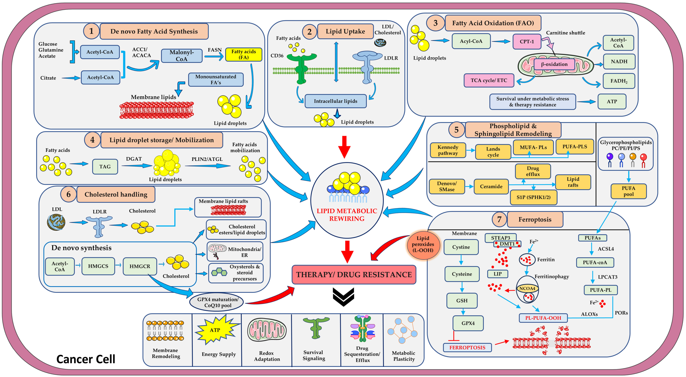
Under metabolic and therapeutic stress, tumor cells can increase their reliance on extracellular lipids (Figure 1, panel 2). Intracellular and extracellular complex lipids are broken down into free fatty acids (FFAs), which expand the cellular fatty acid pool and serve as membrane components, energy reserves, and signaling molecules [32]. Cholesterol, LDL, and fatty acids enter cells through the low-density lipoprotein receptor (LDLR) [32,33], members of the LDL receptor-related protein (LRP) and Solute carrier family 27 (SLC27) families, ABC transporters, and scavenger receptors [32,34,35], and disruption of these systems contributes to tumor plasticity and resistance [32,33,34,35,36,37,38,39]. Importantly, much of this extracellular lipid is supplied by the TME, which makes uptake a key point where cancer cells and stromal cells interact [32,40].
2.2.1. Adipocytes as Lipid Donors: Evidence for Plasticity and Progression
Adipocytes are a major source of exogenous lipids, and several studies show that adipocyte-derived factors promote aggressive phenotypes through cluster of differentiation 36 (CD36) dependent uptake. When ovarian cancer cells were co-cultured with human adipocytes, CD36 expression increased, promoting fatty acid and cholesterol uptake and greater dependence on exogenous lipids. Silencing CD36 reduced lipid accumulation, migration, invasion, and metastatic dissemination, and metastatic ovarian lesions expressed more CD36 than primary tumors, in agreement with Oncomine analyses (a web-based cancer genomics data-mining platform) [41]. In breast cancer, the adipocyte secretome increased CD36 expression, fatty acid uptake, and lipid droplet (LD) accumulation, thereby promoting migration and invasion, and CD36 inhibition attenuated these effects [42]. The response is context-dependent: Adipocyte conditioned medium (ACM) from adipose stem cells (ASCs) of breast cancer patients contained more palmitic and arachidonic acids and less oleic acid than ACM from healthy donors; SUM159 cells showed increased motility and invasion with CD36 upregulation but without significant lipid accumulation, whereas MCF-7 cells accumulated lipids after CD36 elevation without a change in migration [42]. In HER2-positive breast cancer, CD36 is upregulated in cells with acquired lapatinib resistance, increasing exogenous fatty acid uptake and metabolic plasticity. Genetic or pharmacological inhibition of CD36 suppresses the growth of lapatinib-resistant cells in vitro and in vivo, while increased CD36 expression following anti-HER2 therapy in patients correlates with poor prognosis [43]. Adipose-derived lipids can also activate hypoxia-related signaling: ASC conditioned medium elevated hypoxia-inducible factor 1-alpha (HIF-1α) in breast, prostate, and colon cancer cells, and in WiDr colorectal cells, this increased vimentin, zinc finger E-box-binding homeobox 1 (ZEB1), snail family transcriptional repressor 2 (SLUG), and β-catenin while reducing E-cadherin, consistent with EMT. Lipid removal from the medium decreased HIF-1α and migration; lipid replenishment restored them, and HIF-1α silencing reversed lipid-induced metastasis in vivo [44]. Adipose tissue-derived stromal cells promote colorectal cancer stemness, invasion, and metastatic progression by releasing factors that reprogram cancer cells towards a mesenchymal-like phenotype. This study supports the role of adipose tissue in tumor plasticity and progression [45]. Taken together, these studies show that adipocyte-derived lipids act both as substrates and as signals promoting migration, invasion, EMT, and metastasis, while CD36-mediated metabolic rewiring can also contribute directly to resistance to HER2-targeted therapy.
2.2.2. Tumor-Associated Macrophages as Lipid Donors: Direct Evidence of Resistance
In prostate cancer, single-cell studies identified lipid-laden tumor-associated macrophages (TAMs) rich in adipophilin. These lipid-loaded TAMs release CCL6, which enhances tumor cell migration and drives docetaxel resistance, and blocking the macrophage receptor with a collagenous structure (MARCO), the receptor responsible for LDL uptake in these macrophages, depleted their lipid stores and reversed their pro-tumor effects [46]. This example shows that lipid uptake within the TME can reshape immune-cell function and generate signals that support resistance to chemotherapy, so resistance is not exclusively cancer-cell autonomous.
2.2.3. Direct Evidence of Resistance in Tumor Cells
Blocking CD36 with sulfo-N-succinimidyl oleate (SSO), a CD36 inhibitor used to block fatty acid uptake, restored lapatinib sensitivity in resistant rBT474 breast cancer cells, and CD36 silencing promoted apoptosis; the inhibitor JC63.1 re-sensitized resistant xenografts to lapatinib in immunodeficient NSG mice [43]. This directly links therapeutic pressure, lipid uptake, and resistance; lapatinib-resistant tumors depend on CD36-mediated lipid utilization, and disrupting this adaptation restores sensitivity. A second line of evidence comes from acquired resistance to the oxysterol 27-hydroxycholesterol (27HC), which suppresses estrogen receptor-negative breast tumor growth. Resistant lines (4T1, Py230, HCC1954, BPD6, and B16F10) show higher lipid content and fatty acid uptake, supported by increased very-low-density lipoprotein receptor (VLDLR), SLC27, and fatty acid-binding protein (FABP) expression [47,48]. In HCC1954-27HCR cells, cholesterol, phospholipids, neutral lipids, sphingolipids, and FFAs were all elevated, and delipidated serum reduced migration, indicating dependence on extracellular lipids [48]. Here, resistance to 27HC is accompanied by increased lipid uptake and a more migratory, metastatic phenotype in animal models, although the migratory and metastatic readouts again reflect plasticity rather than resistance to a clinical drug.
Figure 1.
Lipid metabolic reprogramming pathways implicated in cancer therapy resistance. Schematic of the major lipid pathways in a cancer cell: (1) de novo fatty-acid synthesis (ACLY, ACC1/ACACA, FASN, SCD1); (2) lipid uptake via CD36 and LDLR; (3) fatty-acid oxidation (FAO) through CPT1-dependent mitochondrial β-oxidation; (4) lipid-droplet storage (DGAT) and mobilization (PLIN2/ATGL); (5) phospholipid and sphingolipid remodeling; and (6) cholesterol uptake and synthesis (HMGCS/HMGCR) (7) Ferroptosis. These pathways may converge on lipid metabolic rewiring, supporting membrane remodeling, energy supply, redox adaptation, survival signaling, drug sequestration/efflux, and metabolic plasticity, which has been associated with therapy resistance. Abbreviations: ACLY, ATP-citrate lyase; ACC1/ACACA, acetyl-CoA carboxylase 1; FASN, fatty acid synthase; SCD1, stearoyl-CoA desaturase 1; CD36, cluster of differentiation 36; LDLR, low-density lipoprotein receptor; FAO, fatty-acid oxidation; CPT1, carnitine palmitoyltransferase 1; DGAT, diacylglycerol acyltransferase; PLIN2, perilipin 2; ATGL, adipose triglyceride lipase; HMGCS, 3-hydroxy-3-methylglutaryl-CoA synthase; HMGCR, 3-hydroxy-3-methylglutaryl-CoA reductase.
2.2.4. Lipid Transport and Tumor Progression
Proteomic profiling by Iglesias-Gato et al. showed sixteen lipid transporters upregulated in prostate cancer bone metastases compared with primary tumors, with LDLR and scavenger receptor class B member 1 (SCARB1) consistently elevated across datasets and ABCA2 and SLC27A4 decreased [49]. These data associate altered lipid transport with progression. Once taken up, fatty acids are activated to fatty acyl-CoAs by long-chain acyl-CoA synthetases (ACSLs), which participate in several metabolic processes linked to cancer progression [50]. Dysregulated lipid metabolism has also been implicated in tumor-immune crosstalk [51]. In summary, upregulation of lipid transporters, particularly CD36, increases fatty acid uptake and supports proliferation, migration, and drug resistance (Figure 1, panel 2). Lipid uptake from adipocytes and TAMs links the TME to tumor plasticity, and direct resistance data (lapatinib, docetaxel, 27HC) indicate that uptake can function as an adaptive response to therapy, whereas migration, EMT, and metastasis data show that the same pathways support a more phenotypically adaptable, aggressive state [34,35,40,41,42,46,48].
2.3. Fatty Acid Oxidation and Tumor Cell Plasticity in Drug Resistance
FAO supplies energy and metabolic intermediates that help cancer cells survive, migrate, and spread. Fatty acids released through lipolysis are activated to fatty acyl-CoAs by ACSLs, transported into mitochondria by the carnitine palmitoyl transferase 1/carnitine acylcarnitine translocase/carnitine palmitoyl transferase 2 (CPT1/CACT/CPT2) system, and oxidized to generate ATP [50]. This provides an alternative energy source that is particularly important under metabolic and therapeutic stress, and the ability to shift between lipid utilization and other metabolic processes contributes to the emergence of drug-tolerant phenotypes.
2.3.1. Evidence Linking FAO to Migration, Invasion, and EMT
Adipocytes again provide an important lipid supply. ACM from obese tissue increased expression of angiopoietin-like protein 4 (ANGPTL4), cAMP-responsive element-binding protein 3-like 3 (CREB3L3), and FAO-related genes (CPT1A, SLC25A20, ACAA2), promoting migration and invasion in breast cancer cell lines (MDA-MB-231, HCC38, and E0771), with stronger effects from morbidly obese than overweight donors; silencing ANGPTL4 reduced focal adhesion kinase (FAK) signaling and motility. Adipocyte-derived substrates can therefore enhance FAO and support the energetic and signaling demands of tumor cell movement. Similarly, aggressive prostate cancer cells showed high palmitate oxidation, elevated CPT1A and CACT activity, and low malonyl-CoA, while restoration of miR-124/129/378 suppressed these enzymes and reduced migration [52]. Inhibiting FAO with etomoxir or silencing CPT1A also reversed transforming growth factor beta (TGF-β)-induced EMT in lung (A549) and liver (Hep3B) cancer cells [53]. These studies connect FAO with motility, invasion, and EMT-associated plasticity, but they primarily demonstrate a role in progression rather than in drug resistance.
2.3.2. Fatty Acid Oxidation and Therapeutic Resistance
The retinoid bexarotene decreased FAO, induced a mesenchymal-to-epithelial transition (MET), increased lipid storage, and re-sensitized resistant breast cancer cells to 5-fluorouracil, indicating that FAO helps maintain a mesenchymal, drug-resistant state and that cells redistribute fatty acids between oxidation and storage. Disrupting FAO can therefore weaken this adaptive state and restore treatment sensitivity [53,54], consistent with the chemo- and radio-sensitization achieved by CPT1 inhibition [26]. Overall, FAO is a driver of tumor flexibility and metastasis, and, in selected settings, of drug resistance (Figure 1, panel 3). Importantly, altered lipid utilization is not restricted to tumor-intrinsic changes: primary breast tumors expressing higher levels of genes associated with lipid catabolism, lipid transport, and FAO were associated with an increased risk of lung metastasis [55]. This highlights the contribution of lipid metabolism to metastatic progression and complements evidence that adipocytes and lung-resident neutrophils can supply lipids within the tumor microenvironment.
2.4. Lipid Synthesis, Droplet Storage and Mobilization in Drug Resistance
2.4.1. De Novo Lipogenesis and Storage
De novo lipogenesis (DNL) synthesizes fatty acids from non-lipid precursors such as glucose and glutamine, and its key enzymes {ACLY, ACACA, FASN, SCD1, and 3-Hydroxy-3-methylglutaryl-CoA reductase (HMGCR)} are often dysregulated to enhance plasticity, stemness, invasion, and therapy resistance [56]. Several observations are primarily phenotypic. Protein C receptor (PROCR) signaling in nasopharyngeal carcinoma activates FASN via nuclear factor kappa-light-chain-enhancer of activated B cells (NF-κB) to promote migration, which FASN inhibition reverses; altered lipid composition and citrate metabolism facilitate EMT and metastasis; and lysophosphatidic acid (LPA) and sphingosine-1-phosphate (S1P) promote survival and migration [57,58]. Others are directly drug-related; upregulated lipogenic enzymes maintain stem-like traits and contribute to resistance to temozolomide and BRAF inhibitors in glioblastoma and melanoma [59], and cholesterol-dependent stabilization of receptor tyrosine kinases such as epidermal growth factor receptor (EGFR) sustains oncogenic signaling, with cytochrome P450 family 51 subfamily A member 1 (CYP51A1) inhibition restoring drug sensitivity [30]. In HER2-positive breast cancer cells resistant to trastuzumab, altered lipid metabolism and increased lipid storage have been reported [60]. In non-small-cell lung cancer (NSCLC) cells surviving EGFR-tyrosine kinase inhibitors (EGFR-TKIs), enhanced SCD1 expression and LD accumulation have been associated with drug-tolerant states; the specific mechanistic claims concerning SREBP activation require confirmation against the relevant primary study [59]. In cisplatin- and 5-fluorouracil-resistant HCT116 colorectal cancer cells, LD accumulation was observed, providing experimental evidence of an association between lipid storage and acquired drug resistance [61].
Excessive LD accumulation has also been associated with MDR phenotypes. LDs are dynamic cytoplasmic organelles that regulate intracellular lipid storage and availability. They contain a hydrophobic core enriched in triacylglycerols (TAGs) and cholesteryl esters (CEs), surrounded by a phospholipid monolayer associated with proteins involved in lipid metabolism and droplet dynamics [62,63]. In cancer cells, LDs can serve as reservoirs of fatty acids and other lipids that support metabolic adaptation and survival under environmental stress, although their contribution varies with tumor type and cellular conditions [64]. TAG synthesis by diacylglycerol acyltransferases (DGAT1 and DGAT2) contributes to LD formation and expansion, whereas lipolytic enzymes mobilize stored TAGs to release fatty acids for subsequent metabolic use [65]. Adipose triglyceride lipase {ATGL/patatin-like phospholipase domain-containing protein 2 (PNPLA2)} catalyzes the initial step of TAG hydrolysis, followed by the sequential action of hormone-sensitive lipase {HSL/lipase E, hormone-sensitive (LIPE)} and monoacylglycerol lipase {MAGL/monoglyceride lipase (MGLL)} [66]. Thus, the balance between TAG synthesis and lipolysis helps determine lipid availability and influences oxidative stress and cancer-cell responses to treatment. LDs’ proposed role in sequestering hydrophobic drugs and lowering effective intracellular drug concentrations requires a source that directly demonstrates this mechanism. In ponatinib-resistant fibroblast growth factor receptor 1 (FGFR1)-driven lung cancer cells, increased LD formation enabled sequestration of ponatinib, contributing to both intrinsic and adaptive drug resistance. Pharmacological reduction of LD using triacsin C enhanced the anticancer activity of ponatinib and restored drug sensitivity [67]. In breast cancer, altered lipid profiles have been associated with changes in docetaxel sensitivity [38], while doxorubicin-resistant breast cancer cells have been investigated for resistance mechanisms involving FABP5/peroxisome proliferator-activated receptor gamma (PPARγ) and calcium/calmodulin-dependent protein kinase II (CaMKII) signaling [39]. The specific claim that MCF7R cells show LD enrichment should be retained with [39] only if this finding is confirmed in the original study.
2.4.2. Lipid Mobilization from the Microenvironment
Tumor cells need not rely on their own lipid synthesis or stores. Tumor-adjacent adipocytes undergoing increased lipolysis provide fatty acids that support breast cancer cells in evading lapatinib [43], a direct example of adipocyte-driven drug escape. In addition, lung-resident neutrophils fuel breast cancer metastasis by transferring stored triglycerides to tumor cells, boosting FAO and mitochondrial respiration. Even when lipid synthesis was blocked, neutrophil-derived lipids sustained tumor survival and colonization in the lung; tumors enriched with lipid-loaded neutrophils showed higher metastatic potential, whereas genetic disruption of ATGL regulators (hypoxia-inducible lipid droplet-associated protein {HIPLDA, G0/G1 switch gene 2 (G0S2), cell death-inducing DFFA-like effector C (CIDEC)} reduced metastasis [54,55]. This neutrophil study demonstrates microenvironmental lipid transfer and metastatic fitness rather than drug resistance per se, but it shows how externally supplied lipids can compensate for impaired intrinsic synthesis and maintain the metabolic flexibility required for persistence (Figure 1, panel 4).
2.5. Role of Phospholipid and Sphingolipid Metabolism in Targeted Therapy Resistance
Alterations in phospholipid metabolism are increasingly linked to resistance to targeted therapies as well as chemotherapy. As noted above, tumor-adjacent adipocytes undergoing increased lipolysis provide fatty acids that support breast cancer cells in evading lapatinib, illustrating how lipid handling in the TME influences drug escape.
Glycerophospholipids, the most abundant phospholipids (PC, PE, PS, PG, PI), are synthesized in the ER and Golgi through sourcing and activation of precursors, backbone assembly, and subsequent modification and transport. They form the structural basis of membranes and lipid rafts, anchor proteins, and participate in signaling microdomains, so changes in their composition affect receptor localization, membrane fluidity, and transporter distribution, and can reduce intracellular drug accumulation. Phospholipases liberate bioactive mediators such as DAG, PA, IP3, LPA, and arachidonic acid, which activate PI3K/AKT and MAPK survival pathways [32,68,69]. Specific lipid-modifying enzymes have been directly implicated in resistance. Lysophosphatidylcholine acyltransferase-2 (LPCAT2) contributes to oxaliplatin and 5-fluorouracil resistance in colorectal cancer by activating anti-apoptotic signaling during ER stress and suppressing chemotherapy-induced immunogenic cell death, likely through altered ER and plasma membrane lipid composition. SCD1 converts saturated to monounsaturated fatty acids [37]; progesterone-driven SCD1 upregulation increases membrane fluidity and disrupts cytoskeletal interactions, promoting docetaxel resistance in breast cancer. Long-chain saturated lysophospholipids protect against cisplatin, and LPA acting through lysophosphatidic acid receptor 1 (LPAR1/LPA1) promotes resistance to adriamycin and paclitaxel in triple-negative breast cancer through upregulation of multidrug efflux transporters (MRP1-3, BCRP) and antioxidant enzymes [32].
Sphingolipids (sphingomyelin and glycosphingolipids), based on sphingosine backbones and abundant in neural tissues, are also synthesized in the ER and Golgi. Sphingomyelin breakdown generates ceramide via sphingomyelinases, and ceramide, ceramide-1-phosphate (C1P), sphingosine, and sphingosine-1-phosphate (S1P) are potent signaling lipids governing cell death, stress responses, and survival. Ceramide generally promotes apoptosis, whereas S1P often supports pro-survival signaling; shifts in the balance between these lipids can contribute to resistance to anticancer agents [36,70].
Collectively, dysregulation of phospholipid and sphingolipid metabolism contributes to resistance by altering membrane composition, modulating drug uptake and efflux, rewiring survival signaling, and modifying stress responses. Alterations in lipid-raft cholesterol can also influence AKT survival signaling [71]. These changes complement alterations in fatty acid synthesis, oxidation, and storage, indicating coordinated remodeling across lipid metabolism (Figure 1, panel 5), and phospholipid metabolism is a promising therapeutic target in treatment failure.
2.6. Altered Cholesterol Metabolism Drives Drug Resistance
Dysregulated cholesterol metabolism is a key contributor to drug resistance. Cholesterol-enriched lipid rafts serve as platforms for AKT and EGFR survival signaling, and high cholesterol can alter membrane permeability and transporter activity, reducing intracellular drug accumulation [32,72]. Resistant cells can increase cholesterol synthesis and uptake through HMGCR and LDLR while suppressing efflux via ABCA1, leading to cholesterol accumulation (Figure 1, panel 6); cholesterol accumulation in mitochondrial membranes can also blunt apoptosis [32,72]. Data from The Cancer Genome Atlas show that upregulated cholesterol biosynthesis correlates with poorer survival and more advanced disease [32]. This correlative evidence supports cholesterol reprogramming as a feature associated with treatment failure but is not itself proof of causality.
In castration-resistant prostate cancer (CRPC), macrophage-derived cholesterol supports therapeutic resistance by increasing cholesterol availability to tumor cells and promoting intratumoral androgen biosynthesis. The underlying primary study showed that macrophages can transfer cholesterol to prostate tumor cells, and macrophage depletion reduced androgen levels and improved response to androgen-deprivation therapy [32,73]. This direct evidence of resistance shows that resistant tumor cells can obtain cholesterol from surrounding cells rather than relying exclusively on their own synthesis, in line with the TAM-dependent docetaxel resistance described in Section 2.2.2.
Gefitinib-resistant NSCLC cells show higher intracellular cholesterol than sensitive cells [74]. In breast cancer, elevated endogenous cholesterol biosynthesis in aromatase inhibitor-resistant cells activates estrogen receptor-α (Erα), and statins reduce ERα binding and cell invasion [32]. In pancreatic ductal adenocarcinoma (PDAC), gemcitabine-tolerant cells accumulate cholesteryl esters (CE) relative to sensitive lines, and inhibition of LDLR or ACAT, which esterifies cholesterol, reduces CE storage and may restore gemcitabine response [75]. Resistant cells may therefore redistribute cholesterol between free and stored forms rather than simply increasing total levels.
ABC transporters play a central role in resistance across many cancers [41], and membrane cholesterol influences their localization and activity. P-glycoprotein preferentially localizes to cholesterol-rich microdomains, and altering membrane cholesterol changes its transport function [76]; ABCC1 (MRP1) is similarly regulated, redistributing to higher-density membrane regions and losing activity when membrane cholesterol depletes [76]. Cholesterol thus affects resistance both through intracellular signaling and through membrane-associated drug transport. Together, these findings support a model in which therapeutic pressure promotes cholesterol metabolic and membrane adaptations that maintain survival signaling, alter drug transport, and resist apoptosis, with cholesterol supplied by both tumor cells and the TME. Targeting cholesterol synthesis, uptake, or storage, for example with statins, ACAT inhibitors, or LDLR modulators, may provide complementary strategies for overcoming therapy resistance [32].
2.7. The SCAP/SREBP Axis as a Transcriptional Regulatory Hub Linking Lipid-Metabolic Pathways and Therapy Resistance
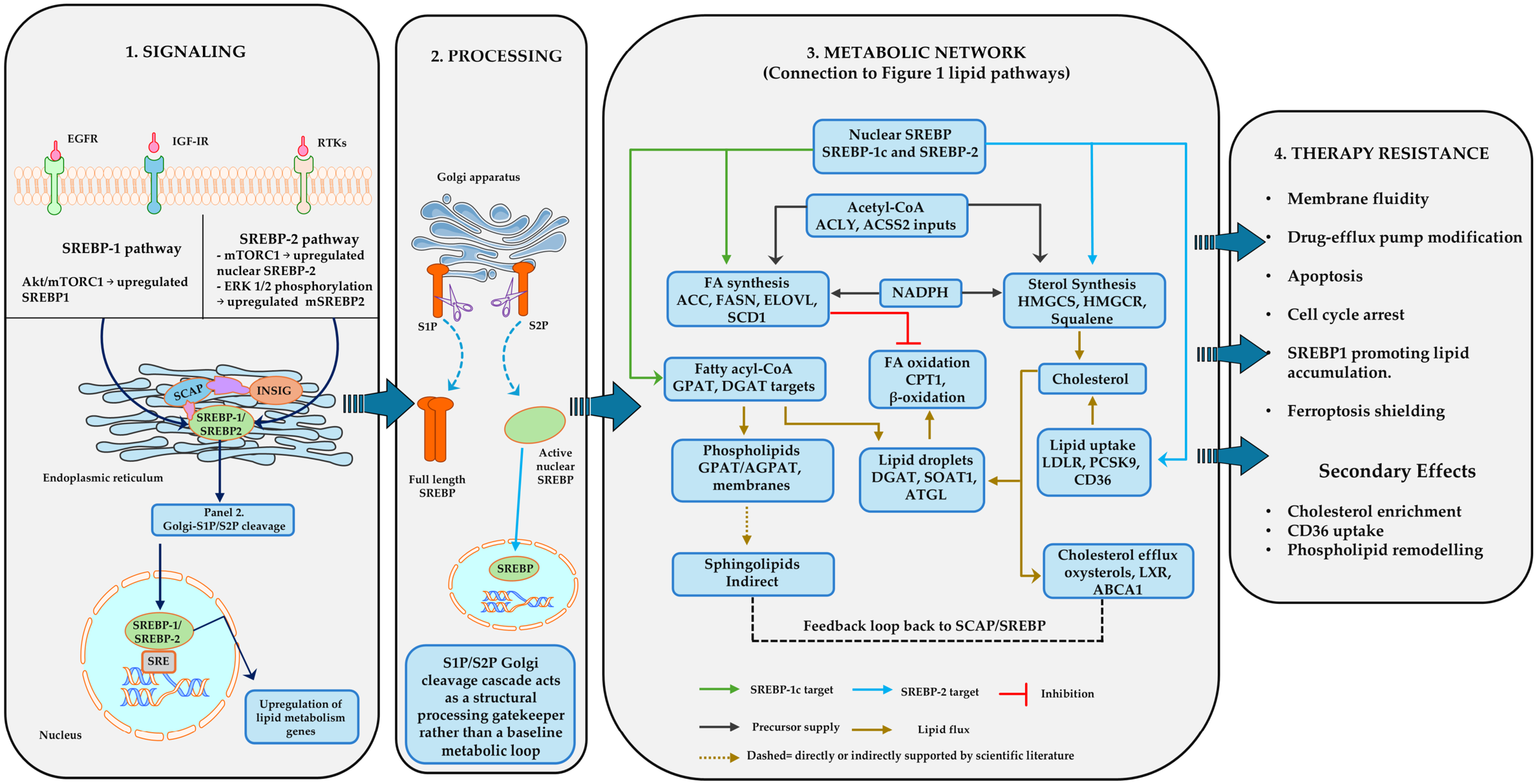
The lipid pathways summarized in Figure 1 are not controlled independently of one another. In many tumors, they are transcriptionally coordinated by the sterol regulatory element-binding protein (SREBP) family and its escort protein, SREBP cleavage-activating protein (SCAP) [13,14,15,16]. Figure 2 organizes this regulatory system into four connected steps, which also structure this section; upstream oncogenic and nutrient signaling that sets SREBP abundance and processing; insulin-induced gene INSIG-SCAP-controlled ER-to-Golgi trafficking and proteolytic activation; the downstream SREBP-regulated lipid-metabolic network; and the consequences of this network for therapy resistance. Throughout, we distinguish regulatory links that are supported by direct experimental evidence from those that are indirect or context-dependent, mirroring the solid and dotted arrows in the Figure 2.
2.7.1. SREBP Isoforms and Upstream Oncogenic and Nutrient Signaling
Mammalian cells express three SREBP isoforms from two genes. SREBP-1a and SREBP-1c are produced from sterol regulatory element-binding transcription factor 1 (SREBF1) through alternative transcription start sites, whereas SREBP-2 is encoded by SREBF2 [14,16]. SREBP-1c preferentially drives fatty-acid and triglyceride synthesis, SREBP-2 preferentially controls cholesterol synthesis and uptake, and SREBP-1a is the most potent and least selective isoform, activating both programmes [14,77]. Several oncogenic pathways converge on SREBP-1, including EGFR, PI3K/AKT/mTOR and mutant p53 [78]. In glioblastoma, EGFR-PI3K-AKT signaling increases SREBP-1 activation and LDLR expression, thereby linking growth-factor signaling to lipid synthesis and cholesterol uptake [79]. Sustained PI3K-AKT-mTORC1 signaling induces SREBP-1 and SCD1-dependent lipogenesis, protects PI3K-mutant and phosphatase and tensin homolog (PTEN)-deficient cancer cells from ferroptosis [80]. MAPK pathway activity acts similarly in BRAF-mutant melanoma: BRAF inhibition reduces SREBP-1 processing and lipogenesis in therapy-sensitive cells, but not in cells with acquired resistance [81]. Signaling can also act on SCAP itself. EGFR-driven glucose uptake promotes N-glycosylation of SCAP, which stabilizes SCAP, weakens its association with INSIG and favours SCAP-SREBP transport to the Golgi [82], and signal transducer and activator of transcription 3 (STAT3) binds the promoters of both SCAP and SREBF1 to raise SCAP and SREBP-1 expression [77].
Two further inputs regulate SREBP-1 expression rather than its proteolytic processing (Figure 2, panel 1). First, in liver and breast cancer cells SREBP-1 induces glutamine synthetase (GS), which increases Sp1 O-GlcNAcylation and thereby SREBP-1 transcription. This feed-forward loop raises acetyl-CoA carboxylase 1 (ACC1) expression and LD formation, including under glutamine deprivation [83]. O-GlcNAc transferase (OGT) activity is itself required to sustain SREBP-1 expression and lipid synthesis in breast cancer cells [84]. Second, insulin-induced c-Myc upregulates thymine DNA glycosylase (TDG), which demethylates the SREBP1 promoter and increases SREBP-1 and ACC1 expression [85]. These mechanisms have been characterized mainly in liver and breast cancer models, and their relevance to other tumor types remains to be established.
SREBP-2 is regulated by partly distinct inputs. Active mTORC1 lowers ER cholesterol by limiting cholesterol delivery to lysosomes, which promotes SREBP-2 activation [86], and mutant p53 associates with sterol-gene promoters through SREBP transcription factors to upregulate the mevalonate pathway [87]. ERK1/2-dependent phosphorylation has also been proposed to enhance the transcriptional activity of mature SREBP-2 [88]; this link is currently the least well supported of the inputs shown in Figure 2.
Figure 2.
The SCAP/SREBP regulatory axis links oncogenic signaling to lipid-metabolic plasticity and therapy resistance. (1) Upstream regulation: EGFR, IGF-1R, RTKs, (Not shown in figure GS-Sp1-OGT, c-Myc–TDG, mutant p53 regulate), AKT/mTORC1, ERK1/2, and SREBP expression, processing, or activity. (2) Processing: under sterol-replete conditions, INSIG retains SCAP-SREBP in the ER. Sterol depletion promotes SCAP-mediated transport to the Golgi, where S1P and S2P sequentially release mature nuclear SREBP, which binds sterol regulatory elements (SREs). (3) Lipid-metabolic network: SREBP-1c and SREBP-2 regulate precursor supply (acetyl-CoA and NADPH) and six modules: fatty-acid synthesis, lipid uptake, fatty-acid oxidation, lipid-droplet storage/mobilization, phospholipid and sphingolipid synthesis, and sterol synthesis/cholesterol efflux. (4) Therapy resistance: lipid-metabolic adaptation supports membrane remodeling, ferroptosis protection, and altered drug handling, reducing treatment sensitivity. Arrow code: Green, SREBP-1c targets; blue, SREBP-2 targets; black, precursor supply; brown, lipid flux; red blunt-ended line, inhibition; dotted brown arrow, indirect or context-dependent links. Abbreviations: ACLY, ATP-citrate lyase; ACC, acetyl-CoA carboxylase; FASN, fatty acid synthase; SCD1, stearoyl-CoA desaturase 1; LDLR, low-density lipoprotein receptor; CPT1, carnitine palmitoyltransferase 1; DGAT, diacylglycerol acyltransferase; SOAT1, sterol O-acyltransferase 1; ATGL, adipose triglyceride lipase; GPAT/AGPAT, glycerol-3-phosphate/acylglycerol-3-phosphate acyltransferase; HMGCS/HMGCR, HMG-CoA synthase/reductase; LXR, liver X receptor; ABCA1, ATP-binding cassette transporter A1; GS, glutamine synthetase; OGT, O-GlcNAc transferase; TDG, thymine DNA glycosylase; MUFA, monounsaturated fatty acid. Solid and dotted arrows distinguish direct experimental evidence from indirect or context-dependent regulatory links.
2.7.2. INSIG-SCAP-Controlled Processing and Nuclear Transcriptional Activation of SREBP
SREBPs are synthesized as ER-membrane precursors bound to SCAP. When ER sterol levels are sufficient, cholesterol-bound SCAP associates with INSIG, and the SREBP-SCAP complex is retained in the ER [13,89]. When ER cholesterol falls below a critical threshold, SCAP undergoes a conformational change that allows the complex to be packaged into coat protein complex II (COPII) vesicles and transported to the Golgi apparatus [89] (Figure 2, panel 2). There, sequential cleavage by site-1 protease (S1P) and site-2 protease (S2P) releases the mature, transcriptionally active N-terminal fragment (mSREBP), which enters the nucleus and binds sterol regulatory elements (SREs) in target-gene promoters [14,15]. Erlins, which are cholesterol-binding ER proteins associated with the SREBP-SCAP-INSIG complex, reinforce this retention. Their depletion activates SREBP target genes through the canonical SCAP-dependent route even when sterols are plentiful [89]. Tumors can uncouple this step from sterol feedback. In glioblastoma cells, sterol depletion alone did not activate SREBPs in glucose-free medium, whereas glucose restored SCAP abundance and SREBP-1/-2 activation, which shows that nutrient and oncogenic inputs can govern trafficking alongside sterol sensing [82]. Because proteolytic maturation is obligatory for SREBP activity, the S1P/S2P cascade acts as a structural processing gatekeeper rather than as a baseline metabolic loop; this is why the signaling inputs in panel 1 act upstream of it, whereas the metabolic effects in Figure 2, panel 3 act downstream.
2.7.3. Direct and Indirect Targets: The SREBP-Regulated Lipid-Metabolic Network
Once in the nucleus, SREBP-1 and SREBP-2 activate overlapping but distinct transcriptional programmes (Figure 2, panel 3; green arrows, SREBP-1c targets; blue arrows, SREBP-2 targets). The network is organized around the supply of acetyl-CoA and NADPH, which is consumed by fatty-acid and sterol synthesis, and around the subsequent fates of the lipids produced; uptake, storage, oxidation, membrane incorporation and efflux. The six numbered modules correspond to the lipid pathways summarized in Figure 1.
Fatty-acid synthesis. SREBP-1 induces genes encoding ACLY, acetyl-CoA carboxylase (ACC; ACACA), FASN and SCD1, together with fatty-acid elongases [14,79]. Desaturation by SCD1 is of particular functional importance. When SREBP activity was attenuated, cancer cells retained long-chain saturated fatty-acid synthesis but lost desaturation capacity. The resulting imbalance between saturated and monounsaturated pools caused lipotoxicity, and supplementation with monounsaturated fatty acids restored growth [90].
Lipid and cholesterol uptake. LDLR mediates cholesterol uptake and is induced by SREBP-2 and, in glioblastoma, downstream of EGFR-PI3K-AKT-SREBP-1 signaling [79,91]. Proprotein convertase subtilisin/kexin type 9 (PCSK9), which limits LDLR availability, is a direct transcriptional target of SREBP1 in pancreatic cancer, where the SREBP1-PCSK9 axis has been linked to programmed death-ligand 1 (PD-L1) regulation and resistance to anti-PD-1 therapy [92]. Uptake of fatty acids through CD36 provides an additional lipid supply, but its dependence on SREBP is context-dependent and should not be assumed [93].
Fatty-acid oxidation. Regulation of fatty-acid oxidation by the SREBP axis is largely indirect. Malonyl-CoA produced by ACC inhibits CPT1, restricting mitochondrial fatty-acid entry and favouring lipid synthesis over oxidation [94] (red inhibitory line in Figure 2). This balance is dynamic and depends on nutrient availability, oncogenic signaling and cellular energy demands. For example, in KRAS-mutant non-small-cell lung cancer, SREBP-1 also influences mitochondrial metabolism, although these effects were not explained by a significant reduction in lipogenesis [95]. However, some drug-resistant cancer cells can maintain or increase fatty-acid oxidation to support metabolic adaptation and survival, indicating that the relationship between SREBP-driven lipogenesis and mitochondrial fatty-acid oxidation is context-dependent.
Lipid-droplet storage and mobilization. Excess fatty acids and cholesterol are stored in lipid droplets as triglycerides and cholesteryl esters, respectively. This process involves DGATs, which catalyze triglyceride synthesis, and sterol O-acyltransferase 1 (SOAT1), which esterifies cholesterol. Stored lipids can subsequently be mobilized by lipases such as ATGL. In cancer cells, SREBP-1 upregulates SOAT1, promoting cholesteryl-ester storage, limiting lipotoxicity and supporting tumor growth [96]. The GS-Sp1-SREBP-1-ACC1 feed-forward loop described above also promotes LD formation [83]. SREBP-1 can additionally regulate LD metabolism through context-dependent transcriptional regulation of DGAT1, although direct transcriptional control of DGAT2 and ATGL by SREBP-1 remains less clearly established. Accordingly, Figure 2 distinguishes experimentally supported regulatory links from the broader processes of lipid storage and mobilization.
Phospholipid and sphingolipid remodeling. Fatty acyl-CoAs are incorporated into phospholipids through glycerolipid synthesis pathways involving glycerol-3-phosphate acyltransferases (GPATs) and 1-acylglycerol-3-phosphate O-acyltransferases (AGPATs). Thus, SREBP-dependent lipogenesis can influence membrane fatty-acid composition and saturation. DNL promotes membrane lipid saturation, which can protect cancer cells against free radicals and chemotherapeutic agents [97]. In gefitinib-resistant EGFR-mutant lung cancer cells, sustained SREBP-1-dependent lipogenesis contributes to the maintenance of altered lipid composition, while SREBP-1 inhibition with fatostatin increases oxidative stress and sensitizes cells to gefitinib [98]. The connection to sphingolipid metabolism is represented by a dotted arrow because it is indirect. Sphingolipid synthesis involves a broader network, including serine palmitoyltransferase and sphingosine kinases, which should not be considered uniform direct SREBP targets without pathway-specific evidence.
Cholesterol synthesis and efflux. SREBP-2, and SREBP-1a to a lesser extent, induces genes involved in cholesterol synthesis, including those encoding HMG-CoA synthase (HMGCS), HMG-CoA reductase (HMGCR) and squalene synthase [14]. Because cholesterol and fatty-acid synthesis share acetyl-CoA and NADPH as metabolic resources, these pathways are metabolically interconnected (Figure 2). Feedback mechanisms restrain cholesterol accumulation at several levels. Sterol-dependent association of SCAP with INSIG retains the SREBP-SCAP complex in the endoplasmic reticulum, limiting further SREBP activation. In parallel, oxysterol-activated liver X receptors (LXRs) promote ABCA1-mediated cholesterol efflux. In glioblastoma models, LXR activation also disrupts EGFR–AKT-SREBP-1-LDLR-dependent lipid-survival signaling, promoting tumor-cell death [91].
2.7.4. Contribution of SREBP-Regulated Programmes to Therapy Resistance
In treated tumors, the network shown in Figure 2, panel 3 supports resistance through several recurring but context-dependent mechanisms. In several models, sustained SREBP-1 activity during therapy, rather than SREBP-1 expression at baseline, distinguishes resistant from sensitive cells. In BRAF-mutant melanoma, vemurafenib reduced SREBP-1 processing and de novo lipogenesis in sensitive cells but not in resistant cells, and pharmacological SREBP-1 inhibition re-sensitized resistant tumors in vitro and in a patient-derived xenograft model [81]. A similar pattern was reported for gefitinib-resistant EGFR-mutant lung cancer, in which fatostatin combined with gefitinib suppressed xenograft growth that gefitinib alone did not [98].
Membrane remodeling and altered drug response. Changes in fatty-acid synthesis and desaturation alter membrane lipid saturation and thereby susceptibility to oxidative damage and chemotherapeutics [97]. In both BRAF and EGFR inhibitor-resistant models, SREBP-1 inhibition shifted membrane phospholipids towards higher polyunsaturation and re-sensitized cells to the targeted agent [81,98]. The direction and consequences of these changes depend on the lipid species, membrane compartment and drug involved, and reduced membrane fluidity should not be assumed to decrease drug permeability uniformly.
Redox adaptation and ferroptosis resistance. SREBP-1-dependent monounsaturated fatty-acid synthesis through SCD1 can be incorporated into membrane phospholipids and reduce the availability of oxidation-sensitive polyunsaturated species. In cancer cells with hyperactive PI3K-AKT-mTOR signaling, this mechanism confers ferroptosis resistance, and genetic or pharmacological ablation of SREBP1 or SCD1 sensitized them [80]. SREBP-1 inhibition also increased reactive oxygen species (ROS) and malondialdehyde in gefitinib-resistant cells [98]. The relationship between SREBP activity, SCD1 expression and ferroptosis sensitivity nevertheless varies between tumor types and requires direct validation in each setting [93].
Cholesterol-dependent survival and drug handling. In HCC, caspase-3-mediated SREBP2 activation promoted cholesterol biosynthesis, cancer stem-cell properties and resistance to sorafenib and lenvatinib; simvastatin suppressed tumor growth and sensitized resistant models to sorafenib [99]. The SREBF2-STAR-related lipid transfer protein 4 (STARD4) axis also contributed to sorafenib resistance by increasing mitochondrial cholesterol accumulation and limiting cytochrome C release, highlighting the importance of intracellular cholesterol distribution, rather than total cholesterol abundance alone [100]. A recent review further discusses SREBP2-associated cholesterol metabolic reprogramming in HCC progression and treatment resistance [101]. Cholesterol may also influence membrane organization and membrane-protein function, including that of ABC transporters. However, increased cholesterol does not necessarily enhance ABCB1-mediated drug efflux, and direct transcriptional regulation of ABCA1 or ABCB1 by SREBP2 should not be assumed without tumor-specific evidence. Thus, the contribution of sterol homeostasis to multidrug resistance must be evaluated in the context of the tumor type, transporter and therapeutic agent.
Metabolic adaptation to cellular stress. SREBP-1 can buffer therapeutic stress by coupling glucose and lipid metabolism. In NSCLC, SREBP-1 increased glucose uptake and sustained tumor growth during lysosome inhibition with chloroquine, and blocking glucose transport overcame this resistance by inducing mitochondrial damage and oxidative stress [102]. In glioblastoma, resistance to the lysosome inhibitor pimozide involved SREBP-1-driven glutamine consumption and lipogenesis, with ammonia from glutamine metabolism further intensifying SREBP-1 activation [103]. These findings support a role for SREBP-1 in stress adaptation, but whether the same mechanism operates against other lysosomal or autophagy-targeting agents requires pathway-specific evidence.
Tumor microenvironment and immune context. Beyond tumor-cell-intrinsic effects, SREBP1 has been linked to epithelial–mesenchymal transition, immune regulation, cell-cycle control and the tumor microenvironment [15]. For example, the SREBP1-PCSK9 axis contributes to immune evasion and anti-PD-1 resistance in pancreatic cancer [92]. As panel 4 indicates, inhibition of SREBP1 can therefore promote apoptosis, cell-cycle arrest, or ferroptosis, whereas its activity promotes lipid accumulation and proliferation, with secondary supporting effects through cholesterol enrichment, CD36-mediated uptake and phospholipid remodeling.
2.8. Ferroptosis Involvement in Lipid Metabolism and Drug Resistance
Ferroptosis is fed by PUFA-containing phospholipids formed through ACSL4/LPCAT3 (Figure 1, panel 5), with PUFA supply from uptake (Figure 1, panel 2) and LD release (Figure 1, panel 4), and is restrained by SREBP-1/SCD1-derived MUFAs (Figure 1, panel 1) and cholesterol-pathway intermediates (Figure 1, panel 6), while GPX4 and FSP1 act as the main defences. Where the PUFAs in membranes come from. Cells cannot make the essential PUFAs linoleic acid (ω6) and α-linolenic acid (ω3), so they take them up from the diet and circulation, for example through the fatty-acid transporter CD36 (Section 2.2). Inside the cell, these are elongated and desaturated into longer PUFAs such as AA and AdA. Free PUFAs are kept in two main places: stored in triglycerides within lipid droplets, from which lipases can release them (Section 2.4), and built into membrane glycerophospholipids such as PC, PE, PI and PS (Section 2.5). Glycerophospholipids are made by the Kennedy pathway and then remodelled by the Lands cycle, in which phospholipase A2 removes a fatty acid and acyltransferases such as LPCAT3 add another. This remodeling decides whether a membrane is rich in PUFAs, which makes it vulnerable to ferroptosis, or in monounsaturated fatty acids (MUFAs) made by SCD1, which protect it. Sphingolipids are a separate branch. Their fatty-acid chains are mostly saturated or monounsaturated, so they are not a major PUFA source. They may influence ferroptosis indirectly, through ceramide and sphingosine-1-phosphate signaling, membrane rafts and survival pathways, and this role is context-dependent (Section 2.5). More details on ferroptosis can be found in Section 3.8.
Ferroptosis suppression as a route to drug resistance. Many anticancer agents kill cells partly through lipid peroxidation, so tumour cells that strengthen their ferroptosis defences, or that reduce the supply of PUFA-containing phospholipids, can survive treatment. The cell states that resist therapy are often the ones most dependent on this protection. Mesenchymal, therapy-resistant cells and drug-tolerant persister cells rely on GPX4 to avoid lipid-peroxide-induced death, which is why GPX4 inhibition selectively kills them [104,105]. Lowering ACSL4 is another route. In sorafenib-resistant HCC, ETS1-driven miR-23a-3p reduces ACSL4 and limits ferroptosis, and inhibiting this miRNA restores ACSL4 and sorafenib sensitivity [106]. ACSL4 expression also predicts ferroptosis sensitivity in basal-like breast cancer cells [107].
Antioxidant transcriptional programmes add a further layer. In HCC cells, activation of the p62-Keap1-NRF2 pathway protects against ferroptosis, and NRF2 inhibition enhances the effect of erastin [108]. Sorafenib induces metallothionein-1G (MT-1G) in an NRF2-dependent manner; MT-1G suppresses ferroptosis and contributes to sorafenib resistance, and genetic or pharmacological inhibition of MT-1G enhances sorafenib activity in vitro and in xenografts [109]. In head and neck cancer, activation of the NRF2-antioxidant response element pathway contributes to resistance to GPX4 inhibitors in cisplatin-resistant cells [110]. These mechanisms are also documented in biliary cancers. In intrahepatic cholangiocarcinoma (iCCA), the lncRNA PAX8-AS1 raises GPX4 through NRF2 activation and by stabilizing GPX4 mRNA, suppresses gemcitabine- and cisplatin-induced ferroptosis, and promotes chemoresistance [111]. Consistent with a role for GPX4 in biliary resistance, intrahepatic cholangiocarcinoma cell lines were more sensitive to RSL3 than extrahepatic lines, which showed higher GPX4 expression and greater resistance [112]. In biliary tract cancer cells, sensitivity to IKE and RSL3 correlates with CD71 and SLC7A11 expression [113]. Immune pressure acts through the same lipid machinery; IFNγ from CD8+ T cells, together with fatty acids, induces ACSL4-dependent ferroptosis in tumour cells [114], so low ACSL4 activity may weaken this form of immune killing. Finally, lipid pathways discussed earlier feed into the same defence. SREBP-1/SCD1-derived MUFAs displace oxidation-prone PUFAs from membrane phospholipids [17,80], and 7-dehydrocholesterol, an intermediate of cholesterol biosynthesis (Section 2.6), has been identified as an endogenous suppressor of ferroptosis [115]. Together, these findings indicate that ferroptosis evasion is a recurring feature of resistant cells, and that its strength depends on the PUFA-phospholipid pool, GPX4 activity and NRF2-driven antioxidant programmes.
3. Targeting Lipid Metabolic Plasticity: Pharmacological and miRNA-Based Strategies
3.1. Lipid Metabolism as an Adaptive Hub: Two Layers of Intervention
Section 2.1, Section 2.2, Section 2.3, Section 2.4, Section 2.5, Section 2.6, Section 2.7 and Section 2.8 portray lipid metabolism as an adaptive hub that connects therapeutic stress with tumor-cell plasticity and drug resistance. Its interconnected processes, including lipid synthesis, uptake, oxidation, storage, membrane remodeling, and cholesterol handling, are influenced by both tumor cells and the tumor microenvironment. Each process can be targeted at two levels: directly, using small-molecule inhibitors against metabolic enzymes, transporters, or receptors; and upstream, through microRNAs (miRNAs). MiRNAs are small non-coding RNAs, typically approximately 22 nucleotides long, that regulate gene expression by binding to complementary sequences in target mRNAs, leading to mRNA degradation or inhibition of translation. Depending on their targets, miRNAs can function as tumor suppressors or oncogenic regulators, making them potential therapeutic tools through miRNA replacement or inhibition strategies [116]. Each process can be targeted at two levels: directly, using small-molecule inhibitors or antibodies against metabolic enzymes, transporters, or receptors; and upstream, through microRNAs (miRNAs). Their therapeutic potential includes inhibiting disease-promoting miRNAs or restoring the expression of tumor-suppressive miRNAs. Chemically modified oligonucleotides, including antagomirs and locked nucleic acid (LNA)-based inhibitors, have demonstrated the feasibility of miRNA silencing in vivo [116,117,118].
MiRNAs can influence treatment response by regulating genes involved in lipid metabolism and other cellular processes associated with drug resistance. In pancreatic ductal adenocarcinoma, miRNA-based approaches have been investigated as potential strategies to overcome chemotherapy resistance [119]. In breast cancer, altered exosomal miRNA profiles and their associated target-gene networks have been linked to response to neoadjuvant chemotherapy, highlighting their potential as predictive biomarkers and therapeutic targets [120]. More specifically, miR-328-3p has been linked to CPT1A-associated fatty-acid β-oxidation, cancer stemness, and metastasis in breast cancer [121]. In gastric cancer, miR-145-5p promotes fatty-acid oxidation-dependent stemness and chemoresistance through the long non-coding RNA MACC1-AS1 [122]. These findings illustrate how non-coding RNAs can influence metabolic adaptation and treatment response, although the mechanisms and strength of evidence differ between cancer types. Evidence from lipid-regulatory studies also demonstrates that miRNAs can coordinate multiple aspects of lipid homeostasis. For example, miR-33a-5p suppresses ABCA1- and ABCG1-mediated cholesterol efflux in THP-1 macrophages under inflammatory stress [123]. This finding provides mechanistic evidence for miRNA regulation of cholesterol handling, although it does not directly establish a role in cancer drug resistance. More broadly, changes in lipid homeostasis can affect membrane dynamics, receptor signaling, mitochondrial metabolism, oxidative-stress responses, and drug-transporter activity, processes implicated in cancer drug resistance [32,59].
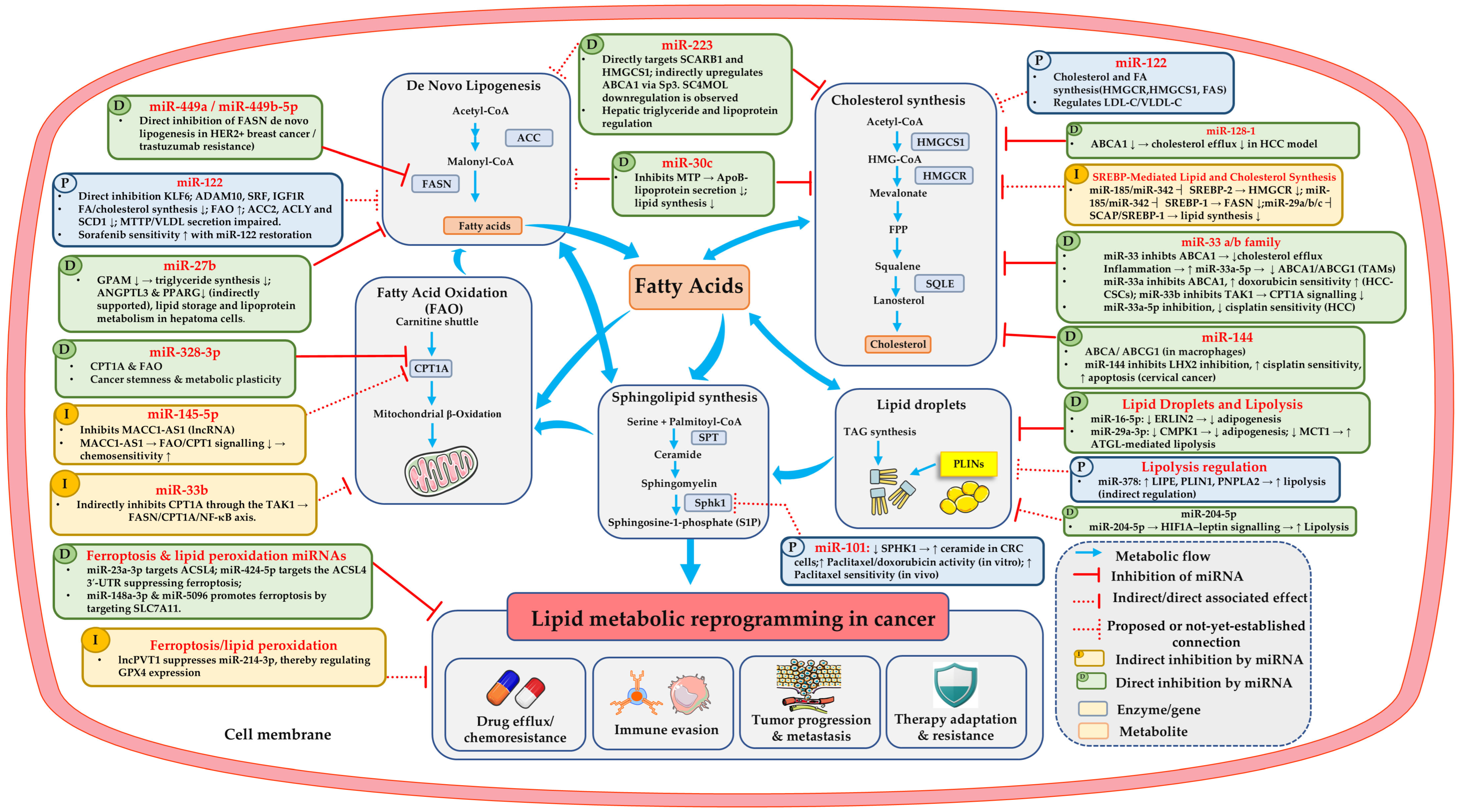
Accordingly, each subsection below considers miRNA regulators (Table 1 and Figure 3) alongside pharmacological inhibitors (Supplementary Table S1 and Figure 4) targeting the same metabolic node. The miRNAs are classified according to whether they act through direct (D), indirect (I), or proposed (P) inhibition of the listed targets. Because lipid-metabolic pathways are interconnected, inhibition of one node may trigger compensatory changes in other pathways. This capacity for metabolic rewiring may limit the effectiveness of single-pathway inhibition and provides a rationale for investigating combination strategies.
3.2. Targeting De Novo Lipogenesis (ACLY, ACC, FASN, SCD1)
Oncogenic signaling and de novo fatty-acid synthesis in cancers such as HER2-positive breast cancer: sustained upregulation of DNL contributes to metabolic reprogramming and tumor aggressiveness. This process is closely linked to hyperactivation of PI3K-AKT signaling, a pathway frequently dysregulated through growth-factor receptor tyrosine kinases (RTKs), including HER2 and the insulin receptor, or oncogenic mutations in PIK3CA. More than 80% of HER2-amplified breast tumors have been reported to exhibit increased AKT phosphorylation at Ser473 and Thr308. Activated AKT directly phosphorylates and stimulates ACLY, thereby enhancing cytosolic acetyl-CoA production [124]. This provides a metabolic link between oncogenic signaling and lipid biosynthesis. In the DNL pathway, glucose-derived pyruvate enters mitochondrial metabolism and contributes to citrate production through the tricarboxylic acid (TCA) cycle. Citrate is subsequently exported into the cytosol, where ACLY, a cytosolic homotetrameric enzyme, converts it into acetyl-CoA [59,125]. Acetyl-CoA is then carboxylated by ACC to generate malonyl-CoA. ACC comprises two isoforms, ACC1 and ACC2, of which ACC1 primarily supports DNL, whereas ACC2 contributes to the regulation of FAO. ACC1 catalyzes the rate-limiting step of fatty-acid synthesis, supplying malonyl-CoA as the substrate for FASN, which produces palmitate, the primary end product of DNL. Palmitate can subsequently undergo elongation and desaturation to generate longer-chain and unsaturated fatty acids, including stearic, palmitoleic and oleic acids [125]. FASN overexpression has also been associated with clinical outcomes in several tumor types, although its prognostic significance varies according to tumor context. In HER2-positive breast cancer, high FASN expression has been associated with poorer relapse-free and distant metastasis-free survival [126]. Similarly, in gastric cancer, FASN overexpression has been associated with poorer overall survival and increased recurrence [127]. However, findings in colon cancer suggest that the relationship between FASN expression and survival may vary according to patient characteristics, including body mass index [128]. These observations support the clinical relevance of FASN while highlighting the context-dependent nature of its prognostic significance.
Figure 3.
miRNA regulatory networks controlling lipid metabolism in cancer. Schematic of the core li-pid-metabolic pathways (de novo lipogenesis, cholesterol synthesis, FAO, sphingolipid synthesis and lipid droplets) and the miRNAs reported to regulate them. miRNAs acting on ferroptosis and lipid peroxidation (ACSL4, SLC7A11, GPX4), including one lncRNA–miRNA axis (lncPVT1/miR-214-3p), are shown separately. Blue arrows denote metabolic flow; red bar-headed lines denote miRNA-mediated inhibition; dotted red lines denote indirect or associated effects. Green boxes (D) mark direct inhibition of the listed targets, yellow boxes (I) indirect inhibition, and blue boxes (P) proposed inhibition (see Table 1). These interactions converge on lipid metabolic reprogramming, which drives drug efflux/chemoresistance, immune evasion, tumour progression and metastasis, and therapy ad-aptation/resistance. Abbreviations: ABCA1/ABCG1, ATP-binding cassette transporters A1/G1; ACC, acetyl-CoA carboxylase; ACSL4, acyl-CoA synthetase long-chain family member 4; ANGPTL3, angiopoietin-like 3; ApoB, apolipoprotein B; ATGL, adipose triglyceride lipase; CPT1A, carnitine palmitoyltransferase 1A; CRC, colorectal cancer; FAO, fatty acid oxi-dation; FASN, fatty acid synthase; FPP, farnesyl pyrophosphate; GPAM, glycerol-3-phosphate acyltransferase, mi-tochondrial; GPX4, glutathione peroxidase 4; HCC, HCC-CSCs, hepatocellular carcinoma cancer stem cells; HER2, human epidermal growth factor receptor 2; HMGCR, HMG-CoA reductase; HMGCS1, HMG-CoA synthase 1; LDL-C/VLDL-C, low-/very-low-density lipoprotein cholesterol; LDLR, low-density lipoprotein receptor; LHX2, LIM homeobox 2; lncPVT1, long non-coding RNA PVT1; MTP, microsomal triglyceride transfer protein; PLINs, perili-pins; PPARG, peroxisome proliferator-activated receptor gamma; SC4MOL, sterol-C4-methyl oxidase-like; SCAP, SREBP cleavage-activating protein; SLC7A11, solute carrier family 7 member 11; SPHK1, sphingosine kinase 1; SPT, serine palmitoyltransferase; S1P, sphingosine-1-phosphate; SQLE, squalene epoxidase; SREBP, sterol regulatory el-ement-binding protein; TAG, triacylglycerol; TAK1, TGF-β-activated kinase 1; TAMs, tumour-associated macro-phages; UTR, untranslated region.
In addition to its role in fatty-acid synthesis, malonyl-CoA inhibits CPT1, thereby restricting mitochondrial fatty-acid uptake and β-oxidation. ACC activity is regulated, in part, by AMP-activated protein kinase (AMPK), a key metabolic sensor involved in cancer progression. AMPK-mediated phosphorylation inhibits ACC activity, thereby limiting fatty-acid synthesis, and also regulates SREBP-1c, a transcriptional regulator of lipogenic genes [14]. Together, these signaling and metabolic mechanisms coordinate lipid anabolism and catabolism, influencing the balance between fatty-acid synthesis and oxidation.
miRNA regulation. MiRNAs regulate gene expression primarily through post-transcriptional mechanisms, including binding to target mRNAs to promote their degradation or inhibit translation. Their effects on lipid metabolism may arise through direct targeting of metabolic-gene transcripts or indirectly through upstream regulators and broader changes in cellular metabolism. However, the miRNAs discussed below should not be interpreted as direct inhibitors of ACLY, ACC, FASN, or SCD1 unless direct targeting of the relevant transcript has been demonstrated (Figure 3). The liver-enriched miR-122 regulates hepatic lipid homeostasis by promoting lipid synthesis and limiting fatty acid oxidation. Its inhibition in mice reduced hepatic fatty acid and cholesterol synthesis, increased fatty acid oxidation and AMPK activation, and improved plasma cholesterol and hepatic steatosis [129]. LNA-mediated miRNA silencing in non-human primates further demonstrated the feasibility of inhibiting miRNAs in vivo [117]. Conversely, miR-122 deletion caused hepatic steatosis, inflammation and spontaneous HCC development, whereas miR-122 restoration suppressed tumor development in a MYC-driven HCC model [130]. Similarly, miR-122a knockout induced steatohepatitis, fibrosis and spontaneous HCC, partly through reduced microsomal triglyceride transfer protein (MTTP) expression and impaired VLDL secretion; restoration of MTTP reversed steatosis, while miR-122a re-expression reduced liver pathology and tumor incidence. Kruppel-like factor 6 (KLF6) was also identified as a direct miR-122a target contributing to fibrosis [131]. In human HCC, miR-122 was frequently downregulated, and its restoration suppressed malignant properties and enhanced sorafenib sensitivity through direct targeting of of A disintegrin and metalloproteinase 10 (ADAM10), serum response factor (SRF) and insulin-like growth factor 1 receptor (IGF1R) [132]. These findings support a role for miR-122 in coordinating lipid homeostasis, but do not establish direct binding to the mRNAs encoding ACLY, ACC1, FASN, or SCD1. Moreover, miR-122 has important physiological and tumor-suppressive functions in the liver: miR-122-deficient mice develop liver inflammation and hepatocellular carcinoma [130,131]. Conversely, restoration of miR-122 suppresses tumorigenic properties and sensitizes hepatocellular carcinoma cells to sorafenib [132].
In Huh7 hepatoma cells, miR-27b regulates triglyceride metabolism by directly targeting glycerol-3-phosphate acyltransferase (GPAM), a key enzyme in triglyceride synthesis. It also downregulates ANGPTL3, although direct targeting remains unconfirmed, and influences PPARG-associated lipid-regulatory signaling [133]. These findings illustrate how miRNA-mediated metabolic regulation intersects with liver homeostasis and cancer treatment response, although the contribution of individual lipogenic enzymes to these effects requires further investigation.
Pharmacological inhibitors. In contrast to miRNAs, pharmacological inhibitors generally act at the protein level by suppressing the catalytic activity or function of metabolic enzymes, including ACLY, ACC, FASN, and SCD1. ACLY inhibitors include the prototype SB-204990 [134] and the allosteric inhibitor BMS-303141 [135]. SB-204990 inhibited ACL activity and glucose-dependent lipid synthesis, reducing phospholipid production and mitochondrial membrane potential in Bcl-xL-overexpressing murine pro-B lymphocytes; it also suppressed NSCLC and prostate cancer cell growth in vitro and tumor growth in xenografts, predominantly through cytostatic effects [134]. BMS-303141 suppressed hepatocellular carcinoma cell proliferation and colony formation by inducing ER stress-mediated, eIF2α/ATF4/CHOP-dependent apoptosis, and enhanced sorafenib efficacy in HCC xenograft models [135]. Glutathione S-transferase Mu 4 (GSTM4) was downregulated in prostate cancer and exerted tumor-suppressive effects by promoting tripartite motif-containing protein 27 (TRIM27)-mediated ACLY degradation, thereby reducing prostate cancer cell proliferation and migration. GSTM4 overexpression also inhibited tumor growth in subcutaneous and orthotopic xenograft models and reduced bone metastasis in vivo. In contrast, ETC-1002 (bempedoic acid) reversed the effects of GSTM4 knockdown on prostate cancer cell proliferation and migration [136]. The allosteric ACC inhibitor ND-646 prevents ACC1/ACC2 dimerization, suppressing fatty-acid synthesis and tumor growth in preclinical NSCLC xenograft and genetically engineered mouse models, both as a single agent and in combination with carboplatin [137].
Another important rate-limiting enzyme in long-chain fatty acid synthesis, ACACA, which plays a critical role in cellular energy storage and lipid synthesis, is a therapeutic target. TOFA (5-tetradecyloxy-2-furoic acid), an allosteric inhibitor of ACACA/ACC1, suppresses fatty-acid synthesis and induces apoptosis through depletion of cellular long-chain fatty acids, as evidenced by PARP cleavage, DNA fragmentation and Annexin V staining. ACACA silencing produced similar apoptotic effects, while palmitate (16:0) supplementation rescued cell death in breast, lung and colon cancer cells, supporting the role of ACACA inhibition in this response. Additional TOFA treatment following ACACA silencing did not significantly increase cell death. TOFA findings were in alignment with findings from soraphen A inducing apoptosis by inhibiting the biotin carboxylase (BC) domain of ACACA [138]. Soraphen A inhibits ACC, suppressing fatty-acid synthesis, reducing phospholipid content and inducing apoptosis or autophagy in prostate cancer cells, while palmitate supplementation rescues cell survival [139].
FASN is another extensively investigated therapeutic target. The selective FASN inhibitor Fasnall (a thiophenopyrimidine) induces substantial lipidomic alterations, including increased ceramide, diacylglycerol and unsaturated fatty-acid levels, alongside enhanced exogenous palmitate uptake and its preferential incorporation into neutral lipids rather than phospholipids. The increase in ceramide levels contributes to apoptosis induction. Consistent with these effects on lipid metabolism, Fasnall demonstrated antitumor activity in the MMTV-Neu model of HER2-positive breast cancer and enhanced the efficacy of carboplatin in preclinical experiments [140]. Targeting the -ketoacyl reductase (KR) domain of FASN with the specific inhibitor GSK2194069 blocks de novo lipid synthesis and restricts cancer cell growth. Isotopic labeling with 13C-acetate combined with NMR analysis confirms that GSK2194069 treatment sharply reduces fatty acid production in NSCLC and gastric cancer cell lines [141]. In recurrent high-grade astrocytoma, where FASN is frequently upregulated to drive lipid biosynthesis, the clinical-stage FASN inhibitor denifanstat (TVB-2640) combined with bevacizumab demonstrated favorable safety and marked clinical efficacy in a Phase II trial. The combination achieved an overall response rate of 56% and a 6-month progression-free survival (PFS6) rate of 31.4%, significantly outperforming historical bevacizumab monotherapy. Treatment was well tolerated with predominantly Grade 1–2 adverse events, successfully meeting its primary endpoint [142,143]. FASN inhibitors, including denifanstat and other investigational compounds, have been reviewed in detail [143]. The SCD1 inhibitor A939572, a small-molecule inhibitor of SCD1, demonstrated antitumor activity in clear-cell renal cell carcinoma (ccRCC). Pharmacological inhibition of SCD1 reduced tumor-cell proliferation and induced apoptosis, accompanied by activation of ER stress signaling, consistent with disrupted lipid desaturation and altered cellular lipid homeostasis. Notably, combining A939572 with the mTOR inhibitor temsirolimus synergistically suppressed tumor growth in vitro and in vivo, supporting SCD1 inhibition as a potential therapeutic strategy for advanced ccRCC [144]. Overall, targeting de novo fatty-acid synthesis may disrupt tumor lipid homeostasis and limit cancer progression. However, the therapeutic effects of inhibiting ACLY, ACC, FASN, or SCD1 appear to be tumor- and context-dependent, highlighting the need for further clinical validation.
3.3. Targeting Cholesterol Biosynthesis, Esterification, Uptake, and Efflux
Cholesterol biosynthesis begins with acetyl-CoA, which is converted into HMG-CoA through the sequential actions of HMGCS1. HMG-CoA is subsequently converted into mevalonate by HMGCR, the rate-limiting enzyme of the mevalonate pathway. Mevalonate is further metabolized to generate farnesyl pyrophosphate (FPP), which serves as a precursor for squalene synthesis. Squalene is then converted into SQLE, followed by cyclization through lanosterol synthase (LSS), also known as oxidosqualene cyclase, to produce lanosterol. Subsequent enzymatic reactions generate cholesterol [33]. In addition to de novo synthesis, intracellular cholesterol levels are regulated through uptake, storage and efflux. SOAT1 (also referred to as ACAT1) in the cholesterol-metabolism literature esterifies excess free cholesterol to generate cholesteryl esters, which are stored in lipid droplets. This process buffers potentially toxic free-cholesterol accumulation and supports intracellular lipid storage. Conversely, LXRs are cholesterol-responsive nuclear receptors that regulate cholesterol homeostasis and promote cholesterol efflux, particularly through transcriptional regulation of transporters such as ABCA1 and ABCG1. LXR signaling can also influence tumor-cell lipid metabolism and survival pathways. Collectively, these biosynthetic and regulatory components contribute to cholesterol availability, storage and cellular signaling, making them potential therapeutic targets in cancer [145]. These biosynthetic steps provide several potential pharmacological targets, particularly HMGCR and LSS, which can be inhibited to disrupt cholesterol production. miRNAs and chemical inhibitors targeting these enzymes and regulatory proteins, and their implications for cancer therapy, are discussed in the following sections.
miRNA regulation. As illustrated in Table 1 and Figure 3, miR-223 and miR-30c are presented as proposed inhibitors of cholesterol biosynthesis and lipoprotein handling, respectively; miR-144 and miR-128-1 are shown as direct inhibitors of ABCA1.
Synthesis and lipoprotein handling. miR-223 coordinates cholesterol homeostasis by regulating cholesterol uptake, biosynthesis and efflux through distinct molecular mechanisms. In Huh7 hepatoma cells, miR-223 directly targets the 3′-UTRs of SCARB1 (encoding scavenger receptor class B type I, SR-BI) and HMGCS1, reducing SR-BI expression and HDL-cholesterol uptake while suppressing HMGCS1-mediated cholesterol biosynthesis. These effects were supported by 3′-UTR luciferase reporter assays, in which disruption of the predicted miRNA-binding sites attenuated repression. In contrast, miR-223 indirectly increases ABCA1 expression through the transcription factor Sp3, thereby promoting cellular cholesterol efflux. Consistent with these findings, hepatic miR-223 expression was elevated in hypercholesterolaemic Apoe−/− mice, whereas genetic deletion of miR-223 increased HDL-cholesterol levels and particle size, as well as hepatic and plasma total cholesterol. These findings demonstrate that miR-223 regulates multiple components of cholesterol metabolism through both direct and indirect mechanisms [146]. miR-30c directly targets microsomal triglyceride transfer protein (MTP) mRNA, reducing MTP expression and activity and thereby suppressing hepatic ApoB-containing lipoprotein secretion. In human hepatoma cells, miR-30c also reduced the expression of lipid-synthesis-related genes and inhibited fatty acid, phospholipid and triglyceride synthesis independently of MTP. Consistently, hepatic miR-30c overexpression in Western diet-fed mice reduced plasma cholesterol by 16–27% and decreased the production of triglyceride-rich lipoproteins, demonstrating its role in regulating hepatic lipid synthesis and lipoprotein secretion [147]. These findings support roles for miR-223 and miR-30c in cholesterol and lipoprotein homeostasis, although the cited studies do not establish that these effects directly mediate cancer drug resistance.
Efflux. Although ABCA1 and ABCG1 are cholesterol-transport proteins rather than classical multidrug-resistance pumps, cholesterol levels and membrane lipid composition can influence lipid-raft organization, receptor signaling, and drug-transporter localization or function [72,74,76]. The miR-128-1 directly targets the ABCA1 3′-UTR, suppressing ABCA1 expression and consequently reducing cholesterol efflux in HepG2 hepatoma cells and J774A.1 macrophages [148]. The miR-33 family regulates cholesterol homeostasis by repressing cholesterol-efflux pathways, including ABCA1. Under inflammatory conditions, miR-33a-5p has been associated with reduced ABCA1 and ABCG1 expression, impaired cholesterol efflux and increased intracellular lipid accumulation in macrophages [123]. This mechanism may be relevant to tumor-associated macrophages (TAMs); however, the cited study did not directly investigate TAMs [101,123]. In HCC, miR-33a-mediated ABCA1 repression in stem-like cells was associated with increased doxorubicin sensitivity [149]. Separately, in HCC cell models, reduced miR-33a-5p expression was associated with cisplatin resistance. Restoring miR-33a-5p increased cisplatin sensitivity, whereas its inhibition reduced sensitivity [150]. Together, these studies indicate that miR-33a can influence chemotherapy response in HCC. However, the contribution of ABCA1-mediated cholesterol transport to the cisplatin-response effects was not established [150]. Together, these studies indicate that miR-33a can influence chemotherapy response in HCC. However, the contribution of ABCA1-mediated cholesterol transport to the cisplatin-response effects reported in the second study was not established. Similarly, miR-144 represses ABCA1 expression and reduces cholesterol efflux in macrophages, contributing to cholesterol homeostasis [151]. The extent to which miR-144 regulates ABCA1 or ABCG1 in tumor cells remains unclear. In cervical cancer, miR-144 increased cisplatin sensitivity by directly targeting LHX2, reducing cell viability, migration, and invasion while promoting apoptosis [152]. This cancer-specific effect should be distinguished from miR-144-mediated cholesterol regulation in macrophages. Separately, ABCA1 itself has been implicated in cancer-cell survival, supporting the potential relevance of cholesterol efflux to tumor biology [153]. Overall, these studies suggest that miRNAs can influence cholesterol homeostasis and chemotherapy response through overlapping or distinct mechanisms, depending on the cellular context.
Pharmacological inhibitors. The details are provided in Supplementary Table S1 and Figure 4. Statins, the classical HMGCR inhibitors, and oxidosqualene cyclase inhibitors reduce tumor growth, angiogenesis, and metastasis in preclinical models [154,155]. Statins inhibited Rho prenylation, tumor growth and spontaneous metastasis, improving survival in a melanoma model [154]. The oxidosqualene cyclase (OSC) inhibitor Ro 48-8071 suppressed tumor angiogenesis, growth and metastasis in pancreatic and colorectal cancer models. It inhibited Akt phosphorylation and enhanced the antitumor and antimetastatic effects of 5-fluorouracil (5-FU) in HCT116 xenografts [155]. In prostate cancer, the LXR agonist T0901317 increased ABCG1-mediated cholesterol efflux, disrupting lipid rafts and suppressing AKT signaling, thereby inducing apoptosis and inhibiting tumor growth in vitro and in xenograft models [156]. In glioblastoma, the LXR agonist GW3965 promoted inducible degrader of LDLR (IDOL)-mediated LDLR degradation and increased ABCA1 expression, disrupting EGFRvIII-PI3K-SREBP-1-dependent cholesterol uptake and promoting tumor-cell death in vitro and in xenograft models [91]. The LXR inverse agonist SR9243 suppressed glycolysis and de novo lipogenesis, inducing apoptosis and inhibiting tumor growth in prostate, colorectal and lung cancer models in vitro and in vivo, without evident systemic toxicity [157]. In high-grade prostate cancer and metastases, PTEN loss was associated with aberrant cholesteryl ester accumulation in lipid droplets through enhanced PI3K/AKT signaling, which promoted exogenous lipoprotein uptake and cholesterol esterification. Pharmacological inhibition of SOAT1 using avasimibe or Sandoz 58-035 reduced cholesteryl ester accumulation and prostate cancer cell viability. Avasimibe additionally induced cell-cycle arrest and apoptosis and suppressed tumor growth in xenograft models [158]. In adrenocortical carcinoma, mitotane inhibits SOAT1, causing toxic lipid accumulation and ER stress, which promote adrenal-specific cytotoxicity and suppress steroidogenesis. SOAT1 expression in ACC tissues correlated with response to mitotane treatment, identifying SOAT1 as a key target of this clinically used drug [159]. Cholesterol can also be depleted directly. Methyl-β-cyclodextrin (MβCD) depletes membrane cholesterol and inhibits tumor growth in preclinical models. In melanoma, MβCD enhanced tamoxifen uptake and tamoxifen-induced cell-cycle arrest and apoptosis, reduced AKT/ERK signaling, and was associated with reduced Bcl-2 and caveolin-1 and increased Bax. Combined MβCD and tamoxifen also reduced tumor volume and weight in a mouse isograft model, and cholesterol supplementation reversed this effect. These findings suggest that cholesterol depletion may sensitize melanoma to tamoxifen, although MβCD remains primarily an experimental tool [160]. Together, these agents target several steps of cholesterol handling, but the evidence is almost entirely preclinical, and their value against drug-resistant tumors remains to be established clinically.
3.4. Targeting Fatty Acid Oxidation and Exogenous Fatty Acid Uptake
FAO supports cancer cell proliferation, survival, drug resistance and metastasis by providing energy through mitochondrial β-oxidation. The carnitine shuttle, comprising CPT1, CACT and CPT2, facilitates the mitochondrial import of long-chain fatty acids: CPT1 converts fatty acyl-CoA into fatty acyl-carnitine at the outer mitochondrial membrane. These fatty acyl-CoAs available on the cytosolic side of the outer mitochondrial membrane can originate from DNL, extracellular fatty acid uptake or intracellular lipid stores. They are then converted into fatty acyl-carnitine by CPT1 for mitochondrial transport and β-oxidation. CACT transports these fatty acyl-carnitines across the inner membrane, and CPT2 regenerates these fatty acyl-CoAs in the mitochondrial matrix for β-oxidation [26]. FABP4 regulates lipid metabolism through interactions with PPARγ and CD36. Its interaction with PPARγ modulates the expression of genes involved in lipid metabolism and adipogenesis. FABP4 also contributes to intracellular fatty-acid trafficking, potentially directing fatty acids towards mitochondrial β-oxidation or ER lipid synthesis, thereby linking lipid availability to energy production and lipid storage [161]. The following sections discuss miRNAs and chemical inhibitors targeting this pathway.
Figure 4.
Schematic of the lipid-metabolic routes that converge on a shared fatty acid/cholesterol pool and support the drug-resistant phenotype. Each node is annotated with representative agents reported to target it: ace-tyl-CoA/upstream lipogenesis (ACLY, ACC), fatty acid synthesis (FASN) and desaturation (SCD1), cholester-ol/isoprenoid metabolism (HMGCR, SQLE, LXR, SOAT1/ACAT1), SREBP processing and activation (SCAP, site-1 protease), FAO (CPT1A, CPT1, CPT2), fatty acid/cholesterol uptake and transport (LDLR, CD36, FABP4), li-pid-droplet/TAG storage and lipolysis (DGAT1, ATGL), sphingolipid signalling (SphK1, S1P), and ferroptosis/lipid peroxidation (system xc−, GPX4, ACSL4). Ferroptosis inducers and inhibitors are both shown. Blue arrows indicate metabolic flow; red bar-headed lines indicate inhibition by a pharmacological agent; red arrows indicate contribu-tion to the drug-resistant phenotype; the blue dotted arrow indicates peroxidation of PUFA-containing phospho-lipids. Abbreviations: ACAT1, ACC, acetyl-CoA carboxylase; ACLY, ATP-citrate lyase; ACSL4, acyl-CoA synthetase long-chain family member 4; ATGL, adipose triglyceride lipase; CD36, cluster of differentiation 36 (fatty acid translocase); CoQ10, coenzyme Q10; CPT1A/CPT1/CPT2, carnitine palmitoyltransferase 1A/1/2; DGAT1, diacyl-glycerol O-acyltransferase 1; ELOVL, elongation of very long-chain fatty acids; FABP, fatty acid-binding protein; FADS2, fatty acid desaturase 2; FAO, fatty acid oxidation; FASN, fatty acid synthase; FPP, farnesyl pyrophosphate; GPX4, glutathione peroxidase 4; HMGCR, HMG-CoA reductase; HMGCS1, HMG-CoA synthase 1; IKE, imidazole ketone erastin; LDLR, low-density lipoprotein receptor; LXR, liver X receptor; MβCD, methyl-β-cyclodextrin; MUFA, monounsaturated fatty acids; PL, phospholipid; PLINs, perilipins; PNPLA2, patatin-like phospholipase do-main-containing 2; PUFA, polyunsaturated fatty acids; S1P, sphingosine-1-phosphate; SCAP, SREBP cleav-age-activating protein; SCD1, stearoyl-CoA desaturase 1; SFA, saturated fatty acids; SOAT1, sterol O-acyltransferase 1; SphK1, sphingosine kinase 1; SPT, serine palmitoyltransferase; SQLE, squalene epoxidase; SREBP, sterol regulatory element-binding protein; SSO, sulfosuccinimidyl oleate; TAG, triacylglycerol; TOFA, 5-(tetradecyloxy)-2-furoic acid; xCT, cystine/glutamate antiporter (SLC7A11).
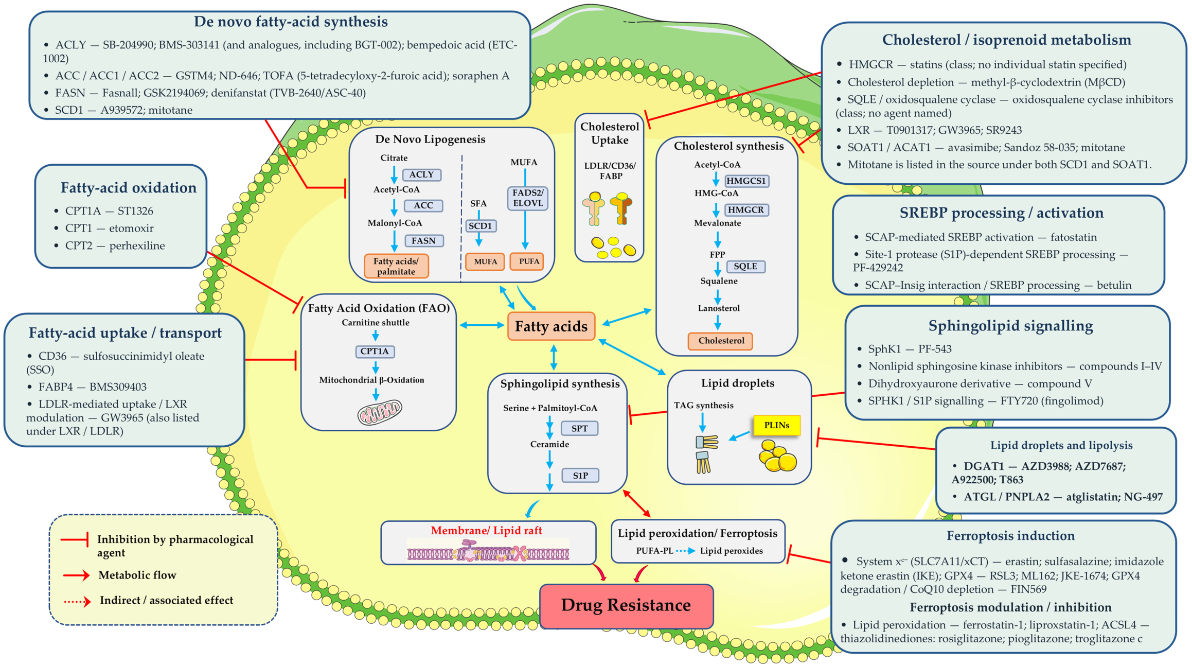
miRNA regulation. Three miRNAs converge on CPT1A, the rate-limiting gatekeeper of mitochondrial β-oxidation, which is the target of the CPT1 inhibitors above. As noted, miR-33 has been reported to repress CPT1A, a rate-limiting enzyme of mitochondrial FAO. However, cancer cells adapt their metabolism to the tumor microenvironment, and this regulation may be offset in resistant cells. In platinum-resistant ovarian cancer cells, for example, de novo lipogenesis and glucose uptake are reduced, whereas fatty-acid uptake and β-oxidation are increased, supporting survival under cisplatin-induced oxidative stress [22]. Whether such reprogramming overrides miR-33-mediated CPT1A repression in resistant tumors has not been tested directly. miR-33b directly targets transforming Growth Factor-β-Activated Kinase 1 (TAK1), thereby indirectly suppressing FASN and CPT1A, reducing lipid turnover through FAO activities and inhibiting NF-κB signaling through the TAK1/FASN/CPT1A/NF-κB axis [162]. miR-328-3p directly targets the 3′-UTR of CPT1A, reducing CPT1A expression and fatty acid β-oxidation (FAO). This regulation disrupts the CPT1A/FAO/stemness axis, thereby suppressing cancer stemness and breast cancer metastasis. In breast cancer, miR-328-3p was downregulated, particularly in patients with metastatic disease, and its expression is negatively correlated with tumor metastasis. Overexpression of miR-328-3p suppressed the invasive capacity of highly metastatic breast cancer cells, whereas its inhibition promoted metastatic behavior. Mechanistically, miR-328-3p directly targets the 3′-UTR of CPT1A, reducing CPT1A expression and FAO. This regulation disrupts the CPT1A/FAO/stemness axis, thereby suppressing cancer stemness and breast cancer metastasis [121]. In gastric cancer cells, miR-145-5p plays a role in metastasis-associated colon cancer 1 antisense RNA 1 (MACC1-AS1) induced stemness and chemoresistance by regulating fatty acid oxidation (FAO). MACC1-AS1 acts as a molecular sponge for miR-145-5p, thereby promoting the expression of FAO-related enzymes, including CPT1. Restoration of miR-145-5p suppresses fatty acid uptake, FAO activity and ATP production while increasing reactive oxygen species (ROS) generation. These metabolic changes reduce cancer stemness and enhance sensitivity to 5-fluorouracil (5-FU) and oxaliplatin. Importantly, CPT1 is a downstream component of this regulatory pathway, not a direct target of miR-145-5p [122].
Pharmacological inhibitors. Mitochondrial FAO, supported by the CPT1-dependent carnitine shuttle, can sustain ATP production and redox homeostasis under metabolic stress. ST1326 is a selective CPT1A inhibitor, targeting the enzyme responsible for the rate-limiting step of long-chain fatty acid transport into mitochondria for FAO. ST1326 inhibited FAO, inducing dose- and time-dependent growth arrest, mitochondrial damage and apoptosis, with particular cytotoxicity against primary acute myeloid leukemia cells [163]. Burkitt’s lymphoma. ST1326 inhibited long- and short-chain FAO, inducing cytotoxicity in Burkitt’s lymphoma cells, lipid accumulation and impaired mitochondrial fatty acid channeling. It also prevented tumor formation in Eµ-myc mice by selectively suppressing c-Myc-overexpressing B cells [164]. Gastrointestinal cancers exhibit increased CPT1B and CPT2 expression following oxaliplatin treatment, with elevated levels associated with poorer chemotherapy outcomes. NFATc3-mediated CPT2 upregulation supports NADPH production and redox homeostasis. Perhexiline inhibits CPT2, increases ROS and promotes apoptosis; it also suppresses GC and CRC cell proliferation. Combined with oxaliplatin, perhexiline reduces tumor growth in xenograft and PDX models [165]. Etomoxir, a prototype CPT1 inhibitor, has off-target effects at high concentrations, including mitochondrial complex I inhibition. As CPT1 may also support cancer-cell proliferation independently of β-oxidation, genetic validation is essential when interpreting pharmacological FAO inhibition [166]. In conclusion, FAO supports cancer-cell survival, proliferation and migration under TME stress, promotes tumor growth and therapy resistance and modulates immune and stromal cell functions. Therefore, targeting FAO may help overcome tumor progression and therapeutic resistance [167]. Tumor cells acquire fatty acids from their microenvironment through lipid transporters, particularly CD36, and lipid-binding proteins, supporting fatty-acid uptake and promoting tumor proliferation, migration and drug resistance [93]. Inhibiting CD36 has been one of the therapeutic strategies for the treatment of cancer and drug resistance. In oral squamous cell carcinoma (OSCC), CD36 inhibition with SSO suppressed tumor-cell proliferation, induced apoptosis and enhanced T-cell responses. In vivo, SSO reduced tumor growth, increased CD4+ and CD8+ T cells and dendritic cells, and decreased myeloid-derived suppressor cells (MDSCs) and Tregs, indicating direct antitumor and immunomodulatory effects [168]. In ovarian cancer, adipocyte-induced FABP4 expression promotes metastasis and carboplatin resistance, while the FABP4 inhibitor BMS309403 reduced tumor burden and improved carboplatin response in preclinical models [169]. These findings identify fatty-acid uptake and intracellular lipid handling as potential therapeutic vulnerabilities, although further validation is needed to establish their clinical utility.
3.5. Targeting Lipid-Droplet Storage and Lipolysis
Intracellular lipid balance depends on the regulation of TG synthesis and breakdown at LDs. Excess fatty acids can be converted into TGs by DGAT1/2 and stored in lipid droplets, protecting tumor cells from lipotoxicity and oxidative stress. DGAT1 and DGAT2 promote TG synthesis and storage, protecting cells from excess fatty acids, whereas ATGL initiates TG breakdown, releasing fatty acids for energy production. Cancer cells can alter this balance to manage metabolic stress and support survival [170].
miRNA regulation. LDs provide a dynamic reservoir of fatty acids that can be mobilized during metabolic stress, so miRNA-mediated changes in enzymes controlling lipid synthesis, storage, or utilization may allow tumor cells to switch between uptake, synthesis, storage, and oxidation according to the demands imposed by therapy. Several cancer-associated miRNAs have been shown to promote LD breakdown and lipolysis. In pancreatic cancer-derived exosomes, miR-16-5p and miR-29a-3p promote adipose atrophy by inhibiting adipogenesis through ERLIN2 and CMPK1, respectively, while miR-29a-3p enhances lipolysis by suppressing MCT1 and increasing ATGL activity. These effects contribute to cancer-associated fat loss and LD mobilization in adipose tissue; their roles in tumor-cell LD turnover and drug resistance remain unestablished [171]. miR-378 increases catecholamine-stimulated lipolysis by elevating the expression of the key lipolytic genes LIPE (HSL), PLIN1, and PNPLA2 (ATGL) in human cancer cachexia, human adipose tissue and differentiated adipocytes [172]. In addition, breast-cancer-derived exosomal miR-204-5p drives lipolysis and white-adipose-tissue browning through a HIF1A-leptin signaling axis [173]. Because LDs and fatty acids serve as metabolic substrates during nutrient stress, miRNA-mediated regulation of autophagy may also intersect with lipid mobilization and energy production, a currently hypothetical connection to LD turnover in PDAC.
Pharmacological inhibitors. In glioblastoma, DGAT1 is upregulated, and its inhibition disrupts LD formation, redirects excess fatty acids toward mitochondrial oxidation, and induces ROS-associated mitochondrial damage and apoptosis, suppressing xenograft growth, which was inhibited by ROS scavenger N-acetyl-cysteine (NAC) [174]. Pharmacological inhibition of DGAT1 using AZD3988, AZD7687, A922500 and T863 suppressed proliferation and cell-cycle progression in DGAT1-high or DGAT1-amplified melanoma cells. DGAT1 inhibition also induced oxidative stress, while combined inhibition of DGAT1 and superoxide dismutase 1 (SOD1) markedly suppressed tumor growth. These findings highlight DGAT1 as a potential metabolic target in melanoma [175]. In prostate cancer models, DGAT1 inhibition reduced LD abundance and suppressed tumor growth in vitro and in vivo, while also impairing cancer-cell migration [176]. Atglistatin (murine ATGL inhibitor) was studied in castration-resistant prostate cancer (CRPC). Atglistatin reduced prostate cancer cell growth and colony formation in a dose-dependent manner. ATGL inhibition also increased neutral lipid accumulation and impaired the growth of Hi-MYC mouse-derived prostate cancer organoids, with stronger effects under low-glucose conditions [177]. NG-497 is a selective, reversible and competitive inhibitor of human ATGL. Unlike Atglistatin, which inhibits murine ATGL, NG-497 inhibits human and rhesus monkey ATGL. In HepG2 hepatocellular carcinoma cells, NG-497 increased triglyceride accumulation and reduced fatty-acid-dependent mitochondrial respiration under galactose conditions. However, its anticancer efficacy and role in drug resistance were not investigated [178]. Overall, these studies highlight the therapeutic potential of targeting lipid storage and mobilization in cancer. However, the effects of these inhibitors vary across tumor types and metabolic contexts, and most evidence remains preclinical. Further studies are needed to establish their selectivity, efficacy in human cancer models and potential to overcome therapeutic resistance.
3.6. Targeting Sphingolipid Signaling, Membrane Organization, and Drug Efflux
Sphingolipid metabolism begins with the condensation of serine and palmitoyl-CoA by serine palmitoyltransferase (SPT) to form 3-keto-dihydrosphingosine, which is subsequently reduced by 3-ketodihydrosphingosine reductase (KDSR) to dihydrosphingosine (sphinganine). Dihydrosphingosine is then acetylated by ceramide synthases (CerS 1-6) to form dihydroceramide, and further desaturated by dihydroceramide desaturase (DES) to yield ceramide. Ceramide serves as a central hub that can be glycosylated to form glucosylceramides or galactosylceramides, acquire a phosphocholine headgroup via sphingomyelin synthase (SMS) to produce sphingomyelin, or be phosphorylated by ceramide kinase (CK) to form ceramide 1-phosphate (C1P). Alternatively, ceramidases (CDase) break down ceramide to generate sphingosine, which can either be recycled back into ceramide by CerS or phosphorylated by sphingosine kinases (SPHK1/2) to form sphingosine-1-phosphate (S1P) [36,70]. The sphingolipid rheostat dictates cell fate by balancing a pro-apoptotic ceramide/C1P branch, which drives stress responses, cell cycle arrest, and inflammatory signaling, against a pro-survival S1P axis. Extracellular S1P engages specific G protein-coupled receptors (S1PR1-5) to activate major survival cascades, including the PI3K/AKT and MAPK/ERK pathways, thereby promoting cell proliferation, anti-apoptotic signals, and tumor angiogenesis [36,70].
miRNA regulation. Sphingolipid metabolism can influence cancer-cell responses to chemotherapy through the balance between pro-apoptotic ceramide and sphingosine-1-phosphate (S1P). In colorectal cancer cells, miR-101 downregulates sphingosine kinase 1 (SPHK1), increases ceramide levels, and enhances sensitivity to paclitaxel and doxorubicin. In HCT-116 xenograft models, miR-101 overexpression inhibited tumor growth and enhanced paclitaxel activity in vivo [179]. Membrane lipid composition determines the localization and function of drug-efflux transporters such as ABCC1 [76]; miRNAs regulating drug-efflux pumps can link membrane organization to chemoresistance.
Pharmacological inhibitors. The sphingolipid axis, initiated by serine palmitoyltransferase (SPT), balances pro-apoptotic ceramide and pro-survival S1P. SphK1, which generates S1P, is inhibited by PF-543, a sphingosine-competitive inhibitor that depletes cellular S1P by up to 10-fold [180]. SphK1 activity has been linked to chemoresistance (for example, olaparib resistance in ovarian cancer via NF-κB/NRF2/ferroptosis signaling) when using PF-543 [181]. Novel nonlipid sphingosine kinase (SK) inhibitors suppressed proliferation and induced apoptosis in bladder and breast cancer cells, including multidrug-resistant cells overexpressing MRP1 or P-glycoprotein. The aurone derivative compound V also reduced tumor growth by more than 50% in a syngeneic mouse mammary adenocarcinoma model. These findings support SK inhibition as a potential strategy against drug-resistant cancer, although reversal of resistance to specific chemotherapeutic agents was not demonstrated [182]. FTY720 (fingolimod), an FDA-approved multiple sclerosis drug, inhibits SPHK1 activity and promotes its degradation, disrupting S1P-mediated survival signaling. In ovarian cancer models, FTY720 reduced proliferation, invasion and angiogenesis while increasing apoptosis. Combined with SPHK1 knockdown, it further suppressed tumor cell growth and invasion in chemotherapy-sensitive and -resistant models. FTY720 also reduced tumor growth in cell-derived and patient-derived xenografts [183]. PF-543 also sensitized TRAIL-resistant colorectal cancer cells to TRAIL-induced apoptosis, in association with SPHK1/S1PR1/STAT3 signaling and activation of the mitochondrial apoptosis pathway [184]. FTY720 inhibited SphK1 in hormone-refractory prostate cancer cells and increased radiosensitivity in mouse prostate tumor models, independently of S1P receptors [185]. Together, these studies support SphK1 inhibition as a strategy to overcome treatment resistance, although the evidence remains preclinical.
3.7. Targeting SREBP-1/SREBP-2 and the SREBP Pathway
SREBPs are master transcription factors of lipid biosynthesis. SREBP-1 mainly drives fatty-acid and triglyceride synthesis (including FASN), whereas SREBP-2 mainly controls cholesterol synthesis and uptake (including HMGCR). SREBPs are synthesized as ER-bound precursors that bind SCAP, travel with it to the Golgi, and are cleaved by Site-1 and Site-2 proteases, releasing the active form that enters the nucleus. Because this pathway coordinates the enzymes discussed in Section 2.7, it can be targeted at the level of transcription (miRNAs) or at the level of SREBP processing and trafficking (small molecules), as described below.
miRNA regulation. Several miRNAs directly restrain the SREBP axis. miR-185 and miR-342 target the 3′-UTRs of both SREBP-1 and SREBP-2, suppressing their expression and downstream genes, including FASN and HMGCR. This reduces de novo lipogenesis and cholesterogenesis and inhibits tumorigenicity in prostate cancer cells [186]. In parallel, the miR-29 family (miR-29a/b/c) forms a negative-feedback loop with SCAP/SREBP-1: SREBP-1 transcriptionally induces miR-29, which in turn targets the 3′-UTRs of SCAP and SREBP-1, limiting further pathway activation and lipid synthesis [187]. These findings demonstrate how post-transcriptional regulation can restrict SREBP-dependent metabolic programs that support cancer-cell growth, although their direct contribution to therapy resistance requires further investigation.
Pharmacological inhibitors. Small-molecule inhibitors target distinct steps of SREBP activation. Fatostatin, a synthetic diarylthiazole, inhibits SREBP activation by blocking SCAP-mediated ER-to-Golgi translocation. This reduces the expression of lipogenic genes involved in fatty acid and cholesterol biosynthesis [188]. PF-429242, a competitive inhibitor of site-1 protease (S1P), suppresses SREBP processing and reduces the expression of SREBP target genes. It inhibited cholesterol synthesis in hepatoma cells and reduced hepatic cholesterol and fatty acid synthesis, suppressing proteolytic processing of SREBPs in the Golgi [189]. Betulin, a naturally occurring triterpene, inhibits SREBP maturation by promoting the interaction between SCAP and INSIG, thereby suppressing cholesterol and fatty acid biosynthesis. In hepatoma cells, betulin reduced SREBP activity and downregulated lipogenic genes, including FASN, ACC and HMGCR. It also reduced cellular cholesterol and neutral lipid levels. Betulin promoted the interaction between SCAP and INSIG, retaining SREBP precursors in the endoplasmic reticulum and reducing their maturation and nuclear translocation [190]. Together, these compounds provide complementary pharmacological approaches to suppress SREBP-dependent lipid synthesis, although their therapeutic efficacy against cancer drug resistance remains to be established.
3.8. Targeting Lipid Peroxidation and Ferroptosis
Lipid peroxidation is a key feature and a necessary step in ferroptosis, an iron-dependent form of regulated cell death that differs from apoptosis, necrosis and autophagy [191,192,193]. Ferroptosis occurs when oxidised lipids, called lipid peroxides, build up in cell membranes. These peroxides form through a chain reaction involving free radicals. Iron-derived radicals remove a hydrogen atom from a polyunsaturated fatty acid (PUFA), forming a lipid radical (L•). This radical reacts with oxygen to form a lipid peroxyl radical (LOO•), which attacks nearby lipids and produces lipid hydroperoxides (LOOH), continuing the chain reaction until antioxidants such as vitamin E stop it [192]. Lipids containing PUFAs especially arachidonic acid (AA) and adrenic acid (AdA), are particularly vulnerable to oxidation because of their multiple double bonds. Lipid peroxidation can occur through self-propagating radical reactions or with the help of enzymes such as 15-lipoxygenase (15-LOX) and cytochrome P450 oxidoreductase (POR) [192]. Peroxidised PUFA-PLs disrupt membrane structure, while their breakdown products make the membrane leaky and cause cell rupture [115]. ACSL4 links lipid metabolism to ferroptosis by promoting the incorporation of PUFAs into membrane phospholipids. The ACSL family includes five members (ACSL1, 3, 4, 5 and 6) that use ATP to attach coenzyme A to long-chain fatty acids, preparing them for energy production or lipid synthesis. Each member has different substrate preferences and functions. ACSL4 mainly acts on PUFAs, while ACSL5 and ACSL6 also contribute to fatty-acid metabolism [192]. ACSL4 is the isoform with a direct role in ferroptosis [107,192]. It converts arachidonic acid (AA) and adrenic acid (AdA) into AA-CoA and AdA-CoA, which LPCAT3 esterifies into phosphatidylethanolamines (PE-AA, PE-AdA), the principal peroxidation substrates [192]. Loss of ACSL4 depletes long ω6 PUFA from membranes and confers ferroptosis resistance, so ACSL4 expression and activity indicate ferroptosis susceptibility [107,192]. As described in Section 2.7, lipid synthesis and remodeling determine membrane PUFA levels, while SREBP-1/SCD1-driven MUFA production can protect against ferroptosis [17]. Although ACSL4 is highly expressed in several cancers, including liver, colorectal, breast and prostate cancers, some cancer stem cells downregulate it to reduce lipid peroxidation and resist ferroptosis [193]. However, ACSL4 dependence varies with the trigger; it is generally lower with erastin or cystine depletion than with GPX4 inhibitors. LPCAT3 and, in p53-mediated ferroptosis, ALOX12 can bypass it [193]. The main defence against ferroptosis is glutathione peroxidase 4 (GPX4), which uses glutathione (GSH) to convert toxic phospholipid hydroperoxides into non-toxic alcohols [194,195]. GSH synthesis depends on cysteine supplied by system xc−, a transporter formed by SLC7A11 (xCT) and SLC3A2. This system exchanges extracellular cystine for intracellular glutamate. Inside the cell, cystine is reduced to cysteine, which combines with glutamate and glycine to form GSH [191,192]. SLC7A11 therefore helps maintain antioxidant defences and is also regulated by miRNAs (miR-148a-3p, miR-5096; Table 1). Erastin and sulfasalazine inhibit system xc−, reducing GSH levels, whereas RSL3 directly inhibits GPX4 [191,194]. This pathway is relevant to drug resistance because mesenchymal, therapy-resistant cancer cells and drug-tolerant persister cells can depend on GPX4 for survival. Inhibiting GPX4 can kill these cancer cells and prevent tumour relapse in mouse models [104,105]. Ferroptosis can be therapeutically targeted in cancer and drug-resistant cancer cells. The key therapeutic targets discussed below are SLC7A11 (system xc−), GPX4, and ACSL4, which can be modulated by miRNAs or pharmacological agents.
miRNA regulation. In sorafenib-resistant hepatocellular carcinoma (HCC), ETS1-driven miR-23a-3p directly targets ACSL4, suppressing ferroptosis and contributing to treatment resistance; its inhibition restores ACSL4 expression and sorafenib sensitivity [106]. In colorectal cancer (CRC), miR-148a-3p targets SLC7A11, promoting ferroptosis alongside increased lipid peroxidation, reduced GPX4 expression and altered iron-handling proteins [196]. Exosomal miR-148a-3p directly targets SLC7A11, promoting M1 polarization and ferroptosis-related characteristics in recipient macrophages [197]. In liver cancer, lncPVT1 sponges miR-214-3p, sustaining GPX4 expression and limiting ferroptosis; ketamine-induced disruption of this axis promotes ferroptosis in preclinical models [198]. In breast cancer, miR-5096 directly targets SLC7A11, reducing glutathione and increasing lipid ROS and intracellular iron, thereby promoting ferroptosis [199]. Conversely, miR-424-5p directly targets ACSL4 in ovarian cancer, reducing ferroptosis induced by erastin or RSL3; its inhibition or ACSL4 restoration increases ferroptotic sensitivity [200].
Pharmacological inhibitors. Ferroptosis inhibitors act at three levels.
- (i)
- System xc− inhibitors. Erastin and sulfasalazine inhibit cystine uptake, depleting GSH and compromising GPX4-mediated antioxidant defense [191,201]. Erastin induces ferroptosis in fibrosarcoma cells [191], while system xCT inhibition induces ferroptosis across cancer models [201]. Sulfasalazine suppresses triple-negative breast tumor growth in mice [202]. In HCC, NRF2 inhibition enhances erastin anticancer effects in vitro and in xenografts [108]. The metabolically stable erastin analog imidazole ketone erastin (IKE) slows tumor growth in a diffuse large B-cell lymphoma (DLBCL) xenograft [203] and reduces biliary tract cancer cell viability, with sensitivity correlating with CD71 and SLC7A11 expression [113].
- (ii)
- Direct GPX4 inhibitors. RSL3 and ML162 are covalent GPX4 inhibitors that induce lipid peroxidation [194,204]. RSL3 suppresses xenograft growth of RAS-transformed tumor cells [194], induces ferroptosis in colorectal cancer cells, an effect reversed by GPX4 overexpression [205], and targets GPX4-dependent clear-cell renal cell carcinoma [206]. In cholangiocarcinoma, RSL3 reduces viability and induces lipid peroxidation in intrahepatic cell lines, whereas extrahepatic lines show greater resistance and higher GPX4 expression [112]. ML210 and JKE-1674 are masked prodrugs converted intracellularly into electrophiles that covalently modify GPX4; ML210 showed activity comparable to RSL3 and ML162 across 821 cancer cell lines, including GPX4-dependent LOX-IMVI melanoma cells [204]. FIN56 promotes GPX4 degradation and depletes coenzyme Q10 [207,208], inducing ferroptosis in bladder cancer cells [208] and glioblastoma cells, where it also suppresses tumor growth in vivo [114]. However, the poor pharmacokinetics of current covalent GPX4 inhibitors limit their in vivo application [204].
- (iii)
- Lipid Peroxidation Inhibitors and upstream modulators. Ferrostatin-1, identified as a suppressor of erastin-induced cell death, and liproxstatin-1 inhibit lipid-peroxidation chain reactions by trapping radicals; iron chelators such as deferoxamine also suppress ferroptosis [191,209]. These agents help confirm ferroptotic cell death experimentally, including in biliary tract cancer cells treated with IKE or RSL3 [113]. Thiazolidinediones, including rosiglitazone, pioglitazone and troglitazone, inhibit ACSL4 and protect against ferroptosis [107,192]. Inhibiting LPCAT3, 15-LOX, or POR may also limit the formation or oxidation of peroxidizable phospholipids [192]. ACSL4 expression predicts ferroptosis sensitivity in basal-like breast cancer cells [107], while experimental work in melanoma supports ACSL4-dependent ferroptosis induced by IFNγ and fatty acids and demonstrates inhibition by rosiglitazone [114]. The relevant information is also summarized in Supplementary Table S1 and Figure 4. These findings highlight the therapeutic potential of targeting system xc−, GPX4 and the ACSL4-PUFA-phospholipid pathway. GPX4 dependence in mesenchymal, therapy-resistant and drug-tolerant cancer cells provides a rationale for targeting ferroptosis to overcome resistance [104,105,210]. Preclinical studies support this approach across breast, colorectal, liver, biliary tract, kidney, bladder and brain cancers, as well as melanoma and lymphoma.Table 1. MicroRNAs regulating lipid-metabolic pathways and their links to cancer drug resistance. Evidence class (miRNA → target): Direct, regulation of the listed target shown experimentally (e.g., 3′-UTR reporter assay); Indirect, regulation through an intermediate (e.g., lncRNA sponge, signaling axis); Proposed, lipid-pathway regulation supported by expression or functional data, but direct targeting not established. Links to drug resistance are given in the “Resistance phenotype/drug” column. Classes match Figure 3 (D, I, P). miRNAs are listed in ascending order of the lowest reference number cited.
3.9. Exploiting Ferroptosis to Overcome Therapy Resistance
Because ferroptosis kills through iron-dependent lipid peroxidation rather than through the canonical apoptotic machinery, it can bypass resistance to apoptosis-based therapies. Resistant tumors commonly evade it by relying on GPX4 and system xc− to neutralize lipid peroxides, and this dependence is also a vulnerability. Targeting the GPX4-regulated pathway, or inhibiting NRF2-driven antioxidant defense, can sensitize resistant tumors to ferroptosis inducers such as erastin and RSL3 [104,105,108,110,191,194]. A recent review summarizes how ferroptosis is linked to chemoresistance and how inducing it can reverse resistance [210]. Preclinical studies are summarized below by the node that is targeted.
In cisplatin-resistant head and neck cancer cells, pharmacological inhibition of the cystine/glutamate antiporter with sulfasalazine, and its genetic silencing, induced ferroptosis and increased cisplatin sensitivity in vitro and in mouse xenografts [211]. Erastin, sulfasalazine and the more stable analog IKE act at this node and reduce tumor growth in several models [201,202,203]. In biliary tract cancer cells, the efficacy of IKE and RSL3 correlates with CD71 and SLC7A11 expression, which suggests a possible biomarker for patient selection [113]. GPX4 inhibition. In drug-tolerant persister cells across several cancer models, GPX4 inhibition with RSL3 or ML210 selectively induced ferroptosis and prevented tumor relapse in mice [105]. In iCCA, the PAX8-AS1/GPX4 axis drives resistance to gemcitabine and cisplatin, and the GPX4 inhibitor JKE-1674 combined with these drugs showed synergy in patient-derived organoids and improved antitumor effects in xenograft and orthotopic models of resistant cells [111]. Masked prodrugs such as ML210 and JKE-1674 are more suitable for in vivo work than RSL3, which has poor pharmacokinetics [204]. In bladder cancer cells, FIN56-induced ferroptosis acts synergistically with mTOR inhibition [207,208]. Targeting NRF2 and related antioxidant buffers. In head and neck cancer, inhibiting NRF2 or silencing p62 sensitized resistant cells to RSL3, and trigonelline sensitized cisplatin-resistant cells to RSL3 in a xenograft model [110]. NRF2 inhibition also enhances the anticancer effects of erastin in HCC [108], and inhibiting MT-1G enhances sorafenib activity [109]. Restoring the PUFA-phospholipid substrate and miRNA-based strategies. Inhibiting miR-23a-3p restores ACSL4 and sensitizes sorafenib-resistant HCC cells [106]. miR-148a-3p targets SLC7A11 in colorectal cancer cells and promotes ferroptosis, which supports restoring this miRNA as a possible strategy [196]. HDAC2/EZH2-mediated miR-148a suppression upregulated PDK1, promoting glycolysis, EMT, invasion and Adriamycin resistance in breast cancer cells [212]. The strength of the miRNA evidence varies, and many links remain proposed or indirect. Combination with immunotherapy. Because IFNγ-driven tumor ferroptosis depends on ACSL4 [114], approaches that preserve ferroptosis sensitivity may complement T-cell-based therapies, although this requires direct testing in resistant tumors.
Limitations. The evidence is largely preclinical. Current covalent GPX4 inhibitors have poor pharmacokinetics [204], ACSL4 dependence varies with the ferroptosis trigger [192], and sensitivity differs between tumor types and even between intrahepatic and extrahepatic biliary lines [112]. Reliable biomarkers of ferroptosis sensitivity, such as ACSL4, SLC7A11, CD71 and GPX4 levels, are needed to identify patients who may benefit [107,113]. But, recently, a reducing reagent-resistant TfR1 dimer (3R dimer) was identified as a novel marker of lipid peroxidation, offering a potential approach for detecting ferroptosis in cells, tissues and circulating extracellular vesicles [213].
3.10. Compensatory Rewiring, Intercellular miRNA Signaling, and Combination Strategies
3.10.1. Compensatory Rewiring Limits Single-Pathway Inhibition
A recurring lesson from Section 3.2, Section 3.3, Section 3.4, Section 3.5, Section 3.6, Section 3.7, Section 3.8 and Section 3.9 is that the lipid hub adapts when one node is blocked. Statin monotherapy is limited by compensatory LDLR-mediated cholesterol uptake [154,155]; ACC inhibition (e.g., ND-646) is buffered by compensatory fatty acid oxidation [137]; DGAT1 inhibition diverts excess fatty acids into mitochondrial oxidation [174]; and the anticancer effects of CPT1 inhibition by etomoxir may not depend solely on β-oxidation [166]. Lipid-targeted therapy therefore risks selecting for cells that reroute flux through an unblocked node. miRNAs are likely part of this adaptive layer. The relationship is probably reciprocal, because therapy-induced metabolic changes may in turn alter miRNA expression, which may help explain how cancer cells maintain survival despite prolonged exposure to anticancer drugs. miRNAs also contribute to multidrug resistance more broadly through drug transporters, DNA repair, apoptosis, cell cycle, autophagy, and metabolism, while influencing cellular responses within genetically heterogeneous tumors [34,35]. Together, these observations provide a strong rationale for combination strategies that address each node together with its compensatory routes. Because inhibitors and miRNAs often converge on the same targets, the strategies are discussed below by metabolic node (Table 1, Supplementary Table S1).
3.10.2. Combination Strategies Targeting Lipid Metabolism and Drug Resistance
In adrenocortical carcinoma, rosuvastatin potentiated the effects of mitotane by reducing cell viability, inducing apoptosis, increasing intracellular free cholesterol levels, decreasing the expression of HMGCR and ABCA1, genes involved in cholesterol metabolism, and inhibiting steroidogenesis [214]. PCSK9 inhibition can enhance anti-PD-1 immunotherapy by preventing MHC-I degradation, increasing its cell-surface expression and promoting intratumoral cytotoxic T-cell infiltration. Separately, SOAT1 (ACAT1)-mediated cholesterol esterification buffers excess free cholesterol, protecting tumor cells from cholesterol-induced toxicity [215]. In diethylnitrosamine-induced HCC, high-fat diet (HFD) feeding increased the expression of SOAT1, which promotes fatty-acid storage, and CPT1A, which mediates FAO. These pathways formed a compensatory feedback loop: SOAT1 inhibition enhanced CPT1A-mediated mitochondrial fatty-acid oxidation, whereas CPT1A inhibition promoted SOAT1-dependent LD formation. Accordingly, combined SOAT1 and CPT1A inhibition using avasimibe and etomoxir disrupted this metabolic compensation, producing synergistic anticancer effects in vitro and in vivo [216]. In lipid synthesis and desaturation, ACLY knockdown using shRNA-ACLY increased sorafenib sensitivity in resistant HCC cells, supporting ACLY as a potential target for overcoming resistance [217]. In ccRCC, the SCD1 inhibitor A939572 synergized with the mTOR inhibitor temsirolimus to inhibit tumor growth in vitro and in vivo [144]. In ovarian cancer, adipocyte-induced FABP4 promoted metastasis and carboplatin resistance, whereas FABP4 inhibition with BMS309403 reduced tumor burden and increased carboplatin sensitivity in preclinical models [169]. Sphingolipid signaling provides another therapeutic opportunity. In TRAIL-resistant colorectal cancer cells, the SPHK1 inhibitor PF-543 enhanced TRAIL-induced apoptosis and reduced stem-like properties [184]. In drug-resistant glioblastoma models, pharmacological inhibition of SPHK1 with PF-543 in combination with temozolomide (and further enhanced by vemurafenib-mediated ATF4 suppression) significantly improved therapeutic efficacy, supporting further exploration of sphingolipid pathway targeting in combination therapy [218]. At the membrane level, molecular dynamics simulations suggest that membrane lipids enter the P-gp/ABCB1 binding cavity and support tariquidar binding, potentially enhancing P-gp inhibition, increasing chemotherapeutic drug retention, and restoring drug cytotoxicity in resistant cells [219]. In multidrug-resistant breast cancer models, an endogenous magnetic LD-mediated sonodynamic strategy increased doxorubicin sensitivity, disrupted lysosomal sequestration and suppressed ATP-dependent drug efflux [220]. Separately, phospholipid-conjugated porphyrins and nanoliposomal formulations, including porphyrin-lipid nanovesicles, bypassed P-gp- and ABCG2-mediated efflux, overcoming photodynamic therapy (PDT) resistance in breast cancer cell models. These porphyrin-lipid nanovesicles evade the P-gp and ABCG2 pumps implicated in the development of resistance to PDT, restoring PDT efficacy [221]. Ferroptosis provides a further link between lipid metabolism and treatment response. In sorafenib-resistant HCC, inhibition of the ETS1/miR-23a-3p axis restored ACSL4 expression and ferroptotic sensitivity, supporting miR-23a-3p inhibition as a potential strategy to resensitize cells to sorafenib [106]. GPX4 inhibition with RSL3 or ML210 selectively induced ferroptosis in drug-tolerant persister cells across several cancer models and prevented tumor relapse in mice [105]. System xc− inhibitors, including erastin, sulfasalazine and imidazole ketone erastin (IKE) [108,184,201,208], provide complementary means of inducing ferroptosis by limiting cystine uptake and glutathione-dependent antioxidant defense; however, their combination effects must be attributed to the specific cancer model and study in which they were tested.
miRNA-based modulation: opportunities and liabilities. Despite promising preclinical findings, miRNA therapeutics face major translational barriers, particularly inefficient delivery, toxicity, immune activation and limited target specificity, because individual miRNAs can regulate multiple transcripts. These properties also make functional validation complex and time-consuming. To date, no miRNA-based therapeutic has received approval from the US Food and Drug Administration (FDA), and none has progressed to Phase III clinical trials [222,223]. MRX34, an miR-34a mimic and the first miRNA-based cancer therapeutic to enter a Phase I trial in 2013, MRX34, a liposomal miR-34a mimic, was tested in 85 patients with refractory solid tumors in a phase 1 trial (NCT01829971). It delivered miR-34a to tumors and modulated target genes, with three partial responses and 16 cases of stable disease lasting at least four cycles. The trial was closed early after severe immune-mediated adverse events, including four deaths [222,224]. TargomiRs, EGFR-targeted minicells loaded with a miR-16 mimic, showed an acceptable safety profile in mesothelioma but only one partial response among 22 assessable patients, with cardiac events among the dose-limiting toxicities [225]. Other programs have also encountered setbacks, including the miR-29b mimic program for fibrosis, an anti-miR-21 program for Alport syndrome, and anti-miR-122 approaches for hepatitis C, which were discontinued or terminated during clinical development [222].
Nevertheless, newer programs with improved delivery platforms offer encouraging signals. INT-1B3 is a lipid nanoparticle-formulated mimic of the tumor suppressor miR-193a-3p. In cell lines from triple-negative breast cancer, NSCLC, melanoma, colon cancer and HCC, it raised PTEN pathway activity, dampened oncogenic pathways, suppressed proliferation, cell cycle progression and migration and induced apoptosis, senescence and DNA damage despite differing genetic backgrounds. It was delivered safely and efficiently to tumors in orthotopic mouse models, and a phase 1/1b first-in-human trial (ClinicalTrials.gov identifier: NCT04675996) was designed to assess safety, pharmacokinetics and preliminary activity in advanced solid tumors [226]. The same company is developing a second mimic, INT-5A2, for HCC and glioblastoma. TTX-MC138 is an LNA-based inhibitor of miR-10b, an oncogenic driver in metastatic breast cancer and other tumors including glioblastoma, conjugated to dextran-coated iron oxide nanoparticles (ClinicalTrials.gov identifier: NCT01849952). It lowered miR-10b expression in breast cancer models, and the earlier inhibitor RGLS5579 extended survival with temozolomide in an orthotopic glioblastoma model. Preclinical testing is also reported in glioblastoma, pancreatic cancer, SCLC and osteosarcoma. In its phase 1a trial, 16 patients with advanced cancer had no dose-limiting toxicities, and the recommended phase 2 dose was 4.8 mg/kg. The company reported stable disease lasting six months in 9 of 14 evaluable patients, including a marked fall in thyroglobulin in one patient with metastatic thyroid cancer, and a phase 2a trial in ctDNA-positive colorectal cancer began in May 2026 [226].
These experiences highlight the need for improved delivery systems, rigorous off-target and immune-toxicity assessment, and careful patient selection before miRNA-based strategies can be translated into effective cancer treatments.
4. Discussion
Lipid metabolism is a highly interconnected network that undergoes extensive remodeling during malignant transformation and therapeutic adaptation. Lipids are not only a source of membrane components or stored energy. They also take part in signaling, membrane organization, mitochondrial metabolism, redox regulation, and communication between tumor cells and the surrounding microenvironment. Under therapeutic pressure, cancer cells can exploit this flexibility to maintain cellular integrity, sustain energy production, activate survival pathways, and reduce their sensitivity to anticancer agents. Metabolic plasticity is a defining feature of lipid-associated drug resistance, enabling cancer cells to maintain lipid homeostasis by rerouting metabolic flux when a particular pathway is inhibited. For example, statin treatment can trigger compensatory LDLR-mediated cholesterol uptake [154,155], ACC inhibition can increase reliance on fatty acid oxidation [137], and DGAT1 inhibition can redirect excess fatty acids towards mitochondrial oxidation [174]. Furthermore, the anticancer effects of CPT1 inhibition may involve mechanisms beyond the suppression of β-oxidation [166]. SREBP-1 contributes to this adaptive capacity by coordinating transcriptional programs that couple glucose, glutamine and lipid metabolism under therapeutic stress. In NSCLC, SREBP-1 increases glucose uptake and sustains tumor growth during lysosome inhibition with chloroquine; blocking glucose transport overcomes this resistance by inducing mitochondrial damage and oxidative stress [102]. In glioblastoma, resistance to the lysosome inhibitor pimozide involves SREBP-1-driven glutamine consumption and lipogenesis, with ammonia generated from glutamine metabolism further intensifying SREBP-1 activation [103]. Critically, it is often the sustained activity of SREBP-1 during therapy, rather than its baseline expression, that distinguishes resistant from sensitive cells. In BRAF-mutant melanoma, vemurafenib reduced SREBP-1 processing and de novo lipogenesis in sensitive cells but not in resistant cells [81,91]; a parallel pattern occurs in gefitinib-resistant EGFR-mutant lung cancer [98]. Beyond simple upregulation, SREBP-1 enables plasticity through membrane remodeling and redox adaptation. Changes in fatty-acid synthesis and desaturation alter membrane lipid saturation, thereby modifying susceptibility to oxidative damage and chemotherapeutics [97]. In both BRAF- and EGFR-inhibitor-resistant models, SREBP-1 inhibition shifts membrane phospholipids towards higher polyunsaturation and restores drug sensitivity [81,98]. Cholesterol handling provides an additional layer of plasticity: caspase-3-mediated SREBP2 activation promotes cholesterol biosynthesis and cancer-stem-cell properties in hepatocellular carcinoma, while the SREBF2-STARD4 axis specifically increases mitochondrial cholesterol accumulation to limit cytochrome c release [99,100]. Concurrently, SREBP-1-dependent monounsaturated fatty-acid synthesis via SCD1 reduces the availability of oxidation-sensitive polyunsaturated species, conferring ferroptosis resistance in cells with hyperactive PI3K-AKT-mTOR signaling [80]. These examples illustrate that SREBP-regulated programs support metabolic plasticity not merely by increasing lipid production, but by dynamically adjusting membrane composition, redox balance and subcellular lipid distribution according to the nature of the therapeutic stress. Cancer cells frequently acquire drug resistance by actively suppressing ferroptosis. Parallel GPX4-independent antioxidant systems, including the FSP1-CoQ10-NAD(P)H pathway [227,228], the mitochondrial DHODH-CoQ pathway [229], and the GCH1-BH4 pathway [230], further allow cells to compensate when the canonical cystine-GSH-GPX4 axis is impaired, illustrating how coordinated remodeling of lipid substrates and redox capacity contributes to ferroptosis resistance under therapeutic stress. Cancer cells can also limit ferroptosis by restricting the labile iron pool (LIP). Upregulation of ferritin (FTH1/FTL) sequesters excess iron and thereby reduces the availability of redox-active Fe2+ required for lipid peroxidation, contributing to ferroptosis resistance [231].
As discussed in Section 3.10.2, several combination approaches have successfully disrupted the compensatory metabolic pathways that sustain therapy-resistant states. Dual inhibition of SOAT1 and CPT1A with avasimibe and etomoxir broke the feedback loop between cholesterol esterification and fatty-acid oxidation, producing synergistic anticancer effects in vitro and in vivo [216]. Concurrent SCD1 and mTOR inhibition with A939572 and temsirolimus suppressed tumor growth in ccRcc models [144], while FABP4 inhibition with BMS309403 restored carboplatin sensitivity and reduced metastatic burden in ovarian cancer [169]. ACLY knockdown increased sorafenib sensitivity in resistant HCC cells [217], PF-543-mediated SPHK1 inhibition induces cytotoxicity and programmed necrosis in colorectal cancer cells, suppresses HCT-116 xenograft growth, and enhances TRAIL- and temozolomide-based therapies in resistant colorectal cancer and glioblastoma, respectively [184,211,232]. Ferroptosis-inducing strategies, including GPX4 inhibition or restoration of ACSL4 expression, further eliminated drug-tolerant persister cells and re-sensitized resistant tumors [105,106].
Collectively, these findings illustrate that simultaneous targeting of interdependent lipid pathways prevents cancer cells from simply rerouting metabolic flux through alternative routes. Consequently, the identification of reliable predictive biomarkers that can report on these compensatory states will be essential for selecting patients most likely to benefit from combination strategies and for advancing ferroptosis- and lipid-targeted therapies toward clinical application.
5. Conclusions and Future Perspectives
Collectively, the evidence reviewed here establishes lipid metabolic reprogramming as a central contributor to cancer drug resistance that extends well beyond bioenergetic adaptation. Cancer cells rewire fatty acid synthesis, uptake, oxidation, and storage while remodeling phospholipid, sphingolipid, and cholesterol pools, forming an interconnected network that sustains survival signaling, preserves membrane integrity, and limits intracellular drug accumulation [32]. Because microRNAs sit upstream of many of these pathways, miRNA-lipid crosstalk emerges as a comparatively underexplored regulatory axis linking post-transcriptional control to metabolic resistance [106,116,121,122]. Clinical translation nevertheless remains limited. Lipid-targeting agents include FASN inhibitors [140,141,142,143], LXR modulators [156,157], CPT1 inhibitors [163,164,166,231], and cholesterol-lowering drugs [154,155], SREBP pathway inhibitors [98,186,187,188,189,190]. Ferroptosis pathways [106,196,198,199,200] are constrained by toxicity, compensatory rewiring, and lack of tumor selectivity [32,59], and lipid dependency varies between and within tumor types [126,127,128]. Single-node inhibition is therefore unlikely to overcome a redundant resistance program, which argues for rational combinations with chemotherapy, targeted agents, or immunotherapy [135,140,144,169,214,215,216,217]. Priorities going forward include integrating lipidomic, transcriptomic, and miRNA profiling to identify lipid-dependent tumors and actionable vulnerabilities [21,29,30,120]; targeting the lipid supply provided by adipocytes, macrophages, and neutrophils in the TME [46,55,168,169]; and developing miRNA mimics and inhibitors of lipid regulators [116,117,118,129,132], a promising but early-stage avenue that will require improved delivery, stability, and tumor specificity [222,223,224,225,226]. A further direction is the role of lipid metabolism in immune function, where lipid reprogramming in tumor cells may deplete or alter the lipids available to infiltrating immune cells, promote CD8+ T-cell dysfunction and exhaustion, and shape immune checkpoint expression, thereby linking chemoresistance to immune escape [92,114,215].
6. Review Methodology and Literature Search Strategy
A literature search was conducted primarily using PubMed, with additional searches in Scopus, Web of Science, and Google Scholar. Search terms included “lipid metabolic reprogramming,” “lipid metabolism and cancer drug resistance,” “fatty acid metabolism in cancer,” “cholesterol metabolism in cancer,” and “multidrug resistance.” Studies published between 2000 and 2026 were considered, prioritizing recent research while including seminal publications where relevant. Original research and review articles were selected based on their relevance to lipid synthesis, uptake, storage, oxidation, and membrane lipid remodeling, particularly their roles in cancer progression and resistance to chemotherapy, targeted therapy, and immunotherapy. Studies providing mechanistic, experimental, or clinically relevant evidence were prioritized, while publications outside the scope of the review were excluded. The findings were critically evaluated and synthesized thematically to examine the mechanisms linking lipid metabolic reprogramming to cancer drug resistance.
Supplementary Materials
The following supporting information can be downloaded at: https://www.mdpi.com/article/10.3390/ijms272210316/s1.
Author Contributions
S.N.C., V.E. and C.R. designed the overall concept and outline of the manuscript; S.N.C. wrote the manuscript; V.E. and C.R. edited and thoroughly reviewed the manuscript. All authors have read and agreed to the published version of the manuscript.
Funding
This work was supported by the Italian Association for Cancer Research (AIRC, IG 23117).
Institutional Review Board Statement
Not applicable (no human/animal data in a review).
Informed Consent Statement
Not applicable.
Data Availability Statement
No new data were created or analyzed in this study. Data sharing is not applicable to this article.
Conflicts of Interest
The authors declare no conflict of interest.
Abbreviations
ABCA1—ATP-binding cassette subfamily A member 1; ABCA2—ATP-binding cassette subfamily A member 2; ABCC1—ATP-binding cassette subfamily C member 1; ACAA2—Acetyl-CoA acyltransferase 2; ACACA—Acetyl-CoA carboxylase alpha (ACC1); ACACB—Acetyl-CoA carboxylase beta (ACC2); ACM—Adipocyte-conditioned medium; ACAT—Acetyl-CoA acetyltransferase/acyl-CoA:cholesterol acyltransferase; ACLY—ATP citrate lyase; ACSL—Acyl-CoA synthetase long-chain family; ACSL1—Acyl-CoA synthetase long-chain family member 1; ACSL4—Acyl-CoA synthetase long-chain family member 4; ADAM10—A disintegrin and metalloproteinase 10; AGPAT—1-Acylglycerol-3-phosphate O-acyltransferase; AGPAT1—1-Acylglycerol-3-phosphate O-acyltransferase 1; AKT—Protein kinase B; AMPK—AMP-activated protein kinase; ANGPTL3—Angiopoietin-like protein 3; ANGPTL4—Angiopoietin-like protein 4; ATP—Adenosine triphosphate; ATF4—Activating transcription factor 4; ATGL—Adipose triglyceride lipase; BCRP—Breast cancer resistance protein; BRAF—B-Raf proto-oncogene, serine/threonine kinase; CACT—Carnitine-acylcarnitine translocase; CAMKII—Calcium/calmodulin-dependent protein kinase II; C1P—Ceramide-1-phosphate; CCL6—C-C motif chemokine ligand 6; CD36—Cluster of differentiation 36; CD71—Cluster of differentiation 71 (transferrin receptor 1); CDDP—Cis-diamminedichloroplatinum (cisplatin); CE—Cholesteryl ester; CIDEC—Cell death-inducing DFFA-like effector C; COPII—Coat protein complex II; COQ10—Coenzyme Q10; CPT1—Carnitine palmitoyltransferase 1; CPT1A—Carnitine palmitoyltransferase 1A; CPT2—Carnitine palmitoyltransferase 2; CREB3L3—cAMP-responsive element-binding protein 3-like protein 3; CRPC—Castration-resistant prostate cancer; DAG—Diacylglycerol; DHODH—Dihydroorotate dehydrogenase; DGAT—Diacylglycerol acyltransferase; DGAT1—Diacylglycerol acyltransferase 1; DGAT2—Diacylglycerol acyltransferase 2; DNA—Deoxyribonucleic acid; eIF2α—Eukaryotic translation initiation factor 2 alpha, ELOVL—Elongation of very-long-chain fatty acids protein; EMT—Epithelial–mesenchymal transition; ER—Endoplasmic reticulum; ERα—Estrogen receptor alpha; ERK1/2—Extracellular signal-regulated kinases 1 and 2; ETS1—ETS proto-oncogene 1, TF-transcription factor; FAO—Fatty acid oxidation; FABP—Fatty acid-binding protein; FABP5—Fatty acid-binding protein 5; FAK—Focal adhesion kinase; FASN—Fatty acid synthase; FTH1—Ferritin heavy chain 1; FTL—Ferritin light chain; GPAM—Glycerol-3-phosphate acyltransferase, mitochondrial; GPAT—Glycerol-3-phosphate acyltransferase; GPX4—Glutathione peroxidase 4; GSH—Glutathione; GS—Glutamine synthetase; GSTM4—Glutathione S-transferase mu 4; HCC—Hepatocellular carcinoma; HIF-1α—Hypoxia-inducible factor 1 alpha; HILPDA—Hypoxia-inducible lipid droplet-associated protein; HMG-CoA—3-Hydroxy-3-methylglutaryl coenzyme A; HMGCR—3-Hydroxy-3-methylglutaryl-CoA reductase; HMGCS—3-Hydroxy-3-methylglutaryl-CoA synthase; HMGCS1—3-Hydroxy-3-methylglutaryl-CoA synthase 1; HSL—Hormone-sensitive lipase; IGF1R—Insulin-like growth factor 1 receptor; IL-6—Interleukin 6; IL-8—Interleukin 8; INSIG—Insulin-induced gene; IP3—Inositol 1,4,5-trisphosphate; KLF6—Krüppel-like factor 6; KRAS—KRAS proto-oncogene, GTPase; LNA—Locked nucleic acid; LDL—Low-density lipoprotein; LDLR—Low-density lipoprotein receptor; LHX2—LIM homeobox 2; LIP—labile iron pool; LPA—Lysophosphatidic acid; LPA1—Lysophosphatidic acid receptor 1; LPAR1—Lysophosphatidic acid receptor 1; LPCAT2—Lysophosphatidylcholine acyltransferase 2; LRP—LDL receptor-related protein; LXR—Liver X receptor; MACC1-AS1—Metastasis-associated in colon cancer 1 antisense RNA 1; MAPK—Mitogen-activated protein kinase; MARCO—Macrophage receptor with collagenous structure; MHC-I—Major histocompatibility complex class I; miRNA—MicroRNA; MRP1—Multidrug resistance-associated protein 1; MRP2—Multidrug resistance-associated protein 2; MRP3—Multidrug resistance-associated protein 3; mTOR—Mechanistic target of rapamycin; MTP—Microsomal triglyceride transfer protein; MUFA—Monounsaturated fatty acid; MYC—MYC proto-oncogene, bHLH transcription factor; NADPH—Nicotinamide adenine dinucleotide phosphate (reduced form); NF-κB—Nuclear factor kappa B; NRF2—Nuclear factor erythroid 2-related factor 2; OGT—O-linked N-acetylglucosamine transferase; p53—Tumor protein p53; PC—Phosphatidylcholine; PCSK9—Proprotein convertase subtilisin/kexin type 9; PDAC—Pancreatic ductal adenocarcinoma; PE—Phosphatidylethanolamine; P-gp—P-glycoprotein (drug resistance protein); PI—Phosphatidylinositol; PI3K—Phosphoinositide 3-kinase; PL—Phospholipid; PLIN2—Perilipin 2; PNPLA2—Patatin-like phospholipase domain-containing protein 2; PPARG—Peroxisome proliferator-activated receptor gamma; PROCR—Protein C receptor; PS—Phosphatidylserine; PTEN—Phosphatase and tensin homolog; PUFA—Polyunsaturated fatty acid; RNA—Ribonucleic acid; ROS—Reactive oxygen species; RTK—Receptor tyrosine kinase; S1P—Sphingosine-1-phosphate; S1P/S2P—Site-1 protease/site-2 protease; SCAP—SREBP cleavage-activating protein; SCARB1—Scavenger receptor class B member 1; SCD1—Stearoyl-CoA desaturase 1; SLC7A11—Solute carrier family 7 member 11; SLC25A20—Solute carrier family 25 member 20; SLC27—Solute carrier family 27 (fatty acid transport proteins); SLC27A4—Solute carrier family 27 member 4; SLUG—Snail family transcriptional repressor 2; SOAT1—Sterol O-acyltransferase 1; SQLE—Squalene epoxidase; SPT—Serine palmitoyltransferase; SPHK1—Sphingosine kinase 1; SRF—Serum response factor; SREBP-1—Sterol regulatory element-binding protein 1; SREBP-2—Sterol regulatory element-binding protein 2; STAT3—Signal transducer and activator of transcription 3; TAG—Triacylglycerol; TAK1—Transforming growth factor beta-activated kinase 1; TAM—Tumor-associated macrophage; TDG—thymine DNA glycosylase; TFR1—Transferrin receptor 1; TFRC—Transferrin receptor; TME—Tumor microenvironment; TRIM27—Tripartite motif-containing 27; UTR—Untranslated region; VHL—Von Hippel–Lindau tumor suppressor; VLDL—Very-low-density lipoprotein; VLDLR—Very-low-density lipoprotein receptor; WAT—White adipose tissue; ZEB1—Zinc finger E-box-binding homeobox 1; 15-LOX—15-Lipoxygenase; 27HC—27-Hydroxycholesterol. Italic on gene names here: ACACA, ACACB, BRAF, INSIG, KRAS, LPAR1, MACC1-AS1, MYC, PNPLA2, SLC25A20, TFRC.
References
- Hanahan, D.; Weinberg, R.A. Hallmarks of cancer: The next generation. Cell 2011, 144, 646–674. [Google Scholar] [CrossRef] [Scilit] [PubMed]
- Hanahan, D. Hallmarks of cancer: New dimensions. Cancer Discov. 2022, 12, 31–46. [Google Scholar] [CrossRef] [Scilit] [PubMed]
- Hanahan, D. Hallmarks of cancer—Then and now, and beyond. Cell 2026, 189, 2254–2277. [Google Scholar] [CrossRef] [Scilit] [PubMed]
- Chabner, B.A.; Roberts, T.G. Chemotherapy and the war on cancer. Nat. Rev. Cancer 2005, 5, 65–72. [Google Scholar] [CrossRef] [Scilit] [PubMed]
- Baskar, R.; Lee, K.A.; Yeo, R.; Yeoh, K.W. Cancer and radiation therapy: Current advances and future directions. Int. J. Med. Sci. 2012, 9, 193–199. [Google Scholar] [CrossRef] [Scilit] [PubMed]
- Lumachi, F.; Brunello, A.; Maruzzo, M.; Basso, U.; Basso, S.M.M. Treatment of estrogen receptor-positive breast cancer. Curr. Med. Chem. 2013, 20, 596–604. [Google Scholar] [CrossRef] [Scilit] [PubMed]
- Mellman, I.; Coukos, G.; Dranoff, G. Cancer immunotherapy comes of age. Nature 2011, 480, 480–489. [Google Scholar] [CrossRef] [Scilit] [PubMed]
- Smith, C.E.P.; Prasad, V. Targeted cancer therapies. Am. Fam. Physician 2021, 103, 155–163. [Google Scholar] [PubMed]
- Khaddour, K.; Hana, C.K.; Mewawalla, P. Hematopoietic stem cell transplantation. In StatPearls; StatPearls Publishing: Treasure Island, FL, USA, 2023. [Google Scholar]
- Gottesman, M.M.; Ling, V. The molecular basis of multidrug resistance in cancer: The early years of P-glycoprotein research. FEBS Lett. 2006, 580, 998–1009. [Google Scholar] [CrossRef] [Scilit] [PubMed]
- Wang, X.; Zhang, H.; Chen, X. Drug resistance and combating drug resistance in cancer. Cancer Drug Resist. 2019, 2, 141–160. [Google Scholar] [CrossRef] [Scilit] [PubMed]
- Wang, W.; Bai, L.; Li, W.; Cui, J. The lipid metabolic landscape of cancers and new therapeutic perspectives. Front. Oncol. 2020, 10, 605154. [Google Scholar] [CrossRef] [Scilit] [PubMed]
- Goldstein, J.L.; DeBose-Boyd, R.A.; Brown, M.S. Protein Sensors for Membrane Sterols. Cell 2006, 124, 35–46. [Google Scholar] [CrossRef] [Scilit] [PubMed]
- Horton, J.D.; Goldstein, J.L.; Brown, M.S. SREBPs: Activators of the Complete Program of Cholesterol and Fatty Acid Synthesis in the Liver. J. Clin. Investig. 2002, 109, 1125–1131. [Google Scholar] [CrossRef] [Scilit] [PubMed]
- Zou, X.; Li, Y.; Yang, M.; Liu, L.; Wu, R.; Cui, G.; Dai, J. SREBPs in Metabolic Reprogramming and Disease: Mechanisms and Therapeutic Potential. FASEB J. 2026, 40, e72037. [Google Scholar] [CrossRef] [Scilit] [PubMed]
- He, Y.; Qi, S.; Chen, L.; Zhu, J.; Liang, L.; Chen, X.; Zhang, H.; Zhuo, L.; Zhao, S.; Liu, S.; et al. The Roles and Mechanisms of SREBP1 in Cancer Development and Drug Response. Genes Dis. 2024, 11, 100987. [Google Scholar] [CrossRef] [Scilit] [PubMed]
- Tesfay, L.; Paul, B.T.; Konstorum, A.; Deng, Z.; Cox, A.O.; Lee, J.; Furdui, C.M.; Hegde, P.; Torti, F.M.; Torti, S.V. Stearoyl-CoA Desaturase 1 Protects Ovarian Cancer Cells from Ferroptotic Cell Death. Cancer Res. 2019, 79, 5355–5366. [Google Scholar] [CrossRef] [Scilit] [PubMed]
- Troost, J.; Lindenmaier, H.; Haefeli, W.E.; Weiss, J. Modulation of Cellular Cholesterol Alters P-Glycoprotein Activity in Multidrug-Resistant Cells. Mol. Pharmacol. 2004, 66, 1332–1339. [Google Scholar] [CrossRef] [Scilit] [PubMed]
- Wu, Y.; Si, R.; Tang, H.; He, Z.; Zhu, H.; Wang, L.; Fan, Y.; Xia, S.; He, Z.; Wang, Q. Cholesterol Reduces the Sensitivity to Platinum-Based Chemotherapy via Upregulating ABCG2 in Lung Adenocarcinoma. Biochem. Biophys. Res. Commun. 2015, 457, 465–470. [Google Scholar] [CrossRef] [Scilit] [PubMed]
- Wang, C.-W.; Huang, C.-C.; Chou, P.-H.; Chang, Y.-P.; Wei, S.; Guengerich, F.P.; Chou, Y.-C.; Wang, S.-F.; Lai, P.-S.; Souček, P.; et al. 7-Ketocholesterol and 27-Hydroxycholesterol Decreased Doxorubicin Sensitivity in Breast Cancer Cells: Estrogenic Activity and mTOR Pathway. Oncotarget 2017, 8, 66033–66050. [Google Scholar] [CrossRef] [Scilit] [PubMed]
- Fernández, L.P.; Gómez de Cedrón, M.; Ramírez de Molina, A. Alterations of lipid metabolism in cancer: Implications in prognosis and treatment. Front. Oncol. 2020, 10, 577420. [Google Scholar] [CrossRef] [Scilit] [PubMed]
- Tan, Y.; Li, J.; Zhao, G.; Huang, K.-C.; Cardenas, H.; Wang, Y.; Matei, D.; Cheng, J.-X. Metabolic Reprogramming from Glycolysis to Fatty Acid Uptake and Beta-Oxidation in Platinum-Resistant Cancer Cells. Nat. Commun. 2022, 13, 4554. [Google Scholar] [CrossRef] [Scilit] [PubMed]
- Harayama, T.; Riezman, H. Understanding the diversity of membrane lipid composition. Nat. Rev. Mol. Cell Biol. 2018, 19, 281–296, Correction in Nat. Rev. Mol. Cell Biol. 2019, 20, 715. https://doi.org/10.1038/nrm.2017.138. [Google Scholar] [CrossRef] [Scilit] [PubMed]
- Currie, E.; Schulze, A.; Zechner, R.; Walther, T.C.; Farese, R.V., Jr. Cellular fatty acid metabolism and cancer. Cell Metab. 2013, 18, 153–161. [Google Scholar] [CrossRef] [Scilit] [PubMed]
- Walther, T.C.; Chung, J.; Farese, R.V., Jr. Lipid droplet biogenesis. Annu. Rev. Cell Dev. Biol. 2017, 33, 491–510. [Google Scholar] [CrossRef] [Scilit] [PubMed]
- Wang, J.; Xiang, H.; Lu, Y.; Wu, T.; Ji, G. The role and therapeutic implication of CPTs in fatty acid oxidation and cancers progression. Am. J. Cancer Res. 2021, 11, 2477–2494. [Google Scholar] [PubMed]
- Guillaumond, F.; Bidaut, G.; Ouaissi, M.; Servais, S.; Gouirand, V.; Olivares, O.; Lac, S.; Borge, L.; Roques, J.; Gayet, O.; et al. Cholesterol uptake disruption, in association with chemotherapy, is a promising combined metabolic therapy for pancreatic adenocarcinoma. Proc. Natl. Acad. Sci. USA 2015, 112, 2473–2478. [Google Scholar] [CrossRef] [Scilit] [PubMed]
- Vasseur, S.; Guillaumond, F. LDL receptor: An open route to feed pancreatic tumor cells. Mol. Cell. Oncol. 2016, 3, e1033586. [Google Scholar] [CrossRef] [Scilit] [PubMed]
- Dória, M.L.; Cotrim, C.Z.; Simões, C.; Macedo, B.; Domingues, P.; Domingues, M.R.; Helguero, L.A. Lipidomic analysis of phospholipids from human mammary epithelial and breast cancer cell lines. J. Cell. Physiol. 2013, 228, 590–598. [Google Scholar] [CrossRef] [Scilit] [PubMed]
- Taware, R.; More, T.H.; Bagadi, M.; Taunk, K.; Mane, A.; Rapole, S. Lipidomics investigations into the tissue phospholipidomic landscape of invasive ductal carcinoma of the breast. RSC Adv. 2020, 10, 39831–39845. [Google Scholar] [CrossRef] [Scilit] [PubMed]
- Kiebish, M.A.; Han, X.; Cheng, H.; Chuang, J.H.; Seyfried, T.N. Cardiolipin and electron transport chain abnormalities in mouse brain tumor mitochondria: Lipidomic evidence supporting the Warburg theory of cancer. J. Lipid Res. 2008, 49, 2545–2556. [Google Scholar] [CrossRef] [Scilit] [PubMed]
- Wang, Z.; Wang, Y.; Li, Z.; Xue, W.; Hu, S.; Kong, X. Lipid metabolism as a target for cancer drug resistance: Progress and prospects. Front. Pharmacol. 2023, 14, 1274335. [Google Scholar] [CrossRef] [Scilit] [PubMed]
- Yan, A.; Jia, Z.; Qiao, C.; Wang, M.; Ding, X. Cholesterol metabolism in drug-resistant cancer. Int. J. Oncol. 2020, 57, 1103–1115. [Google Scholar] [CrossRef] [Scilit] [PubMed]
- Emran, T.B.; Shahriar, A.; Mahmud, A.R.; Rahman, T.; Abir, M.H.; Siddiquee, M.F.R.; Ahmed, H.; Rahman, N.; Nainu, F.; Wahyudin, E.; et al. Multidrug resistance in cancer: Understanding molecular mechanisms, immunoprevention and therapeutic approaches. Front. Oncol. 2022, 12, 891652. [Google Scholar] [CrossRef] [Scilit] [PubMed]
- Bukowski, K.; Kciuk, M.; Kontek, R. Mechanisms of multidrug resistance in cancer chemotherapy. Int. J. Mol. Sci. 2020, 21, 3233. [Google Scholar] [CrossRef] [Scilit] [PubMed]
- Ponnusamy, S.; Meyers-Needham, M.; Senkal, C.E.; Saddoughi, S.A.; Sentelle, D.; Selvam, S.P.; Salas, A.; Ogretmen, B. Sphingolipids and cancer: Ceramide and sphingosine-1-phosphate in the regulation of cell death and drug resistance. Future Oncol. 2010, 6, 1603–1624. [Google Scholar] [CrossRef] [Scilit] [PubMed]
- Cotte, A.K.; Aires, V.; Ghiringhelli, F.; Delmas, D. LPCAT2 controls chemoresistance in colorectal cancer. Mol. Cell. Oncol. 2018, 5, e1448245. [Google Scholar] [CrossRef] [Scilit] [PubMed]
- Schlaepfer, I.R.; Hitz, C.A.; Gijón, M.A.; Bergman, B.C.; Eckel, R.H.; Jacobsen, B.M. Progestin modulates the lipid profile and sensitivity of breast cancer cells to docetaxel. Mol. Cell. Endocrinol. 2012, 363, 111–121. [Google Scholar] [CrossRef] [Scilit] [PubMed]
- Chen, N.N.; Ma, X.D.; Miao, Z.; Zhang, X.M.; Han, B.Y.; Almaamari, A.A.; Huang, J.M.; Chen, X.Y.; Liu, Y.J.; Su, S.W. Doxorubicin resistance in breast cancer is mediated via the activation of FABP5/PPARγ and CaMKII signaling pathway. Front. Pharmacol. 2023, 14, 1150861. [Google Scholar] [CrossRef] [Scilit] [PubMed]
- Cao, Y. Adipocyte and lipid metabolism in cancer drug resistance. J. Clin. Investig. 2019, 129, 3006–3017. [Google Scholar] [CrossRef] [Scilit] [PubMed]
- Ladanyi, A.; Mukherjee, A.; Kenny, H.A.; Johnson, A.; Mitra, A.K.; Sundaresan, S.; Nieman, K.M.; Pascual, G.; Benitah, S.A.; Montag, A.; et al. Adipocyte-induced CD36 expression drives ovarian cancer progression and metastasis. Oncogene 2018, 37, 2285–2301. [Google Scholar] [CrossRef] [Scilit] [PubMed]
- Zaoui, M.; Morel, M.; Ferrand, N.; Fellahi, S.; Bastard, J.P.; Lamazière, A.; Larsen, A.K.; Béréziat, V.; Atlan, M.; Sabbah, M. Breast-associated adipocytes secretome induce fatty acid uptake and invasiveness in breast cancer cells via CD36 independently of body mass index, menopausal status and mammary density. Cancers 2019, 11, 2012. [Google Scholar] [CrossRef] [Scilit] [PubMed]
- Feng, W.W.; Wilkins, O.; Bang, S.; Ung, M.; Li, J.; An, J.; del Genio, C.; Canfield, K.; DiRenzo, J.; Wells, W.; et al. CD36-mediated metabolic rewiring of breast cancer cells promotes resistance to HER2-targeted therapies. Cell Rep. 2019, 29, 3405–3420.e5. [Google Scholar] [CrossRef] [Scilit] [PubMed]
- Seo, J.; Kim, K.S.; Park, J.W.; Cho, J.Y.; Chang, H.; Fukuda, J.; Hong, K.Y.; Chun, Y.S. Metastasis-on-a-chip reveals adipocyte-derived lipids trigger cancer cell migration via HIF-1α activation in cancer cells. Biomaterials 2021, 269, 120622. [Google Scholar] [CrossRef] [Scilit] [PubMed]
- Di Franco, S.; Bianca, P.; Sardina, D.S.; Turdo, A.; Gaggianesi, M.; Veschi, V.; Nicotra, A.; Mangiapane, L.R.; Lo Iacono, M.; Pillitteri, I.; et al. Adipose stem cell niche reprograms the colorectal cancer stem cell metastatic machinery. Nat. Commun. 2021, 12, 5006. [Google Scholar] [CrossRef] [Scilit] [PubMed]
- Masetti, M.; Carriero, R.; Portale, F.; Marelli, G.; Morina, N.; Pandini, M.; Iovino, M.; Partini, B.; Erreni, M.; Ponzetta, A.; et al. Lipid-loaded tumor-associated macrophages sustain tumor growth and invasiveness in prostate cancer. J. Exp. Med. 2022, 219, e20210564. [Google Scholar] [CrossRef] [Scilit] [PubMed]
- Baek, A.E.; Yu, Y.R.A.; He, S.; Wardell, S.E.; Chang, C.Y.; Kwon, S.; Pillai, R.V.; McDowell, H.B.; Thompson, J.W.; Dubois, L.G.; et al. The cholesterol metabolite 27-hydroxycholesterol facilitates breast cancer metastasis through its actions on immune cells. Nat. Commun. 2017, 8, 864. [Google Scholar] [CrossRef] [Scilit] [PubMed]
- Liu, W.; Chakraborty, B.; Safi, R.; Kazmin, D.; Chang, C.Y.; McDonnell, D.P. Dysregulated cholesterol homeostasis results in resistance to ferroptosis increasing tumorigenicity and metastasis in cancer. Nat. Commun. 2021, 12, 5103. [Google Scholar] [CrossRef] [Scilit] [PubMed]
- Iglesias-Gato, D.; Thysell, E.; Tyanova, S.; Crnalic, S.; Santos, A.; Lima, T.S.; Geiger, T.; Cox, J.; Widmark, A.; Bergh, A.; et al. The proteome of prostate cancer bone metastasis reveals heterogeneity with prognostic implications. Clin. Cancer Res. 2018, 24, 5433–5444. [Google Scholar] [CrossRef] [Scilit] [PubMed]
- Tang, Y.; Zhou, J.; Hooi, S.C.; Jiang, Y.M.; Lu, G.D. Fatty acid activation in carcinogenesis and cancer development: Essential roles of long-chain acyl-CoA synthetases (Review). Oncol. Lett. 2018, 16, 1390–1396. [Google Scholar] [CrossRef] [Scilit] [PubMed]
- Siltari, A.; Syvälä, H.; Lou, Y.R.; Gao, Y.; Murtola, T.J. Role of lipids and lipid metabolism in prostate cancer progression and the tumor’s immune environment. Cancers 2022, 14, 4293. [Google Scholar] [CrossRef] [Scilit] [PubMed]
- Geneste, A.; Duong, M.N.; Molina, L.; Conilh, L.; Beaumel, S.; Cleret, A.; Chettab, K.; Lachat, M.; Jordheim, L.P.; Matera, E.L.; et al. Adipocyte-conditioned medium induces resistance of breast cancer cells to lapatinib. BMC Pharmacol. Toxicol. 2020, 21, 61. [Google Scholar] [CrossRef] [Scilit] [PubMed]
- Loo, S.Y.; Toh, L.P.; Xie, W.H.; Pathak, E.; Tan, W.; Ma, S.; Lee, M.Y.; Shatishwaran, S.; Yeo, J.Z.Z.; Yuan, J.; et al. Fatty acid oxidation is a druggable gateway regulating cellular plasticity for driving metastasis in breast cancer. Sci. Adv. 2021, 7, eabh2443. [Google Scholar] [CrossRef] [Scilit] [PubMed]
- Camarda, R.; Zhou, A.Y.; Kohnz, R.A.; Balakrishnan, S.; Mahieu, C.; Anderton, B.; Eyob, H.; Kajimura, S.; Tward, A.; Krings, G.; et al. Inhibition of fatty acid oxidation as a therapy for MYC-overexpressing triple-negative breast cancer. Nat. Med. 2016, 22, 427–432. [Google Scholar] [CrossRef] [Scilit] [PubMed]
- Li, P.; Lu, M.; Shi, J.; Gong, Z.; Hua, L.; Li, Q.; Lim, B.; Zhang, X.H.F.; Chen, X.; Li, S.; et al. Lung mesenchymal cells elicit lipid storage in neutrophils that fuel breast cancer lung metastasis. Nat. Immunol. 2020, 21, 1444–1455. [Google Scholar] [CrossRef] [Scilit] [PubMed]
- Kuo, C.Y.; Ann, D.K. When fats commit crimes: Fatty acid metabolism, cancer stemness and therapeutic resistance. Cancer Commun. 2018, 38, 47. [Google Scholar] [CrossRef] [Scilit] [PubMed]
- Zhang, P.; He, Q.; Wang, Y.; Zhou, G.; Chen, Y.; Tang, L.; Zhang, Y.; Hong, X.; Mao, Y.; He, Q.; et al. Protein C receptor maintains cancer stem cell properties via activating lipid synthesis in nasopharyngeal carcinoma. Signal Transduct. Target. Ther. 2022, 7, 46. [Google Scholar] [CrossRef] [Scilit] [PubMed]
- Li, H.; Feng, Z.; He, M.L. Lipid metabolism alteration contributes to and maintains the properties of cancer stem cells. Theranostics 2020, 10, 7053–7069. [Google Scholar] [CrossRef] [Scilit] [PubMed]
- Yang, R.; Yi, M.; Xiang, B. Novel insights on lipid metabolism alterations in drug resistance in cancer. Front. Cell Dev. Biol. 2022, 10, 875318. [Google Scholar] [CrossRef] [Scilit] [PubMed]
- Cortese, K.; Ponassi, M.; Profumo, A.; Coronel Vargas, G.; Iervasi, E.; Gagliani, M.C.; Bellese, G.; Tavella, S.; Castagnola, P. Lipid metabolism reprogramming and trastuzumab resistance in breast cancer cell lines overexpressing the ERBB2 membrane receptor. Membranes 2023, 13, 540. [Google Scholar] [CrossRef] [Scilit] [PubMed]
- Chang, S.N.; Kang, S.C. Decursinol Angelate Inhibits Glutamate Dehydrogenase 1 Activity and Induces Intrinsic Apoptosis in MDR-CRC Cells. Cancers 2023, 15, 3541. [Google Scholar] [CrossRef] [Scilit] [PubMed]
- Khor, V.K.; Shen, W.J.; Kraemer, F.B. Lipid droplet metabolism. Curr. Opin. Clin. Nutr. Metab. Care 2013, 16, 632–637. [Google Scholar] [CrossRef] [Scilit] [PubMed]
- Wilfling, F.; Haas, J.T.; Walther, T.C.; Farese, R.V., Jr. Lipid droplet biogenesis. Curr. Opin. Cell Biol. 2014, 29, 39–45. [Google Scholar] [CrossRef] [Scilit] [PubMed]
- Koizume, S.; Miyagi, Y. Lipid droplets: A key cellular organelle associated with cancer cell survival under normoxia and hypoxia. Int. J. Mol. Sci. 2016, 17, 1430. [Google Scholar] [CrossRef] [Scilit] [PubMed]
- Sachdev, V.; Leopold, C.; Bauer, R.; Patankar, J.V.; Iqbal, J.; Obrowsky, S.; Boverhof, R.; Doktorova, M.; Scheicher, B.; Goeritzer, M.; et al. Novel role of a triglyceride-synthesizing enzyme: DGAT1 at the crossroad between triglyceride and cholesterol metabolism. Biochim. Biophys. Acta 2016, 1861, 1132–1141. [Google Scholar] [CrossRef] [Scilit] [PubMed]
- Zechner, R.; Kienesberger, P.C.; Haemmerle, G.; Zimmermann, R.; Lass, A. Adipose triglyceride lipase and the lipolytic catabolism of cellular fat stores. J. Lipid Res. 2009, 50, 3–21. [Google Scholar] [CrossRef] [Scilit] [PubMed]
- Englinger, B.; Laemmerer, A.; Moser, P.; Kallus, S.; Röhrl, C.; Pirker, C.; Baier, D.; Niederstaetter, L.; Meier-Menches, S.M.; Gerner, C.; et al. Lipid droplet-mediated scavenging as novel intrinsic and adaptive resistance factor against the multikinase inhibitor ponatinib. Int. J. Cancer 2020, 147, 1680–1693. [Google Scholar] [CrossRef] [Scilit] [PubMed]
- Yang, Y.; Lee, M.; Fairn, G.D. Phospholipid subcellular localization and dynamics. J. Biol. Chem. 2018, 293, 6230–6240. [Google Scholar] [CrossRef] [Scilit] [PubMed]
- Hishikawa, D.; Hashidate, T.; Shimizu, T.; Shindou, H. Diversity and function of membrane glycerophospholipids generated by the remodeling pathway in mammalian cells. J. Lipid Res. 2014, 55, 799–807. [Google Scholar] [CrossRef] [Scilit] [PubMed]
- Bataller, M.; Sánchez-García, A.; Garcia-Mayea, Y.; Mir, C.; Rodriguez, I.; LLeonart, M.E. The role of sphingolipids metabolism in cancer drug resistance. Front. Oncol. 2021, 11, 807636. [Google Scholar] [CrossRef] [Scilit] [PubMed]
- Yu, S.; Wang, L.; Che, D.; Zhang, M.; Li, M.; Naito, M.; Xin, W.; Zhou, L. Targeting CRABP-II overcomes pancreatic cancer drug resistance by reversing lipid raft cholesterol accumulation and AKT survival signaling. J. Exp. Clin. Cancer Res. 2022, 41, 63. [Google Scholar] [CrossRef] [Scilit] [PubMed]
- Abdulla, N.; Vincent, C.T.; Kaur, M. Mechanistic insights delineating the role of cholesterol in epithelial mesenchymal transition and drug resistance in cancer. Front. Cell Dev. Biol. 2021, 9, 728325. [Google Scholar] [CrossRef] [Scilit] [PubMed]
- Chen, Q.; Pan, Z.; Zhao, M.; Wang, Q.; Qiao, C.; Miao, L.; Ding, X. High cholesterol in lipid rafts reduces the sensitivity to EGFR-TKI therapy in non-small cell lung cancer. J. Cell. Physiol. 2018, 233, 6722–6732. [Google Scholar] [CrossRef] [Scilit] [PubMed]
- Li, J.; Qu, X.; Tian, J.; Zhang, J.T.; Cheng, J.X. Cholesterol esterification inhibition and gemcitabine synergistically suppress pancreatic ductal adenocarcinoma proliferation. PLoS ONE 2018, 13, e0193318. [Google Scholar] [CrossRef] [Scilit] [PubMed]
- Kamau, S.W.; Krämer, S.D.; Günthert, M.; Wunderli-Allenspach, H. Effect of the modulation of the membrane lipid composition on the localization and function of P-glycoprotein in MDR1-MDCK cells. Vitr. Cell. Dev. Biol. Anim. 2005, 41, 207–216. [Google Scholar] [CrossRef] [Scilit] [PubMed]
- El-Kenawi, A.; Dominguez-Viqueira, W.; Liu, M.; Awasthi, S.; Abraham-Miranda, J.; Keske, A.; Steiner, K.K.; Noel, L.; Serna, A.N.; Dhillon, J.; et al. Macrophage-Derived Cholesterol Contributes to Therapeutic Resistance in Prostate Cancer. Cancer Res. 2021, 81, 5477–5490. [Google Scholar] [CrossRef] [Scilit] [PubMed]
- Fan, Y.; Zhang, R.; Wang, C.; Pan, M.; Geng, F.; Zhong, Y.; Su, H.; Kou, Y.; Mo, X.; Lefai, E.; et al. STAT3 Activation of SCAP-SREBP-1 Signaling Upregulates Fatty Acid Synthesis to Promote Tumor Growth. J. Biol. Chem. 2024, 300, 107351. [Google Scholar] [CrossRef] [Scilit] [PubMed]
- Zhao, Q.; Lin, X.; Wang, G. Targeting SREBP-1-Mediated Lipogenesis as Potential Strategies for Cancer. Front. Oncol. 2022, 12, 952371. [Google Scholar] [CrossRef] [Scilit] [PubMed]
- Guo, D.; Bell, E.H.; Mischel, P.; Chakravarti, A. Targeting SREBP-1-Driven Lipid Metabolism to Treat Cancer. Curr. Pharm. Des. 2014, 20, 2619–2626. [Google Scholar] [CrossRef] [Scilit] [PubMed]
- Yi, J.; Zhu, J.; Wu, J.; Thompson, C.B.; Jiang, X. Oncogenic Activation of PI3K-AKT-mTOR Signaling Suppresses Ferroptosis via SREBP-Mediated Lipogenesis. Proc. Natl. Acad. Sci. USA 2020, 117, 31189–31197. [Google Scholar] [CrossRef] [Scilit] [PubMed]
- Talebi, A.; Dehairs, J.; Rambow, F.; Rogiers, A.; Nittner, D.; Derua, R.; Vanderhoydonc, F.; Duarte, J.A.G.; Bosisio, F.; Eynde, K.V.D.; et al. Sustained SREBP-1-Dependent Lipogenesis as a Key Mediator of Resistance to BRAF-Targeted Therapy. Nat. Commun. 2018, 9, 2500. [Google Scholar] [CrossRef] [Scilit] [PubMed]
- Cheng, C.; Ru, P.; Geng, F.; Liu, J.; Yoo, J.Y.; Wu, X.; Cheng, X.; Euthine, V.; Hu, P.; Guo, J.Y.; et al. Glucose-Mediated N-Glycosylation of SCAP Is Essential for SREBP-1 Activation and Tumor Growth. Cancer Cell 2015, 28, 569–581. [Google Scholar] [CrossRef] [Scilit] [PubMed]
- Jhu, J.-W.; Yan, J.-B.; Lin, Z.-H.; Lin, S.-C.; Peng, I.-C. SREBP1-Induced Glutamine Synthetase Triggers a Feedforward Loop to Upregulate SREBP1 through Sp1 O-GlcNAcylation and Augments Lipid Droplet Formation in Cancer Cells. Int. J. Mol. Sci. 2021, 22, 9814. [Google Scholar] [CrossRef] [Scilit] [PubMed]
- Sodi, V.L.; Bacigalupa, Z.A.; Ferrer, C.M.; Lee, J.V.; Gocal, W.A.; Mukhopadhyay, D.; Wellen, K.E.; Ivan, M.; Reginato, M.J. Nutrient Sensor O-GlcNAc Transferase Controls Cancer Lipid Metabolism via SREBP-1 Regulation. Oncogene 2018, 37, 924–934. [Google Scholar] [CrossRef] [Scilit] [PubMed]
- Yan, J.-B.; Lai, C.-C.; Jhu, J.-W.; Gongol, B.; Marin, T.L.; Lin, S.-C.; Chiu, H.-Y.; Yen, C.-J.; Wang, L.-Y.; Peng, I.-C. Insulin and Metformin Control Cell Proliferation by Regulating TDG-Mediated DNA Demethylation in Liver and Breast Cancer Cells. Mol. Ther. Oncolytics 2020, 18, 282–294. [Google Scholar] [CrossRef] [Scilit] [PubMed]
- Eid, W.; Dauner, K.; Courtney, K.C.; Gagnon, A.; Parks, R.J.; Sorisky, A.; Zha, X. mTORC1 Activates SREBP-2 by Suppressing Cholesterol Trafficking to Lysosomes in Mammalian Cells. Proc. Natl. Acad. Sci. USA 2017, 114, 7999–8004. [Google Scholar] [CrossRef] [Scilit] [PubMed]
- Freed-Pastor, W.A.; Mizuno, H.; Zhao, X.; Langerød, A.; Moon, S.-H.; Rodriguez-Barrueco, R.; Barsotti, A.; Chicas, A.; Li, W.; Polotskaia, A.; et al. Mutant p53 Disrupts Mammary Tissue Architecture via the Mevalonate Pathway. Cell 2012, 148, 244–258. [Google Scholar] [CrossRef] [Scilit] [PubMed]
- Kotzka, J.; Lehr, S.; Roth, G.; Avci, H.; Knebel, B.; Müller-Wieland, D. Insulin-Activated Erk-Mitogen-Activated Protein Kinases Phosphorylate Sterol Regulatory Element-Binding Protein-2 at Serine Residues 432 and 455 in Vivo. J. Biol. Chem. 2004, 279, 22404–22411. [Google Scholar] [CrossRef] [Scilit] [PubMed]
- Huber, M.D.; Vesely, P.W.; Datta, K.; Gerace, L. Erlins Restrict SREBP Activation in the ER and Regulate Cellular Cholesterol Homeostasis. J. Cell Biol. 2013, 203, 427–436. [Google Scholar] [CrossRef] [Scilit] [PubMed]
- Williams, K.J.; Argus, J.P.; Zhu, Y.; Wilks, M.Q.; Marbois, B.N.; York, A.G.; Kidani, Y.; Pourzia, A.L.; Akhavan, D.; Lisiero, D.N.; et al. An Essential Requirement for the SCAP/SREBP Signaling Axis to Protect Cancer Cells from Lipotoxicity. Cancer Res. 2013, 73, 2850–2862. [Google Scholar] [CrossRef] [Scilit] [PubMed]
- An LXR Agonist Promotes Glioblastoma Cell Death through Inhibition of an EGFR/AKT/SREBP-1/LDLR-Dependent Pathway. Cancer Discov. 2011, 1, 442–456, Correction in Cancer Discov. 2012, 2, 190–191. https://doi.org/10.1158/2159-8274.CD-12-0005. [Google Scholar] [CrossRef] [Scilit] [PubMed]
- Lao, M.; Zhang, X.; Li, Z.; Sun, K.; Yang, H.; Wang, S.; He, L.; Chen, Y.; Zhang, H.; Shi, J.; et al. Lipid Metabolism Reprograming by SREBP1-PCSK9 Targeting Sensitizes Pancreatic Cancer to Immunochemotherapy. Cancer Commun. 2025, 45, 1010–1037. [Google Scholar] [CrossRef] [Scilit] [PubMed]
- Martin-Perez, M.; Urdiroz-Urricelqui, U.; Bigas, C.; Benitah, S.A. The Role of Lipids in Cancer Progression and Metastasis. Cell Metab. 2022, 34, 1675–1699. [Google Scholar] [CrossRef] [Scilit] [PubMed]
- McGarry, J.D.; Foster, D.W. Regulation of Hepatic Fatty Acid Oxidation and Ketone Body Production. Annu. Rev. Biochem. 1980, 49, 395–420. [Google Scholar] [CrossRef] [Scilit] [PubMed]
- Ruiz, C.F.; Montal, E.D.; Haley, J.A.; Bott, A.J.; Haley, J.D. SREBP1 Regulates Mitochondrial Metabolism in Oncogenic KRAS Expressing NSCLC. FASEB J. 2020, 34, 10574–10589. [Google Scholar] [CrossRef] [Scilit] [PubMed]
- Zhong, Y.; Mazik, L.; Su, H.; Chiang, C.-Y.; Geng, F.; Mo, X.; Wang, Q.; Kou, Y.; Chen, W.; Li, Z.; et al. SREBP-1 Upregulates SOAT1 to Promote Tumor Growth by Preventing Lipotoxicity. Cell Rep. 2026, 45, 116896. [Google Scholar] [CrossRef] [Scilit] [PubMed]
- Rysman, E.; Brusselmans, K.; Scheys, K.; Timmermans, L.; Derua, R.; Munck, S.; Van Veldhoven, P.P.; Waltregny, D.; Daniëls, V.W.; Machiels, J.; et al. De Novo Lipogenesis Protects Cancer Cells from Free Radicals and Chemotherapeutics by Promoting Membrane Lipid Saturation. Cancer Res. 2010, 70, 8117–8126. [Google Scholar] [CrossRef] [Scilit] [PubMed]
- Xu, C.; Zhang, L.; Wang, D.; Jiang, S.; Cao, D.; Zhao, Z.; Huang, M.; Jin, J. Lipidomics Reveals That Sustained SREBP-1-Dependent Lipogenesis Is a Key Mediator of Gefitinib-Acquired Resistance in EGFR-Mutant Lung Cancer. Cell Death Discov. 2021, 7, 353. [Google Scholar] [CrossRef] [Scilit] [PubMed]
- Mok, E.H.K.; Leung, C.O.N.; Zhou, L.; Lei, M.M.L.; Leung, H.W.; Tong, M.; Wong, T.L.; Lau, E.Y.T.; Ng, I.O.L.; Ding, J.; et al. Caspase-3-Induced Activation of SREBP2 Drives Drug Resistance via Promotion of Cholesterol Biosynthesis in Hepatocellular Carcinoma. Cancer Res. 2022, 82, 3102–3115. [Google Scholar] [CrossRef] [Scilit] [PubMed]
- Yue, X.; Kong, Y.; Zhang, Y.; Sun, M.; Liu, S.; Wu, Z.; Gao, L.; Liang, X.; Ma, C. SREBF2-STARD4 Axis Confers Sorafenib Resistance in Hepatocellular Carcinoma by Regulating Mitochondrial Cholesterol Homeostasis. Cancer Sci. 2023, 114, 477–489. [Google Scholar] [CrossRef] [Scilit] [PubMed]
- Nagati, V.; Tenugu, S.; Salazar, M.; Tripathi, M.K. Metabolic Reprogramming and Drug Resistance in Hepatocellular Carcinoma: The Emerging Role of SREBP2-Driven Cholesterol Metabolism. Drug Discov. Today 2026, 31, 104788. [Google Scholar] [CrossRef] [Scilit] [PubMed]
- Zhong, Y.; Geng, F.; Su, H.; Mazik, L.; Yin, X.; Pan, M.; Li, N.; Chiang, C.-Y.; Tonniges, J.R.; Mo, X.; et al. SREBP-1 Increases Glucose Uptake to Promote Tumor Resistance to Lysosome Inhibition. Sci. Transl. Med. 2026, 18, eadx6873. [Google Scholar] [CrossRef] [Scilit] [PubMed]
- Zhong, Y.; Geng, F.; Mazik, L.; Yin, X.; Becker, A.P.; Mohammed, S.; Su, H.; Xing, E.; Kou, Y.; Chiang, C.-Y.; et al. Combinatorial Targeting of Glutamine Metabolism and Lysosomal-Based Lipid Metabolism Effectively Suppresses Glioblastoma. Cell Rep. Med. 2024, 5, 101706. [Google Scholar] [CrossRef] [Scilit] [PubMed]
- Viswanathan, V.S.; Ryan, M.J.; Dhruv, H.D.; Gill, S.; Eichhoff, O.M.; Seashore-Ludlow, B.; Kaffenberger, S.D.; Eaton, J.K.; Shimada, K.; Aguirre, A.J.; et al. Dependency of a Therapy-Resistant State of Cancer Cells on a Lipid Peroxidase Pathway. Nature 2017, 547, 453–457. [Google Scholar] [CrossRef] [Scilit] [PubMed]
- Hangauer, M.J.; Viswanathan, V.S.; Ryan, M.J.; Bole, D.; Eaton, J.K.; Matov, A.; Galeas, J.; Dhruv, H.D.; Berens, M.E.; Schreiber, S.L.; et al. Drug-Tolerant Persister Cancer Cells Are Vulnerable to GPX4 Inhibition. Nature 2017, 551, 247–250. [Google Scholar] [CrossRef] [Scilit] [PubMed]
- Lu, Y.; Chan, Y.-T.; Tan, H.-Y.; Zhang, C.; Guo, W.; Xu, Y.; Sharma, R.; Chen, Z.-S.; Zheng, Y.-C.; Wang, N.; et al. Epigenetic regulation of ferroptosis via ETS1/miR-23a-3p/ACSL4 axis mediates sorafenib resistance in human hepatocellular carcinoma. J. Exp. Clin. Cancer Res. 2022, 41, 3. [Google Scholar] [CrossRef] [Scilit] [PubMed]
- Doll, S.; Proneth, B.; Tyurina, Y.Y.; Panzilius, E.; Kobayashi, S.; Ingold, I.; Irmler, M.; Beckers, J.; Aichler, M.; Walch, A.; et al. ACSL4 Dictates Ferroptosis Sensitivity by Shaping Cellular Lipid Composition. Nat. Chem. Biol. 2017, 13, 91–98. [Google Scholar] [CrossRef] [Scilit] [PubMed]
- Sun, X.; Ou, Z.; Chen, R.; Niu, X.; Chen, D.; Kang, R.; Tang, D. Activation of the p62-Keap1-NRF2 Pathway Protects against Ferroptosis in Hepatocellular Carcinoma Cells. Hepatology 2016, 63, 174–184, Erratum in Hepatology 2025, 82, E38–E39. https://doi.org/10.1097/HEP.0000000000001384. [Google Scholar] [CrossRef] [Scilit] [PubMed]
- Sun, X.; Niu, X.; Chen, R.; He, W.; Chen, D.; Kang, R.; Tang, D. Metallothionein-1G Facilitates Sorafenib Resistance through Inhibition of Ferroptosis. Hepatology 2016, 64, 488–500. [Google Scholar] [CrossRef] [Scilit] [PubMed]
- Shin, D.; Kim, E.H.; Lee, J.; Roh, J.-L. Nrf2 Inhibition Reverses Resistance to GPX4 Inhibitor-Induced Ferroptosis in Head and Neck Cancer. Free Radic. Biol. Med. 2018, 129, 454–462. [Google Scholar] [CrossRef] [Scilit] [PubMed]
- Chen, Z.-W.; Shan, J.-J.; Chen, M.; Wu, Z.; Zhao, Y.-M.; Zhu, H.-X.; Jin, X.; Wang, Y.-X.; Wu, Y.-B.; Xiang, Z.; et al. Targeting GPX4 to Induce Ferroptosis Overcomes Chemoresistance Mediated by the PAX8-AS1/GPX4 Axis in Intrahepatic Cholangiocarcinoma. Adv. Sci. 2025, 12, e01042. [Google Scholar] [CrossRef] [Scilit] [PubMed]
- Lee, Y.R.; Seo, C.; Abdullah, M.; Baek, S.H.; Lee, S.J. Characterization of Differential GPX4 Essentiality Between Intrahepatic and Extrahepatic Cholangiocarcinoma via Leveraging of a Large-Scale Functional Genomic Screen. Int. J. Mol. Sci. 2025, 26, 11990. [Google Scholar] [CrossRef] [Scilit] [PubMed]
- Bekric, D.; Kiesslich, T.; Ocker, M.; Winklmayr, M.; Ritter, M.; Dobias, H.; Beyreis, M.; Neureiter, D.; Mayr, C. The Efficacy of Ferroptosis-Inducing Compounds IKE and RSL3 Correlates with the Expression of Ferroptotic Pathway Regulators CD71 and SLC7A11 in Biliary Tract Cancer Cells. PLoS ONE 2024, 19, e0302050. [Google Scholar] [CrossRef] [Scilit] [PubMed]
- Liao, P.; Wang, W.; Wang, W.; Kryczek, I.; Li, X.; Bian, Y.; Sell, A.; Wei, S.; Grove, S.; Johnson, J.K.; et al. CD8+ T Cells and Fatty Acids Orchestrate Tumor Ferroptosis and Immunity via ACSL4. Cancer Cell 2022, 40, 365–378. [Google Scholar] [CrossRef] [Scilit] [PubMed]
- Freitas, F.P.; Alborzinia, H.; dos Santos, A.F.; Nepachalovich, P.; Pedrera, L.; Zilka, O.; Inague, A.; Klein, C.; Aroua, N.; Kaushal, K.; et al. 7-Dehydrocholesterol Is an Endogenous Suppressor of Ferroptosis. Nature 2024, 626, 401–410, Correction in Nature 2026, 652, E5. https://doi.org/10.1038/s41586-026-10403-z. [Google Scholar] [CrossRef] [Scilit] [PubMed]
- Rupaimoole, R.; Slack, F.J. MicroRNA therapeutics: Towards a new era for the management of cancer and other diseases. Nat. Rev. Drug Discov. 2017, 16, 203–222. [Google Scholar] [CrossRef] [Scilit] [PubMed]
- Elmén, J.; Lindow, M.; Schütz, S.; Lawrence, M.; Petri, A.; Obad, S.; Lindholm, M.; Hedtjärn, M.; Hansen, H.F.; Berger, U.; et al. LNA-mediated microRNA silencing in non-human primates. Nature 2008, 452, 896–899. [Google Scholar] [CrossRef] [Scilit] [PubMed]
- Krützfeldt, J.; Rajewsky, N.; Braich, R.; Rajeev, K.G.; Tuschl, T.; Manoharan, M.; Stoffel, M. Silencing of microRNAs in vivo with ‘antagomirs’. Nature 2005, 438, 685–689. [Google Scholar] [CrossRef] [Scilit] [PubMed]
- Vahabi, M.; Dehni, B.; Antomás, I.; Giovannetti, E.; Peters, G.J. Targeting miRNA and using miRNA as potential therapeutic options to bypass resistance in pancreatic ductal adenocarcinoma. Cancer Metastasis Rev. 2023, 42, 725–740. [Google Scholar] [CrossRef] [Scilit] [PubMed]
- Kim, M.W.; Moon, S.; Lee, S.; Lee, H.; Kim, Y.; Kim, J.Y.; Kim, J.Y.; Kim, S.I. Exploring miRNA-target gene profiles associated with drug resistance in patients with breast cancer receiving neoadjuvant chemotherapy. Oncol. Lett. 2024, 27, 158. [Google Scholar] [CrossRef] [Scilit] [PubMed]
- Zeng, F.; Yao, M.; Wang, Y.; Zheng, W.; Liu, S.; Hou, Z.; Cheng, X.; Sun, S.; Li, T.; Zhao, H.; et al. Fatty acid β-oxidation promotes breast cancer stemness and metastasis via the miRNA-328-3p-CPT1A pathway. Cancer Gene Ther. 2022, 29, 383–395. [Google Scholar] [CrossRef] [Scilit] [PubMed]
- He, W.; Liang, B.; Wang, C.; Li, S.; Zhao, Y.; Huang, Q.; Liu, Z.; Yao, Z.; Wu, Q.; Liao, W.; et al. MSC-regulated lncRNA MACC1-AS1 promotes stemness and chemoresistance through fatty acid oxidation in gastric cancer. Oncogene 2019, 38, 4637–4654. [Google Scholar] [CrossRef] [Scilit] [PubMed]
- Mao, M.; Lei, H.; Liu, Q.; Chen, Y.; Zhao, L.; Li, Q.; Luo, S.; Zuo, Z.; He, Q.; Huang, W.; et al. Effects of miR-33a-5p on ABCA1/G1-mediated cholesterol efflux under inflammatory stress in THP-1 macrophages. PLoS ONE 2014, 9, e109722. [Google Scholar] [CrossRef] [PubMed]
- Koundouros, N.; Poulogiannis, G. Reprogramming of fatty acid metabolism in cancer. Br. J. Cancer 2020, 122, 4–22. [Google Scholar] [CrossRef] [Scilit] [PubMed]
- Song, Z.; Xiaoli, A.M.; Yang, F. Regulation and metabolic significance of de novo lipogenesis in adipose tissues. Nutrients 2018, 10, 1383. [Google Scholar] [CrossRef] [Scilit] [PubMed]
- Orominas-Faja, B.; Vellon, L.; Cuyàs, E.; Buxó, M.; Martin-Castillo, B.; Serra, D.; García, J.; Lupu, R.; Menendez, J.A. Clinical and Therapeutic Relevance of the Metabolic Oncogene Fatty Acid Synthase in HER2+ Breast Cancer. Histol. Histopathol. 2017, 32, 687–698. [Google Scholar] [CrossRef] [Scilit] [PubMed]
- Duan, J.; Sun, L.; Huang, H.; Wu, Z.; Wang, L.; Liao, W. Overexpression of Fatty Acid Synthase Predicts a Poor Prognosis for Human Gastric Cancer. Mol. Med. Rep. 2016, 13, 3027–3035. [Google Scholar] [CrossRef] [Scilit] [PubMed]
- Ogino, S.; Nosho, K.; Meyerhardt, J.A.; Kirkner, G.J.; Chan, A.T.; Kawasaki, T.; Giovannucci, E.L.; Loda, M.; Fuchs, C.S. Cohort Study of Fatty Acid Synthase Expression and Patient Survival in Colon Cancer. J. Clin. Oncol. 2008, 26, 5713–5720. [Google Scholar] [CrossRef] [Scilit] [PubMed]
- Esau, C.; Davis, S.; Murray, S.F.; Yu, X.X.; Pandey, S.K.; Pear, M.; Watts, L.; Booten, S.L.; Graham, M.; McKay, R.; et al. miR-122 regulation of lipid metabolism revealed by in vivo antisense targeting. Cell Metab. 2006, 3, 87–98. [Google Scholar] [CrossRef] [Scilit] [PubMed]
- Hsu, S.H.; Wang, B.; Kota, J.; Yu, J.; Costinean, S.; Kutay, H.; Yu, L.; Bai, S.; La Perle, K.; Chivukula, R.R.; et al. Essential metabolic, anti-inflammatory, and anti-tumorigenic functions of miR-122 in liver. J. Clin. Investig. 2012, 122, 2871–2883. [Google Scholar] [CrossRef] [Scilit] [PubMed]
- Tsai, W.C.; Hsu, S.D.; Hsu, C.S.; Lai, T.C.; Chen, S.J.; Shen, R.; Huang, Y.; Chen, H.C.; Lee, C.H.; Tsai, T.F.; et al. MicroRNA-122 plays a critical role in liver homeostasis and hepatocarcinogenesis. J. Clin. Investig. 2012, 122, 2884–2897. [Google Scholar] [CrossRef] [Scilit] [PubMed]
- Bai, S.; Nasser, M.W.; Wang, B.; Hsu, S.H.; Datta, J.; Kutay, H.; Yadav, A.; Nuovo, G.; Kumar, P.; Ghoshal, K. MicroRNA-122 inhibits tumorigenic properties of hepatocellular carcinoma cells and sensitizes these cells to sorafenib. J. Biol. Chem. 2009, 284, 32015–32027. [Google Scholar] [CrossRef] [Scilit] [PubMed]
- Vickers, K.C.; Shoucri, B.M.; Levin, M.G.; Wu, H.; Pearson, D.S.; Osei-Hwedieh, D.; Collins, F.S.; Remaley, A.T.; Sethupathy, P. MicroRNA-27b is a regulatory hub in lipid metabolism and is altered in dyslipidemia. Hepatology 2013, 57, 533–542. [Google Scholar] [CrossRef] [Scilit] [PubMed]
- Hatzivassiliou, G.; Zhao, F.; Bauer, D.E.; Andreadis, C.; Shaw, A.N.; Dhanak, D.; Hingorani, S.R.; Tuveson, D.A.; Thompson, C.B. ATP citrate lyase inhibition can suppress tumor cell growth. Cancer Cell 2005, 8, 311–321. [Google Scholar] [CrossRef] [Scilit] [PubMed]
- Zheng, Y.; Zhou, Q.; Zhao, C.; Li, J.; Yu, Z.; Zhu, Q. ATP citrate lyase inhibitor triggers endoplasmic reticulum stress to induce hepatocellular carcinoma cell apoptosis via p-eIF2α/ATF4/CHOP axis. J. Cell. Mol. Med. 2021, 25, 1468–1479. [Google Scholar] [CrossRef] [Scilit] [PubMed]
- Xiong, K.P.; Liu, F.; Wang, W.; Wang, G.; Qian, K.; Tu, S.; Chen, S.; Xu, H.; Zhang, S.; Li, Y.; et al. GSTM4 suppresses prostate cancer progression by targeting TRIM27-mediated ACLY ubiquitination and lipid metabolism. Adv. Sci. 2026, 13, e77394. [Google Scholar] [CrossRef] [Scilit] [PubMed]
- Svensson, R.U.; Parker, S.J.; Eichner, L.J.; Kolar, M.J.; Wallace, M.; Brun, S.N.; Lombardo, P.S.; Van Nostrand, J.L.; Hutchins, A.; Vera, L.; et al. Inhibition of acetyl-CoA carboxylase suppresses fatty acid synthesis and tumor growth of non-small-cell lung cancer in preclinical models. Nat. Med. 2016, 22, 1108–1119. [Google Scholar] [CrossRef] [Scilit] [PubMed]
- Wang, C.; Xu, C.; Sun, M.; Luo, D.; Liao, D.-F.; Cao, D. Acetyl-CoA carboxylase-α inhibitor TOFA induces human cancer cell apoptosis. Biochem. Biophys. Res. Commun. 2009, 385, 302–306. [Google Scholar] [CrossRef] [Scilit] [PubMed]
- Beckers, A.; Organe, S.; Timmermans, L.; Scheys, K.; Peeters, A.; Brusselmans, K.; Verhoeven, G.; Swinnen, J.V. Chemical Inhibition of Acetyl-CoA Carboxylase Induces Growth Arrest and Cytotoxicity Selectively in Cancer Cells. Cancer Res. 2007, 67, 8180–8187. [Google Scholar] [CrossRef] [Scilit] [PubMed]
- Alwarawrah, Y.; Hughes, P.; Loiselle, D.; Carlson, D.A.; Darr, D.B.; Jordan, J.L.; Xiong, J.; Hunter, L.M.; Dubois, L.G.; Thompson, J.W.; et al. Fasnall, a selective FASN inhibitor, shows potent anti-tumor activity in the MMTV-Neu model of HER2+ breast cancer. Cell Chem. Biol. 2016, 23, 678–688. [Google Scholar] [CrossRef] [Scilit] [PubMed]
- Hardwicke, M.A.; Rendina, A.R.; Williams, S.P.; Moore, M.L.; Wang, L.; A Krueger, J.; Plant, R.N.; Totoritis, R.D.; Zhang, G.; Briand, J.; et al. A human fatty acid synthase inhibitor binds β-ketoacyl reductase in the keto-substrate site. Nat. Chem. Biol. 2014, 10, 774–779. [Google Scholar] [CrossRef] [Scilit] [PubMed]
- Kelly, W.J.; Diaz Duque, A.E.; Michalek, J.; Konkel, B.; Caflisch, L.; Chen, Y.; Pathuri, S.C.; Madhusudanannair-Kunnuparampil, V.; Floyd, J.; Brenner, A. Phase II investigation of TVB-2640 (denifanstat) with bevacizumab in patients with first relapse high-grade astrocytoma. Clin. Cancer Res. 2023, 29, 2419–2425. [Google Scholar] [CrossRef] [Scilit] [PubMed]
- Nuzzo, P.V.; Rodrigues, S.; Ribeiro, C.F.; Teixeira, I.F.; Fanelli, G.N.; Bleve, S.; Ravera, F.; Pakula, H.; Pederzoli, F.; Nanus, D.M.; et al. Targeting cancer metabolism: Therapeutic potential of the fatty acid synthase (FASN) inhibitors. Crit. Rev. Oncol. Hematol. 2025, 214, 104910. [Google Scholar] [CrossRef] [Scilit] [PubMed]
- Von Roemeling, C.A.; Marlow, L.A.; Wei, J.J.; Cooper, S.J.; Caulfield, T.R.; Wu, K.; Tan, W.W.; Tun, H.W.; Copland, J.A. Stearoyl-CoA desaturase 1 is a novel molecular therapeutic target for clear cell renal cell carcinoma. Clin. Cancer Res. 2013, 19, 2368–2380. [Google Scholar] [CrossRef] [Scilit] [PubMed]
- Huang, B.; Song, B.L.; Xu, C. Cholesterol metabolism in cancer: Mechanisms and therapeutic opportunities. Nat. Metab. 2020, 2, 132–141. [Google Scholar] [CrossRef] [Scilit] [PubMed]
- Vickers, K.C.; Landstreet, S.R.; Levin, M.G.; Shoucri, B.M.; Toth, C.L.; Taylor, R.C.; Palmisano, B.T.; Tabet, F.; Cui, H.L.; Rye, K.A.; et al. MicroRNA-223 coordinates cholesterol homeostasis. Proc. Natl. Acad. Sci. USA 2014, 111, 14518–14523. [Google Scholar] [CrossRef] [Scilit] [PubMed]
- Soh, J.; Iqbal, J.; Queiroz, J.; Fernandez-Hernando, C.; Hussain, M.M. MicroRNA-30c reduces hyperlipidemia and atherosclerosis in mice by decreasing lipid synthesis and lipoprotein secretion. Nat. Med. 2013, 19, 892–900. [Google Scholar] [CrossRef] [Scilit] [PubMed]
- Wagschal, A.; Najafi-Shoushtari, S.H.; Wang, L.; Goedeke, L.; Sinha, S.; deLemos, A.S.; Black, J.C.; Ramírez, C.M.; Li, Y.; Tewhey, R.; et al. Genome-wide identification of microRNAs regulating cholesterol and triglyceride homeostasis. Nat. Med. 2015, 21, 1290–1297. [Google Scholar] [CrossRef] [Scilit] [PubMed]
- Hou, H.; Kang, Y.; Li, Y.; Zeng, Y.; Ding, G.; Shang, J. miR-33a expression sensitizes Lgr5+ HCC-CSCs to doxorubicin via ABCA1. Neoplasma 2017, 64, 81–91. [Google Scholar] [CrossRef] [Scilit] [PubMed]
- Meng, W.; Tai, Y.; Zhao, H.; Fu, B.; Zhang, T.; Liu, W.; Li, H.; Yang, Y.; Zhang, Q.; Feng, Y.; et al. Downregulation of miR-33a-5p in Hepatocellular Carcinoma: A Possible Mechanism for Chemotherapy Resistance. Med. Sci. Monit. 2017, 23, 1295–1304. [Google Scholar] [CrossRef] [Scilit] [PubMed]
- Ramírez, C.M.; Rotllan, N.; Vlassov, A.V.; Dávalos, A.; Li, M.; Goedeke, L.; Aranda, J.F.; Cirera-Salinas, D.; Araldi, E.; Salerno, A.; et al. Control of cholesterol metabolism and plasma high-density lipoprotein levels by microRNA-144. Circ. Res. 2013, 112, 1592–1601. [Google Scholar] [CrossRef] [Scilit] [PubMed]
- Shi, F.; Su, J.; Liu, Z.; Wang, J.; Wang, T. miR-144 reverses cisplatin resistance in cervical cancer via targeting LHX2. J. Cell. Biochem. 2019, 120, 15018–15026. [Google Scholar] [CrossRef] [Scilit] [PubMed]
- Smith, B.; Land, H. Anticancer activity of the cholesterol exporter ABCA1 gene. Cell Rep. 2012, 2, 580–590. [Google Scholar] [CrossRef] [Scilit] [PubMed]
- Tsubaki, M.; Takeda, T.; Tomonari, Y.; Mashimo, K.; Kameda, Y.; Itoh, T.; Imano, M.; Satou, T.; Nishio, K.; Nishida, S. Statins improve survival by inhibiting spontaneous metastasis and tumor growth in a mouse melanoma model. Am. J. Cancer Res. 2015, 5, 3186–3197. [Google Scholar] [PubMed]
- Maione, F.; Oliaro-Bosso, S.; Meda, C.; Di Nicolantonio, F.; Bussolino, F.; Balliano, G.; Viola, F.; Giraudo, E. The cholesterol biosynthesis enzyme oxidosqualene cyclase is a new target to impair tumour angiogenesis and metastasis dissemination. Sci. Rep. 2015, 5, 9054. [Google Scholar] [CrossRef] [Scilit] [PubMed]
- Pommier, A.J.C.; Alves, G.; Viennois, E.; Bernard, S.; Communal, Y.; Sion, B.; Marceau, G.; Damon, C.; Mouzat, K.; Caira, F.; et al. Liver X Receptor activation downregulates AKT survival signaling in lipid rafts and induces apoptosis of prostate cancer cells. Oncogene 2010, 29, 2712–2723. [Google Scholar] [CrossRef] [Scilit] [PubMed]
- Flaveny, C.A.; Griffett, K.; El-Gendy, B.E.; Kazantzis, M.; Sengupta, M.; Amelio, A.L.; Chatterjee, A.; Walker, J.; Solt, L.A.; Kamenecka, T.M.; et al. Broad anti-tumor activity of a small molecule that selectively targets the Warburg effect and lipogenesis. Cancer Cell 2015, 28, 42–56. [Google Scholar] [CrossRef] [Scilit] [PubMed]
- Yue, S.; Li, J.; Lee, S.Y.; Lee, H.J.; Shao, T.; Song, B.; Cheng, L.; Masterson, T.A.; Liu, X.; Ratliff, T.L.; et al. Cholesteryl ester accumulation induced by PTEN loss and PI3K/AKT activation underlies human prostate cancer aggressiveness. Cell Metab. 2014, 19, 393–406. [Google Scholar] [CrossRef] [Scilit] [PubMed]
- Sbiera, S.; Leich, E.; Liebisch, G.; Sbiera, I.; Schirbel, A.; Wiemer, L.; Matysik, S.; Eckhardt, C.; Gardill, F.; Gehl, A.; et al. Mitotane inhibits sterol-O-acyl-transferase 1 triggering lipid-mediated endoplasmic reticulum stress and apoptosis in adrenocortical carcinoma cells. Endocrinology 2015, 156, 3895–3908. [Google Scholar] [CrossRef] [Scilit] [PubMed]
- Mohammad, N.; Malvi, P.; Meena, A.S.; Singh, S.V.; Chaube, B.; Vannuruswamy, G.; Kulkarni, M.J.; Bhat, M.K. Cholesterol depletion by methyl-β-cyclodextrin augments tamoxifen induced cell death by enhancing its uptake in melanoma. Mol. Cancer 2014, 13, 204. [Google Scholar] [CrossRef] [Scilit] [PubMed]
- Wang, M.; Hawanga, M.; Wan, S.; Wu, C.; Liang, C.; Zhang, X.; Zhang, D.; Hu, F.; Liu, Y.; Li, Z.; et al. FABP4 in Lipid Metabolism and the Tumor Microenvironment: Mechanisms and Therapeutic Potential. Lipids Health Dis. 2026, 25, 29. [Google Scholar] [CrossRef] [Scilit] [PubMed]
- Wang, X.; Yung, M.M.H.; Sharma, R.; Chen, F.; Poon, Y.T.; Lam, W.Y.; Li, B.; Ngan, H.Y.S.; Chan, K.K.L.; Chan, D.W. Epigenetic Silencing of miR-33b Promotes Peritoneal Metastases of Ovarian Cancer by Modulating the TAK1/FASN/CPT1A/NF-κB Axis. Cancers 2021, 13, 4795. [Google Scholar] [CrossRef] [Scilit] [PubMed]
- Ricciardi, M.R.; Mirabilii, S.; Allegretti, M.; Licchetta, R.; Calarco, A.; Torrisi, M.R.; Foà, R.; Nicolai, R.; Peluso, G.; Tafuri, A. Targeting the leukemia cell metabolism by the CPT1a inhibition: Functional preclinical effects in leukemias. Blood 2015, 126, 1925–1929. [Google Scholar] [CrossRef] [Scilit] [PubMed]
- Pacilli, A.; Calienni, M.; Margarucci, S.; D’apolito, M.; Petillo, O.; Rocchi, L.; Pasquinelli, G.; Nicolai, R.; Koverech, A.; Calvani, M.; et al. Carnitine-acyltransferase system inhibition, cancer cell death, and prevention of myc-induced lymphomagenesis. J. Natl. Cancer Inst. 2013, 105, 489–498. [Google Scholar] [CrossRef] [Scilit] [PubMed]
- Wang, Y.; Lu, J.-H.; Wang, F.; Wang, Y.-N.; He, M.-M.; Wu, Q.-N.; Lu, Y.-X.; Yu, H.-E.; Chen, Z.-H.; Zhao, Q.; et al. Inhibition of fatty acid catabolism augments the efficacy of oxaliplatin-based chemotherapy in gastrointestinal cancers. Cancer Lett. 2020, 473, 10–21. [Google Scholar] [CrossRef] [Scilit] [PubMed]
- Yao, C.H.; Liu, G.Y.; Wang, R.; Moon, S.H.; Gross, R.W.; Patti, G.J. Identifying off-target effects of etomoxir reveals that carnitine palmitoyltransferase I is essential for cancer cell proliferation independent of β-oxidation. PLoS Biol. 2018, 16, e2003782. [Google Scholar] [CrossRef] [Scilit] [PubMed]
- Rashed, N.; Liu, W.; Luo, X. The Role of Fatty Acid Oxidation in the Tumor Microenvironment: Implications for Cancer Progression and Therapeutic Strategies. Biochim. Biophys. Acta Rev. Cancer 2025, 1880, 189474. [Google Scholar] [CrossRef] [Scilit] [PubMed]
- Takaichi, M.; Tachinami, H.; Takatsuka, D.; Yonesi, A.; Sakurai, K.; Rasul, M.I.; Imaue, S.; Yamada, S.-I.; Ruslin, M.; Yamazaki, M.; et al. Targeting CD36-Mediated Lipid Metabolism by Selective Inhibitor-Augmented Antitumor Immune Responses in Oral Cancer. Int. J. Mol. Sci. 2024, 25, 9438. [Google Scholar] [CrossRef] [Scilit] [PubMed]
- Mukherjee, A.; Chiang, C.Y.; Daifotis, H.A.; Nieman, K.M.; Fahrmann, J.F.; Lastra, R.R.; Romero, I.L.; Fiehn, O.; Lengyel, E. Adipocyte-induced FABP4 expression in ovarian cancer cells promotes metastasis and mediates carboplatin resistance. Cancer Res. 2020, 80, 1748–1761. [Google Scholar] [CrossRef] [Scilit] [PubMed]
- Yen, C.-L.E.; Stone, S.J.; Koliwad, S.; Harris, C.; Farese, R.V. Thematic Review Series: Glycerolipids. DGAT Enzymes and Triacylglycerol Biosynthesis. J. Lipid Res. 2008, 49, 2283–2301. [Google Scholar] [CrossRef] [Scilit] [PubMed]
- Tien, S.-C.; Chang, C.-C.; Huang, C.-H.; Peng, H.-Y.; Chang, Y.-T.; Chang, M.-C.; Lee, W.-H.; Hu, C.-M. Exosomal miRNA 16-5p/29a-3p from pancreatic cancer induce adipose atrophy by inhibiting adipogenesis and promoting lipolysis. iScience 2024, 27, 110346. [Google Scholar] [CrossRef] [Scilit] [PubMed]
- Kulyté, A.; Lorente-Cebrián, S.; Gao, H.; Mejhert, N.; Agustsson, T.; Arner, P.; Rydén, M.; Dahlman, I. MicroRNA profiling links miR-378 to enhanced adipocyte lipolysis in human cancer cachexia. Am. J. Physiol. Endocrinol. Metab. 2014, 306, E267–E274. [Google Scholar] [CrossRef] [Scilit] [PubMed]
- Hu, Y.; Liu, L.; Chen, Y.; Zhang, X.; Zhou, H.; Hu, S.; Li, X.; Li, M.; Li, J.; Cheng, S.; et al. Cancer-cell-secreted miR-204-5p induces leptin signalling pathway in white adipose tissue to promote cancer-associated cachexia. Nat. Commun. 2023, 14, 5179. [Google Scholar] [CrossRef] [Scilit] [PubMed]
- Cheng, X.; Geng, F.; Pan, M.; Wu, X.; Zhong, Y.; Wang, C.; Tian, Z.; Cheng, C.; Zhang, R.; Puduvalli, V.; et al. Targeting DGAT1 ameliorates glioblastoma by increasing fat catabolism and oxidative stress. Cell Metab. 2020, 32, 229–242.e8. [Google Scholar] [CrossRef] [Scilit] [PubMed]
- Wilcock, D.J.; Badrock, A.P.; Wong, C.W.; Owen, R.; Guerin, M.; Southam, A.D.; Johnston, H.; Telfer, B.A.; Fullwood, P.; Watson, J.; et al. Oxidative stress from DGAT1 oncoprotein inhibition in melanoma suppresses tumor growth when ROS defenses are also breached. Cell Rep. 2022, 40, 111223. [Google Scholar] [CrossRef] [Scilit] [PubMed]
- Nardi, F.; Franco, O.E.; Fitchev, P.; Morales, A.; Vickman, R.E.; Hayward, S.W.; Crawford, S.E. DGAT1 Inhibitor Suppresses Prostate Tumor Growth and Migration by Regulating Intracellular Lipids and Non-Centrosomal MTOC Protein GM130. Sci. Rep. 2019, 9, 3035. [Google Scholar] [CrossRef] [Scilit] [PubMed]
- Awad, D.; Cao, P.H.A.; Pulliam, T.L.; Spradlin, M.; Subramani, E.; Tellman, T.V.; Ribeiro, C.F.; Muzzioli, R.; Jewell, B.E.; Pakula, H.; et al. Adipose Triglyceride Lipase Is a Therapeutic Target in Advanced Prostate Cancer That Promotes Metabolic Plasticity. Cancer Res. 2024, 84, 703–724. [Google Scholar] [CrossRef] [Scilit] [PubMed]
- Grabner, G.F.; Guttenberger, N.; Mayer, N.; Migglautsch-Sulzer, A.K.; Lembacher-Fadum, C.; Fawzy, N.; Bulfon, D.; Hofer, P.; Züllig, T.; Hartig, L.; et al. Small-Molecule Inhibitors Targeting Lipolysis in Human Adipocytes. J. Am. Chem. Soc. 2022, 144, 6237–6250. [Google Scholar] [CrossRef] [Scilit] [PubMed]
- Chen, M.-B.; Wei, M.-X.; Han, J.-Y.; Wu, X.-Y.; Li, C.; Wang, J.; Shen, W.; Lu, P.-H. MicroRNA-101 down-regulates sphingosine kinase 1 in colorectal cancer cells. Biochem. Biophys. Res. Commun. 2015, 463, 954–960. [Google Scholar] [CrossRef] [Scilit] [PubMed]
- Schnute, M.E.; McReynolds, M.D.; Kasten, T.; Yates, M.; Jerome, G.; Rains, J.W.; Hall, T.; Chrencik, J.; Kraus, M.; Cronin, C.N.; et al. Modulation of cellular S1P levels with a novel, potent and specific inhibitor of sphingosine kinase-1. Biochem. J. 2012, 444, 79–88. [Google Scholar] [CrossRef] [Scilit] [PubMed]
- Teng, K.; Ma, H.; Gai, P.; Zhao, X.; Qi, G. SPHK1 Enhances Olaparib Resistance in Ovarian Cancer through the NF-κB/NRF2/Ferroptosis Pathway. Cell Death Discov. 2025, 11, 29. [Google Scholar] [CrossRef] [Scilit] [PubMed]
- French, K.J.; Schrecengost, R.S.; Lee, B.D.; Zhuang, Y.; Smith, S.N.; Eberly, J.L.; Yun, J.K.; Smith, C.D. Discovery and evaluation of inhibitors of human sphingosine kinase. Cancer Res. 2003, 63, 5962–5969. [Google Scholar] [PubMed]
- Pyne, N.J.; Tonelli, F.; Lim, K.G.; Long, J.; Edwards, J.; Pyne, S. Targeting Sphingosine Kinase 1 in Cancer. Adv. Biol. Regul. 2012, 52, 31–38. [Google Scholar] [CrossRef] [Scilit] [PubMed]
- Kim, S.L.; Shin, M.; Jin, B.C.; Seo, S.Y.; Kim, S.W. The Combination of PF-543 and TRAIL Effectively Induces Apoptotic Cell Death and Inhibits Stem Cell-Like Properties Through the SPHK1/S1PR1/STAT3 Pathway in TRAIL-Resistant Colorectal Cancer Cells. Dig. Dis. Sci. 2025, 70, 3041–3055. [Google Scholar] [CrossRef] [Scilit] [PubMed]
- Pchejetski, D.; Bohler, T.; Brizuela, L.; Sauer, L.; Doumerc, N.; Golzio, M.; Salunkhe, V.; Teissié, J.; Malavaud, B.; Waxman, J.; et al. FTY720 (Fingolimod) Sensitizes Prostate Cancer Cells to Radiotherapy by Inhibition of Sphingosine Kinase-1. Cancer Res. 2010, 70, 8651–8661. [Google Scholar] [CrossRef] [Scilit] [PubMed]
- Li, X.; Chen, Y.-T.; Josson, S.; Mukhopadhyay, N.K.; Kim, J.; Freeman, M.R.; Huang, W.-C. MicroRNA-185 and 342 Inhibit Tumorigenicity and Induce Apoptosis through Blockade of the SREBP Metabolic Pathway in Prostate Cancer Cells. PLoS ONE 2013, 8, e70987. [Google Scholar] [CrossRef] [Scilit] [PubMed]
- Ru, P.; Hu, P.; Geng, F.; Mo, X.; Cheng, C.; Yoo, J.Y.; Cheng, X.; Wu, X.; Guo, J.Y.; Nakano, I.; et al. Feedback Loop Regulation of SCAP/SREBP-1 by miR-29 Modulates EGFR Signaling-Driven Glioblastoma Growth. Cell Rep. 2016, 16, 1527–15353, Erratum in Cell Rep. 2017, 18, 1076–1077. https://doi.org/10.1016/j.celrep.2017.01.016. [Google Scholar] [CrossRef] [Scilit] [PubMed]
- Kamisuki, S.; Mao, Q.; Abu-Elheiga, L.; Gu, Z.; Kugimiya, A.; Kwon, Y.; Shinohara, T.; Kawazoe, Y.; Sato, S.-I.; Asakura, K.; et al. A Small Molecule That Blocks Fat Synthesis by Inhibiting the Activation of SREBP. Chem. Biol. 2009, 16, 882–892. [Google Scholar] [CrossRef] [Scilit] [PubMed]
- Hawkins, J.L.; Robbins, M.D.; Warren, L.C.; Xia, D.; Petras, S.F.; Valentine, J.J.; Varghese, A.H.; Wang, I.-K.; Subashi, T.A.; Shelly, L.D.; et al. Pharmacologic Inhibition of Site 1 Protease Activity Inhibits Sterol Regulatory Element-Binding Protein Processing and Reduces Lipogenic Enzyme Gene Expression and Lipid Synthesis in Cultured Cells and Experimental Animals. J. Pharmacol. Exp. Ther. 2008, 326, 801–808. [Google Scholar] [CrossRef] [Scilit] [PubMed]
- Tang, J.-J.; Li, J.-G.; Qi, W.; Qiu, W.W.; Li, P.S.; Li, B.L.; Song, B.L. Inhibition of SREBP by a Small Molecule, Betulin, Improves Hyperlipidemia and Insulin Resistance and Reduces Atherosclerotic Plaques. Cell Metab. 2011, 13, 44–56, Erratum in Cell Metab. 2021, 33, 222. https://doi.org/10.1016/j.cmet.2020.11.013. [Google Scholar] [CrossRef] [Scilit] [PubMed]
- Dixon, S.J.; Lemberg, K.M.; Lamprecht, M.R.; Skouta, R.; Zaitsev, E.M.; Gleason, C.E.; Patel, D.N.; Bauer, A.J.; Cantley, A.M.; Yang, W.S.; et al. Ferroptosis: An Iron-Dependent Form of Nonapoptotic Cell Death. Cell 2012, 149, 1060–1072. [Google Scholar] [CrossRef] [Scilit] [PubMed]
- Jiang, Y.; Zhang, M.; Sun, M. ACSL4 at the Helm of the Lipid Peroxidation Ship: A Deep-Sea Exploration towards Ferroptosis. Front. Pharmacol. 2025, 16, 1594419. [Google Scholar] [CrossRef] [Scilit] [PubMed]
- Shimada, K.; Skouta, R.; Kaplan, A.; Yang, W.S.; Hayano, M.; Dixon, S.J.; Brown, L.M.; A Valenzuela, C.; Wolpaw, A.J.; Stockwell, B.R. Global Survey of Cell Death Mechanisms Reveals Metabolic Regulation of Ferroptosis. Nat. Chem. Biol. 2016, 12, 497–503. [Google Scholar] [CrossRef] [Scilit] [PubMed]
- Yang, W.S.; SriRamaratnam, R.; Welsch, M.E.; Shimada, K.; Skouta, R.; Viswanathan, V.S.; Cheah, J.H.; Clemons, P.A.; Shamji, A.F.; Clish, C.B.; et al. Regulation of Ferroptotic Cancer Cell Death by GPX4. Cell 2014, 156, 317–331. [Google Scholar] [CrossRef] [Scilit] [PubMed]
- Ingold, I.; Berndt, C.; Schmitt, S.; Doll, S.; Poschmann, G.; Buday, K.; Roveri, A.; Peng, X.; Porto Freitas, F.P.; Seibt, T.; et al. Selenium Utilization by GPX4 Is Required to Prevent Hydroperoxide-Induced Ferroptosis. Cell 2018, 172, 409–422. [Google Scholar] [CrossRef] [Scilit] [PubMed]
- Martino, E.; Balestrieri, A.; Aragona, F.; Bifulco, G.; Mele, L.; Campanile, G.; Balestrieri, M.L.; D’onofrio, N. miR-148a-3p Promotes Colorectal Cancer Cell Ferroptosis by Targeting SLC7A11. Cancers 2023, 15, 4342. [Google Scholar] [CrossRef] [Scilit] [PubMed]
- Liang, J.; Cai, Y.; Shao, Y. Exosomal miR-148a-3p from LPS-Activated Macrophages Promotes M1 Polarization and Ferroptosis-Related Characteristics of Recipient Macrophages by Reducing SLC7A11. Infect. Drug Resist. 2026, 19, 590998. [Google Scholar] [CrossRef] [Scilit] [PubMed]
- He, G.N.; Bao, N.R.; Wang, S.; Xi, M.; Zhang, T.H.; Chen, F.S. Ketamine Induces Ferroptosis of Liver Cancer Cells by Targeting lncRNA PVT1/miR-214-3p/GPX4. Drug Des. Dev. Ther. 2021, 15, 3965–3978. [Google Scholar] [CrossRef] [Scilit] [PubMed]
- Yadav, P.; Sharma, P.; Sundaram, S.; Venkatraman, G.; Bera, A.K.; Karunagaran, D. SLC7A11/xCT Is a Target of miR-5096 and Its Restoration Partially Rescues miR-5096-Mediated Ferroptosis and Anti-Tumour Effects in Human Breast Cancer Cells. Cancer Lett. 2022, 540, 215752. [Google Scholar] [CrossRef] [Scilit] [PubMed]
- Ma, L.L.; Liang, L.; Zhou, D.; Wang, S.W. Tumor Suppressor miR-424-5p Abrogates Ferroptosis in Ovarian Cancer through Targeting ACSL4. Neoplasma 2021, 68, 380–391. [Google Scholar] [CrossRef] [Scilit] [PubMed]
- Dixon, S.J.; Patel, D.N.; Welsch, M.; Skouta, R.; Lee, E.D.; Hayano, M.; Thomas, A.G.; Gleason, C.E.; Tatonetti, N.P.; Slusher, B.S.; et al. Pharmacological Inhibition of Cystine–Glutamate Exchange Induces Endoplasmic Reticulum Stress and Ferroptosis. eLife 2014, 3, e02523. [Google Scholar] [CrossRef] [Scilit] [PubMed]
- Timmerman, L.A.; Holton, T.; Yuneva, M.; Louie, R.J.; Padró, M.; Daemen, A.; Hu, M.; Chan, D.A.; Ethier, S.P.; van‘t Veer, L.J.; et al. Glutamine Sensitivity Analysis Identifies the xCT Antiporter as a Common Triple-Negative Breast Tumor Therapeutic Target. Cancer Cell 2013, 24, 450–465. [Google Scholar] [CrossRef] [Scilit] [PubMed]
- Zhang, Y.; Tan, H.; Daniels, J.D.; Zandkarimi, F.; Liu, H.; Brown, L.M.; Uchida, K.; O’Connor, O.A.; Stockwell, B.R. Imidazole Ketone Erastin Induces Ferroptosis and Slows Tumor Growth in a Mouse Lymphoma Model. Cell Chem. Biol. 2019, 26, 623–633.e9. [Google Scholar] [CrossRef] [Scilit] [PubMed]
- Eaton, J.K.; Furst, L.; Ruberto, R.A.; Moosmayer, D.; Hilpmann, A.; Ryan, M.J.; Zimmermann, K.; Cai, L.L.; Niehues, M.; Badock, V.; et al. Selective Covalent Targeting of GPX4 Using Masked Nitrile-Oxide Electrophiles. Nat. Chem. Biol. 2020, 16, 497–506. [Google Scholar] [CrossRef] [Scilit] [PubMed]
- Sui, X.; Zhang, R.; Liu, S.; Duan, T.; Zhai, L.; Zhang, M.; Han, X.; Xiang, Y.; Huang, X.; Lin, H.; et al. RSL3 Drives Ferroptosis Through GPX4 Inactivation and ROS Production in Colorectal Cancer. Front. Pharmacol. 2018, 9, 1371. [Google Scholar] [CrossRef] [Scilit] [PubMed]
- Zou, Y.; Palte, M.J.; Deik, A.A.; Li, H.; Eaton, J.K.; Wang, W.; Tseng, Y.-Y.; Deasy, R.; Kost-Alimova, M.; Dančík, V.; et al. A GPX4-Dependent Cancer Cell State Underlies the Clear-Cell Morphology and Confers Sensitivity to Ferroptosis. Nat. Commun. 2019, 10, 1617. [Google Scholar] [CrossRef] [Scilit] [PubMed]
- Sun, Y.; Berleth, N.; Wu, W.; Schlütermann, D.; Deitersen, J.; Stuhldreier, F.; Berning, L.; Friedrich, A.; Akgün, S.; Mendiburo, M.J.; et al. Fin56-Induced Ferroptosis Is Supported by Autophagy-Mediated GPX4 Degradation and Functions Synergistically with mTOR Inhibition to Kill Bladder Cancer Cells. Cell Death Dis. 2021, 12, 1028. [Google Scholar] [CrossRef] [Scilit] [PubMed]
- Zhang, X.; Guo, Y.; Li, H.; Han, L. FIN56, a Novel Ferroptosis Inducer, Triggers Lysosomal Membrane Permeabilization in a TFEB-Dependent Manner in Glioblastoma. J. Cancer 2021, 12, 6610–6619. [Google Scholar] [CrossRef] [Scilit] [PubMed]
- Zilka, O.; Shah, R.; Li, B.; Friedmann Angeli, J.P.; Griesser, M.; Conrad, M.; Pratt, D.A. On the Mechanism of Cytoprotection by Ferrostatin-1 and Liproxstatin-1 and the Role of Lipid Peroxidation in Ferroptotic Cell Death. ACS Cent. Sci. 2017, 3, 232–243. [Google Scholar] [CrossRef] [Scilit] [PubMed]
- Zhang, C.; Liu, X.; Jin, S.; Chen, Y.; Guo, R. Ferroptosis in Cancer Therapy: A Novel Approach to Reversing Drug Resistance. Mol. Cancer 2022, 21, 47. [Google Scholar] [CrossRef] [Scilit] [PubMed]
- Roh, J.-L.; Kim, E.H.; Jang, H.J.; Park, J.Y.; Shin, D. Induction of Ferroptotic Cell Death for Overcoming Cisplatin Resistance of Head and Neck Cancer. Cancer Lett. 2016, 381, 96–103. [Google Scholar] [CrossRef] [Scilit] [PubMed]
- Xie, Y.; Shi, Z.; Qian, Y.; Jiang, C.; Liu, W.; Liu, B.; Jiang, B. HDAC2- and EZH2-Mediated Histone Modifications Induce PDK1 Expression through miR-148a Downregulation in Breast Cancer Progression and Adriamycin Resistance. Cancers 2022, 14, 3600. [Google Scholar] [CrossRef] [Scilit] [PubMed]
- He, J.; Kelleher, M.; Veeckmans, G.; Zhang, Y.; Reynolds, L.; Tanczos, H.M.; Huang, Z.; McLaughlin, B.P.; Molina, M.A.M.; Lightburn, A.; et al. The Rise of Reducing Reagent-Resistant TfR1 Dimer Marks Ferroptosis. Nat. Cell Biol. 2026, 28, 2263–2273. [Google Scholar] [CrossRef] [Scilit] [PubMed]
- Boulate, G.; Amazit, L.; Naman, A.; Seck, A.; Paci, A.; Lombes, A.; Pussard, E.; Baudin, E.; Lombes, M.; Hescot, S. Potentiation of mitotane action by rosuvastatin: New insights for adrenocortical carcinoma management. Int. J. Oncol. 2019, 54, 2149–2156. [Google Scholar] [CrossRef] [Scilit] [PubMed]
- Liu, X.; Bao, X.; Hu, M.; Chang, H.; Jiao, M.; Cheng, J.; Xie, L.; Huang, Q.; Li, F.; Li, C.-Y. Inhibition of PCSK9 potentiates immune checkpoint therapy for cancer. Nature 2020, 588, 693–698. [Google Scholar] [CrossRef] [Scilit] [PubMed]
- Ren, M.; Xu, H.; Xia, H.; Tang, Q.; Bi, F. Simultaneously targeting SOAT1 and CPT1A ameliorates hepatocellular carcinoma by disrupting lipid homeostasis. Cell Death Discov. 2021, 7, 125. [Google Scholar] [CrossRef] [Scilit] [PubMed]
- Sun, H.; Wang, F.; Huang, Y.; Wang, J.; Zhang, L.; Shen, Y.; Lin, C.; Guo, P. Targeted inhibition of ACLY expression to reverse the resistance of sorafenib in hepatocellular carcinoma. J. Cancer 2022, 13, 951–964. [Google Scholar] [CrossRef] [Scilit] [PubMed]
- Lan, B.; Zhuang, Z.; Zhang, J.; He, Y.; Wang, N.; Deng, Z.; Mei, L.; Li, Y.; Gao, Y. Triggering of endoplasmic reticulum stress via ATF4-SPHK1 signaling promotes glioblastoma invasion and chemoresistance. Cell Death Dis. 2024, 15, 552. [Google Scholar] [CrossRef] [Scilit] [PubMed]
- Kapoor, K.; Pant, S.; Tajkhorshid, E. Active participation of membrane lipids in inhibition of multidrug transporter P-glycoprotein. Chem. Sci. 2021, 12, 6293–6306. [Google Scholar] [CrossRef] [Scilit] [PubMed]
- Shi, Z.; Zeng, Y.; Luo, J.; Wang, X.; Ma, G.; Zhang, T.; Huang, P. Endogenous magnetic lipid droplet-mediated cascade-targeted sonodynamic therapy as an approach to reversing breast cancer multidrug resistance. ACS Nano 2024, 18, 28659–28674. [Google Scholar] [CrossRef] [Scilit] [PubMed]
- Baglo, Y.; Liang, B.J.; Robey, R.W.; Ambudkar, S.V.; Gottesman, M.M.; Huang, H.-C. Porphyrin-lipid assemblies and nanovesicles overcome ABC transporter-mediated photodynamic therapy resistance in cancer cells. Cancer Lett. 2019, 457, 110–118. [Google Scholar] [CrossRef] [Scilit] [PubMed]
- What Will It Take to Get miRNA Therapies to Market? Nat. Biotechnol. 2024, 42, 1623–1624. [Google Scholar] [CrossRef] [Scilit] [PubMed]
- Seyhan, A.A. Trials and Tribulations of MicroRNA Therapeutics. Int. J. Mol. Sci. 2024, 25, 1469. [Google Scholar] [CrossRef] [Scilit] [PubMed]
- Hong, D.S.; Kang, Y.K.; Borad, M.; Sachdev, J.; Ejadi, S.; Lim, H.Y.; Brenner, A.J.; Park, K.; Lee, J.-L.; Kim, T.-Y.; et al. Phase 1 Study of MRX34, a Liposomal miR-34a Mimic, in Patients with Advanced Solid Tumours. Br. J. Cancer 2020, 122, 1630–1637. [Google Scholar] [CrossRef] [Scilit] [PubMed]
- Van Zandwijk, N.; Pavlakis, N.; Kao, S.C.; Linton, A.; Boyer, M.J.; Clarke, S.; Huynh, Y.; Chrzanowska, A.; Fulham, M.J.; Bailey, D.L.; et al. Safety and Activity of MicroRNA-Loaded Minicells in Patients with Recurrent Malignant Pleural Mesothelioma: A First-in-Man, Phase 1, Open-Label, Dose-Escalation Study. Lancet Oncol. 2017, 18, 1386–1396. [Google Scholar] [CrossRef] [Scilit] [PubMed]
- Kim, T.; Croce, C.M. MicroRNA: Trends in Clinical Trials of Cancer Diagnosis and Therapy Strategies. Exp. Mol. Med. 2023, 55, 1314–1321. [Google Scholar] [CrossRef] [Scilit] [PubMed]
- Bersuker, K.; Hendricks, J.M.; Li, Z.; Magtanong, L.; Ford, B.; Tang, P.H.; Roberts, M.A.; Tong, B.; Maimone, T.J.; Zoncu, R.; et al. The CoQ Oxidoreductase FSP1 Acts Parallel to GPX4 to Inhibit Ferroptosis. Nature 2019, 575, 688–692. [Google Scholar] [CrossRef] [Scilit] [PubMed]
- Doll, S.; Freitas, F.P.; Shah, R.; Aldrovandi, M.; da Silva, M.C.; Ingold, I.; Grocin, A.G.; da Silva, T.N.X.; Panzilius, E.; Scheel, C.H.; et al. FSP1 Is a Glutathione-Independent Ferroptosis Suppressor. Nature 2019, 575, 693–698. [Google Scholar] [CrossRef] [Scilit] [PubMed]
- Mao, C.; Liu, X.; Zhang, Y.; Lei, G.; Yan, Y.; Lee, H.; Koppula, P.; Wu, S.; Zhuang, L.; Fang, B.; et al. DHODH-Mediated Ferroptosis Defence Is a Targetable Vulnerability in Cancer. Nature 2021, 596, 586–590, Correction in Nature 2021, 596, E13. https://doi.org/10.1038/s41586-021-03820-9. [Google Scholar] [CrossRef] [Scilit] [PubMed]
- Kraft, V.A.N.; Bezjian, C.T.; Pfeiffer, S.; Ringelstetter, L.; Müller, C.; Zandkarimi, F.; Merl-Pham, J.; Bao, X.; Anastasov, N.; Kössl, J.; et al. GTP Cyclohydrolase 1/Tetrahydrobiopterin Counteract Ferroptosis through Lipid Remodeling. ACS Cent. Sci. 2020, 6, 41–53. [Google Scholar] [CrossRef] [Scilit] [PubMed]
- Zhang, Y.Y.; Han, Y.; Tan, Y.T.; Lu, Y.-X.; Ma, M.-Y.; Zheng, Y.-Q.; Chen, H.-J.; Fu, H.-Y.; Mo, H.-Y.; Wu, Q.-N.; et al. Peritumoural Adipose Tissue Promotes Ferroptosis Resistance by 3-Hydroxykynurenine-Mediated Suppression of Ferritinophagy. Nat. Cell Biol. 2026, 28, 783–796. [Google Scholar] [CrossRef] [Scilit] [PubMed]
- Ju, T.; Gao, D.; Fang, Z.Y. Targeting Colorectal Cancer Cells by a Novel Sphingosine Kinase 1 Inhibitor PF-543. Biochem. Biophys. Res. Commun. 2016, 470, 728–734. [Google Scholar] [CrossRef] [Scilit] [PubMed]
Disclaimer/Publisher’s Note: The statements, opinions and data contained in all publications are solely those of the individual author(s) and contributor(s) and not of MDPI and/or the editor(s). MDPI and/or the editor(s) disclaim responsibility for any injury to people or property resulting from any ideas, methods, instructions or products referred to in the content. |
© 2026 by the authors. Licensee MDPI, Basel, Switzerland. This article is an open access article distributed under the terms and conditions of the Creative Commons Attribution (CC BY) license.