


Abstract
Arsenic-induced myocardial injury (AIMI) is a cardiovascular consequence of arsenic exposure. Astragaloside IV (AS-IV), a primary bioactive compound from Astragalus membranaceus, is known for its cardioprotective properties, yet its efficacy against AIMI remains unexplored. This study combined network toxicology, molecular docking, molecular dynamics simulations, and in vivo experiments to assess AS-IV’s protective effects and potential mechanisms in AIMI. We identified 101 overlapping targets between AS-IV and AIMI. Functional enrichment implicated apoptosis-related processes and inflammatory pathways in AS-IV’s actions. Five hub genes—LGALS3, MMP9, IL2, AR, and BCL2L1—were identified. Docking and simulations predicted potential interactions of AS-IV with MMP-9 and galectin-3 (encoded by MMP9 and LGALS3). In vivo, AS-IV ameliorated arsenic-induced histopathological damage and reduced cTnI levels. Arsenic exposure decreased myocardial GSH-Px activity but increased T-SOD activity, whereas MDA levels showed no statistically significant changes across groups. AS-IV restored GSH-Px activity and further increased T-SOD activity without significantly altering MDA levels. In addition, AS-IV attenuated cardiomyocyte apoptosis, as evidenced by reduced TUNEL positivity and cleaved caspase-3 expression. Notably, arsenic upregulated galectin-3, which AS-IV dose-dependently reduced, while MMP-9 remained unchanged in this early-stage model, consistent with minimal fibrosis. Further bioinformatic analyses linked galectin-3 to early immune-inflammatory and extracellular matrix remodeling signals. In conclusion, AS-IV attenuated arsenic-induced myocardial injury and cardiomyocyte apoptosis in mice. The protective effects were accompanied by changes in oxidative stress-related enzyme activities and reduced myocardial Galectin-3 expression. These findings suggest that galectin-3 may represent a candidate target associated with AS-IV treatment, although its causal involvement in the cardioprotective effects of AS-IV requires further investigation.
1. Introduction
Arsenic (As) is a globally recognized environmental contaminant that poses a serious threat to public health. Human exposure to arsenic primarily occurs through the consumption of contaminated groundwater, dietary intake of arsenic-containing foods, and occupational or industrial activities. Owing to its widespread distribution and long-term persistence in the environment, arsenic exposure has become a major global health concern. Increasing epidemiological and experimental evidence has demonstrated that chronic arsenic exposure exerts significant cardiovascular toxicity and is associated with an increased risk of various cardiovascular disorders, including hypertension, atherosclerosis, arrhythmias, and myocardial injury [1,2,3]. Among these adverse outcomes, arsenic-induced myocardial injury (AIMI) has attracted growing attention as a cardiovascular consequence of arsenic exposure [4]. However, targeted therapeutic approaches for AIMI remain limited. Previous studies have shown that apoptosis and inflammatory responses are critical contributors to the pathogenesis of arsenic-induced cardiovascular toxicity [5]. Arsenic exposure can trigger excessive intracellular reactive oxygen species (ROS) generation, leading to mitochondrial dysfunction and subsequent activation of caspase-dependent apoptotic pathways, ultimately resulting in extensive cardiomyocyte apoptosis and impaired cardiac contractile function [6]. Concurrently, arsenic exposure markedly activates the NF-κB signaling pathway and promotes the production of pro-inflammatory cytokines, including IL-1β, IL-6, and TNF-α. The resulting inflammatory response may further exacerbate the vicious cycle between oxidative stress and apoptosis, thereby accelerating myocardial injury and pathological remodeling [6,7]. Several bioactive compounds, such as naringin, have been reported to alleviate arsenic-induced cardiotoxicity by scavenging ROS, restoring mitochondrial enzyme activity, and suppressing caspase activation, thereby reducing cardiomyocyte apoptosis and improving cardiac function [6]. Furthermore, evidence from other environmental toxicant models demonstrates that natural polyphenolic compounds, such as resveratrol, can attenuate toxicant-induced tissue injury by enhancing protective intracellular pathways, including SIRT1-mediated deacetylation and anti-inflammatory signaling [8]. More directly, a recent study demonstrated that AS-IV attenuated septic myocardial injury through DUSP1-PHB2-associated mitochondrial quality control and ER-autophagy [9]. These findings suggest that targeting apoptosis- and inflammation-related signaling pathways may represent a promising strategy for preventing AIMI. Beyond the heart, chronic arsenic exposure disrupts the liver–microbiota–gut axis in chickens [10], while polystyrene microplastics exacerbate arsenic-induced ferroptosis and lipid metabolic disruption in chicken hepatocytes [11]. Whether AS-IV can protect against toxicity induced by combined exposure to arsenic and environmental co-contaminants remains unknown and warrants future investigation.
Given this background, the identification of natural multi-target compounds capable of simultaneously suppressing apoptosis and inflammation is of considerable therapeutic interest. Astragaloside IV (AS-IV), a major bioactive saponin isolated from Astragalus membranaceus, has been widely reported to exert cardioprotective effects in various pathological conditions, particularly against myocardial ischemia–reperfusion injury [12]. Pharmacological studies have demonstrated that AS-IV possesses diverse biological activities, including antioxidant, anti-inflammatory, and anti-apoptotic properties, highlighting its substantial therapeutic potential in cardiovascular diseases [9,13]. Although AS-IV has shown beneficial effects in multiple cardiovascular disorders, its therapeutic potential and the underlying molecular mechanisms in AIMI have not yet been systematically investigated. Building upon these pharmacological properties, exploring its multi-target mechanisms in AIMI is of significant scientific value.
Among the molecules involved in inflammatory regulation and tissue remodeling, galectin-3, encoded by the LGALS3 gene, has emerged as an important mediator linking inflammation, cell stress, and fibrotic responses. Previous studies have implicated galectin-3 in myocardial injury and cardiac remodeling [14,15,16,17,18]. Aberrant galectin-3 expression is also closely associated with myocardial remodeling, heart failure, cardiac fibrosis, and inflammatory cardiac injury, and it has been recognized as a potential biomarker for adverse cardiovascular outcomes. Given its involvement in coordinating inflammatory and apoptotic processes, galectin-3 may represent a potential mediator of environmentally induced myocardial injury.
Therefore, the present study aimed to systematically investigate the protective effects of AS-IV against AIMI and elucidate its potential molecular mechanisms. An integrated strategy combining network pharmacology, network toxicology, molecular docking, molecular dynamics simulation, and experimental validation was employed to identify candidate therapeutic targets and signaling pathways associated with AS-IV intervention in AIMI. Furthermore, an arsenic-induced myocardial injury model was established in male Kunming mice to evaluate the protective effects of AS-IV against arsenic-induced myocardial damage. Particular attention was given to the role of LGALS3/galectin-3, a candidate target identified through computational analyses, in the anti-apoptotic and cardioprotective actions of AS-IV. This study provides new insights into the molecular basis of arsenic cardiotoxicity and may contribute to the development of novel therapeutic strategies for arsenic-related cardiovascular injury.
2. Results
2.1. ADMET-Based Toxicity Prediction of Astragaloside IV
To preliminarily evaluate the safety profile of Astragaloside IV (AS-IV), in silico toxicity assessment was performed using ADMETlab 2.0 and ProTox 3.0. The predicted toxicity parameters are summarized in Table 1, while detailed absorption, distribution, metabolism, excretion, and toxicity (ADMET) properties are provided in Table S1. Overall, AS-IV exhibited a favorable safety profile with low predicted toxicity risks, supporting its suitability for subsequent investigations in arsenic-induced myocardial injury (AIMI).
Table 1.
Toxicity prediction of astragaloside IV (AS-IV) using ADMETlab 2.0 and ProTox 3.0.
2.2. Network Toxicology Identifies an LGALS3-Centered Hub Gene Network in AIMI
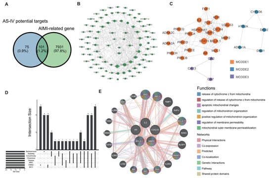
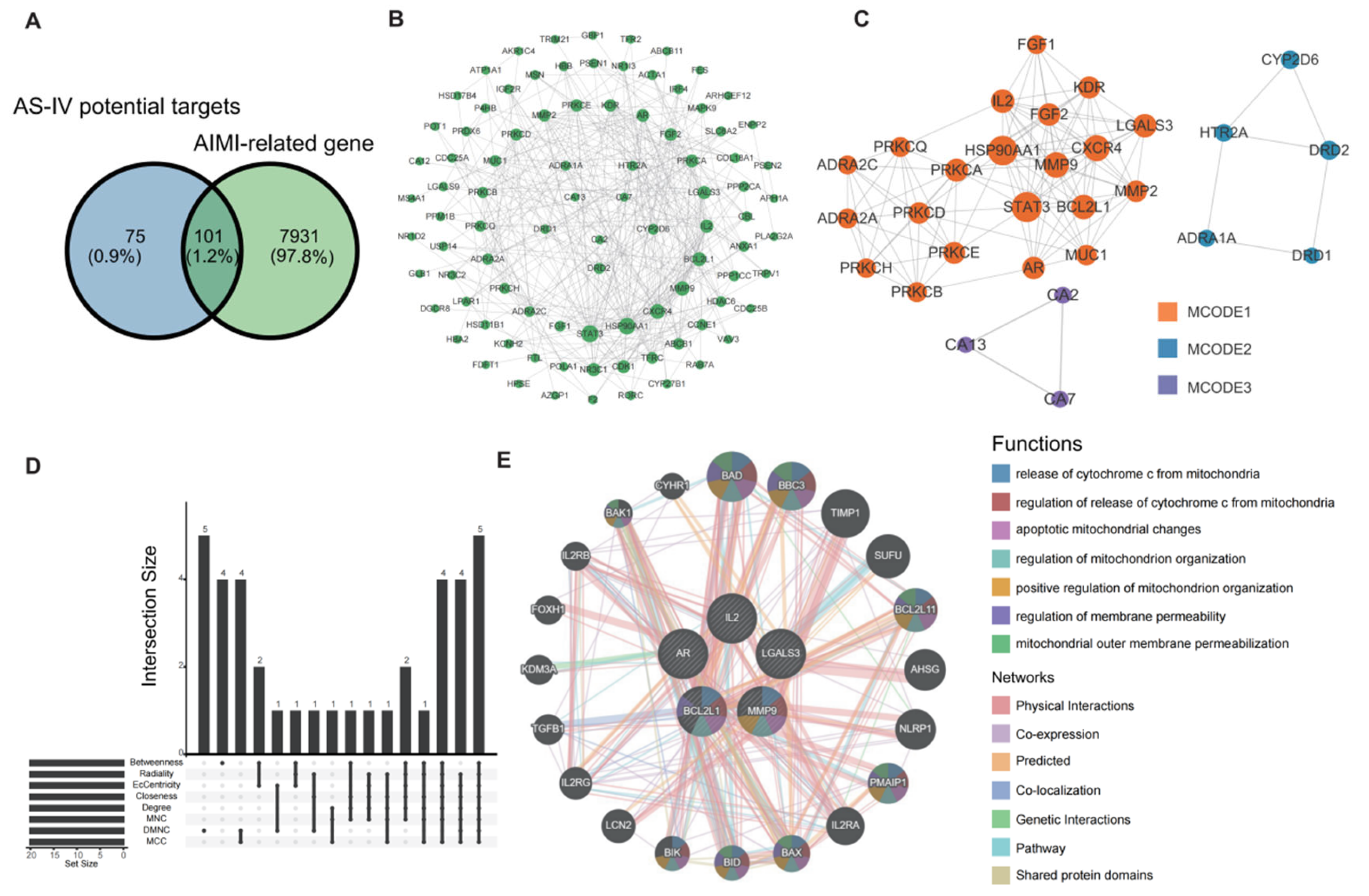
A total of 176 putative AS-IV targets were identified from BATMAN-TCM, SwissTargetPrediction, and PharmMapper after removing duplicates (Table S2). Meanwhile, 8032 AIMI-related genes were collected from GeneCards, Online Mendelian Inheritance in Man (OMIM), and the Comparative Toxicogenomics Database (CTD) (Table S3). Intersection analysis identified 101 overlapping genes, representing potential therapeutic targets of AS-IV against AIMI (Figure 1A; Table S4).
Figure 1.
Identification of candidate targets and hub genes of AS-IV against arsenic-induced myocardial injury (AIMI). (A) Venn diagram showing the intersection between predicted Astragaloside IV (AS-IV) targets and AIMI-related genes, yielding 101 overlapping candidate targets. (B) Protein–protein interaction (PPI) network of the overlapping targets constructed using STRING and visualized in Cytoscape (version 3.10.2). (C) Three densely connected modules identified by Molecular Complex Detection (MCODE) clustering analysis. (D) UpSet plot showing hub genes screened by eight cytoHubba topological algorithms. (E) GeneMANIA network illustrating the functional associations of the identified hub genes, associated with mitochondrial regulation and apoptosis-related processes.
The protein–protein interaction (PPI) network constructed using STRING revealed extensive interactions among these candidate targets (Figure 1B; Table S5). MCODE analysis identified three highly interconnected functional modules (Figure 1C), which were mainly associated with cell proliferation, apoptosis, inflammatory regulation, and cellular homeostasis.
To identify key regulatory nodes, eight topological algorithms in the cytoHubba plugin were applied. Five overlapping hub genes were consistently identified across different algorithms, including LGALS3, MMP9, IL2, AR, and BCL2L1 (Figure 1D; Table S6). GeneMANIA analysis indicated that the hub genes were primarily involved in mitochondrial cytochrome c release, regulation of mitochondrial membrane permeability, and mitochondria-mediated apoptotic signaling, highlighting mitochondrial dysfunction and apoptosis as potential targets of AS-IV in AIMI (Figure 1E).
2.3. Functional and Regulatory Network Analysis Implicates Apoptosis and Immune Regulation in AIMI
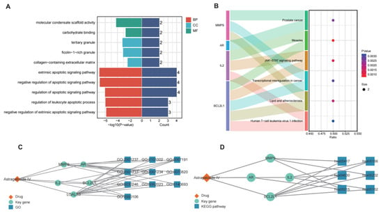
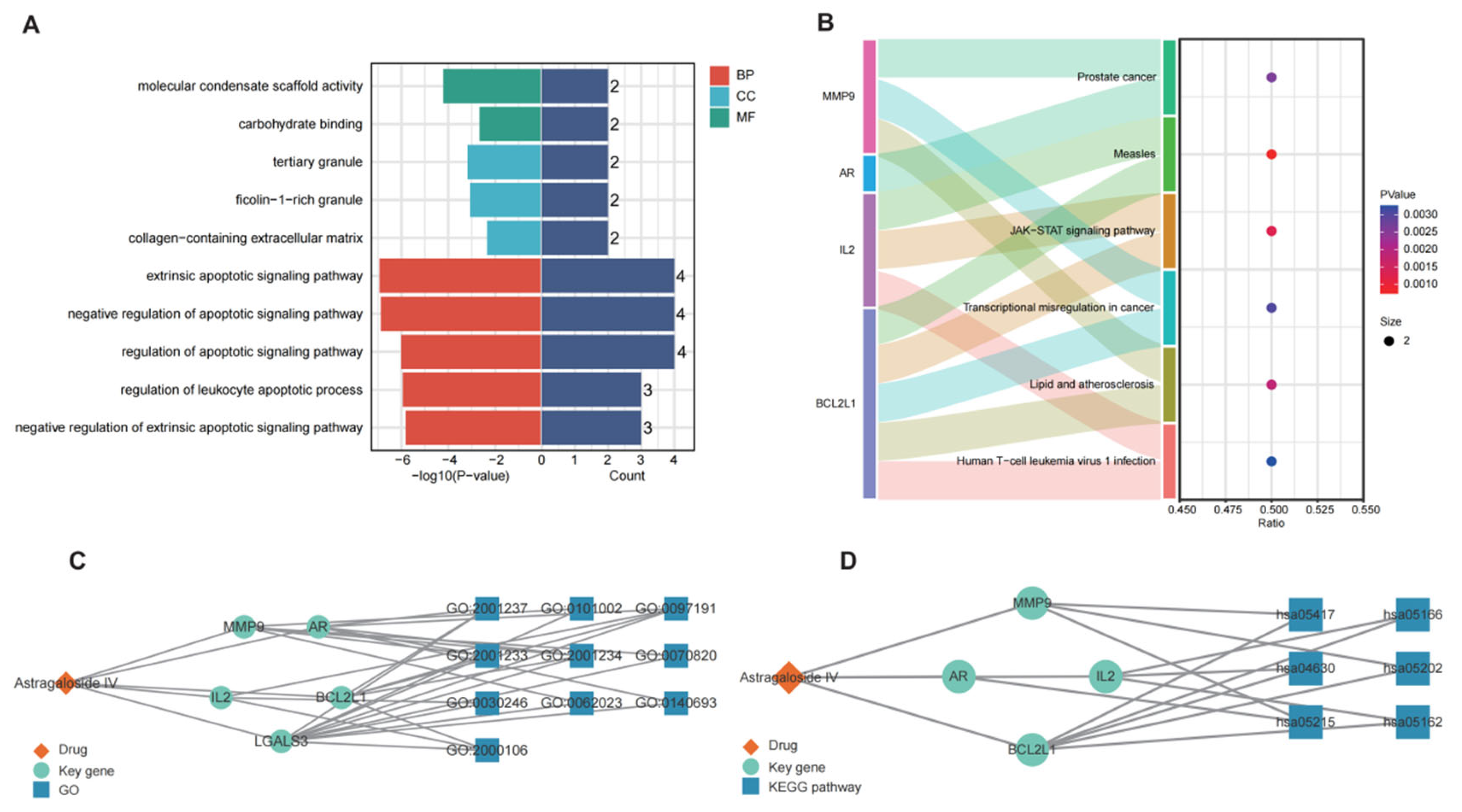
Gene Ontology (GO) and Kyoto Encyclopedia of Genes and Genomes (KEGG) enrichment analyses identified 416 significantly enriched GO terms, including 378 biological process (BP), 31 molecular function (MF), and 7 cellular component (CC) terms (Tables S7 and S8). GO analysis showed that the target genes were mainly enriched in apoptosis-related processes, including regulation of extrinsic apoptotic signaling and leukocyte apoptosis (Figure 2A). KEGG analysis revealed significant enrichment in immune- and inflammation-related pathways, particularly the JAK–STAT signaling pathway (Figure 2B).
Figure 2.
Functional enrichment analysis and network visualization of AS-IV-related hub genes. (A) Gene Ontology (GO) enrichment analysis of the hub genes, including biological process (BP), cellular component (CC), and molecular function (MF) terms. (B) Kyoto Encyclopedia of Genes and Genomes (KEGG) pathway enrichment analysis, showing hub gene–pathway associations and enrichment results. Dot size represents the number of mapped genes, and color indicates the P value. (C) AS-IV–hub gene–GO term network. (D) AS-IV–hub gene–KEGG pathway network.
Functional analysis of AS-IV–hub gene networks pointed to apoptosis, immune response and inflammation as major biological processes (Figure 2C,D). Consistent enrichment of apoptotic pathways suggests that inhibition of cardiomyocyte apoptosis may contribute to the protective effects of AS-IV against AIMI. Furthermore, putative transcription factors and upstream regulatory networks predicted via ChEA3 and competing endogenous RNA (ceRNA) databases provided hypothesis-generating computational clues regarding the multilayered regulation of the identified hub genes (Figure S1, Tables S9 and S10).
2.4. Molecular Docking and Molecular Dynamics Simulations Suggest Galectin-3 as a Potential Binding Target of AS-IV
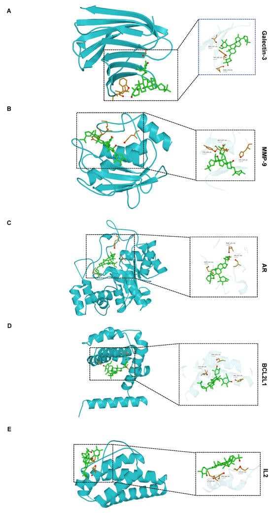
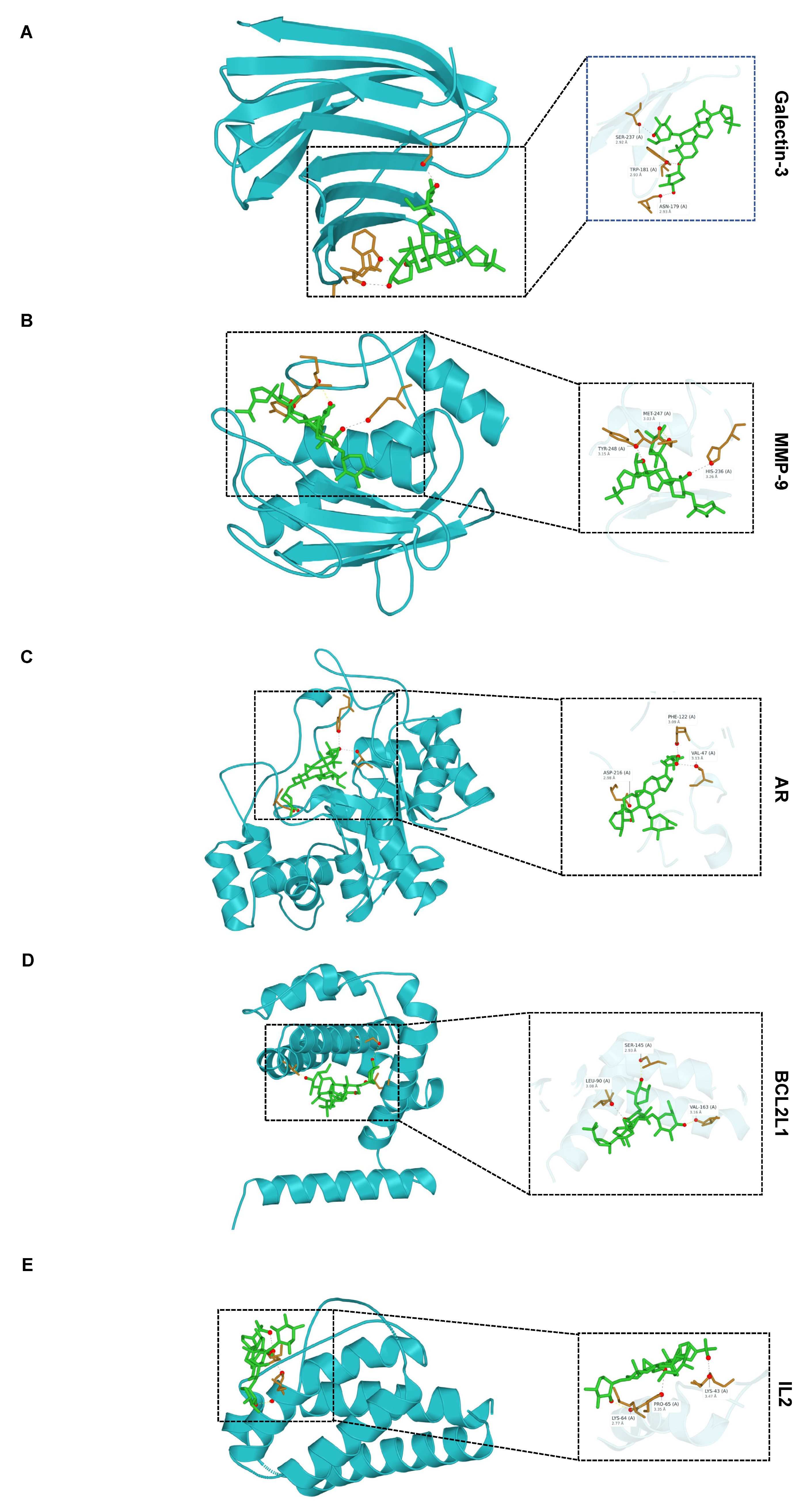
To predict potential interactions between AS-IV and the identified hub proteins, molecular docking was performed using proteins encoded by the five hub genes via AutoDock 4 and AutoDock Vina (Table 2). Considering the docking results together with the biological relevance identified in the preceding bioinformatics analyses, matrix metalloproteinase-9 (MMP-9) and galectin-3 were selected for further investigation. Representative docking models generated using AutoDock Vina demonstrated favorable interactions between AS-IV and the active binding pockets of both proteins through multiple hydrogen bonds and hydrophobic interactions (Figure 3). Subsequently, molecular dynamics simulations were performed to further investigate the conformational behavior and stability of these protein–ligand complexes.
Table 2.
Binding energies of molecular docking between AS-IV and target proteins.
Figure 3.
Molecular docking of AS-IV with five hub proteins using AutoDock Vina. Representative docking conformations of AS-IV with (A) galectin-3, (B) MMP-9, (C) AR protein, (D) BCL2L1 protein, and (E) IL-2, based on initial AutoDock4 and cross-validation via AutoDock Vina. The left panels show the overall binding conformations, and the right panels show enlarged views of the predicted binding pockets. Proteins are displayed as green cartoons, and AS-IV is shown as yellow sticks. Key interacting residues and hydrogen-bond distances are indicated in (A).
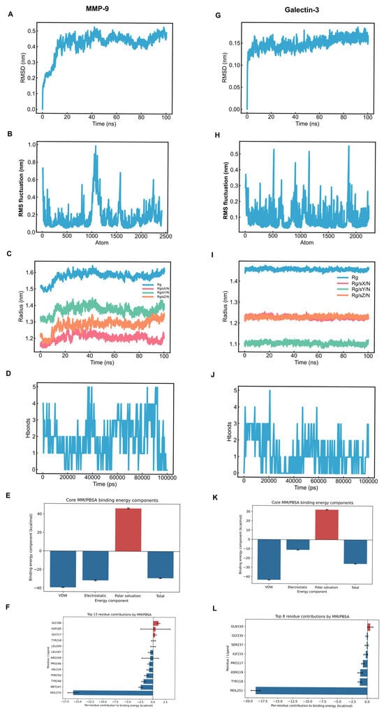
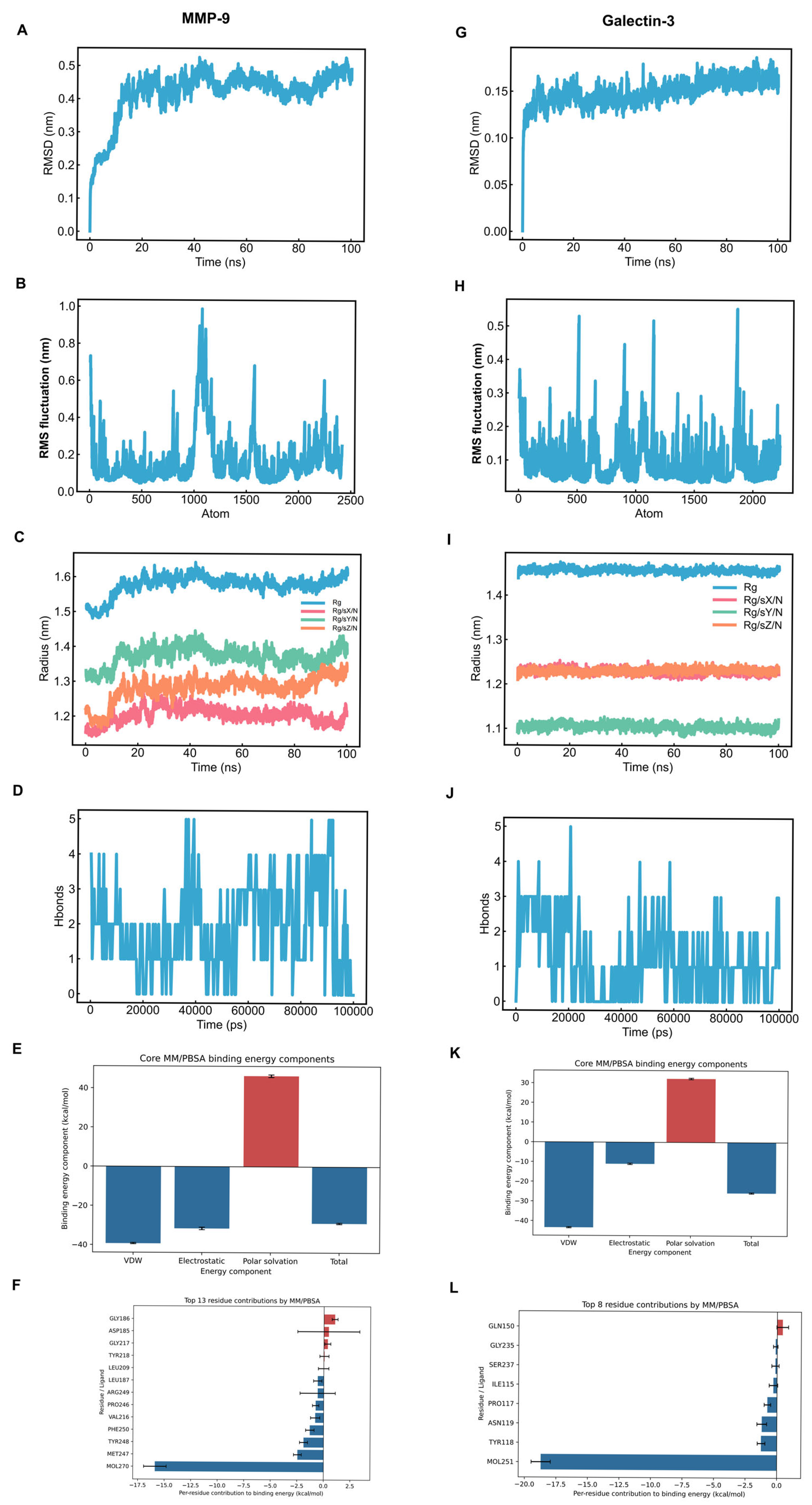
To assess the structural stability and dynamic behavior of the AS-IV complexes, 100-ns molecular dynamics simulations were performed for MMP-9 and galectin-3 (Figure 4). For the MMP-9–AS-IV complex, the protein root-mean-square deviation (RMSD) stabilized between 0.22 and 0.26 nm after an initial adjustment (30–40 ns), while most residues exhibited low local fluctuations (RMSF < 0.2 nm) with a peak at residue 179 (~0.58 nm) (Figure 4A,B). The radius of gyration (Rg) remained stable around 1.50 nm, and an average of ~3.4 hydrogen bonds was maintained throughout the simulation (Figure 4C,D). Furthermore, Molecular mechanics/Poisson–Boltzmann surface area (MM/PBSA) binding free energy calculations over the 50–100 ns trajectories yielded an estimated value of −28.90 ± 0.33 kcal/mol for the MMP-9–AS-IV complex (with van der Waals interactions as the major favorable energy component and Met247, Tyr248, and Phe250 contributing significantly), as shown in Figure 4E,F.
Figure 4.
Molecular dynamics simulation and molecular mechanics/Poisson–Boltzmann surface area (MM/PBSA) analyses of AS-IV complexes with MMP-9 and galectin-3. (A–F) Analyses for the AS-IV–MMP-9 complex: (A) RMSD, (B) RMSF, (C) radius of gyration (Rg), and (D) hydrogen-bond number; (E) Core MM/PBSA binding energy components and (F) per-residue energy decomposition. (G–L) The corresponding analyses for the AS-IV–Galectin-3 complex: (G) RMSD, (H) RMSF, (I) Rg, and (J) hydrogen-bond number; (K) Core MM/PBSA binding energy components and (L) per-residue energy decomposition. RMSD reflects overall conformational deviation during the simulation, RMSF indicates local residue flexibility, Rg describes the compactness of the protein structure, and the hydrogen-bond number represents the dynamic hydrogen-bond interactions between the protein and ligand. For MM/PBSA calculations, VDW represents van der Waals energy, Electrostatic represents electrostatic energy, Polar solvation represents polar solvation energy, and Total indicates the total estimated binding energy.
For the galectin-3–AS-IV complex, the protein RMSD remained stable (0.15–0.18 nm) during the final 30 ns, with low overall residue fluctuations (RMSF mostly < 0.2 nm) and a stable Rg averaging ~1.46 nm (Figure 4G–I). An average of ~1.2 hydrogen bonds was observed, indicating dynamic intermolecular interactions (Figure 4J). Furthermore, MM/PBSA binding free energy calculations over the 50–100 ns trajectories yielded estimated values of −28.90 ± 0.33 kcal/mol for MMP-9–AS-IV and −25.75 ± 0.28 kcal/mol for galectin-3–AS-IV, supported predominantly by favorable van der Waals interactions. MM/PBSA calculations for the galectin-3–AS-IV complex yielded a binding free energy of −25.75 ± 0.28 kcal/mol, supported predominantly by van der Waals interactions, with key favorable contributions from Tyr118, Asn119, and Pro117 (Figure 4K,L).
2.5. AS-IV Alleviates Arsenic-Induced Myocardial Injury in Mice
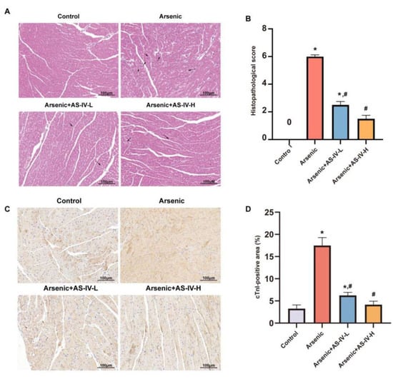
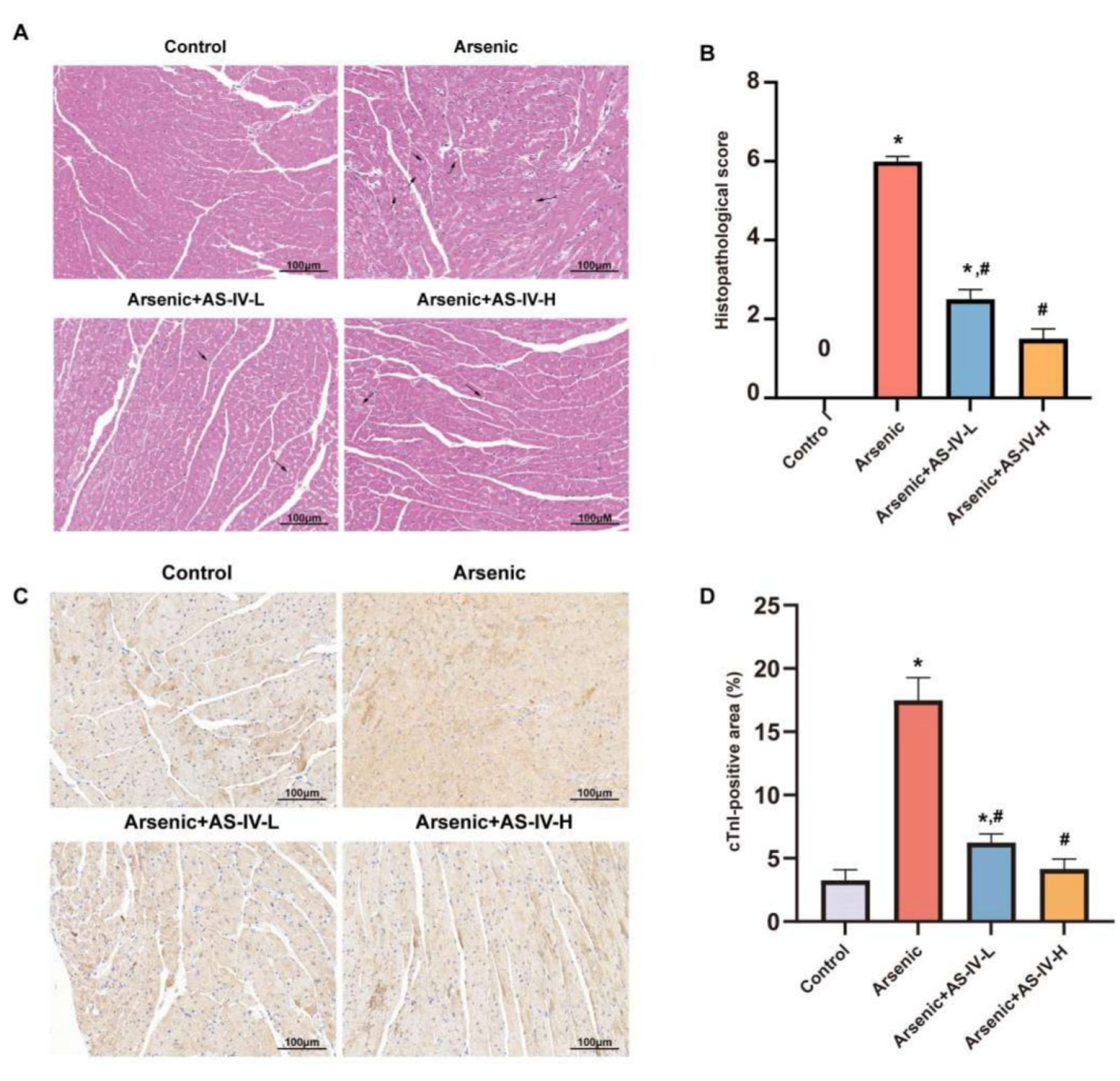
No mortality or significant differences in body weight or heart coefficient were observed among groups during the experimental period (Figure S2A,B). Histopathological examination revealed marked myocardial injury in arsenic-exposed mice, characterized by cardiomyocyte swelling, myofibrillar disorganization, and nuclear pyknosis. AS-IV treatment alleviated these pathological alterations, with the high-dose group exhibiting nearly normal myocardial architecture (Figure 5A,B). Consistently, cTnI immunoreactivity was significantly increased in the arsenic group, whereas high-dose AS-IV markedly reduced cTnI expression to a level comparable to controls (Figure 5C,D). These findings indicate that AS-IV effectively attenuates arsenic-induced myocardial injury.
Figure 5.
AS-IV attenuates arsenic-induced myocardial injury in mice. (A) Representative hematoxylin and eosin (H&E)-stained myocardial sections. Arrows indicate scattered cardiomyocytes with swelling and vacuolar degeneration. (B) Quantification of myocardial injury scores. (C) Representative immunohistochemical staining of cardiac troponin I (cTnI) in myocardial tissue. (D) Quantification of the cTnI-positive area. Scale bars = 100 μm. Data are presented as mean ± standard deviation (SD) (n = 9). * p < 0.05 compared with the Control group; # p < 0.05 compared with the Arsenic group. AS-IV-L and AS-IV-H denote AS-IV at 10 and 20 mg/kg/day, respectively.
Notably, Masson’s trichrome staining (Figure S2C) revealed no overt myocardial fibrosis following 21 days of arsenic exposure. Consistently, immunohistochemistry for MMP-9 (Figure S2D,E) showed no meaningful intergroup differences. These findings indicate myocardial injury without overt fibrosis after 21 days of arsenic exposure. Histopathological assessment of the liver, kidney, spleen, and lung by H&E and Masson’s trichrome staining showed no evident morphological alterations or fibrotic changes among the groups (Figures S3 and S4).
2.6. Changes in Oxidative Stress-Related Markers and Attenuation of Arsenic-Induced Cardiomyocyte Apoptosis by AS-IV
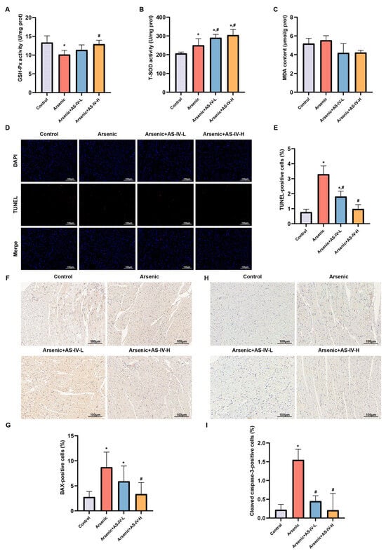
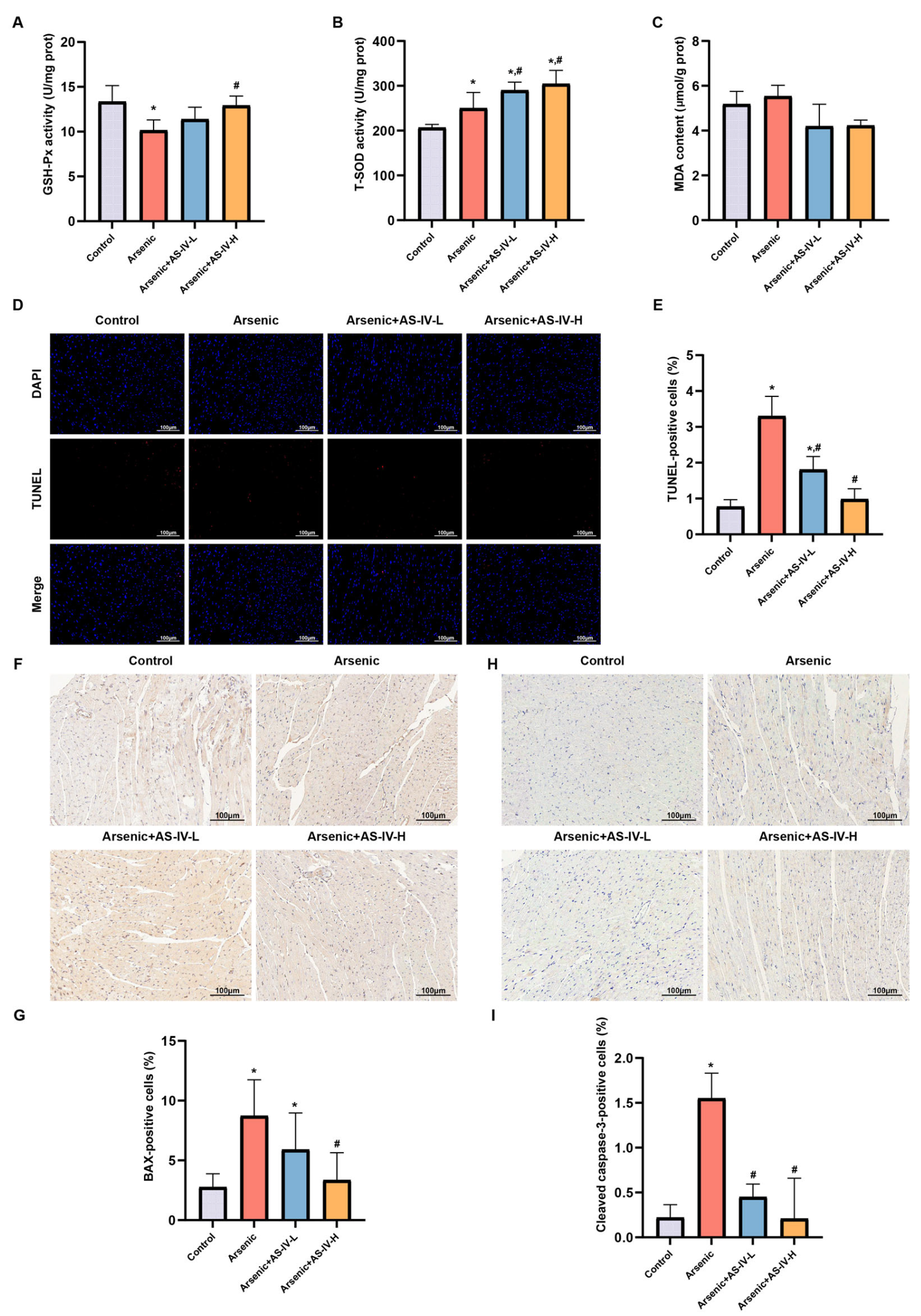
Oxidative stress-related markers and apoptotic indices were further evaluated in myocardial tissue. Compared with the control group, arsenic exposure significantly reduced GSH-Px activity, whereas AS-IV treatment significanlty increased GSH-Px activity (Figure 6A). T-SOD activity was elevated following arsenic exposure and was further enhanced by AS-IV treatment (Figure 6B). No statistically significant differences in myocardial malondialdehyde (MDA) levels were observed among the groups (Figure 6C). These findings indicate alterations in antioxidant enzyme activities following arsenic exposure and AS-IV treatment.
Figure 6.
Changes in oxidative stress-related markers and attenuation of cardiomyocyte apoptosis by AS-IV in arsenic-exposed mice. (A–C) Myocardial oxidative stress-related parameters, including GSH-Px activity, T-SOD activity, and MDA content, respectively. (D) Representative TUNEL staining of myocardial sections. Nuclei were counterstained with DAPI. (E) Quantification of TUNEL-positive cells. (F,G) Representative immunohistochemical staining and quantification of BAX in myocardial tissue. (H,I) Representative immunohistochemical staining and quantification of cleaved caspase-3 in myocardial tissue. Scale bars = 100 μm. Data are presented as mean ± SD or median (IQR) (n = 9), as appropriate. * p < 0.05 compared with the Control group; # p < 0.05 compared with the Arsenic group. AS-IV-L and AS-IV-H denote AS-IV at 10 and 20 mg/kg/day, respectively.
Arsenic exposure induced marked cardiomyocyte apoptosis, as demonstrated by increased cells positive for terminal deoxynucleotidyl transferase dUTP nick end labeling (TUNEL)-positive cells (Figure 6D,E), elevated BAX expression (Figure 6F,G), and enhanced cleaved caspase-3 expression (Figure 6H,I). Compared with the arsenic group, both AS-IV doses reduced TUNEL positivity and cleaved caspase-3 immunoreactivity, whereas BAX immunoreactivity was significantly lower only at the higher AS-IV dose. Collectively, these findings suggest that AS-IV protects against arsenic-induced myocardial injury, in association with altered antioxidant enzyme activities and reduced cardiomyocyte apoptosis.
2.7. Galectin-3 Expression Is Associated with AS-IV-Mediated Protection and Early Immune–Remodeling Responses in AIMI
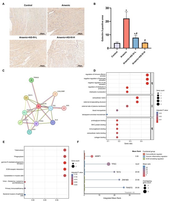
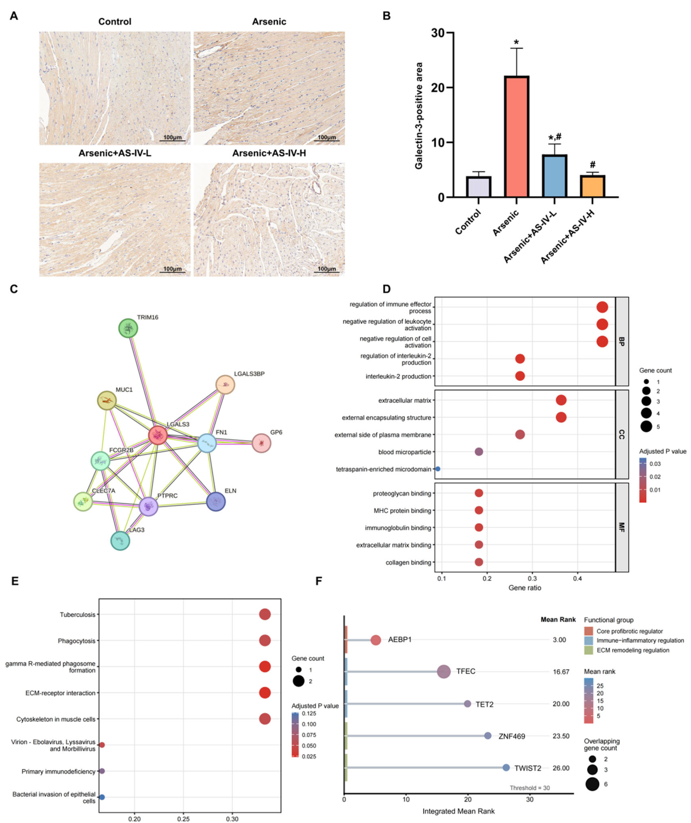
Candidate hub genes LGALS3 (encoding galectin-3) and MMP9 were prioritized for experimental validation based on our network toxicology and molecular docking analyses. Among them, only galectin-3 was markedly upregulated after arsenic exposure and was dose-dependently reduced by AS-IV treatment, with the high-dose AS-IV group showing levels comparable to the control group (Figure 7A,B). In contrast, other examined transcripts (AR, IL-2, and BCL2L1) showed no consistent regulatory patterns (Figure S5), highlighting LGALS3 as a candidate AS-IV-responsive target.
Figure 7.
Experimental validation and bioinformatic characterization of LGALS3 in arsenic-induced myocardial injury. (A) Representative immunohistochemical staining of galectin-3 in myocardial tissue. Scale bars = 100 μm. (B) Quantification of LGALS3-positive area. Data are presented as mean ± SD (n = 9). * p < 0.05 vs Control; # p < 0.05 vs Arsenic. (C) Protein–protein interaction network of LGALS3 and its interacting partners constructed using STRING. (D) GO enrichment analysis of LGALS3-associated proteins. (E) KEGG pathway enrichment analysis of LGALS3-associated proteins. (F) ChEA3-based prediction of upstream transcriptional regulators of the LGALS3 network. The dashed line indicates the Mean Rank cutoff of 30; node size represents overlapping gene count, and color indicates integrated Mean Rank score. AS-IV-L and AS-IV-H denote AS-IV at 10 and 20 mg/kg/day, respectively.
To characterize the molecular context of LGALS3, a STRING-based protein–protein interaction network was constructed, identifying a module of ten interacting proteins, including FN1, ELN, MUC1, LGALS3BP, CLEC7A, and PTPRC (Figure 7C; Table S11). Functional enrichment analysis indicated that this network was associated with immune regulation and extracellular matrix-related processes, while KEGG analysis highlighted extracellular matrix (ECM)–receptor interaction and phagocytosis pathways (Figure 7D,E; Tables S12 and S13). To explore potential upstream regulation, ChEA3 analysis of the LGALS3-centered network identified several candidate transcription factors, including AEBP1, TFEC, TET2, ZNF469, and TWIST2 (Mean Rank < 30) (Figure 7F; Table S14). These findings represent computational predictions that may provide hypotheses for future investigation, but do not establish direct transcriptional regulation of LGALS3 or its associated network.
Collectively, these findings indicate that galectin-3 expression is responsive to arsenic exposure and AS-IV treatment, suggesting that LGALS3 may represent a candidate molecular marker associated with AS-IV intervention in arsenic-induced myocardial injury.
3. Discussion
This study integrated network toxicology, molecular docking, molecular dynamics simulations, and in vivo validation to investigate the protective effects and potential molecular mechanisms of AS-IV against AIMI. The findings demonstrate that AS-IV alleviated arsenic-induced myocardial pathological injury and reduced cTnI levels, altered myocardial antioxidant enzyme activities, suppressed cardiomyocyte apoptosis, and significantly decreased galectin-3 expression.
Oxidative stress and apoptosis have been implicated in arsenic-induced cardiotoxicity. Previous studies have shown that arsenic exposure induces excessive ROS generation, mitochondrial dysfunction, and activation of caspase-dependent apoptotic pathways, ultimately leading to cardiomyocyte injury and dysfunction [6]. In the present study, arsenic exposure significantly reduced GSH-Px activity but increased T-SOD activity, whereas MDA levels showed no statistically significant change. The elevated T-SOD activity may reflect an adaptive or compensatory response to arsenic-induced cellular stress, while the decreased GSH-Px activity may indicate an alteration in endogenous antioxidant defense. Differences in arsenic exposure dose, duration, and experimental model characteristics may contribute to variations in oxidative stress-related responses. Therefore, the observed changes in these markers suggest alterations in antioxidant enzyme activities rather than providing conclusive evidence of overt oxidative damage. AS-IV treatment restored GSH-Px activity and further increased T-SOD activity; however, these changes alone are insufficient to establish an antioxidant mechanism underlying its cardioprotective effects. In contrast, the anti-apoptotic effects of AS-IV were supported by reduced TUNEL positivity and decreased BAX and cleaved caspase-3 expression. Wang et al. reported increased myocardial MDA and reduced antioxidant enzyme activities in sodium arsenite-exposed rats [19]. Differences between their findings and ours may be related to variations in arsenic exposure conditions and experimental models.
Among the candidate hub targets identified by network toxicology, LGALS3/galectin-3 emerged as the most biologically consistent target in the present model. Molecular docking and molecular dynamics simulations predicted potential interactions between AS-IV and galectin-3, while in vivo validation showed that galectin-3 was significantly upregulated after arsenic exposure and dose-dependently reduced by AS-IV treatment. In contrast, MMP-9 expression remained unchanged across the experimental groups. Notably, the computational analyses yielded more favorable predicted interaction energies for MMP-9 than for galectin-3. However, MMP-9 expression did not show a corresponding response to arsenic exposure or AS-IV treatment in the present 21-day model, in which no evident myocardial collagen accumulation was observed by Masson’s trichrome staining. Conversely, galectin-3 showed more consistent changes in vivo in association with arsenic exposure and AS-IV intervention, despite its less favorable predicted interaction energies. These findings suggest that computationally predicted interaction energies alone do not determine the biological relevance of a candidate target in a specific pathological context. Furthermore, although AR, IL-2, and BCL2L1 were identified as hub genes through network topology analysis, their mRNA and immunohistochemical expression patterns were not consistently associated with myocardial injury or AS-IV treatment. Collectively, these findings identify galectin-3 as an AS-IV-responsive candidate associated with arsenic-induced myocardial injury in this model.
Unlike previous AS-IV studies of ischemia–reperfusion injury [12,13] and septic myocardial injury [9], which investigated distinct acute injury settings, the present study extends the investigation of AS-IV to an arsenic-induced myocardial injury model by integrating network toxicology, computational analyses, and in vivo experiments. Notably, AS-IV was administered during ongoing sodium arsenite exposure (days 15–21), rather than before arsenic exposure, allowing us to investigate its effects during the development of arsenic-associated myocardial injury.
Galectin-3 expression increased in injured myocardium without overt fibrosis in the present 21-day model. Previous evidence suggests that galectin-3 is upregulated during the early molecular phase of pressure overload-related cardiac remodeling, well before advanced structural damage becomes fully established [15]. Additionally, either genetic deletion or pharmacological inhibition of galectin-3 attenuates myocardial fibrosis, adverse ventricular remodeling, and inflammatory pro-fibrotic cascades [16,17,18]. However, Al-Salam et al. observed that galectin-3 deficiency exacerbated myocardial injury and apoptosis in acute doxorubicin cardiotoxicity [14]. This context-dependent role indicates that the observed reduction in galectin-3 expression in our study reflects an association with AS-IV intervention rather than establishing galectin-3 as a confirmed mediator or mechanism of protection.
In the present study, no animal mortality, overt clinical signs of severe toxicity, or significant differences in body weight and heart weight index were observed among the experimental groups over the 21-day period. These results indicate that under the current experimental parameters, arsenic exposure did not produce gross toxicity or alter basic organ weight ratios. Chen et al. reported similar body-weight trajectories in ICR mice receiving sodium arsenite at 20 mg/kg by gavage, five days per week for eight weeks, despite increased stimulus-induced vascular leakage [20]. This prior finding serves as background evidence that preserved body weight can coexist with underlying arsenic-induced biological alterations. However, differences in mouse strain, dosing schedule, exposure duration, and evaluated endpoints preclude direct comparisons. Importantly, the lack of significant changes in body weight or heart weight index in our study cannot rule out potential systemic toxicity in other organs or organ systems, nor does it imply that the observed biological effects are exclusively restricted to the myocardium.
Several methodological and biological limitations should be noted. The subacute 21-day exposure reflects an early injury window, meaning findings cannot be directly extrapolated to chronic environmental poisoning or late-stage structural remodeling; moreover, the use of male mice only restricts generalizability across sexes. Mechanistically, biochemical alterations in GSH-Px, T-SOD, and MDA lack direct ROS measurements to confirm overt oxidative stress, and network-based predictions remain exploratory without direct functional assays. In addition, computational docking and 100-ns simulations—conducted without co-crystallized ligand redocking—provide only preliminary structural insights rather than absolute thermodynamic binding constants. Finally, while galectin-3 expression correlated strongly with AS-IV treatment, the lack of genetic or pharmacological intervention precludes definitive causal attribution.
4. Materials and Methods
4.1. Animals and Experimental Design
Male SPF Kunming mice (4–5 weeks old, 15–18 g) were assigned to four groups (n = 9/group) using a randomized block design based on body weight: Control, Arsenic, Arsenic + low-dose AS-IV (AS-IV-L, 10 mg/kg/day), and Arsenic + high-dose AS-IV (AS-IV-H, 20 mg/kg/day). Mice in the arsenic-exposed groups received NaAsO2 (20 mg/kg/day, gavage) for 21 consecutive days. This dose was selected based on a previous study demonstrating significant systemic vascular responses in ICR mice following oral gavage of NaAsO2 at 10–20 mg/kg/day, while the 21-day exposure duration was established as a subacute model to induce reproducible myocardial injury based on established toxicological paradigms [20]. From days 15–21, mice in the AS-IV-L and AS-IV-H groups were co-administered AS-IV intraperitoneally at 10 and 20 mg/kg/day, respectively. The 10 mg/kg/day dose was chosen based on reported cardioprotective efficacy in mouse models [21], while the 20 mg/kg/day dose was included to evaluate dose-dependent effects during ongoing arsenic exposure. Control mice received vehicle only, and arsenic-treated mice received the corresponding vehicle during the AS-IV intervention period.
Body weight and general condition were monitored throughout the experiment. After the experiment, mice were placed in an anesthesia induction chamber and anesthetized with 3%–5% isoflurane delivered in oxygen at a flow rate of 1–2 L/min for approximately 3–5 min until deep anesthesia was achieved. After confirming the absence of the toe-pinch reflex and complete loss of pain reflex, mice were euthanized by cervical dislocation. Then, heart, liver, kidney, spleen, and lung tissues were collected. Organ weights were recorded, and organ coefficients were calculated as organ weight/body weight (mg/g). All animal procedures were approved by the Institutional Animal Care and Use Committee of Hunan Prevention and Treatment Institute for Occupational Diseases (Approval No. 2025-010, approval date 16 October 2025).
4.2. Histological Staining
Heart, liver, kidney, spleen, and lung tissues were fixed in neutral buffered fixative, embedded in paraffin, and sectioned at approximately 5 μm. For H&E staining, sections were deparaffinized, rehydrated, stained with hematoxylin and eosin, dehydrated, cleared, and mounted. H&E-stained sections were used to evaluate general tissue morphology, including myocardial fiber arrangement, intercellular space, cardiomyocyte swelling, and nuclear morphology. In addition, myocardial injury was semi-quantitatively evaluated based on three features (cardiomyocyte swelling, myofibrillar disorganization, and nuclear pyknosis) across four myocardial regions (subepicardial, middle, subendocardial/papillary, and interventricular septum) [22]. Each of the 12 items was graded on a 0–3 scale, and the scores were summed and divided by 4 to yield a final myocardial injury score (range: 0–9).
Masson’s trichrome staining was performed using a commercial kit (Servicebio, Wuhan, China). Collagen fibers were stained blue, while muscle fibers and erythrocytes were stained red. H&E and Masson staining were used for morphological observation and representative image presentation, with semi-quantitative scoring applied to H&E evaluations, whereas no collagen area quantification was performed owing to minimal fibrosis. Histological evaluation and image selection were conducted by investigators blinded to the experimental groups.
4.3. Immunohistochemistry
Immunohistochemistry (IHC) was performed on paraffin-embedded myocardial sections to evaluate the expression of cTnI, MMP-9, galectin-3, BAX, and cleaved caspase-3. Briefly, sections were deparaffinized, rehydrated, and subjected to heat-induced antigen retrieval in Tris-EDTA buffer (pH 8.0) for 30 min. Endogenous peroxidase activity was quenched with 3% hydrogen peroxide, followed by blocking with 3% bovine serum albumin (BSA).
Sections were incubated overnight at 4 °C with the following primary antibodies from Servicebio (Wuhan, China): anti-cTnI (GB122408, 1:400), anti-MMP-9 (GB15132, 1:500), anti-galectin-3 (GB151145, 1:500), anti-BAX (GB154122, 1:500), and anti-cleaved caspase-3 (GB11532, 1:500). After washing, sections were incubated with appropriate polymer-based secondary antibodies (S-vision system, Servicebio, Wuhan, China).
Immunoreactivity was visualized using diaminobenzidine (DAB), and nuclei were counterstained with hematoxylin. Positive staining appeared as brown-yellow signals. Images were acquired under identical microscope settings. Quantitative analysis was performed using ImageJ software (version 1.54) [23] by an investigator blinded to the experimental groups. For each mouse (n = 9 per group), five randomly selected non-overlapping fields were analyzed, and the mean value was calculated as a single biological replicate. The percentage of positive staining area or the number of positive cells was quantified according to the staining characteristics of each marker.
4.4. TUNEL Staining
Cardiomyocyte apoptosis was detected using a commercial TUNEL assay kit (Servicebio, Wuhan, China; CF488 fluorescence kit). Paraffin-embedded myocardial sections were deparaffinized, rehydrated, and treated with proteinase K at 37 °C for 20 min. After permeabilization and equilibration, sections were incubated with TUNEL reaction mixture at 37 °C for 1 h in the dark. Nuclei were counterstained with DAPI, and sections were mounted with antifade mounting medium. TUNEL-positive cells showed green fluorescence, while nuclei showed blue fluorescence.
Images were captured using a fluorescence microscope under identical exposure settings. For each mouse, nine randomly selected non-overlapping fields were analyzed using ImageJ software. The apoptotic index was calculated as the percentage of TUNEL-positive cells among total DAPI-positive nuclei.
4.5. Assessment of Oxidative Stress Markers
Myocardial oxidative stress was assessed by detecting MDA concentration, T-SOD and GSH-Px activities with commercial kits (G4302, G4306, G4310; Servicebio, Wuhan, China) according to the manufacturer’s instructions. Briefly, cardiac tissues were homogenized in PBS (1:9, w/v), centrifuged at 10,000× g for 10–15 min at 4 °C, and supernatants were harvested. Total protein concentration was measured using a BCA kit (G2026, Servicebio, Wuhan, China). All biochemical indices were normalized to protein content, with MDA expressed as μmol/g protein and T-SOD and GSH-Px enzyme activities as U/mg protein.
4.6. Quantitative Real-Time PCR
Total RNA was extracted from frozen myocardial tissue using RNA extraction reagent. RNA concentration and purity were assessed using a NanoDrop spectrophotometer. cDNA was synthesized using SweScript All-in-One RT SuperMix for qPCR according to the manufacturer’s instructions. Quantitative real-time PCR was performed using 2× Universal Blue SYBR Green qPCR Master Mix on a Bio-Rad CFX Connect system. The PCR program was as follows: 95 °C for 30 s, followed by 40 cycles of 95 °C for 15 s and 60 °C for 30 s. Melting curve analysis was performed to confirm amplification specificity. Gapdh was used as the internal reference gene, and relative mRNA expression was calculated using the 2−ΔΔCt method [24]. The primer sequences used in this study are listed in Table 3.
Table 3.
The primer sequences.
4.7. Toxicity Prediction of AS-IV
The chemical structure and SMILES string of AS-IV were obtained from PubChem. The SMILES information was submitted to ProTox 3.0 and ADMETlab 2.0 for in silico toxicity and ADMET prediction, including acute toxicity, blood–brain barrier permeability, oral bioavailability, and potential adverse effects.
4.8. Target Collection and Intersection Analysis
DrugBank, BindingDB, ChEMBL, and TCMSP were additionally searched for experimentally validated AS-IV targets; however, no suitable direct target records met the predefined inclusion criteria. Therefore, BATMAN-TCM, SwissTargetPrediction, and PharmMapper were used as complementary prediction platforms. The predicted targets from these databases were integrated, and duplicate entries were removed to generate a non-redundant AS-IV-related target set. Arsenic-induced myocardial injury-related targets were retrieved from GeneCards, OMIM, and the Comparative Toxicogenomics Database (CTD). In CTD, the terms “arsenic” and “myocardial injury” were searched separately, and the overlapping genes were retained as an initial candidate gene set for subsequent PPI-based prioritization, rather than confirmed specific targets of arsenic-induced myocardial injury. These genes were subsequently intersected with the predicted AS-IV target set to identify potential therapeutic targets shared by AS-IV and arsenic-induced myocardial injury.
4.9. PPI Network and Hub Gene Identification
Candidate targets were imported into the STRING database to construct a protein–protein interaction network, with the species set as Homo sapiens and the minimum interaction score set to 0.40. The resulting network was visualized in Cytoscape. Key modules were identified using the MCODE plugin. Hub genes were further prioritized using the cytoHubba plugin based on eight topological algorithms: maximal clique centrality (MCC), density of maximum neighborhood component (DMNC), maximum neighborhood component (MNC), Degree, Eccentricity, Closeness, Radiality, and Betweenness. All nodes in the PPI network were independently scored and ranked by each algorithm. The top 20 genes from each algorithm were selected, and the intersection of the eight top-20 gene sets was defined as the consensus hub gene set. No additional numerical score cutoff was applied.
4.10. Functional Enrichment and Regulatory Network Analysis
GO and KEGG enrichment analyses were performed using the R package clusterProfiler [25]. Terms with p < 0.05 and FDR-adjusted q < 0.2 were considered significantly enriched. A compound-target-pathway network was constructed using Cytoscape. Potential transcription factors were predicted using iRegulon, and miRNA- and lncRNA-mediated regulatory networks were constructed using miRDB, miRNet, and ENCORI.
4.11. Molecular Docking and Molecular Dynamics Simulation
Three-dimensional protein structures of hub targets were obtained from the RCSB PDB database, and the structure of AS-IV was downloaded from PubChem in SDF format. Molecular docking was primarily performed using AutoDock Vina (version 1.2.7) to evaluate the potential interactions between AS-IV and the hub target proteins. AutoDock 4 was also used to provide complementary docking results for comparison. The best binding conformations were visualized using PyMOL (version 2.5.x). More negative predicted binding affinity scores indicated more favorable ligand–protein interactions.
Molecular dynamics simulations were performed using GROMACS (version 2025.2) to assess the stability of selected AS-IV-protein complexes. Protein topologies were generated using the AMBER99SB-ILDN force field, and ligand parameters and topology files were generated using Sobtop (version 1.0 dev5). The systems were solvated with the TIP3P water model in a cubic box, neutralized with counter-ions, energy-minimized using the steepest descent and conjugate gradient methods, equilibrated under NVT at 300 K for 100 ps and NPT equilibration at 1 bar for 100 ps ensembles, and subjected to 100 ns production simulations. RMSD, RMSF, radius of gyration, and the number of hydrogen bonds were analyzed using DuIvyTools (version 0.6.0) to evaluate complex stability.
To further evaluate the binding affinity, binding free energy calculations were performed using gmx_MMPBSA (version 1.6.5) for the MMP9–AS-IV and LGALS3–AS-IV complexes. Trajectories from 50 to 100 ns were extracted at 0.2-ns intervals, yielding 251 snapshots for each complex, to calculate the van der Waals, electrostatic, and solvation energy contributions (including polar solvation energy calculated using the Poisson–Boltzmann implicit solvent model and nonpolar contributions). Per-residue energy decomposition was additionally conducted to identify key residue contributions to the ligand–protein interaction. Because conformational entropy was not included, the calculated MM/PBSA values were interpreted as approximate binding energy estimates rather than complete binding free energies.
4.12. Protein Interaction Network and Upstream Regulatory Analysis of LGALS3
To further characterize the biological context of galectin-3, an LGALS3-centered protein–protein interaction (PPI) network was constructed using the STRING database [26]. The human galectin-3 protein identifier ENSP00000254301 was queried with the organism set to Homo sapiens, and the minimum required interaction score was set to 0.40. Direct interaction partners were extracted for downstream functional enrichment analysis. GO and KEGG pathway enrichment analyses were performed using the R package clusterProfiler, with statistical significance defined as p < 0.05.
For upstream regulatory analysis, genes from the LGALS3-centered PPI network were submitted to the ChEA3 database [27]. Candidate transcription factors were ranked according to the integrated Mean Rank score, and those with Mean Rank < 30 were selected for further interpretation. ChEA3 results were considered hypothesis-generating computational predictions; no ChIP-qPCR or luciferase reporter assays were performed to validate these transcription factor associations.
4.13. Statistical Analysis
Statistical analyses were performed using SPSS 26.0 and GraphPad Prism 10.0. Data are expressed as mean ± standard deviation (SD) or median (interquartile range, IQR) for normally or non-normally distributed variables, respectively. Normality was assessed by the Shapiro–Wilk test and homogeneity of variances by Levene’s test. For multiple group comparisons, one-way ANOVA followed by Tukey’s post hoc test was applied when parametric assumptions were met; otherwise, the Kruskal–Wallis test followed by Dunn’s post hoc test was used (specifically for BAX immunohistochemical data, which did not meet parametric assumptions). For animal experiments, n = 9 per group. For IHC analysis, five randomly selected non-overlapping fields were analyzed for each mouse, whereas nine randomly selected non-overlapping fields were analyzed for TUNEL staining. The mean value of these fields was used as one biological replicate for statistical analysis. Statistical significance was set at p < 0.05.
5. Conclusions
In this short-term mouse model, AS-IV attenuated arsenic-induced myocardial injury, suppressed cardiomyocyte apoptosis, and modulated oxidative stress-related enzyme activities. These protective effects were accompanied by a marked reduction in myocardial galectin-3 expression. Although galectin-3 emerges as a prominent candidate target associated with AS-IV intervention, its precise causal role in mediating cardioprotection warrants future functional verification.
Supplementary Materials
The following supporting information can be downloaded at: https://www.mdpi.com/article/10.3390/ijms272210142/s1.
Author Contributions
Conceptualization, T.C.; methodology, R.D., L.Z. and S.C.; validation, R.D.; formal analysis, R.D.; investigation, X.L., R.D., Y.X., L.Z., X.Z., S.C. and T.L.; resources, T.C.; data curation, X.L. and Y.X.; writing—original draft preparation, X.L. and R.D.; writing—review and editing, T.C.; visualization, X.L.; supervision, T.C. All authors have read and agreed to the published version of the manuscript.
Funding
This research was funded by the Science and Technology Program of Hunan Province (No. 2025JJ80781); Health Research Project of Hunan Provincial Health Commission (No. 20255944); and the Huxiang Young Talent Project (No. 2024RC3153).
Institutional Review Board Statement
All experimental procedures were reviewed and approved by the Institutional Animal Care and Use Committee of Hunan Prevention and Treatment Institute for Occupational Diseases (Ethics Approval No. 2025-010, approval date 16 October 2025).
Informed Consent Statement
Not applicable.
Data Availability Statement
The original contributions presented in this study are included in the article and Supplementary Materials. Further inquiries and requests for the raw experimental data may be directed to the corresponding author.
Acknowledgments
We sincerely appreciate Jianfeng Chen from the Toxicology Laboratory of Hunan Prevention and Treatment Institute for Occupational Diseases for providing experimental space and personnel support to complete this study.
Conflicts of Interest
The authors declare no conflicts of interest.
References
- Lee, H.; Jo, Y.; Jung, M.; Lee, J.H.; Kim, T.H.; Lee, J.; Kim, D.J.; Rahmati, M.; Smith, L.; Pizzol, D.; et al. Heavy metal exposure and all health outcomes: An umbrella review of meta-analyses. J. Hazard. Mater. 2026, 503, 141141. [Google Scholar] [CrossRef] [Scilit]
- Medgyesi, D.N.; Bangia, K.; Spielfogel, E.S.; Fisher, J.A.; Madrigal, J.M.; Jones, R.R.; Ward, M.H.; Lacey, J.V., Jr.; Sanchez, T.R. Long-Term Exposure to Arsenic in Community Water Supplies and Risk of Cardiovascular Disease among Women in the California Teachers Study. Environ. Health Perspect. 2024, 132, 107006. [Google Scholar] [CrossRef] [Scilit]
- Kagawa, T.; He, T.; Ohgami, N.; Chen, D.; Tazaki, A.; Li, H.; Naito, H.; Tamura, T.; Wakai, K.; Kato, M. Emerging hazard: Exposure to fish-derived arsenobetaine increases a latent risk of hypertension in humans and mice. Environ. Int. 2026, 214, 110365. [Google Scholar] [CrossRef] [Scilit]
- Yuan, Y.; Xiao, Y.; Feng, W.; Liu, Y.; Yu, Y.; Zhou, L.; Qiu, G.; Wang, H.; Liu, B.; Liu, K.; et al. Plasma Metal Concentrations and Incident Coronary Heart Disease in Chinese Adults: The Dongfeng-Tongji Cohort. Environ. Health Perspect. 2017, 125, 107007. [Google Scholar] [CrossRef] [Scilit]
- Pan, Z.; Gong, T.; Liang, P. Heavy Metal Exposure and Cardiovascular Disease. Circ. Res. 2024, 134, 1160–1178. [Google Scholar] [CrossRef] [Scilit]
- Adil, M.; Kandhare, A.D.; Ghosh, P.; Bodhankar, S.L. Sodium arsenite-induced myocardial bruise in rats: Ameliorative effect of naringin via TGF-beta/Smad and Nrf/HO pathways. Chem. Biol. Interact. 2016, 253, 66–77. [Google Scholar] [CrossRef] [Scilit]
- Wang, Y.; Hou, J.; Yu, Y.; He, K.; Guo, H.; Liu, N.; Wang, H.; Song, Y.; Ma, D. Improvement effect of Shengxian Decoction on cardiac toxicity induced by arsenic trioxide in rats. J. Ethnopharmacol. 2025, 352, 120163. [Google Scholar] [CrossRef] [Scilit]
- Yang, Q.; Han, B.; Li, S.; Wang, X.; Wu, P.; Liu, Y.; Li, J.; Han, B.; Deng, N.; Zhang, Z. The link between deacetylation and hepatotoxicity induced by exposure to hexavalent chromium. J. Adv. Res. 2022, 35, 129–140. [Google Scholar] [CrossRef] [Scilit]
- Wang, J.; Pu, X.; Zhuang, H.; Guo, Z.; Wang, M.; Yang, H.; Li, C.; Chang, X. Astragaloside IV alleviates septic myocardial injury through DUSP1-Prohibitin 2 mediated mitochondrial quality control and ER-autophagy. J. Adv. Res. 2025, 75, 561–580. [Google Scholar] [CrossRef] [Scilit]
- Li, J.; Guo, C.; Liu, Y.; Han, B.; Lv, Z.; Jiang, H.; Li, S.; Zhang, Z. Chronic arsenic exposure-provoked biotoxicity involved in liver-microbiota-gut axis disruption in chickens based on multi-omics technologies. J. Adv. Res. 2025, 67, 373–386. [Google Scholar] [CrossRef] [Scilit]
- Ma, J.; Han, B.; Li, J.; Liu, Y.; Lv, Z.; Song, K.; Li, M.; Liu, D.; Ji, S.; Zhang, Z. Polystyrene Microplastics Exacerbate Arsenic-Induced Ferroptosis and Lipid Metabolism Dysregulation in Chicken Hepatocytes through SIRT7-SIRT1 Axis-Associated Transcriptomic Alterations. Environ. Sci. Technol. 2026, 60, 25221–25242. [Google Scholar] [CrossRef] [Scilit]
- Zhai, P.; Chen, Q.; Wang, X.; Ouyang, X.; Yang, M.; Dong, Y.; Li, J.; Li, Y.; Luo, S.; Liu, Y.; et al. The combination of Tanshinone IIA and Astragaloside IV attenuates myocardial ischemia-reperfusion injury by inhibiting the STING pathway. Chin. Med. 2024, 19, 34. [Google Scholar] [CrossRef] [Scilit]
- Zeng, R.Y.; Qiu, Z.C.; Zhao, S.T.; Lai, S.Q.; Qiu, R.B.; Wu, Z.M.; Xu, Z.Q.; Li, J.N.; Luo, X.D.; Wang, B.L.; et al. Astragaloside IV mitigates myocardial ischemia/reperfusion injury by modulating the 14-3-3eta/GPX4 axis, reducing mitochondrial dysfunction, ferroptosis, and apoptosis. Phytomedicine 2026, 155, 158082. [Google Scholar] [CrossRef] [Scilit]
- Al-Salam, S.; Kandhan, K.; Sudhadevi, M.; Yasin, J.; Tariq, S. Early Doxorubicin Myocardial Injury: Inflammatory, Oxidative Stress, and Apoptotic Role of Galectin-3. Int. J. Mol. Sci. 2022, 23, 12479. [Google Scholar] [CrossRef] [Scilit]
- Arrieta, V.; Martinez-Martinez, E.; Ibarrola, J.; Alvarez, V.; Sadaba, R.; Garcia-Pena, A.; Fernandez-Celis, A.; Cachofeiro, V.; Rossignol, P.; Lopez-Andres, N. A role for galectin-3 in the development of early molecular alterations in short-term aortic stenosis. Clin. Sci. 2017, 131, 935–949. [Google Scholar] [CrossRef] [Scilit]
- Wang, X.; Gaur, M.; Mounzih, K.; Rodriguez, H.J.; Qiu, H.; Chen, M.; Yan, L.; Cooper, B.A.; Narayan, S.; Derakhshandeh, R.; et al. Inhibition of galectin-3 post-infarction impedes progressive fibrosis by regulating inflammatory profibrotic cascades. Cardiovasc. Res. 2023, 119, 2536–2549. [Google Scholar] [CrossRef] [Scilit]
- Vlachou, F.; Varela, A.; Stathopoulou, K.; Ntatsoulis, K.; Synolaki, E.; Pratsinis, H.; Kletsas, D.; Sideras, P.; Davos, C.H.; Capetanaki, Y.; et al. Galectin-3 interferes with tissue repair and promotes cardiac dysfunction and comorbidities in a genetic heart failure model. Cell. Mol. Life Sci. 2022, 79, 250. [Google Scholar] [CrossRef] [Scilit]
- Yu, L.; Ruifrok, W.P.; Meissner, M.; Bos, E.M.; van Goor, H.; Sanjabi, B.; van der Harst, P.; Pitt, B.; Goldstein, I.J.; Koerts, J.A.; et al. Genetic and pharmacological inhibition of galectin-3 prevents cardiac remodeling by interfering with myocardial fibrogenesis. Circ. Heart Fail. 2013, 6, 107–117. [Google Scholar] [CrossRef] [Scilit]
- Wang, L.; Shi, W.; Gao, X.; SreeHarsha, N.; Zhang, D. Cardioprotective role of metformin against sodium arsenite-induced oxidative stress, inflammation, and apoptosis. IUBMB Life 2020, 72, 749–757. [Google Scholar] [CrossRef] [Scilit]
- Chen, S.C.; Chang, C.Y.; Lin, M.L. Vascular Hyperpermeability Response in Animals Systemically Exposed to Arsenic. Int. J. Med. Sci. 2018, 15, 425–429. [Google Scholar] [CrossRef] [Scilit]
- Zhang, W.; Zhang, L.; Zhou, H.; Li, C.; Shao, C.; He, Y.; Yang, J.; Wan, H. Astragaloside IV Alleviates Infarction Induced Cardiomyocyte Injury by Improving Mitochondrial Morphology and Function. Front. Cardiovasc. Med. 2022, 9, 810541. [Google Scholar] [CrossRef] [Scilit]
- Varghese, M.V.; Abhilash, M.; Alex, M.; Paul, M.V.S.; Prathapan, A.; Raghu, K.G.; Nair, R.H. Attenuation of arsenic trioxide induced cardiotoxicity through flaxseed oil in experimental rats. Redox Rep. 2017, 22, 346–352. [Google Scholar] [CrossRef] [Scilit]
- Schneider, C.A.; Rasband, W.S.; Eliceiri, K.W. NIH Image to ImageJ: 25 years of image analysis. Nat. Methods 2012, 9, 671–675. [Google Scholar] [CrossRef] [Scilit]
- Livak, K.J.; Schmittgen, T.D. Analysis of relative gene expression data using real-time quantitative PCR and the 2−ΔΔCT Method. Methods 2001, 25, 402–408. [Google Scholar] [CrossRef] [Scilit]
- Yu, G.; Wang, L.G.; Han, Y.; He, Q.Y. clusterProfiler: An R package for comparing biological themes among gene clusters. OMICS 2012, 16, 284–287. [Google Scholar] [CrossRef] [Scilit]
- Szklarczyk, D.; Kirsch, R.; Koutrouli, M.; Nastou, K.; Mehryary, F.; Hachilif, R.; Gable, A.L.; Fang, T.; Doncheva, N.T.; Pyysalo, S.; et al. The STRING database in 2023: Protein-protein association networks and functional enrichment analyses for any sequenced genome of interest. Nucleic Acids Res. 2023, 51, D638–D646. [Google Scholar] [CrossRef] [Scilit]
- Keenan, A.B.; Torre, D.; Lachmann, A.; Leong, A.K.; Wojciechowicz, M.L.; Utti, V.; Jagodnik, K.M.; Kropiwnicki, E.; Wang, Z.; Ma’ayan, A. ChEA3: Transcription factor enrichment analysis by orthogonal omics integration. Nucleic Acids Res. 2019, 47, W212–W224. [Google Scholar] [CrossRef] [Scilit]

Disclaimer/Publisher’s Note: The statements, opinions and data contained in all publications are solely those of the individual author(s) and contributor(s) and not of MDPI and/or the editor(s). MDPI and/or the editor(s) disclaim responsibility for any injury to people or property resulting from any ideas, methods, instructions or products referred to in the content. |
© 2026 by the authors. Licensee MDPI, Basel, Switzerland. This article is an open access article distributed under the terms and conditions of the Creative Commons Attribution (CC BY) license.