


Abstract
Coronary artery disease (CAD) remains a leading cause of morbidity and mortality worldwide. Its development results from a complex interaction between environmental risk factors and genetic susceptibility. Evidence indicates that oxidative stress, defined as a state in which free radical production exceeds antioxidant defenses, plays an important role in atherogenesis by promoting endothelial dysfunction, lipid oxidation, vascular inflammation and plaque instability. This review summarizes current knowledge on polymorphisms of the most important genes involved in reactive oxygen species generation and antioxidant defense. Several variants have been associated with CAD, hypertension, carotid atherosclerosis and other cardiovascular phenotypes, although the direction and strength of associations often differ across populations. In many cases, functional effects include altered enzyme activity, changes in promoter activity, reduced nitric oxide bioavailability or impaired antioxidant capacity. The evidence is heterogeneous: many candidate-gene studies are small, differ in ancestry, phenotype definitions and genetic models and are affected by limited replication and publication bias. Among the variants discussed, NOS3 rs3918226 and GPX1 rs1050450, rs1800668 and rs3811699 showed genome-wide significant associations with CAD in a large-scale genome-wide association study. The variants reviewed have no established role in CAD risk stratification and their added value beyond established clinical risk factors and validated genome-wide CAD polygenic risk scores has not been tested.
1. Introduction
Coronary artery disease (CAD) remains one of the leading causes of morbidity and mortality worldwide [1]. Despite significant advances in pharmacological interventions and revascularization strategies, the identification of individuals at elevated cardiovascular risk prior to clinical manifestation remains a challenge [2]. Its etiopathogenesis is highly complex and multifactorial, resulting from the interaction between environmental exposures and genetic determinants that influence disease susceptibility both independently and through gene–environment interactions [3]. While traditional risk factors, including dyslipidemia, hypertension, diabetes mellitus, smoking and obesity, explain a large proportion of CAD cases, a significant heritable component suggests that genetic factors play a major role in disease susceptibility [4].
In recent years, growing attention has been directed toward the role of oxidative stress as one of the key mechanisms leading to the development of atherosclerosis and its clinical complications [5]. Reactive oxygen species (ROS) are generated as a consequence of normal cellular metabolism and serve essential physiological functions in cell signaling and host defense. However, when ROS production exceeds the neutralizing capacity of endogenous antioxidant systems, a state of oxidative stress arises [6]. To counteract ROS, cells rely on enzymatic and non-enzymatic antioxidant defense systems [7]. The enzymatic antioxidant system includes several families of proteins that eliminate ROS and their reactive metabolites [8].
Susceptibility to oxidative stress varies considerably between individuals and is partly genetically determined [9]. Genetic polymorphisms in genes encoding antioxidant enzymes can affect enzyme expression, activity or substrate specificity, thereby modifying individual capacity to counteract oxidative stress and consequently susceptibility to atherosclerotic cardiovascular disease (CVD). Such genetic variations may not only influence baseline cardiovascular risk but also modify the impact of environmental factors, contributing to heterogeneity in clinical presentation and disease progression among individuals with similar exposure profiles [10].
Single nucleotide polymorphisms (SNPs), insertion/deletion variants and copy number variations (CNVs) in genes regulating oxidative stress have been investigated as potential genetic determinants of CAD risk. However, associations reported for many oxidative stress-related variants have not been consistently replicated. Differences in allele frequencies, population ancestry, environmental exposures, CAD definitions and sample size complicate comparisons between studies [11]. Furthermore, most candidate gene studies have focused only on analyses of single polymorphisms, whereas the integrated function of the antioxidant defense system suggests that analysis of multiple variants may improve risk assessment [12].
The aim of this paper is to review current evidence regarding the relationship between polymorphisms in genes involved in the regulation of oxidative stress and the risk of developing CAD. We summarize findings from case–control studies and meta-analyses, discuss the functional consequences of the most extensively studied variants and examine potential gene–environment interactions relevant to CAD risk. Particular emphasis is placed on determining whether candidate-gene associations are supported by genome-wide evidence.
2. Oxidative Stress
Oxidative stress results from an imbalance between extensive production of ROS and the capacity of endogenous antioxidant defense systems to neutralize them [6]. ROS comprise a heterogeneous group of oxygen-derived molecules, including both free radical species, such as superoxide (O2•−), hydroxyl (•OH), peroxyl (RO2•) and alkoxyl (RO•) radicals and non-radical oxidants capable of generating free radicals, including hydrogen peroxide (H2O2), hypochlorous acid (HOCl), ozone (O3) and singlet oxygen (1O2) [13].
The major cellular sources of ROS in the vasculature include the mitochondrial electron transport chain and nicotinamide adenine dinucleotide phosphate (NADPH) oxidases (NOX). Additionally, ROS are generated by other enzymatic systems such as xanthine oxidoreductase (XOR), uncoupled endothelial nitric oxide synthase (eNOS), cyclooxygenases, cytochrome P450 enzymes, and lipoxygenases, as well as within cellular organelles such as peroxisomes and the endoplasmic reticulum [14].
To counterbalance ROS production, cells possess coordinated enzymatic antioxidant systems that ensure both the efficient elimination of oxidizing agents and the limitation of their harmful effects. The major antioxidant enzymes include superoxide dismutase (SOD), catalase (CAT) and glutathione peroxidase (GPx), which act sequentially on ROS, whereas glutathione S-transferases (GST) constitute a second tier of defense, conjugating glutathione with the electrophilic end-products of lipid peroxidation [7,15]. A schematic representation of the balance between ROS production and antioxidant defenses is presented in Figure 1.
Figure 1.
Schematic representation of the balance between reactive oxygen species (ROS) production and antioxidant defenses. Legend: •OH, hydroxyl radical; CAT, catalase; Cl−, chloride ion; COX, cyclooxygenases; DUOX, dual oxidase; eNOS, endothelial nitric oxide synthase; Fe2+, ferrous iron; Fe3+, ferric iron; GPx, glutathione peroxidase; GR, glutathione reductase; GSH, reduced glutathione; GSSG, oxidized glutathione; GST, glutathione S-transferases; H2O, water; H2O2, hydrogen peroxide; HOCl, hypochlorous acid; LOX, lipoxygenases; MPO, myeloperoxidase; NADPH, nicotinamide adenine dinucleotide phosphate, reduced form; NO, nitric oxide; NOX, NADPH oxidase; O2, molecular oxygen; O2•−, superoxide anion radical; ONOO−, peroxynitrite; ONOOH, peroxynitrous acid; ROS, reactive oxygen species; SOD, superoxide dismutase; TRX, thioredoxin; XO, xanthine oxidase.
2.1. Consequences of Oxidative Stress
ROS play important physiological roles when maintained at controlled levels. They function as signaling molecules involved in the regulation of cellular processes such as gene expression, cell proliferation, differentiation and adaptive stress responses. In the vascular system, nitric oxide (NO) acts as a key mediator of vasodilation and helps maintain endothelial homeostasis. Free radicals are also essential components of innate immune defense, where they contribute to the destruction of pathogens by activated phagocytes [14,16]. However, overproduction or accumulation of ROS leads to oxidative stress, resulting in oxidative damage to cells. The primary targets of ROS include membrane lipids, which undergo peroxidation, leading to loss of membrane integrity and altered permeability. Proteins undergo denaturation, oxidative modification of amino acids, loss of enzymatic activity, structural changes and degradation. In the case of nucleic acids, oxidative damage causes strand breaks and base modifications, mutations, deletions or translocations [6,14,17]. The pathological role of ROS is summarized in Figure 2.
Figure 2.
Pathological roles of reactive oxygen species (ROS) and oxidative stress. Legend: MDA, malondialdehyde; oxLDL, oxidized low-density lipoprotein; VSMCs, vascular smooth muscle cells; 4-HNE, 4-hydroxynonenal; 8-oxoG, 8-oxoguanine.
2.2. Role of Oxidative Stress in the Pathogenesis of Atherosclerosis
CVD is influenced by a wide range of risk factors that can be broadly classified as non-modifiable and modifiable. Non-modifiable risk factors include increasing age, male sex, genetic predisposition and family history of premature CVD. Modifiable risk factors include arterial hypertension, elevated plasma lipid levels, diabetes mellitus, cigarette smoking, obesity, physical inactivity, and unhealthy dietary patterns [18]. Emerging evidence also highlights the role of chronic inflammation, oxidative stress and endothelial dysfunction in the development and progression of CVD [19].
Oxidative stress initiates a cascade of pathological events that contribute to the development and progression of CVD. Excessive ROS production induces endothelial dysfunction by promoting vascular inflammation, mitochondrial dysfunction and eNOS uncoupling, leading to reduced NO bioavailability and impaired endothelial signaling. This results in increased monocyte adhesion and phenotypic switching of vascular smooth muscle cells (VSMCs) from a contractile to a synthetic phenotype that contributes to dysregulation of vascular tone and development of CVD [20].
Although endothelial dysfunction alone may not account for all pathological features of CVD, sustained oxidative stress acts as a trigger of atherosclerosis by inducing the oxidative modification of low-density lipoproteins (LDL). Oxidized LDL (ox-LDL) particles are preferentially taken up by macrophages via scavenger receptors, resulting in foam cell formation and lipid accumulation within the arterial wall. Furthermore, oxidized LDL stimulates an inflammatory response—dysfunctional endothelial cells increase the expression of cytokines and adhesion molecules, facilitating recruitment of additional monocytes and T-lymphocytes into the sub-endothelial space. Production of mitogens, cytokines and chemoattractants recruits VSMCs to the intima, where they contribute to the production of extracellular matrix components which form a fibrous cap around the fatty core of the plaque. Furthermore, apoptosis of foam cells and VSMCs contributes to the formation of a necrotic core [21,22]. In advanced lesions, enhanced oxidative stress promotes fibrous cap weakening through activation of matrix metalloproteinases (MMP), thereby increasing plaque vulnerability and the risk of rupture and subsequent coronary events [23]. A summary of oxidative stress-driven mechanisms of atherosclerosis progression is presented in Figure 3.
Figure 3.
Oxidative stress-driven mechanisms of atherosclerosis progression. Legend: eNOS, endothelial nitric oxide synthase; ICAM-1, intercellular adhesion molecule 1; IL, interleukin; LDL, low-density lipoprotein; MAPK, mitogen-activated protein kinase; MCP-1, monocyte chemoattractant protein 1; oxLDL, oxidized low-density lipoprotein; ROS, reactive oxygen species; TNF, tumor necrosis factor; VSMCs, vascular smooth muscle cells.
3. Role of Single-Nucleotide Polymorphisms
CAD is a complex, multifactorial disorder in which genetic susceptibility results from the cumulative effects of numerous common variants rather than from rare mutations with large individual effects [9].
SNPs, substitutions of a single base pair at a defined genomic position, represent the most abundant form of human genetic variation. Of the more than 88 million variants cataloged by the 1000 Genomes Project, 84.7 million were SNPs, compared with 3.6 million short indels and 60,000 structural variants, and a typical genome differs from the reference at approximately 4–5 million sites [24]. Unlike monogenic cardiovascular conditions such as familial hypercholesterolemia, where disease manifestation follows Mendelian inheritance patterns driven by high-penetrance mutations, CAD susceptibility is shaped by the aggregate contribution of hundreds to thousands of low-penetrance variants, each associated with only a small increase in relative risk—typically corresponding to per-allele odds ratios of approximately 1.04–1.30 [25,26].
The heritability of CAD, estimated at approximately 40–60% in twin and family studies, indicates a significant genetic contribution to disease susceptibility [4,27,28]. Genome-wide association studies (GWAS) have transformed the mapping of this contribution [29]. Analysis of millions of SNPs across the genome has identified over 300 independent loci associated with coronary atherosclerosis, but these loci explain only part of its heritability [28]. Most of these variants map to non-coding regions (intergenic sequences, introns and regulatory elements) rather than to protein-coding exons [30]. This observation suggests that genetic susceptibility to CAD is mediated mainly through altered regulation of gene expression, including transcription factor binding, enhancer activity and non-coding RNA function, rather than through structural changes in protein sequence. Functional analyses have linked many CAD-associated loci to lipid metabolism, vascular inflammation, endothelial function and smooth muscle cell biology [31,32,33].
Because individual SNPs exert only marginal effects on disease risk, newer methods aggregate many variants into polygenic risk scores (PRS) [34]. These scores combine the effects of numerous risk variants, creating a range of genetic risk levels across the population [35]. Individuals in the highest percentiles of this distribution may carry a CAD risk comparable to that of monogenic mutation carriers, despite the absence of any single high-impact variant [36]. Integrating PRS with established clinical risk factors (age, sex, hypertension, diabetes, dyslipidemia and smoking status) should improve risk stratification compared with current models and allow earlier identification of individuals who might benefit from intensified prevention [37]. Adding PRS to the pooled cohort equations has produced only modest improvements in risk prediction, with meaningful reclassification observed in a limited number of individuals [38]. This is consistent with the observation that most of the population carries a similar number of risk alleles, which limits the separation achievable by score-based stratification [39].
A substantial fraction of CAD heritability nevertheless remains unexplained, the so-called ‘missing heritability’ problem [40]. Potential explanations include rare variants of larger effect not captured by common SNP arrays, structural variation such as CNVs and insertions/deletions, gene–gene and gene–environment interactions and epigenetic modification [41].
Several methodological considerations are essential when interpreting genetic association studies. Effect sizes must be evaluated in the context of allele frequencies because common variants with modest effects may have greater population-attributable risk than rare variants with larger effects [42]. Replication in independent cohorts with different ancestral backgrounds is required to distinguish true associations from false positives caused by population stratification, multiple testing or genotyping error [43]. Meta-analyses increase statistical power and can reveal effect modification by ethnicity, sex or environmental exposure [44]. Finally, validating mechanisms through in vitro studies, animal models and Mendelian randomization (MR) remains necessary to confirm the biological pathways underlying statistical associations [45].
Candidate gene studies provide a complementary approach by focusing on variants with known functional relevance in specific biological pathways. This allows potential mechanisms underlying genetic associations to be examined directly rather than inferred retrospectively. This may be particularly relevant for oxidative stress pathways, where individual variants may have insignificant effects that do not reach genome-wide significance thresholds but may become more evident in the presence of increased oxidative stress, such as in smokers or patients with diabetes or hypercholesterolemia. However, candidate gene studies are heterogeneous and have historically been affected by limited replication. Therefore, the findings discussed below should be interpreted with appropriate caution [46].
4. Literature Search and Evidence Assessment
This article was designed as a narrative review. Relevant literature was identified through searches of PubMed/MEDLINE, Scopus, Web of Science and Google Scholar and supplemented by manual screening of reference lists of relevant original studies and reviews. Publications in English and Polish were considered. Searches were conducted from 1 December 2025 to October 2026 using combinations of terms related to oxidative stress, CAD, genetic polymorphisms, SNPs, GWAS and the individual genes and variants discussed in this review. Priority was given to original genetic association studies, meta-analyses, GWAS and functional studies directly relevant to CAD. Evidence from related cardiovascular and selected non-coronary phenotypes was considered as complementary context for the interpretation of individual variants. Studies were included if they examined the oxidative-stress genes or variants covered in this review and reported genetic associations with CAD, CHD, or MI, or relevant functional evidence.
For the purposes of the present study, the current nomenclature and allele notation of the investigated polymorphisms were adopted according to the NCBI dbSNP database rather than from the original historical publications, as standardized dbSNP allele notation is also commonly used in large-scale genetic studies, including GWAS [47]. Historical nomenclature was generally based on the position of a variant relative to a gene, promoter, transcript, or coding sequence (e.g., -463G>A, C242T, or G894T) and, for coding variants, sometimes on the resulting amino acid substitution (e.g., Glu298Asp). Because such designations may depend on the reference sequence, transcript, numbering system and DNA-strand orientation used in a particular study, historical allele notation may differ from the current dbSNP representation. In particular, the same biallelic variant may be reported on opposite DNA strands (e.g., A>G on one strand corresponds to T>C on the complementary strand). Therefore, throughout the present study, the current NCBI dbSNP notation was used consistently, while the historical designation and/or allele notation reported in the literature was additionally provided for each SNP, where available. Genomic allele notation follows NCBI dbSNP on the GRCh38 forward strand in reference-to-alternative (REF > ALT) order. A comparison of the historical nomenclature and allele notation with the current NCBI dbSNP representation for all investigated polymorphisms is provided in Supplementary Table S1. Minor allele frequencies in the European population, presented in Supplementary Table S1, were obtained from the NCBI Allele Frequency Aggregator (ALFA; European population, BioSample: SAMN10492695) [48].
In addition to the conventional literature search, the major candidate variants discussed in the review were reassessed against contemporary genome-wide evidence using the NHGRI-EBI GWAS Catalog. Direct variant-CAD associations and genome-wide significant CAD signals within ±500 kb of each candidate variant were examined, using p < 5 × 10⁻⁸ as the threshold for genome-wide significance. Absence of a variant-CAD association from the GWAS Catalog was interpreted as absence of a reported genome-wide significant association in the records examined, rather than evidence that the variant has no effect. We also examined the full GWAS summary statistics, such as β (reported effect estimate), SE (standard error), OR (odds ratio) with 95% CI (confidence interval), and p-value, for all SNPs reported in the present review using data from the largest CAD study by Aragam et al. [49], which included approximately 1.17 million participants of predominantly European ancestry. These data are provided in Table S2. As CIs were not provided in the source data, they were calculated from the reported β and SE values using the formula:
where a nearby genome-wide significant CAD signal was identified, linkage disequilibrium (LD) with the candidate variant was additionally evaluated, using the LDpair module of LDlink [50] with the 1000 Genomes Project Phase 3 European (EUR: CEU, Utah residents with Northern and Western European ancestry; TSI, Tuscans from Italy; FIN, Finnish from Finland; GBR, British from England and Scotland; IBS, Iberian populations from Spain) and East Asian (EAS: CHB, Han Chinese from Beijing; JPT, Japanese from Tokyo; CHS, Southern Han Chinese; CDX, Chinese Dai from Xishuangbanna; KHV, Kinh from Ho Chi Minh City, Vietnam) populations. The EUR and EAS reference panels were used for the LDpair comparisons across the examined genes. Data from the LDmatrix tool, another module of LDlink, using the CEU (U.S. Utah residents with ancestry from northern and western Europe) population as a reference. R2 was used as the main measure of correlation, with D′ considered as complementary information. These targeted GWAS Catalog queries and LDlink analyses constituted secondary in silico evidence assessments and no new participant-level genetic association analyses or genome-wide association studies were performed.
95% CI = exp[β ± 1.96 × SE]
Additionally, we performed an in silico analysis of expression quantitative trait loci (eQTLs), based on data obtained from the Genotype-Tissue Expression (GTEx) portal (release v10) [51], for selected polymorphisms analyzed in the present study. The genotype-specific expression of a given gene was examined in the following tissues: coronary artery, aorta, left ventricle, tibial artery, atrial appendage, and whole blood. These results are presented in Supplementary Table S3.
As this was a narrative synthesis, no formal protocol was registered and PRISMA-based study selection, duplicate screening, standardized data extraction, quantitative meta-analysis and formal risk-of-bias assessment were not undertaken. Study findings were integrated qualitatively with particular attention to consistency across populations, study design, functional evidence and support from contemporary genome-wide data.
During the preparation of this manuscript, the authors used ChatGPT (GPT-5, OpenAI 2026),Codex (GPT-5 based, OpenAI, 2026) and Claude (Claude Sonnet 5.5 and Claude Fable 5.1, Anthropic 2026) for language editing, grammar, structure, spelling, punctuation and formatting. The authors reviewed and edited the output and take full responsibility for the content of this publication.
5. Gene Polymorphisms of ROS-Producing Systems and CVD
5.1. NADPH Oxidases
NADPH oxidases constitute a distinct enzyme family whose role is the generation of superoxide. They are multisubunit complexes composed of two membrane-bound subunits: the membrane protein p22phox and a catalytic NOX protein, together with several cytoplasmic components. The NADPH oxidase family includes seven catalytic isoforms, NOX1-NOX5 and DUOX1-DUOX2, which produce superoxide and/or hydrogen peroxide depending on the specific isoform [52]. NOX1-NOX4 function in association with p22phox, whereas NOX5 and the DUOX enzymes are regulated through distinct structural mechanisms. In the NOX2 complex, NOX2 together with p22phox forms cytochrome b558, while activation requires the cytosolic components p47phox, p67phox and p40phox, as well as Rac GTPases [53]. Numerous studies indicate that NADPH oxidases play a role in the pathogenesis of atherosclerosis-related cardiovascular disorders, including CAD [54,55,56].
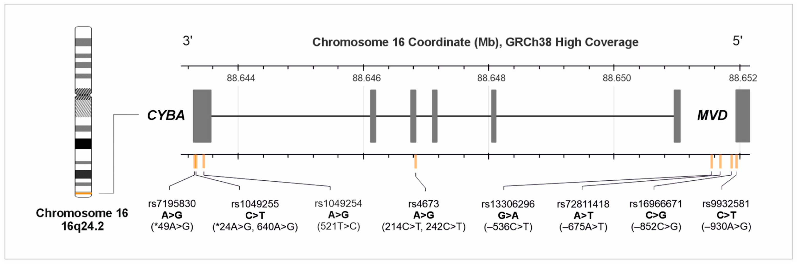
p22phox expression has been associated with oxidative stress and has been reported to be increased in atherosclerotic plaques as well as in myocardial infarction and stroke. The CYBA gene (16q24), which encodes the p22phox subunit, contains multiple polymorphic loci that have been extensively investigated for associations with CVD susceptibility [57,58,59,60]. The location of common SNPs in the CYBA gene is shown in Figure 4, based on data from LDlink.
Figure 4.
Location of single-nucleotide polymorphisms in the CYBA gene. Legend: CYBA, cytochrome b-245 alpha chain gene; GRCh38, Genome Reference Consortium Human Build 38; Mb, megabase; MVD, mevalonate diphosphate decarboxylase gene. Figure 4 uses current variant nomenclature and allele orientation; historical variant names used in the original publications are provided in parentheses.
5.1.1. rs4673
Rs4673 A>G (c.214C>T, 242C>T) results in the replacement of histidine by tyrosine at amino acid position 72. This substitution has been proposed to reduce enzymatic activity, potentially by altering heme binding [61]. Several functional studies indicate that the A allele is associated with lower NADPH oxidase activity, although findings differ between tissues and experimental systems. In saphenous veins from 110 patients with CAD, carriers of the A allele had lower NADPH oxidase activity, independently of other risk factors, with both basal and NADH-stimulated superoxide production reduced by about 30% [61]. In neutrophils from 90 healthy adults, superoxide production was 8.7, 7.9 and 5.9 µmol/L/min per 106 cells for the GG, GA and AA genotypes [62]. In lymphoblastoid cell lines from 185 patients with cardiovascular disease, superoxide production was lower in carriers of the A allele, while p22phox mRNA and protein levels did not differ between genotypes [63]. Hypertensive carriers of the GG genotype also had higher phagocytic NADPH oxidase activity and higher plasma von Willebrand factor levels than AA carriers [64]. In the coronary circulation, among 93 patients, GG homozygotes had blunted flow-dependent dilation of epicardial arteries, independently of other risk factors, while dilation to nitroglycerin showed only a trend [65].
Other data are not consistent. One study found the opposite: in neutrophils from 23 healthy subjects, superoxide production after PMA stimulation was higher in carriers of the A allele, although basal production did not differ and HL-60 cells transfected with the variant construct also produced more superoxide [66]. In granulocytes of 81 healthy volunteers and 51 more in a confirmatory study, rs4673 had no consistent effect on enzyme activity [67]. Findings for circulating oxidation markers are also mixed: in 134 patients with type 2 diabetes, oxidized HDL was lower in GA/AA subjects, oxidized LDL was slightly higher and TBARS (thiobarbituric acid reactive substances) were unchanged [68].
In lymphoblastoid cells from 50 healthy individuals, Bedard et al. analyzed the three common CYBA variants both within haplotypes and individually. When considered separately, rs4673 showed no significant association with ROS generation. However, the haplogroup consistently associated with significantly reduced ROS generation was haplogroup C—GGT (rs4673, rs1049254, and rs1049255), which contained all three variants, including rs4673. The authors concluded that the ROS phenotype was better explained by the haplotype than by any single SNP [69]. In the GTEx data assessed for the present review (Table S3), rs4673 was associated with CYBA expression in the aorta and tibial artery, with lower expression associated with the G allele [51].
Individual studies regarding CAD have produced inconsistent results [70,71]. A meta-analysis of CAD (CAD cases defined as CHD, CAD, MI, AMI or unstable angina) including 20 study populations (8845 cases/6855 controls) found no overall association of the A allele with CAD in the allelic model with substantial between-study heterogeneity (OR = 0.93, 95% CI 0.82–1.05; I2 = 67.9%). Stratification by ancestry indicated a lower risk in Asian populations in carriers of the A allele (allelic model: OR = 0.75, 95% CI 0.67–0.84; dominant model: OR = 0.69, 95% CI 0.61–0.79; 7 studies) but no association in Caucasians (OR = 1.04, 95% CI 0.95–1.14; 10 studies) [72]. An earlier meta-analysis reported a marginal CAD risk increase in Caucasians under a recessive model (AA versus GA + GG: OR = 1.21, 95% CI 1.00–1.46; I2 = 29.1%) [73]. In this meta-analysis, CAD was defined as previous MI, angina pectoris or ≥50% luminal obstruction of at least one major epicardial vessel, so a clinical diagnosis without angiography was allowed. The authors attributed the Caucasian signal to confounding by BMI and study design: after adjustment for BMI in meta-regression, ethnicity was no longer significant (p = 0.067). A separate meta-analysis regarding ACS defined as acute MI or unstable angina (10 studies, 6102 cases/8669 controls) found a reduced risk in carriers of the A allele in Asian populations (allelic comparison: OR = 0.73, 95% CI 0.64–0.83; I2 = 39%) with no association among Caucasians (OR = 1.06, 95% CI 0.99–1.14) [74]. None of these meta-analyses detected publication bias by funnel plot or Egger’s test.
The variant has been evaluated in a prospective 7-year follow-up of 1197 patients with angiographically confirmed coronary atherosclerosis and the AA genotype was an independent protective factor against all-cause mortality (HR = 0.47, 95% CI 0.28–0.82) but an independent risk factor for elective revascularization (HR = 2.04, 95% CI 1.18–3.52), while no association with major adverse cardiovascular events (MACE; death, acute coronary syndrome by ESC criteria or stroke) was observed [75]. In 663 non-diabetic patients from a post-infarction cohort, the GG genotype was associated with recurrent coronary events, but only within a subgroup defined by high C-reactive protein and hypercholesterolemia (GG versus GA + AA: HR 3.14, 95% CI 1.34–7.35, p = 0.0084) [76].
5.1.2. rs1049255
Another commonly investigated polymorphism, rs1049255 C>T (A640G), is located within the 3′ untranslated region of CYBA. Direct measurements of enzyme activity are contradictory. No effect on the respiratory burst was found in neutrophils from 90 healthy adults [62]. In contrast, CC carriers produced 28% less superoxide than TT individuals in two independent cohorts of healthy volunteers with an allele-dose effect that remained significant after Bonferroni correction [67]. The same group later reported a consistent set of results for the C allele: lower NADPH oxidase activity in granulocytes of 125 healthy volunteers, lower CYBA mRNA and protein expression, reduced transcript stability and lower activity of an allele-specific luciferase reporter construct in transfected cells [77]. A study in patients with type 2 diabetes found a non-significant trend towards higher superoxide production in CC carriers only in the diabetic group [78].
Bedard et al. found that rs1049255 was associated with reduced ROS generation when analyzed as an individual SNP in both native lymphoblastoid cells and p22phox-deficient cells rescued with CYBA variants. However, haplotype analysis suggested that the observed single-SNP association was better explained by haplogroup C, which contained rs4673, rs1049254 and rs1049255, than by an independent effect of rs1049255 alone. In contrast, a luciferase reporter assay provided evidence of an allele-specific regulatory effect, with the rs1049255 T allele reducing reporter activity (p = 0.0055) [69].
A meta-analysis of eight studies (3904 cases/3498 controls) reported a reduced risk of CHD, although the association reached significance in only one of five genetic models tested (TC versus TT: OR = 0.86, 95% CI 0.77–0.96; I2 = 48.1%) and was restricted to Caucasians (OR = 0.77, 95% CI 0.66–0.91) [79]. CHD was not defined uniformly across the pooled studies: the diagnostic criteria ranged from at least 75% stenosis and at least 50% stenosis to “single- or multi-vessel disease”, myocardial infarction or unstable angina and acute myocardial infarction alone, and the pooled estimates are not stratified by endpoint [79]. In a separate meta-analysis, a sensitivity analysis excluding one study left five studies and showed protective associations under the allelic (OR = 0.84, 95% CI 0.75–0.93), dominant (OR = 0.77, 95% CI 0.64–0.92) and recessive (OR = 0.82, 95% CI 0.69–0.97) models, with no publication bias detected [72]. No association with death, MACE or revascularization was observed for rs1049255 in a 7-year prospective cohort of 1197 patients [75].
5.1.3. rs7195830
Evidence concerning rs7195830 A>G (*49A>G), located in the 3′UTR, also remains limited. The variant lies 25 bp downstream of rs1049255 and was selected as a HapMap tag SNP to represent common genetic variation within CYBA, rather than because a functional effect had already been established [80]. Fan et al. proposed that rs7195830 lies within a miRNA-binding region and suggested that allelic variation at this site might affect miRNA-mediated regulation of CYBA, although this hypothesis was not tested experimentally [80]. In a genetic analysis of renal cell carcinoma, HaploReg annotation placed rs7195830 and the variants in LD with it in an open chromatin region characterized by enhancer histone marks, DNase I hypersensitivity and RNA polymerase II binding, suggesting that this region may influence transcription factor binding and CYBA expression. In GTEx data, analyzed in the same study, the A allele was associated with higher CYBA expression in the terminal ileum (p = 0.027) with a similar but non-significant pattern in the renal cortex (p = 0.31) [81].
The G allele was associated with premature CAD in a Polish case–control study of 481 subjects (242 patients with CAD with angiographic confirmation and 239 controls—blood donors without signs or family history of CAD who did not undergo angiography; GG+AG versus AA: OR = 1.70, 95% CI 1.04–2.76; G versus A: OR = 1.40, 95% CI 1.08–1.83) with a stronger effect among women (GG+AG versus AA: OR = 4.35, 95% CI 1.50–13.20) [82]. The association remained after adjustment for conventional cardiovascular risk factors. The same study also reported associations of the G allele with hypertension and left ventricular hypertrophy [82]. In an analysis of 490 participants from Poland (245 CAD patients and 245 controls), the rs9932581 C/rs7195830 G diplotype was associated with premature CAD (OR 2.29, 95% CI 1.42–3.72, Bonferroni-corrected) and the association was stronger in women (OR 4.08, 95% CI 1.38–12.04) than in men (OR 1.98, 95% CI 1.14–3.45). A second diplotype, rs72811418 A/rs7195830 G, was more frequent in the CAD group but reached significance only in the multivariate model (OR 1.95, 95% CI 1.12–3.40) and the association remained significant after Bonferroni correction [83]. The opposite combination, rs9932581 T/rs7195830 A, was more frequent in healthy women but was not confirmed after adjustment [83].
5.1.4. rs9932581
The rs9932581 C>T (-930A>G) variant lies in the CYBA promoter 930 bp upstream of the ATG codon [84], within a potential binding site for C/EBP transcription factors [85]. In a luciferase reporter assay in VSMCs obtained from spontaneously hypertensive rats, the promoter carrying the C allele had 30% higher reporter expression than the promoter carrying the T allele [84]. In phagocytic cells from 70 patients with essential hypertension and 70 normotensive controls, hypertensive carriers of the CC genotype had higher p22phox mRNA and protein levels, measured by Northern and Western blotting, higher NADPH oxidase activity and lower serum levels of nitric oxide metabolites than hypertensive TT/TC carriers [85]. In the same study, transfection experiments were carried out in three systems: the A7r5 cell line, VSMCs from normotensive rats and cells from spontaneously hypertensive rats. The T-to-C substitution increased reporter gene expression by 30% only in the hypertensive cells and had no effect in the normotensive cells or in the A7r5 line [85]. However, other studies found no effect on superoxide production at all [62,63,67]. Kokubo et al. reported a significant relationship between rs9932581 and hypertension, but the effect was observed exclusively in male patients [86]. Moreover, the rs9932581 C allele has further been linked to elevated brachial and aortic blood pressure [87].
In the context of CAD, the C allele was associated with premature CAD in a case–control study of 480 subjects (240 patients with angiographically confirmed premature CAD vs. 240 blood donors) in a Polish population (C allele carriers versus TT: OR = 2.03, 95% CI 1.21–3.44; after adjustment for traditional risk factors: OR = 2.22, 95% CI 1.17–4.20) [88]. This direction is consistent with the higher NADPH oxidase activity attributed to the C allele in hypertensive individuals. In the same cohort, overweight or obesity was found to be a risk factor for CAD in C allele carriers but not in TT homozygotes [88]. rs9932581 was also shown to modify the association between cigarette smoking and intima-media thickness (IMT) in young healthy adults [89]. On the other hand, the C allele was reported to have a protective effect against premature MI (acute myocardial infarction, STEMI or NSTEMI) in a study of 302 subjects (102 patients aged ≤40 years, 200 age- and gender-matched hospital controls) in an Austrian population (log-additive/per-allele model: unadjusted OR = 0.63, 95% CI 0.45–0.90; adjusted OR = 0.50, 95% CI 0.30–0.81) [90]. In a prospective 7-year follow-up of 1197 patients with angiographically confirmed coronary atherosclerosis, no association of rs9932581 with death, MACE or elective revascularization was observed in either dominant or recessive models [75].
5.1.5. rs1049254
Rs1049254 A>G (c.521T>C; historically 549C>T) is a non-synonymous variant in exon 6 of CYBA that results in a valine-to-alanine substitution at codon 174 (p.Val174Ala; reported in the literature also as Ala174Val). It is one of three variants forming the CYBA haplogroup C, together with rs4673 and rs1049255 [69]. In Epstein–Barr virus-transformed B lymphocytes from 50 healthy individuals, haplogroup C generated significantly less ROS than the most common haplogroup and the same difference was reproduced after transduction of the variants into p22phox-deficient cells. When the three variants were analyzed separately, rs1049254 was associated with ROS generation in the rescued cells only and not in the native cell lines [69]. In a study of multiple sclerosis, NOX2-dependent ROS production was measured by chemiluminescence in blood myeloid cells from 55 healthy donors genotyped at rs1049254 and rs4673. Production was lower in carriers of the rs1049254 G allele, whereas the rs4673 A allele showed only a trend. The association was stronger when both variants were considered together, with the lowest ROS in donors carrying rs1049254/GG and rs4673/AA and the highest in those having the opposite combination [91].
Evidence for coronary disease is limited to a single case–control study in a Chinese population (297 patients with CAD defined as >50% stenosis of at least one major coronary artery on angiography, or symptoms and ECG changes (angina, arrhythmia), or symptoms, cardiac enzyme elevation and ECG changes (MI) and 343 controls without angiographic confirmation). Carriers of the AA and GA genotypes had a higher risk of CAD after adjustment for conventional risk factors (OR = 1.57, 95% CI 1.01–2.46, p = 0.047) [92].
5.1.6. Other CYBA Polymorphisms
Several additional polymorphisms have been examined, although the available evidence is limited and derives largely from single case–control studies with heterogeneous endpoints.
The rs72811418 A>T (-675A>T) promoter variant was predicted to affect HIF-1α binding. Co-transfection experiments supported an allele-specific effect, as HIF-1α increased luciferase activity in the A allele construct, whereas no change was observed for the T allele construct [93]. Subjects with the AA genotype also showed higher NADPH oxidase-dependent superoxide production and greater carotid IMT than carriers of the T allele, although the variant explained only about 1% of the variability in IMT [93]. These results should be interpreted with caution because the T allele was relatively rare (frequency <0.10) and no TT homozygotes were identified among hypertensive subjects [93]. The variant was not independently associated with CAD, although an association was observed when it occurred in specific diplotypes with rs9932581 [83]. In a Polish case–control study of 490 subjects (CAD in this study was angiographically documented in 245 patients aged 27–55 years), analysis identified the rs9932581 C/rs72811418 A diplotype as a CAD risk factor, whereas the rs9932581 T/rs72811418 A diplotype was associated with reduced CAD risk in women. rs72811418 A/rs7195830 G was more frequent in the CAD group, but the difference reached significance only after multivariate adjustment (OR 1.95, 95% CI 1.12–3.40) and with a stronger effect in women (OR 15.98, 95% CI 3.20–79.44) [83]. In a prospective study of 276 Polish patients with angiographically confirmed CAD, no TT homozygotes were identified. Compared with AA homozygotes, TA heterozygotes had a higher risk of cardiovascular death both at 5 years (HR = 3.27, 95% CI 1.01–10.61) and at 10 years (HR = 2.37, 95% CI 1.01–5.53) [94].
Other variants have been examined in relation to peripheral vascular disease and the extent of coronary atherosclerosis. Within the Polish cohort, the GG genotype of rs13306296 G>A (-536C>T) occurred more frequently in patients with peripheral artery disease and carriage of the G allele of rs16966671 C>G (-852C>G) was associated with multivessel stenosis [83]. These findings concern different vascular phenotypes and cannot be equated with evidence of angiographically confirmed disease or incident coronary events. In the GTEx data assessed for the present review (Table S3), rs13306296 was associated with CYBA expression in the aorta, tibial artery, atrial appendage, left ventricle and whole blood, with lower expression associated with the A allele, and rs16966671 was associated with CYBA expression in the aorta, tibial artery and left ventricle, with higher expression associated with the G allele [51].
Overall, studies of CYBA polymorphisms provide evidence for their potential functional importance, but their association with CAD remains inconsistent. Reported associations vary between populations, differ by sex and clinical endpoints and many findings come from single studies without independent replication or confirmation in meta-analyses.
None of the analyzed CYBA variants have been reported as a variant directly associated with CAD in the GWAS Catalog. In the primary-discovery CAD GWAS summary statistics of Aragam et al., rs4673 showed no significant association: genomic-forward-strand G versus A, OR 0.9917 (95% CI 0.9814–1.0021), p = 0.1181 [49]. For rs4673, genome-wide significant associations were reported, but they concerned blood-pressure phenotypes rather than CAD. Screening of the region 500 kb upstream and downstream of each variant (chr16: approximately 88.15–89.15 Mb) identified a single genome-wide significant CAD lead variant, rs55880988 (chr16:88,479,845; p = 5 × 10⁻⁹; mapped gene ZFPM1), reported in a multi-ancestry association study of CAD [95]. All eight variants were in linkage equilibrium with rs55880988 in the European reference population (R2 ≤ 0.010; D′ ≤ 0.24), and the same result was obtained in East Asians (R2 ≤ 0.004). These results suggest that rs55880988 is unlikely to explain the reported CYBA associations through LD, but they do not establish an independent effect of the CYBA variants on CAD.
In MR analyses, rs1049254, used as the lead cis-eQTL for CYBA expression, was not associated with MI [96]. rs13306296, used as an instrument for CYBA expression, was likewise not associated with CAD or coronary atherosclerosis [97,98].
A summary of data on CYBA gene polymorphisms, their impact on the molecular phenotype, and CYBA gene expression in tissues of the cardiovascular system, as well as their association with the risk of cardiovascular diseases, is presented in Figure 5. Additional information can be found in Supplementary Table S4.
Figure 5.
Summary of data on CYBA gene polymorphisms and their association with molecular and clinical phenotype of CAD. Legend: CAD, coronary artery disease; CHD, coronary heart disease; CV, cardiovascular; HT, hypertension; MACE, major adverse cardiovascular events; SNP, single-nucleotide polymorphism. Figure 5 uses current variant nomenclature and allele orientation; historical variant names used in the original publications are provided in parentheses. The direction of the arrows indicates an increase or decrease in NADPH oxidase activity, CYBA gene expression, or the risk of developing the phenotype or experiencing a cardiovascular event. Full bibliographic details are provided in the main text and Supplementary Table S4.
5.2. Xanthine Oxidoreductase
XOR is encoded by the XDH gene located on chromosome 2p23.1 and catalyzes the terminal steps of purine catabolism—converting hypoxanthine to xanthine and xanthine to uric acid [99,100]. The enzyme exists in two interconvertible forms: xanthine dehydrogenase (XDH), which transfers electrons to NAD+ and xanthine oxidase (XO), which transfers electrons to molecular oxygen, generating superoxide and hydrogen peroxide. Conversion of the dehydrogenase to the oxidase form occurs reversibly through oxidation and irreversibly through proteolysis, under hypoxic, ischemic and inflammatory conditions [101]. Circulating XO binds to vascular endothelial cells, where it increases ROS production and reduces NO bioavailability [99].
Published XDH genetic association studies have primarily examined blood pressure or hypertension, not CAD susceptibility. Associations involving rs1042039, rs1054889 and rs2073316 were reported in Han Chinese participants [102], rs2295475, rs207454 and rs45547640 in Japanese men [103], rs2043013, rs11904439 and rs148756340 in European cohorts [104], and rs206860 in Korean women [105]. These findings concern non-coronary blood-pressure phenotypes and do not constitute direct evidence of XDH-associated CAD risk.
This limited CAD-specific evidence should also be considered in the context of the genetic determinants of serum urate and the post-translational regulation of XOR activity. The causal role of urate in CVD remains uncertain. Large-scale GWAS indicate that serum urate levels are primarily influenced by urate transport genes, particularly SLC2A9 and ABCG2, which account for 3.4% of the 7% variance explained by 26 identified loci [106]. Although an initial Mendelian randomization study suggested a causal association between serum urate and CHD, this was not supported after accounting for pleiotropy [107], while a subsequent study found no clinically relevant causal association with CHD or MI [108]. Similarly, the ALL-HEART trial found no reduction in major cardiovascular events with allopurinol in patients with ischemic heart disease [109]. Furthermore, XDH variants may not adequately reflect XOR activity, which is also regulated post-translationally under oxidative, ischemic and inflammatory conditions.
None of the ten XDH variants analyzed (rs1042039, rs1054889, rs2073316, rs2295475, rs207454, rs45547640, rs2043013, rs11904439, rs148756340, rs206860) has been reported in the GWAS Catalog as directly associated with CAD and no genome-wide significant CAD or MI lead variant was identified in their overlapping ±500 kb region. The only additional GWAS evidence found concerned an association between rs2073316 and circulating interleukin-18 levels [110]. Therefore, the available genetic evidence does not establish XDH variation as a CAD susceptibility factor.
The available studies regarding XDH polymorphisms mainly concern hypertension and other non-coronary phenotypes. Evidence regarding CAD from candidate-gene studies and large GWAS remains insufficient.
5.3. Myeloperoxidase
Myeloperoxidase (MPO) is a heme-containing enzyme stored in azurophilic granules of neutrophils and monocytes. After leukocyte activation, MPO is released into the extracellular space, where it uses hydrogen peroxide generated during the respiratory burst to produce highly reactive oxidants, particularly hypochlorous acid (HOCl). Although this system has an important antimicrobial role, sustained MPO activity within the vascular wall may contribute to oxidative modification of lipoproteins and progression of atherosclerosis [111].
MPO-derived oxidants can modify both LDL and HDL particles. In vitro, the MPO-H2O2-nitrite system induced apoB-100 nitration and LDL lipid peroxidation, producing modified LDL that promoted macrophage cholesterol accumulation and foam-cell formation [112]. HDL isolated from human atherosclerotic lesions contains higher levels of 3-chlorotyrosine than plasma HDL, supporting its modification by MPO-derived chlorinating oxidants [113]. MPO binds to apolipoprotein A-I (apoA-I), and MPO-mediated oxidative modification of the protein can impair ABCA1-dependent cholesterol efflux from macrophages [114]. Modified apoA-I also has a reduced ability to activate lecithin:cholesterol acyltransferase (LCAT) [115]. Together, impaired ABCA1-dependent cholesterol efflux and reduced LCAT activation may compromise early steps in reverse cholesterol transport [114,115]. In endothelial cell experiments, MPO-modified HDL also lost anti-inflammatory activity and promoted inflammatory signaling [116].
The best-characterized functional polymorphism of the MPO gene is the promoter variant rs2333227 C>T (-463G>A). The polymorphic site lies within an Alu regulatory element containing an SP1-binding motif. The C allele creates a strong SP1 binding site and is associated with higher transcriptional activity, whereas substitution with the T allele disrupts this site and results in lower promoter activity and reduced MPO expression. Reporter gene assays showed higher transcriptional activity of the C promoter allele compared with the T allele [117].
Although reporter assays support higher transcriptional activity of the C allele, studies in primary human cells have produced less consistent results. Rutgers et al. reported genotype-dependent differences in MPO activity in selected age- and sex-specific subgroups [118], whereas Videm et al. found no association of rs2333227 with plasma MPO concentration, neutrophil degranulation or stimulated reactive oxygen metabolite production [119]. These findings suggest that the transcriptional effect of rs2333227 may be context-dependent and does not necessarily translate into measurable differences in MPO release or oxidant production in circulating neutrophils.
The association of rs2333227 with CAD has been investigated in several populations, with a protective effect of the T allele reported in several of them. In a French-Canadian case–control study of 229 patients with angiographically confirmed CAD (at least one coronary stenosis of ≥30% of luminal diameter) and 217 controls (no history of CAD, stroke or TIA who did not undergo angiography), the T allele was less frequent among patients. In a recessive model, TT homozygotes had a significantly reduced CAD risk compared with carriers of the C allele (TT vs. TC + CC; OR 0.138, 95% CI 0.040–0.474). A protective association was also observed in a dominant model comparing carriers of the T allele with CC homozygotes (TT + TC vs. CC; OR 0.639, 95% CI 0.436–0.937) [120]. In a Chinese study of premature CAD (diagnosis before age 55 in men or 65 in women and by ≥50% stenosis of at least one major coronary artery on angiography or a previous MI according to WHO criteria), the TT genotype was similarly associated with substantially lower disease risk (OR 0.172, 95% CI 0.057–0.526) [121].
A 2013 meta-analysis of five studies including 1938 CAD cases and 1990 controls found no significant association between rs2333227 and overall CAD risk under any model, with significant heterogeneity in the additive, recessive and allelic models that the authors attributed to differences in age and sex distribution [122]. This meta-analysis applied no single CAD definition: it included studies with CAD diagnosed on the basis of angiographic features, or clinical or laboratory findings. A later meta-analysis of 9 studies including 4744 subjects, with CAD diagnosed according to the 1979 WHO criteria, reported a protective association for the T allele in the allelic model (T versus C: OR = 0.68, 95% CI 0.54–0.85), as well as under dominant (TT + TC versus CC: OR = 0.68, 95% CI 0.53–0.87) and recessive models (TT versus TC + CC: OR = 0.41, 95% CI 0.22–0.76). In ancestry-stratified analyses, the allelic association was present in Chinese populations but not in non-Chinese populations [123]. Another meta-analysis included five case–control studies of rs2333227 (1881 cases and 5554 controls) and found a protective association in the dominant model in the overall analysis (TT + TC versus CC: OR = 0.872, 95% CI 0.77–0.99). No significant association with CAD was found in Caucasian populations under any genetic model. However, in Asian populations the TT genotype was associated with lower CAD risk in both the homozygous comparison (TT versus CC: OR = 0.353, 95% CI 0.16–0.78) and the recessive model (TT versus TC + CC: OR = 0.372, 95% CI 0.17–0.81) [124]. The pooled CAD endpoint combined angiographically confirmed CAD (stenosis threshold ≥50% or ≥30%, depending on the study) with MI diagnosed by WHO criteria (enzymes, ECG, symptoms). The largest meta-analysis of 19 studies with 4008 cases and 3460 controls evaluated CHD based on diagnoses reported in the included case–control studies with separate analyses of stable angina and acute coronary syndrome and similarly reported protective associations across several models in Asian populations (13 studies) and no association in Caucasian populations (6 studies) with no evidence of publication bias [125].
Additionally, in a small study of 49 young healthy men, CC homozygotes had lower coronary flow reserve and lower adenosine-stimulated myocardial blood flow than TT/TC carriers after adjustment for age, body mass index, smoking and family history of cardiovascular disease [126]. In the Helsinki Sudden Death Study among men aged <53 years, carriers of the rs2333227 T allele (TT/TC) had a larger area of fibrotic and calcified lesions in the abdominal aorta than CC homozygotes. In men aged ≥53 years, CC homozygotes had more extensive fibrotic lesions in the thoracic aorta, but this association was not independent of other atherosclerosis risk factors [127]. The endpoint was the percentage of the aortic surface covered by fatty streaks, fibrotic and calcified lesions, measured by computerized planimetry of autopsy specimens from 266 men. In contrast, another angiographic study found no association between rs2333227 genotypes and CAD severity, although a weak association was observed between plasma MPO levels and the extent of CAD [128]. The endpoint was the percentage of the aortic surface covered by fatty streaks, and fibrotic and calcified lesions, measured by computerized planimetry of autopsy specimens from 266 men.
A second MPO promoter polymorphism, rs34097845 C>T (-129G>A), has been investigated in relation to circulating MPO concentrations and neutrophil enzyme activity. In 82 healthy families from the STANISLAS cohort, the T allele was associated with lower serum MPO concentrations [129]. Lower neutrophil MPO activity was also reported for rs34097845 in a study of 243 healthy individuals [118]. In 305 healthy blood donors, the T allele was associated with lower MPO concentrations in unstimulated blood (p < 0.05), although the differences between genotypes were small and the variant did not affect degranulation or reactive oxygen metabolite production after neutrophil stimulation [119]. More recently, a molecular quantitative trait locus analysis identified rs34097845 as significantly associated with both MPO gene expression and circulating MPO protein levels, with consistent directions of effect [130].
Associations with coronary outcomes have been inconsistent. In a Turkish case–control study of 201 patients with angiographically confirmed CAD (defined as >50% atherosclerotic stenosis of at least one major coronary vessel on coronary angiography) and 201 controls (healthy on clinical examination, ECG and echocardiography but did not undergo angiography), the T allele was more frequent among patients and CT heterozygotes had higher CAD odds than CC homozygotes in the unadjusted analysis (OR = 1.94, 95% CI 1.30–2.89). However, the association was no longer significant after adjustment for age, sex, smoking, hypertension, diabetes and hypercholesterolemia (adjusted OR = 1.23, 95% CI 0.69–2.18) [131]. A meta-analysis of three studies with 1610 cases and 1739 controls found no overall association with CAD under the dominant model (TT + TC versus CC: OR = 0.906, 95% CI 0.74–1.10; I2 = 31.5%). The authors noted that the limited number of studies and participants reduced the ability to detect a modest association [124]. Although rs34097845 has been associated with MPO concentrations and neutrophil activity, the available evidence does not establish a consistent relationship with CAD susceptibility.
In the GWAS Catalog search performed for this review, neither rs2333227 nor rs34097845 was identified as directly associated with CAD. Reported associations concerned MPO measurements rather than coronary outcomes. However, in the full CAD GWAS summary statistics, rs2333227 showed a nominal association (the effect allele T (corresponding to the MPO promoter -463A allele), OR 1.022, 95% CI 1.010–1.035; p = 4.53 × 10⁻⁴; Table S2), below the genome-wide significance threshold [49]. The direction of this association was opposite to the protective effect of the T allele reported in several earlier studies and meta-analyses. Furthermore, no genome-wide significant lead variant for CAD, MI, ACS or IHD was identified within the ±500 kb region surrounding both variants [132].
In cis-pQTL MR, rs34097845, the main protein QTL for plasma MPO, was not associated with CHD (OR 1.03, 95% CI 0.93–1.15) [133]. Null results for CAD and MI were also reported in proteome-wide MR studies using this variant [134,135]. A six-variant cis instrument for plasma MPO that included rs2333227 showed a nominal association with CHD (OR 1.08, 95% CI 1.02–1.14; p = 0.011) [136].
Overall, evidence linking MPO promoter variants with CAD remains heterogeneous. For rs2333227, a protective association of the T allele has been reported more consistently in Asian than in non-Asian populations, but this is not supported by genome-wide evidence, where the nominal association in the large CAD GWAS was pointed in the opposite direction. For rs34097845, although associations with MPO expression and circulating levels have been reported, evidence for CAD susceptibility remains inconclusive and available MR findings are largely null.
6. Role of Antioxidant Gene Variants in Susceptibility to CAD and CVD
Endogenous antioxidants can be broadly divided into two categories: enzymatic antioxidants, including SOD, CAT and GPx and non-enzymatic antioxidants, for example: glutathione, uric acid and vitamins C and E [137]. Polymorphisms within genes encoding enzymatic antioxidants have been investigated as potential modulators of oxidative stress burden and as factors that may influence individual susceptibility to CVD.
6.1. Superoxide Dismutases
Superoxide dismutases are enzymes that catalyze the dismutation of superoxide anions to hydrogen peroxide (H2O2). Three distinct isoenzymes have been defined: SOD1, also termed copper-zinc superoxide dismutase (CuZn-SOD); SOD2, known as manganese superoxide dismutase (Mn-SOD); and SOD3 or extracellular superoxide dismutase (EC-SOD). They are respectively encoded by the SOD1, SOD2 and SOD3 genes [137,138]. SOD1 limits intracellular superoxide accumulation and contributes to the maintenance of vascular function, as shown by impaired endothelium-dependent vasodilation in SOD1-deficient mice [139]. In neutrophils, SOD1 also helps limit oxidative damage during the respiratory burst while supporting MPO-dependent oxidant production and antimicrobial activity [140]. SOD3 protects nitric oxide (NO) in the extracellular vascular compartment by removing superoxide, thereby reducing NO inactivation and limiting peroxynitrite formation [141,142]. In patients with CAD, lower endothelium-bound EC-SOD activity was associated with impaired endothelium-dependent vasodilation [143]. SOD2 acts within mitochondria, where it limits superoxide accumulation and helps preserve mitochondrial function. Partial SOD2 deficiency in hyperlipidemic mice increased vascular oxidative stress, atherosclerotic burden and features of plaque instability [144]. A summary of the data on SOD gene polymorphisms and their association with CVD risk is presented in Table 1.
6.1.1. Copper-Zinc Superoxide Dismutase
The 50 bp insertion/deletion polymorphism rs36232792 is located within a SOD1 promoter region containing two predicted SP1, two C/EBP and one AP2 binding sites. Functional assays showed that the deletion reduced SOD1 promoter activity by approximately 40–50% in several human and rodent cell lines [145]. EMSA demonstrated in vitro SP1 binding to the wild-type 50 bp sequence, and ChIP analysis confirmed this interaction in vivo, with no binding signal in deletion homozygotes. These findings indicate that the deletion removes functional SP1 binding sites and reduces SOD1 promoter activity [145].
Measurements in human tissue show effects in the same direction but of a smaller magnitude. In peripheral blood cells of 47 healthy Iranian volunteers, heterozygotes had about 33% less SOD1 mRNA than Ins/Ins individuals, and no Del/Del carriers were present in that sample [146]. In erythrocytes of 282 Swedish control subjects, the 50 bp deletion allele was associated with a 5% reduction in SOD1 enzymatic activity, whereas no corresponding correlation was found in 510 patients with amyotrophic lateral sclerosis (ALS) [147]. In 471 participants of the MASHAD cohort, serum SOD activity was inversely related to the number of deletion alleles: 2.66 ± 1.42 U/ml in Ins/Ins, 2.34 ± 1.19 U/ml in Ins/Del and 1.65 ± 1.07 U/ml in Del/Del (p = 0.003) [148].
However, quantification of mRNA in peripheral blood mononuclear cells (PBMCs) of 48 Italian patients with sporadic ALS showed no significant difference between genotypes (p = 0.3), although mean values decreased from 0.33 in individuals homozygous for the non-deleted allele to 0.30 in heterozygotes and 0.21 in the three subjects homozygous for the deletion [149]. In an Iranian keratoconus study, individuals with the Del/Del genotype had lower SOD1 activity, lower total antioxidant capacity and higher malondialdehyde concentrations (p < 0.05), although the Del/Del genotype was present in only 0.69% of the 733 participants [150]. Interpretation of these findings is limited by the small number of deletion homozygotes.
In an Iranian case–control study (200 CVD cases/200 controls), both the Ins/Del and Del/Del genotypes were associated with increased CVD risk (OR = 2.10, 95% CI 1.34–3.29 and OR = 4.81, 95% CI 1.73–13.35, respectively), and the estimates were similar after adjustment for age and sex (OR = 2.29, 95% CI 1.41–3.70 and OR = 5.90, 95% CI 2.06–16.88). Cases and controls differed significantly in ancestry, with Balouch subjects accounting for 40.9% of cases and 10.5% of controls and Afghan subjects for 1.0% and 24.0%, respectively (p < 0.001) [151]. Cases were defined by the occurrence of MI, unstable angina with ischemic electrocardiographic changes, or death from CHD or atherothrombotic stroke, so the endpoint was not restricted to coronary events [151]. In the prospective MASHAD cohort, including 200 participants with incident CVD events and 271 event-free participants, the Del allele was associated with an increased risk of CVD at 120 months (RR = 7.16, 95% CI 3.81–13.43). A significant association was also observed for the Del/Del genotype (RR = 5.59, 95% CI 1.04–29.99), whereas the Ins/Del genotype was not significantly associated with CVD risk at this time point [148]. CVD events were described as stable and unstable angina, MI and sudden cardiovascular death.
An analysis of GTEx data by Gómez-Meda et al. linked the rs1041740 T allele to lower SOD1 expression in both sun-exposed and non-exposed skin [152]. In silico analysis placed the variant within predicted binding sites for OSR2, ZNF652, PRDM6 and GLI2 and within interaction sites for C/EBPβ and p300, but none of these predictions have been tested experimentally.
In the prospective, community-based Yamagata (Takahata) Study, 2799 Japanese participants aged >40 years were followed for approximately 10 years. Among the analyzed SOD1 variants, rs1041740 showed an association—homozygous TT carriers had a higher risk of both all-cause mortality (adjusted HR = 1.53, 95% CI 1.01–2.30) and cardiovascular mortality (adjusted HR = 2.07, 95% CI 1.10–3.91) compared with CC carriers, after adjustment for conventional cardiovascular risk factors [153]. When the rs1041740 TT genotype was included alongside established risk factors, discrimination of cardiovascular mortality improved, with a net reclassification index of 0.108 (95% CI 0.010–0.207, p = 0.03) and an integrated discrimination index of 0.015 (95% CI 0.0001–0.030, p = 0.05) [153]. Haplotype analysis (five genotyped SOD1 variants: rs4998557, rs2070424, rs1041740, rs4817420 and rs17880487) pointed in the same direction: haplotype 2 (GATTC), carrying the minor T allele of rs1041740, was associated with increased susceptibility to cardiovascular mortality, whereas haplotype 4 (AGCCT), carrying the minor T allele of rs17880487, was associated with lower risk. The authors describe the two haplotypes as complementary, since they carry opposite alleles at each of the five genotyped positions. Consequently, haplotype 4 also carries the low-risk C allele of rs1041740 and the two haplotype results cannot be regarded as independent findings [153]. For rs17880487, no separate estimate was obtained, as no cardiovascular death occurred among the 29 TT carriers (1% of the cohort) [153].
Lastly, a cohort of 3744 European Caucasian patients with type 2 diabetes was followed for 6 years. Seven variants across the SOD1 region were analyzed: rs9974610 and rs2173962, located approximately 13.6 kb and 9.9 kb upstream of the transcription start site, rs10432782 in intron 2, rs2070424 in intron 3, rs1041740 in intron 4, and rs17880135 and rs202449, located approximately 0.7 kb and 5.0 kb downstream of the end of exon 5. These were selected as tag SNPs capturing about 90% of the allelic variation in polymorphisms with a minor allele frequency of at least 5% and r2 > 0.8 within the haplotype blocks containing SOD1 [154]. Little functional information is available for the two upstream and intronic variants that emerged from this analysis: genotypes at rs9974610 were compared for SOD1 mRNA expression in Epstein–Barr virus-transformed lymphoblasts, serum SOD1 mass and serum SOD activity in the DCCT/EDIC study and no differences were detected [155]. For rs10432782, GG homozygotes showed lower plasma SOD1 activity than TT homozygotes among noise-sensitive workers, whereas plasma malondialdehyde levels were higher in GG than in TT homozygotes among noise-resistant workers; neither association was reproduced in the other susceptibility group [156]. In the diabetic cohort, TT homozygotes at rs1041740 had an increased risk of death from cardiovascular causes, defined as sudden death or fatal MI or stroke (HR = 1.78, 95% CI 1.10–2.78, p = 0.02, recessive model), as did carriers of the minor allele of rs10432782 (HR = 1.71, 95% CI 1.16–2.48, p = 0.007), whereas carriers of the minor allele of rs9974610 had a decreased risk (HR = 0.64, 95% CI 0.46–0.88, p = 0.005). For rs2173962, the hazard ratio did not reach the significance threshold adopted by the authors (HR = 1.80, 95% CI 1.06–2.90, p = 0.03; threshold p ≤ 0.02), and the remaining three variants showed no association. All estimates were adjusted for sex, age, body mass index, duration of diabetes, HbA1c, lipid parameters, urinary albumin excretion, hypertension, previous MI and treatment allocation and no association with the incidence of non-fatal MI or stroke was observed over the same follow-up [154].
Across studies, effects were reported in opposing directions for different SOD1 variants, were largely restricted to mortality endpoints without corresponding associations with incident cardiovascular events, and were obtained in cohorts of modest size, one of them restricted to patients with type 2 diabetes.
None of the SOD1 variants analyzed in this review have been reported in the GWAS Catalog in association with CAD. All map to a narrow interval on chromosome 21q22.11 (chr21: approximately 31.65–31.67 Mb), so their ±500 kb windows overlap almost completely (chr21: approximately 31.15–32.17 Mb). Although this region contains numerous reported GWAS signals, none of them concern CAD, MI, ACS or IHD. In the GTEx data, summarized in Table S3, SOD1 rs2173962 was associated with genotype-dependent SOD1 expression in whole blood (p = 8.18 × 10⁻⁵).
In summary, data-based MR (SMR) across GTEx tissues, using rs4998557 as an SOD1 expression instrument, was not associated with coronary atherosclerosis (p = 0.107) [98].
Taken together, these findings do not currently support a consistent role for SOD1 variation in coronary risk.
6.1.2. Manganese Superoxide Dismutase
One of the most frequently investigated SNPs in the SOD2 gene is rs4880 A>G (Ala16Val), a transition in exon 2. It replaces alanine with valine in the mitochondrial targeting sequence. This sequence carries the MnSOD precursor into the mitochondrion. Inside the matrix, it is cleaved off, and four monomers assemble into the active enzyme [157]. Computational models predicted an α-helical structure for the Ala targeting sequence and a β-sheet structure for the Val variant [157]. Molecular dynamics simulations indicated that the alanine variant maintains a uniform α-helix, whereas the valine variant loses it rapidly [158]. When both precursors were imported into isolated rat liver mitochondria, the Val precursor was partly arrested within the inner mitochondrial membrane. The import of the Ala precursor gave about 30% higher MnSOD activity and 40% more mature tetramer [157]. In human hepatoma cells transfected with the two variants, mature exogenous protein levels and MnSOD activity were four-fold higher for the Ala construct, and unimported precursor was shown to be degraded by the proteasome; although transcript levels were identical 8 h after transfection, they were two-fold higher for the Ala transcript at 36 h, indicating an additional effect on transcript stability [159].
These experiments used introduced constructs, not the native gene. In a Tunisian case–control study, erythrocyte SOD activity decreased across genotypes, from 1392.6 ± 219.3 U/gHb in GG carriers to 1285.3 ± 188.9 in GA and 979.4 ± 220.4 in AA (p < 10−4) [160]. In a Japanese study, mitochondrial MnSOD activity in leukocytes was 542 U/mg protein in GG carriers, 302 in GA and 134 in AA (p < 0.0001). Cytosolic MnSOD and Cu/ZnSOD activity did not differ between genotypes, so the effect was limited to the mitochondrial compartment. Macrophages from AA carriers were also more likely to die after exposure to oxidized LDL, and apoptosis decreased as mitochondrial MnSOD activity increased [161]. Only two subjects in that study were GG.
Findings in relation to coronary endpoints are inconsistent. A meta-analysis reported a reduced CAD risk associated with the G allele in a dominant model (REM: OR = 0.62, 95% CI 0.49–0.78; 8 studies, 2256 cases/6518 controls; I2 = 69.0%) [162]. After excluding the study whose controls deviated from Hardy–Weinberg equilibrium, the fixed-effect estimate was OR = 0.69 (95% CI 0.61–0.78), with heterogeneity reduced to I2 = 34%. The test for small-study effects was significant before that exclusion. Six of the eight studies were conducted in Asian populations; three recruited patients with diabetes, and the endpoints ranged from angiographic CAD to vasospastic angina. In their own Chinese case–control sample, the protective effect was present for premature CAD (adjusted OR = 0.59, 95% CI 0.40–0.87) but not for late-onset CAD (0.61, 0.33–1.13) [162].
A Tunisian case–control study (164 cases/203 controls) found the association in men only: AA carriers were at about twofold higher risk of CHD than G allele carriers (adjusted OR 1.89, 95% CI 1.18–3.42) [160]. In the same study, SOD activity decreased across genotypes (1392.6, 1285.3 and 979.4 U/gHb for GG, GA and AA, p < 10−4), and the A allele was associated with a higher number of stenosed vessels.
A Japanese case–control study compared 498 patients with angiographically defined CAD (diameter stenosis ≥75%) with 627 healthy subjects recruited at a routine health examination, aged 37–87 and 38–85 years, respectively. The A allele was more frequent among patients (88.5% vs. 83.3%), and the authors found that the AA genotype was associated with higher CAD risk independently of other coronary risk factors (adjusted OR 1.49, 95% CI 1.06–2.11, p = 0.03). Among the 87 patients with acute MI, the association was stronger (adjusted OR 2.89, 95% CI 1.47–5.68, p = 0.002) [161].
In contrast, a Taiwanese case–control study with angiographic CAD confirmation (447 patients with ≥50% stenosis, 210 controls) reported the opposite direction, with G allele carriers at increased risk of CAD (AG + GG vs. AA, adjusted OR 1.86, 95% CI 1.15–3.01), and the association was observed in one- and two-vessel disease but not in three-vessel disease [163]. Only 29 of the 210 controls carried a G allele, so this estimate was based on a small number of carriers. The effect was larger among ever-smokers (OR = 2.23, 95% CI 1.02–4.88) than among never-smokers (OR = 1.79, 95% CI 0.95–3.37), but the interaction with smoking was not significant (p = 0.55) [163].
An Iranian case–control study found no association in any genetic model (90 patients with angiographically defined CAD and without type 2 diabetes vs. 90 controls; dominant model, adjusted OR 1.34, 95% CI 0.69–2.59) [164].
Prospective data for rs4880 are limited to cohorts with diabetes and with composite cardiovascular rather than coronary endpoints. In a Danish cohort of patients with type 1 diabetes, carriers of the A allele were at higher risk of diabetic nephropathy (OR 1.7) and of cardiovascular events (stroke, MI, CABG and PCI) during follow-up of 8.7 years (HR 1.6, 95% CI 1.0–2.5), independently of nephropathy status [165].
The rs4880 GG genotype has also been linked to an increased risk of hypertriglyceridemia combined with low high-density lipoprotein cholesterol (HDL-C), although only 25 subjects in that study had this genotype. Combining the risk genotypes of SOD2 rs4880, SOD3 rs2536512 and rs2855262, GPX3 rs3828599 and the GSTT1 deletion into a genetic risk score strengthened the association (score ≥6 versus 0–2, adjusted OR 5.73, 95% CI 2.22–14.77). None of the variants was associated with the severity of coronary atherosclerosis assessed by the Gensini score, but the high triglyceride-to-HDL cholesterol ratio was (OR 2.26, 95% CI 1.17–4.34). This led the authors to propose that these variants act on CAD through the lipid phenotype rather than directly [166]. rs4880 was also reported as a weak determinant of carotid intima-media thickness [167].
Lastly, the rs4880 variant has no reported genome-wide significant association with CAD or related coronary endpoints in the GWAS Catalog and in the Aragam et al. primary-discovery CAD summary statistics; rs4880 had OR 1.0030 per genomic-forward-strand G allele versus A (95% CI 0.9934–1.0127), p = 0.5399 [49]. In the region 500 kb upstream and downstream of the variant (chr6:159,192,840–160,192,840), several genome-wide significant lead variants for coronary phenotypes were identified, mapped to IGF2R, AIRN, SLC22A1, SLC22A2, MAS1 and PNLDC1, including rs6932293 (p = 2 × 10−37), rs144078421 (p = 1 × 10⁻²⁶), rs9456508 (p = 2 × 10⁻¹⁷), rs9457761 (p = 8 × 10⁻¹⁷), rs688359 (p = 5 × 10⁻¹⁵) and rs2297374 (p = 1 × 10⁻⁸). Despite this dense clustering of CAD signals, rs4880 was in linkage equilibrium with every one of them in the European reference population (R2 ≤ 0.012) and in East Asians (R2 ≤ 0.012). Therefore, the coronary associations reported in this region are unlikely to account for any association observed for rs4880.
6.1.3. Extracellular Superoxide Dismutase
The most extensively studied functional variant of SOD3 is rs1799895 C>G (R213G, p.Arg213Gly), a C to G transversion converting codon 213 from arginine to glycine within the heparin-binding domain [168]. In a study of 504 Swedish blood donors, 11 individuals (2.2%) carried the G allele, all as heterozygotes. Carriers had approximately 8–10-fold higher plasma EC-SOD concentrations than non-carriers, while the specific enzymatic activity of the variant protein remained unchanged [169]. In an independent Japanese study, serum EC-SOD concentrations increased according to genotype, from approximately 186 ng/mL in CC non-carriers to 1009 ng/mL in CG heterozygotes and 2531 ng/mL in the three GG homozygotes identified, all of whom were hemodialysis patients [170]. CG heterozygotes in a French cohort of patients with type 1 diabetes had an approximately seven-fold higher plasma EC-SOD concentration than C/C non-carriers [171].
The marked increase in circulating EC-SOD reflects altered tissue distribution rather than increased catalytic activity. EC-SOD isolated from heterozygous and homozygous carriers showed reduced affinity for heparin, and binding of mutant EC-SOD to bovine aortic endothelial cells was approximately 50-fold lower than that of the normal protein [170]. Subsequent experiments demonstrated that normal EC-SOD is internalized by endothelial cells through a clathrin-mediated pathway, whereas no detectable endocytosis of the variant protein encoded by the rs1799895 G allele was observed. Endocytosed normal EC-SOD showed minimal or no nuclear localization [172]. These findings support a redistribution mechanism in which reduced binding to heparan sulfate proteoglycans and impaired endothelial uptake increase circulating EC-SOD while decreasing its retention within the vascular wall. This may reduce local antioxidant protection despite high plasma concentrations of the enzyme [169,172,173].
Subsequent experiments demonstrated that normal EC-SOD is internalized by endothelial cells through a clathrin-mediated pathway, whereas no detectable endocytosis of the variant protein encoded by the rs1799895 G allele was observed. Low levels were independently associated with a greater likelihood of previous MI (OR = 2.04, 95% CI 1.10–3.82). Plasma EC-SOD concentrations were also lower in current smokers than in non-smokers and lower in men than in women [174]. Importantly, EC-SOD concentration was not associated with the angiographic severity of coronary atherosclerosis.
In the Copenhagen City Heart Study, 9188 participants were followed for a median of 21 years, during which 956 developed IHD (data from the national hospital discharge and cause-of-death registers, ICD-8 410–414/ICD-10 I20-I25). Of the participants, 221 (2.4%) were CG heterozygotes and 2 (0.02%) were GG homozygotes. Heterozygotes had a higher risk of IHD than CC non-carriers (age- and sex-adjusted relative risk 1.5, 95% CI 1.1–2.1) [173]. A later study from the same population genotyped 95,871 individuals, of whom 4498 had diabetes. No association was seen overall (HR 1.06, 95% CI 0.88–1.28), and the effect was limited to participants with diabetes (HR 2.32, 95% CI 1.44–3.75, versus 0.97, 95% CI 0.80–1.19 in non-diabetic participants; p for interaction 0.002). The endpoint was a composite of cardiovascular death, MI and ischemic stroke [175].
Other SOD3 variants have been investigated less extensively. The promoter variant rs2284659 T>G (-1257G>T), located about 2.3 kb upstream of the start of exon 1, was examined in two pooled cohorts of type 1 diabetes (GENEDIAB/GENESIS) and one cohort of type 2 diabetes (DIABHYCAR). In these cohorts (GENEDIAB, GENESIS, DIABHYCAR), MI was defined as at least two of the following: chest pain lasting ≥20 min, raised creatine phosphokinase and/or troponin, or typical ECG changes. In the type 1 diabetes cohorts, the minor T allele was associated with a lower prevalence of MI at baseline (OR 0.48, 95% CI 0.29–0.78, codominant model) and with a lower incidence during follow-up (HR 0.58, 95% CI 0.40–0.83, dominant model), as well as with lower cardiovascular (HR 0.33, 95% CI 0.08–0.74) and all-cause mortality (HR 0.44, 95% CI 0.21–0.73, recessive model). In the DIABHYCAR cohort, the T allele was again associated with a lower incidence of MI (HR 0.75, 95% CI 0.59–0.94) and all-cause mortality (HR 0.87, 95% CI 0.79–0.97), both in a dominant model. In GENEDIAB, T allele carriers had higher plasma EC-SOD concentrations than GG homozygotes (p = 0.007), whereas TT homozygotes had lower AOPP concentrations than G-allele carriers (nominal p = 0.02) [171].
The associations observed for rs2284659 are unlikely to be explained by rs1799895, because LD between the two variants was very low (r2 = 0.009, D′ = 0.687). The molecular mechanism underlying rs2284659 remains uncertain. The authors note that the variant does not appear to alter known transcription-factor binding sites and is in complete LD with two other SOD3 promoter variants, rs2159757 and rs13435617, which fall within predicted binding sites for the PBX, HLF and E4BP4 transcription factors, but this has not been tested experimentally [171].
Two further variants, rs2536512 G>A (Ala58Thr; Ala40Thr) and rs2855262 T>C (5982C>T), were examined together with SOD2 rs4880, GPX3 rs3828599 and the GSTT1 deletion in 396 Thai subjects at high risk of CAD [166]. rs2536512 replaces alanine with threonine at position 58 of SOD3, and GG carriers have been reported to have lower total plasma SOD activity than carriers of the other genotypes [176]. The rs2536512 AA genotype was associated with hypertriglyceridemia and hypertriglyceridemia combined with low HDL-C, and the rs2855262 CC genotype was associated with the same combined phenotype. The authors noted that the two variants are in strong LD and therefore should not be interpreted as independent genetic signals. None of the variants was associated with the severity of coronary atherosclerosis assessed by the Gensini score [166]. A later study found no association between rs2536512 and angiographically defined CAD in 279 patients and 165 subjects without it. The variant was then combined with GPX3 rs3828599, PON1 rs705379 and the GSTT1 deletion into a genetic risk score, calculated as the number of risk alleles carried and ranging from 0 to 8. Compared with subjects carrying two or fewer risk alleles, those carrying six or more had a higher risk of CAD (adjusted OR 3.26, 95% CI 1.29–8.25, p = 0.012) and of multivessel disease (adjusted OR 4.36, 95% CI 1.36–14.00, p = 0.013), but not of single-vessel disease [177].
Table 1.
Summary of superoxide dismutase gene polymorphisms and their association with cardiovascular disease risk. Abbreviations: AMI—acute myocardial infarction; AP2—activator protein 2; bp—base pair; CAD—coronary artery disease; C/EBP—CCAAT/enhancer-binding protein; CHD—coronary heart disease; CV—cardiovascular; CVD—cardiovascular disease; EC-SOD—extracellular superoxide dismutase; eQTL—expression quantitative trait locus; GRS—genetic risk score; GWAS—genome-wide association study; HR—hazard ratio; IHD—ischemic heart disease; Ins/Del—insertion/deletion; MASHAD—Mashhad Stroke and Heart Atherosclerotic Disorder study; MDA—malondialdehyde; MI—myocardial infarction; MnSOD—manganese superoxide dismutase; mRNA—messenger RNA; OR—odds ratio; oxLDL—oxidized low-density lipoprotein; RR—relative risk; SOD—superoxide dismutase; SP1—specificity protein 1. The direction of the arrows indicates an increase (↑) or decrease (↓) in the given parameter (e.g., promoter activity, mRNA expression, enzyme activity) or in the risk of the phenotype or cardiovascular event.
rs1799895 was not reported in the GWAS Catalog as a variant directly associated with CAD, although several studies reported genome-wide significant associations with circulating EC-SOD levels [178,179] consistent with the known functional effect of this substitution on enzyme binding to the vascular endothelium. Nevertheless, CAD GWAS summary statistics from Aragam et al. (2022) showed nominal associations for SOD3 rs1799895 (effect allele C, OR 0.947, 95% CI 0.901–0.996; p = 0.0327), rs2159757 (A, OR 0.981, 95% CI 0.970–0.992; p = 7.24 × 10⁻⁴) and rs2284659 (T, OR 1.014, 95% CI 1.004–1.025; p = 0.00768; Table S2) [49]. None of these associations met the genome-wide threshold, and the differences in outcomes and populations limit direct comparisons. Screening of the region 500 kb upstream and downstream of the variant (chr4:24,300,212–25,300,212) identified no lead variant reaching genome-wide significance for CAD or MI. Thus, an association involving rs1799895 would be unlikely to be explained by known coronary signals in the surrounding region.
In proteome-wide MR, rs1799895 used as an instrument for plasma SOD3 was nominally associated with MI (OR 1.04, 95% CI 1.01–1.07; p = 0.020), but this did not replicate [96]. In the same study, colocalization of the SOD3 protein signal with MI was inconclusive (PP.H4 0.55 in discovery, 0.15 in replication) [96].
6.2. Catalase
CAT is an antioxidant enzyme found primarily in peroxisomes that catalyzes the dismutation of hydrogen peroxide (H2O2) into water and molecular oxygen. By removing H2O2, catalase protects cells from oxidative damage and helps maintain redox homeostasis, particularly in tissues with high metabolic activity and abundant ROS production [180]. Van Empel et al. demonstrated that EUK-8, a synthetic superoxide dismutase and catalase mimetic, reduced cardiac oxidative stress and improved survival in pressure-overloaded harlequin mutant mice with mitochondrial antioxidant deficiency, preventing progression to heart failure [181]. Evidence that hydrogen peroxide contributes to adverse myocardial remodeling comes from animal models: cardiac-specific catalase overexpression prevented progression from compensated hypertrophy to heart failure in Gαq transgenic mice [182].
Three CAT promoter polymorphisms have been studied: rs1001179, rs7943316 and rs769214. In a small cohort of healthy Iranian subjects, rs7943316 and rs1001179 were in LD (D’ = 1.0, r2 = 0.18) [183]. The rs1001179 C>T (C-262T) polymorphism of the CAT gene is located 262 base pairs upstream of the transcription start site in the 5′-flanking region [184]. The two promoter alleles also showed different nuclear protein-binding patterns in electrophoretic mobility shift assays [184]. In 47 healthy Iranian subjects, CAT mRNA levels increased with the number of rs1001179 T alleles (r = 0.501, p < 0.001). When rs7943316 and rs1001179 were considered together, the number of variant alleles was also positively correlated with CAT mRNA expression (r = 0.598, p < 0.001) [183].
A limited number of studies have examined rs1001179 in relation to coronary endpoints, and none found an association. In a Taiwanese case–control study with angiographic confirmation in both groups, the combined C/T + T/T genotype group was not associated with CAD (adjusted OR 1.51, 95% CI 0.77–2.95) and showed no relation to the number of diseased vessels [163]. No association was found with IHD (defined as angina or possible MI on the WHO cardiovascular questionnaire and/or ECG abnormalities and/or fixed or reversible perfusion defects on myocardial scintigraphy, without coronary angiography) in 520 Caucasian-Brazilian patients with type 2 diabetes, with genotype and allele frequencies being similar in patients with and without IHD [185]. Similarly, no association was found with in-stent restenosis (defined as >50% narrowing of the stented segment) in 101 men followed by angiography six months after bare-metal stent implantation (44 with and 57 without restenosis). Seven antioxidant-gene variants were tested, but only GPX1 and NOS3 variants reached significance; no effect estimate was reported for CAT [186]. In 168 Chinese patients with type 2 diabetes, 85 of whom had a history of ischemic CVD, the genotype distribution of rs1001179 did not differ between those with and without such a history [187]. History of ischemic CVD (previous MI, angina or CABG) was taken from the medical history without angiographic verification or stated diagnostic criteria, and the comparison group had type 2 diabetes for ≥10 years without such events [187].
Data on rs769214 G>A (-844A/G) remain limited and raise concern for incident hypertension and perioperative MI. In a study of progression to home-measured hypertension over 12 years in 403 Japanese adults, rs769214 was one of four significant predictors among 51 SNPs tested without correction for multiple comparisons (adjusted OR 1.6, 95% CI 1.0–2.5, p = 0.04, dominant model) [188]. It has also been studied in relation to perioperative MI after cardiac surgery [189].
Another CAT variant, rs7943316 A>T (A-21T, -89A/T), has not been studied in relation to CVD on its own. It appears only as part of a promoter haplotype with rs769214 and rs1049982 T>C (-20C/T): CAT1 (rs769214 G/rs7943316 A/rs1049982 T) and CAT2 (rs769214 A/rs7943316 T/rs1049982 C). CAT2 carriers showed no difference in carotid IMT or baseline carotid diameter but had a greater carotid diameter in interaction with older age and higher systolic blood pressure, and a greater number of carotid atheromatous plaques [190]. Interestingly, many of these CAT polymorphisms have a significant effect on gene expression, both in cardiovascular tissues and in whole blood, as evidenced by GTEx data [51]. Figure 6 presents SNPs in the CAT gene in the context of haplotype block formation (A), based on data from LDlink and gene expression (B, C), using the rs769214 variant as a representative example of the haplotype characterized by high LD and, consequently, a highly similar expression profile of the linked alleles.
Figure 6.
LD of CAT gene common single nucleotide polymorphisms (A) and CAT gene expression of rs769214 (B) and rs1001179 (C) SNP genotypes in aorta, left ventricle and whole blood. Legend: CAT, catalase; D′, normalized linkage disequilibrium coefficient; NES, normalized effect size; R2, squared correlation coefficient. The color intensity in panel A indicates the degree of LD between alleles.
None of the four CAT variants analyzed (rs1001179, rs7943316, rs769214, or rs1049982) has been reported in the GWAS Catalog as directly associated with CAD. For rs1001179, a strong association with type 2 diabetes was reported (p = 9 × 10⁻²⁵) [191]. All four map to a narrow interval on chromosome 11p13, and their ±500 kb windows are largely overlapping. Within this region, the only coronary-related signal identified was rs12285326, reported for coronary atherosclerosis/increased number of diseased vessels, but it did not reach genome-wide significance (p = 5 × 10⁻⁶). None of the CAT variants studied lies within an established coronary risk locus, and no genome-wide significant coronary association was identified in the region examined.
In SMR, rs769214, used as a CAT expression instrument, was not associated with coronary atherosclerosis (p = 0.098) [98]. Serum catalase colocalized with CAD only at a trans locus on chromosome 1p13 (PP.H4 0.993), unrelated to the CAT variants discussed here [192].
Overall, some CAT promoter variants have been associated with differences in CAT expression, but evidence linking individual variants to CAD or IHD is limited and largely null. No CAT variant discussed here has established genome-wide support for CAD, and the available MR and colocalization findings do not implicate these variants in coronary atherosclerosis. Therefore, their role in CAD remains not established.
6.3. Glutathione Peroxidase-1
Glutathione peroxidase 1 (GPx1) is a selenium-dependent antioxidant enzyme that catalyzes the reduction of hydrogen peroxide and organic hydroperoxides, using reduced glutathione as an electron donor. Encoded by the GPX1 gene located in chromosomal region 3p21.3, this enzyme represents a first-line defense against oxidative damage [193]. Reduced GPx1 activity leads to accumulation of hydrogen peroxide and lipid hydroperoxides within the vascular wall, promoting oxidative modification of LDL, and endothelial dysfunction through diminished NO bioavailability and activation of redox-sensitive transcription factors such as NF-κB that promote pro-inflammatory and pro-atherogenic gene expression [194].
Animal studies support this mechanism—heterozygous GPx-1-deficient mice showed impaired endothelium-dependent vascular relaxation in resistance vessels, as well as abnormal diastolic function in hearts subjected to ischemia–reperfusion injury [195]. Moreover, double-knockout mice (GPx-1-/-ApoE-/-) developed atherosclerosis more rapidly than control apolipoprotein E-deficient mice [196]. In patients with CAD, those in the highest quartile of GPx-1 activity had a lower risk of cardiovascular events than those in the lowest (HR 0.29, 95% CI 0.15–0.58, p < 0.001) [197].
The most extensively studied functional polymorphism in GPX1 is rs1050450 G>A (Pro198Leu), which results in substitution of proline with leucine at codon 198. In a case–control study of breast cancer involving 754 Danish postmenopausal women (377 cases and 377 controls), erythrocyte GPX activity fell by 5% for each additional copy of the A allele (p = 0.0003), and this relationship was present both in cases and in controls [198]. In 405 Polish participants, erythrocyte GPx1 activity did not differ between genotypes at all [199]. These findings suggest that the effect of rs1050450 on basal GPx1 activity is not fully consistent across human studies.
It has been implicated in CAD susceptibility across diverse populations. However, individual case–control studies have yielded inconsistent results, likely reflecting population heterogeneity.
Meta-analytic evidence supports an association in Asian populations, although the strength and robustness of the overall association differ between analyses. Ye et al. demonstrated a significant association between rs1050450 and CHD in Chinese and Indian populations (OR 1.61, 95% CI 1.25–2.07, p = 0.0002, fixed-effect model, 4 studies, 1560 cases and 978 controls, I2 = 0%; participants were classified as cases or controls based on coronary angiography) [200]. Zhang et al. pooled 10 studies involving 1430 cases and 3767 controls. The association between rs1050450 and CVD was stronger in East Asians (OR 1.84, 95% CI 1.39–2.43) than in the overall population (OR 1.36, 95% CI 1.08–1.70). The association was not evident in the non-East Asian subgroup (OR 1.08, 95% CI 0.95–1.23). Moreover, the overall association lost statistical significance after adjustment for potential publication bias, whereas no substantial publication bias was detected in the East Asian subgroup [201]. The pooled CVD included CHD, MI, stroke, peripheral vascular disease, in-stent restenosis, coronary artery calcification and metabolic syndrome and only two included studies used angiographically defined CHD. The A allele of rs1050450 was reported to increase susceptibility to in-stent restenosis [186].
In contrast, several studies have not confirmed this association between rs1050450 and CAD, underscoring the variability across studies. In an Iranian angiographic cohort (95 patients with ≥50% coronary artery stenosis and 55 controls), Najafi et al. found no association between rs1050450 and coronary artery stenosis (≥50% in at least one of the RCA, LCX or LAD; controls <5%, assessed by CT coronary angiography) (adjusted OR 0.79, 95% CI 0.28–2.2, p = 0.6). Erythrocyte GPx1 activity also did not differ between cases and controls. This lack of association was consistent with the authors’ bioinformatic analysis, which suggested that the Pro198Leu substitution lies outside the functional regions of GPx1 and is unlikely to affect its stability or function [202]. Further analyses likewise found no association of rs1050450 with CHD risk in Tunisian [160] and Taiwanese populations [163]. In 168 Chinese patients with type 2 diabetes, 85 of whom had a history of ischemic CVD, the genotype distribution of rs1050450 did not differ between those with and without such a history [187].
Importantly, the functional impact of this polymorphism may be modulated by selenium status through a gene-nutrient interaction. In 405 Polish participants, the correlation between erythrocyte GPx1 activity and plasma selenium was strongest in GG homozygotes and weakest in AA. The difference between the GG and AA genotype-specific relationships was formally tested and was statistically significant (p = 0.034) [199]. The authors therefore concluded that rs1050450 may modify the responsiveness of GPx1 activity to selenium rather than determine its basal activity [199]. A further genotype-dependent effect was reported in the Danish cohort, where alcohol intake correlated with higher GPX activity in GG and GA carriers, whereas no increase was observed in AA homozygotes [198].
Beyond rs1050450, other GPX1 variants such as rs1800668, rs3448, rs8179164 and rs3811699 were evaluated, but mainly in functional studies or limited association analyses unrelated to CVD, which limits the ability to draw definitive conclusions and underscores the need for larger, well-controlled replication studies.
A single ±500 kb window covered all five GPX1 variants analyzed, which lie within about 1.5 kb of one another at 3p21.31 (GRCh38). The GWAS Catalog did not identify any of these variants as a reported direct CAD or MI lead signal. However, in the CAD GWAS summary statistics, Aragam et al. (2022) found that rs1050450 (effect allele A: OR 0.967, 95% CI 0.957–0.977; p = 3.27 × 10⁻¹⁰), rs1800668 (A: OR 0.967, 95% CI 0.957–0.977; p = 3.08 × 10⁻¹⁰) and rs3811699 (T: OR 1.034, 95% CI 1.023–1.045; p = 3.05 × 10⁻¹⁰) exceeded the genome-wide significance threshold (Table S2) [49]. The surrounding region contains eleven genome-wide (p = 1 × 10⁻¹³ to 7 × 10⁻⁸) lead variants for coronary phenotypes, mapped to eight genes: TRAIP, LAMB2, RHOA, QRICH1, BSN, DALRD3, CDHR4 and MST1/RNF123. In Europeans, rs1800668 and rs3811699 were strongly correlated with rs11718165 (R2 = 0.92), a coronary lead variant mapped to BSN. Associations involving either of these GPX1 variants could therefore reflect the neighboring coronary signal rather than an independent effect attributable to GPX1. By contrast, rs3448 and rs8179164 showed little correlation with any of the coronary lead variants identified in this region (R2 ≤ 0.141 and ≤ 0.013, respectively), making it unlikely that associations involving these variants would be explained by the established coronary signals at 3p21.31.
In SMR analysis based on blood eQTL data, higher genetically predicted GPX1 expression was associated with increased CAD risk (OR 1.46, 95% CI 1.21–1.77). However, colocalization support was weak (PPH4 0.60), and the association observed in FinnGen (OR 1.24, 95% CI 1.08–1.44; p = 0.003) did not remain significant after FDR correction [203]. Another study reported locus-level colocalization between GPX1 expression and CAD (PP.H4 = 0.80–0.94), although none of the GPX1 variants reviewed here was identified as the lead variant [204]. Similarly, a two-sample MR analysis using blood cis-eQTL instruments from eQTLGen (31,684 samples) predicted that genetically higher GPX1 expression in blood was associated with greater CAD risk (per 1 SD: OR 1.30, 95% CI 1.17–1.44; p = 7.37 × 10⁻⁷), supported by consistent weighted-median estimates [205]. In the same analysis, the rs1050450 A allele was associated with lower CAD risk (OR 0.97; p = 2.06 × 10⁻⁶). A multi-omics MR study, methylation at cg05126514 within BSN was causally associated with CAD through central nervous system- and lifestyle-related traits [206], suggesting a possible alternative mechanism at the 3p21.31 locus that is independent of GPX1.
Overall, evidence linking GPX1 variation to CAD remains heterogeneous and may vary by ancestry and environmental context. Candidate-gene studies and meta-analyses report increased risk mainly in East Asian populations, whereas findings in non-East Asian populations are largely null or inconsistent [200,201,202]. Although CAD GWAS show genome-wide-significant associations for rs1050450, rs1800668 and rs3811699 [49], the latter two are strongly correlated with a neighboring coronary lead variant at the BSN locus, so their associations cannot be confidently attributed specifically to GPX1. MR and colocalization findings are suggestive of a possible relationship between GPX1 expression and CAD but do not establish a causal effect.
6.4. Glutathione System
6.4.1. Glutathione Synthesis
Glutathione (γ-L-glutamyl-L-cysteinyl-glycine; GSH) is the principal intracellular antioxidant, participating in detoxification of electrophilic xenobiotics via GSTs, maintaining protein thiol status and contributing to regulation of NO homeostasis in the vasculature [207].
GSH biosynthesis occurs through two sequential ATP-dependent enzymatic reactions. The first step is catalyzed by glutamate-cysteine ligase (GCL), a heterodimeric enzyme composed of a catalytic subunit (GCLC, encoded by GCLC on chromosome 6p12.1) and a modifier subunit (GCLM, encoded by GCLM on chromosome 1p22.1). GCLC has all catalytic activity and is subject to feedback inhibition by GSH, while GCLM modulates enzyme kinetics by lowering the Km for glutamate and raising the Ki for GSH. The second step is catalyzed by glutathione synthase (GS), which adds glycine to γ-glutamylcysteine to complete GSH synthesis [207,208].
Both GCLC and GCLM promoters contain antioxidant or electrophile response elements (ARE/EpREs) mediating transcriptional upregulation by NF-E2-related factor 2 (Nrf2) in response to oxidative stress and other electrophilic stimuli [207]. Given the central role of GSH in vascular redox homeostasis, genetic polymorphisms affecting GCL expression represent potential candidates for CVD susceptibility.
Nakamura et al. identified a C>T transition at position -588 in the GCLM promoter (rs41303970 G>A; also reported as -590C>T). In reporter assays, constructs carrying the A allele showed reduced oxidant-induced promoter activation. Under oxidant exposure, luciferase activity was approximately 40–50% of that observed with the G allele in HUVECs, THP-1 and HeLa cells. Basal promoter activity was similar between the alleles, suggesting that the variant mainly affected the response to oxidative stress rather than baseline transcription [209]. In monocyte-derived macrophages, the increase in GCLM mRNA after oxidant exposure was lower in GA than in GG subjects. Carriers of the A allele also had significantly lower plasma GSH levels in a subgroup of 84 subjects matched for traditional risk factors and previous MI (48 GG and 36 GA + AA) [209]. The proportion of A-allele carriers was higher among Japanese MI patients than among angiographically defined controls (GA and AA genotypes: 31.5% of 429 patients vs. 19.2% of 428 controls; p < 0.001), and multiple logistic regression analysis confirmed that the A allele of the rs41303970 polymorphism was an independent risk factor for MI after adjustment for traditional cardiovascular risk factors (OR 1.98, 95% CI 1.38–2.83; p < 0.001) [209].
A subsequent clinical study demonstrated that rs41303970 was associated with impaired endothelium-dependent coronary vasomotor function. Analysis in 157 consecutive subjects with angiographically normal coronary arteries showed that carriers of the A allele had significantly impaired dilation (or enhanced constriction) of epicardial coronary arteries and blunted increases in coronary blood flow in response to intracoronary acetylcholine infusion. These associations remained significant after multivariate adjustment for traditional risk factors. In a subgroup of 59 subjects, A-allele carriers also showed a reduced response to intracoronary NG-monomethyl-L-arginine, suggesting lower basal coronary NO activity, while the response to nitrate was unchanged. These findings support the role of the GCL-GSH axis in endothelial NO bioactivity [210]. In type 2 diabetic patients (n = 1746), Katakami et al. demonstrated that the GCLM rs41303970 polymorphism was significantly associated with carotid IMT, with mean IMT rising across genotypes (0.87 ± 0.19 mm for GG, 0.91 ± 0.23 mm for GA and 0.93 ± 0.35 mm for AA; r = 0.090, p = 0.0008), an association that persisted in stepwise multivariable regression (F = 11.8, p = 0.0006). In the same cohort, the accumulation of pro-oxidant alleles across four oxidative stress-related genes, including the GCLM A allele, was an independent risk factor for carotid atherosclerosis (r = 0.108, p < 0.0001; F = 13.5, p = 0.0003) [10].
Similarly, Racis et al. included this polymorphism in a genetic risk score analysis and found that the cumulative number of pro-oxidant alleles from eight variants in six oxidative stress-related genes was independently associated with the extent of coronary atherosclerosis as quantified by the Gensini score (Gensini score ≥1, every stenosis of 25–100% scored) when the highest-risk group was compared with the lowest (9–12 risk alleles vs. 1–2: OR 2.47, 95% CI 1.19–5.23, p = 0.014; the GRS was also independently associated with a higher Gensini score in multivariable analysis). However, rs41303970 analyzed individually showed no association with the Gensini score (A allele carriers vs. GG: OR 1.00, 95% CI 0.78–1.31, p = 0.953) [211].
Two further studies in Japanese patients with type 2 diabetes did not confirm an independent association of rs41303970 with coronary outcomes. In 3819 subjects (226 patients with and 3593 without MI), rs41303970 was not associated with the prevalence of MI (ascertained from clinical records, characteristic ECG changes and coronary angiography or echocardiography findings) when analyzed alone, and only the cumulative number of pro-oxidant alleles across five variants was an independent risk factor (OR per allele 1.16, 95% CI 1.01–1.34, p = 0.034) [212]. The authors noted that the study was underpowered to exclude modest effects of individual polymorphisms and therefore did not interpret the null result as definitive evidence of no effect [212]. In a prospective analysis of 1977 subjects (with no history of CVD at baseline) followed for a median of 7.5 years, the A allele was not associated with CHD events (coronary death, MI, angina and coronary revascularization diagnosed by cardiologists from symptoms, ECG, cardiac enzymes, angiography and/or echocardiography) (HR 1.36, 95% CI 0.96–1.94, p = 0.086) or with cardiovascular events (HR 1.09, 95% CI 0.85–1.40, p = 0.506), whereas carriers of eight or more pro-oxidant alleles had a markedly higher risk of CHD (HR 2.92, 95% CI 1.50–5.67, p = 0.002) [213].
An additional independent case–control study from northern Sweden provides an important counterpoint. Engström et al. genotyped rs41303970 and rs17883901 in 458 cases of first-ever MI and 569 matched controls. Neither the previously reported association of rs41303970 nor that of rs17883901 with MI was confirmed. Interestingly, rs41303970 AA carriers tended to have a lower rather than higher MI risk across most categories of EPA+DHA levels, although the AA genotype was rare and the estimates were not statistically significant [214].
Koide et al. identified the rs17883901 G>A (-129C>T) polymorphism in the GCLC promoter region. The A allele was associated with a reduction in promoter activity in response to hydrogen peroxide in human endothelial cells (50% to 60% of the activity of the G allele), and in 62 male subjects (31 with and 31 without the A allele, matched for age), endothelium-dependent dilation of coronary arteries in response to acetylcholine was impaired in A allele carriers. A case–control analysis in 255 male patients with MI (defined by symptoms, typical ECG changes and CK >2 × the upper limit of normal, confirmed by coronary angiography and left ventriculography) and 179 male controls (with angiographically normal coronary arteries (<10% stenosis after nitrate) without acetylcholine-induced spasm) revealed the A allele to be significantly more frequent in patients with MI (GA and AA genotypes: 28.2% vs. 17.3%; allele-based OR 1.90, 95% CI 1.23–2.92, p < 0.005) and to increase MI risk independently of conventional coronary risk factors (OR 1.81, 95% CI 1.08–3.03; p = 0.03) [215].
Butticaz et al. showed that the effect of the A allele on GCLC expression depended on a nearby GAG trinucleotide-repeat polymorphism in the 5′-UTR. The A allele reduced reporter expression when combined with the 7-GAG repeat, but not with 8-, 9- or 10-repeat alleles. The combination of the 7/7 GAG genotype with the rs17883901 GA genotype was also associated with lower GCLC protein levels in primary fibroblasts. This suggests that the effect of rs17883901 on GCLC expression may depend on the surrounding genetic background rather than being the same in all individuals [216].
Skvortsova et al. investigated GCLM rs41303970 and GCLC rs17883901 polymorphisms in a mixed Kazakhstan population with 360 IHD patients (diagnosed clinically according to WHO criteria (stable angina CCS class I-IV, class II-III chronic HF or post-infarction cardiosclerosis) by exercise testing or coronary angiography) and 341 controls (healthy blood donors without angiographic verification). Both polymorphisms were significantly associated with IHD risk in the Kazakhstan population: the GCLM A allele (OR 1.53, 95% CI 1.15–2.03, p = 0.003) and the GCLC A allele (OR 1.48, 95% CI 1.09–2.03, p = 0.013) under a multiplicative model with corresponding estimates for carriers of at least one A allele (GA + AA vs. GG) of 1.57 (1.14–2.17) and 1.47 (1.04–2.08). The associations were stronger in the Kazakh subgroup and were not significant among Russian participants. All estimates were unadjusted, and the cohorts were ethnically mixed. Therefore, the findings require replication in a genetically homogeneous population before they can be regarded as independent evidence [217].
In SMR, rs41303970 used as a GCLM expression instrument was not associated with coronary atherosclerosis (p = 0.518) [98].
Overall, GCLC rs17883901 and GCLM rs41303970 are among the few oxidative stress gene variants for which a coherent chain of evidence exists, linking reduced promoter activity to lower GSH availability, impaired endothelium-dependent coronary vasomotion, and increased MI risk. The original functional and MI association data for both variants came largely from one Japanese research group, whereas subsequent Japanese diabetic cohorts failed to demonstrate significant individual associations with MI or incident CHD, a Swedish case–control study did not replicate either MI association, and a Kazakhstan study reported positive associations in an ethnically heterogeneous population with limited adjustment for confounding. For rs17883901, later experimental data also indicate that its transcriptional effect may depend on the neighboring GAG-repeat genotype. Neither variant has been reported in the GWAS Catalog for coronary or for any other related phenotype, and neither of the two windows of 500 kb upstream and downstream (chr1:93,409,753–94,409,753 and chr6:53,045,239–54,045,239) contained a lead variant reaching genome-wide significance for CAD or MI. The functional evidence is more consistent than the epidemiological evidence, although for rs17883901, even that effect proved to be context-dependent. Neither variant can currently be regarded as having a consistently replicated independent association with coronary disease.
6.4.2. Glutathione S-Transferases
GSTs constitute a superfamily of phase II detoxification enzymes that catalyze the conjugation of electrophilic molecules, including ROS metabolites and lipid peroxidation products, with reduced glutathione, thereby facilitating their cellular elimination [218]. The GSTs are encoded by multiple gene families, among which GSTM1 (chromosome 1p13.3), GSTT1 (chromosome 22q11) and GSTP1 (chromosome 11q13) have been most extensively studied in relation to CVD. GSTM1 and GSTT1 are subject to copy number variation (CNV), and individuals homozygous for the deletion produce no functional enzyme of the corresponding isoform [219,220].
Evidence on the GST deletion polymorphisms comes almost entirely from retrospective case–control studies and from meta-analyses of those studies. Studies differ in what they count as disease, from angiographically confirmed CAD through clinically defined CHD to any ischemic vascular event. Substantial between-study heterogeneity is present in several meta-analyses. In proteome-wide MR, genetically higher plasma GSTT1 protein was associated with lower CHD risk (OR 0.93, 95% CI 0.91–0.94) [221].
The meta-analysis by Wang et al. pooled 19 case–control studies (8020 cases and 11,501 controls) with heterogeneous endpoints (MI, angiographic CAD, carotid atherosclerosis, CVD in systemic sclerosis). According to the published erratum, the GSTM1 null genotype was associated with CHD with a corrected pooled OR of 1.38 (95% CI 1.01–1.87) [222]. Subsequent meta-analyses have often reported a similar association, although effect sizes and robustness have varied. Yang et al., analyzing 13 case–control studies—10 CAD studies and 3 myocardial infarction studies with 5453 cases and 5068 controls, reported an adjusted OR of 1.26 (95% CI 1.11–1.43) with minimal heterogeneity (I2 = 3%), with significantly elevated risk observed specifically in Caucasian populations (OR 1.22, 95% CI 1.06–1.41) and MI subgroups (OR 1.19, 95% CI 1.01–1.40), whereas no association was found in Asians (OR 1.20, 95% CI 0.85–1.71) [223].
The most recent meta-analysis by Raina et al., incorporating 20 studies with 6631 CAD cases and 5910 controls, also reported an association between the GSTM1 deletion polymorphism and CAD risk, with significant estimates under the allele contrast (OR 1.63, 95% CI 1.40–1.90), dominant (OR 1.76, 95% CI 1.46–2.11) and recessive models (OR 1.93, 95% CI 1.43–2.60) [224]. The meta-analysis required only clinically confirmed CAD, without a uniform definition, whereas the authors’ own North Indian case–control sample (220 cases, 240 controls) defined CAD as >50% stenosis in at least one epicardial coronary artery on angiography. Similarly, Song et al., pooling data from 37 studies (16,684 CHD cases and 36,510 controls), reported an association of the GSTM1 null genotype with CHD risk in the total population (OR 1.38, 95% CI 1.15–1.65; I2 = 93.8%), in Caucasians (OR 1.34, 95% CI 1.04–1.72) and in Asians (OR 1.40, 95% CI 1.11–1.77). A case-only analysis initially included 12 studies; after exclusion of one study contributing substantially to heterogeneity, the remaining 11 studies supported a multiplicative interaction with smoking (OR 1.27, 95% CI 1.08–1.50; I2 = 0%) [225]. Across the included studies, CHD-related outcomes were reported as MI (including acute MI), CAD (including early-onset CAD), and atherosclerosis (including coronary atherosclerosis). The overall association showed substantial between-study heterogeneity, whereas the case-only smoking interaction showed no detectable heterogeneity (I2 = 0%).
Zhou et al. combined 26 case–control studies with 13,929 CHD cases (with confirmation by coronary angiography in 25 of the 26 studies) and 33,667 controls (47,596 subjects in total). Under a random-effects model, the GSTM1 null genotype was associated with CHD risk (OR 1.35, 95% CI 1.00–1.83; I2 = 97.4%), but after exclusion of studies identified by heterogeneity analyses as major outliers, a fixed-effects model showed no association in the total population (OR 1.01, 95% CI 0.95–1.07). The authors concluded that there was no overall association between GSTM1 genotype and CHD risk, although the null genotype might contribute to susceptibility in individuals with a smoking history [226]. After the studies responsible for the heterogeneity were excluded and the analysis was repeated, the association was weaker but did not disappear (OR 1.17, 95% CI 1.05–1.31; I2 = 40.2%) [225].
The strongest population-based evidence is negative. Nørskov et al. examined CNV in GSTM1 and GSTT1 in two general-population cohorts and two case–control studies (6557 ischemic vascular disease cases and 16,502 controls) and found no association of either with IHD, MI, ischemic cerebrovascular disease, ischemic stroke or any ischemic vascular event. No interaction with smoking was detected [227]. In their meta-analysis (13,196 cases and 33,228 controls), the GSTM1 null genotype was associated with risk (OR 1.28, 95% CI 1.03–1.59), but the association disappeared after removing one study with an extreme estimate. Among participants of European ancestry, the OR estimate was 1.06 (95% CI 0.95–1.19) for GSTM1 and 1.06 (95% CI 0.95–1.18) for GSTT1 [227].
The association between the GSTT1 null genotype and CVD appears weaker and less consistent than that observed for GSTM1. Wang et al. reported a non-significant association with CHD (OR 1.26, 95% CI 0.90–1.75) [222], whereas Raina et al. demonstrated a significant association under allele contrast and dominant models (OR 1.38, 95% CI 1.17–1.64 and OR 2.66, 95% CI 2.11–3.36, respectively) but not under the recessive model (OR 0.88, 95% CI 0.54–1.41) [224]. On the other hand, in a small North Indian case–control study (197 cases and 198 controls), the GSTT1 null genotype was significantly less frequent among CAD patients (confirmed angiographically as >50% stenosis in one of the coronary arteries), leading the authors to conclude that it was protective in that population and no association was found for GSTM1 in the same cohort [228]. In the population-based analyses by Nørskov et al., no association was found for GSTT1 either [227].
Wang et al., pooling data from eight studies (2909 cases and 3745 controls), reported a statistically significant increase in CHD risk for individuals carrying combined deletions in both genes (OR 2.38, 95% CI 1.03–5.48; I2 = 96%) [222]. In the case–control sample of Raina et al., carriers of both null genotypes were rare and showed no increased CAD risk [224].
Gene–environment interactions between GST polymorphisms and smoking have been extensively studied; however, most of the evidence is based on subgroup, stratified and case-only analyses, rather than on replication in large, well-powered prospective cohorts. Wang et al., stratifying by smoking status across five studies, found elevated risks among smokers with null genotypes (GSTM1 OR 2.21, 95% CI 1.24–3.92; GSTT1 OR 3.29, 95% CI 1.49–7.26) [222]. Similar associations were reported in subsequent meta-analyses, with Song et al. concluding that smoking-associated CHD risk appeared higher among carriers of the GSTM1 null genotype (OR among smokers 1.64, 95% CI 1.12–2.40; among non-smokers 1.26, 95% CI 0.70–2.27) [225]. Liu et al. found an association between the GSTM1 null genotype and CAD in smokers (OR 1.48, 95% CI 1.02–2.15), whereas the overall estimate did not reach significance (OR 1.24, 95% CI 1.00–1.55) [229]. Inclusion required a CAD diagnosed by angiography. Taken together, these findings suggest possible effect modification by smoking, but this interaction is supported mainly by retrospective and case-only analyses and was not reproduced in the large population studies [227]. Direct evidence for an effect on the progression of atherosclerosis comes from a two-year study of 189 male lifelong smokers, in which carriers of the GSTM1 null genotype showed greater progression of common carotid intima-media thickness than carriers of the functional genotype (difference in two-year increase 0.042 mm, 95% CI 0.006–0.078, p = 0.02). The genotype-related difference was significant in the placebo group and was not observed in participants receiving vitamin E [230].
Ge et al., in a meta-analysis of 14 case–control studies, found no association between the GSTM1 null genotype and hypertension risk, with similar findings for GSTT1 across 13 studies [218]. In contrast, Eslami & Sahebkar, analyzing 12 studies, concluded that both GSTM1 and GSTT1 null genotypes may be predisposing factors for essential hypertension [231]. An ethnicity-stratified meta-analysis by Rong et al., examining 22 case–control studies for each genotype, revealed that the GSTT1 null genotype was associated with increased hypertension risk specifically in Indian populations [232].
The GSTP1 gene harbors a functionally significant SNP, rs1695 A>G (Ile105Val), in exon 5, which affects substrate-binding affinity and catalytic efficiency [233]. Position 105 lies within the substrate-binding site, and the amino acid present there affects its geometry. The Ile105Val substitution changes catalytic efficiency, but whether it raises or lowers it depends on the substrate. Differences in thermal stability have also been reported [233,234,235]. Therefore, its impact on protection against oxidative injury in vivo remains uncertain. The functional consequences of the variant were examined in a subgroup of 109 patients with heart failure (HF) due to CAD (with angiographic confirmation), drawn from a study of 194 HF patients and 274 controls. Carriers of at least one G allele had higher ICAM-1 concentrations (p = 0.041), whereas malondialdehyde levels and both endothelium-dependent and endothelium-independent vasodilation of the brachial artery did not differ between genotypes [236]. The GTEx eQTL analysis (Table S3) showed an association of rs1695 with GSTP1 expression in the aorta, coronary and tibial arteries, atrial appendage and left ventricle (all p values below the tissue-specific significance thresholds). GSTP1 rs1695, the main protein QTL for GSTP1, was not associated with CHD (OR 0.91, 95% CI 0.81–1.02) or MI (OR 0.93, 95% CI 0.83–1.06) [133].
Clinical studies have likewise failed to show a consistent association with coronary risk. The most recent meta-analysis, by Song et al., pooled 15 case–control studies (MI, angiographic and other CAD, early-onset CAD, perioperative MI, incident CHD, ischemic HF and carotid atherosclerosis) comprising 5451 CHD cases and 5561 controls and found no statistically significant association under any of five genetic models (allele model OR 1.05, 95% CI 0.93–1.18; dominant model OR 1.14, 95% CI 0.97–1.33; recessive model OR 1.04, 95% CI 0.83–1.30). Subgroup and sensitivity analyses confirmed these null findings [237]. Eslami et al. (2014) found no significant association between the rs1695 polymorphism and hypertension risk in their meta-analysis [231]. Consistently, the Copenhagen population studies also found no association of GSTP1 rs1695 with ischemic vascular disease [227].
Individual studies have also produced conflicting results, although they address different phenotypes and are not directly comparable. In a small study of young South Africans of Indian ancestry (102 cases and 100 controls), Phulukdaree et al. found the GSTP1 AA genotype more frequently in patients with early-onset CAD (stable, angiographically confirmed CAD in adults aged <45 years) than in controls (matched for age, sex and ancestry but did not undergo angiography) (65% versus 48%), and the G allele correspondingly less frequently in patients (20% versus 29.5%; OR 0.60, 95% CI 0.38–0.95, p = 0.038). The increase in risk among smokers carrying the AA genotype did not reach statistical significance (RR 1.38, 95% CI 0.96–1.99). In the same cohort, the GSTM1 null genotype was more strongly associated with CAD (OR 2.59, 95% CI 1.35–4.97) [238].
Overall, the GSTM1 null genotype has been associated with CAD in several meta-analyses of case–control studies, but this association was not confirmed in the large Copenhagen population studies. Evidence for GSTT1 is inconsistent, with differences in both the strength and direction of reported associations. For rs1695, functional effects have been described, but no consistent association with coronary risk has been demonstrated at the population level. None of these variants currently has an established role in CAD risk stratification.
7. Endothelial Nitric Oxide Synthase
NO plays a central role in vascular relaxation, inhibits platelet aggregation and adhesion, prevents leukocyte adhesion to the endothelium and migration into the vascular wall, and limits smooth muscle cell proliferation and migration. Three NOS isoforms have been described: neuronal NOS (nNOS), inducible NOS (iNOS) and endothelial NOS (eNOS), which is the main source of NO in endothelial cells [239,240]. eNOS, encoded by the NOS3 gene located on chromosome 7q35-36, catalyzes the conversion of L-arginine to NO and L-citrulline in a reaction requiring molecular oxygen, NADPH and tetrahydrobiopterin (BH4) as cofactors [241]. Apolipoprotein E (apoE)/eNOS double-knockout mice developed more extensive atherosclerosis than apoE-knockout controls, supporting a protective role for endothelial NO [242].
Conversely, under conditions of substrate (L-arginine) or cofactor (BH4) deficiency, eNOS may become “uncoupled,” producing superoxide anion (O2•−) rather than NO. This phenomenon transforms eNOS from a vasoprotective enzyme into a source of harmful ROS, exacerbating oxidative stress and accelerating endothelial dysfunction. Uncoupled eNOS has been implicated in the pathogenesis of hypertension, diabetes-associated vascular complications and atherosclerosis [243].
Given the critical role of NO in cardiovascular physiology, genetic variants affecting eNOS expression, activity, or stability have emerged as candidate susceptibility factors for CAD, MI, hypertension and vasospasm. A summary table of NOS3 gene polymorphisms and their association with CVD risk is presented in Table 2.
7.1. rs1799983 (G894T, Glu298Asp)
The rs1799983 T>G polymorphism in the NOS3 gene, also referred to as the G894T variant or Glu298Asp, results in a guanine-to-thymine substitution at position +894 of the coding sequence in exon 7, which leads to the replacement of glutamic acid by aspartic acid at codon 298 [241].
The Asp298 variant was originally reported to undergo selective proteolytic cleavage, which was later shown to be an artifact of sample preparation [244,245,246]. Purified recombinant eNOS shows no difference between the two variants in catalytic activity, cofactor requirements or dimer formation [247], and no difference in enzyme activity or protein localization was found in human endothelial cells [246]. A different mechanism has since been proposed. In primary human endothelial cells genotyped for the variant, the T allele was associated with reduced abundance of eNOS in the caveolar membrane fraction, weaker association with caveolin-1, impaired dissociation from caveolin-1 in response to shear stress, and lower NO production [248]. Evidence from studies in humans is inconsistent. Carriers of the T allele showed a reduced forearm blood flow response to L-NMMA, indicating lower basal NO production [249]. Schneider et al. found no association of rs1799983 with endothelium-dependent vasodilation or basal NO release [250]. Plasma nitrite concentrations also did not differ between rs1799983 genotypes, although differences were observed between NOS3 haplotypes [251]. The functional data therefore remain inconsistent.
Studies and meta-analyses have examined rs1799983 in relation to hypertension and coronary artery spasm [252,253,254]. A large meta-analysis reported that rs1799983 is associated with an increased risk of essential hypertension across multiple genetic models. The TT genotype has also been associated with elevated systolic and diastolic blood pressure, and the T allele frequency is significantly higher among hypertensive patients compared to normotensive controls in several populations, including Middle Eastern and Asian cohorts [252,253].
A strong association with coronary artery spasm has been reported, particularly in Japanese populations, with Yoshimura et al. demonstrating a significantly elevated T allele frequency in patients with documented coronary spasm compared with controls [254].
The association for coronary endpoints is less consistent and appears to depend on ancestry and on age at disease onset. Several studies and meta-analyses have reported associations of rs1799983 with CAD or MI [255,256,257]. In a prospective study of 2726 Japanese adults, carriers of the rs1799983 GT/TT genotypes had a higher risk of cardiovascular death or non-fatal myocardial infarction during a median follow-up of 16 years [258]. Other investigations have failed to confirm a significant relationship or have detected effects only within selected patient subgroups [259,260,261].
Most of these studies were subsequently included in the meta-analyses, and results generally support an association between this polymorphism and CAD, although effect estimates vary by ancestry. However, they are largely based on overlapping sets of primary studies, so agreement between them reflects a shared evidence base rather than independent replication. They also differ in endpoint definition, in the genetic models tested and in how ancestry was assigned, which limits direct comparison of their effect estimates.
Across these analyses, the association is consistently weakest in European populations. Rai et al. combined 60 case–control studies of rs1799983 (15,957 CAD cases vs. 14,075 controls) and reported an association with CAD in all three genetic models (allelic OR 1.29, 1.19–1.40; recessive OR 1.52, 1.30–1.79; dominant OR 1.28, 1.17–1.40; all p < 0.00001) with an I2 of 58–72% [262]. The endpoint was CAD diagnosed by coronary angiography or clinical assessment, with controls not being CAD patients [262]. The association was strongest in the Middle Eastern subgroup (allelic OR 1.54, 1.24–1.91) and weakest in the European subgroup (allelic OR 1.15, 1.05–1.26). Luo et al., in a meta-analysis of 34 case–control studies comprising 21,068 subjects (8229 MI cases, 12,839 controls), reported that the T allele was associated with increased MI risk under the homozygous (TT vs. GG: OR = 1.41, 95% CI 1.08–1.84; I2 = 67%, p = 0.012), recessive (TT vs. GT/GG: OR = 1.35, 95% CI 1.06–1.70; I2 = 61%, p = 0.014) and dominant (TT/GT vs. GG: OR = 1.18, 95% CI 1.04–1.34; I2 = 66%, p = 0.009) genetic models. The effect was restricted to the Asian subgroup (homozygous OR = 3.44, 95% CI = 2.15–5.49; I2 = 19%, p < 0.001), with no association among non-Asians (homozygous OR 1.18, 95% CI = 0.89–1.55; I2 = 73%, p = 0.250) [263]. The authors note that the TT genotype was rare in the Asian studies and the confidence intervals in that subgroup were correspondingly wide. Rai et al. pooled 39 studies from 37 articles (11,516 cases, 15,925 controls), reported an association between rs1799983 and susceptibility to ACS or premature CAD (PCAD) under both the dominant (OR 1.16, 95% CI 1.05–1.28; p = 0.002) and allelic (OR 1.20, 95% CI 1.09–1.32; p = 0.0001) models, driven by the “all other ancestries combined” subgroup (dominant OR 1.36, 1.13–1.63, p = 0.001; allelic OR 1.36, 1.14–1.61, p = 0.0005). In the European-ancestry subgroup, the dominant model was null (OR 1.08, 0.97–1.21; p = 0.16) and only the allelic model reached nominal significance (OR 1.13, 1.01–1.26; p = 0.03) [264]. Each included study supplied its own definition: cases had acute coronary syndrome or premature CAD “with varied definitions from one study to another”, “premature” ranging from under 35 to under 65 years, and controls were free of CAD on electrocardiography or angiography. Zhu et al., in 10 studies of premature CAD (1894 cases, 2096 controls), found a pooled association under the allelic (OR 1.31, 95% CI 1.01–1.71; p = 0.045) and recessive (OR 1.67, 1.08–2.58; p = 0.022) but not the dominant model (OR 1.28, 0.97–1.69; p = 0.09), and localized the association to a three-study “mixed population” subgroup of Mexican, Egyptian and Brazilian cohorts (411 cases/403 controls; allelic OR 1.62, 1.16–2.27; p = 0.005), with no association in the larger five-study Caucasian subgroup (1238 cases/1497 controls; allelic OR 1.32, 0.90–1.93; p = 0.160) [265]. In a further subgroup analysis, the variant was associated with premature MI (diagnosed by ECG, clinical and laboratory data) (allelic OR 1.34, 1.03–1.76; p = 0.032) but not with premature CAD (diagnosed by coronary angiography) without MI (allelic OR 1.29, 0.83–2.01; p = 0.261) [265].
This pattern can be partly attributed to the age of disease onset. The negative results in European populations come mainly from analyses restricted to premature or early-onset disease. Rai et al. (2023) studied ACS or PCAD, and their sub-analysis restricted to the MI endpoint (30 studies from 29 articles; 9039 cases, 13,677 controls) reproduced the same results (European dominant OR 1.08, 0.95–1.24, p = 0.24 vs. all other ancestries OR 1.24, 1.08–1.42, p = 0.002) [264]. An age-stratified meta-analysis (123 studies diagnosing coronary heart disease based on American Heart Association (AHA) criteria, 29,040 cases and 26,694 controls in total for rs1799983 and rs2070744 combined; for rs1799983 in cases aged over 55 years: 48 studies, 14,318 cases and 13,038 controls) found that in individuals over 55 years, rs1799983 was significantly associated with increased CHD risk (allele contrast OR 1.34, 95% CI 1.22–1.47, FDR: 1.7 × 10⁻⁸; I2 63–78%) both overall and in the East Asian (OR 1.50, 95% CI 1.26–1.77), European (OR 1.15, 95% CI 1.02–1.29) and Greater Middle Eastern (OR 1.46, 95% CI 1.14–1.87) subgroups, but not in the American subgroup (OR 1.13, 0.93–1.37), whereas associations in the younger age stratum did not remain significant after correction for multiple testing (allelic OR 1.27, 95% CI 1.07–1.50; dominant OR 1.26, 95% CI 1.05–1.51; FDR-adjusted p = 0.056 for both), and no ancestry subgroup retained significance [266].
Two additional factors should be considered when interpreting these estimates. First, the strength of the association varies between genetic models. Casas et al., pooling 14 studies of rs1799983 (6036 cases, 6106 controls), reported an association for TT homozygosity (OR 1.31, 95% CI 1.13–1.51, recessive model; 14 studies), but not under a dominant model (OR 1.06, 0.97–1.15) [255] with heterozygosity not associated with disease. IHD was defined as MI by the World Health Organization (MONICA) criteria or angiographic coronary artery occlusion of more than 50% of the luminal diameter. Between-study heterogeneity in that analysis (p = 0.0002) was largely accounted for by a single UK study. The random-effects estimate (OR 1.34, 1.00–1.79) and the estimate obtained after excluding that study (OR 1.17, 1.00–1.36) remained significant [255]. Second, evidence of publication bias was inconsistent across meta-analyses. Rai et al. (2023) detected asymmetry in the European subgroup for both models (p < 0.0001), but not in the other ancestry groups (p = 0.99 and 0.97), and regarded the nominally significant European allelic association (OR 1.13, 95% CI 1.01–1.26) as potentially biased [264]. Earlier analyses by Rai et al. (2014) and Casas et al. found no evidence of asymmetry [255,262], while signals observed in the age-stratified analysis did not remain significant after correction for multiple testing [266].
The age-stratified findings suggest that the association may differ between age groups, but they do not establish that age or cumulative environmental exposure explains the differences between studies [266].
7.2. rs2070744 (T-786C)
Another widely investigated NOS3 polymorphism is rs2070744 C>T (-786T>C), a substitution of thymine with cytosine at position -786 within the 5′-flanking (promoter) region of the gene. Nakayama et al. demonstrated that the C allele reduces promoter activity compared to the T allele [267]. Miyamoto et al. showed that replication protein A1 (RPA1) binds preferentially to the C-containing promoter sequence and represses NOS3 transcription. They also reported lower serum nitrite/nitrate concentrations in C-allele carriers [268], although this association was not consistently reproduced [259].
In vitro studies by Asif et al. demonstrated that endothelial cells homozygous for the rs2070744 variant (CC genotype) have reduced shear stress-induced eNOS expression and diminished NO synthesis capacity compared to TT cells. The CC cells showed an upregulation of MnSOD, suggesting an adaptive response to protect limited NO from rapid degradation by superoxide [269].
Findings for rs2070744 have varied across studies and meta-analyses, which assessed both coronary disease and cardiovascular mortality [255,259,270,271,272,273].
As a cis-pQTL for plasma eNOS, rs2070744 was associated with lower MI risk (OR 0.44, 95% CI 0.27–0.71; p = 0.00082) [96]. However, the HEIDI test suggested pleiotropy or linkage (p = 0.0018), and the result did not replicate in the Million Veteran Program (OR 0.83; p = 0.33) [96].
Rai et al., pooling 29 case–control studies (7043 CAD cases, 10,409 controls), reported a significant association between the rs2070744 polymorphism and CAD (definitions ranging over CAD, MI, acute MI, premature MI, premature CAD, ACS and CVD) in all three genetic models (allelic OR 1.34, 95% CI 1.20–1.49; dominant OR 1.42, 1.25–1.62; recessive OR 1.36, 1.20–1.53; all p < 0.00001), with an I2 of 35–69%. The association was strongest in the Asian subgroup (allelic OR 1.61, 1.24–2.10; six studies) and weaker in the European subgroup (allelic OR 1.21, 1.05–1.40; 15 studies). However, rs2070744 showed evidence of publication bias (Egger’s p = 0.002 pooled, 0.004 in the European subgroup in the recessive model) in this analysis [262]. Kong et al. reported an association with MI risk, particularly among Asians, by pooling studies labeled as MI or acute MI, including premature MI and late-onset MI [274]. On the other hand, Casas et al., in their meta-analysis, included 7 studies of rs2070744 (2377 cases, 7702 controls) and found no association with IHD (defined as MI by World Health Organization criteria or angiographic coronary artery occlusion of more than 50% of the luminal diameter) under the recessive (OR, 1.06; 95% CI, 0.89–1.25) or the dominant model (OR 1.10, 0.99–1.24). Egger’s test gave no indication of publication bias (p = 0.54) [255]. Given the limited statistical power (73% to detect an OR of 1.2 at α = 0.05), the authors acknowledged that a very small effect on IHD risk remained possible [255]. In the age- and ancestry-stratified meta-analysis, rs2070744 was not significantly associated with CHD (American Heart Association criteria) below the age of 55 in either of the two ancestries for which sufficient studies were available (European and Greater Middle Eastern; all FDR > 0.05), whereas above 55 years an association was present in East Asian (allele contrast OR 1.65, 95% CI 1.28–2.14; FDR 6.4 × 10⁻⁴) and European (OR 1.33, 1.07–1.64; FDR 0.017) populations [266]. Combined analyses suggest that in Asian populations the C allele may confer increased susceptibility to carotid atherosclerosis under allelic and recessive inheritance models [275].
7.3. rs61722009/Intron 4 VNTR
The rs61722009 polymorphism/intron-4 variable number tandem repeat (VNTR; 4a/4b) is a 27-base-pair repeat polymorphism located within intron 4 of the eNOS gene [241]. Its role in NO homeostasis is not yet fully understood [259]. The larger allele (4b) consists of five tandem 27 bp repeats, while the smaller allele (4a) contains only four repeats [241,276]. Despite its intronic location, this polymorphism has potentially functional consequences: carriers of the 4a allele have been reported to have lower plasma NO concentrations and lower eNOS protein expression [276]. A synthetic 27-nt RNA duplex reduced eNOS transcription by 63% and lowered eNOS mRNA and protein by more than 64% in endothelial cells [277]. The 27 bp repeat region generates a short intronic small RNA that suppresses NOS3 transcription, and cells carrying five repeats produce more siRNA and less NOS3 mRNA than cells with four repeats [278]. This mechanism therefore acts as a negative feedback regulator of eNOS expression. However, its direction does not match the reported NO data, since fewer repeats should mean more eNOS. Plasma NO reflects whole-body production rather than endothelial NOS3 transcription. The association of the 4a allele with reduced NOx and higher cardiovascular risk may instead reflect LD with regulatory variants elsewhere in NOS3 or haplotype-dependent effects on transcriptional efficiency [276,277].
Evidence from meta-analyses is inconsistent. Rai et al., pooling 43 case–control studies (12,477 CAD cases based on coronary angiography, 9274 controls), reported an association with CAD in all three models (allelic OR 1.20, 95% CI 1.09–1.32; dominant OR 1.19, 1.07–1.32; recessive OR 1.41, 1.19–1.67), with an I2 of 17–61%; the association was strongest in the Middle Eastern subgroup (allelic OR 1.71, 1.47–1.98; I2 = 0%) and was also present in the Asian subgroup (OR 1.25, 1.08–1.44), whereas the European subgroup showed no association under any model (allelic OR 0.99, 0.93–1.06) [262]. In the meta-analysis by Casas et al., which included 16 studies of this variant, homozygosity for the intron 4 a allele was associated with increased risk of IHD under a recessive model (OR 1.34, 95% CI 1.03–1.75) with no association under a dominant model (OR 1.06, 0.97–1.16). However, this was the only one of the three variants for which Egger’s test indicated publication bias (p = 0.03), which the authors note may have led to overestimation of this association [255]. Li et al. included 19 studies of this variant (11,206 cases of CAD diagnosed by coronary angiography, 9077 controls) and found no association in the allelic model (OR 0.98, 95% CI 0.87–1.10) in either the non-Asian (15 studies; OR 1.00, 0.88–1.13) or Asian (4 studies; OR 0.89, 0.64–1.10) subgroup [279]. Rai et al. attribute the difference between their result and that of Li et al. to the number of studies available, since more than twenty case–control studies of this variant were published after 2010 [262]. Lastly, allelic and dominant models of the eNOS 4a/4b polymorphism have been associated with an increased risk of carotid atherosclerosis in Asian populations (allelic OR 1.49, 1.07–2.07) [275].
7.4. Haplotype Effects and LD
The three NOS3 variants are not inherited independently, and haplotype-based analyses may therefore be more informative than single-variant approaches. Strong LD, as assessed by D′, was reported between rs2070744 and the intron 4 VNTR (rs61722009) and between the VNTR and rs1799983 [280]. The LD estimates come from an Indian case–control study of dilated cardiomyopathy (115 patients, 454 controls), without significant coronary artery disease. However, in this study the rs1799983 genotypes of controls deviate strongly from Hardy–Weinberg equilibrium (p ≈ 1.6 × 10⁻⁶), and the r2 and LOD values are mutually inconsistent. In a combined-genotype analysis of angiographically defined CAD, individuals with the TT genotype and at least one rs2070744 C allele had an OR of 4.0 (95% CI 1.7–9.1), compared with ORs of 2.5 and 2.6 for the individual variants [271]. The direction of these effects is not uniform. In African-Brazilian patients with systolic heart failure, the rs2070744 C/4b/rs1799983 T haplotype was associated with lower all-cause mortality [281]. This indicates that haplotype effects in NOS3 are both endpoint- and ancestry-dependent and that a given haplotype cannot be simply labeled as ‘at risk’ or ‘protective’.
7.5. GWAS and Functional Genomic Context
None of the evaluated variants (rs1799983, rs2070744 and rs61722009) was identified as a CAD-associated signal at the NOS3 locus, although rs1799983 was included in a European-ancestry GWAS meta-analysis for any stroke [282]. Although neither variant reached genome-wide significance, full CAD GWAS summary statistics from study by Aragam et al. (2022) showed nominal associations for rs1799983 (effect allele T, OR 1.020, 95% CI 1.009–1.031; p = 4.27 × 10⁻⁴) and rs2070744 (effect allele T, OR 0.976, 95% CI 0.966–0.986; p = 4.44 × 10⁻⁶; Table S2) [49]. These directions are broadly consistent with earlier studies associating rs1799983 T and rs2070744 C with higher coronary risk, but the nominal GWAS findings do not establish independent causal effects. Across the ±500 kb regions surrounding rs1799983, rs2070744, and rs61722009, the strongest genome-wide significant CAD signal was observed at rs3918226, another NOS3 variant. The rs3918226 variant is discussed in Section 7.6. In Europeans, rs1799983 and rs2070744 showed high or complete D′ with rs3918226 but only modest or weak correlation (D′ = 0.954, R2 = 0.189 and D′ = 1.0, R2 = 0.140, respectively), indicating that neither variant strongly tags the rs3918226 CAD signal.
7.6. rs3918226 Polymorphism (-665 C>T)
The rs3918226 C>T (-665C>T) variant is a substitution of cytosine by thymine at position -665 of the NOS3 promoter [283]. A GWAS of essential hypertension identified rs3918226 as a susceptibility locus (OR 1.34, 95% CI 1.25–1.44). Sequence analysis predicted a binding site for ETS transcription factors one nucleotide away from the variant, and the authors proposed that the substitution lowers NOS3 transcription [284]. In cells transfected with the eNOS promoter, the T allele gave about 20% less transcription than the C allele in HeLa cells and about 40% less in HEK293T cells. However, both are non-endothelial cell lines [285]. In expression quantitative trait locus data, the T allele was associated with lower NOS3 expression, and the effect was stronger in the tibial artery than in the coronary artery or aorta [283]. In the GTEx data assessed for the present review (Table S3), rs3918226 was also associated with NOS3 expression in atrial appendage tissue (p = 2.90 × 10⁻⁷), with lower expression associated with the T allele.
Unlike the three variants discussed above, rs3918226 has a genome-wide significant association with CAD. It was initially identified as the lead SNP of a novel locus in the NOS3 region in the 1000 Genomes-based GWAS meta-analysis of approximately 185,000 CAD cases and controls (OR 1.14, 95% CI 1.09–1.19; p = 1.7 × 10⁻⁹) and had previously been associated with essential hypertension and tentatively with CAD in a candidate-gene meta-analysis [284,286]. The variant had been missed by earlier GWAS for technical reasons and was only moderately well imputed. However, the association was supported by directly genotyped data for contributing cohorts, indicating that the signal is unlikely to be an imputation artifact [286]. In a meta-analysis of 181,522 CAD cases among 1165,690 participants, rs3918226 showed a genome-wide significant association with CAD (OR = 1.118, 95% CI 1.098–1.139; p = 5 × 10⁻³²), and gene prioritization placed NOS3 among the genes with the strongest support in the whole analysis, alongside PCSK9, LDLR and SORT1 [49]. In the fine-mapping analysis by van der Harst and Verweij, rs3918226 was prioritized at the NOS3 locus with a posterior probability of 1.00 (GWAS p = 1.4 × 10⁻²⁰) [287].
These findings provide stronger genome-wide evidence for a CAD association of rs3918226 than for the other NOS3 variants discussed here. Consistent with these findings, Cañadas-Garre et al. proposed rs3918226 as a potential marker of cardiovascular susceptibility, reporting associations with increased risk of CVD, CAD, hypertension and elevated systolic and diastolic blood pressure, alongside reduced serum cholesterol and LDL levels [288]. The coexistence of increased cardiovascular risk with lower cholesterol and LDL levels suggests that non-lipid pathways may be involved, potentially including effects on endothelial NO signaling. In the GenHAT substudy, participants were 30,269 older hypertensive ALLHAT patients with additional cardiovascular risk factors, and CHD (fatal CHD or non-fatal MI) was reported by clinical investigators over a mean follow-up of 4.9 years. CT/TT carriers had a modestly higher risk of the composite endpoint of fatal CHD or non-fatal MI than CC homozygotes (HR 1.12, 95% CI 1.00–1.26; nominal p = 0.048) [289].
In GTEx-based enloc analyses, the lead variant of the NOS3 expression-CAD colocalization corresponded to rs3918226, with regional colocalization probabilities of 0.998 in the lung, 0.93 in the aorta and 0.83 in the coronary artery [290]. With UK Biobank coronary outcomes, the arterial values were low (0.02–0.09) [290]. In contrast, coloc analysis did not support colocalization of NOS3 expression with CAD in the aorta or coronary artery (PP.H4 0.12 and 0.24) [291]. In STARNET tissues, NOS3 expression colocalized with CAD in liver and visceral fat (PP.H4 0.94 and 0.97) but not in arterial tissue [292].
Additional associations with other polymorphisms have been reported, but these observations derive primarily from a limited number of case–control studies and should be interpreted cautiously pending further replication in larger, well-designed cohorts.
Table 2.
Summary table of NOS3 gene polymorphisms and their association with cardiovascular disease risk. Abbreviations: ACS—acute coronary syndrome; CAD—coronary artery disease; CHD—coronary heart disease; eNOS—endothelial nitric oxide synthase; eQTL—expression quantitative trait locus; ETS—E26 transformation-specific transcription factor family; GWAS—genome-wide association study; IHD—ischemic heart disease; MI—myocardial infarction; mRNA—messenger RNA; NO—nitric oxide; OR—odds ratio; SNP—single-nucleotide polymorphism; VNTR—variable number tandem repeat. The direction of the arrows indicates an increase (↑) or decrease (↓) in NOS3 promoter activity, eNOS expression or enzyme activity, NO production, or the risk of developing a phenotype or experiencing a cardiovascular event.
8. Conclusions
Evidence linking oxidative stress-related polymorphisms to CAD remains heterogeneous and is dominated by candidate-gene studies. Many reported associations come from small or moderate-sized cohorts and may be vulnerable to chance findings, selective publication, and limited replication. Findings from a single study should not be regarded as established without independent replication. Differences in ancestry, phenotype definitions and genetic models further limit comparability, while publication bias and overlap between primary studies in meta-analyses can make apparently consistent findings less independent than they seem [262,263,264,265,266]. Among the variants discussed in this review, NOS3 rs3918226 and GPX1 rs1050450, rs1800668 and rs3811699 are supported by genome-wide significant variant-level associations with CAD in the large GWAS summary statistics of Aragam et al. (2022) [49,286]. For variants without such support, candidate-gene studies and meta-analyses provide primarily biological and hypothesis-generating evidence. The nominal associations seen for selected MPO, SOD3 and NOS3 variants do not meet the genome-wide significance threshold. The significant GPX1 findings do not, by themselves, establish independent causal variants or the causal gene. The analyses reported in this review indicate that rs3918226 shows only modest or weak correlation with rs1799983 and rs2070744 despite high D′ values; these two variants have been much more extensively investigated in candidate-gene studies. Figure 7 summarizes the overall strength of evidence and the current clinical relevance of the variants discussed in this review.
Figure 7.
Summary of the evidence supporting oxidative stress-related genetic variants in CAD and their current clinical relevance.
Candidate-gene studies and GWAS address related but different questions. Candidate-gene studies are hypothesis-driven and start from a biologically defined pathway [46]. They are often conducted in single-ancestry cohorts; some use well-characterized clinical phenotypes and standardized genotyping and risk-factor adjustment within each cohort; however, phenotype definitions and ascertainment criteria vary across studies. Many collect molecular data on gene or protein expression, superoxide production, or enzyme activity in addition to the clinical endpoint. Such data show how a variant works, not only that it is associated with disease. This is why the function of variants such as CYBA rs4673 [61,64] or GCLM rs41303970 [210] is better understood than that of most loci found by GWAS [45]. These advantages depend on the individual study design, but at the same time, these studies are often too small to detect weak associations, and positive results are more likely to be published than negative ones [46]. Findings from small studies should be interpreted with particular caution: with limited sample sizes, statistically significant effect estimates are often inflated or represent false-positive findings.
Independent replication helps determine whether an association extends beyond the population in which it was first reported. Meta-analyses increase statistical power and improve the precision of effect estimates while allowing differences between populations and study subgroups to be examined. Nevertheless, their conclusions depend on the quality and comparability of the included studies. Differences in phenotype definitions, ancestry, genetic models and adjustment strategies can complicate interpretation, and pooling results does not eliminate bias in the original data. Deviation from Hardy–Weinberg equilibrium in controls, which may indicate genotyping error or population stratification, is a further source of false-positive associations—for example, in the SOD2 rs4880 meta-analysis, excluding the study whose controls deviated from equilibrium reduced heterogeneity from I2 = 69.0% to 34% [162]. Furthermore, several meta-analyses reviewed here included overlapping sets of primary studies, so agreement between their findings should not be treated as independent replication [262,263,264,265,266].
GWAS offer broad genomic coverage and the statistical power to detect associations with small effects without restricting the analysis to genes selected on the basis of existing biological knowledge. However, analyzing millions of variants does not necessarily mean that participants are better characterized clinically or that the biological effects of the variants are better understood. Large GWAS often combine data from studies that differ in how participants are selected, how disease is defined and how genetic data are obtained. Including populations of different ancestries can help identify new associations, but differences in allele frequencies, LD and effect sizes need to be taken into account [43,44,286]. A genome-wide significant association does not by itself identify the causal variant or the gene involved, nor does it explain how they affect disease risk. Further functional studies are needed to answer these questions [49]. A genome-wide significant association does not by itself identify the causal variant or explain its biological effects [41,42,45]. Candidate-gene studies remain informative for hypothesis generation and for investigating the biological effects of variants, but findings without genome-wide support or independent replication should be considered unconfirmed. The evidence for each variant should therefore be judged by the consistency of functional, epidemiological and genome-wide findings, without treating biological plausibility alone as confirmation of a CAD association.
The observed heterogeneity could also reflect gene–environment interactions [3]. For example, the functional consequences of GPX1 rs1050450 [198,199] appear to vary according to selenium status and alcohol intake, while associations involving GSTM1 and GSTT1 null genotypes have been reported more consistently among smokers [222,225,226,229,230]. Such findings may be explained by the greater impact of reduced antioxidant capacity under conditions of increased oxidative stress. However, most of these observations are based on subgroup analyses and independent replication in large, well-powered cohorts remains limited. They should therefore be considered suggestive rather than definitive evidence of effect modification.
PRSs combine the effects of large numbers of variants across the genome rather than assess candidate polymorphisms individually [35,39]. Most variants included in CAD PRSs lie outside classical antioxidant pathways, and the biological roles of many are still unknown. Adding a PRS to a conventional clinical risk model can improve risk stratification, although the gain in discrimination is usually modest [36,37,38].
Several of the discussed variants have been included in small genetic risk scores. For example, SOD3 rs2536512 was not associated with angiographically defined CAD when analyzed alone. However, a four-gene score combining it with GPX3 rs3828599, PON1 rs705379 and the GSTT1 deletion was associated with both CAD and multivessel disease [177]. For GCLM rs41303970, there was no individual association with the Gensini score. In the same study, the total number of pro-oxidant alleles across eight polymorphisms in six oxidative stress-related genes was independently associated with the extent of coronary atherosclerosis [211]. In a Japanese cohort with type 2 diabetes, a five-variant allele count that included rs41303970 was associated with MI, but rs41303970 alone was not [212]. In each case, the signal resulted from aggregation rather than from any single variant, which suggests that the predictive information carried by individual oxidative stress-related polymorphisms is rather small and detectable mainly in combination [10,12].
These pathway-specific scores differ from validated genome-wide CAD PRSs [35,39]. They include only a few preselected variants and were evaluated in cohorts ranging from several hundred to several thousand participants. Independent external validation and comparison with contemporary genome-wide CAD PRSs remain necessary. The oxidative stress-related candidate variants reviewed here therefore have no established role in routine CAD risk stratification [11]. Their added value beyond established clinical risk factors and validated genome-wide CAD PRSs has not been tested, and the current evidence does not support their use for clinical risk prediction. Similarly, genotype-guided antioxidant treatment, such as selenium supplementation according to GPX1 genotype, is not supported by current evidence [198,199].
Future studies should focus on large, well-characterized cohorts with consistent definitions of CAD and relevant environmental exposures [3]. Candidate-gene studies can help explain a variant’s biological effects, while GWAS assess its association with disease in large populations [45,46]. Such an approach will help distinguish meaningful CAD associations from findings that are more likely to depend on the studied population, the assessed phenotype or the study design.
Supplementary Materials
The following supporting information can be downloaded at https://www.mdpi.com/article/10.3390/ijms272210081/s1.
Author Contributions
Conceptualization, K.C., T.N., P.N. and K.M.-S.; methodology, K.C. and T.N.; validation, K.C., T.N., P.N. and K.M.-S.; investigation, K.C.; writing—original draft preparation, K.C.; writing—review and editing, K.C., T.N., P.N. and K.M.-S.; visualization, P.N. and K.C.; supervision, T.N., P.N. and K.M.-S. All authors have read and agreed to the published version of the manuscript.
Funding
This research received no external funding.
Institutional Review Board Statement
Not applicable.
Informed Consent Statement
Not applicable.
Data Availability Statement
No new data were created or analyzed in this study. Data sharing is not applicable to this article.
Acknowledgments
During the preparation of this manuscript, the authors used ChatGPT (GPT-5, OpenAI, 2026), Codex (GPT-5-based, OpenAI, 2026), and Claude (Claude Sonnet 5.5 and Claude Fable 5.1, Anthropic, 2026) for language editing, grammar, structure, spelling, punctuation, and formatting. The authors reviewed and edited the output and take full responsibility for the content of this publication.
Conflicts of Interest
The authors declare no conflicts of interest.
Abbreviations
The following abbreviations are used in this manuscript:
| ACS | acute coronary syndrome |
| CAD | coronary artery disease |
| CAT | catalase |
| CHD | coronary heart disease |
| CI | confidence interval |
| CNV | copy number variation |
| CuZn-SOD | copper-zinc superoxide dismutase (SOD1 protein) |
| CVD | cardiovascular disease |
| EC-SOD | extracellular superoxide dismutase (SOD3 protein) |
| eNOS | endothelial nitric oxide synthase |
| GCL | glutamate-cysteine ligase |
| GCLC | glutamate-cysteine ligase catalytic subunit |
| GCLM | glutamate-cysteine ligase modifier subunit |
| GPx | glutathione peroxidase |
| GPx1 | glutathione peroxidase 1 (protein) |
| GSH | reduced glutathione |
| GST | glutathione S-transferase |
| GWAS | genome-wide association study |
| HDL-C | high-density lipoprotein cholesterol |
| IHD | ischemic heart disease |
| IMT | intima-media thickness |
| LD | linkage disequilibrium |
| LDL | low-density lipoprotein |
| MI | myocardial infarction |
| MMP | matrix metalloproteinase |
| MnSOD | manganese superoxide dismutase (SOD2 protein) |
| NADPH | nicotinamide adenine dinucleotide phosphate |
| NO | nitric oxide |
| NOS | nitric oxide synthase |
| NOX | NADPH oxidase |
| NOx | nitrite/nitrate |
| OR | odds ratio |
| ox-LDL | oxidized low-density lipoprotein |
| PCAD | premature coronary artery disease |
| PRS | polygenic risk score |
| ROS | reactive oxygen species |
| SNP | single nucleotide polymorphism |
| SOD | superoxide dismutase |
| T2D | type 2 diabetes |
| TG | triglycerides |
| VNTR | variable number tandem repeat |
| VSMC | vascular smooth muscle cell |
| XDH | xanthine dehydrogenase |
| XO | xanthine oxidase |
| XOR | xanthine oxidoreductase |
References
- Di Cesare, M.; Perel, P.; Taylor, S.; Kabudula, C.; Bixby, H.; Gaziano, T.A.; McGhie, D.V.; Mwangi, J.; Pervan, B.; Narula, J.; et al. The Heart of the World. Glob. Heart 2024, 19, 11. [Google Scholar] [CrossRef] [Scilit] [PubMed]
- van Daalen, K.R.; Zhang, D.; Kaptoge, S.; Paige, E.; Di Angelantonio, E.; Pennells, L. Risk estimation for the primary prevention of cardiovascular disease: Considerations for appropriate risk prediction model selection. Lancet Glob. Health 2024, 12, e1343–e1358. [Google Scholar] [CrossRef] [Scilit] [PubMed]
- Hartiala, J.A.; Hilser, J.R.; Biswas, S.; Lusis, A.J.; Allayee, H. Gene-Environment Interactions for Cardiovascular Disease. Curr. Atheroscler. Rep. 2021, 23, 75. [Google Scholar] [CrossRef] [Scilit] [PubMed]
- Vinkhuyzen, A.A.E.; Wray, N.R.; Yang, J.; Goddard, M.E.; Visscher, P.M. Estimation and Partition of Heritability in Human Populations Using Whole-Genome Analysis Methods. Annu. Rev. Genet. 2013, 47, 75–95. [Google Scholar] [CrossRef] [Scilit] [PubMed]
- Förstermann, U.; Xia, N.; Li, H. Roles of Vascular Oxidative Stress and Nitric Oxide in the Pathogenesis of Atherosclerosis. Circ. Res. 2017, 120, 713–735. [Google Scholar] [CrossRef] [Scilit] [PubMed]
- Birben, E.; Sahiner, U.M.; Sackesen, C.; Erzurum, S.; Kalayci, O. Oxidative Stress and Antioxidant Defense. World Allergy Organ. J. 2012, 5, 9–19. [Google Scholar] [CrossRef] [Scilit] [PubMed]
- D’Oria, R.; Schipani, R.; Leonardini, A.; Natalicchio, A.; Perrini, S.; Cignarelli, A.; Laviola, L.; Giorgino, F. The Role of Oxidative Stress in Cardiac Disease: From Physiological Response to Injury Factor. Oxid. Med. Cell. Longev. 2020, 2020, 5732956. [Google Scholar] [CrossRef] [Scilit] [PubMed]
- Awad, M.A.; Aldosari, S.R.; Abid, M.R. Genetic Alterations in Oxidant and Anti-Oxidant Enzymes in the Vascular System. Front. Cardiovasc. Med. 2018, 5, 107. [Google Scholar] [CrossRef] [Scilit] [PubMed]
- Schunkert, H.; König, I.R.; Kathiresan, S.; Reilly, M.P.; Assimes, T.L.; Holm, H.; Preuss, M.; Stewart, A.F.R.; Barbalic, M.; Gieger, C.; et al. Large-scale association analysis identifies 13 new susceptibility loci for coronary artery disease. Nat. Genet. 2011, 43, 333–338. [Google Scholar] [CrossRef] [Scilit] [PubMed]
- Katakami, N.; Sakamoto, K.; Kaneto, H.; Matsuhisa, M.; Shimizu, I.; Ishibashi, F.; Osonoi, T.; Kashiwagi, A.; Kawamori, R.; Hori, M.; et al. Combined effect of oxidative stress-related gene polymorphisms on atherosclerosis. Biochem. Biophys. Res. Commun. 2009, 379, 861–865. [Google Scholar] [CrossRef] [Scilit] [PubMed]
- Fiatal, S.; Ádány, R. Application of Single-Nucleotide Polymorphism-Related Risk Estimates in Identification of Increased Genetic Susceptibility to Cardiovascular Diseases: A Literature Review. Front. Public Health 2018, 5, 358. [Google Scholar] [CrossRef] [Scilit] [PubMed]
- Racis, M.; Stanisławska-Sachadyn, A.; Sobiczewski, W.; Wirtwein, M.; Krzemiński, M.; Rynkiewicz, A.; Wasąg, B.; Jaguszewski, M.; Gruchała, M. Total impact of oxidative stress genes on cardiovascular events-a 7-year follow-up study. J. Appl. Genet. 2023, 64, 319–327. [Google Scholar] [CrossRef] [Scilit] [PubMed]
- Senoner, T.; Dichtl, W. Oxidative Stress in Cardiovascular Diseases: Still a Therapeutic Target? Nutrients 2019, 11, 2090. [Google Scholar] [CrossRef] [Scilit] [PubMed]
- Pizzino, G.; Irrera, N.; Cucinotta, M.; Pallio, G.; Mannino, F.; Arcoraci, V.; Squadrito, F.; Altavilla, D.; Bitto, A. Oxidative Stress: Harms and Benefits for Human Health. Oxid. Med. Cell. Longev. 2017, 2017, 8416763. [Google Scholar] [CrossRef] [Scilit] [PubMed]
- Alnasser, S.M. The role of glutathione S-transferases in human disease pathogenesis and their current inhibitors. Genes Dis. 2025, 12, 101482. [Google Scholar] [CrossRef] [Scilit] [PubMed]
- Di Meo, S.; Venditti, P. Evolution of the Knowledge of Free Radicals and Other Oxidants. Oxid. Med. Cell. Longev. 2020, 2020, 9829176. [Google Scholar] [CrossRef] [Scilit] [PubMed]
- Hajam, Y.A.; Rani, R.; Ganie, S.Y.; Sheikh, T.A.; Javaid, D.; Qadri, S.S.; Pramodh, S.; Alsulimani, A.; Alkhanani, M.F.; Harakeh, S.; et al. Oxidative Stress in Human Pathology and Aging: Molecular Mechanisms and Perspectives. Cells 2022, 11, 552. [Google Scholar] [CrossRef] [Scilit] [PubMed]
- Visseren, F.L.J.; Mach, F.; Smulders, Y.M.; Carballo, D.; Koskinas, K.C.; Bäck, M.; Benetos, A.; Biffi, A.; Boavida, J.-M.; Capodanno, D.; et al. 2021 ESC Guidelines on cardiovascular disease prevention in clinical practice. Eur. Heart J. 2021, 42, 3227–3337. [Google Scholar] [CrossRef] [Scilit] [PubMed]
- Steven, S.; Frenis, K.; Oelze, M.; Kalinovic, S.; Kuntic, M.; Bayo Jimenez, M.T.; Vujacic-Mirski, K.; Helmstädter, J.; Kröller-Schön, S.; Münzel, T.; et al. Vascular Inflammation and Oxidative Stress: Major Triggers for Cardiovascular Disease. Oxid. Med. Cell. Longev. 2019, 2019, 7092151. [Google Scholar] [CrossRef] [Scilit] [PubMed]
- Shaito, A.; Aramouni, K.; Assaf, R.; Parenti, A.; Orekhov, A.; El Yazbi, A.; Pintus, G.; Eid, A.H. Oxidative Stress-Induced Endothelial Dysfunction in Cardiovascular Diseases. Front. Biosci. Landmark 2022, 27, 105. [Google Scholar] [CrossRef] [Scilit] [PubMed]
- Moore, K.J.; Tabas, I. Macrophages in the Pathogenesis of Atherosclerosis. Cell 2011, 145, 341–355. [Google Scholar] [CrossRef] [Scilit] [PubMed]
- Batty, M.; Bennett, M.R.; Yu, E. The Role of Oxidative Stress in Atherosclerosis. Cells 2022, 11, 3843. [Google Scholar] [CrossRef] [Scilit] [PubMed]
- Xu, X.-P.; Meisel, S.R.; Ong, J.M.; Kaul, S.; Cercek, B.; Rajavashisth, T.B.; Sharifi, B.; Shah, P.K. Oxidized Low-Density Lipoprotein Regulates Matrix Metalloproteinase-9 and Its Tissue Inhibitor in Human Monocyte-Derived Macrophages. Circulation 1999, 99, 993–998. [Google Scholar] [CrossRef] [Scilit] [PubMed]
- The 1000 Genomes Project Consortium. A global reference for human genetic variation. Nature 2015, 526, 68–74. [Google Scholar] [CrossRef] [Scilit] [PubMed]
- Sayols-Baixeras, S.; Lluís-Ganella, C.; Lucas, G.; Elosua, R. Pathogenesis of coronary artery disease: Focus on genetic risk factors and identification of genetic variants. Appl. Clin. Genet. 2014, 7, 15–32. [Google Scholar] [CrossRef] [Scilit] [PubMed]
- Peden, J.F.; Farrall, M. Thirty-five common variants for coronary artery disease: The fruits of much collaborative labour. Hum. Mol. Genet. 2011, 20, R198–R205. [Google Scholar] [CrossRef] [Scilit] [PubMed]
- Zdravkovic, S.; Wienke, A.; Pedersen, N.L.; Marenberg, M.E.; Yashin, A.I.; De Faire, U. Heritability of death from coronary heart disease: A 36-year follow-up of 20 966 Swedish twins. J. Intern. Med. 2002, 252, 247–254. [Google Scholar] [CrossRef] [Scilit] [PubMed]
- Iyer, K.R.; Clarke, S.L.; Guarischi-Sousa, R.; Gjoni, K.; Heath, A.S.; Young, E.P.; Stitziel, N.O.; Laurie, C.; Broome, J.G.; Khan, A.T.; et al. Unveiling the Genetic Landscape of Coronary Artery Disease Through Common and Rare Structural Variants. J. Am. Heart Assoc. 2025, 14, e036499. [Google Scholar] [CrossRef] [Scilit] [PubMed]
- Visscher, P.M.; Wray, N.R.; Zhang, Q.; Sklar, P.; McCarthy, M.I.; Brown, M.A.; Yang, J. 10 Years of GWAS Discovery: Biology, Function, and Translation. Am. J. Hum. Genet. 2017, 101, 5–22. [Google Scholar] [CrossRef] [Scilit] [PubMed]
- Brænne, I.; Civelek, M.; Vilne, B.; Di Narzo, A.; Johnson, A.D.; Zhao, Y.; Reiz, B.; Codoni, V.; Webb, T.R.; Foroughi Asl, H.; et al. Prediction of Causal Candidate Genes in Coronary Artery Disease Loci. Arterioscler. Thromb. Vasc. Biol. 2015, 35, 2207–2217. [Google Scholar] [CrossRef] [Scilit] [PubMed]
- Turner, A.W.; Wong, D.; Dreisbach, C.N.; Miller, C.L. GWAS Reveal Targets in Vessel Wall Pathways to Treat Coronary Artery Disease. Front. Cardiovasc. Med. 2018, 5, 72. [Google Scholar] [CrossRef] [Scilit] [PubMed]
- Deloukas, P.; Kanoni, S.; Willenborg, C.; Farrall, M.; Assimes, T.L.; Thompson, J.R.; Ingelsson, E.; Saleheen, D.; Erdmann, J.; Goldstein, B.A.; et al. Large-scale association analysis identifies new risk loci for coronary artery disease. Nat. Genet. 2013, 45, 25–33. [Google Scholar] [CrossRef] [Scilit] [PubMed]
- Erbilgin, A.; Civelek, M.; Romanoski, C.E.; Pan, C.; Hagopian, R.; Berliner, J.A.; Lusis, A.J. Identification of CAD candidate genes in GWAS loci and their expression in vascular cells. J. Lipid Res. 2013, 54, 1894–1905. [Google Scholar] [CrossRef] [Scilit] [PubMed]
- Inouye, M.; Abraham, G.; Nelson, C.P.; Wood, A.M.; Sweeting, M.J.; Dudbridge, F.; Lai, F.Y.; Kaptoge, S.; Brozynska, M.; Wang, T.; et al. Genomic Risk Prediction of Coronary Artery Disease in 480,000 Adults: Implications for Primary Prevention. J. Am. Coll. Cardiol. 2018, 72, 1883–1893. [Google Scholar] [CrossRef] [Scilit] [PubMed]
- Sun, L.; Pennells, L.; Kaptoge, S.; Nelson, C.P.; Ritchie, S.C.; Abraham, G.; Arnold, M.; Bell, S.; Bolton, T.; Burgess, S.; et al. Polygenic risk scores in cardiovascular risk prediction: A cohort study and modelling analyses. PLoS Med. 2021, 18, e1003498. [Google Scholar] [CrossRef] [Scilit] [PubMed]
- Marston, N.A.; Vassy, J.L. A Tale of Two Scores: Comparing Coronary Artery Calcium and Polygenic Risk Scores for the Prediction of Coronary Heart Disease Events. Clin. Chem. 2024, 70, 358–360. [Google Scholar] [CrossRef] [Scilit] [PubMed]
- Busby, G.B.; Kulm, S.; Bolli, A.; Kintzle, J.; Di Domenico, P.; Bottà, G. Ancestry-specific polygenic risk scores are risk enhancers for clinical cardiovascular disease assessments. Nat. Commun. 2023, 14, 7105. [Google Scholar] [CrossRef] [Scilit] [PubMed]
- Elliott, J.; Bodinier, B.; Bond, T.A.; Chadeau-Hyam, M.; Evangelou, E.; Moons, K.G.M.; Dehghan, A.; Muller, D.C.; Elliott, P.; Tzoulaki, I. Predictive Accuracy of a Polygenic Risk Score-Enhanced Prediction Model vs a Clinical Risk Score for Coronary Artery Disease. JAMA 2020, 323, 636–645. [Google Scholar] [CrossRef] [Scilit] [PubMed]
- Schunkert, H.; Di Angelantonio, E.; Inouye, M.; Patel, R.S.; Ripatti, S.; Widen, E.; Sanderson, S.C.; Kaski, J.P.; McEvoy, J.W.; Vardas, P.; et al. Clinical utility and implementation of polygenic risk scores for predicting cardiovascular disease: A clinical consensus statement of the ESC Council on Cardiovascular Genomics, the ESC Cardiovascular Risk Collaboration, and the European Association of Preventive Cardiology. Eur. Heart J. 2025, 46, 1372–1383. [Google Scholar] [CrossRef] [Scilit] [PubMed]
- Rocheleau, G.; Clarke, S.L.; Auguste, G.; Hasbani, N.R.; Morrison, A.C.; Heath, A.S.; Bielak, L.F.; Iyer, K.R.; Young, E.P.; Stitziel, N.O.; et al. Rare variant contribution to the heritability of coronary artery disease. Nat. Commun. 2024, 15, 8741. [Google Scholar] [CrossRef] [Scilit] [PubMed]
- Clarke, A.J.; Cooper, D.N. GWAS: Heritability missing in action? Eur. J. Hum. Genet. 2010, 18, 859–861. [Google Scholar] [CrossRef] [Scilit] [PubMed]
- Gibson, G. Rare and common variants: Twenty arguments. Nat. Rev. Genet. 2012, 13, 135–145. [Google Scholar] [CrossRef] [Scilit] [PubMed]
- Oetjens, M.T.; Brown-Gentry, K.; Goodloe, R.; Dilks, H.H.; Crawford, D.C. Population Stratification in the Context of Diverse Epidemiologic Surveys Sans Genome-Wide Data. Front. Genet. 2016, 7, 76. [Google Scholar] [CrossRef] [Scilit] [PubMed]
- Mägi, R.; Horikoshi, M.; Sofer, T.; Mahajan, A.; Kitajima, H.; Franceschini, N.; McCarthy, M.I.; COGENT-Kidney Consortium; T2D-GENES Consortium; Morris, A.P. Trans-ethnic meta-regression of genome-wide association studies accounting for ancestry increases power for discovery and improves fine-mapping resolution. Hum. Mol. Genet. 2017, 26, 3639–3650. [Google Scholar] [CrossRef] [Scilit] [PubMed]
- Dourlen, P.; Chapuis, J.; Lambert, J.-C. Using High-Throughput Animal or Cell-Based Models to Functionally Characterize GWAS Signals. Curr. Genet. Med. Rep. 2018, 6, 107–115. [Google Scholar] [CrossRef] [Scilit] [PubMed]
- Tabor, H.K.; Risch, N.J.; Myers, R.M. Candidate-gene approaches for studying complex genetic traits: Practical considerations. Nat. Rev. Genet. 2002, 3, 391–397. [Google Scholar] [CrossRef] [Scilit] [PubMed]
- Phan, L.; Zhang, H.; Wang, Q.; Villamarin, R.; Hefferon, T.; Ramanathan, A.; Kattman, B. The evolution of dbSNP: 25 years of impact in genomic research. Nucleic Acids Res. 2025, 53, D925–D931. [Google Scholar] [CrossRef] [Scilit] [PubMed]
- Phan, L.; Jin, Y.; Zhang, H.; Qiang, W.; Shekhtman, E.; Shao, D.; Revoe, D.; Villamarin, R.; Ivanchenko, E.; Kimura, M.; et al. ALFA: Allele Frequency Aggregator. National Center for Biotechnology Information, U.S. National Library of Medicine. 2020. Available online: https://www.ncbi.nlm.nih.gov/snp/docs/gsr/alfa/ (accessed on 29 October 2026).
- Aragam, K.G.; Jiang, T.; Goel, A.; Kanoni, S.; Wolford, B.N.; Atri, D.S.; Weeks, E.M.; Wang, M.; Hindy, G.; Zhou, W.; et al. Discovery and systematic characterization of risk variants and genes for coronary artery disease in over a million participants. Nat. Genet. 2022, 54, 1803–1815. [Google Scholar] [CrossRef] [Scilit] [PubMed]
- Machiela, M.J.; Chanock, S.J. LDlink: A web-based application for exploring population-specific haplotype structure and linking correlated alleles of possible functional variants. Bioinformatics 2015, 31, 3555–3557. [Google Scholar] [CrossRef] [Scilit] [PubMed]
- GTEx Portal. Genotype-Tissue Expression (GTEx) Project. Available online: https://gtexportal.org/home/ (accessed on 16 September 2026).
- Vermot, A.; Petit-Härtlein, I.; Smith, S.M.E.; Fieschi, F. NADPH Oxidases (NOX): An Overview from Discovery, Molecular Mechanisms to Physiology and Pathology. Antioxidants 2021, 10, 890. [Google Scholar] [CrossRef] [Scilit] [PubMed]
- Lassègue, B.; San Martín, A.; Griendling, K.K. Biochemistry, Physiology, and Pathophysiology of NADPH Oxidases in the Cardiovascular System. Circ. Res. 2012, 110, 1364–1390. [Google Scholar] [CrossRef] [Scilit] [PubMed]
- Poznyak, A.V.; Grechko, A.V.; Orekhova, V.A.; Khotina, V.; Ivanova, E.A.; Orekhov, A.N. NADPH Oxidases and Their Role in Atherosclerosis. Biomedicines 2020, 8, 206. [Google Scholar] [CrossRef] [Scilit] [PubMed]
- Kračun, D.; Lopes, L.R.; Cifuentes-Pagano, E.; Pagano, P.J. NADPH oxidases: Redox regulation of cell homeostasis and disease. Physiol. Rev. 2025, 105, 1291–1428. [Google Scholar] [CrossRef] [Scilit] [PubMed]
- Bedard, K.; Krause, K.-H. The NOX Family of ROS-Generating NADPH Oxidases: Physiology and Pathophysiology. Physiol. Rev. 2007, 87, 245–313. [Google Scholar] [CrossRef] [Scilit] [PubMed]
- Ushio-Fukai, M.; Zafari, A.M.; Fukui, T.; Ishizaka, N.; Griendling, K.K. p22phox Is a Critical Component of the Superoxide-generating NADH/NADPH Oxidase System and Regulates Angiotensin II-induced Hypertrophy in Vascular Smooth Muscle Cells. J. Biol. Chem. 1996, 271, 23317–23321. [Google Scholar] [CrossRef] [Scilit] [PubMed]
- Sorescu, D.; Weiss, D.; Lassègue, B.; Clempus, R.E.; Szöcs, K.; Sorescu, G.P.; Valppu, L.; Quinn, M.T.; Lambeth, J.D.; Vega, J.D.; et al. Superoxide Production and Expression of Nox Family Proteins in Human Atherosclerosis. Circulation 2002, 105, 1429–1435. [Google Scholar] [CrossRef] [Scilit] [PubMed]
- Azumi, H.; Inoue, N.; Ohashi, Y.; Terashima, M.; Mori, T.; Fujita, H.; Awano, K.; Kobayashi, K.; Maeda, K.; Hata, K.; et al. Superoxide Generation in Directional Coronary Atherectomy Specimens of Patients With Angina Pectoris: Important Role of NAD(P)H Oxidase. Arterioscler. Thromb. Vasc. Biol. 2002, 22, 1838–1844. [Google Scholar] [CrossRef] [Scilit] [PubMed]
- Kahles, T.; Brandes, R.P. Which NADPH Oxidase Isoform Is Relevant for Ischemic Stroke? The Case for Nox 2. Antioxid. Redox Signal. 2013, 18, 1400–1417. [Google Scholar] [CrossRef] [Scilit] [PubMed]
- Guzik, T.J.; West, N.E.J.; Black, E.; McDonald, D.; Ratnatunga, C.; Pillai, R.; Channon, K.M. Functional Effect of the C242T Polymorphism in the NAD(P)H Oxidase p22phox Gene on Vascular Superoxide Production in Atherosclerosis. Circulation 2000, 102, 1744–1747. [Google Scholar] [CrossRef] [Scilit] [PubMed]
- Wyche, K.E.; Wang, S.S.; Griendling, K.K.; Dikalov, S.I.; Austin, H.; Rao, S.; Fink, B.; Harrison, D.G.; Zafari, A.M. C242T CYBA Polymorphism of the NADPH Oxidase Is Associated With Reduced Respiratory Burst in Human Neutrophils. Hypertension 2004, 43, 1246–1251. [Google Scholar] [CrossRef] [Scilit] [PubMed]
- Mehranpour, P.; Wang, S.S.; Blanco, R.R.; Li, W.; Song, Q.; Lassègue, B.; Dikalov, S.I.; Austin, H.; Zafari, A.M. The C242T CYBA Polymorphism as a Major Determinant of NADPH Oxidase Activity in Patients with Cardiovascular Disease. Cardiovasc. Hematol. Agents Med. Chem. 2009, 7, 251–259. [Google Scholar] [CrossRef] [Scilit] [PubMed]
- Moreno, M.U.; San José, G.; Fortuño, A.; Beloqui, O.; Díez, J.; Zalba, G. The C242T CYBA polymorphism of NADPH oxidase is associated with essential hypertension. J. Hypertens. 2006, 24, 1299–1306. [Google Scholar] [CrossRef] [Scilit] [PubMed]
- Schächinger, V.; Britten, M.B.; Dimmeler, S.; Zeiher, A.M. NADH/NADPH oxidase p22phox gene polymorphism is associated with improved coronary endothelial vasodilator function. Eur. Heart J. 2001, 22, 96–101. [Google Scholar] [CrossRef] [Scilit] [PubMed]
- Shimo-Nakanishi, Y.; Hasebe, T.; Suzuki, A.; Mochizuki, H.; Nomiyama, T.; Tanaka, Y.; Nagaoka, I.; Mizuno, Y.; Urabe, T. Functional Effects of NAD(P)H Oxidase p22phox C242T Mutation in Human Leukocytes and Association with Thrombotic Cerebral Infarction. Atherosclerosis 2004, 175, 109–115. [Google Scholar] [CrossRef] [Scilit] [PubMed]
- Schirmer, M.; Hoffmann, M.; Kaya, E.; Tzvetkov, M.; Brockmöller, J. Genetic polymorphisms of NAD(P)H oxidase: Variation in subunit expression and enzyme activity. Pharmacogenom. J. 2008, 8, 297–304. [Google Scholar] [CrossRef] [Scilit] [PubMed]
- Nakano, T.; Matsunaga, S.; Nagata, A.; Maruyama, T. NAD(P)H oxidase p22phox Gene C242T polymorphism and lipoprotein oxidation. Clin. Chim. Acta 2003, 335, 101–107. [Google Scholar] [CrossRef] [Scilit] [PubMed]
- Bedard, K.; Attar, H.; Bonnefont, J.; Jaquet, V.; Borel, C.; Plastre, O.; Stasia, M.-J.; Antonarakis, S.E.; Krause, K.-H. Three common polymorphisms in the CYBA gene form a haplotype associated with decreased ROS generation. Hum. Mutat. 2009, 30, 1123–1133. [Google Scholar] [CrossRef] [Scilit] [PubMed]
- Niemiec, P.; Zak, I.; Wita, K. The 242T variant of the CYBA gene polymorphism increases the risk of coronary artery disease associated with cigarette smoking and hypercholesterolemia. Coron. Artery Dis. 2007, 18, 339–346, Erratum in Coron. Artery Dis. 2007, 18(7):587. [Google Scholar] [CrossRef] [Scilit] [PubMed]
- Cahilly, C.; Ballantyne, C.M.; Lim, D.-S.; Gotto, A.; Marian, A.J. A Variant of p22phox, Involved in Generation of Reactive Oxygen Species in the Vessel Wall, Is Associated With Progression of Coronary Atherosclerosis. Circ. Res. 2000, 86, 391–395. [Google Scholar] [CrossRef] [Scilit] [PubMed]
- Xu, Q.; Yuan, F.; Shen, X.; Wen, H.; Li, W.; Cheng, B.; Wu, J. Polymorphisms of C242T and A640G in CYBA Gene and the Risk of Coronary Artery Disease: A Meta-Analysis. PLoS ONE 2014, 9, e84251. [Google Scholar] [CrossRef] [Scilit] [PubMed]
- Wu, Z.; Lou, Y.; Jin, W.; Liu, Y.; Lu, L.; Chen, Q.; Xie, Y.; Lu, G. Relationship of the p22phox (CYBA) Gene Polymorphism C242T with Risk of Coronary Artery Disease: A Meta-Analysis. PLoS ONE 2013, 8, e70885, Erratum in PLoS ONE. 2013, 8(12). [Google Scholar] [CrossRef] [Scilit] [PubMed]
- Hu, P.; Huang, M.-Y.; Hu, X.-Y.; Xie, X.-J.; Xiang, M.-X.; Liu, X.-B.; Wang, J.-A. Meta-analysis of C242T polymorphism in CYBA genes: Risk of acute coronary syndrome is lower in Asians but not in Caucasians. J. Zhejiang Univ. Sci. B 2015, 16, 370–379. [Google Scholar] [CrossRef] [Scilit] [PubMed]
- Racis, M.; Sobiczewski, W.; Stanisławska-Sachadyn, A.; Wirtwein, M.; Bluj, E.; Nedoszytko, M.; Borzyszkowska, J.; Limon, J.; Rynkiewicz, A.; Gruchała, M. NADPH Oxidase Gene Polymorphism is Associated with Mortality and Cardiovascular Events in 7-Year Follow-Up. J. Clin. Med. 2020, 9, 1475. [Google Scholar] [CrossRef] [Scilit] [PubMed]
- Corsetti, J.P.; Ryan, D.; Moss, A.J.; Zareba, W.; Sparks, C.E. NAD(P)H oxidase polymorphism (C242T) and high HDL cholesterol associate with recurrent coronary events in postinfarction patients. Atherosclerosis 2008, 196, 461–468. [Google Scholar] [CrossRef] [Scilit] [PubMed]
- Hoffmann, M.; Schirmer, M.A.; Tzvetkov, M.V.; Kreuz, M.; Ziepert, M.; Wojnowski, L.; Kube, D.; Pfreundschuh, M.; Trümper, L.; Loeffler, M.; et al. A Functional Polymorphism in the NAD(P)H Oxidase Subunit CYBA Is Related to Gene Expression, Enzyme Activity, and Outcome in Non-Hodgkin Lymphoma. Cancer Res. 2010, 70, 2328–2338. [Google Scholar] [CrossRef] [Scilit] [PubMed]
- Moreno, M.U.; San José, G.; Fortuño, A.; Miguel-Carrasco, J.L.; Beloqui, O.; Díez, J.; Zalba, G. The A640G CYBA polymorphism associates with subclinical atherosclerosis in diabetes. Front. Biosci. 2011, 3, 1467–1474. [Google Scholar] [CrossRef] [Scilit] [PubMed]
- Liang, B.; Wei, Q.; Shen, T.; Su, L.; Yan, Y.; Wu, G.; Lu, J.; Gu, L. The A640G polymorphism in the NAD(P)H oxidase p22phox gene (CYBA) is associated with risk reduction of coronary heart disease: A meta-analysis. Clin. Biochem. 2014, 47, 409–416. [Google Scholar] [CrossRef] [Scilit] [PubMed]
- Fan, K.-L.; Zhang, H.-F.; Zhu, Z.-Y.; Yao, W.-M.; Shen, J.; Liang, N.-X.; Gong, L.; Yin, Z.-J.; Cao, K.-J.; Li, X.-L. Association of CYBA rs7195830 polymorphism with estimated glomerular filtration rate in an adult Han sample from Jiangsu province, China. Chin. Med. J. 2013, 126, 3311–3315. [Google Scholar] [CrossRef] [Scilit]
- Chang, C.-F.; Huang, S.-P.; Hsueh, Y.-M.; Chen, P.-L.; Lee, C.-H.; Geng, J.-H.; Huang, C.-Y.; Bao, B.-Y. CYBA as a Potential Biomarker for Renal Cell Carcinoma: Evidence from an Integrated Genetic Analysis. Cancer Genom. Proteom. 2023, 20, 469–475. [Google Scholar] [CrossRef] [Scilit] [PubMed]
- Nowak, T.; Niemiec, P.; Górczyńska-Kosiorz, S.; Balcerzyk, A.; Iwanicki, T.; Krauze, J.; Grzeszczak, W.; Ochalska-Tyka, A.; Iwanicka, J.; Zak, I. The CYBA Gene *49A>G Polymorphism (rs7195830) Is Associated with Hypertension in Patients with Coronary Artery Disease. BioMed Res. Int. 2016, 2016, 1539671. [Google Scholar] [CrossRef] [Scilit] [PubMed]
- Nowak, T.; Niemiec, P.; Iwanicki, T.; Balcerzyk, A.; Krauze, J.; Ochalska-Tyka, A.; Zak, I. Analysis of selected promoter polymorphisms and haplotypes of the CYBA gene encoding the p22phox, subunit of NADPH oxidases, in patients with coronary artery disease. Free Radic. Res. 2018, 52, 1132–1139. [Google Scholar] [CrossRef] [Scilit] [PubMed]
- Moreno, M.U.; San José, G.; Orbe, J.; Páramo, J.A.; Beloqui, O.; Díez, J.; Zalba, G. Preliminary characterisation of the promoter of the human p22phox gene: Identification of a new polymorphism associated with hypertension. FEBS Lett. 2003, 542, 27–31, Erratum in FEBS Lett. 2010, 584(22):4709.. [Google Scholar] [CrossRef] [Scilit] [PubMed]
- San José, G.; Moreno, M.U.; Oliván, S.; Beloqui, O.; Fortuño, A.; Díez, J.; Zalba, G. Functional Effect of the p22phox -930 A/G Polymorphism on p22phox Expression and NADPH Oxidase Activity in Hypertension. Hypertension 2004, 44, 163–169. [Google Scholar] [CrossRef] [Scilit] [PubMed]
- Kokubo, Y.; Iwai, N.; Tago, N.; Inamoto, N.; Okayama, A.; Yamawaki, H.; Naraba, H.; Tomoike, H. Association Analysis Between Hypertension and CYBA, CLCNKB, and KCNMB1 Functional Polymorphisms in the Japanese Population-The Suita Study-. Circ. J. 2005, 69, 138–142. [Google Scholar] [CrossRef] [Scilit] [PubMed]
- Xaplanteris, P.; Vlachopoulos, C.; Baou, K.; Vassiliadou, C.; Dima, I.; Ioakeimidis, N.; Stefanadis, C. The effect of p22phox -930A/G, A640G and C242T polymorphisms of NADPH oxidase on peripheral and central pressures in healthy, normotensive individuals. Hypertens. Res. 2010, 33, 814–818. [Google Scholar] [CrossRef] [Scilit] [PubMed]
- Niemiec, P.; Nowak, T.; Iwanicki, T.; Krauze, J.; Gorczynska-Kosiorz, S.; Grzeszczak, W.; Ochalska-Tyka, A.; Zak, I. The -930A>G polymorphism of the CYBA gene is associated with premature coronary artery disease. A case-control study and gene-risk factors interactions. Mol. Biol. Rep. 2014, 41, 3287–3294. [Google Scholar] [CrossRef] [Scilit] [PubMed]
- Fan, M.; Raitakari, O.T.; Kähönen, M.; Juonala, M.; Hutri-Kähönen, N.; Pörsti, I.; Viikari, J.; Lehtimäki, T. The Association Between Cigarette Smoking and Carotid Intima-Media Thickness Is Influenced by the -930A/G CYBA Gene Polymorphism: The Cardiovascular Risk in Young Finns Study. Am. J. Hypertens. 2009, 22, 281–287. [Google Scholar] [CrossRef] [Scilit] [PubMed]
- Goliasch, G.; Wiesbauer, F.; Grafl, A.; Ponweiser, E.; Blessberger, H.; Tentzeris, I.; Wojta, J.; Schillinger, M.; Huber, K.; Maurer, G.; et al. The effect of p22-PHOX (CYBA) polymorphisms on premature coronary artery disease (≤40 years of age). Thromb. Haemost. 2011, 105, 529–534. [Google Scholar] [CrossRef] [Scilit] [PubMed]
- Törnell, A.; Kiffin, R.; Haghighi, S.; Mossberg, N.; Andersen, O.; Hellstrand, K.; Martner, A. Impact of CYBA genotypes on severity and progression of multiple sclerosis. Eur. J. Neurol. 2022, 29, 1457–1464. [Google Scholar] [CrossRef] [Scilit] [PubMed]
- Liu, T.-T.; Wang, L.-L.; Fang, S.-X.; Jia, C.-Q. Association of nicotinamide adenine dinucleotide phosphate oxidase p22phox gene 549C>T polymorphism with coronary artery disease. Chin. Med. J. 2012, 125, 1416–1419. [Google Scholar] [PubMed]
- Moreno, M.U.; San José, G.; Fortuño, A.; Beloqui, O.; Redón, J.; Chaves, F.J.; Corella, D.; Díez, J.; Zalba, G. A novel CYBA variant, the -675A/T polymorphism, is associated with essential hypertension. J. Hypertens. 2007, 25, 1620–1626. [Google Scholar] [CrossRef] [Scilit] [PubMed]
- Balcerzyk-Matić, A.; Nowak, T.; Mizia-Stec, K.; Iwanicka, J.; Iwanicki, T.; Bańka, P.; Jarosz, A.; Filipecki, A.; Żak, I.; Krauze, J.; et al. Polymorphic Variants of AGT, ABCA1, and CYBA Genes Influence the Survival of Patients with Coronary Artery Disease: A Prospective Cohort Study. Genes 2022, 13, 2148. [Google Scholar] [CrossRef] [Scilit] [PubMed]
- Tcheandjieu, C.; Zhu, X.; Hilliard, A.T.; Clarke, S.L.; Napolioni, V.; Ma, S.; Lee, K.M.; Fang, H.; Chen, F.; Lu, Y.; et al. Large-scale genome-wide association study of coronary artery disease in genetically diverse populations. Nat. Med. 2022, 28, 1679–1692. [Google Scholar] [CrossRef] [Scilit] [PubMed]
- Zha, C.; Weng, M.; Xu, C.; Yuan, Y.; He, Y.; Qi, Y.; Shi, P.; Shen, X. Integrated network pharmacology and Mendelian randomization analysis to elucidate potential mechanisms of curcumin and resveratrol in myocardial infarction treatment. J. Pharmacol. Exp. Ther. 2026, 393, 104898. [Google Scholar] [CrossRef] [Scilit] [PubMed]
- Ray, A.; Alabarse, P.; Malik, R.; Sargurupremraj, M.; Bernhagen, J.; Dichgans, M.; Baumeister, S.-E.; Georgakis, M.K. Single-cell transcriptome-wide Mendelian randomization and colocalization analyses uncover cell-specific mechanisms in atherosclerotic cardiovascular disease. Am. J. Hum. Genet. 2025, 112, 1597–1609. [Google Scholar] [CrossRef] [Scilit] [PubMed]
- Hu, X.; Chen, G.; Yang, X.; Cui, J.; Zhang, N. A cross-tissue transcriptome-wide association study identifies WDPCP as a potential susceptibility gene for coronary atherosclerosis. Atheroscler. Plus 2024, 58, 59–74. [Google Scholar] [CrossRef] [Scilit] [PubMed]
- Nelson, K.L.; Voruganti, V.S. Implication of xanthine oxidoreductase in oxidative stress-related chronic diseases. Front. Endocrinol. 2026, 16, 1662037. [Google Scholar] [CrossRef] [Scilit] [PubMed]
- Nishino, T.; Okamoto, K. Mechanistic insights into xanthine oxidoreductase from development studies of candidate drugs to treat hyperuricemia and gout. J. Biol. Inorg. Chem. 2015, 20, 195–207. [Google Scholar] [CrossRef] [Scilit] [PubMed]
- Bortolotti, M.; Polito, L.; Battelli, M.G.; Bolognesi, A. Xanthine oxidoreductase: One enzyme for multiple physiological tasks. Redox Biol. 2021, 41, 101882. [Google Scholar] [CrossRef] [Scilit] [PubMed]
- Wu, B.; Hao, Y.; Shi, J.; Geng, N.; Li, T.; Chen, Y.; Sun, Z.; Zheng, L.; Li, H.; Li, N.; et al. Association between xanthine dehydrogenase tag single nucleotide polymorphisms and essential hypertension. Mol. Med. Rep. 2015, 12, 5685–5690. [Google Scholar] [CrossRef] [Scilit] [PubMed]
- Yang, J.; Kamide, K.; Kokubo, Y.; Takiuchi, S.; Horio, T.; Matayoshi, T.; Yasuda, H.; Miwa, Y.; Yoshii, M.; Yoshihara, F.; et al. Associations of Hypertension and Its Complications with Variations in the Xanthine Dehydrogenase Gene. Hypertens. Res. 2008, 31, 931–940. [Google Scholar] [CrossRef] [Scilit] [PubMed]
- Scheepers, L.E.J.M.; Wei, F.-F.; Stolarz-Skrzypek, K.; Malyutina, S.; Tikhonoff, V.; Thijs, L.; Salvi, E.; Barlassina, C.; Filipovský, J.; Casiglia, E.; et al. Xanthine oxidase gene variants and their association with blood pressure and incident hypertension: A population study. J. Hypertens. 2016, 34, 2147–2154. [Google Scholar] [CrossRef] [Scilit] [PubMed]
- Lee, J.-H.; Go, T.H.; Lee, S.-H.; Kim, J.; Huh, J.H.; Kim, J.Y.; Kang, D.R.; Jeong, S.; Koh, S.-B.; Choi, J.R. Association between Serum Urate and Risk of Hypertension in Menopausal Women with XDH Gene. J. Clin. Med. 2019, 8, 738. [Google Scholar] [CrossRef] [Scilit] [PubMed]
- Köttgen, A.; Albrecht, E.; Teumer, A.; Vitart, V.; Krumsiek, J.; Hundertmark, C.; Pistis, G.; Ruggiero, D.; O’Seaghdha, C.M.; Haller, T.; et al. Genome-wide association analyses identify 18 new loci associated with serum urate concentrations. Nat. Genet. 2013, 45, 145–154. [Google Scholar] [CrossRef] [Scilit] [PubMed]
- White, J.; Sofat, R.; Hemani, G.; Shah, T.; Engmann, J.; Dale, C.; Shah, S.; Kruger, F.A.; Giambartolomei, C.; Swerdlow, D.I.; et al. Plasma urate concentration and risk of coronary heart disease: A Mendelian randomisation analysis. Lancet Diabetes Endocrinol. 2016, 4, 327–336. [Google Scholar] [CrossRef] [Scilit] [PubMed]
- Efstathiadou, A.; Gill, D.; McGrane, F.; Quinn, T.; Dawson, J. Genetically Determined Uric Acid and the Risk of Cardiovascular and Neurovascular Diseases: A Mendelian Randomization Study of Outcomes Investigated in Randomized Trials. J. Am. Heart Assoc. 2019, 8, e012738. [Google Scholar] [CrossRef] [Scilit] [PubMed]
- Mackenzie, I.S.; Hawkey, C.J.; Ford, I.; Greenlaw, N.; Pigazzani, F.; Rogers, A.; Struthers, A.D.; Begg, A.G.; Wei, L.; Avery, A.J.; et al. Allopurinol versus usual care in UK patients with ischaemic heart disease (ALL-HEART): A multicentre, prospective, randomised, open-label, blinded-endpoint trial. Lancet 2022, 400, 1195–1205. [Google Scholar] [CrossRef] [Scilit] [PubMed]
- Ahola-Olli, A.V.; Würtz, P.; Havulinna, A.S.; Aalto, K.; Pitkänen, N.; Lehtimäki, T.; Kähönen, M.; Lyytikäinen, L.-P.; Raitoharju, E.; Seppälä, I.; et al. Genome-wide Association Study Identifies 27 Loci Influencing Concentrations of Circulating Cytokines and Growth Factors. Am. J. Hum. Genet. 2017, 100, 40–50. [Google Scholar] [CrossRef] [Scilit] [PubMed]
- Marsche, G.; Stadler, J.T.; Kargl, J.; Holzer, M. Understanding Myeloperoxidase-Induced Damage to HDL Structure and Function in the Vessel Wall: Implications for HDL-Based Therapies. Antioxidants 2022, 11, 556. [Google Scholar] [CrossRef] [Scilit] [PubMed]
- Podrez, E.A.; Schmitt, D.; Hoff, H.F.; Hazen, S.L. Myeloperoxidase-generated reactive nitrogen species convert LDL into an atherogenic form in vitro. J. Clin. Investig. 1999, 103, 1547–1560. [Google Scholar] [CrossRef] [Scilit] [PubMed]
- Bergt, C.; Pennathur, S.; Fu, X.; Byun, J.; O’Brien, K.; McDonald, T.O.; Singh, P.; Anantharamaiah, G.M.; Chait, A.; Brunzell, J.; et al. The myeloperoxidase product hypochlorous acid oxidizes HDL in the human artery wall and impairs ABCA1-dependent cholesterol transport. Proc. Natl. Acad. Sci. USA 2004, 101, 13032–13037. [Google Scholar] [CrossRef] [Scilit] [PubMed]
- Shao, B.; Oda, M.N.; Bergt, C.; Fu, X.; Green, P.S.; Brot, N.; Oram, J.F.; Heinecke, J.W. Myeloperoxidase impairs ABCA1-dependent cholesterol efflux through methionine oxidation and site-specific tyrosine chlorination of apolipoprotein A-I. J. Biol. Chem. 2006, 281, 9001–9004. [Google Scholar] [CrossRef] [Scilit] [PubMed]
- Shao, B.; Oda, M.N.; Oram, J.F.; Heinecke, J.W. Myeloperoxidase: An Oxidative Pathway for Generating Dysfunctional High-Density Lipoprotein. Chem. Res. Toxicol. 2010, 23, 447–454. [Google Scholar] [CrossRef] [Scilit] [PubMed]
- Undurti, A.; Huang, Y.; Lupica, J.A.; Smith, J.D.; DiDonato, J.A.; Hazen, S.L. Modification of high density lipoprotein by myeloperoxidase generates a pro-inflammatory particle. J. Biol. Chem. 2009, 284, 30825–30835. [Google Scholar] [CrossRef] [Scilit] [PubMed]
- Piedrafita, F.J.; Molander, R.B.; Vansant, G.; Orlova, E.A.; Pfahl, M.; Reynolds, W.F. An Alu element in the myeloperoxidase promoter contains a composite SP1-thyroid hormone-retinoic acid response element. J. Biol. Chem. 1996, 271, 14412–14420. [Google Scholar] [CrossRef] [Scilit] [PubMed]
- Rutgers, A.; Heeringa, P.; Giesen, J.E.H.M.; Theunissen, R.T.; Jacobs, H.; Cohen Tervaert, J.W. Neutrophil myeloperoxidase activity and the influence of two single-nucleotide promoter polymorphisms. Br. J. Haematol. 2003, 123, 536–538. [Google Scholar] [CrossRef] [Scilit] [PubMed]
- Videm, V.; Olsen, G.D. No relationship between neutrophil granulocyte activation and the myeloperoxidase gene -129 G>A and -463 G>A promoter polymorphisms: Implications for investigations of cardiovascular disease. Coron. Artery Dis. 2009, 20, 446–452. [Google Scholar] [CrossRef] [Scilit] [PubMed]
- Nikpoor, B.; Turecki, G.; Fournier, C.; Théroux, P.; Rouleau, G.A. A functional myeloperoxidase polymorphic variant is associated with coronary artery disease in French-Canadians. Am. Heart J. 2001, 142, 336–339. [Google Scholar] [CrossRef] [Scilit] [PubMed]
- Zhong, C.; Quanzhong, Y.; Genshan, M.; Hua, Z.; Ruolong, Z.; Jiahong, W.; Chunheng, G. Myeloperoxidase gene -463G > A polymorphism and premature coronary artery disease. Genet. Mol. Biol. 2009, 32, 260–263. [Google Scholar] [CrossRef] [Scilit] [PubMed]
- Chang, C.; Gao, B.; Liu, Z.; Mao, J.; Jiang, G. The myeloperoxidase -463G/A polymorphism and coronary artery disease risk: A meta-analysis of 1938 cases and 1990 controls. Clin. Biochem. 2013, 46, 1644–1648. [Google Scholar] [CrossRef] [Scilit] [PubMed]
- Li, Y.-Y.; Wang, H.; Qian, J.; Kim, H.J.; Wu, J.-J.; Wang, L.-S.; Zhou, C.-W.; Yang, Z.-J.; Lu, X.-Z. PRISMA-combined Myeloperoxidase -463G/A gene polymorphism and coronary artery disease: A meta-analysis of 4744 subjects. Medicine 2017, 96, e6461. [Google Scholar] [CrossRef] [Scilit] [PubMed]
- Wang, Y.; Chen, X.-Y.; Wang, K.; Li, S.; Zhang, X.-Y. Myeloperoxidase polymorphism and coronary artery disease risk: A meta-analysis. Medicine 2017, 96, e7280. [Google Scholar] [CrossRef] [Scilit] [PubMed]
- Zhang, Y.-Q.; Jiang, Y.-F.; Chen, M.; Zhang, N.-N.; Zhou, Y.-F. Association between myeloperoxidase rs2333227 polymorphism and susceptibility to coronary heart disease. Arch. Med. Sci. 2020, 16, 1231–1238. [Google Scholar] [CrossRef] [Scilit] [PubMed]
- Mäkelä, R.; Laaksonen, R.; Janatuinen, T.; Vesalainen, R.; Nuutila, P.; Jaakkola, O.; Knuuti, J.; Lehtimäki, T. Myeloperoxidase gene variation and coronary flow reserve in young healthy men. J. Biomed. Sci. 2004, 11, 59–64. [Google Scholar] [CrossRef] [Scilit] [PubMed]
- Mäkelä, R.; Karhunen, P.J.; Kunnas, T.A.; Ilveskoski, E.; Kajander, O.A.; Mikkelsson, J.; Perola, M.; Penttilä, A.; Lehtimäki, T. Myeloperoxidase gene variation as a determinant of atherosclerosis progression in the abdominal and thoracic aorta: An autopsy study. Lab. Investig. 2003, 83, 919–925. [Google Scholar] [CrossRef] [Scilit] [PubMed]
- Wainstein, R.V.; Wainstein, M.V.; Ribeiro, J.P.; Dornelles, L.V.; Tozzati, P.; Ashton-Prolla, P.; Ewald, I.P.; Vietta, G.; Polanczyk, C.A. Association between myeloperoxidase polymorphisms and its plasma levels with severity of coronary artery disease. Clin. Biochem. 2010, 43, 57–62. [Google Scholar] [CrossRef] [Scilit] [PubMed]
- Hoy, A.; Trégouët, D.; Leininger-Muller, B.; Poirier, O.; Maurice, M.; Sass, C.; Siest, G.; Tiret, L.; Visvikis, S. Serum myeloperoxidase concentration in a healthy population: Biological variations, familial resemblance and new genetic polymorphisms. Eur. J. Hum. Genet. 2001, 9, 780–786. [Google Scholar] [CrossRef] [Scilit] [PubMed]
- Brown, A.A.; Fernandez-Tajes, J.J.; Hong, M.-G.; Brorsson, C.A.; Koivula, R.W.; Davtian, D.; Dupuis, T.; Sartori, A.; Michalettou, T.-D.; Forgie, I.M.; et al. Genetic analysis of blood molecular phenotypes reveals common properties in the regulatory networks affecting complex traits. Nat. Commun. 2023, 14, 5062. [Google Scholar] [CrossRef] [Scilit] [PubMed]
- Arslan, S.; Berkan, Ö.; Bayyurt, B.; Beton, O.; Şahin, N.Ö.; Aydemir, E.I. Effects of MPO-463G/A and -129G/A polymorphisms on coronary artery disease risk and patient survival in a Turkish population. Biomed. Rep. 2017, 7, 547–552. [Google Scholar] [CrossRef] [Scilit] [PubMed]
- Sollis, E.; Mosaku, A.; Abid, A.; Buniello, A.; Cerezo, M.; Gil, L.; Groza, T.; Güneş, O.; Hall, P.; Hayhurst, J.; et al. The NHGRI-EBI GWAS Catalog: Knowledgebase and deposition resource. Nucleic Acids Res. 2023, 51, D977–D985. [Google Scholar] [CrossRef] [Scilit] [PubMed]
- Sun, Z.; Yun, Z.; Lin, J.; Sun, X.; Wang, Q.; Duan, J.; Li, C.; Zhang, X.; Xu, S.; Wang, Z.; et al. Comprehensive mendelian randomization analysis of plasma proteomics to identify new therapeutic targets for the treatment of coronary heart disease and myocardial infarction. J. Transl. Med. 2024, 22, 404. [Google Scholar] [CrossRef] [Scilit] [PubMed]
- Lind, L.; Mazidi, M.; Clarke, R.; Bennett, D.A.; Zheng, R. Measured and genetically predicted protein levels and cardiovascular diseases in UK Biobank and China Kadoorie Biobank. Nat. Cardiovasc. Res. 2024, 3, 1189–1198. [Google Scholar] [CrossRef] [Scilit] [PubMed]
- Zheng, J.; Haberland, V.; Baird, D.; Walker, V.; Haycock, P.C.; Hurle, M.R.; Gutteridge, A.; Erola, P.; Liu, Y.; Luo, S.; et al. Phenome-wide Mendelian randomization mapping the influence of the plasma proteome on complex diseases. Nat. Genet. 2020, 52, 1122–1131. [Google Scholar] [CrossRef] [Scilit] [PubMed]
- Yao, C.; Chen, G.; Song, C.; Keefe, J.; Mendelson, M.; Huan, T.; Sun, B.B.; Laser, A.; Maranville, J.C.; Wu, H.; et al. Genome-wide mapping of plasma protein QTLs identifies putatively causal genes and pathways for cardiovascular disease. Nat. Commun. 2018, 9, 3268, Erratum in Nat. Commun. 2018, 9(1):3853. [Google Scholar] [CrossRef] [Scilit] [PubMed]
- Panova, I.G.; Tatikolov, A.S. Endogenous and Exogenous Antioxidants as Agents Preventing the Negative Effects of Contrast Media (Contrast-Induced Nephropathy). Pharmaceuticals 2023, 16, 1077. [Google Scholar] [CrossRef] [Scilit] [PubMed]
- Cai, Q.; Shu, X.-O.; Wen, W.; Cheng, J.-R.; Dai, Q.; Gao, Y.-T.; Zheng, W. Genetic polymorphism in the manganese superoxide dismutase gene, antioxidant intake, and breast cancer risk: Results from the Shanghai Breast Cancer Study. Breast Cancer Res. 2004, 6, R647–R655. [Google Scholar] [CrossRef] [Scilit] [PubMed]
- Didion, S.P.; Ryan, M.J.; Didion, L.A.; Fegan, P.E.; Sigmund, C.D.; Faraci, F.M. Increased Superoxide and Vascular Dysfunction in CuZnSOD-Deficient Mice. Circ. Res. 2002, 91, 938–944. [Google Scholar] [CrossRef] [Scilit] [PubMed]
- Brinkmann, V.; Goosmann, C.; Maier, A.; Meyer, T.; Foti, A. SOD1 controls neutrophil oxidative burst and microbial killing. J. Immunol. 2025, 214, 2015–2028. [Google Scholar] [CrossRef] [Scilit] [PubMed]
- Jung, O.; Marklund, S.L.; Geiger, H.; Pedrazzini, T.; Busse, R.; Brandes, R.P. Extracellular Superoxide Dismutase Is a Major Determinant of Nitric Oxide Bioavailability: In Vivo and Ex Vivo Evidence From ecSOD-Deficient Mice. Circ. Res. 2003, 93, 622–629. [Google Scholar] [CrossRef] [Scilit] [PubMed]
- Guzik, T.J.; West, N.E.J.; Pillai, R.; Taggart, D.P.; Channon, K.M. Nitric Oxide Modulates Superoxide Release and Peroxynitrite Formation in Human Blood Vessels. Hypertension 2002, 39, 1088–1094. [Google Scholar] [CrossRef] [Scilit] [PubMed]
- Landmesser, U.; Merten, R.; Spiekermann, S.; Büttner, K.; Drexler, H.; Hornig, B. Vascular Extracellular Superoxide Dismutase Activity in Patients With Coronary Artery Disease: Relation to Endothelium-Dependent Vasodilation. Circulation 2000, 101, 2264–2270. [Google Scholar] [CrossRef] [Scilit] [PubMed]
- Vendrov, A.E.; Stevenson, M.D.; Alahari, S.; Pan, H.; Wickline, S.A.; Madamanchi, N.R.; Runge, M.S. Attenuated Superoxide Dismutase 2 Activity Induces Atherosclerotic Plaque Instability During Aging in Hyperlipidemic Mice. J. Am. Heart Assoc. 2017, 6, e006775. [Google Scholar] [CrossRef] [Scilit] [PubMed]
- Broom, W.J.; Greenway, M.; Sadri-Vakili, G.; Russ, C.; Auwarter, K.E.; Glajch, K.E.; Dupre, N.; Swingler, R.J.; Purcell, S.; Hayward, C.; et al. 50bp deletion in the promoter for superoxide dismutase 1 (SOD1) reduces SOD1 expression in vitro and may correlate with increased age of onset of sporadic amyotrophic lateral sclerosis. Amyotroph. Lateral Scler. 2008, 9, 229–237. [Google Scholar] [CrossRef] [Scilit] [PubMed]
- Saify, K.; Saadat, M. Influence of a 50bp Ins/Del polymorphism at promoter of the superoxide dismutase-1 on gene expression and risk of heroin dependency. Environ. Health Prev. Med. 2017, 22, 4. [Google Scholar] [CrossRef] [Scilit] [PubMed]
- Ingre, C.; Wuolikainen, A.; Marklund, S.L.; Birve, A.; Press, R.; Andersen, P.M. A 50 bp deletion in the SOD1 promoter lowers enzyme expression but is not associated with ALS in Sweden. Amyotroph. Lateral Scler. Front. Degener. 2016, 17, 452–457. [Google Scholar] [CrossRef] [Scilit] [PubMed]
- Darroudi, S.; Tajbakhsh, A.; Esmaily, H.; Ghazizadeh, H.; Zamani, P.; Sadabadi, F.; Tayefi, M.; Tayefi, B.; Fereydouni, N.; Mouhebati, M.; et al. 50 bp deletion in promoter superoxide dismutase 1 gene and increasing risk of cardiovascular disease in Mashhad stroke and heart atherosclerotic disorder cohort study. BioFactors 2020, 46, 55–63. [Google Scholar] [CrossRef] [Scilit] [PubMed]
- Milani, P.; Gagliardi, S.; Bongioanni, P.; Grieco, G.S.; Dezza, M.; Bianchi, M.; Cova, E.; Ceroni, M.; Cereda, C. Effect of the 50 bp deletion polymorphism in the SOD1 promoter on SOD1 mRNA levels in Italian ALS patients. J. Neurol. Sci. 2012, 313, 75–78. [Google Scholar] [CrossRef] [Scilit] [PubMed]
- Sedaghat, M.-R.; Shiri, H.; Tavakkol-Afshari, J.; Norouzmahani, M.-E.; Bahri, F.; Fooladi, S.; Momeni-Moghaddam, H.; Danesh, Z.; Nikpoor, A.R.; Momeni-Moghaddam, M.A.; et al. Impact of a 50 bp insertion/deletion polymorphism of the superoxide dismutase-1 on oxidative stress status and risk of keratoconus. Exp. Eye Res. 2024, 238, 109742. [Google Scholar] [CrossRef] [Scilit] [PubMed]
- Eskandari-Nasab, E.; Kharazi-Nejad, E.; Nakhaee, A.; Afzali, M.; Tabatabaei, S.P.; Tirgar-Fakheri, K.; Hashemi, M. 50-bp Ins/Del polymorphism of SOD1 is associated with increased risk of cardiovascular disease. Acta Med. Iran. 2014, 52, 591–595. [Google Scholar] [PubMed]
- Gómez-Meda, B.C.; Zúñiga-González, G.M.; Bañuelos-Díaz, L.D.; Oceguera-Angel, M.P.; Galindo-Gómez, A.; Sánchez-Parada, M.G.; Garibaldi-Ríos, A.F.; Torres-Mendoza, B.M.; Zamora-Perez, A.L.; Delgado-Saucedo, J.I.; et al. Association of SOD1 gene variants (50 bp Ins/Del, rs4817415, rs2070424, rs1041740, rs17880135) with plasma protein levels in vitiligo patients and their analysis in silico. Eur. Rev. Med. Pharmacol. Sci. 2024, 28, 2464–2482. [Google Scholar] [CrossRef] [Scilit] [PubMed]
- Otaki, Y.; Watanabe, T.; Nishiyama, S.; Takahashi, H.; Arimoto, T.; Shishido, T.; Miyamoto, T.; Konta, T.; Shibata, Y.; Sato, H.; et al. The Impact of Superoxide Dismutase-1 Genetic Variation on Cardiovascular and All-Cause Mortality in a Prospective Cohort Study: The Yamagata (Takahata) Study. PLoS ONE 2016, 11, e0164732. [Google Scholar] [CrossRef] [Scilit] [PubMed]
- Neves, A.L.; Mohammedi, K.; Emery, N.; Roussel, R.; Fumeron, F.; Marre, M.; Velho, G. Allelic variations in superoxide dismutase-1 (SOD1) gene and renal and cardiovascular morbidity and mortality in type 2 diabetic subjects. Mol. Genet. Metab. 2012, 106, 359–365. [Google Scholar] [CrossRef] [Scilit] [PubMed]
- Al-Kateb, H.; Boright, A.P.; Mirea, L.; Xie, X.; Sutradhar, R.; Mowjoodi, A.; Bharaj, B.; Liu, M.; Bucksa, J.M.; Arends, V.L.; et al. Multiple superoxide dismutase 1/splicing factor serine alanine 15 variants are associated with the development and progression of diabetic nephropathy: The Diabetes Control and Complications Trial/Epidemiology of Diabetes Interventions and Complications Genetics study. Diabetes 2008, 57, 218–228. [Google Scholar] [CrossRef] [Scilit] [PubMed]
- Liu, Y.-M.; Li, X.-D.; Guo, X.; Liu, B.; Lin, A.-H.; Rao, S.-Q. Association between polymorphisms in SOD1 and noise-induced hearing loss in Chinese workers. Acta Otolaryngol. 2010, 130, 477–486. [Google Scholar] [CrossRef] [Scilit] [PubMed]
- Sutton, A.; Khoury, H.; Prip-Buus, C.; Cepanec, C.; Pessayre, D.; Degoul, F. The Ala16Val genetic dimorphism modulates the import of human manganese superoxide dismutase into rat liver mitochondria. Pharmacogenetics 2003, 13, 145–157. [Google Scholar] [PubMed]
- Broz, M.; Furlan, V.; Lešnik, S.; Jukič, M.; Bren, U. The Effect of the Ala16Val Mutation on the Secondary Structure of the Manganese Superoxide Dismutase Mitochondrial Targeting Sequence. Antioxidants 2022, 11, 2348. [Google Scholar] [CrossRef] [Scilit] [PubMed]
- Sutton, A.; Imbert, A.; Igoudjil, A.; Descatoire, V.; Cazanave, S.; Pessayre, D.; Degoul, F. The manganese superoxide dismutase Ala16Val dimorphism modulates both mitochondrial import and mRNA stability. Pharmacogenet. Genom. 2005, 15, 311–319. [Google Scholar] [CrossRef] [Scilit] [PubMed]
- Souiden, Y.; Mallouli, H.; Meskhi, S.; Chaabouni, Y.; Rebai, A.; Chéour, F.; Mahdouani, K. MnSOD and GPx1 polymorphism relationship with coronary heart disease risk and severity. Biol. Res. 2016, 49, 22. [Google Scholar] [CrossRef] [Scilit] [PubMed]
- Fujimoto, H.; Taguchi, J.-I.; Imai, Y.; Ayabe, S.; Hashimoto, H.; Kobayashi, H.; Ogasawara, K.; Aizawa, T.; Yamakado, M.; Nagai, R.; et al. Manganese superoxide dismutase polymorphism affects the oxidized low-density lipoprotein-induced apoptosis of macrophages and coronary artery disease. Eur. Heart J. 2008, 29, 1267–1274. [Google Scholar] [CrossRef] [Scilit] [PubMed]
- Tian, C.; Liu, T.; Fang, S.; Du, X.; Jia, C. Association of C47T polymorphism in SOD2 gene with coronary artery disease: A case-control study and a meta-analysis. Mol. Biol. Rep. 2012, 39, 5269–5276. [Google Scholar] [CrossRef] [Scilit] [PubMed]
- Yeh, H.-L.; Kuo, L.-T.; Sung, F.-C.; Yeh, C.-C. Association between Polymorphisms of Antioxidant Gene (MnSOD, CAT, and GPx1) and Risk of Coronary Artery Disease. BioMed Res. Int. 2018, 2018, 5086869. [Google Scholar] [CrossRef] [Scilit] [PubMed]
- Yari, A.; Karam, Z.M.; Meybodi, S.M.E.; Sargazi, M.L.; Saeidi, K. CDKN2B-AS (rs2891168), SOD2 (rs4880), and PON1 (rs662) polymorphisms and susceptibility to coronary artery disease and type 2 diabetes mellitus in Iranian patients: A case-control study. Health Sci. Rep. 2023, 6, e1717. [Google Scholar] [CrossRef] [Scilit] [PubMed]
- Möllsten, A.; Jorsal, A.; Lajer, M.; Vionnet, N.; Tarnow, L. The V16A polymorphism in SOD2 is associated with increased risk of diabetic nephropathy and cardiovascular disease in type 1 diabetes. Diabetologia 2009, 52, 2590–2593. [Google Scholar] [CrossRef] [Scilit] [PubMed]
- Decharatchakul, N.; Settasatian, C.; Settasatian, N.; Komanasin, N.; Kukongviriyapan, U.; Intharaphet, P.; Senthong, V. Association of genetic polymorphisms in SOD2, SOD3, GPX3, and GSTT1 with hypertriglyceridemia and low HDL-C level in subjects with high risk of coronary artery disease. PeerJ 2019, 7, e7407. [Google Scholar] [CrossRef] [Scilit] [PubMed]
- Kakko, S.; Päivänsalo, M.; Koistinen, P.; Kesäniemi, Y.A.; Kinnula, V.L.; Savolainen, M.J. The signal sequence polymorphism of the MnSOD gene is associated with the degree of carotid atherosclerosis. Atherosclerosis 2003, 168, 147–152. [Google Scholar] [CrossRef] [Scilit] [PubMed]
- Krishnamurthy, H.K.; Rajavelu, I.; Pereira, M.; Jayaraman, V.; Krishna, K.; Wang, T.; Bei, K.; Rajasekaran, J.J. Inside the genome: Understanding genetic influences on oxidative stress. Front. Genet. 2024, 15, 1397352. [Google Scholar] [CrossRef] [Scilit] [PubMed]
- Sandström, J.; Nilsson, P.; Karlsson, K.; Marklund, S.L. 10-fold increase in human plasma extracellular superoxide dismutase content caused by a mutation in heparin-binding domain. J. Biol. Chem. 1994, 269, 19163–19166. [Google Scholar] [CrossRef] [Scilit]
- Adachi, T.; Yamada, H.; Yamada, Y.; Morihara, N.; Yamazaki, N.; Murakami, T.; Futenma, A.; Kato, K.; Hirano, K. Substitution of glycine for arginine-213 in extracellular-superoxide dismutase impairs affinity for heparin and endothelial cell surface. Biochem. J. 1996, 313, 235–239. [Google Scholar] [CrossRef] [Scilit] [PubMed]
- Mohammedi, K.; Bellili-Muñoz, N.; Marklund, S.L.; Driss, F.; Le Nagard, H.; Patente, T.A.; Fumeron, F.; Roussel, R.; Hadjadj, S.; Marre, M.; et al. Plasma extracellular superoxide dismutase concentration, allelic variations in the SOD3 gene and risk of myocardial infarction and all-cause mortality in people with type 1 and type 2 diabetes. Cardiovasc. Diabetol. 2015, 14, 845. [Google Scholar] [CrossRef] [Scilit] [PubMed]
- Chu, Y.; Piper, R.; Richardson, S.; Watanabe, Y.; Patel, P.; Heistad, D.D. Endocytosis of Extracellular Superoxide Dismutase Into Endothelial Cells: Role of the Heparin-Binding Domain. Arterioscler. Thromb. Vasc. Biol. 2006, 26, 1985–1990. [Google Scholar] [CrossRef] [Scilit] [PubMed]
- Juul, K.; Tybjærg-Hansen, A.; Marklund, S.; Heegaard, N.H.H.; Steffensen, R.; Sillesen, H.; Jensen, G.; Nordestgaard, B.G. Genetically Reduced Antioxidative Protection and Increased Ischemic Heart Disease Risk: The Copenhagen City Heart Study. Circulation 2004, 109, 59–65. [Google Scholar] [CrossRef] [Scilit] [PubMed]
- Wang, X.L.; Adachi, T.; Sim, A.S.; Wilcken, D.E.L. Plasma Extracellular Superoxide Dismutase Levels in an Australian Population With Coronary Artery Disease. Arterioscler. Thromb. Vasc. Biol. 1998, 18, 1915–1921. [Google Scholar] [CrossRef] [Scilit] [PubMed]
- Kobylecki, C.J.; Afzal, S.; Nordestgaard, B.G. Genetically Low Antioxidant Protection and Risk of Cardiovascular Disease and Heart Failure in Diabetic Subjects. EBioMedicine 2015, 2, 2010–2015. [Google Scholar] [CrossRef] [Scilit] [PubMed]
- Dong, X.; Li, D.; Liu, H.; Zhao, Y. SOD3 and eNOS genotypes are associated with SOD activity and NOx. Exp. Ther. Med. 2014, 8, 328–334. [Google Scholar] [CrossRef] [Scilit] [PubMed]
- Decharatchakul, N.; Settasatian, C.; Settasatian, N.; Komanasin, N.; Kukongviriyapan, U.; Intharapetch, P.; Senthong, V.; Sawanyawisuth, K. Association of combined genetic variations in SOD3, GPX3, PON1, and GSTT1 with hypertension and severity of coronary artery disease. Heart Vessel. 2020, 35, 918–929. [Google Scholar] [CrossRef] [Scilit] [PubMed]
- Pietzner, M.; Wheeler, E.; Carrasco-Zanini, J.; Cortes, A.; Koprulu, M.; Wörheide, M.A.; Oerton, E.; Cook, J.; Stewart, I.D.; Kerrison, N.D.; et al. Mapping the proteo-genomic convergence of human diseases. Science 2021, 374, eabj1541. [Google Scholar] [CrossRef] [Scilit] [PubMed]
- Katz, D.H.; Tahir, U.A.; Bick, A.G.; Pampana, A.; Ngo, D.; Benson, M.D.; Yu, Z.; Robbins, J.M.; Chen, Z.-Z.; Cruz, D.E.; et al. Whole Genome Sequence Analysis of the Plasma Proteome in Black Adults Provides Novel Insights Into Cardiovascular Disease. Circulation 2022, 145, 357–370. [Google Scholar] [CrossRef] [Scilit] [PubMed]
- Ścibior, D.; Czeczot, H. Catalase: Structure, properties, functions. Postep. Hig. Med. Dosw. 2006, 60, 170–180. [Google Scholar]
- van Empel, V.P.M.; Bertrand, A.T.; van Oort, R.J.; van der Nagel, R.; Engelen, M.; van Rijen, H.V.; Doevendans, P.A.; Crijns, H.J.; Ackerman, S.L.; Sluiter, W.; et al. EUK-8, a Superoxide Dismutase and Catalase Mimetic, Reduces Cardiac Oxidative Stress and Ameliorates Pressure Overload-Induced Heart Failure in the Harlequin Mouse Mutant. J. Am. Coll. Cardiol. 2006, 48, 824–832. [Google Scholar] [CrossRef] [Scilit] [PubMed]
- Qin, F.; Lennon-Edwards, S.; Lancel, S.; Biolo, A.; Siwik, D.A.; Pimentel, D.R.; Dorn, G.W.; Kang, Y.J.; Colucci, W.S. Cardiac-Specific Overexpression of Catalase Identifies Hydrogen Peroxide-Dependent and -Independent Phases of Myocardial Remodeling and Prevents the Progression to Overt Heart Failure in Gαq-Overexpressing Transgenic Mice. Circ. Heart Fail. 2010, 3, 306–313. [Google Scholar] [CrossRef] [Scilit] [PubMed]
- Saify, K.; Saadat, I.; Saadat, M. Influence of A-21T and C-262T genetic polymorphisms at the promoter region of the catalase (CAT) on gene expression. Environ. Health Prev. Med. 2016, 21, 382–386. [Google Scholar] [CrossRef] [Scilit] [PubMed]
- Forsberg, L.; Lyrenäs, L.; de Faire, U.; Morgenstern, R. A common functional C-T substitution polymorphism in the promoter region of the human catalase gene influences transcription factor binding, reporter gene transcription and is correlated to blood catalase levels. Free Radic. Biol. Med. 2001, 30, 500–505. [Google Scholar] [CrossRef] [Scilit] [PubMed]
- dos Santos, K.G.; Canani, L.H.; Gross, J.L.; Tschiedel, B.; Souto, K.E.P.; Roisenberg, I. The Catalase -262C/T Promoter Polymorphism and Diabetic Complications in Caucasians with Type 2 Diabetes. Dis. Markers 2006, 22, 355–359. [Google Scholar] [CrossRef] [Scilit] [PubMed]
- Shuvalova, Y.A.; Kaminnyi, A.I.; Meshkov, A.N.; Shirokov, R.O.; Samko, A.N. Association between polymorphisms of eNOS and GPx-1 genes, activity of free-radical processes and in-stent restenosis. Mol. Cell. Biochem. 2012, 370, 241–249. [Google Scholar] [CrossRef] [Scilit] [PubMed]
- Chen, H.; Yu, M.; Li, M.; Zhao, R.; Zhu, Q.; Zhou, W.; Lu, M.; Lu, Y.; Zheng, T.; Jiang, J.; et al. Polymorphic variations in manganese superoxide dismutase (MnSOD), glutathione peroxidase-1 (GPX1), and catalase (CAT) contribute to elevated plasma triglyceride levels in Chinese patients with type 2 diabetes or diabetic cardiovascular disease. Mol. Cell. Biochem. 2012, 363, 85–91. [Google Scholar] [CrossRef] [Scilit] [PubMed]
- Watanabe, Y.; Metoki, H.; Ohkubo, T.; Katsuya, T.; Tabara, Y.; Kikuya, M.; Hirose, T.; Sugimoto, K.; Asayama, K.; Inoue, R.; et al. Accumulation of common polymorphisms is associated with development of hypertension: A 12-year follow-up from the Ohasama study. Hypertens. Res. 2010, 33, 129–134. [Google Scholar] [CrossRef] [Scilit] [PubMed]
- Podgoreanu, M.V.; White, W.D.; Morris, R.W.; Mathew, J.P.; Stafford-Smith, M.; Welsby, I.J.; Grocott, H.P.; Milano, C.A.; Newman, M.F.; Schwinn, D.A.; et al. Inflammatory Gene Polymorphisms and Risk of Postoperative Myocardial Infarction After Cardiac Surgery. Circulation 2006, 114, I275–I281. [Google Scholar] [CrossRef] [Scilit] [PubMed]
- Nivet-Antoine, V.; Labat, C.; El Shamieh, S.; Dulcire, X.; Cottart, C.-H.; Beaudeux, J.-L.; Zannad, F.; Visvikis-Siest, S.; Benetos, A. Relationship between catalase haplotype and arterial aging. Atherosclerosis 2013, 227, 100–105. [Google Scholar] [CrossRef] [Scilit] [PubMed]
- Pipal, K.V.; Mamtani, M.; Patel, A.A.; Jaiswal, S.G.; Jaisinghani, M.T.; Kulkarni, H. Susceptibility Loci for Type 2 Diabetes in the Ethnically Endogamous Indian Sindhi Population: A Pooled Blood Genome-Wide Association Study. Genes 2022, 13, 1298. [Google Scholar] [CrossRef] [Scilit] [PubMed]
- Gudjonsson, A.; Gudmundsdottir, V.; Axelsson, G.T.; Gudmundsson, E.F.; Jonsson, B.G.; Launer, L.J.; Lamb, J.R.; Jennings, L.L.; Aspelund, T.; Emilsson, V.; et al. A genome-wide association study of serum proteins reveals shared loci with common diseases. Nat. Commun. 2022, 13, 480. [Google Scholar] [CrossRef] [Scilit] [PubMed]
- Brigelius-Flohé, R.; Maiorino, M. Glutathione peroxidases. Biochim. Biophys. Acta Gen. Subj. 2013, 1830, 3289–3303. [Google Scholar] [CrossRef] [Scilit] [PubMed]
- Lubos, E.; Loscalzo, J.; Handy, D.E. Glutathione Peroxidase-1 in Health and Disease: From Molecular Mechanisms to Therapeutic Opportunities. Antioxid. Redox Signal. 2011, 15, 1957–1997. [Google Scholar] [CrossRef] [Scilit] [PubMed]
- Forgione, M.A.; Cap, A.; Liao, R.; Moldovan, N.I.; Eberhardt, R.T.; Lim, C.C.; Jones, J.; Goldschmidt-Clermont, P.J.; Loscalzo, J. Heterozygous Cellular Glutathione Peroxidase Deficiency in the Mouse: Abnormalities in vascular and cardiac function and structure. Circulation 2002, 106, 1154–1158. [Google Scholar] [CrossRef] [Scilit] [PubMed]
- Torzewski, M.; Ochsenhirt, V.; Kleschyov, A.L.; Oelze, M.; Daiber, A.; Li, H.; Rossmann, H.; Tsimikas, S.; Reifenberg, K.; Cheng, F.; et al. Deficiency of Glutathione Peroxidase-1 Accelerates the Progression of Atherosclerosis in Apolipoprotein E-Deficient Mice. Arterioscler. Thromb. Vasc. Biol. 2007, 27, 850–857. [Google Scholar] [CrossRef] [Scilit] [PubMed]
- Blankenberg, S.; Rupprecht, H.J.; Bickel, C.; Torzewski, M.; Hafner, G.; Tiret, L.; Smieja, M.; Cambien, F.; Meyer, J.; Lackner, K.J. AtheroGene Investigators. Glutathione Peroxidase 1 Activity and Cardiovascular Events in Patients with Coronary Artery Disease. N. Engl. J. Med. 2003, 349, 1605–1613. [Google Scholar] [CrossRef] [Scilit] [PubMed]
- Ravn-Haren, G.; Olsen, A.; Tjønneland, A.; Dragsted, L.O.; Nexø, B.A.; Wallin, H.; Overvad, K.; Raaschou-Nielsen, O.; Vogel, U. Associations between GPX1 Pro198Leu polymorphism, erythrocyte GPX activity, alcohol consumption and breast cancer risk in a prospective cohort study. Carcinogenesis 2006, 27, 820–825. [Google Scholar] [CrossRef] [Scilit] [PubMed]
- Jablonska, E.; Gromadzinska, J.; Reszka, E.; Wasowicz, W.; Sobala, W.; Szeszenia-Dabrowska, N.; Boffetta, P. Association between GPx1 Pro198Leu polymorphism, GPx1 activity and plasma selenium concentration in humans. Eur. J. Nutr. 2009, 48, 383–386. [Google Scholar] [CrossRef] [Scilit] [PubMed]
- Ye, H.; Li, X.; Wang, L.; Liao, Q.; Xu, L.; Huang, Y.; Xu, L.; Xu, X.; Chen, C.; Wu, H.; et al. Genetic associations with coronary heart disease: Meta-analyses of 12 candidate genetic variants. Gene 2013, 531, 71–77. [Google Scholar] [CrossRef] [Scilit] [PubMed]
- Zhang, J.X.; Wang, Z.M.; Zhang, J.J.; Zhu, L.L.; Gao, X.F.; Chen, S.L. Association of glutathione peroxidase-1 (GPx-1) rs1050450 Pro198Leu and Pro197Leu polymorphisms with cardiovascular risk: A meta-analysis of observational studies. J. Geriatr. Cardiol. 2014, 11, 141–150. [Google Scholar] [CrossRef] [PubMed]
- Najafi, M.; Ghasemi, H.; Roustazadeh, A.; Farajollahi, M. Lack of association between glutathione peroxidase1 (GPx1) activity, Pro198Leu polymorphism and stenosis of coronary arteries: A population-based prediction. Meta Gene 2014, 2, 722–729. [Google Scholar] [CrossRef] [Scilit] [PubMed]
- Fang, Q.; Fan, H.; Li, Q.; Zhang, M.; Zhou, Z.; Du, J.; Huang, J. Multi-Omic Insight Into the Molecular Networks in the Pathogenesis of Coronary Artery Disease. J. Am. Heart Assoc. 2025, 14, e037203. [Google Scholar] [CrossRef] [Scilit] [PubMed]
- Qian, Q.; Xue, R.; Xu, C.; Wang, F.; Zeng, J.; Xiao, J. CVD Atlas: A multi-omics database of cardiovascular disease. Nucleic Acids Res. 2025, 53, D1348–D1355. [Google Scholar] [CrossRef] [Scilit] [PubMed]
- Bon-Baret, V.; Chignon, A.; Boulanger, M.-C.; Li, Z.; Argaud, D.; Arsenault, B.J.; Thériault, S.; Bossé, Y.; Mathieu, P. System Genetics Including Causal Inference Identify Immune Targets for Coronary Artery Disease and the Lifespan. Circ. Genom. Precis. Med. 2021, 14, e003196. [Google Scholar] [CrossRef] [Scilit] [PubMed]
- Nikpay, M.; Soubeyrand, S.; Tahmasbi, R.; McPherson, R. Multiomics Screening Identifies Molecular Biomarkers Causally Associated With the Risk of Coronary Artery Disease. Circ. Genom. Precis. Med. 2020, 13, e002876. [Google Scholar] [CrossRef] [Scilit] [PubMed]
- Lu, S.C. Glutathione synthesis. Biochim. Biophys. Acta Gen. Subj. 2013, 1830, 3143–3153. [Google Scholar] [CrossRef] [Scilit] [PubMed]
- Dalton, T.P.; Chen, Y.; Schneider, S.N.; Nebert, D.W.; Shertzer, H.G. Genetically altered mice to evaluate glutathione homeostasis in health and disease. Free Radic. Biol. Med. 2004, 37, 1511–1526. [Google Scholar] [CrossRef] [Scilit] [PubMed]
- Nakamura, S.-I.; Kugiyama, K.; Sugiyama, S.; Miyamoto, S.; Koide, S.-I.; Fukushima, H.; Honda, O.; Yoshimura, M.; Ogawa, H. Polymorphism in the 5′-Flanking Region of Human Glutamate-Cysteine Ligase Modifier Subunit Gene Is Associated With Myocardial Infarction. Circulation 2002, 105, 2968–2973. [Google Scholar] [CrossRef] [Scilit] [PubMed]
- Nakamura, S.-I.; Sugiyama, S.; Fujioka, D.; Kawabata, K.-I.; Ogawa, H.; Kugiyama, K. Polymorphism in Glutamate-Cysteine Ligase Modifier Subunit Gene Is Associated With Impairment of Nitric Oxide-Mediated Coronary Vasomotor Function. Circulation 2003, 108, 1425–1427. [Google Scholar] [CrossRef] [Scilit] [PubMed]
- Racis, M.; Stanisławska-Sachadyn, A.; Sobiczewski, W.; Wirtwein, M.; Krzemiński, M.; Krawczyńska, N.; Limon, J.; Rynkiewicz, A.; Gruchała, M. Association of Genes Related to Oxidative Stress with the Extent of Coronary Atherosclerosis. Life 2020, 10, 210. [Google Scholar] [CrossRef] [Scilit] [PubMed]
- Katakami, N.; Kaneto, H.; Matsuoka, T.-A.; Takahara, M.; Imamura, K.; Ishibashi, F.; Kanda, T.; Kawai, K.; Osonoi, T.; Kashiwagi, A.; et al. Accumulation of gene polymorphisms related to oxidative stress is associated with myocardial infarction in Japanese type 2 diabetic patients. Atherosclerosis 2010, 212, 534–538. [Google Scholar] [CrossRef] [Scilit] [PubMed]
- Katakami, N.; Kaneto, H.; Matsuoka, T.-A.; Takahara, M.; Osonoi, T.; Saitou, M.; Kawai, K.; Ishibashi, F.; Kashiwagi, A.; Kawamori, R.; et al. Accumulation of oxidative stress-related gene polymorphisms and the risk of coronary heart disease events in patients with type 2 diabetes—An 8-year prospective study. Atherosclerosis 2014, 235, 408–414. [Google Scholar] [CrossRef] [Scilit] [PubMed]
- Engström, K.S.; Wennberg, M.; Strömberg, U.; Bergdahl, I.A.; Hallmans, G.; Jansson, J.-H.; Lundh, T.; Norberg, M.; Rentschler, G.; Vessby, B.; et al. Evaluation of the impact of genetic polymorphisms in glutathione-related genes on the association between methylmercury or n-3 polyunsaturated long chain fatty acids and risk of myocardial infarction: A case-control study. Environ. Health 2011, 10, 33. [Google Scholar] [CrossRef] [Scilit] [PubMed]
- Koide, S.-I.; Kugiyama, K.; Sugiyama, S.; Nakamura, S.-I.; Fukushima, H.; Honda, O.; Yoshimura, M.; Ogawa, H. Association of polymorphism in glutamate-cysteine ligase catalytic subunit gene with coronary vasomotor dysfunction and myocardial infarction. J. Am. Coll. Cardiol. 2003, 41, 539–545. [Google Scholar] [CrossRef] [Scilit] [PubMed]
- Butticaz, C.; Gysin, R.; Cuénod, M.; Do, K.Q. Interaction of GAG trinucleotide repeat and C-129T polymorphisms impairs expression of the glutamate-cysteine ligase catalytic subunit gene. Free Radic. Biol. Med. 2011, 50, 617–623. [Google Scholar] [CrossRef] [Scilit] [PubMed]
- Skvortsova, L.; Perfelyeva, A.; Khussainova, E.; Mansharipova, A.; Forman, H.J.; Djansugurova, L. Association of GCLM -588C/T and GCLC -129T/C Promoter Polymorphisms of Genes Coding the Subunits of Glutamate Cysteine Ligase with Ischemic Heart Disease Development in Kazakhstan Population. Dis. Markers 2017, 2017, 4209257. [Google Scholar] [CrossRef] [Scilit] [PubMed]
- Ge, B.; Song, Y.; Zhang, Y.; Liu, X.; Wen, Y.; Guo, X. Glutathione S-Transferase M1 (GSTM1) and T1 (GSTT1) Null Polymorphisms and the Risk of Hypertension: A Meta-Analysis. PLoS ONE 2015, 10, e0118897. [Google Scholar] [CrossRef] [Scilit] [PubMed]
- Strange, R.C.; Spiteri, M.A.; Ramachandran, S.; Fryer, A.A. Glutathione-S-transferase family of enzymes. Mutat. Res. Fundam. Mol. Mech. Mutagen. 2001, 482, 21–26. [Google Scholar] [CrossRef] [Scilit] [PubMed]
- Hayes, J.D.; Strange, R.C. Glutathione S-Transferase Polymorphisms and Their Biological Consequences. Pharmacology 2000, 61, 154–166. [Google Scholar] [CrossRef] [Scilit] [PubMed]
- de Ruiter, S.C.; van Vugt, M.; Finan, C.; Grobbee, D.E.; de Kleijn, D.P.V.; Pasterkamp, G.; den Ruijter, H.M.; Benavente, E.D.; Peters, S.A.E.; Schmidt, A.F. Integrating multi-modal omics to identify therapeutic atherosclerosis pathways for coronary heart disease. EBioMedicine 2025, 121, 105966. [Google Scholar] [CrossRef] [Scilit] [PubMed]
- Wang, J.; Zou, L.; Huang, S.; Lu, F.; Lang, X.; Han, L.; Song, Z.; Xu, Z. Genetic polymorphisms of glutathione S-transferase genes GSTM1, GSTT1 and risk of coronary heart disease. Mutagenesis 2010, 25, 365–369, Correction in Mutagenesis 2011, 26, 357. https://doi.org/10.1093/mutage/geq106. [Google Scholar] [CrossRef] [Scilit] [PubMed]
- Yang, M.; Zhao, J.; Xing, L.; Shi, L. Association Between GSTM1 Null Genotype and Coronary Artery Disease Risk: A Meta-Analysis. Med. Sci. Monit. 2014, 20, 1550–1555. [Google Scholar] [CrossRef] [Scilit] [PubMed]
- Raina, J.K.; Sharma, R.; Kumar, N.; Ali, S.A.; Panjaliya, R.K.; Bakaya, A.; Kumar, P. Association of Glutathione S-transferase gene polymorphism with coronary artery disease (CAD) in North Indian population (Jammu and Kashmir): Evidence from a case-control study and an updated meta-analysis. Inhal. Toxicol. 2025, 37, 133–145. [Google Scholar] [CrossRef] [Scilit] [PubMed]
- Song, Y.; Shan, Z.; Liu, X.; Chen, X.; Luo, C.; Chen, L.; Wang, Y.; Gong, L.; Liu, L.; Liang, J. An updated meta-analysis showed smoking modify the association of GSTM1 null genotype on the risk of coronary heart disease. Biosci. Rep. 2021, 41, BSR20200490. [Google Scholar] [CrossRef] [Scilit] [PubMed]
- Zhou, D.; Hu, W.; Wang, Q.; Jin, Y. Glutathione S-transferase M1 Polymorphism and Coronary Heart Disease Susceptibility: A Meta-analysis Involving 47,596 Subjects. Heart Lung Circ. 2014, 23, 578–585. [Google Scholar] [CrossRef] [Scilit] [PubMed]
- Nørskov, M.S.; Frikke-Schmidt, R.; Loft, S.; Sillesen, H.; Grande, P.; Nordestgaard, B.G.; Tybjærg-Hansen, A. Copy Number Variation in Glutathione S-Transferases M1 and T1 and Ischemic Vascular Disease: Four Studies and Meta-Analyses. Circ. Cardiovasc. Genet. 2011, 4, 418–428. [Google Scholar] [CrossRef] [Scilit] [PubMed]
- Girisha, K.M.; Gilmour, A.; Mastana, S.; Singh, V.P.; Sinha, N.; Tewari, S.; Ramesh, V.; Sankar, V.H.; Agrawal, S. T1 and M1 polymorphism in glutathione S-transferase gene and coronary artery disease in North Indian population. Indian J. Med. Sci. 2004, 58, 520–526. [Google Scholar] [PubMed]
- Liu, M.; Gu, Y.; Ma, J.-N.; Bao, K.-N.; Ao, L.; Ni, X. An updated analysis on the association of GSTM1 polymorphism and smoking exposure with the increased risk of coronary heart disease. J. Int. Med. Res. 2022, 50, 03000605221123697. [Google Scholar] [CrossRef] [Scilit] [PubMed]
- de Waart, F.G.; Kok, F.J.; Smilde, T.J.; Hijmans, A.; Wollersheim, H.; Stalenhoef, A.F.H. Effect of glutathione S-transferase M1 genotype on progression of atherosclerosis in lifelong male smokers. Atherosclerosis 2001, 158, 227–231. [Google Scholar] [CrossRef] [Scilit] [PubMed]
- Eslami, S.; Sahebkar, A. Glutathione-S-Transferase M1 and T1 Null Genotypes Are Associated with Hypertension Risk: A Systematic Review and Meta-Analysis of 12 Studies. Curr. Hypertens. Rep. 2014, 16, 432. [Google Scholar] [CrossRef] [Scilit] [PubMed]
- Rong, S.-L.; Zhou, X.-D.; Wang, Z.-K.; Wang, X.-L.; Wang, Y.-C.; Xue, C.-S.; Li, B. Glutathione S-Transferase M1 and T1 polymorphisms and hypertension risk: An updated meta-analysis. J. Hum. Hypertens. 2019, 33, 454–465. [Google Scholar] [CrossRef] [Scilit] [PubMed]
- Hu, X.; Ji, X.; Srivastava, S.K.; Xia, H.; Awasthi, S.; Nanduri, B.; Awasthi, Y.C.; Zimniak, P.; Singh, S.V. Mechanism of Differential Catalytic Efficiency of Two Polymorphic Forms of Human Glutathione S-Transferase P1-1 in the Glutathione Conjugation of Carcinogenic Diol Epoxide of Chrysene. Arch. Biochem. Biophys. 1997, 345, 32–38. [Google Scholar] [CrossRef] [Scilit] [PubMed]
- Zimniak, P.; Nanduri, B.; Pikuła, S.; Bandorowicz-Pikuła, J.; Singhal, S.S.; Srivastava, S.K.; Awasthi, S.; Awasthi, Y.C. Naturally Occurring Human Glutathione S-transferase GSTP1-1 Isoforms with Isoleucine and Valine in Position 104 Differ in Enzymic Properties. Eur. J. Biochem. 1994, 224, 893–899. [Google Scholar] [CrossRef] [Scilit] [PubMed]
- Johansson, A.-S.; Stenberg, G.; Widersten, M.; Mannervik, B. Structure-activity relationships and thermal stability of human glutathione transferase P1-1 governed by the H-site residue 105. J. Mol. Biol. 1998, 278, 687–698. [Google Scholar] [CrossRef] [Scilit] [PubMed]
- Simeunovic, D.; Odanovic, N.; Pljesa-Ercegovac, M.; Radic, T.; Radovanovic, S.; Coric, V.; Milinkovic, I.; Matic, M.; Djukic, T.; Ristic, A.; et al. Glutathione Transferase P1 Polymorphism Might Be a Risk Determinant in Heart Failure. Dis. Markers 2019, 2019, 6984845. [Google Scholar] [CrossRef] [Scilit] [PubMed]
- Song, Y.; Liu, X.; Luo, C.; Chen, L.; Gong, L.; Yu, H.; Wang, B.; Liu, E.; Xu, H.; Liang, J. Association of GSTP1 Ile105Val polymorphism with the risk of coronary heart disease: An updated meta-analysis. PLoS ONE 2021, 16, e0254738. [Google Scholar] [CrossRef] [Scilit] [PubMed]
- Phulukdaree, A.; Khan, S.; Moodley, D.; Chuturgoon, A.A. GST polymorphisms and early-onset coronary artery disease in young South African Indians. S. Afr. Med. J. 2012, 102, 627–630. [Google Scholar] [CrossRef] [Scilit] [PubMed]
- Li, H.; Förstermann, U. Nitric oxide in the pathogenesis of vascular disease. J. Pathol. 2000, 190, 244–254. [Google Scholar] [CrossRef] [Scilit]
- Napoli, C.; de Nigris, F.; Williams-Ignarro, S.; Pignalosa, O.; Sica, V.; Ignarro, L.J. Nitric oxide and atherosclerosis: An update. Nitric Oxide 2006, 15, 265–279. [Google Scholar] [CrossRef] [Scilit] [PubMed]
- Oliveira-Paula, G.H.; Lacchini, R.; Tanus-Santos, J.E. Endothelial nitric oxide synthase: From biochemistry and gene structure to clinical implications of NOS3 polymorphisms. Gene 2016, 575, 584–599. [Google Scholar] [CrossRef] [Scilit] [PubMed]
- Kuhlencordt, P.J.; Gyurko, R.; Han, F.; Scherrer-Crosbie, M.; Aretz, T.H.; Hajjar, R.; Picard, M.H.; Huang, P.L. Accelerated Atherosclerosis, Aortic Aneurysm Formation, and Ischemic Heart Disease in Apolipoprotein E/Endothelial Nitric Oxide Synthase Double-Knockout Mice. Circulation 2001, 104, 448–454. [Google Scholar] [CrossRef] [Scilit] [PubMed]
- Janaszak-Jasiecka, A.; Płoska, A.; Wierońska, J.M.; Dobrucki, L.W.; Kalinowski, L. Endothelial dysfunction due to eNOS uncoupling: Molecular mechanisms as potential therapeutic targets. Cell. Mol. Biol. Lett. 2023, 28, 21. [Google Scholar] [CrossRef] [Scilit] [PubMed]
- Tesauro, M.; Thompson, W.C.; Rogliani, P.; Qi, L.; Chaudhary, P.P.; Moss, J. Intracellular processing of endothelial nitric oxide synthase isoforms associated with differences in severity of cardiopulmonary diseases: Cleavage of proteins with aspartate vs. glutamate at position 298. Proc. Natl. Acad. Sci. USA 2000, 97, 2832–2835. [Google Scholar] [CrossRef] [Scilit] [PubMed]
- Fairchild, T.A.; Fulton, D.; Fontana, J.T.; Gratton, J.-P.; McCabe, T.J.; Sessa, W.C. Acidic Hydrolysis as a Mechanism for the Cleavage of the Glu298 → Asp Variant of Human Endothelial Nitric-oxide Synthase. J. Biol. Chem. 2001, 276, 26674–26679. [Google Scholar] [CrossRef] [Scilit] [PubMed]
- McDonald, D.M.; Alp, N.J.; Channon, K.M. Functional comparison of the endothelial nitric oxide synthase Glu298Asp polymorphic variants in human endothelial cells. Pharmacogenetics 2004, 14, 831–839. [Google Scholar] [CrossRef] [Scilit] [PubMed]
- Golser, R.; Gorren, A.C.F.; Mayer, B.; Schmidt, K. Functional characterization of Glu298Asp mutant human endothelial nitric oxide synthase purified from a yeast expression system. Nitric Oxide 2003, 8, 7–14. [Google Scholar] [CrossRef] [Scilit] [PubMed]
- Joshi, M.S.; Mineo, C.; Shaul, P.W.; Bauer, J.A. Biochemical consequences of the NOS3 Glu298Asp variation in human endothelium: Altered caveolar localization and impaired response to shear. FASEB J. 2007, 21, 2655–2663. [Google Scholar] [CrossRef] [Scilit] [PubMed]
- Veldman, B.A.; Spiering, W.; Doevendans, P.A.; Vervoort, G.; Kroon, A.A.; de Leeuw, P.W.; Smits, P. The Glu298Asp polymorphism of the NOS 3 gene as a determinant of the baseline production of nitric oxide. J. Hypertens. 2002, 20, 2023–2027. [Google Scholar] [CrossRef] [Scilit] [PubMed]
- Schneider, M.P.; Erdmann, J.; Delles, C.; Fleck, E.; Regitz-Zagrosek, V.; Schmieder, R.E. Functional gene testing of the Glu298Asp polymorphism of the endothelial NO synthase. J. Hypertens. 2000, 18, 1767–1773. [Google Scholar] [CrossRef] [Scilit] [PubMed]
- Metzger, I.F.; Sertório, J.T.C.; Tanus-Santos, J.E. Modulation of nitric oxide formation by endothelial nitric oxide synthase gene haplotypes. Free Radic. Biol. Med. 2007, 43, 987–992. [Google Scholar] [CrossRef] [Scilit] [PubMed]
- Niu, W.; Qi, Y. An Updated Meta-Analysis of Endothelial Nitric Oxide Synthase Gene: Three Well-Characterized Polymorphisms with Hypertension. PLoS ONE 2011, 6, e24266. [Google Scholar] [CrossRef] [Scilit] [PubMed]
- Shi, J.; Liu, S.; Guo, Y.; Liu, S.; Xu, J.; Pan, L.; Hu, Y.; Liu, Y.; Cheng, Y. Association between eNOS rs1799983 polymorphism and hypertension: A meta-analysis involving 14,185 cases and 13,407 controls. BMC Cardiovasc. Disord. 2021, 21, 385. [Google Scholar] [CrossRef] [Scilit] [PubMed]
- Yoshimura, M.; Yasue, H.; Nakayama, M.; Shimasaki, Y.; Sumida, H.; Sugiyama, S.; Kugiyama, K.; Ogawa, H.; Ogawa, Y.; Saito, Y.; et al. A missense Glu298Asp variant in the endothelial nitric oxide synthase gene is associated with coronary spasm in the Japanese. Hum. Genet. 1998, 103, 65–69. [Google Scholar] [CrossRef] [Scilit] [PubMed]
- Casas, J.P.; Bautista, L.E.; Humphries, S.E.; Hingorani, A.D. Endothelial Nitric Oxide Synthase Genotype and Ischemic Heart Disease: Meta-Analysis of 26 Studies Involving 23028 Subjects. Circulation 2004, 109, 1359–1365. [Google Scholar] [CrossRef] [Scilit] [PubMed]
- Hingorani, A.D.; Liang, C.F.; Fatibene, J.; Lyon, A.; Monteith, S.; Parsons, A.; Haydock, S.; Hopper, R.V.; Stephens, N.G.; O’Shaughnessy, K.M.; et al. A Common Variant of the Endothelial Nitric Oxide Synthase (Glu298→Asp) Is a Major Risk Factor for Coronary Artery Disease in the UK. Circulation 1999, 100, 1515–1520. [Google Scholar] [CrossRef] [Scilit] [PubMed]
- Colombo, M.G.; Andreassi, M.G.; Paradossi, U.; Botto, N.; Manfredi, S.; Masetti, S.; Rossi, G.; Clerico, A.; Biagini, A. Evidence for association of a common variant of the endothelial nitric oxide synthase gene (Glu298→Asp polymorphism) to the presence, extent, and severity of coronary artery disease. Heart 2002, 87, 525–528. [Google Scholar] [CrossRef] [Scilit] [PubMed]
- Saito, Y.; Otaki, Y.; Watanabe, T.; Wanezaki, M.; Kutsuzawa, D.; Kato, S.; Tamura, H.; Nishiyama, S.; Arimoto, T.; Takahashi, H.; et al. Effects of Nitric Oxide Synthase 3 Gene Polymorphisms on Cardiovascular Events in a General Japanese Population—The Yamagata (Takahata) Study. Circ. Rep. 2022, 4, 222–229. [Google Scholar] [CrossRef] [Scilit] [PubMed]
- Jeerooburkhan, N.; Jones, L.C.; Bujac, S.; Cooper, J.A.; Miller, G.J.; Vallance, P.; Humphries, S.E.; Hingorani, A.D. Genetic and Environmental Determinants of Plasma Nitrogen Oxides and Risk of Ischemic Heart Disease. Hypertension 2001, 38, 1054–1061. [Google Scholar] [CrossRef] [Scilit] [PubMed]
- Gupta, M.D.; Akkarappatty, C.; Kunal, S.; Mp, G.; Bansal, A.; Batra, V.; Tyagi, S. Endothelial nitric oxide synthase (eNOS) gene polymorphism (Glu298asp) and nitric oxide (NO) levels in patients with ST-segment elevation myocardial infarction (STEMI). Indian Heart J. 2024, 76, 67–70. [Google Scholar] [CrossRef] [Scilit] [PubMed]
- Rossi, G.P.; Cesari, M.; Zanchetta, M.; Colonna, S.; Maiolino, G.; Pedon, L.; Cavallin, M.; Maiolino, P.; Pessina, A.C. The T-786C endothelial nitric oxide synthase genotype is a novel risk factor for coronary artery disease in Caucasian patients of the GENICA study. J. Am. Coll. Cardiol. 2003, 41, 930–937. [Google Scholar] [CrossRef] [Scilit] [PubMed]
- Rai, H.; Parveen, F.; Kumar, S.; Kapoor, A.; Sinha, N. Association of Endothelial Nitric Oxide Synthase Gene Polymorphisms with Coronary Artery Disease: An Updated Meta-Analysis and Systematic Review. PLoS ONE 2014, 9, e113363. [Google Scholar] [CrossRef] [Scilit] [PubMed]
- Luo, J.-Q.; Wen, J.-G.; Zhou, H.-H.; Chen, X.-P.; Zhang, W. Endothelial Nitric Oxide Synthase Gene G894T Polymorphism and Myocardial Infarction: A Meta-Analysis of 34 Studies Involving 21068 Subjects. PLoS ONE 2014, 9, e87196. [Google Scholar] [CrossRef] [Scilit] [PubMed]
- Rai, H.; Fitzgerald, S.; Coughlan, J.J.; Spence, M.; Colleran, R.; Joner, M.; Byrne, R.A. Glu298Asp variant of the endothelial nitric oxide synthase gene and acute coronary syndrome or premature coronary artery disease: A systematic review and meta-analysis. Nitric Oxide 2023, 138–139, 85–95. [Google Scholar] [CrossRef] [Scilit] [PubMed]
- Zhu, B.; Si, X.; Gong, Y.; Yan, G.; Wang, D.; Qiao, Y.; Liu, B.; Hou, J.; Tang, C. An association between the endothelial nitric oxide synthase gene G894T polymorphism and premature coronary artery disease: A meta-analysis. Oncotarget 2017, 8, 77990–77998. [Google Scholar] [CrossRef] [Scilit] [PubMed]
- Jafarinejad-Farsangi, S.; Asgarian, S.; Ghahari, S.; Najd-Hassan-Bonab, L.; Moazzam-Jazi, M. Ancestry- and Age-Dependent Effects of NOS3 Polymorphisms on Coronary Heart Disease Risk: A Meta-analysis. Cardiovasc. Toxicol. 2025, 25, 1650–1672. [Google Scholar] [CrossRef] [Scilit] [PubMed]
- Nakayama, M.; Yasue, H.; Yoshimura, M.; Shimasaki, Y.; Kugiyama, K.; Ogawa, H.; Motoyama, T.; Saito, Y.; Ogawa, Y.; Miyamoto, Y.; et al. T-786→C Mutation in the 5′-Flanking Region of the Endothelial Nitric Oxide Synthase Gene Is Associated With Coronary Spasm. Circulation 1999, 99, 2864–2870. [Google Scholar] [CrossRef] [Scilit] [PubMed]
- Miyamoto, Y.; Saito, Y.; Nakayama, M.; Shimasaki, Y.; Yoshimura, T.; Yoshimura, M.; Harada, M.; Kajiyama, N.; Kishimoto, I.; Kuwahara, K.; et al. Replication protein A1 reduces transcription of the endothelial nitric oxide synthase gene containing a -786T->C mutation associated with coronary spastic angina. Hum. Mol. Genet. 2000, 9, 2629–2637. [Google Scholar] [CrossRef] [Scilit] [PubMed]
- Asif, A.R.; Oellerich, M.; Armstrong, V.W.; Hecker, M.; Cattaruzza, M. T-786C Polymorphism of the nos-3 Gene and the Endothelial Cell Response to Fluid Shear Stress-A Proteome Analysis. J. Proteome Res. 2009, 8, 3161–3168. [Google Scholar] [CrossRef] [Scilit] [PubMed]
- Rossi, G.P.; Maiolino, G.; Zanchetta, M.; Sticchi, D.; Pedon, L.; Cesari, M.; Montemurro, D.; De Toni, R.; Zavattiero, S.; Pessina, A.C. The T-786C Endothelial Nitric Oxide Synthase Genotype Predicts Cardiovascular Mortality in High-Risk Patients. J. Am. Coll. Cardiol. 2006, 48, 1166–1174. [Google Scholar] [CrossRef] [Scilit] [PubMed]
- Colombo, M.G.; Paradossi, U.; Andreassi, M.G.; Botto, N.; Manfredi, S.; Masetti, S.; Biagini, A.; Clerico, A. Endothelial Nitric Oxide Synthase Gene Polymorphisms and Risk of Coronary Artery Disease. Clin. Chem. 2003, 49, 389–395. [Google Scholar] [CrossRef] [Scilit] [PubMed]
- Alp, E.; Menevse, S.; Tulmac, M.; Kan, D.; Yalcin, R.; Erkan, A.F.; Cengel, A. Lack of Association Between Matrix Metalloproteinase-9 and Endothelial Nitric Oxide Synthase Gene Polymorphisms and Coronary Artery Disease in Turkish Population. DNA Cell Biol. 2009, 28, 343–350. [Google Scholar] [CrossRef] [Scilit] [PubMed]
- Kincl, V.; Vašků, A.; Meluzín, J.; Panovský, R.; Seménka, J.; Groch, L. Association of the eNOS 4a/b and -786T/C Polymormphisms with Coronary Artery Disease, Obesity and Diabetes Mellitus. Folia Biol. 2009, 55, 187–191. [Google Scholar] [CrossRef] [Scilit] [PubMed]
- Kong, X.-Z.; Zhang, Z.-Y.; Wei, L.-H.; Li, R.; Yu, J. The Endothelial Nitric Oxide Synthase Gene T-786C Polymorphism Increases Myocardial Infarction Risk: A Meta-Analysis. Med. Sci. Monit. 2017, 23, 759–766. [Google Scholar] [CrossRef] [Scilit] [PubMed]
- Chen, Y.; Chen, L.; Zhou, Q. Genetic association between eNOS gene polymorphisms and risk of carotid atherosclerosis: A meta-analysis. Herz 2021, 46, 253–264. [Google Scholar] [CrossRef] [Scilit] [PubMed]
- Ramírez-Patiño, R.; Figuera, L.E.; Puebla-Pérez, A.M.; Delgado-Saucedo, J.I.; Legazpí-Macias, M.M.; Mariaud-Schmidt, R.P.; Ramos-Silva, A.; Gutiérrez-Hurtado, I.A.; Gómez Flores-Ramos, L.; Zúñiga-González, G.M.; et al. Intron 4 VNTR (4a/b) Polymorphism of the Endothelial Nitric Oxide Synthase Gene Is Associated with Breast Cancer in Mexican Women. J. Korean Med. Sci. 2013, 28, 1587–1594. [Google Scholar] [CrossRef] [Scilit] [PubMed]
- Zhang, M.-X.; Ou, H.; Shen, Y.H.; Wang, J.; Wang, J.; Coselli, J.; Wang, X.L. Regulation of endothelial nitric oxide synthase by small RNA. Proc. Natl. Acad. Sci. USA 2005, 102, 16967–16972. [Google Scholar] [CrossRef] [Scilit] [PubMed]
- Zhang, M.-X.; Zhang, C.; Shen, Y.H.; Wang, J.; Li, X.N.; Zhang, Y.; Coselli, J.; Wang, X.L. Biogenesis of Short Intronic Repeat 27-Nucleotide Small RNA from Endothelial Nitric-oxide Synthase Gene. J. Biol. Chem. 2008, 283, 14685–14693. [Google Scholar] [CrossRef] [Scilit] [PubMed]
- Li, J.; Wu, X.; Li, X.; Feng, G.; He, L.; Shi, Y. The Endothelial Nitric Oxide Synthase Gene Is Associated with Coronary Artery Disease: A Meta-Analysis. Cardiology 2010, 116, 271–278. [Google Scholar] [CrossRef] [Scilit] [PubMed]
- Matsa, L.S.; Rangaraju, A.; Vengaldas, V.; Latifi, M.; Jahromi, H.M.; Ananthapur, V.; Nallari, P. Haplotypes of NOS3 Gene Polymorphisms in Dilated Cardiomyopathy. PLoS ONE 2013, 8, e70523. [Google Scholar] [CrossRef] [Scilit] [PubMed]
- Martinelli, N.C.; Santos, K.G.; Biolo, A.; La Porta, V.L.; Cohen, C.R.; Silvello, D.; Andrades, M.E.; Clausell, N.; Rohde, L.E. Polymorphisms of endothelial nitric oxide synthase gene in systolic heart failure: An haplotype analysis. Nitric Oxide 2012, 26, 141–147. [Google Scholar] [CrossRef] [Scilit] [PubMed]
- Malik, R.; Rannikmäe, K.; Traylor, M.; Georgakis, M.K.; Sargurupremraj, M.; Markus, H.S.; Hopewell, J.C.; Debette, S.; Sudlow, C.L.M.; Dichgans, M. MEGASTROKE consortium and the International Stroke Genetics Consortium. Genome-wide meta-analysis identifies 3 novel loci associated with stroke. Ann. Neurol. 2018, 84, 934–939. [Google Scholar] [CrossRef] [Scilit] [PubMed]
- Zhabin, S.; Lazarenko, V.; Azarova, I.; Klyosova, E.; Bashkatov, D.; Kononov, S.; Dolgintsev, M.; Churnosov, M.; Solodilova, M.; Polonikov, A. The Promoter Polymorphism rs3918226 of the Endothelial Nitric Oxide Synthase Gene as a Novel Susceptibility Marker for Peripheral Artery Disease. Ann. Vasc. Surg. 2024, 108, 557–563. [Google Scholar] [CrossRef] [Scilit] [PubMed]
- Salvi, E.; Kutalik, Z.; Glorioso, N.; Benaglio, P.; Frau, F.; Kuznetsova, T.; Arima, H.; Hoggart, C.; Tichet, J.; Nikitin, Y.P.; et al. Genomewide Association Study Using a High-Density Single Nucleotide Polymorphism Array and Case-Control Design Identifies a Novel Essential Hypertension Susceptibility Locus in the Promoter Region of Endothelial NO Synthase. Hypertension 2012, 59, 248–255. [Google Scholar] [CrossRef] [Scilit] [PubMed]
- Salvi, E.; Kuznetsova, T.; Thijs, L.; Lupoli, S.; Stolarz-Skrzypek, K.; D’Avila, F.; Tikhonoff, V.; De Astis, S.; Barcella, M.; Seidlerová, J.; et al. Target sequencing, cell experiments, and a population study establish endothelial nitric oxide synthase (eNOS) gene as hypertension susceptibility gene. Hypertension 2013, 62, 844–852. [Google Scholar] [CrossRef] [Scilit] [PubMed]
- Nikpay, M.; Goel, A.; Won, H.-H.; Hall, L.M.; Willenborg, C.; Kanoni, S.; Saleheen, D.; Kyriakou, T.; Nelson, C.P.; Hopewell, J.C.; et al. A comprehensive 1000 Genomes-based genome-wide association meta-analysis of coronary artery disease. Nat. Genet. 2015, 47, 1121–1130. [Google Scholar] [CrossRef] [Scilit] [PubMed]
- van der Harst, P.; Verweij, N. Identification of 64 Novel Genetic Loci Provides an Expanded View on the Genetic Architecture of Coronary Artery Disease. Circ. Res. 2018, 122, 433–443. [Google Scholar] [CrossRef] [Scilit] [PubMed]
- Cañadas-Garre, M.; Maqueda, J.J.; Baños-Jaime, B.; Hill, C.; Skelly, R.; Cappa, R.; Brennan, E.; Doyle, R.; Godson, C.; Maxwell, A.P.; et al. Mitochondrial related variants associated with cardiovascular traits. Front. Physiol. 2024, 15, 1395371. [Google Scholar] [CrossRef] [Scilit] [PubMed]
- Zhang, X.; Lynch, A.I.; Davis, B.R.; Ford, C.E.; Boerwinkle, E.; Eckfeldt, J.H.; Leiendecker-Foster, C.; Arnett, D.K. Pharmacogenetic Association of NOS3 Variants with Cardiovascular Disease in Patients with Hypertension: The GenHAT Study. PLoS ONE 2012, 7, e34217. [Google Scholar] [CrossRef] [Scilit] [PubMed]
- Hukku, A.; Pividori, M.; Luca, F.; Pique-Regi, R.; Im, H.K.; Wen, X. Probabilistic colocalization of genetic variants from complex and molecular traits: Promise and limitations. Am. J. Hum. Genet. 2021, 108, 25–35. [Google Scholar] [CrossRef] [Scilit] [PubMed]
- Lee, P.C.; Jung, I.-H.; Thussu, S.; Patel, V.; Wagoner, R.; Burks, K.H.; Amrute, J.; Elenbaas, J.S.; Kang, C.J.; Young, E.P.; et al. Instrumental variable and colocalization analyses identify endotrophin and HTRA1 as potential therapeutic targets for coronary artery disease. iScience 2024, 27, 110104. [Google Scholar] [CrossRef] [Scilit] [PubMed]
- Hao, K.; Ermel, R.; Sukhavasi, K.; Cheng, H.; Ma, L.; Li, L.; Amadori, L.; Koplev, S.; Franzén, O.; d’Escamard, V.; et al. Integrative Prioritization of Causal Genes for Coronary Artery Disease. Circ. Genom. Precis. Med. 2022, 15, e003365. [Google Scholar] [CrossRef] [Scilit] [PubMed]
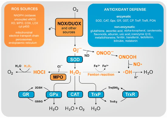
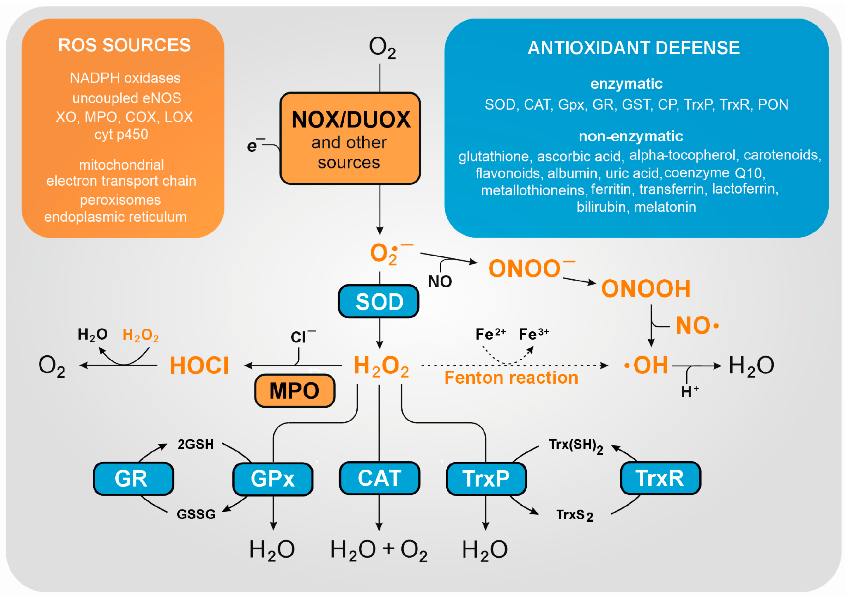
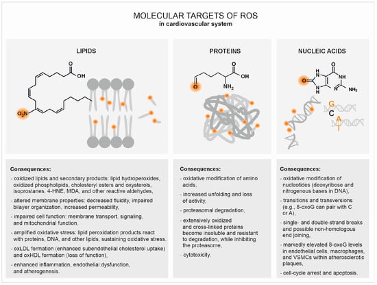
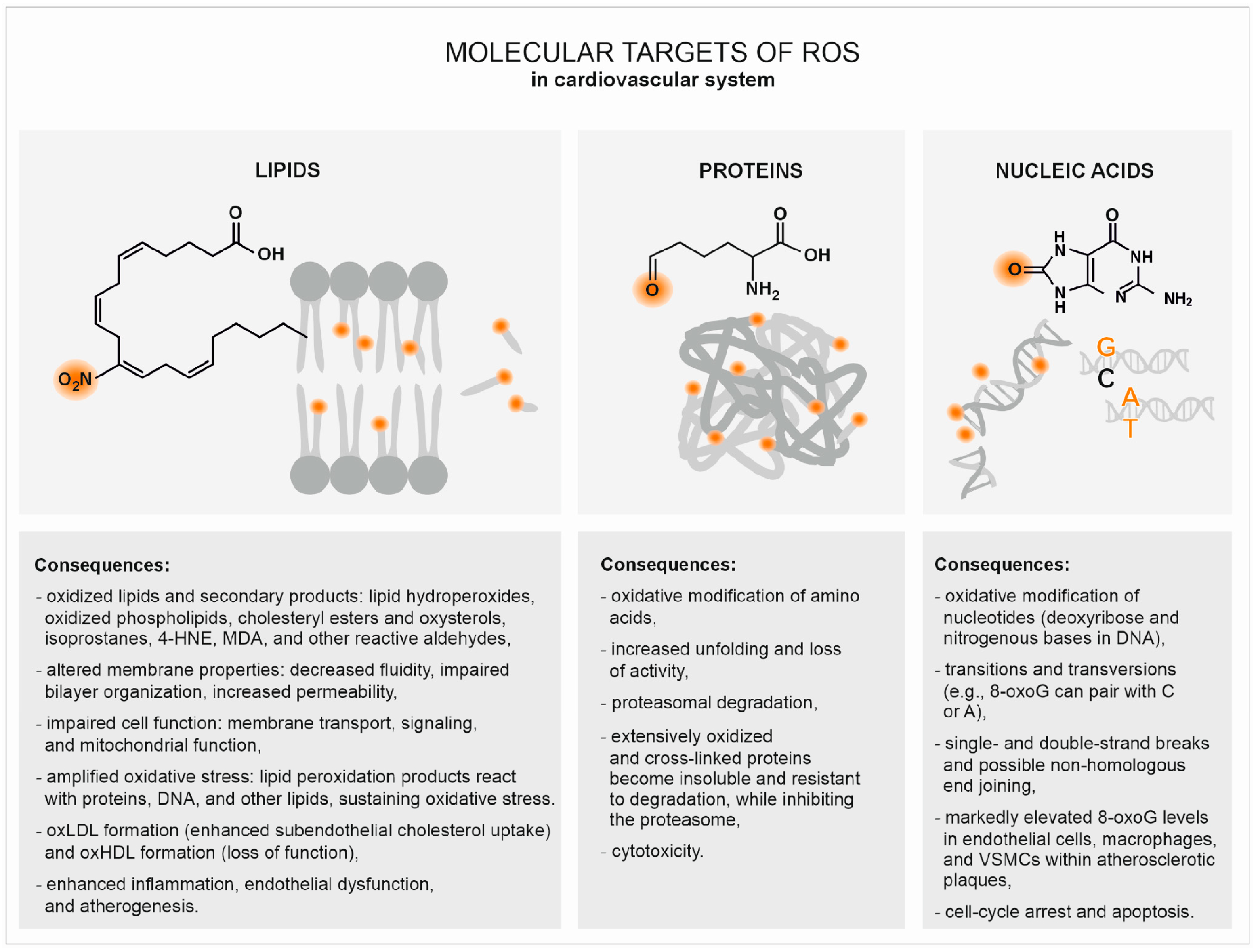
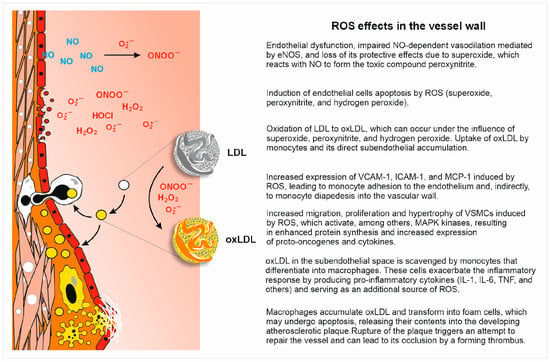
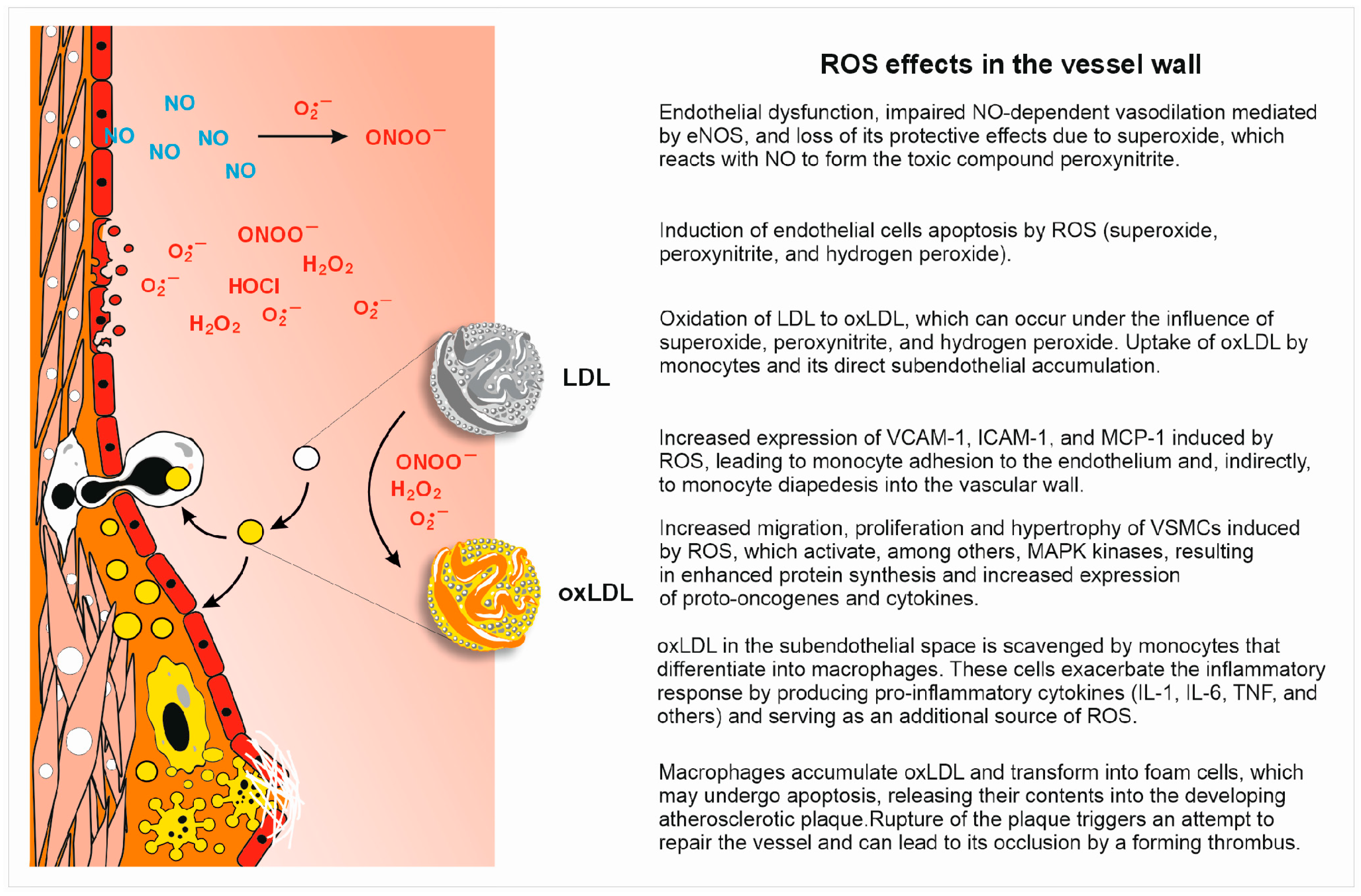
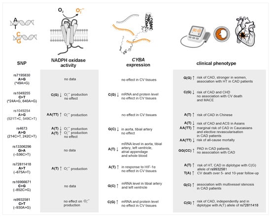
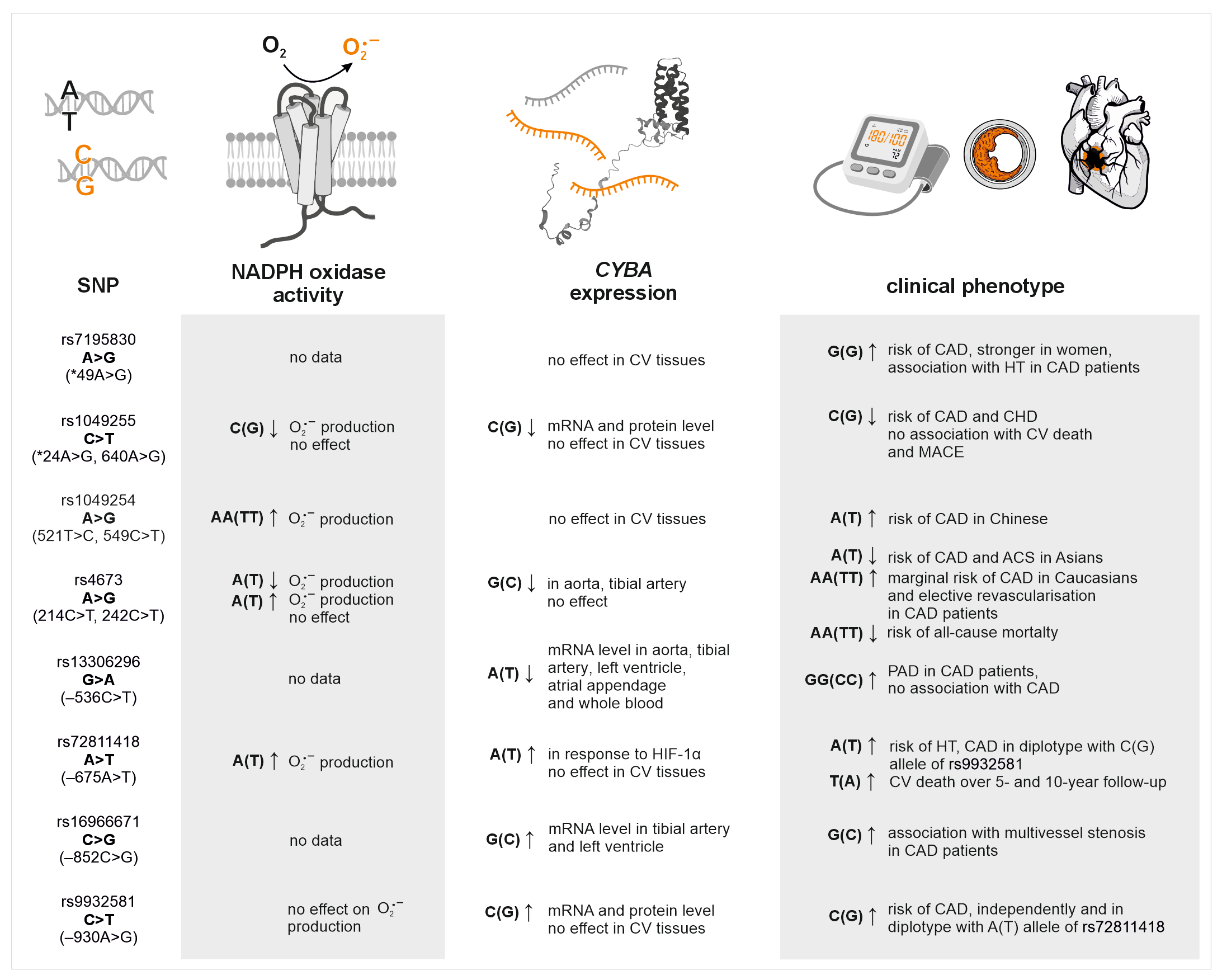
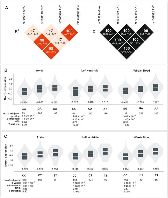
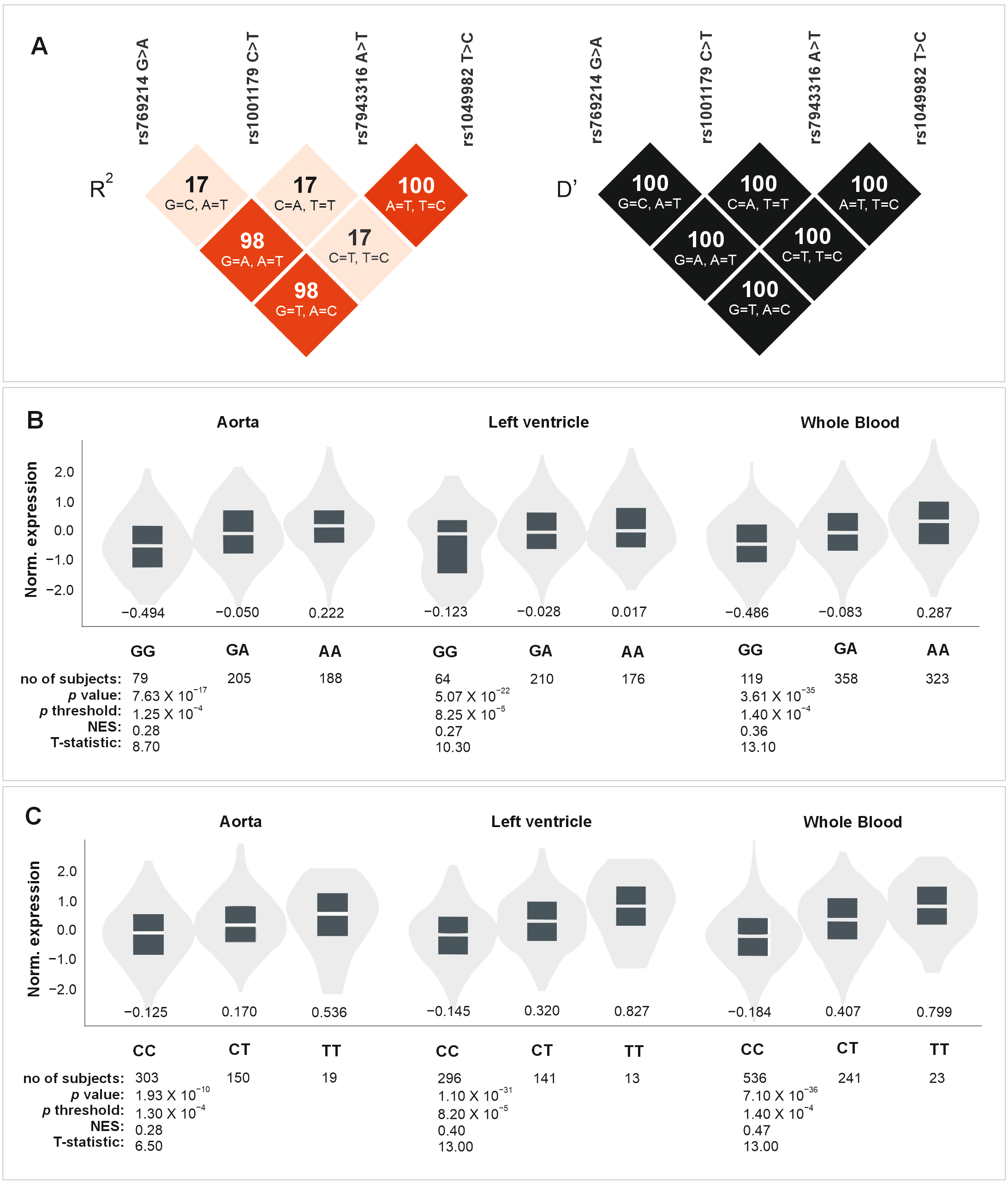
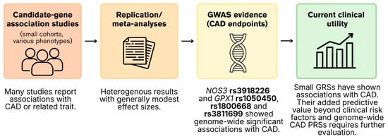
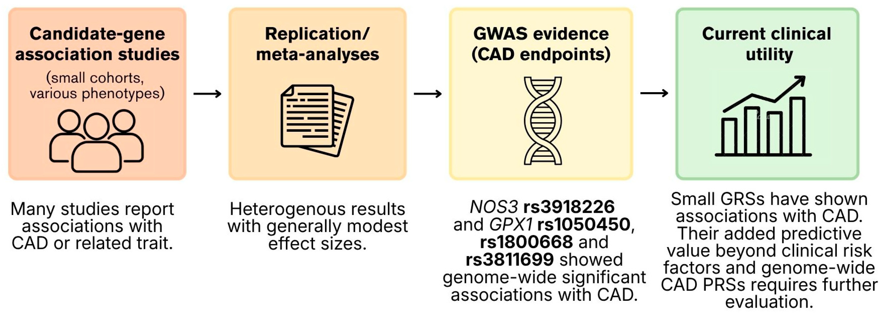
Disclaimer/Publisher’s Note: The statements, opinions and data contained in all publications are solely those of the individual author(s) and contributor(s) and not of MDPI and/or the editor(s). MDPI and/or the editor(s) disclaim responsibility for any injury to people or property resulting from any ideas, methods, instructions or products referred to in the content. |
© 2026 by the authors. Licensee MDPI, Basel, Switzerland. This article is an open access article distributed under the terms and conditions of the Creative Commons Attribution (CC BY) license.