


Abstract
Conjunctival melanoma (CM), a rare malignant tumor of the ocular surface, is characterized by high invasiveness and a poor prognosis. Despite aggressive local control measures such as complete surgical resection combined with cryotherapy, patients still face a high risk of local recurrence (60%) and distant metastasis (20–30%). This article provides a narrative review of the pathogenesis and targeted therapies for CM, focusing on its core molecular mechanisms, with particular attention to therapeutic strategies targeting key driver genes and pathways. It also summarizes the clinical evidence and latest breakthroughs in immunotherapy and emerging treatment strategies. By integrating evidence from basic research and clinical translation, this review provides a theoretical basis for optimizing the diagnostic and therapeutic pathways for CM.
1. Introduction
1.1. Background and Clinical Significance of CM
Conjunctival melanoma (CM) originates from melanocytes in the basal layer of the conjunctival epithelium [1,2] and accounts for 2% of all ocular tumors [3,4]. It occurs predominantly in middle-aged and older individuals, with reported median ages at diagnosis ranging from 55 to 65 years [4,5,6]. It is more common in fair-skinned individuals and may be related to ultraviolet (UV) exposure [2,7].
CM arises through three main pathways, approximately 70% originating from primary acquired melanosis (PAM) with atypia, while a small proportion arise from malignant transformation of a conjunctival nevus or de novo mutations [8]. In modern pathological classification, PAM with atypia is now designated as conjunctival melanocytic intraepithelial lesion (C-MIL), which is graded as low-grade and high-grade; high-grade C-MIL corresponds to what was previously termed PAM with severe atypia. The risk of PAM with severe atypia progressing to melanoma ranges from 13% to 50% [8,9]. Notably, a case report described rapid malignant transformation of a conjunctival nevus into conjunctival melanoma after deucravacitinib initiation, suggesting a possible temporal association [10]. The tumor can involve various regions of the conjunctiva, most commonly the bulbar conjunctiva, followed by the forniceal conjunctiva, palpebral conjunctiva, plica semilunaris, and caruncle [2,11]. The typical presentation is a tan-brown or darkly pigmented lesion that is nodular or flat [5,12]. It is often asymptomatic in the early stages, but as the lesion enlarges, it may cause a foreign body sensation, irritation, redness, or visual changes [12,13]. Local invasion often involves the eyelids or the orbit [14]. About 20–30% of patients develop metastases, with common sites of metastasis including the lung, brain, and liver [15].
In current treatment strategies, complete surgical resection with 2–4 mm margins combined with cryotherapy remains the gold standard [5,7], and a “no-touch” technique is recommended intraoperatively to reduce iatrogenic seeding of tumor cells [1,14]. Nevertheless, postoperative local recurrence remains a major clinical challenge, and rates as high as 60% have been reported in the literature [1,16]. For patients with residual postoperative intraepithelial disease, diffuse PAM or multifocal PAM with atypia, or positive or poorly defined surgical margins, topical chemotherapy is a common adjuvant approach [1,4,17], and adjuvant radiotherapy can delay local recurrence [3,8]. However, current combination therapies are still associated with a high risk of recurrence and metastasis. Therefore, a deeper understanding of the molecular drivers of CM and the exploration of targeted therapies are essential to overcome the current clinical challenges of recurrence and metastasis.
1.2. Comparison with Cutaneous Melanoma and Uveal Melanoma
Ocular melanomas, including uveal melanoma (UM) and CM, are the most common primary intraocular and ocular surface malignancies in adults [18]. Although they share a common melanocytic origin with cutaneous melanoma, their molecular characteristics differ significantly (summarized in Table 1). Notably, the incidence of CM has significantly increased, roughly paralleling the increasing incidence of cutaneous melanoma, whereas the incidence of UM remains relatively stable [11]. Cutaneous melanoma is primarily driven by UV radiation and frequently harbors BRAF (especially V600E), NRAS, or NF1 mutations, exhibiting a high mutational burden. In contrast, UM is characterized by a near-universal absence of BRAF mutations and lacks a clear UV etiologic link [18]. It is typically driven by mutually exclusive activating mutations in GNAQ or GNA11, while secondary alterations in BAP1, SF3B1, and EIF1AX dictate metastatic risk [18]. Consequently, UM exhibits a low mutational burden and a poor response to immune checkpoint inhibitors (ICIs) [19].
Table 1.
Molecular and clinical characteristics of major melanoma subtypes.
CM represents a molecular bridge between these two extremes. Although evidence regarding whether CM shares the exact same risk factors as cutaneous melanoma remains controversial [13], the conjunctiva is the only mucosal tissue on the ocular surface directly exposed to UV radiation, and UV exposure is considered a critical etiologic factor for CM [6,7,11,14,20]. Molecularly, CM is similar to cutaneous melanoma, typically involving BRAF, NRAS, NF1, and TERT promoter mutations [18]. However, CM also harbors hotspot mutations traditionally associated with UM (including BAP1, SF3B1, and GNAQ/GNA11), which are correlated with disease progression, increased metastatic risk, and higher mortality [1]. Its metastatic pattern is similar to that of cutaneous melanoma, with 20% to 30% of patients developing lymphatic and hematogenous metastasis [11].
Given the genetic similarities to cutaneous melanoma, checkpoint inhibitors have successfully induced remission in some cases of metastatic CM [1,21]; furthermore, similar to cutaneous melanoma, CM exhibits an inadequate response to external beam radiotherapy, limiting its role [22]. In summary, in-depth analysis of the molecular pathogenesis of CM and its targeted therapeutic strategies holds significant value for overcoming the current diagnostic and therapeutic bottlenecks of CM.
For this narrative review, relevant publications were retrieved from PubMed, Web of Science and other databases. Articles were selected according to their relevance to the molecular pathogenesis and clinical management of CM, with priority given to recent original studies, clinical trials and authoritative reviews.
2. CM-Related Genetic Alterations
2.1. Gene Alterations Related to the MAPK/PI3K Signaling Pathway
The MAPK (RAS/RAF/MEK/ERK) and PI3K/AKT/mTOR pathways are frequently dysregulated in CM. The former regulates differentiation and proliferation by transmitting growth signals to the nucleus, whereas the latter contributes to tumorigenesis through aberrant activation of proto-oncogenes and inactivation of tumor suppressor genes [23,24].
BRAF V600E is the most common BRAF mutation, accounting for about 80% of BRAF mutations [1,2]. The V600K variant is more common in tumors associated with chronic sun damage. In cutaneous melanoma, this variant is often accompanied by a higher tumor mutational burden (TMB), which may predict greater sensitivity to ICIs. However, in CM, this association has not been validated, and clinical studies indicate that V600E is the predominant BRAF mutation subtype [25]. BRAF mutations occur early in tumorigenesis and are more common in younger male patients and in bulbar conjunctival lesions, suggesting that UV exposure may be a potential risk factor [2,23,26]. BRAF mutations are also more frequent in CMs arising from nevi [1,24], and are associated with a higher risk of recurrence [24,27].
Over 90% of NRAS mutations are point mutations at codon 61 [18], resulting in constitutive activation of the NRAS protein [24]. Unlike normal melanocytes, NRAS-mutant melanomas activate MAPK signaling primarily through CRAF rather than BRAF, which may be one of the mechanisms underlying primary resistance to BRAF inhibitors [18]. NRAS and BRAF mutations are usually mutually exclusive [24,27]. In addition, mutations in other RAS family members, such as HRAS and KRAS, are occasionally found in CM [26].
NF1 is the most commonly mutated gene in CM, mutated in approximately one-third of cases, with nonsense or frameshift mutations predominating [18,23,25]. NF1 mutations often exhibit UV-induced mutational signatures, such as C → T transitions and CC → TT tandem mutations. They are commonly associated with a higher TMB [24] and are more prevalent in older patients [26]. Although NF1 mutations can coexist with BRAF or NRAS mutations, such coexistence is uncommon [18,23,24].
KIT mutations are uncommon in CM. However, KIT overexpression is observed more frequently than KIT mutations [24,25], and KIT alterations are more frequent in Asian than in Caucasian populations [25]. KIT mutations are often mutually exclusive with BRAF and NRAS mutations but can coexist with NF1 mutations [24]. Some CMs harbor mutations in both KIT and SF3B1 (splicing factor 3B subunit 1), suggesting that these cases may represent a distinct subtype of mucosal melanoma [26,28].
Reduced or absent PTEN expression is common in CM and is accompanied by upregulation of mTOR pathway signaling; nuclear PTEN loss is more pronounced in CM than in conjunctival nevi. However, among different subtypes, nuclear PTEN expression levels are higher in amelanotic CM than in pigmented CM [29].
2.2. Genes Related to Telomere Maintenance and Chromatin Remodeling
Telomere maintenance and chromatin remodeling play important roles in CM progression. Tumor cells can acquire unlimited replicative capacity by activating telomerase, whose catalytic subunit is encoded by TERT, or by inducing alternative lengthening of telomeres (ALT) through mechanisms such as ATRX loss [24,30].
TERT promoter mutations are not observed in conjunctival nevi [23,24], occur in approximately 8% of PAM with atypia [18,31], and are detected in 32–43% of CMs [23,31]. These mutations often exhibit a UV-induced mutational signature [32,33]. Clinically, TERT promoter mutations are closely associated with CM metastasis and shorter metastasis-free survival [30,34], and are considered markers of tumor invasiveness and poor prognosis [31]. TERT promoter mutations can coexist with BRAF or NRAS mutations [23,24], but are mutually exclusive with ATRX mutations, suggesting that telomerase reactivation and ALT represent two distinct telomere maintenance mechanisms [30].
ATRX typically binds heterochromatin through its ADD domain and, together with the DAXX complex, deposits histone variant H3.3 at telomeres to maintain telomere stability. Loss-of-function mutations in ATRX or loss of ATRX protein expression can disrupt this process, thereby inducing ALT [30]. ATRX inactivation often coexists with NF1 mutations [24,35], and is more common in lesions at non-sun-exposed sites [23]. ATRX mutations and TERT promoter mutations are usually mutually exclusive. Both alterations are frequently detected in metastatic CM, underscoring their roles as early high-risk driver events [23]. However, ATRX-mutant CM (often arising in non-sun-exposed sites) may exhibit a relatively less aggressive clinical behavior compared to other subtypes, although it still retains metastatic potential [23,24]. Most metastatic CM cases harbor ATRX loss or TERT promoter mutations; both alterations are detectable in precancerous lesions but are absent in benign lesions, suggesting that they may represent early events in CM progression [23,30].
2.3. UV-Induced Genomic Features
UV-induced DNA damage plays a key role in the development of CM [26], typically manifesting as a mutational pattern characterized predominantly by C → T transitions, particularly CC → TT tandem mutations, suggesting that UV radiation is an important etiologic factor in a subset of CMs [20,21,22].
CMs harboring a UV-induced mutational signature usually have a high TMB [24], ranging from 1.1 to 15.6 mutations/Mb, with levels intermediate between those of choroidal melanoma without UV exposure and typical cutaneous melanoma [32]. This feature is closely related to anatomical location; bulbar conjunctival melanomas, which are directly exposed to UV, have the highest TMB and the most pronounced UV mutational signature [25].
Notably, UV-driven CMs not only exhibit a high TMB and a UV-induced mutational signature similar to that of cutaneous melanoma, but also harbor many large-scale structural variants commonly found in mucosal melanoma, such as insertions, deletions, and copy number alterations [26]. This mixed phenotype suggests that CM development is driven by both the accumulation of structural variants and UV-mediated mutational processes.
2.4. Chromosomal Structural Variants and Copy Number Alterations
The genome of CM exhibits widespread chromosomal instability, including copy number variations (CNVs) involving entire chromosomes or chromosome arms, as well as polyploidy and aneuploidy [24]. Amplification of 6p is the most frequent alteration [23].
These alterations affect key pathways and processes, including MAPK signaling, cell cycle regulation, and telomere maintenance. At the gene level, copy number gains or amplifications frequently affect BRAF, NRAS, TERT, KIT, CCND1, CDKN1A, and RUNX2, whereas losses or deletions affect tumor suppressor genes such as NF1, TP53, CBL, CDKN2A, and CDKN2B [24]. These alterations overlap substantially with the genes that are frequently mutated, as described above, indicating that multiple genetic mechanisms converge on the same oncogenic pathways.
Furthermore, the frequency of CDKN2A alterations is approximately 31%, and approximately 6% of cases harbor CDK4 R24C hotspot mutations and SF3B1 missense mutations. Notably, a small cohort study (n = 14) did not detect GNAQ, GNA11, or BAP1 mutations [25]. While GNAQ and GNA11 mutations are virtually absent in CM, BAP1 mutations are rare but have been occasionally reported in a small subset of cases [1,2].
Specific CNVs have well-defined clinical significance. Deletions of 10q involving tumor suppressor genes such as PTEN are significantly associated with increased tumor thickness, lymphatic invasion, and an increased risk of metastasis [18,24,35], and are more frequent in BRAF-mutant tumors [18,24]. CNV patterns vary depending on genetic background. BRAF/NRAS wild-type tumors show relatively high CNV frequencies [23,24]. In BRAF-mutant tumors, 10q loss is more common, whereas gains of 1q, 3p, and 17q are relatively rare [24].
2.5. Epigenetic Dysregulation
Epigenetic dysregulation plays an important role in the development and progression of CM, involving multiple levels such as DNA methylation, histone modification, and non-coding RNA networks.
With respect to DNA methylation, the tumor suppressor genes p16INK4a and RASSF1A are downregulated owing to hypermethylation of CpG islands in their promoter regions [36,37]. Downregulation of p16INK4a is particularly helpful for differentiating melanoma from conjunctival nevi in histopathological diagnosis [37]. Furthermore, mutations in epigenetic regulatory genes such as TET2, SETD2, IDH1, members of the KMT2 family, and ARID2 are relatively common, suggesting that epigenetic dysregulation is an important mechanism in CM pathogenesis [24].
At the non-coding RNA level, differential expression of microRNAs (miRNAs) is associated with the invasion, metastasis, and diagnosis of CM. Downregulation of miR-204/211 is associated with increased invasiveness, whereas aberrant expression of miR-21/146b is associated with metastasis and may serve as potential prognostic markers. The interaction between miR-146a and NOTCH proteins is also involved in early tumorigenesis [23]. In terms of diagnosis, expression profiles of miR-9-5p, miR-196b-5p, and miR-615-3p are helpful in distinguishing benign from malignant lesions. In addition, upregulation of miR-20b can promote tumor progression by inhibiting PTEN, whereas inhibition of miR-506 and miR-509 can reduce tumor cell proliferation and invasion. Regarding circular RNAs (circRNAs), more than 9300 circRNAs have been identified in CM tissues [38], among which circMTUS1 is upregulated in tumors and can promote tumorigenesis by sponging hsa-miR-622 and hsa-miR-1208 [23].
2.6. Other Related Genetic Alterations
In addition to the classic MAPK/PI3K pathway, alterations in other signaling pathways are also implicated in CM. In cell cycle regulation, mutations or deletions in TP53 and CDKN2A lead to loss of cell cycle control [18,24]. In the Wnt signaling pathway, CTNNB1 mutations are present in approximately 15% of cases, especially in tumors arising from nevi [26,28,35], and these mutations are usually mutually exclusive with other oncogenic mutations [26]. The FAT4 gene, which encodes an upstream transmembrane receptor in the Hippo signaling pathway, has a relatively high mutation frequency in a Chinese patient cohort. FAT4 mutations exhibit a UV-induced mutational signature, and low FAT4 expression is closely associated with poor prognosis [27,32].
In studies of receptor tyrosine kinases, mutations in genes such as ALK and MET have been reported [24], along with rare actionable mutations such as ERBB2 S310F and MET T1010I [26]. Although infrequent, these mutations may provide potential directions for personalized treatment of CM.
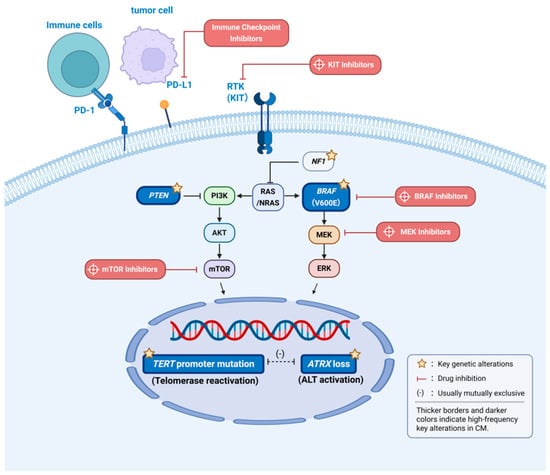
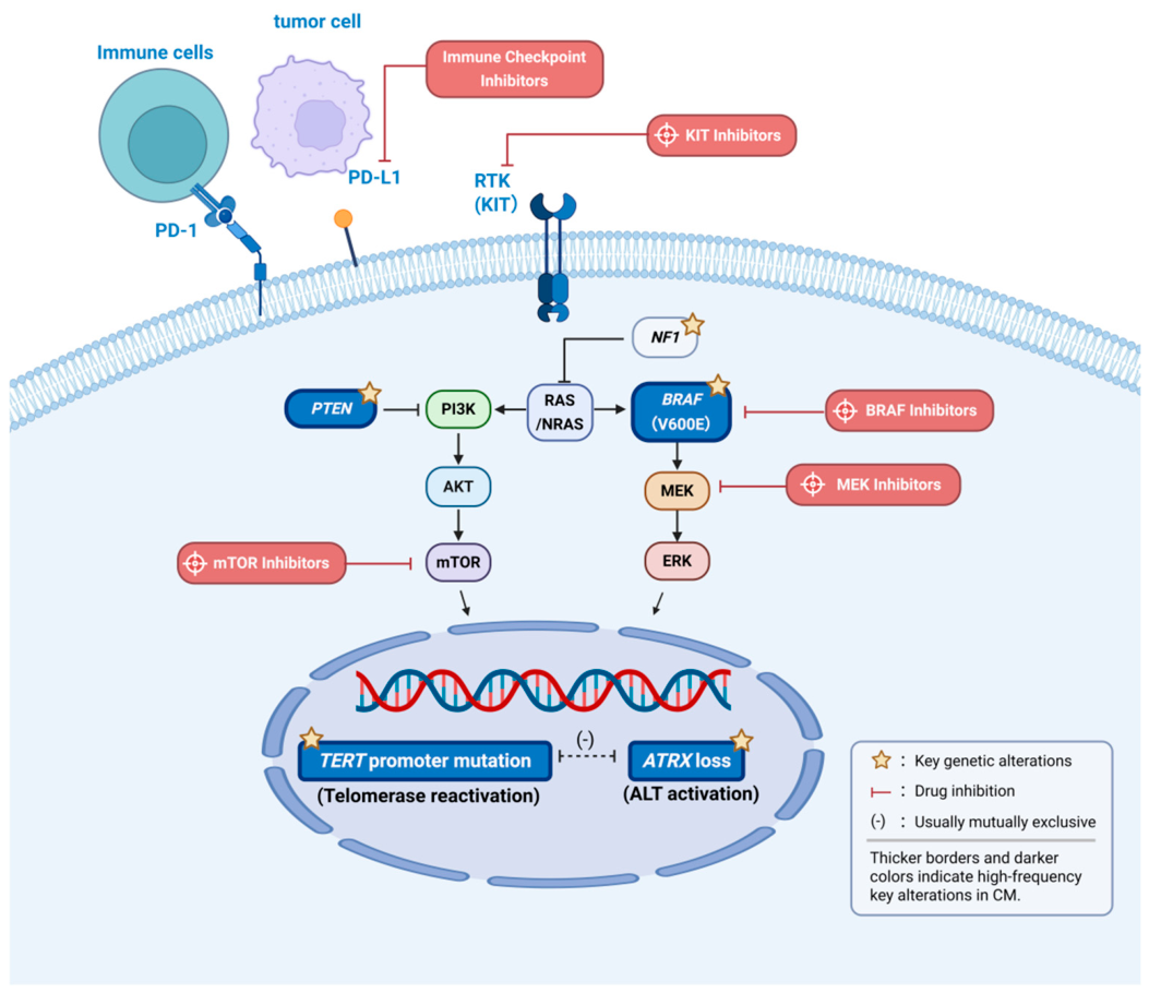
Notably, some CM cases harbor SF3B1 and BAP1 mutations but lack GNAQ/GNA11 mutations, suggesting that their tumorigenesis differs from that of classic choroidal melanoma [26,27,33]. Furthermore, missense variants involving β-TrCP-binding motifs, which are also present in other tumors, have been observed in CM, supporting their oncogenic role [26]. The above molecular alterations can be summarized as the core network shown in Figure 1.
Figure 1.
Schematic overview of core molecular mechanisms and targeted therapies in conjunctival melanoma (CM).
The diagram illustrates the MAPK/PI3K signaling pathways, telomere maintenance mechanisms, and the immune microenvironment specific to CM. Key genetic alterations are highlighted with yellow stars, and high-frequency events (e.g., BRAF V600E, PTEN loss, TERT promoter mutations, ATRX loss) are visually emphasized with thicker borders and darker colors. The mutual exclusivity of TERT promoter mutations and ATRX loss is indicated by (-). For targeted therapy, inhibitors are depicted in red, linking directly to their respective molecular targets. Notably, KIT-targeted agents are generally not considered first-line treatment for CM. For immunotherapy, PD-1 inhibitors are shown to block the PD-1/PD-L1 interaction.
3. Advances in Molecular Targeted Therapy
Treatment of CM is shifting from traditional surgery to personalized strategies guided by genetic characteristics. This shift is driven largely by increasing understanding of the molecular pathogenesis of CM and has also been facilitated by the genetic similarities between CM and cutaneous melanoma [23,32]. For locally advanced or metastatic CM, although no unified first-line standard regimen has been established, personalized strategies based on targeted therapy and immunotherapy are emerging as a new therapeutic paradigm [23]. Table 2 provides a comprehensive summary of key clinical studies, patient populations, sample sizes, and clinical outcomes in CM.
Table 2.
Summary of clinical studies on treatments and outcomes in conjunctival melanoma.
3.1. Targeted Therapy
Unlike choroidal melanoma, CM has a higher BRAF mutation rate and tends to metastasize to lymph nodes rather than the liver, making the MAPK pathway an ideal therapeutic target [32]. Most CMs harbor mutations in MAPK pathway genes, such as BRAF, RAS, KIT, and NF1, and are therefore considered targetable malignancies [23,28]. BRAF inhibitors (e.g., vemurafenib and dabrafenib) and MEK inhibitors (e.g., trametinib and cobimetinib) have been used to control unresectable locally advanced disease, avoid orbital exenteration, and treat distant metastases [23,25]. Combination therapy with BRAF and MEK inhibitors improves efficacy and delays resistance compared with monotherapy [1,23,44]. In a small study of Chinese patients, BRAF inhibitors showed clinical activity in metastatic BRAF-mutant tumors [45]. A previous review by Zeng et al. summarized 9 individual cases of advanced CM treated with BRAF/MEK inhibitors, reporting a 33.3% (3/9) complete response rate and a 33.3% (3/9) local control rate [46]. Another study showed a disease control rate of 37.5%, a median progression-free survival (PFS) of 12.6 months, and a median overall survival (OS) of 29.1 months; however, these findings require validation in larger studies due to the limited sample size [39]. Common adverse events include arthralgia, diarrhea, rash, fever, weight loss, vomiting, headache, and elevated transaminase levels [32].
In addition to BRAF/MEK inhibitor combination therapy, other combination strategies are also being explored. Extrapolating from non-CM cohorts (e.g., NRAS-mutant melanoma), pan-RAF inhibitor plus MEK inhibitor combinations, such as naporafenib plus trametinib, have achieved objective response rates of 40–46.7% in clinical trials [44]. Based on preclinical and non-CM clinical data, tumors harboring alterations in CDKN2A, CDK4, or CCND1 are more sensitive to the combination of CDK4/6 inhibitors (e.g., ribociclib) and MEK inhibitors [44]. Similarly, preclinical and early-phase clinical data suggest that autophagy inhibition (e.g., with hydroxychloroquine plus MEK inhibitors), and receptor tyrosine kinase (RTK)-targeted combination therapies, such as combinations involving sorafenib or tivantinib, have shown potential in NRAS-mutant tumors [44]. In addition, although CM-specific data are very limited, imatinib and other KIT-targeted agents are not considered a first-line treatment for CM, and their efficacy needs to be confirmed in further studies [1].
Drug resistance remains a major challenge. Resistance to BRAF inhibitor monotherapy often emerges within one year [23], and is mediated by MAPK pathway reactivation, epigenetic remodeling, and feedback from the tumor microenvironment [23,44]. Frequent loss of PTEN in CM may reduce the efficacy of BRAF inhibitors and contribute to the development of resistance [23]. In vitro studies have shown that the sensitivity of CM cell lines to BRAF inhibitors, MEK inhibitors, and PI3K/mTOR inhibitors depends in part on BRAF V600E or NRAS Q61L mutational status [35]. MEK inhibitors, such as AZD8330, have high efficacy in BRAF-mutant cell lines, whereas inhibition of the PI3K/mTOR axis is more effective in NRAS-mutant cell lines [35].
3.2. Immunotherapy
ICIs restore T-cell activation by targeting immune checkpoint pathways such as CTLA-4 and PD-1/PD-L1, and have become an important treatment modality for several melanoma subtypes [23]. The use of ICIs in CM is based on the molecular similarities between CM and cutaneous melanoma, as well as UV exposure-related characteristics [25]. ICIs have shown considerable promise in locally advanced and metastatic CM, with small studies suggesting response rates of up to 50% in metastatic disease and up to 70% in locally advanced disease [25], and they are considered a first-line therapy for metastatic melanoma [21]. Case series have shown that PD-1 inhibitors, such as nivolumab and pembrolizumab, or the CTLA-4 inhibitor ipilimumab, can induce complete or partial tumor regression in some patients [39]. In 26 patients with metastatic CM who received first-line therapy, the one-year progression-free survival rate was 42% [39].
Regarding treatment response prediction, although TMB is a well-established predictive biomarker in cutaneous melanoma, its predictive value in CM remains to be fully elucidated and lacks prospective clinical validation. ICIs may still have a role in patients with locally advanced or metastatic disease, especially those with higher TMB [23,25]. PD-L1 is expressed in approximately 19% of CMs [28] and is associated with distant metastasis and poorer survival [1]; however, PD-L1 testing is not routinely recommended to guide treatment in clinical practice [28].
ICI regimens for CM generally follow established protocols for cutaneous melanoma and mainly include CTLA-4 inhibitors and PD-1 inhibitors [23]. PD-1 inhibitor monotherapy is more effective than CTLA-4 inhibitor monotherapy, whereas their combination produces synergistic effects but is associated with higher rates of immune-related adverse events compared to PD-1 monotherapy [28]. While CM-specific randomized trials are lacking, evidence extrapolated from cutaneous melanoma trials shows that nivolumab combined with ipilimumab prolonged median OS compared with monotherapy (72.1 months vs. 36.9 months), providing a rationale for this combination in CM [47]. Furthermore, for subtypes lacking effective targeted therapies, such as NRAS-mutant tumors, PD-1 inhibitors have been recognized by the National Comprehensive Cancer Network (NCCN) and the European Society for Medical Oncology (ESMO) as a first-line treatment [44].
Case reports suggest that ICIs can effectively reduce tumor volume in locally advanced CM [48], potentially avoiding orbital exenteration. However, ICIs may also cause pseudoprogression and immune-related adverse events, such as colitis, dermatitis, pneumonitis, and hepatotoxicity [23,32]. In rare cases, it may also induce cytokine release syndrome (CRS) [49].
Beyond systemic ICIs, local and traditional therapies remain crucial. For instance, a Spanish cohort of 27 primary CM patients receiving surgery plus tailored adjuvant therapy reported a 5-year OS of 89%, although the recurrence rate remained at 33% [41]. Additionally, a small series demonstrated that adjuvant IFNα-2b eye drops after surgical resection achieved complete remission in 4 out of 5 CM patients [40]. Furthermore, local immunomodulatory therapy has also been explored. A case series evaluating 5% imiquimod ointment included 5 patients: 3 cases of ocular surface squamous neoplasia (OSSN) and 2 cases of melanocytic lesions. Specifically, the diffuse melanoma in situ (MIS) case demonstrated a partial clinical response with complete histological response, while the PAM case achieved complete clinical and histological resolution [50].
In summary, ICIs provide an important therapeutic option for locally advanced or metastatic CM. However, because CM is rare, efficacy data are mainly derived from case reports and small case series, and prospective randomized trials are lacking.
3.3. Other Treatments
In addition to targeted therapy and immunotherapy, several emerging strategies are also being explored. Importantly, the current evidence for these strategies is derived primarily from other melanoma subtypes or preclinical models, rather than CM patients.
Dendritic cell (DC) vaccines loaded with tumor antigens activate antitumor T-cell responses [23]. In clinical trials in patients with metastatic cutaneous melanoma, enhanced intratumoral CD8+ T-cell infiltration and prolonged median OS have been observed [23]. Although no human studies have yet been conducted in CM, DC vaccines combined with the conventional type 2 DC (cDC2) subset and osteopontin blockade have shown anti-angiogenic and immunostimulatory activity in animal models of ocular melanoma, suggesting their translational potential across melanoma subtypes [23].
Epigenetic strategies reverse the silencing of tumor suppressor genes or the activation of oncogenes by modulating DNA methylation and histone modifications. DNA methyltransferase inhibitors, such as decitabine, can induce DNA hypomethylation.
When combined with ipilimumab in patients with unresectable (primarily cutaneous) melanoma, they upregulate HLA class I expression and expand intratumoral CD8+PD-1+ T cells. Additionally, Panobinostat has also been shown to promote differentiation and G1-phase arrest in UM models [23]. However, to date, these epigenetic strategies remain entirely untested in CM, and their clinical translation for CM requires extensive investigation.
Electrochemotherapy (ECT) enhances the cytotoxicity of chemotherapeutic agents by applying electric pulses. Preclinical studies using experimental tumor models have shown that repeated ECT (3 cycles; 3 × ECT) significantly increases cytotoxicity and reduces the risk of tumor regrowth in experimental settings compared with a single ECT session [51]. The advantages of ECT include high local drug concentrations, low systemic toxicity, a low risk of acquired resistance, and cost-effectiveness and ease of performance [51]. Future efforts should aim to establish standardized ECT protocols for CM to balance therapeutic efficacy with the safety of adjacent ocular tissues, and clinical studies are required to validate these preclinical findings.
Common genetic alterations and their clinical significance in conjunctival melanoma are summarized in Table 3.
Table 3.
Key genetic alterations in conjunctival melanoma: molecular functions, prognostic significance, and therapeutic implications.
4. Controversies and Future Perspectives
Despite recent advances in research on the molecular mechanisms and treatment of CM, controversies persist in clinical practice because of the rarity of the disease and the complexity of its molecular subtypes. In recent years, the differential distribution of molecular markers such as TERT promoter mutations and ATRX loss between CM and benign lesions has opened new avenues for early diagnosis. However, no consensus has yet been reached on standardized clinical cutoff values for these markers or their association with histopathologic grading.
With respect to treatment, the value of combined BRAF/MEK inhibitor therapy in BRAF-mutant CM has been increasingly recognized, but the optimal timing of treatment remains controversial. Furthermore, a critical limitation is that, as discussed in Section 3.1, the long-term efficacy of targeted therapies is frequently hampered by acquired resistance driven by molecular heterogeneity, such as MAPK pathway reactivation or PTEN loss. In addition, ICIs have shown considerable promise in CM. Their use currently largely follows the standards established for cutaneous melanoma. However, given the distinct mixed phenotype of CM, it remains unclear whether a unique immunotherapy strategy is required.
CM is associated with a high local recurrence rate and a substantial risk of metastasis, making early and precise intervention essential. However, because of the low incidence of CM, available evidence is derived largely from case reports and small retrospective series, and prospective randomized controlled trials with large sample sizes are lacking. This fundamental lack of CM-specific clinical evidence underscores the urgent need to critically validate extrapolated data rather than blindly applying cutaneous melanoma protocols. Moreover, no unified molecular classification system or standardized diagnostic and therapeutic protocols have been established, making it difficult to define a widely accepted optimal treatment strategy.
By summarizing the current molecular landscape and synthesizing the clinical outcomes of both CM-specific and extrapolated treatments, this review highlights the specific therapeutic challenges for distinct molecular subtypes and the need for tailored clinical strategies. We hope that future multicenter, prospective cohort studies will continuously deepen our understanding of the molecular genetic mechanisms of CM and further identify novel biomarkers associated with treatment response, drug resistance, and prognosis, thereby providing higher-quality evidence for the precision diagnosis and treatment of CM and ultimately improving patients’ quality of life and long-term prognosis.
Author Contributions
Conceptualization, A.Z., L.G., Q.J., H.L. and C.X.; investigation (literature search and data analysis), A.Z., L.G., Q.J. and H.L.; writing—original draft preparation, A.Z.; writing—review and editing, A.Z., L.G., Q.J., H.L. and C.X.; visualization, L.G.; supervision, C.X. and H.L.; project administration, C.X. and H.L. All authors have read and agreed to the published version of the manuscript.
Funding
This research received no external funding.
Data Availability Statement
No new data were created or analyzed in this study. Data sharing is not applicable to this article.
Acknowledgments
We thank the guidance and strong support of mentors and colleagues during the collaboration on this paper.
Conflicts of Interest
The authors declare no conflicts of interest.
References
- Li, W.J.; Venkateswaran, N.; Antonietti, M.; Qureshi, S.; Galor, A.; Karp, C.L. Emerging Techniques in the Treatment of Conjunctival Melanoma. Curr. Ophthalmol. Rep. 2025, 13, 7. [Google Scholar] [CrossRef] [Scilit]
- Okongwu, C.C.; Adewara, B.A.; Olaofe, O.O.; Soremekun, A.I.; Ayodele, S.O.; Abdullahi, Y.O.; Ewoye, E.E.; Oladele, J.O. Malignant Melanoma of the Conjunctiva Metastasizing to the Submandibular Gland: A Case Report and Review of the Literature. BMC Ophthalmol. 2025, 25, 130. [Google Scholar] [CrossRef] [Scilit]
- Veisi, A.; Dastborhan, Z.; Dastmardi, M.; Rezaie Kanavi, M.; Rezaei, S. Lacrimal Drainage System and Nasal Cavity Melanoma after Complete Treatment of Conjunctival Melanoma. Case Rep. Ophthalmol. Med. 2024, 2024, 1034939. [Google Scholar] [CrossRef] [Scilit]
- Kharmach, I.; Rezzoug, F.; Moukhlissi, M.; Al Jarroudi, O.; Brahmi, S.A.; Afqir, S. Malignant Transformation of Reese’s Melanosis: A Case of Conjunctival Melanoma and Related Therapeutic Modalities. Cureus 2024, 16, e62331. [Google Scholar] [CrossRef] [Scilit]
- Menna, F.; Tschopp, M.; Meyer, P.; Papazoglou, A. A Case of Conjunctival Melanoma Presenting as a Squamous Cell Carcinoma. Case Rep. Ophthalmol. 2024, 15, 742–750. [Google Scholar] [CrossRef] [Scilit]
- Alhamood, M.; Sbh, B.; Muhammad, A.; Saadeh, I. Conjunctival Malignant Melanoma in a Young Immunocompetent Male Patient: Clinical Presentation and Management—A Case Report. J. Med. Case Rep. 2025, 19, 355. [Google Scholar] [CrossRef] [Scilit]
- Ribeiro, M.; Marques-Couto, P.; Gama-Castro, A.; Dinah-Bragança, T.; Barbosa-Breda, J. A Case Report and an Overview of the Differential Diagnosis of Pigmented Lesions of the Conjunctiva: Just a Freckle or Something to Heckle? Cureus 2024, 16, e66559. [Google Scholar] [CrossRef] [Scilit]
- Vora, G.K.; Demirci, H.; Marr, B.; Mruthyunjaya, P. Advances in the Management of Conjunctival Melanoma. Surv. Ophthalmol. 2017, 62, 26–42. [Google Scholar] [CrossRef] [Scilit]
- Jimura, H.; Mishima, Y.; Sotozono, C.; Watanabe, A.; Asai, J.; Ohbayashi, C.; Ogata, N. Case of Rapidly Expanding Conjunctival Malignant Melanoma Initially from Primary Acquired Melanosis Diagnosed 14 Years Earlier. Int. Med. Case Rep. J. 2021, 14, 361–364. [Google Scholar] [CrossRef] [Scilit]
- Whitcomb, N.; Lee, H.J. Conversion of Conjunctival Nevus to Melanoma in a Patient Taking the TYK2 Inhibitor Deucravacitinib. Cornea 2026, 45, 105–107. [Google Scholar] [CrossRef] [Scilit]
- Kaštelan, S.; Gverović Antunica, A.; Beketić Orešković, L.; Salopek Rabatić, J.; Kasun, B.; Bakija, I. Conjunctival Melanoma—Epidemiological Trends and Features. Pathol. Oncol. Res. 2018, 24, 787–796. [Google Scholar] [CrossRef] [Scilit]
- Englisch, C.N.; Berger, T.; Flockerzi, F.; Bofferding, M.; Seitz, B. Conjunctival Melanoma with Pronounced Central Corneal Invasion: One-Year Relapse Free Follow-Up. Am. J. Ophthalmol. Case Rep. 2024, 36, 102162. [Google Scholar] [CrossRef] [Scilit]
- Ayala, M.; Erripi, K.; Johansson, I. Unilateral Amelanotic Conjunctival Malignant Melanoma: A Case Report. J. Med. Case Rep. 2024, 18, 403. [Google Scholar] [CrossRef] [Scilit]
- Kenworthy, M.K.; Kenworthy, S.J.; De Guzman, P.; Morlet, N. Conjunctival Amelanotic Melanoma Presenting as a Multifocal Pink Lesion. BMJ Case Rep. 2022, 15, e250682. [Google Scholar] [CrossRef] [Scilit]
- Tan, C.H.; Sayegh, Y.; Fasih-Ahmad, S.; Tse, D.T.; Karp, C.L.; Dubovy, S.R. Periocular Granulomatous Inflammatory Lesions Mimicking Conjunctival Melanoma Recurrence in the Setting of Systemic Nivolumab Treatment. Am. J. Ophthalmol. Case Rep. 2024, 36, 102025. [Google Scholar] [CrossRef] [Scilit]
- Zhu, T.; Zong, C.; Xu, S.; Ge, S.; Fan, X.; Jia, R. Relationship between histopathological features, Ki-67 expression and prognosis of conjunctival melanoma. J. Shanghai Jiao Tong Univ. (Med. Sci.) 2022, 42, 617–623. [Google Scholar]
- Cekic, S.P.; Kovacevic, P.T.; Krstic, M.S. Giant Conjunctival Melanoma Treated with Excision and Primary Reconstruction Using a Partial Thickness Skin Graft. Niger. J. Clin. Pract. 2023, 26, 1204–1207. [Google Scholar] [CrossRef] [Scilit]
- Iavarone, L.; Franco, R.; Zito Marino, F.; D’Abbronzo, G.; Argenziano, G.; Scharf, C.; Nucci, G.; Ronchi, A.; Cazzato, G. Ocular Melanoma: A Comprehensive Review with a Focus on Molecular Biology. Int. J. Mol. Sci. 2025, 26, 9799. [Google Scholar] [CrossRef] [Scilit]
- Kaštelan, S.; Gilevska, F.; Tomić, Z.; Živko, J.; Nikuševa-Martić, T. Precision Oncology in Ocular Melanoma: Integrating Molecular and Liquid Biopsy Biomarkers. Curr. Issues Mol. Biol. 2026, 48, 131. [Google Scholar] [CrossRef] [Scilit]
- An, Y.; Ming, Y. A rare case of conjunctival melanoma. J. Clin. Ophthalmol. 2024, 32, 555–557. [Google Scholar]
- Chang, M.; Lally, S.E.; Dalvin, L.A.; Orloff, M.M.; Shields, C.L. Conjunctival Melanoma with Orbital Invasion and Liver Metastasis Managed with Systemic Immune Checkpoint Inhibitor Therapy. Indian J. Ophthalmol. 2019, 67, 2071–2073. [Google Scholar] [CrossRef] [Scilit]
- Matsuo, T.; Yamasaki, O.; Tanaka, T.; Katsui, K.; Waki, T. Proton Beam Therapy Followed by Pembrolizumab for Giant Ocular Surface Conjunctival Malignant Melanoma: A Case Report. Mol. Clin. Oncol. 2022, 16, 12. [Google Scholar] [CrossRef] [Scilit]
- Kaštelan, S.; Zimak, D.M.; Ivić, L.; Antunica, A.G.; Nikuševa-Martić, T. Conjunctival Melanoma: Comprehensive Insights into Clinical Features, Genetic Alterations, and Modern Treatment Approaches. Pathol. Oncol. Res. 2025, 31, 1612085. [Google Scholar] [CrossRef] [Scilit]
- Chang, E.; Demirci, H.; Demirci, F.Y. Genetic Aspects of Conjunctival Melanoma: A Review. Genes 2023, 14, 1668. [Google Scholar] [CrossRef] [Scilit]
- Dimitriou, F.; Wu, X.; Nagarajan, P.; Glitza, I.C.; Zhao, L.; Ning, J.; Patel, S.P.; Wargo, J.A.; Futreal, A.; Woodman, S.; et al. Conjunctival Melanoma Genomic Analysis Reveals Intermediate Tumor Mutation Burden and Genomic Overlap with Cutaneous Melanoma. npj Precis. Oncol. 2025, 9, 365. [Google Scholar] [CrossRef] [Scilit]
- Gardrat, S.; Houy, A.; Brooks, K.; Cassoux, N.; Barnhill, R.; Dayot, S.; Bièche, I.; Raynal, V.; Baulande, S.; Marais, R.; et al. Definition of Biologically Distinct Groups of Conjunctival Melanomas According to Etiological Factors and Implications for Precision Medicine. Cancers 2021, 13, 3836. [Google Scholar] [CrossRef] [Scilit]
- Shi, H.; Tian, H.; Zhu, T.; Chen, J.; Jia, S.; Zong, C.; Liao, Q.; Ruan, J.; Ge, S.; Rao, Y.; et al. Genetic Landscape and Prognosis of Conjunctival Melanoma in Chinese Patients. Br. J. Ophthalmol. 2024, 108, 1306–1312. [Google Scholar] [CrossRef] [Scilit]
- Nahon-Estève, S.; Bertolotto, C.; Picard-Gauci, A.; Gastaud, L.; Baillif, S.; Hofman, P.; Groulier, A.; Maschi, C.; Caujolle, J.-P.; Lassalle, S.; et al. Small but Challenging Conjunctival Melanoma: New Insights, Paradigms and Future Perspectives. Cancers 2021, 13, 5691. [Google Scholar] [CrossRef] [Scilit]
- Lally, S.E.; Milman, T.; Orloff, M.; Dalvin, L.A.; Eberhart, C.G.; Heaphy, C.M.; Rodriguez, F.J.; Lin, C.-C.; Dockery, P.W.; Shields, J.A.; et al. Mutational Landscape and Outcomes of Conjunctival Melanoma in 101 Patients. Ophthalmology 2022, 129, 679–693. [Google Scholar] [CrossRef] [Scilit]
- van Ipenburg, J.A.; van den Bosch, Q.C.C.; Paridaens, D.; Dubbink, H.J.; Kiliç, E.; Naus, N.; Verdijk, R.M. ATRX Loss in the Development and Prognosis of Conjunctival Melanoma. Int. J. Mol. Sci. 2023, 24, 12988. [Google Scholar] [CrossRef] [Scilit]
- Butt, K.; Hussain, R.; Coupland, S.E.; Krishna, Y. Conjunctival Melanoma: A Clinical Review and Update. Cancers 2024, 16, 3121. [Google Scholar] [CrossRef] [Scilit]
- Wang, L.-C.; Tsai, C.-C.; Lin, T.-C.; Rao, N.A. Current Findings of Genetic Alterations and Associated Targeted Therapies of Conjunctival Melanocytic Neoplasms. Taiwan J. Ophthalmol. 2025, 15, 26–33. [Google Scholar] [CrossRef] [Scilit]
- van Poppelen, N.M.; van Ipenburg, J.A.; van den Bosch, Q.; Vaarwater, J.; Brands, T.; Eussen, B.; Magielsen, F.; Dubbink, H.J.; Paridaens, D.; Brosens, E.; et al. Molecular Genetics of Conjunctival Melanoma and Prognostic Value of TERT Promoter Mutation Analysis. Int. J. Mol. Sci. 2021, 22, 5784. [Google Scholar] [CrossRef] [Scilit]
- van Ipenburg, J.A.; Naus, N.C.; Dubbink, H.J.; van Ginderdeuren, R.; Missotten, G.S.; Paridaens, D.; Verdijk, R.M. Prognostic Value of TERT Promoter Mutations in Conjunctival Melanomas in Addition to Clinicopathological Features. Br. J. Ophthalmol. 2021, 105, 1454–1461. [Google Scholar] [CrossRef] [Scilit]
- Nardou, K.; Nicolas, M.; Kuttler, F.; Cisarova, K.; Celik, E.; Quinodoz, M.; Riggi, N.; Michielin, O.; Rivolta, C.; Turcatti, G.; et al. Identification of New Vulnerabilities in Conjunctival Melanoma Using Image-Based High Content Drug Screening. Cancers 2022, 14, 1575. [Google Scholar] [CrossRef] [Scilit]
- Yang, L.; Wang, G.; Shi, H.; Jia, S.; Ruan, J.; Cui, R.; Ge, S. Methylation-Driven Gene DLL3 Is a Potential Prognostic Biomarker in Ocular Melanoma Correlating with Metastasis. Front. Oncol. 2022, 12, 964902. [Google Scholar] [CrossRef] [Scilit]
- Sarrasin, E.; Lalys, E.; Nardou, K.; Celik, E.; Quinodoz, M.; Schalenbourg, A.; Riggi, N.; Rivolta, C.; Berger, A.; Moulin, A. Downregulation of Cyclin Kinase Inhibitors p16INK4a and P27 in Conjunctival Melanomas. Investig. Ophthalmol. Vis. Sci. 2026, 67, 59. [Google Scholar] [CrossRef] [Scilit]
- Shang, Q.; Li, Y.; Wang, H.; Ge, S.; Jia, R. Altered Expression Profile of Circular RNAs in Conjunctival Melanoma. Epigenomics 2019, 11, 787–804. [Google Scholar] [CrossRef] [Scilit]
- Lodde, G.C.; Jansen, P.; Möller, I.; Sucker, A.; Hassel, J.C.; Forschner, A.; Eckardt, J.; Meier, F.; Reinhardt, L.; Kähler, K.C.; et al. Genetic Characterization of Advanced Conjunctival Melanoma and Response to Systemic Treatment. Eur. J. Cancer 2022, 166, 60–72. [Google Scholar] [CrossRef] [Scilit]
- Kikuchi, I.; Kase, S.; Ishijima, K.; Ishida, S. Long-Term Follow-up of Conjunctival Melanoma Treated with Topical Interferon Alpha-2b Eye Drops as Adjunctive Therapy Following Surgical Resection. Graefe’s Arch. Clin. Exp. Ophthalmol. 2017, 255, 2271–2276. [Google Scholar] [CrossRef] [Scilit]
- Pacheco, G.E.; Garcia-Onrubia, L.; Garcia-Alvarez, C.; Muñoz, M.F.; Garcia-Lagarto, E.; Perez, D.M.; Alonso, P.; Saornil, M.A. A Retrospective Review of Conjunctival Melanoma: Presentation, Treatment and Evolution. Arch. Soc. Esp. Oftalmol. (Engl. Ed.) 2019, 94, 218–224. [Google Scholar] [CrossRef] [Scilit]
- Tanabe, M.; Funatsu, N.; Akiyama, M.; Takaki, K.-I.; Fujii, Y.; Seki, E.; Yamana, K.; Yoshikawa, H.; Sonoda, K.-H. Clinical Features and Prognosis of Conjunctival Melanoma in Japanese Patients. Jpn. J. Ophthalmol. 2024, 68, 463–471. [Google Scholar] [CrossRef] [Scilit]
- Goto, H.; Asakage, M.; Niidime, E.; Yamakawa, N.; Komatsu, H.; Tsubota, K.; Umazume, K.; Usui, Y.; Mori, H. Demographics, Clinical Features and Prognosis of Patients with Primary Malignant Conjunctival Tumors at a Tertiary Hospital in Japan. Jpn. J. Ophthalmol. 2025, 69, 316–325. [Google Scholar] [CrossRef] [Scilit]
- Fang, X.; Wang, S.; Fu, S. Dissecting the MAPK Signaling Landscape in Malignant Melanoma: From BRAF and NRAS Mutations to Precision Combination Therapies. Front. Cell Dev. Biol. 2026, 13, 1723066. [Google Scholar] [CrossRef] [Scilit]
- Liu, X.; Yue, H.; Jiang, S.; Kong, L.; Xu, Y.; Chen, Y.; Wang, C.; Wang, Y.; Zhu, X.; Kong, Y.; et al. Clinical Features and Prognosis of Patients with Metastatic Ocular and Orbital Melanoma: A Bi-Institutional Study. Cancer Med. 2023, 12, 16163–16172. [Google Scholar] [CrossRef] [Scilit]
- Zeng, Y.; Hu, C.; Shu, L.; Pan, Y.; Zhao, L.; Pu, X.; Wu, F. Clinical Treatment Options for Early-Stage and Advanced Conjunctival Melanoma. Surv. Ophthalmol. 2021, 66, 461–470. [Google Scholar] [CrossRef] [Scilit]
- Wolchok, J.D.; Chiarion-Sileni, V.; Gonzalez, R.; Grob, J.-J.; Rutkowski, P.; Lao, C.D.; Cowey, C.L.; Schadendorf, D.; Wagstaff, J.; Dummer, R.; et al. Long-Term Outcomes with Nivolumab Plus Ipilimumab or Nivolumab Alone Versus Ipilimumab in Patients with Advanced Melanoma. J. Clin. Oncol. 2022, 40, 127–137. [Google Scholar] [CrossRef] [Scilit]
- Hong, B.Y.-B.; Ford, J.R.; Glitza, I.C.; Torres Cabala, C.A.; Tetzlaff, M.; Prieto, V.G.; Parker, R.; Daniel, C.; Esmaeli, B. Immune Checkpoint Inhibitor Therapy as an Eye-Preserving Treatment for Locally Advanced Conjunctival Melanoma. Ophthalmic Plast. Reconstr. Surg. 2021, 37, e9–e13. [Google Scholar] [CrossRef] [Scilit]
- Suzuki, K.; Uehara, J.; Takeda, K.; Ishizuki, S.; Aoki, S.; Sekiyama, R.; Akiya, M.; Yoshino, K. Cytokine Release Syndrome Induced by Combination Therapy with Nivolumab and Ipilimumab in Lower Palpebral Conjunctival Melanoma: A Case Report. J. Dermatol. 2026, 53, 1537–1541. [Google Scholar] [CrossRef] [Scilit]
- Bowen, R.C.; Bryar, P.J.; Gerami, P. Imiquimod Treatment for Conjunctival Melanoma In Situ or Ocular Surface Squamous Neoplasia. JAMA Ophthalmol. 2026, 144, 42–51. [Google Scholar] [CrossRef] [Scilit]
- Heinzelmann, J.; Hecht, S.; Vogt, A.R.; Siebolts, U.; Kaatzsch, P.; Viestenz, A. Repetitive Bleomycin-Based Electrochemotherapy Improves Antitumor Effectiveness in 3D Tumor Models of Conjunctival Melanoma. J. Clin. Med. 2023, 12, 1087. [Google Scholar] [CrossRef] [Scilit]
- Chou, L.T.; Lozeau, D.F.; Boyle, N.S. A Rare Case of a Long-Standing, Extensive and Invasive Conjunctival Melanoma without Systemic Metastasis. Am. J. Ophthalmol. Case Rep. 2023, 30, 101837. [Google Scholar] [CrossRef] [Scilit]

Disclaimer/Publisher’s Note: The statements, opinions and data contained in all publications are solely those of the individual author(s) and contributor(s) and not of MDPI and/or the editor(s). MDPI and/or the editor(s) disclaim responsibility for any injury to people or property resulting from any ideas, methods, instructions or products referred to in the content. |
© 2026 by the authors. Licensee MDPI, Basel, Switzerland. This article is an open access article distributed under the terms and conditions of the Creative Commons Attribution (CC BY) license.