


Abstract
Basal cell carcinoma (BCC) is the most common type of skin cancer. SMAD-specific E3 ubiquitin protein ligase 1 (SMURF1), a protein involved in regulating the transforming growth factor-beta (TGF-β) signalling pathway, and miR-520d-5p may play a role in the development and progression of BCC. In this study, we investigated the potential roles of SMURF1 and miR-520d-5p, known to affect cancer pathogenesis, in the pathogenesis of BCC. We included 30 BCC tissues and 30 noncancerous skin tissues in the study. Gene expression levels were determined by quantitative polymerase chain reaction qPCR. We performed in silico analyses to identify possible genes and signalling pathways associated with BCC. SMURF1 and miR-520d-5p showed significantly increased expression in BCC tissues compared to controls (p = 0.029 and p = 0.011, respectively). According to in silico analyses, SMURF1 and miR-520d-5p are predicted to be involved in the TGF-β signalling pathway. miR-520d-5p is predicted to regulate multiple target genes in the TGF-β signalling pathway, which requires experimental validation. Our findings indicate that SMURF1 and miR-520d-5p are upregulated in BCC and may be associated with BCC pathogenesis. Both molecules are upregulated in BCC tissues and are implicated in the regulation of the TGF-β signalling pathway. According to in silico analysis, SMURF1, SMAD2, SMAD4, TGFBR1, SMAD7, SMURF2, RUNX2, MAPK1, and TGFB3 are key genes involved in the TGF-β signalling pathway, which may be relevant to BCC pathogenesis.
1. Introduction
Basal cell carcinoma (BCC) is the most common malignant skin tumour. It exhibits local invasion and low metastasis [1]. There is ongoing debate about the cells that originate BCC [2]. Although BCC resembles the basal cell layer of the epidermis, it is thought to arise from cells in the hair follicle. This view is supported by the presence of certain proteins found in the hair follicle in BCC and by its association with the Sonic hedgehog gene. This hypothesis is also strengthened by the fact that BCC is rare in areas where there are no hair follicles. Patched Hedgehog gene mutations are associated with 30–75% of sporadic cases [3]. The cross-talk between the Hedgehog and TGF-β signalling pathways is evident, and genes in the TGF-β signalling pathway exhibit both tumour-suppressive and pro-oncogenic activity [4,5]. Although the Hedgehog/Patched pathway has been reported to be regulated by TGF-β family members, the cellular effects of altered protein levels are not well understood [6,7].
SMURF1 is one of the Nedd4 family proteins expressed in many tissues, including nerve, lung, reproductive organs and bone. First, it was found to regulate bone formation by blocking the intracellular TGF-β/BMP (bone morphogenetic protein) pathway through ubiquitination and degradation of SMAD1 [8,9]. SMURF1 plays an oncogenic role in tumour development, control of cell polarity, tumour cell invasion, and chemotherapy resistance [10]. Specifically, SMURF1 expression in tumours was found to be significantly increased compared with matched normal tumour-adjacent tissue samples. Increased SMURF1 expression was associated with vascular invasion, larger tumour size, distant metastasis, lymph node, advanced tumour-node-metastasis (TNM) stage, as well as decreased survival [11].
The miR-520 family has been reported to act as anti-onco-miRNAs by participating in the regulation of TGF-β and nuclear factor kappa B (NF-κB) signalling pathways in breast cancer [12]. The expression of miR-520d-5p was found to be decreased in cervical cancer tissues and cell lines. It has been shown that miR-520d-5p inhibits the activation of phosphoinositide 3-kinase (PI3K)/AKT signalling [13]. miR-520d-5p suppressed tumour growth and metastasis in colorectal cancer [14]. In addition, miR-520d-5p is predicted to target SMURF1 (miRDB score 83/100), but this interaction has not been experimentally validated. Although previous studies reported changes in SMURF1 and miR-520d-5p expression in different cancers, the roles of these genes for BCC are unclear. This study aimed to elucidate the potential effects of SMURF1 and miR-520d-5p on BCC pathogenesis by molecular and in silico approaches.
2. Results
2.1. SMURF1 and miR-520d-5p Expression Levels
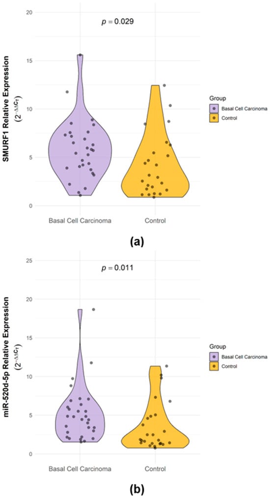
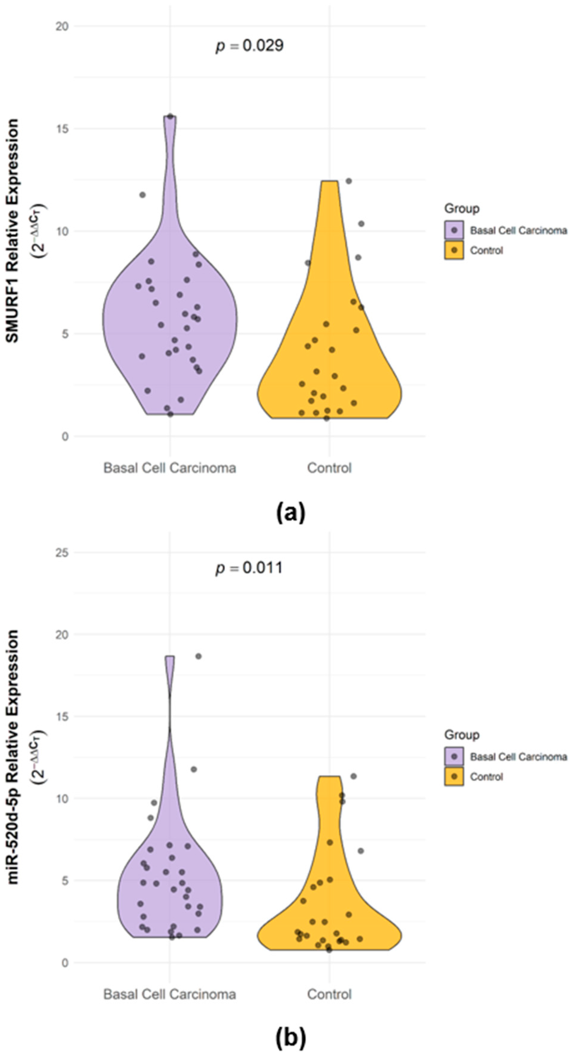
We determined SMURF1 and miR-520d-5p expression levels in skin tissues. SMURF1 and miR-520d-5p expression levels increased in BCC tissues compared to the control group. SMURF1 and miR-520d-5p expression levels were statistically significant between the BCC and control groups (p = 0.029 and p = 0.011, respectively) (Table 1 and Figure 1).
Table 1.
Expression levels of SMURF1 and miR-520d-5p.
Figure 1.
SMURF1 (a) and miR-520d-5p (b) expression levels. The SMURF1 and miR-520d-5p expression levels between BCC tissues and controls differed significantly (p = 0.029 and p = 0.011, respectively).
2.2. Correlation Analyses
To evaluate the relationship between target gene expressions and clinical/demographic parameters, Spearman rank correlation analysis was performed (Table 2). In the BCC group, no statistically significant correlation was observed between SMURF1 and miR-520d-5p expression levels (ρ = 0.090, p = 0.644). Similarly, expression levels of both SMURF1 and miR-520d-5p showed no significant correlation with patient age in either BCC or control groups (p > 0.05). Furthermore, neither SMURF1 nor miR-520d-5p expression was significantly correlated with depth of invasion in BCC tissues (ρ = 0.075, p = 0.727 and ρ = 0.222, p = 0.298, respectively).
Table 2.
Spearman rank correlation analysis between gene expression levels and clinical/demographic parameters in BCC and control groups.
2.3. Subgroup and Multivariable Analyses of SMURF1 Expression According to Age and Sex
Subgroup analyses were performed to evaluate whether SMURF1 expression varied according to age or sex within the BCC and control groups (Table 3). In BCC tissues, SMURF1 expression did not significantly differ between participants aged ≤70 years and those aged >70 years (p = 0.580), and age was not correlated with SMURF1 expression (ρ = −0.005, p = 0.981). In contrast, within the control group, participants aged >70 years exhibited significantly lower SMURF1 levels compared to those aged ≤70 years (median 2.13 vs. 4.92; p = 0.047). However, this age-related difference was not observed in BCC tissues, and the group-by-age interaction in the linear model was not statistically significant (p = 0.675). Regarding sex-related differences, SMURF1 expression showed no significant variation between females and males in BCC tissues (p = 0.214), controls (p = 0.531), or the overall cohort (p = 0.076). Furthermore, age-SMURF1 correlations evaluated separately by sex were non-significant in both females (ρ = 0.033, p = 0.871) and males (ρ = −0.134, p = 0.481). In the multivariable linear regression model adjusting for age and sex, higher SMURF1 expression in the BCC group remained statistically significant (p = 0.027), whereas neither age (p = 0.752) nor sex (p = 0.152) showed an independent significant association. Additionally, no significant group-by-sex (p = 0.573) or group-by-age (p = 0.675) interactions were detected. Given the exploratory nature of these subgroup analyses and the limited subgroup sample sizes, these comparisons were not adjusted for multiple testing.
Table 3.
Subgroup analysis of SMURF1 expression by age and sex in BCC, control, and overall study groups.
2.4. In Silico Analysis
A series of in silico analyses confirmed our experimental hypothesis and findings. We used STRING (https://string-db.org/) for the potential interactions and functional enrichment analysis of SMURF1. The default minimum required interaction score was set to medium confidence (0.400). We determined the gene disease (C4721806) association with DisGeNET (https://www.disgenet.com/). We predicted the potential associations between miR-520d-5p and proteins in the STRING network using miRSystem (http://mirsystem.cgm.ntu.edu.tw/) and miRDB (http://www.mirdb.org/). As part of our study, we identified signalling pathways that could explain the association of hsa-miR-520d-5p with BCC using mirPath from the DIANA collection (https://dianalab.e-ce.uth.gr/).
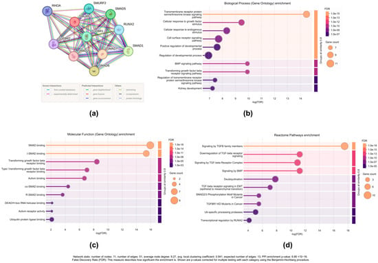
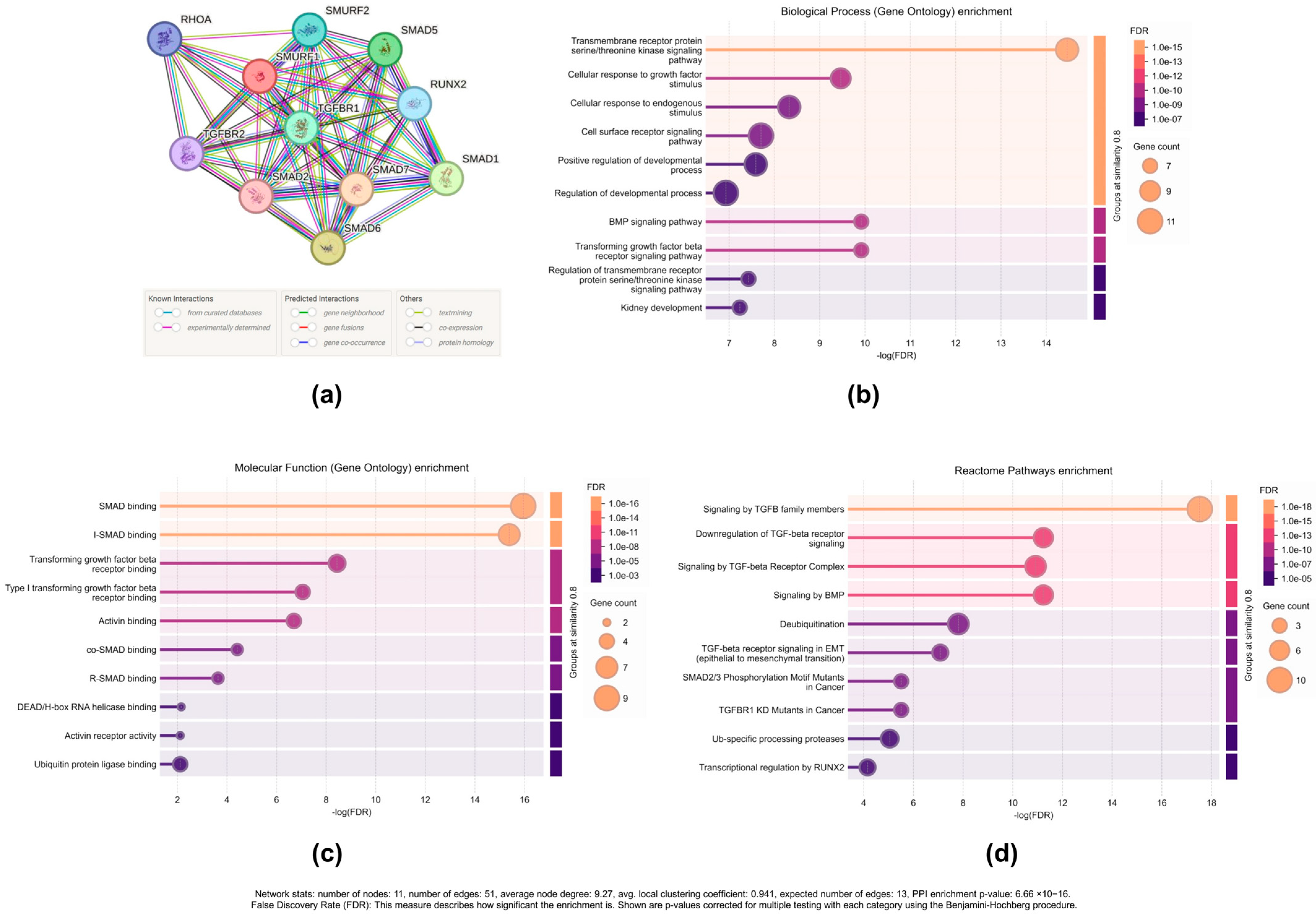
We obtained STRING network and functional enrichment results for SMURF1-interacting proteins (Figure 2). The STRING network analysis revealed a complex interplay among SMURF1 and its interacting proteins, including TGF-beta receptor family members (TGFBR1, TGFBR2), the small GTPase RHOA, and SMAD proteins (SMAD1, SMAD2, SMAD5, SMAD7).
Figure 2.
Functional enrichments of networks based on SMURF1 interactions: (a) interaction network, (b) GO biological process, (c) GO molecular function, (d) Reactome pathway database enrichment analysis results (Network stats: number of nodes: 11, number of edges: 51, average node degree: 9.27, avg. local clustering coefficient: 0.941, expected number of edges: 13, PPI enrichment p-value: 6.66 × 10⁻¹⁶; False Discovery Rate (FDR): This measure describes how significant the enrichment is. Shown are p-values corrected for multiple testing within each category using the Benjamini–Hochberg procedure).
Functional enrichment analysis identified significant associations with biological processes related to cell signalling, particularly transmembrane receptor protein serine/threonine kinase signalling, growth factor signalling, and endogenous stimulus-response. Moreover, Reactome pathway analysis highlighted the involvement of SMURF1 in the TGF-beta signalling pathway, including specific pathways such as “TGF-beta family member signalling” and “TGF-beta receptor signalling.” Additionally, enrichment in “TGF-beta signalling in EMT (epithelial to mesenchymal transition)” points to a potential role for SMURF1 in regulating EMT, a crucial process in cancer metastasis.
Molecular function enrichment revealed significant associations with SMAD binding, TGF-beta receptor binding, and activin binding, indicating that SMURF1 and its interactors directly participate in these processes. Furthermore, the result related to E3 ubiquitin ligase binding supported that SMURF1 may regulate TGF-beta signalling through ubiquitin-mediated protein degradation.
A total of 557 genes associated with BCC (C4721806) were identified in the DisGeNET database (Supplementary File S1). SMAD2, SMAD7, TGFBR1, TGFBR2, and RUNX2 genes in the STRING network were also found to be included in the DisGeNET list. According to miRSystem findings, 782 target genes were predicted for hsa-miR-520d-5p. SMAD4, SMURF2, and SMURF1 genes in the STRING network are miR-520d-5p targets. The TGFB3 gene, which is the DisGeNET list, is a target of miR-520d-5p according to the miRSystem.
We reported a total of 2404 target genes for hsa-miR-520d-5p according to miRDB. These genes include SMURF2 (target score 98/100), SMAD2 (target score 89/100), RUNX2 (target score 61/100), SMURF1 (target score 83/100), TGFBR1 (target score 73/100), and SMAD4 (target score 67/100) genes, located in the STRING network and the TGF-beta and SMAD signalling pathways. MAPK1 (target score 100/100), TGFB3 (target score 85/100), TGFBI (target score 74/100), and TGFB2 (target score 72/100), which are among the BCC-associated genes in the DisGeNET database, are miR-520d-5p targets according to miRDB.
Additionally, mirPath searched the KEGG database using the microT-CDS approach and identified 28 pathways through which hsa-miR-520d-5p may affect genes. Among the prominent signalling pathways that could be associated with BCC, the TGF-beta signalling pathway ranked first. FST, TGFBR1, SMAD2, PPP2CA, SMURF2, SMAD3, INHBA, ID4, ACVR1, ACVR2B, ZFYVE16, SMAD4, SMAD5, BMP2, SP1, ACVR1C, BMPR1A, MAPK1, and BMPR2 genes were targets of miR-520d-5p in the TGF-beta signalling pathway. Also, among the genes of these predicted target pathways, FST, TGFBR1, SMAD2, SMAD4, BMP2, BMPR1A, and MAPK1 were identified as BCC-related genes by DisGeNET.
3. Discussion
Basal cell carcinoma (BCC) is the most common skin cancer, characterised by its local invasiveness and relatively low metastatic potential. The TGF-β pathway plays a critical role in regulating cellular processes such as differentiation, apoptosis, and proliferation [15]. TGF-β signalling regulates Hedgehog pathway activity, which has been shown to affect BCC pathogenesis. In this study, SMURF1 and miR-520d-5p expression was higher in BCC than in non-cancerous controls, and in silico analyses suggest that both may be linked to the TGF-β signalling pathway; whether they contribute to BCC development or progression remains to be determined. Our findings revealed significantly increased expression levels of SMURF1 and miR-520d-5p in BCC tissues compared to non-cancerous controls (p = 0.029 and p = 0.011, respectively). The upregulation of SMURF1 is consistent with previous reports of its oncogenic role in other malignancies [9,10,16,17,18,19], whereas miR-520d-5p has mostly been described as tumour-suppressive in other cancers [13,14,20,21]. In silico analyses suggest that SMURF1 and miR-520d-5p may regulate the TGF-β signalling pathway, a critical pathway affecting cellular processes such as differentiation, apoptosis, and proliferation [16].
SMURF1 has been shown to play an oncogenic role in various cancers by modulating SMAD protein levels through ubiquitination and degradation. SMURF1 directly interacts with Phosphatidylinositol-4-phosphate 5-kinase type I gamma (PIPKIγ), which is highly expressed in lung cancer tissues, leading to its degradation [22]. Inhibition of SMURF1 in breast cancer reduced ER alpha-positive cell proliferation [17]. High SMURF1 expression has been associated with invasion and poor prognosis in glioma [10]. SMURF1 knockdown in breast cancer cell line inhibited mitogen-activated protein kinase kinase 1 (MEK1) and epidermal growth factor (EGF)-induced invasion and migration [18]. SMURF1-mediated ubiquitination of Rho GTPase-activating protein 26 (ARHGAP26) in ovarian cancer promoted migration and invasion [19]. Our results align with the existing literature that positions SMURF1 as a critical regulator of oncogenic processes in various cancers. The upregulation of SMURF1 in BCC underscores its potential involvement in these pathways, although its utility as a biomarker or therapeutic target requires further validation. Our in silico analysis also confirmed that SMURF1 interacts with various SMAD proteins, including SMAD1, SMAD2, SMAD5, and SMAD7, which are important components of the TGF-β signalling cascade. These findings suggest a potential link between SMURF1 and TGF-β/SMAD signalling in BCC; however, the functional significance of this relationship remains to be established. Further investigation of these interactions may reveal novel therapeutic targets for BCC.
Although miR-520d-5p exhibited tumour-suppressive activity in cervical cancer [13], gastric cancer [20], colorectal cancer [14], and hepatoma cancer cells [21], we found higher miR-520d-5p levels in BCC than in controls; this is an association and does not by itself demonstrate oncogenic activity. While miR-520d-5p has been overwhelmingly characterised as a tumour suppressor in most malignancies, reports indicating its potential oncogenic or elevated role in cancers remain extremely limited, with low-grade glioma being a notable exception [23]. Thus, its upregulation in BCC may reflect a context-dependent role, but elevated expression alone cannot be interpreted as evidence of oncogenic potential. Notably, the 3p strand derived from the same precursor, miR-520d-3p, was downregulated in melanoma tissues and suppressed melanoma cell proliferation by targeting anti-silencing function 1B histone chaperone (ASF1B) [24], which shows that members of this miRNA family can have protective, tumour-suppressive effects in skin cancer. Because the two strands have different targets and the cancer types differ, these findings are not directly comparable to ours, but they argue against inferring an oncogenic function from expression levels alone. Many miRNAs act as tumour suppressors or as oncomiRs depending on tissue type, cellular environment and the repertoire of expressed target genes, and a single miRNA may repress both tumour-suppressive and oncogenic transcripts, so that its net effect reflects the balance between them [25]. Our data therefore do not allow us to conclude that miR-520d-5p is oncogenic in BCC. Establishing such a role would require functional validation (e.g., gain- or loss-of-function studies) and analysis of its relationship with clinicopathological features such as histological subtype, perineural or perivascular invasion, mitotic index, and ulceration, which we could not evaluate in this cohort (see limitations).
In silico analyses predict that miR-520d-5p targets a considerable number of genes related to the TGF-β pathway, including SMAD2, SMAD4, SMURF1, SMURF2, and TGFBR1. These molecules are central modulators of TGF-β/SMAD signalling, which can be tumour-suppressive or tumour-promoting, depending on the cellular context [4,5], and interacts with Hedgehog signalling, the main driver pathway of BCC [6,7]. These predictions are, however, hypothesis-generating: target prediction algorithms rely mainly on sequence complementarity and yield many false-positive targets [26], so the increased miR-520d-5p expression in BCC does not demonstrate regulation of these genes or an effect on tumorigenic processes in BCC.
Notably, in silico prediction indicates that miR-520d-5p may target SMURF1, and an inverse relationship would be expected under canonical regulation. However, we observed a parallel elevation of both molecules in BCC tissues, and their expression was not significantly correlated (ρ = 0.090, p = 0.644 in BCC). Weak or absent correlations between miRNAs and their predicted targets have been reported in cancer tissues [27], and miRNA effects on individual targets are generally modest and may be masked in complex tissues [28,29]. Several explanations are possible. First, miR-520d-5p might be induced as part of a compensatory negative feedback response to elevated SMURF1; miRNA-mediated feedback and feedforward loops are recurrent regulatory motifs [30,31], but we have no direct evidence for such a loop in BCC, and this remains a hypothesis. Second, miRNAs often act by inhibiting translation, and their effect may be more evident at the protein level than at the mRNA level [28,29]; we measured SMURF1 mRNA but not SMURF1 protein. Second, SMURF1 is regulated by other miRNAs (for example, miR-17 in mesenchymal stem cells [32]) and by transcriptional mechanisms (for example, androgen receptor signalling [33]), and in BCC these inputs may outweigh the effect of miR-520d-5p. Third, both molecules may be co-upregulated by a shared upstream signal, such as activation of the Hedgehog or TGF-β pathway [6,7]; this is a hypothesis that was not tested in the present study. Finally, bulk formalin-fixed paraffin-embedded (FFPE) tissue contains both tumour and stromal cells, and expression in different cellular compartments may mask a relationship between the two molecules. Protein-level analysis of SMURF1 and luciferase reporter assays are needed to clarify whether miR-520d-5p directly regulates SMURF1 in BCC.
We examined whether expression varied with age and sex, because SMURF1 has been linked to ageing-related pathways [32] and to sex steroid receptor signalling: it facilitates estrogen receptor α signalling in breast cancer cells [17], and androgens upregulate SMURF1 transcription through the androgen receptor in prostate cancer cells [33]. In our cohort, higher SMURF1 expression in BCC was independent of age and sex, with no significant group × age or group × sex interaction. Within BCC, SMURF1 was not related to age or sex. In controls, SMURF1 tended to be lower in participants older than 70 years, but this exploratory observation did not survive multiple-testing correction, depended on the age cut-off, and rested on ten older controls; it therefore requires confirmation. Females showed numerically higher SMURF1 values in all strata, but this was not significant and was not independent of study group, and receptor expression and hormonal status were not assessed, so no conclusion about hormonal regulation can be drawn.
For miR-520d-5p, the difference between BCC and controls was larger at older ages (group × age interaction p = 0.022), reflecting an upward age trend in BCC and a downward trend in controls. Because neither trend was significant on its own, the interaction was not significant after correction for the four interaction tests, and the control group consisted of nevus-margin tissue rather than age-matched healthy skin, this finding is hypothesis-generating. If confirmed, it would suggest that the miR-520d-5p increase in BCC is more pronounced in older, cumulatively sun-exposed skin, which would be consistent with a role of ultraviolet exposure [34,35]. It does not demonstrate an oncogenic function.
This study has several limitations that should be acknowledged. First, the relatively small sample size (30 BCC and 30 non-cancerous skin tissues) may limit the statistical power and generalisability of our findings. Furthermore, all BCC lesions were obtained from the facial region, which may restrict the applicability of our results to tumors arising at other anatomical sites. Second, skin phototype and several clinicopathological characteristics, including histopathological subtype, perineural and perivascular invasion, mitotic index, ulceration, and history of solar-radiation exposure, were not systematically documented in the archived patient records and could not be retrieved retrospectively. Consequently, their potential associations with SMURF1 and miR-520d-5p expression could not be evaluated, and only invasion depth was analysed. In addition, detailed quantitative comparisons among individual BCC subtypes were not feasible because of the limited sample size and the diversity of histological presentations. Although ultraviolet exposure is a major environmental risk factor for BCC [34,35], all lesions in this study originated from chronically sun-exposed facial skin, which may have reduced variability in anatomical site and exposure patterns. Third, our molecular analyses were limited to mRNA expression levels, without protein-level validation or functional assays. Although in silico analyses identified SMURF1 as a potential target of miR-520d-5p, computational predictions alone cannot establish direct regulation, and potential pleiotropic or off-target effects of miR-520d-5p, which is predicted to regulate multiple transcripts within the TGF-β pathway, cannot be excluded. Future studies involving larger and more diverse cohorts, SMURF1 protein-level assessment, dual-luciferase reporter assays, overexpression and knockdown models, and rescue experiments using miRNA mimics or inhibitors in BCC cell lines are warranted to validate these findings and establish the direct regulatory relationship and biological relevance of the miR-520d-5p–SMURF1 axis in BCC.
4. Material and Methods
4.1. Study Groups
Formalin-fixed paraffin-embedded (FFPE) tissues found in the archives of Muğla Sıtkı Koçman University Department of Pathology were used in the study. Cancerous tissues diagnosed with BCC (n = 30) were included as the patient group, and non-lesional surgical margin tissues diagnosed with melanocytic nevus (n = 30) were included as the control group. All of the tissues were taken from the facial area. The BCC group consisted of 16 females and 14 males, with a mean age of 72.42 ± 11.24 years, while the control group included 12 females and 18 males, with a mean age of 69.55 ± 12.66 years. Invasion depth was mean 0.27 ± 0.14 mm for the BCC group. There were no statistically significant differences in gender distribution or age between the two groups (p = 0.301 and p = 0.357, respectively).
The study protocol was approved by the Muğla Sıtkı Koçman University Faculty of Medicine Medical Ethics Committee (Approval number 10; Date: 8 January 2025). All procedures performed in studies involving human participants were in accordance with the ethical standards of the institutional and/or national research committee and with the 1964 Helsinki declaration and its later amendments or comparable ethical standards.
4.2. Deparaffinisation of Tissues
Three sections (each section 10 microns) were taken from the paraffin blocks. Xylene was used to remove paraffin from the tissue sections [36]. 1 mL of xylene was added to the tissues. The tubes were incubated at 60 °C for 5 min and centrifuged at 17,000× g for 2 min. In the final stage, the tissues were washed with 1 mL of 100% ethanol and phosphate-buffered saline (PBS), respectively.
4.3. SMURF1 Gene and miR-520d-5p Expression Analysis
RNA was isolated from FFPE tissues using TRIzol reagent (Invitrogen, Carlsbad, CA, USA). The High-Capacity A.B.T.TM cDNA Synthesis Kit (Cat. No: C03-01-20; Atlas Biyoteknoloji, Ankara, Türkiye) was employed to convert whole RNA into cDNA, following the instructions provided in the kit. The SMURF1 and miR-520d-5p were amplified in accordance with the instructions provided in the Ampliqon RealQ Plus 2× Master Mix Green (Cat. No: A323402; Ampliqon A/S, Odense M, Denmark) reagent kit, using the Applied Biosystems StepOnePlus Real-Time PCR Detection System (Applied Biosystems, Foster City, CA, USA). GAPDH was employed as an endogenous control for SMURF1, while U6 was used for hsa-miR-520d-5p [37]. The 2−ΔΔCT method was used to assess the relative gene expression, which came from a minimum of three separate studies [38]. The primer sequences and genes details are shown in Table 4.
Table 4.
Primer sequences and amplicon sizes.
4.4. Bioinformatic Analyses
No meta-analysis was performed; all in silico analyses were conducted using publicly available web-based tools. Protein–protein interaction (PPI) and functional enrichment analyses of SMURF1 were performed using STRING v12.0 (https://string-db.org/) for Homo sapiens in August 2026. All available interaction sources (text mining, experiments, databases, co-expression, neighbourhood, gene fusion, and co-occurrence) were included, with a minimum required interaction score of 0.400 (medium confidence). Functional enrichment was assessed for Gene Ontology (GO) Biological Process, GO Molecular Function, and Reactome pathways. p-values were adjusted for multiple testing within each category using the Benjamini–Hochberg procedure. Terms with an FDR ≤ 0.05, signal ≥ 0.01, strength ≥ 0.01, and at least two proteins represented in the network were retained. Redundant terms were grouped using a term similarity threshold of 0.8 [39]. Gene–disease associations for basal cell carcinoma (UMLS CUI: C4721806) were retrieved from DisGeNET v21.0 (https://www.disgenet.org/) in August 2026. All available associations were included without applying a minimum gene–disease association (GDA) score threshold; all retrieved associations had scores ≥ 0.01, yielding a total of 557 genes [40]. Potential targets of hsa-miR-520d-5p among the proteins identified in the STRING network were predicted using miRSystem (version 17.0) [41] and miRDB (version 6.0) [42]. The resulting predicted target lists were subsequently intersected with the proteins identified in the STRING network and the DisGeNET disease-associated gene list. Pathway enrichment analysis for hsa-miR-520d-5p was performed using DIANA-miRPath v3.0, with KEGG pathways as the reference database and DIANA-microT-CDS as the target prediction algorithm. Pathways with an FDR-adjusted p-value < 0.05 were considered statistically significant.
4.5. Statistical Analysis
Statistical analyses were performed in R (version 4.5.1; R Foundation for Statistical Computing, Vienna, Austria). All statistical analyses and data visualisation were performed in R (version 4.5.1; R Foundation for Statistical Computing, Vienna, Austria). Data were imported with the readxl package (v1.5.0.1) and summarised with dplyr (v1.2.0). Cliff’s delta was calculated with effsize (v0.8.1). All remaining tests (Shapiro–Wilk, Wilcoxon–Mann–Whitney, Welch’s t-test, Fisher’s exact test, Spearman’s correlation, and linear regression) were performed with base R (stats package). No external reference datasets were used and no meta-analysis was performed; all analyses were based on the qPCR expression data generated in the present study.
Normality was assessed with the Shapiro–Wilk test. Because qPCR expression values were right-skewed and not normally distributed, expression levels were compared between BCC and control groups with the Wilcoxon–Mann–Whitney test and are reported as median [interquartile range, IQR] together with mean ± standard deviation. As effect-size measures we report Cliff’s delta (95% bootstrap CI, 5000 resamples) and the ratio of geometric means (95% CI, from Welch’s t-test on log-transformed values). Age, which was normally distributed, was compared with Welch’s t-test; sex distribution was compared with Fisher’s exact test. Associations between continuous variables (SMURF1, miR-520d-5p, age and invasion depth) were evaluated with Spearman’s rank correlation coefficient (95% bootstrap CI). To examine whether expression differed according to age and sex, participants aged ≤70 years were compared with those aged >70 years (cut-off ≈ the median age of the cohort, 70 years), and females with males, in the BCC group, the control group and the overall cohort, using the Mann–Whitney U test. In addition, linear regression models with log-transformed expression as the dependent variable were fitted to estimate the association of study group (BCC vs. control) with expression after adjustment for age and sex; results are given as fold-changes with 95% CIs. Group × age (continuous, mean-centred) and group × sex interaction terms were tested in separate models, and residual normality was checked. A two-sided p < 0.05 was considered significant.
5. Conclusions
This study investigated the expression of SMURF1 and miR-520d-5p in basal cell carcinoma (BCC). Both molecules were significantly higher in BCC tissues than in non-cancerous controls, and this increase was independent of age and sex. Although SMURF1 is a predicted target of miR-520d-5p, their expression was not inversely correlated, and we did not test this interaction experimentally; the parallel increase therefore remains unexplained and requires functional validation. In silico analyses predict that both molecules are linked to the TGF-β signalling pathway. Our findings indicate an association of SMURF1 and miR-520d-5p with BCC, and they do not establish a causal or therapeutic role. Protein-level analysis, reporter assays, and functional studies in BCC models are needed before these molecules can be considered as potential biomarkers or therapeutic targets.
Supplementary Materials
The following supporting information can be downloaded at: https://www.mdpi.com/article/10.3390/ijms27219900/s1.
Author Contributions
Ş.K.: conceptualisaton, investigation, writing—original draft, visualisation; S.Y.Ç.: resources, investigation, writing—original draft, visualisation; Ç.Ö.: investigation, writing—original draft, writing—review and editing, formal analysis; M.S.A.: investigation, writing—original draft, writing—review and editing, visualisation; T.E.: conceptualisaton, investigation, writing—review and editing. All authors have read and agreed to the published version of the manuscript.
Funding
This research received no external funding.
Institutional Review Board Statement
The study protocol was approved by the Muğla Sıtkı Koçman University Faculty of Medicine Medical Ethics Committee (Approval number 10; Date: 8 January 2025). All procedures performed in studies involving human participants were in accordance with the ethical standards of the institutional and/or national research committee and with the 1964 Helsinki declaration and its later amendments or comparable ethical standards.
Informed Consent Statement
This retrospective study was evaluated and approved by the Muğla Sıtkı Koçman University Faculty of Medicine Medical Ethics Committee (Approval No: 10; Date: 8 January 2025). Due to the retrospective nature of the study using archival paraffin-embedded tissue samples, the requirement for written informed consent was waived by the Ethics Committee.
Data Availability Statement
The data and materials in the current study are available from the corresponding author upon reasonable request.
Conflicts of Interest
The authors declare no conflicts of interest.
References
- Faam, B.; Dadkhahfar, S.; Razzaghi, Z.; Rezaei Tavirani, M.; Robati, R.M. The Significance of DNA Methylation Pattern in the Etiology of Basal Cell Carcinoma. Arch. Dermatol. Res. 2025, 317, 669. [Google Scholar] [CrossRef] [Scilit]
- Dika, E.; Scarfì, F.; Ferracin, M.; Broseghini, E.; Marcelli, E.; Bortolani, B.; Campione, E.; Riefolo, M.; Ricci, C.; Lambertini, M. Basal Cell Carcinoma: A Comprehensive Review. Int. J. Mol. Sci. 2020, 21, 5572. [Google Scholar] [CrossRef] [Scilit]
- Chinem, V.P.; Miot, H.A. Epidemiology of Basal Cell Carcinoma Epidemiologia Do Carcinoma Basocelular. An. Bras. Dermatol. 2011, 86, 292–305. [Google Scholar]
- Syed, V. TGF-β Signaling in Cancer. J. Cell. Biochem. 2016, 117, 1279–1287. [Google Scholar] [CrossRef] [Scilit]
- Inman, G.J. Switching TGFβ from a Tumor Suppressor to a Tumor Promoter. Curr. Opin. Genet. Dev. 2011, 21, 93–99. [Google Scholar] [CrossRef] [Scilit]
- Cameron, M.C.; Lee, E.; Hibler, B.P.; Giordano, C.N.; Barker, C.A.; Mori, S.; Cordova, M.; Nehal, K.S.; Rossi, A.M. Basal Cell Carcinoma: Contemporary Approaches to Diagnosis, Treatment, and Prevention. J. Am. Acad. Dermatol. 2019, 80, 321–339, Erratum in J. Am. Acad. Dermatol. 2019, 81, 310. https://doi.org/10.1016/j.jaad.2019.04.001. [Google Scholar] [CrossRef] [Scilit]
- Dicker, T.; Siller, G.; Saunders, N. Molecular and Cellular Biology of Basal Cell Carcinoma. Australas. J. Dermatol. 2002, 43, 241–246. [Google Scholar] [CrossRef] [Scilit]
- Zhu, H.; Kavsak, P.; Abdollah, S.; Wrana, J.L.; Thomsen, G.H. A SMAD Ubiquitin Ligase Targets the BMP Pathway and Affects Embryonic Pattern Formation. Nature 1999, 400, 687–693. [Google Scholar] [CrossRef] [Scilit]
- Xia, Q.; Li, Y.; Han, D.; Dong, L. SMURF1, a Promoter of Tumor Cell Progression? Cancer Gene Ther. 2021, 28, 551–565. [Google Scholar] [CrossRef] [Scilit]
- Chang, H.; Zhang, J.; Miao, Z.; Ding, Y.; Xu, X.; Zhao, X.; Xu, P.; Wang, Q.; Lin, Y. Suppression of the Smurf1 Expression Inhibits Tumor Progression in Gliomas. Cell. Mol. Neurobiol. 2018, 38, 421–430. [Google Scholar] [CrossRef] [Scilit]
- Tu, J.; Zhao, Z.; Xu, M.; Lu, X.; Chang, L.; Ji, J. NEAT1 Upregulates TGF-β1 to Induce Hepatocellular Carcinoma Progression by Sponging Hsa-Mir-139-5p. J. Cell. Physiol. 2018, 233, 8578–8587. [Google Scholar] [CrossRef] [Scilit]
- Keklikoglou, I.; Koerner, C.; Schmidt, C.; Zhang, J.D.; Heckmann, D.; Shavinskaya, A.; Allgayer, H.; Gückel, B.; Fehm, T.; Schneeweiss, A.; et al. MicroRNA-520/373 Family Functions as a Tumor Suppressor in Estrogen Receptor Negative Breast Cancer by Targeting NF-κB and TGF-β Signaling Pathways. Oncogene 2012, 31, 4150–4163. [Google Scholar] [CrossRef] [Scilit]
- Zhang, L.; Liu, F.; Fu, Y.; Chen, X.; Zhang, D. MiR-520d-5p Functions as a Tumor-Suppressor Gene in Cervical Cancer through Targeting PTK2. Life Sci. 2020, 254, 117558. [Google Scholar] [CrossRef] [Scilit]
- Yan, L.; Yu, J.; Tan, F.; Ye, G.-T.; Shen, Z.-Y.; Liu, H.; Zhang, Y.; Wang, J.-F.; Zhu, X.-J.; Li, G.-X. SP1-Mediated MicroRNA-520d-5p Suppresses Tumor Growth and Metastasis in Colorectal Cancer by Targeting CTHRC1. Am. J. Cancer Res. 2015, 5, 1447–1458. [Google Scholar]
- Zhang, Y.; Alexander, P.B.; Wang, X.F. TGF-β Family Signaling in the Control of Cell Proliferation and Survival. Cold Spring Harb. Perspect. Biol. 2017, 9, a022145. [Google Scholar] [CrossRef] [Scilit]
- Guo, X.; Wang, X. Signaling Cross-Talk between TGF-β/BMP and Other PathWays. Cell Res. 2009, 71–88. [Google Scholar] [CrossRef] [Scilit]
- Yang, H.; Yu, N.; Xu, J.; Ding, X.; Deng, W.; Wu, G.; Li, X.; Hou, Y.; Liu, Z.; Zhao, Y.; et al. SMURF1 Facilitates Estrogen Receptor a Signaling in Breast Cancer Cells. J. Exp. Clin. Cancer Res. 2018, 37, 24. [Google Scholar] [CrossRef] [Scilit]
- Kwon, A.; Lee, H.L.; Woo, K.M.; Ryoo, H.M.; Baek, J.H. SMURF1 Plays a Role in EGF-Induced Breast Cancer Cell Migration and Invasion. Mol. Cells 2013, 36, 548–555. [Google Scholar] [CrossRef] [Scilit]
- Chen, X.; Chen, S.; Li, Y.; Gao, Y.; Huang, S.; Li, H.; Zhu, Y. SMURF1-Mediated Ubiquitination of ARHGAP26 Promotes Ovarian Cancer Cell Invasion and Migration. Exp. Mol. Med. 2019, 51, 1–12. [Google Scholar] [CrossRef] [Scilit]
- Li, T.; Guo, H.; Zhao, X.; Jin, J.; Zhang, L.; Li, H.; Lu, Y.; Nie, Y.; Wu, K.; Shi, Y.; et al. Gastric Cancer Cell Proliferation and Survival Is Enabled by a Cyclophilin B/STAT3/MIR-520d-5p Signaling Feedback Loop. Cancer Res. 2017, 77, 1227–1240. [Google Scholar] [CrossRef] [Scilit]
- Miura, N.; Ishihara, Y.; Miura, Y.; Kimoto, M.; Miura, K. MiR-520d-5p Can Reduce the Mutations in Hepatoma Cancer Cells and IPSCs-Derivatives. BMC Cancer 2019, 19, 587. [Google Scholar] [CrossRef] [Scilit]
- Li, H.; Xiao, N.; Wang, Y.; Wang, R.; Chen, Y.; Pan, W.; Liu, D.; Li, S.; Sun, J.; Zhang, K.; et al. Smurf1 Regulates Lung Cancer Cell Growth and Migration through Interaction with and Ubiquitination of PIPKI. Oncogene 2017, 36, 5668–5680. [Google Scholar] [CrossRef] [Scilit]
- Deshpande, R.P.; Chandra Sekhar, Y.B.V.K.; Panigrahi, M.; Babu, P.P. SIRP Alpha Protein Downregulates in Human Astrocytoma: Presumptive Involvement of Hsa-MiR-520d-5p and Hsa-MiR-520d-3p. Mol. Neurobiol. 2017, 54, 8162–8169. [Google Scholar] [CrossRef] [Scilit]
- Shi, X.; Xu, X.; Shi, N.; Chen, Y.; Fu, M. MicroRNA-520d-3p Suppresses Melanoma Cells Proliferation by Inhibiting the Anti-Silencing Function 1B Histone Chaperone. Bioengineered 2021, 12, 10703–10715. [Google Scholar] [CrossRef] [Scilit]
- Svoronos, A.A.; Engelman, D.M.; Slack, F.J. OncomiR or Tumor Suppressor? The Duplicity of MicroRNAs in Cancer. Cancer Res. 2016, 76, 3666–3670. [Google Scholar] [CrossRef] [Scilit]
- Riffo-Campos, Á.L.; Riquelme, I.; Brebi-Mieville, P. Tools for Sequence-Based MiRNA Target Prediction: What to Choose? Int. J. Mol. Sci. 2016, 17, 1987. [Google Scholar] [CrossRef] [Scilit]
- Li, X.; Yu, X.; He, Y.; Meng, Y.; Liang, J.; Huang, L.; Du, H.; Wang, X.; Liu, W. Integrated Analysis of MicroRNA (MiRNA) and MRNA Profiles Reveals Reduced Correlation between MicroRNA and Target Gene in Cancer. BioMed Res. Int. 2018, 2018, 1972606. [Google Scholar] [CrossRef] [Scilit]
- Guo, H.; Ingolia, N.T.; Weissman, J.S.; Bartel, D.P. Mammalian MicroRNAs Predominantly Act to Decrease Target MRNA Levels. Nature 2010, 466, 835–840. [Google Scholar] [CrossRef] [Scilit]
- Baek, D.; Villén, J.; Shin, C.; Camargo, F.D.; Gygi, S.P.; Bartel, D.P. The Impact of MicroRNAs on Protein Output. Nature 2008, 455, 64–71. [Google Scholar] [CrossRef] [Scilit]
- Tsang, J.; Zhu, J.; van Oudenaarden, A. MicroRNA-Mediated Feedback and Feedforward Loops Are Recurrent Network Motifs in Mammals. Mol. Cell 2007, 26, 753–767. [Google Scholar] [CrossRef] [Scilit]
- Ebert, M.S.; Sharp, P.A. Roles for MicroRNAs in Conferring Robustness to Biological Processes. Cell 2012, 149, 515–524. [Google Scholar] [CrossRef] [Scilit]
- Liu, W.; Qi, M.; Konermann, A.; Zhang, L.; Jin, F.; Jin, Y. The P53/MiR-17/Smurf1 Pathway Mediates Skeletal Deformities in an Age-Related Model via Inhibiting the Function of Mesenchymal Stem Cells. Aging (Albany NY) 2015, 7, 205–218, Erratum in Aging (Albany NY) 2015, 7, 10197–10198. https://doi.org/10.18632/aging.205986. [Google Scholar] [CrossRef] [Scilit]
- Gang, X.; Wang, G.; Huang, H. Androgens Regulate SMAD Ubiquitination Regulatory Factor-1 Expression and Prostate Cancer Cell Invasion. Prostate 2015, 75, 561–572. [Google Scholar] [CrossRef] [Scilit]
- Armstrong, B.K.; Cust, A.E. Sun Exposure and Skin Cancer, and the Puzzle of Cutaneous Melanoma: A Perspective on Fears et al. Mathematical Models of Age and Ultraviolet Effects on the Incidence of Skin Cancer among Whites in the United States. American Journal of Epidemiology 1977: 420–427. Cancer Epidemiol. 2017, 48, 147–156. [Google Scholar] [CrossRef] [Scilit]
- Berman, B.; Cockerell, C.J.; Zografos, P. Pathobiology of Actinic Keratosis: Ultraviolet-Dependent Keratinocyte Proliferation. J. Am. Acad. Dermatol. 2013, 68, S10–S19. [Google Scholar] [CrossRef] [Scilit]
- Kumar, K.; Oli, A.; Hallikeri, K.; Shilpasree, A.S.; Goni, M. An Optimized Protocol for Total RNA Isolation from Archived Formalin-Fixed Paraffin-Embedded Tissues to Identify the Long Non-Coding RNA in Oral Squamous Cell Carcinomas. MethodsX 2022, 9, 101602. [Google Scholar] [CrossRef] [Scilit]
- Ke, Y.; Zhao, W.; Xiong, J.; Cao, R. Downregulation of MiR-16 Promotes Growth and Motility by Targeting HDGF in Non-Small Cell Lung Cancer Cells. FEBS Lett. 2013, 587, 3153–3157. [Google Scholar] [CrossRef] [Scilit]
- Li, H.; Feng, C.; Shi, S. MiR-196b Promotes Lung Cancer Cell Migration and Invasion through the Targeting of GATA6. Oncol. Lett. 2018, 16, 247–252. [Google Scholar] [CrossRef] [Scilit]
- Szklarczyk, D.; Kirsch, R.; Koutrouli, M.; Nastou, K.; Mehryary, F.; Hachilif, R.; Gable, A.L.; Fang, T.; Doncheva, N.T.; Pyysalo, S.; et al. The STRING Database in 2023: Protein–Protein Association Networks and Functional Enrichment Analyses for Any Sequenced Genome of Interest. Nucleic Acids Res. 2022, 51, D638. [Google Scholar] [CrossRef] [Scilit]
- Piñero, J.; Ramírez-Anguita, J.M.; Saüch-Pitarch, J.; Ronzano, F.; Centeno, E.; Sanz, F.; Furlong, L.I. The DisGeNET Knowledge Platform for Disease Genomics: 2019 Update. Nucleic Acids Res. 2020, 48, D845–D855. [Google Scholar] [CrossRef] [Scilit] [PubMed]
- Lu, T.P.; Lee, C.Y.; Tsai, M.H.; Chiu, Y.C.; Hsiao, C.K.; Lai, L.C.; Chuang, E.Y. MiRSystem: An Integrated System for Characterizing Enriched Functions and Pathways of MicroRNA Targets. PLoS ONE 2012, 7, e42390. [Google Scholar] [CrossRef] [Scilit]
- Chen, Y.; Wang, X. miRDB: An Online Database for Prediction of Functional MicroRNA Targets. Nucleic Acids Res. 2020, 48, D127–D131. [Google Scholar] [CrossRef] [Scilit] [PubMed]

Disclaimer/Publisher’s Note: The statements, opinions and data contained in all publications are solely those of the individual author(s) and contributor(s) and not of MDPI and/or the editor(s). MDPI and/or the editor(s) disclaim responsibility for any injury to people or property resulting from any ideas, methods, instructions or products referred to in the content. |
© 2026 by the authors. Licensee MDPI, Basel, Switzerland. This article is an open access article distributed under the terms and conditions of the Creative Commons Attribution (CC BY) license.