


Abstract
Herpesviruses are a large group of double-stranded-DNA viruses that replicate their genomes in the nucleus, forming large replication compartments during lytic infection. Herpesviruses are known to modulate host DNA replication and DNA damage-repair proteins to promote their replication. Ubiquitination is a cellular process that can alter protein stability, localization, and availability, including that of multiple proteins involved in genome maintenance. Ubiquitin-specific proteases (USPs) are a class of deubiquitinating enzymes (DUBs) that counter this process by removing ubiquitin chains, thus acting as another level of regulation. Proteins from each of the three herpesvirus subfamilies (alpha-, beta-, and gammaherpesviruses) have been reported to interact with at least one host USP, and USP7 in particular is reported as a target in representatives of all three subfamilies. In this review, we give an up-to-date summary of herpesviruses–USPs interactions, with special emphasis on DNA replication, and how these interactions could shape infection outcomes. We also examine published proteomic datasets for herpesvirus-driven host-proteome modulation and offer a framework for future directions to prioritize potentially important, understudied USPs. Lastly, we visit herpesvirus-encoded DUBs and highlight their roles in viral replication by remodeling ubiquitin signaling, discussing viral DUBs and virus–USP interfaces as proposed antiviral targets.
1. Introduction
Ubiquitination is a reversible post-translational modification that modulates protein stability, localization, and signaling by attaching ubiquitin (Ub) chains to target proteins in diverse chain architectures. Three types of enzymes, E1 ubiquitin-activating enzyme, E2 ubiquitin-conjugating enzyme, and E3 ubiquitin ligase, perform this function, and the sequential cascades of E1–E2–E3 enzymes have been well reported [1,2,3,4]. Ubiquitin is a 76-amino-acid protein highly conserved across eukaryotes [5]. Protein ubiquitination is essential for the regulation of various eukaryotic processes [6]. Ubiquitin contains seven lysine (K) residues (K6, K11, K27, K29, K33, K48, and K63) and an N-terminal methionine (Met1) that can each serve as linkage points for chain formation, resulting in multiple polyubiquitin linkage types. The linkage composition, chain architecture (including mixed or branched chains), and chain length collectively influence the fate and signaling output of the modified target protein [5]. For instance, K11- and K48-linked polyubiquitin chains lead to proteasome-mediated degradation of the protein substrate via the Ub–proteasome system [7,8], while K63-linked chains are mainly involved in non-degradative roles in cellular pathways including intracellular trafficking of linked proteins, DNA repair, and immune responses [9,10,11,12]. It has been estimated that at least 80% of protein degradation in mammalian cells occurs through the Ub–proteasome system [13].
Meanwhile, deubiquitination is the counteracting process catalyzed by deubiquitinating enzymes (DUB), which hydrolyze Ub–substrate and Ub–Ub peptide or isopeptide bonds and can thereby reverse or reshape the ubiquitin-dependent fate of target proteins [4,14]. The centrality of ubiquitin-dependent regulation to cell biology was highlighted by the 2004 Nobel Prize in Chemistry for the discovery of ubiquitin-mediated protein degradation. The human genome encodes approximately 100 DUBs, which can be divided into seven families: one Zn2+-dependent metalloprotease family and six cysteine protease families. The metalloprotease family contains the JAB1/MPN/MOV34 metalloenzyme (JAMM/MPN+) family, whose catalytic mechanism requires a coordinated metal ion [15,16]. Cysteine protease DUBs are further classified into six major superfamily groups: (1) the Ub-specific protease (USP/ubiquitin-specific processing protease (UBP)) [17], which is the largest superfamily of DUBs; (2) the ovarian tumor proteases (OTUs) [18]; (3) the Machado–Josephin domain proteases (MJD) [19]; (4) the Ub C-terminal hydrolases (UCHs) [20]; (5) MINDY-family DUBs [21], and (6) Zinc Finger With UFM1-Specific Peptidase Domain Protein (ZUFSP/ZUP1) [22,23,24,25,26].
Ubiquitin signaling is a core regulator of various cellular processes, including the DNA damage response (DDR). DUBs, on the other hand, edit ubiquitin modifications to tune protein fate and modify the engagement of repair pathways [27]. Ubiquitin-specific proteases (USPs) are the largest family of human deubiquitinases, with approximately 56 reported USPs [28], and they modulate numerous cellular processes and signaling pathways, including DDR signaling, cellular differentiation, epigenetic regulation, and protein stability [29,30]. For instance, USP1 negatively regulates the Fanconi anemia (FA) DNA repair pathway by deubiquitinating FANCD2, and USP1 associated factor 1 (UAF1) was found to be a stoichiometric USP1 partner in a multi-subunit protein complex that regulates this pathway [31]. Herpesviruses have also been reported to interact with and/or interfere with ubiquitin signaling in ways that could modulate DDR pathways.
Recent reports have identified direct USP–virus interactions at key steps in viral replication and infection, including at viral replication origins, on nascent viral DNA, in DNA-sensing pathways, and in stabilizing viral proteins. In this review we highlight the current evidence for how herpesviruses co-opt defined USP pathways to support or hinder viral replication or fork progression, and to stabilize viral proteins, and how viral DUBs can mimic or counteract host ubiquitin editing to support infection. We conclude that these well-supported herpesvirus–USP examples provide a solid framework for broader investigation. Major gaps remain in pinpointing which USPs are truly rate-limiting across virus families and cell types, and in determining when targeting USP activity could yield antiviral benefit without unacceptable genome instability or immunologic impacts.
2. Ubiquitin-Specific Proteases (USPs) in Genome Maintenance
2.1. USP Architecture and Regulation
USPs are the largest DUB subfamily and adopt a conserved hand-like architecture with palm–thumb–fingers subdomains. The catalytic center lies at the interface between the palm and the thumb, while the fingers engage ubiquitin and help position its C-terminus into the catalytic cleft for hydrolysis of the Ub C-terminal linkage [4,32]. USPs also include various modules at the N- and/or C-terminus that contribute to their substrate specificity, recruitment, and localization. These modules can include TRAF-like domains, ubiquitin-like (UBL) domains, zinc-binding motifs, and large insertions within the catalytic domain [4,33]. UBL domains adopt a ubiquitin-like fold, where they can support intramolecular regulation, partner binding, or subcellular targeting [34]. UBL domains can exist as single or multiple copies in a USP and serve a variety of functions. These can range from driving the localization of USPs to the proteasome, such as with USP14, to autoregulatory functions, such as in USP4, where the UBL domain binds its own catalytic domain, decreasing activity [34].
Many USPs are regulated by conformational changes and cofactor binding that are required for their full catalytic activity [5]. Allosteric regulation is another key layer of USP activity modulation. For instance, the last two UBL domains of USP7 were reported to be sufficient to activate USP7, with an adjacent C-terminal activation peptide being required for full activation. This activity is further enhanced allosterically by GMPS binding to the first three UBL domains (HUBL-123) of USP7 to induce the hyperactivation of USP7 [35]. USPs can also be activated allosterically by binding cofactors at distinct sites distant from the catalytic center, as in UAF1 (WDR48) and WDR20 β-propeller cofactors activation of USP12 [36]. In short, USP regulation can occur through intramolecular mechanisms, where domains within the USP allosterically tune catalytic activity, and through external mechanisms, where post-translational modifications, dedicated adaptor proteins, substrate-assisted conformational activation, and partner complexes modulate USP activity and substrate affinity [37].
2.2. Roles of Key USPs in DNA Damage Signaling and Replication Fork Progression
Ubiquitin signaling is crucial for genome stability, and USPs act as editors by removing or remodeling ubiquitin [27,33]. Multiple USPs have also been reported to play roles at stalled forks and DNA damage lesions. A well-defined genome-maintenance module is the USP1–UAF1 complex in the Fanconi Anemia (FA) pathway and replication-associated repair. RNAi library screening identified USP1 as an FA-pathway component, and USP1 depletion resulted in the hyperaccumulation of monoubiquitinated FANCD2. USP1 was found to physically associate with FANCD2, with chromatin colocalization after DNA damage [38]. A follow-up study showed that UAF1 is a stoichiometric activator of USP1 that increases catalytic turnover (kcat) without increasing substrate affinity (KM), and DNA damage was found to cause immediate transcriptional shutoff of the USP1 gene, resulting in a rapid decline in the USP1/UAF1 complex [31]. USP1 also deubiquitinates PCNA to restrain translesion synthesis (TLS)-associated damage tolerance pathways [39]. The UV-induced autocleavage of USP1 resulted in increased monoubiquitinated PCNA, which promotes TLS [40].
USP7 was also found to be involved in DNA replication progression. Minichromosome maintenance binding protein (MCM-BP) directly interacts with USP7 on chromatin, tethering USP7 to MCM proteins. The knockout of USP7 slowed late S-phase progression and impaired the dissociation of MCM proteins from chromatin in mid- to late S phase [41]. USP37 also aids in replication by deubiquitinating MCM7, thus stabilizing the replication helicase and preventing its disassembly [42].
In homologous recombination (HR), the heterodimer BRCA1/BARD1 is important for response to DNA double-strand breaks (DSBs), both through its E3 ligase function when ubiquitinating, and thus deactivating, RNA pol II, and through antagonistic competition with 53BP1 to direct repair down the homologous recombination pathway [43,44,45]. USP15 was also found to promote homologous recombination (HR) as MDC1 recruits USP15 to DSBs to deubiquitinate the BRCT domain of BARD1, promoting BARD1–HP1γ interaction and BRCA1/BARD1 retention at DSBs [46]. USP11 was also reported to interact with BRCA2 and to participate in DDR functions within the BRCA2 pathway; notably, a catalytically inactive USP11 retained interaction with BRCA2, and USP11 expression did not affect BRCA2 ubiquitination or protein levels [47]. USP28 dimerization was reported to be regulated by 53BP1, which binds and stabilizes USP28 dimers; genotoxic stress diminishes 53BP1–USP28 interaction, promoting USP28 monomerization and aberrant DNA synthesis associated with replication-associated DNA damage [48].
2.3. USPs Shape Cell-Cycle Decisions
USPs are integral regulators of cell cycle progression, as exemplified by the interaction between USP7 and the p53–MDM2 pathway. MDM2 is an E3 Ub ligase that targets p53 for degradation. MDM2 also catalyzes its own ubiquitination, which is counteracted by USP7. The loss of USP7 results in rapid MDM2 degradation and stabilization and the activation of p53 [49]. Structural work mapped the USP7 interaction site for p53 and MDM2 to the N-terminal domain of USP7, and showed that p53 and MDM2 bind competing sites on this domain. Epstein–Barr virus nuclear antigen 1 (EBNA1) also uses the same N-terminal binding pocket, consistent with competitive binding [50]. USP10, a predominantly cytoplasmic USP, translocates to the nucleus following DNA damage to deubiquitinate p53 and counter MDM2-induced nuclear export and degradation [51].
USPs can also be cell cycle-regulated. For example, USP37 expression begins during G1, peaks at the G1/S threshold, and is degraded during mitosis. USP37 binds CDH1 to deubiquitinate cyclin A. Ectopic USP37 expression accelerated S-phase entry after premature cyclin A accumulation in G1 [52]. USP37 also regulates CHK1 stability, and USP37-depleted cells showed higher sensitivity to replication stress and higher levels of DNA damage markers γH2AX and 53BP1 [53].
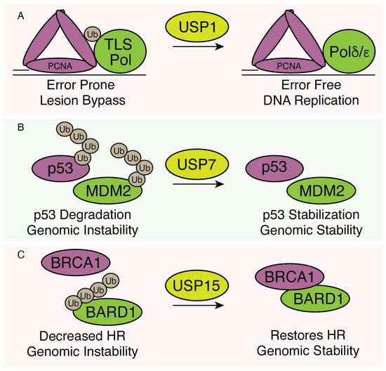
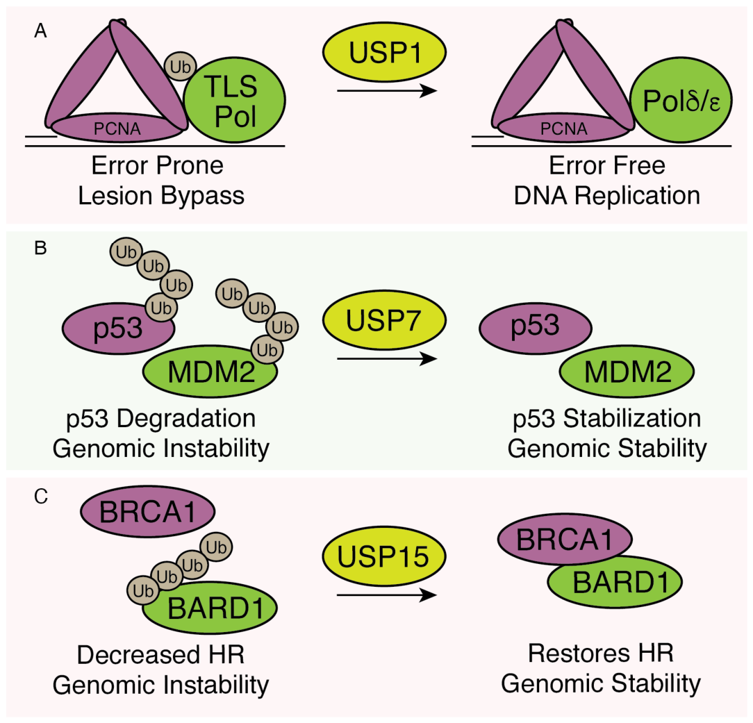
The cellular functions of several USPs are highlighted in Figure 1.
Figure 1.
Cellular functions of select USPs. Schematic summary demonstrating the cellular functions of USP1, USP7, and USP15. (A) PCNA is ubiquitinated to promote the recruitment of error-prone TLS polymerases to bypass DNA lesions. USP1 removes the ubiquitin on PCNA to restore the interaction with error-free replicative polymerases. (B) MDM2 ubiquitinates itself and p53, promoting the rapid turnover of both proteins. USP7 removes the ubiquitin to stabilize p53 to promote genomic stability. (C) Ubiquitination of BARD1 prevents the interaction with BRCA1. USP15 functions to remove the ubiquitin and promote the association of BRCA1 and BARD1 to promote homologous recombination (HR) and the error-free repair of DNA breaks.
3. Ubiquitin-Specific Proteases (USPs) in Herpesvirus Infection
Herpesviruses are large double-strand DNA viruses that establish the life-long infection of their hosts. As nuclear replicating viruses, they must contend with many areas of cell biology regulated by DUBs such as DNA replication, cell cycle control, and DNA repair. We have selected several representative examples of how herpesvirus infections interact with host DUBs and their relevant impact on viral replication, viral protein stability, and overall infection outcome.
To identify these examples, we searched PubMed and Google Scholar with terms including herpesvirus in combination with proteomics, phosphoproteomics, deubiquitinase, ubiquitin-specific protease, iPOND, and aniPOND. No limit was placed on publication date. We retained studies that reported quantitative measurements of protein abundance, phosphorylation, or association with viral DNA in infected cells, and that made those data available in the paper or its supplementary files. Each retained dataset was then examined for USPs showing altered abundance, altered phosphorylation, or enrichment on viral DNA.
3.1. HSV-1: ICP0–USP7 and UL12-USP15
The HSV-1 immediate early protein ICP0 is an E3 ubiquitin ligase that functions by degrading innate antiviral proteins to promote lytic infection [54]. Early studies searching for ICP0-interacting proteins identified a host protein of 135 kDa that bound to the C-terminal region of ICP0 [55]. This protein was later identified as USP7 [56]. The binding was mapped to a short region comprising amino acids 594–633, and an ICP0 mutant with an internal deletion in the region failed to interact with USP7 [57]. Viruses expressing ICP0 mutants that cannot interact with USP7 are unable to stimulate the expression of viral promoters and are attenuated in viral growth [58]. It was later discovered that USP7 was promoting the stabilization of ICP0 by counteracting ICP0-mediated autoubiquitination [59]. Conversely, ICP0 also targets USP7 for degradation [60]. Together, these data indicate that ICP0 and USP7 interaction is responsible for fine-tuning the amount of ICP0 needed for a productive infection and maintaining necessary functions of ICP0 in stimulating gene expression and the correct targeting of cellular proteins.
Subsequent structural work described exactly how ICP0 and USP7 interact. USP7 is a large protein of 1102 amino acids that contains seven domains. These include the N-terminal TRAF-like and catalytic domains as well as five C-terminal UBL domains (UBL1-UBL5) [61]. Biochemical works with protease resistant fragments of USP7 indicate that the C-terminal regions of USP7 are necessary for ICP0 interaction [62]. Two complementary structural papers identified a loop in USP7 UBL2 that binds ICP0. A crystal structure of the UBL1-UBL3 with an ICP0 positively charged peptide encoding amino acids 617–627 showed that ICP0 binds to a negatively charged region in UBL2. This featured salt bridges between ICP0 K620 and K624 with USP7 D762 and D764 [63]. A second study solved solution NMR structures of UBL1, UBL1-UBL2, and UBL3, titrated in ICP0 peptide 617–627, and looked for peak changes in the NMR spectra. They only observed peak changes in UBL1-UBL2 and not in UBL1 or UBL3. They also utilized isothermal calorimetry to identify that UBL1-UBL2 binds ICP0 with a 1:1 stoichiometry [64]. Together, these studies define the exact molecular interactions between ICP0 and USP7 that could be targeted for therapeutic intervention.
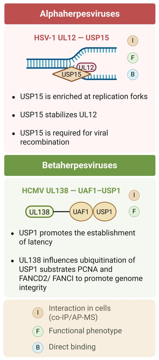
Proteomic studies of replicating HSV-1 DNA identified USP15 as one of the most enriched cellular proteins on nascent DNA [65]. HSV-1 encodes an alkaline nuclease, UL12, that together with ICP8 forms a two-subunit recombinase necessary for homology-directed repair [66]. Proteomic analysis of replicating DNA in a ΔUL12 virus found the complete absence of USP15 associated with DNA. Follow-up studies identified that UL12 directly interacts with USP15 and recruits USP15 to viral DNA [67,68]. USP15 is required for UL12 stability as infection of a ΔUSP15 cell line resulted in reduced amounts of UL12 and a significant reduction in virus yield [68]. UL12 also failed to stimulate the repair of a single strand annealing (SSA) reporter assay in the ΔUSP15 cell line. UL12 protein levels could be rescued with WT and catalytic dead USP15, but SSA was only rescued with WT protein. These data indicate that both WT and catalytic dead USP15 stabilize UL12, but only WT USP15 can rescue single strand annealing, indicating that USP15 likely has other unknown targets necessary for single strand annealing [68]. Biochemical studies mapped the interaction of USP15 to a C-terminal alpha helix in UL12. This helix is conserved in another alphaherpesvirus, PRV UL12, but not in the gammaherpesvirus KSHV SOX protein. Mutations in the UL12 helix abrogate the interaction with USP15 and fail to recruit USP15 [69]. Thus, USP15 is required for efficient replication and recombination during HSV-1 infection.
3.2. HCMV: UL35–USP7 and UL138–UAF1–USP1
The HCMV UL35 gene is required for viral gene expression and efficient replication. The UL35 gene codes for two proteins, UL35 and UL35a. Mass spectrometry analysis of UL35/UL35a-interacting proteins identified USP7 as an interactor of both proteins [70]. UL35 expression was sufficient to alter USP7 subcellular localization. Another interacting protein complex, Cul4DCAF1 E3 ubiquitin ligase complex (DCAF1, DDB1, and DDA1), interacts with UL35, but not UL35a. Mapping with DCAF1 mutants further indicated that the C-terminal region of DCAF1 was required for association with UL35 and mediated the relocalization of DCAF1 into UL35 nuclear bodies [70].
A search for protein interactors of HCMV UL138, a pro-latency protein, identified WDR48 (UAF1) [71]. UAF1 is a scaffold protein necessary for the activity of USP1, USP12, and USP46. UL138, UAF1, and USP1 were all confirmed to interact during HCMV infection. USP1 was required for the continued phosphorylation of STAT1 and for the establishment of latency [71]. USP1-UAF1 are known to regulate the ubiquitination status of PCNA and FANCD2. A follow-up study found that USP1, PCNA, and FANCD2 were all recruited to sites of viral DNA synthesis [72]. The knockdown of USP1 showed a significant increase in virus yields, indicating that USP1 restricts viral replication. Mechanistically, USP1, in conjunction with PCNA and FANCD2, contributes to the genomic integrity of replicating HCMV DNA [72]. Control of the ubiquitination status of PCNA is also important for HCMV genomic integrity as monoubiquitinated PCNA is critical for the recruitment of TLS polymerases, which also promote the integrity of the HCMV genome [73].
3.3. EBV and KSHV: EBNA1–USP7 and vIRF1-USP7
The EBV EBNA1 protein is an essential protein for the replication and maintenance of the latent viral genome. A search for EBNA1-interacting proteins identified USP7 and the interaction was mapped to the N-terminal p53 binding domain of USP7 [62,74]. The interaction with USP7 requires amino acids 436–450 of EBNA1 [75]. This EBNA1 peptide was cocrystalized with the USP7 N-terminal domain. EBNA1 bound to a shallow depression in the surface of USP7 and the interaction was mediated by a combination of hydrophobic and hydrophilic interactions. NMR chemical shift analysis indicated that EBNA1 and p53 compete for the same binding site on USP7. Functionally, this acts to reduce the levels of cellular p53 and make cells more resistant to apoptosis [75]. EBNA1 is also able to cause the disruption of PML nuclear bodies. This disruption requires the interaction with USP7. This disruption impairs the induction of p53, DNA repair, and apoptosis following DNA damage [76].
The KSHV vIRF1, encoded by ORF K9, has homology to cellular IRF3 and IRF7 and functions by disrupting cellular antiviral activity. vIRF interacts with USP7 through a peptide motif of EGPS located at the N-terminus of vIRF. This amino acid motif is identical to the EBNA1 motif required to interact with USP7. Indeed, the cocrystal of vIRF1 peptide with the USP7 N-terminal domain revealed an identical mode of binding to that of EBNA1-USP7 [77]. KSHV vIRF1 and ORF45 also disrupt p53 signaling by competing for binding with USP7 and therefore inducing the destabilization of p53 [77,78]. vIRF2, vIRF3, and vIRF4 also interact with USP7 to regulate innate immune sensing and latency [79,80,81].
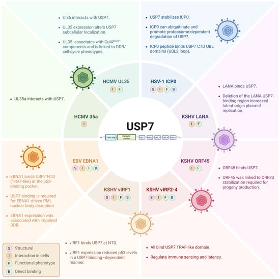
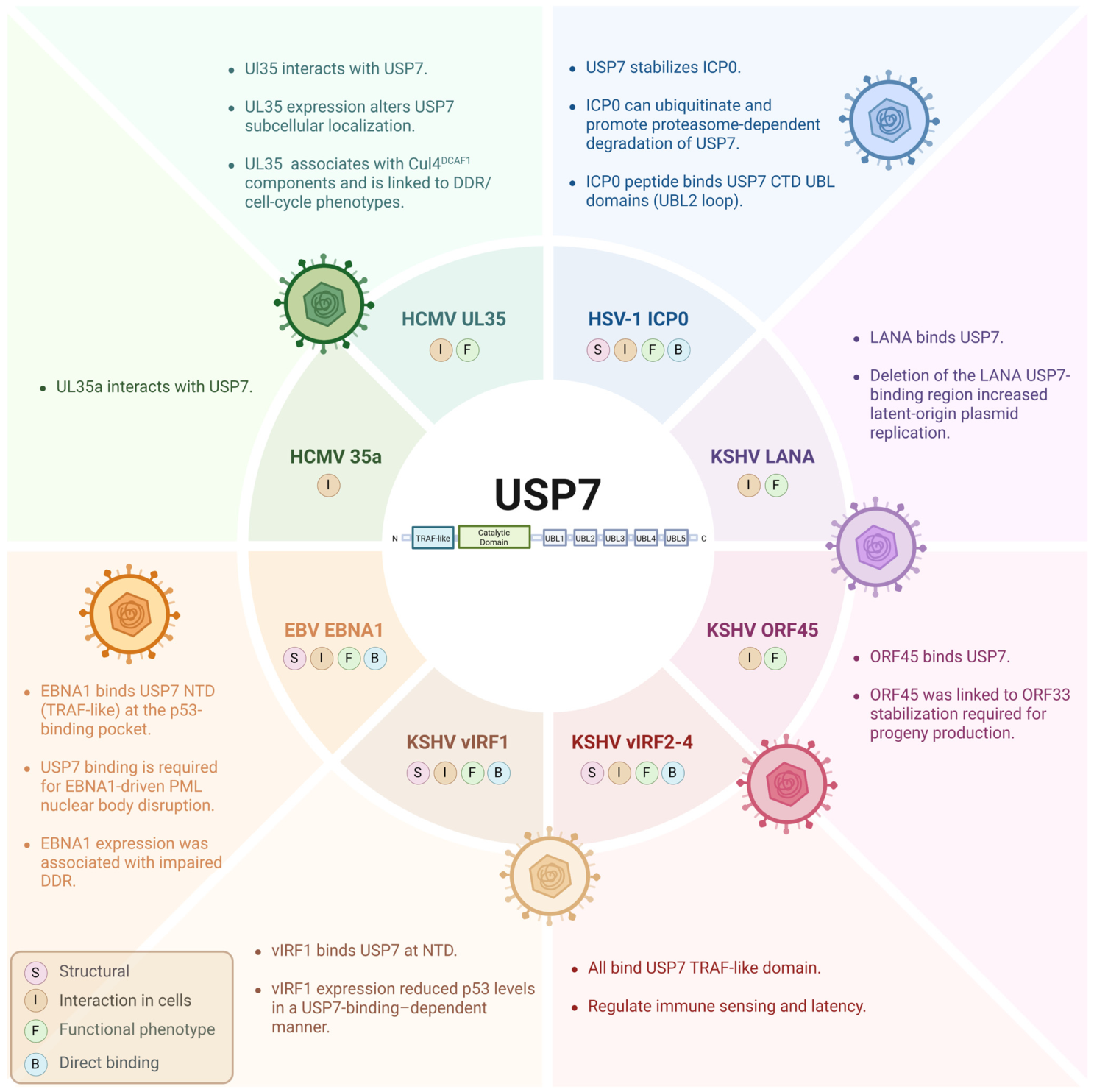
The viral interactions with USP7 are highlighted in Figure 2. Other USPs are highlighted in Figure 3 and Table 1.
Figure 2.
USP7 interfaces targeted by herpesviruses. Schematic summary of reported interactions between host ubiquitin-specific protease 7 (USP7) and herpesvirus proteins across alpha-, beta-, and gammaherpesviruses. Evidence badges indicate the primary support in the cited studies: S, structural interface mapping; B, direct binding demonstrated with purified components; I, interaction detected in cells (e.g., co-IP/AP-MS and/or colocalization); F, functional phenotype reported in the experimental system. USP7 domain annotations indicate cases where studies mapped binding to specific USP7 regions. Created in BioRender. Hasselmann, T. (2026) https://biorender.com/0xwldon.
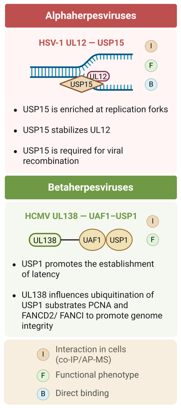
Figure 3.
Validated herpesvirus–host USP interfaces highlighted in this review. Schematic summary of experimentally supported interactions between herpesvirus proteins and host ubiquitin-specific proteases (USPs) other than USP7, grouped by herpesvirus subfamily. Evidence badges indicate the primary support in the cited studies: B, direct binding demonstrated with purified components; I, interaction detected in cells (e.g., co-IP/AP-MS and/or colocalization); F, functional phenotype reported in the experimental system. Created in BioRender. Maarouf, M. (2026) https://biorender.com/nxrtpzc.
Table 1.
Table summarizing viral proteins interacting with USPs.
4. Herpesvirus-Encoded DUBs
All herpesviruses encode a USP encoded at the N-terminus of the large tegument protein that bears no homology to known DUBs or Ub binding proteins. This was first described for HSV-1 UL36 called UL36USP. The enzymatic activity of UL36USP peaks at late times post-infection and appears to require proteolytic cleavage from the rest of the large tegument protein [87]. Other herpesvirus large tegument proteins have very low sequence identity with UL36USP, but all contain the conserved residues of the cysteine protease active site. The purification of the EBV and MCMV USPs reveal that they all retain ubiquitin protease activity [88].
There have been several reported activities for HSV-1 UL36USP. One major target of UL36USP is the host protein PCNA. UL36USP reduces PCNA ubiquitination, inhibits Pol η foci formation, and increases cellular sensitivity to DNA damage [89]. UL36USP also deubiquitinated PCNA more efficiently than USP1/UAF1 [89]. Another host target of UL36USP is TRAF3. The expression of UL36USP disrupts the TRAF3-dependent recruitment/activation of TBK1 and interferes with IFN-β signaling, and an HSV-1 ΔUL36USP virus induces higher IFN-β production [90]. UL36USP also targets IκBα to reduce NF-κB activation and downstream antiviral gene induction [91]. Similarly in animals, HSV-1 ΔUL36USP virus induced higher levels of IFN-β and IFN-stimulated genes (ISGs) in the brain, and identified STING as the target of UL36USP, supporting a role for UL36USP in immune evasion [92]. UL36 was also reported to have an immunomodulatory function independent of its DUB activity, via binding to the IFNAR2 subunit, blocking JAK1–IFNAR2 interaction, and dampening JAK–STAT antiviral signaling, thereby inhibiting IFN-β–induced ISG transcription [93]. Finally, PRV UL36 was shown to be important for initial neural invasion, but not for intraneural transmission [94].
The HCMV homolog of HSV-1 UL36 is UL48. UL48 is also a large tegument protein with an N-terminal USP domain, a conserved active site, and auto-deubiquitinating activity [95,96]. UL48, together with its interacting partner UL45, interacts with and deubiquitinates RIP to inhibit NF-κB signaling [97]. During infection with a UL48 active-site mutant virus, the most affected proteins were UL48 itself and the inner tegument protein pp150 [98]. UL48 also modulates pattern-recognition receptor-mediated type I interferon induction by deubiquitinating TRAF6, TRAF3, IRAK1, IRF7, and STING [99]. The murine cytomegalovirus DUB, M48, is required for the regulation of the viral chemokine, MCK2. The loss of M48 results in elevated levels of MCK2 and unconstrained viral induced inflammation that inhibits replication [100].
The large tegument protein of EBV, BPLF1, also encodes an N-terminal DUB that targets PCNA for deubiquitination [101]. Similar to HSV-1 UL36USP, the expression of the BPLF1 DUB is sufficient to prevent PCNA ubiquitination after DNA damage and prevent recruitment of the TLS polymerase η, suggesting a common feature several herpesviruses need to avoid. Several of the N-terminal DUBs also contain PCNA-interacting peptides, or PIP domains, that regulate their interaction with PCNA [101]. BPLF1 also targets and stabilizes the E3 Ubiquitin ligase that monoubiquitinates PCNA, Rad18. A BPLF1 knockout virus had reduced levels of Rad18 upon reactivation [102]. The KSHV large tegument protein, ORF64, also contains N-terminal DUB activity and is required for efficient viral replication and reactivation from latency [103]. KSHV ORF64 counteracts RIG-I/IFN signaling by reversing TRIM25-mediated K63-linked RIG-I ubiquitination, a key step in RIG-I activation [104]. The inhibition of RIG and type I interferon induction seems to be another conserved function of the herpesvirus DUBs.
5. Proteomic Maps of USPs Remodeling in Herpesvirus Infection
Leveraging large-scale proteomics in the context of herpesvirus infections could help identify candidate host or viral factors that promote viral growth or act as antivirals. Two complementary classes of datasets have been reported for multiple herpesviruses that we will analyze to try to identify understudied USPs with altered accumulation levels following infection: (i) time-resolved whole-cell proteomics that quantifies USP abundance over a defined time post-infection; and (ii) proteomics identifying host factors physically present on viral DNA. Implementing these two approaches can help distinguish USPs whose total cellular abundance changes during infection (which may reflect destabilization or transcriptional downregulation) from USPs enriched on viral DNA, which is more suggestive of a direct role at viral DNA replication sites.
5.1. Time-Resolved Whole-Cell Proteomics
An early study utilized multiplex tandem mass tag (TMT)-based quantitative temporal viromics to profile proteomic changes during HCMV infection over time [105]. The data demonstrate that USP1, USP10, USP22, and USP36 exhibited an early increase (peaking 24 hpi) followed by reduced abundance at later time points (72–96 hpi) relative to mock, whereas USP13 showed an overall decrease from mock across the time course. USP4 was generally reduced post-infection, while USP20, USP33, and USP7 showed increased abundance during infection time points [105].
Follow-up studies used a similar approach with TMT-based temporal proteomic analysis of HSV-1 infection using whole-cell lysates [106]. Of the DUBs observed in this study, USP1 and USP33 were downregulated by 90% and 75%, respectively. They also observed intermediate downregulation of about 40% to 50% for USP7, USP11, USP22, and USP20. USP53 and USP54, which are often annotated as catalytically inactive DUBs, were likewise reduced at late time points [106].
These approaches have also been used to study changes in abundance and posttranslational modifications of host proteins during a time course of HSV-1 infection [107]. USP7 abundance decreased over the time course, while USP5 increased at late time points. USP10, USP24, and USP53 were also reduced over time. They also quantified host phosphosite dynamics, and observed phosphopeptide abundance increased over time for USP15, USP24, USP53, and USP9X. USP34 and USP10 also exhibited increases in phosphorylation at late times post-infection. Conversely, phosphosites on USP8 were downregulated during infection [107].
Another study used TMT-based phosphoproteomic profiling with matched total-proteome analysis in HSV-1-infected cells at 3 hpi. In the total-proteome dataset, USP9X and USP7 showed only modest decreases. In the phosphoproteomic dataset, several USP phosphosites also changed modestly, including decreases in USP8, USP36, and USP10, and increases in USP39 and USP34 [108].
5.2. Host Proteins Physically Present on Viral DNA
Another approach to understand virus biology has involved the application of isolation of proteins on nascent DNA (iPOND) coupled with quantitative mass spectrometry. This technique allows for the identification of proteins physically associated with replicating DNA, and preferentially identifies proteins involved in the transcriptional control of viral genes and in the replication of viral DNA. Several labs have utilized iPOND-MS to identify proteins associated with viral DNA during HSV-1 infection [65,109,110]. Most of the observed USPs were common between virus- and host-replicating DNA, but several were either enriched or under-represented on viral DNA [65]. In particular, USP1, USP10, USP3, USP47, USP48, and USP9X were under-represented after infection with HSV-1, while USP15 and USP39 were greatly enriched [65]. Another approach utilized accelerated native iPOND (aniPOND) to demonstrate that USP39 associates with the incoming viral genome as early as 1 h post infection, and remains associated through the start of DNA replication at 6 h post infection [111].
The application of iPOND has also been utilized to study the viral changes associated with loss of viral proteins or in the presence of small-molecule inhibitors. The comparison of HSV-1 versus ΔICP0 virus was used to identify novel viral restriction factors such as SLFN5. The authors also note that USP7 is enriched on viral DNA in the absence of ICP0 [112]. USP34 and USP48 showed a lower abundance in WT relative to mock and ΔICP0 conditions, whereas USP39 was enriched in WT relative to mock. USP15 showed a higher abundance in ΔICP0 relative to WT and USP1 appeared reduced in ΔICP0 versus WT [112]. The comparison of HSV-1 versus ΔUL12 virus also used iPOND to confirm the recruitment of USP15 to viral DNA observed in other studies, and demonstrate that it was recruited specifically by UL12 [68]. Finally, aniPOND was also used to demonstrate that USP39 is still enriched on viral DNA in the presence of PCNA inhibitors [113].
While changes in USP1 and USP7 have been experimentally demonstrated to be important for several herpesviruses, the other identified understudied USPs highlighted by these datasets remain candidates that require further investigation. Future work will be required to determine whether the enriched USPs highlighted by these datasets have direct roles in in the viral life cycle, or if USPs that are lost or reduced in abundance represent novel restriction factors.
6. Therapeutic Opportunities
The Ub-proteasome system has been proposed as a drug target, and there are currently three FDA-approved drugs that target the proteasome (bortezomib, carfilzomib, ixazomib). The targeting of individual USPs is still in its early stages, and has only started yielding human clinical trial data in a small set of studies [114].
In our current review of herpesvirus–USP interactions, we highlighted USP1 and USP7 as interacting partners for multiple herpesviruses, and USP1 and USP7 have been proposed to have clear translational potential as drug targets. Since USP1 has been reported to be directly involved in DDR pathways [23,115], targeting it may narrow its antiviral applications due to potential side effects. The USP1 inhibitor TNG348 is a potent, highly selective oral allosteric USP1 inhibitor from Tango Therapeutics. It entered Phase 1/2 testing, but the trial was terminated after significant hepatotoxicity/liver function abnormalities (grade 3/4) were observed in initial cohorts after >8 weeks of dosing [116]. A Roche-sponsored USP1-targeting drug, KSQ-4279/RO7623066, is being evaluated in a Phase 1 clinical study in advanced solid tumors as monotherapy and in combination regimens with carboplatin or olaparib [117,118]. KSQ-4279 is reported to bind to a cryptic pocket in USP1 [118]. Whether the USP1–herpes interaction pocket overlaps with this site is an open question. It remains to be seen whether USP1 inhibition could disrupt a herpesvirus–USP1 axis without triggering unacceptable genotoxic stress.
USP7 is another example of a druggable target given its crucial role in regulating p53 levels [119]. Despite several small molecules showing efficacy in vitro in inhibiting USP7, there are currently no clear active clinical trials testing USP7 inhibitors. For instance, FT671 was described as a selective USP7 inhibitor, and its application exhibits a similar biology to the genetic loss of USP7. For example, FT671 increased p53 levels, destabilized MDM2, induced p21, and exhibited in vivo anti-tumor activity [120]. YCH2823 was reported as a next-generation inhibitor of USP7, surpassing FT671 potency by approximately 5-fold [121]. Assuming no adverse safety profiles of USP7 inhibitors, countering USP7 may be considered as an anti-herpesvirus drug with the potential to target multiple viruses. USP7 inhibition would, in principle, interfere with HSV-1 ICP0-USP7 and HCMV UL35/UL35a-USP7 pathways. However, since USP7 is involved in cell-cycle control via the p53–MDM2 axis and other DDR networks, dosing and timing could be critical to achieving antiviral efficacy with minimal cell-cycle disturbance.
7. Conclusions and Open Questions
While targeting host USPs is in the spotlight, another complementary approach would be targeting viral DUBs to ensure higher drug selectivity, while research into herpesvirus DUB inhibitors is relatively scarce [122]. In one reported study, a small-molecule screen identified 10 compounds that inhibited EBV BPLF1 deubiquitinating activity in a pilot assay [123]. Suramin was found to be the top hit, decreasing EBV infectivity in vitro, despite not being selective for the viral DUB. It also inhibited the KSHV DUB, ORF64, in vitro, but did not alter KSHV infectivity or reactivation [123]. This helps establish a framework for targeting viral DUBs, but it also underscores the need to achieve true selectivity while avoiding the cross-inhibition of host USPs. In addition, because many herpesviruses switch between lytic infection and latency, targeting either host USPs or viral DUBs could risk unintended outcomes on genome stability and immune signaling, which is particularly concerning in patients exposed to DNA-damaging agents or in EBV/KSHV-associated malignancies treated with genotoxic chemotherapy. Yet, accumulated knowledge from targeting DUBs in cancer suggests that utilizing similar approaches as antivirals will likely require tight dosing windows with biomarkers for target engagement, so as to reduce inhibitor exposure while maximizing efficacy [114].
Author Contributions
Conceptualization, M.M. and K.N.M.; investigation, M.M., C.S.P. and K.N.M.; writing—original draft preparation, M.M., C.S.P. and K.N.M.; writing—review and editing, M.M., C.S.P. and K.N.M.; visualization, M.M.; supervision, K.N.M.; project administration, K.N.M.; funding acquisition, K.N.M. All authors have read and agreed to the published version of the manuscript.
Funding
National Institutes of Health (NIH) provided funding to Kareem N. Mohni under grants R35GM147134 and R01AI021747.
Institutional Review Board Statement
Not applicable.
Informed Consent Statement
Not applicable.
Data Availability Statement
No new data were created or analyzed in this study.
Acknowledgments
We thank all members of the Mohni Lab for their help and critical discussions.
Conflicts of Interest
The authors declare no conflicts of interest.
Abbreviations
The following abbreviations are used in this manuscript:
| BRCA1/2 | Breast cancer susceptibility gene 1/2 |
| BRCT | BRCA1 C-terminal (domain) |
| CMV | Cytomegalovirus |
| Cul4 | Cullin 4 |
| DCAF1 | DDB1- and CUL4-associated factor 1 |
| DDA1 | DET1- and DDB1-associated protein 1 |
| DDB1 | Damage-specific DNA binding protein 1 |
| DDR | DNA damage response |
| DSB | Double-strand break |
| DUB | Deubiquitinase |
| EBV | Epstein–Barr virus |
| EBNA1 | Epstein–Barr virus nuclear antigen 1 |
| EGPS | Glu–Gly–Pro–Ser (motif) |
| E1 | Ubiquitin-activating enzyme |
| E2 | Ubiquitin-conjugating enzyme |
| E3 | Ubiquitin ligase |
| FA | Fanconi anemia |
| FANCD2/FANCI | Fanconi anemia complementation group D2/I |
| GMPS | GMP synthetase |
| Hpi | Hours post-infection |
| HR | Homologous recombination |
| HCMV | Human cytomegalovirus |
| IFN | Interferon |
| IFNAR2 | Interferon-α/β receptor subunit 2 |
| iPOND | Isolation of proteins on nascent DNA |
| IRAK1 | Interleukin-1 receptor–associated kinase 1 |
| IκBα | Inhibitor of κB alpha |
| ISG | Interferon-stimulated gene |
| JAMM/MPN+ | JAB1/MPN/MOV34 metalloenzyme (family) |
| JAK | Janus kinase |
| KSHV | Kaposi’s sarcoma-associated herpesvirus |
| K (e.g., K48, K63) | Lysine (ubiquitin linkage site) |
| LANA | Latency-associated nuclear antigen |
| M48 | Murine CMV UL48 orthologue |
| MCK2 | Murine CMV chemokine (viral proinflammatory chemokine) |
| MCM | Minichromosome maintenance (complex) |
| MCM-BP | Minichromosome maintenance binding protein |
| MCMV | Murine cytomegalovirus |
| MDM2 | Mouse double minute 2 (E3 ligase regulating p53) |
| Met1 | N-terminal methionine 1 (ubiquitin linkage site) |
| MINDY | Motif interacting with ubiquitin-containing novel DUB family |
| NF-κB | Nuclear factor kappa B |
| NBS1 | Nijmegen breakage syndrome protein 1 |
| ORF | Open reading frame |
| OTU | Ovarian tumor (DUB family) |
| PCNA | Proliferating cell nuclear antigen |
| PML | Promyelocytic leukemia (protein) |
| PML-NB | PML nuclear body |
| PRV | Pseudorabies virus |
| RING | Really interesting new gene |
| S/T | Serine/Threonine |
| S229, T226 (etc.) | Phosphorylation site numbering (Ser/Thr residue position) |
| S-phase | DNA synthesis phase of the cell cycle |
| SSA | Single-strand annealing |
| STAT1/STAT3 | Signal transducer and activator of transcription 1/3 |
| STING | Stimulator of interferon genes |
| TBK1 | TANK-binding kinase 1 |
| TLS | Translesion synthesis |
| TMT | Tandem mass tag |
| TRAF3/6 | TNF receptor–associated factor 3/6 |
| TRIM25 | Tripartite motif-containing protein 25 |
| Ub | Ubiquitin |
| UAF1 | USP1-associated factor 1 (also known as WDR48) |
| UBL | Ubiquitin-like (domain/protein) |
| UCH | Ubiquitin C-terminal hydrolase |
| UL | Unique long (herpesvirus gene region nomenclature) |
| UPS | Ubiquitin–proteasome system |
| USP | Ubiquitin-specific protease |
| vIRF1 | Viral interferon regulatory factor 1 |
| VZV | Varicella-zoster virus |
| WDR48 | WD repeat-containing protein 48 (UAF1) |
| WDR20 | WD repeat-containing protein 20 |
| WT | Wild-type |
| γH2AX | Phosphorylated histone H2AX |
| ZUFSP/ZUP1 | Zinc Finger With UFM1-Specific Peptidase Domain Protein |
| Zn2+ | Zinc ion |
References
- Zheng, N.; Shabek, N. Ubiquitin Ligases: Structure, Function, and Regulation. Annu. Rev. Biochem. 2017, 86, 129–157. [Google Scholar] [CrossRef] [Scilit] [PubMed]
- Deribe, Y.L.; Pawson, T.; Dikic, I. Post-translational modifications in signal integration. Nat. Struct. Mol. Biol. 2010, 17, 666–672. [Google Scholar] [CrossRef] [Scilit] [PubMed]
- Hu, Y.; Bai, X.; Zhang, C.; Chakrabarti, S.; Tang, B.; Xiong, H.; Wang, Z.; Yu, G.; Xu, C.; Chen, Q.; et al. Ubiquitination-activating enzymes UBE1 and UBA6 regulate ubiquitination and expression of cardiac sodium channel Nav1.5. Biochem. J. 2020, 477, 1683–1700. [Google Scholar] [CrossRef] [Scilit] [PubMed]
- Komander, D. The emerging complexity of protein ubiquitination. Biochem. Soc. Trans. 2009, 37, 937–953. [Google Scholar] [CrossRef] [Scilit] [PubMed]
- Komander, D.; Rape, M. The ubiquitin code. Annu. Rev. Biochem. 2012, 81, 203–229. [Google Scholar] [CrossRef] [Scilit] [PubMed]
- Swatek, K.N.; Komander, D. Ubiquitin modifications. Cell Res. 2016, 26, 399–422. [Google Scholar] [CrossRef] [Scilit] [PubMed]
- Jin, L.; Williamson, A.; Banerjee, S.; Philipp, I.; Rape, M. Mechanism of ubiquitin-chain formation by the human anaphase-promoting complex. Cell 2008, 133, 653–665. [Google Scholar] [CrossRef] [Scilit] [PubMed]
- Kerscher, O.; Felberbaum, R.; Hochstrasser, M. Modification of proteins by ubiquitin and ubiquitin-like proteins. Annu. Rev. Cell Dev. Biol. 2006, 22, 159–180. [Google Scholar] [CrossRef] [Scilit] [PubMed]
- Oshiumi, H.; Miyashita, M.; Matsumoto, M.; Seya, T. A distinct role of Riplet-mediated K63-Linked polyubiquitination of the RIG-I repressor domain in human antiviral innate immune responses. PLoS Pathog. 2013, 9, e1003533. [Google Scholar] [CrossRef] [Scilit]
- Okamoto, M.; Kouwaki, T.; Fukushima, Y.; Oshiumi, H. Regulation of RIG-I Activation by K63-Linked Polyubiquitination. Front. Immunol. 2017, 8, 1942. [Google Scholar] [CrossRef] [Scilit] [PubMed]
- Song, G.; Liu, B.; Li, Z.; Wu, H.; Wang, P.; Zhao, K.; Jiang, G.; Zhang, L.; Gao, C. E3 ubiquitin ligase RNF128 promotes innate antiviral immunity through K63-linked ubiquitination of TBK1. Nat. Immunol. 2016, 17, 1342–1351. [Google Scholar] [CrossRef] [Scilit] [PubMed]
- Liu, P.; Gan, W.; Su, S.; Hauenstein, A.V.; Fu, T.M.; Brasher, B.; Schwerdtfeger, C.; Liang, A.C.; Xu, M.; Wei, W. K63-linked polyubiquitin chains bind to DNA to facilitate DNA damage repair. Sci. Signal. 2018, 11, eaar8133. [Google Scholar] [CrossRef] [Scilit]
- Collins, G.A.; Goldberg, A.L. The Logic of the 26S Proteasome. Cell 2017, 169, 792–806. [Google Scholar] [CrossRef] [Scilit] [PubMed]
- Pickart, C.M.; Eddins, M.J. Ubiquitin: Structures, functions, mechanisms. Biochim. Biophys. Acta 2004, 1695, 55–72. [Google Scholar] [CrossRef] [Scilit] [PubMed]
- Verma, R.; Aravind, L.; Oania, R.; McDonald, W.H.; Yates, J.R., 3rd; Koonin, E.V.; Deshaies, R.J. Role of Rpn11 metalloprotease in deubiquitination and degradation by the 26S proteasome. Science 2002, 298, 611–615. [Google Scholar] [CrossRef] [Scilit]
- Maytal-Kivity, V.; Reis, N.; Hofmann, K.; Glickman, M.H. MPN+, a putative catalytic motif found in a subset of MPN domain proteins from eukaryotes and prokaryotes, is critical for Rpn11 function. BMC Biochem. 2002, 3, 28. [Google Scholar] [CrossRef] [Scilit]
- Tobias, J.W.; Varshavsky, A. Cloning and functional analysis of the ubiquitin-specific protease gene UBP1 of Saccharomyces cerevisiae. J. Biol. Chem. 1991, 266, 12021–12028. [Google Scholar] [CrossRef] [Scilit]
- Makarova, K.S.; Aravind, L.; Koonin, E.V. A novel superfamily of predicted cysteine proteases from eukaryotes, viruses and Chlamydia pneumoniae. Trends Biochem. Sci. 2000, 25, 50–52. [Google Scholar] [CrossRef] [Scilit] [PubMed]
- Burnett, B.; Li, F.; Pittman, R.N. The polyglutamine neurodegenerative protein ataxin-3 binds polyubiquitylated proteins and has ubiquitin protease activity. Hum. Mol. Genet. 2003, 12, 3195–3205. [Google Scholar] [CrossRef] [Scilit] [PubMed]
- Johnston, S.C.; Riddle, S.M.; Cohen, R.E.; Hill, C.P. Structural basis for the specificity of ubiquitin C-terminal hydrolases. EMBO J. 1999, 18, 3877–3887. [Google Scholar] [CrossRef] [Scilit] [PubMed]
- Abdul Rehman, S.A.; Kristariyanto, Y.A.; Choi, S.Y.; Nkosi, P.J.; Weidlich, S.; Labib, K.; Hofmann, K.; Kulathu, Y. MINDY-1 Is a Member of an Evolutionarily Conserved and Structurally Distinct New Family of Deubiquitinating Enzymes. Mol. Cell 2016, 63, 146–155. [Google Scholar] [CrossRef] [Scilit] [PubMed]
- Amerik, A.Y.; Hochstrasser, M. Mechanism and function of deubiquitinating enzymes. Biochim. Biophys. Acta 2004, 1695, 189–207. [Google Scholar] [CrossRef] [Scilit] [PubMed]
- Nijman, S.M.; Luna-Vargas, M.P.; Velds, A.; Brummelkamp, T.R.; Dirac, A.M.; Sixma, T.K.; Bernards, R. A genomic and functional inventory of deubiquitinating enzymes. Cell 2005, 123, 773–786. [Google Scholar] [CrossRef] [Scilit] [PubMed]
- Caba, C.; Mohammadzadeh, A.; Tong, Y. On the Study of Deubiquitinases: Using the Right Tools for the Job. Biomolecules 2022, 12, 703. [Google Scholar] [CrossRef] [Scilit] [PubMed]
- Haahr, P.; Borgermann, N.; Guo, X.; Typas, D.; Achuthankutty, D.; Hoffmann, S.; Shearer, R.; Sixma, T.K.; Mailand, N. ZUFSP Deubiquitylates K63-Linked Polyubiquitin Chains to Promote Genome Stability. Mol. Cell 2018, 70, 165–174.e6. [Google Scholar] [CrossRef] [Scilit] [PubMed]
- Kwasna, D.; Abdul Rehman, S.A.; Natarajan, J.; Matthews, S.; Madden, R.; De Cesare, V.; Weidlich, S.; Virdee, S.; Ahel, I.; Gibbs-Seymour, I.; et al. Discovery and Characterization of ZUFSP/ZUP1, a Distinct Deubiquitinase Class Important for Genome Stability. Mol. Cell 2018, 70, 150–164.e6. [Google Scholar] [CrossRef] [Scilit] [PubMed]
- Jackson, S.P.; Durocher, D. Regulation of DNA damage responses by ubiquitin and SUMO. Mol. Cell 2013, 49, 795–807. [Google Scholar] [CrossRef] [Scilit] [PubMed]
- Maurer, S.K.; Mayer, M.P.; Ward, S.J.; Boudjema, S.; Halawa, M.; Zhang, J.; Caulton, S.G.; Emsley, J.; Dreveny, I. Ubiquitin-specific protease 11 structure in complex with an engineered substrate mimetic reveals a molecular feature for deubiquitination selectivity. J. Biol. Chem. 2023, 299, 105300. [Google Scholar] [CrossRef] [Scilit] [PubMed]
- Hoeller, D.; Hecker, C.M.; Dikic, I. Ubiquitin and ubiquitin-like proteins in cancer pathogenesis. Nat. Rev. Cancer 2006, 6, 776–788. [Google Scholar] [CrossRef] [Scilit] [PubMed]
- Senft, D.; Qi, J.; Ronai, Z.A. Ubiquitin ligases in oncogenic transformation and cancer therapy. Nat. Rev. Cancer 2018, 18, 69–88. [Google Scholar] [CrossRef] [Scilit] [PubMed]
- Cohn, M.A.; Kowal, P.; Yang, K.; Haas, W.; Huang, T.T.; Gygi, S.P.; D’Andrea, A.D. A UAF1-containing multisubunit protein complex regulates the Fanconi anemia pathway. Mol. Cell 2007, 28, 786–797. [Google Scholar] [CrossRef] [Scilit] [PubMed]
- Hu, M.; Li, P.; Li, M.; Li, W.; Yao, T.; Wu, J.W.; Gu, W.; Cohen, R.E.; Shi, Y. Crystal structure of a UBP-family deubiquitinating enzyme in isolation and in complex with ubiquitin aldehyde. Cell 2002, 111, 1041–1054. [Google Scholar] [CrossRef] [Scilit] [PubMed]
- Mevissen, T.E.T.; Komander, D. Mechanisms of Deubiquitinase Specificity and Regulation. Annu. Rev. Biochem. 2017, 86, 159–192. [Google Scholar] [CrossRef] [Scilit]
- Faesen, A.C.; Luna-Vargas, M.P.; Sixma, T.K. The role of UBL domains in ubiquitin-specific proteases. Biochem. Soc. Trans. 2012, 40, 539–545. [Google Scholar] [CrossRef] [Scilit] [PubMed]
- Faesen, A.C.; Dirac, A.M.; Shanmugham, A.; Ovaa, H.; Perrakis, A.; Sixma, T.K. Mechanism of USP7/HAUSP activation by its C-terminal ubiquitin-like domain and allosteric regulation by GMP-synthetase. Mol. Cell 2011, 44, 147–159. [Google Scholar] [CrossRef] [Scilit] [PubMed]
- Li, H.; Lim, K.S.; Kim, H.; Hinds, T.R.; Jo, U.; Mao, H.; Weller, C.E.; Sun, J.; Chatterjee, C.; D’Andrea, A.D.; et al. Allosteric Activation of Ubiquitin-Specific Proteases by beta-Propeller Proteins UAF1 and WDR20. Mol. Cell 2016, 63, 249–260. [Google Scholar] [CrossRef] [Scilit] [PubMed]
- Sahtoe, D.D.; Sixma, T.K. Layers of DUB regulation. Trends Biochem. Sci. 2015, 40, 456–467. [Google Scholar] [CrossRef] [Scilit] [PubMed]
- Nijman, S.M.; Huang, T.T.; Dirac, A.M.; Brummelkamp, T.R.; Kerkhoven, R.M.; D’Andrea, A.D.; Bernards, R. The deubiquitinating enzyme USP1 regulates the Fanconi anemia pathway. Mol. Cell 2005, 17, 331–339. [Google Scholar] [CrossRef] [Scilit]
- Ulrich, H.D. Deubiquitinating PCNA: A downside to DNA damage tolerance. Nat. Cell Biol. 2006, 8, 303–305. [Google Scholar] [CrossRef] [Scilit] [PubMed]
- Huang, T.T.; Nijman, S.M.; Mirchandani, K.D.; Galardy, P.J.; Cohn, M.A.; Haas, W.; Gygi, S.P.; Ploegh, H.L.; Bernards, R.; D’Andrea, A.D. Regulation of monoubiquitinated PCNA by DUB autocleavage. Nat. Cell Biol. 2006, 8, 341–347, Comment in Nat. Cell Biol. 2006, 8, 303–305. [Google Scholar] [CrossRef] [Scilit]
- Jagannathan, M.; Nguyen, T.; Gallo, D.; Luthra, N.; Brown, G.W.; Saridakis, V.; Frappier, L. A role for USP7 in DNA replication. Mol. Cell Biol. 2014, 34, 132–145. [Google Scholar] [CrossRef] [Scilit]
- Bolhuis, D.L.; Fleifel, D.; Bonacci, T.; Wang, X.; Mouery, B.L.; Cook, J.G.; Brown, N.G.; Emanuele, M.J. USP37 prevents unscheduled replisome unloading through MCM complex deubiquitination. Nat. Commun. 2025, 16, 4575. [Google Scholar] [CrossRef] [Scilit] [PubMed]
- Michelena, J.; Pellegrino, S.; Spegg, V.; Altmeyer, M. Replicated chromatin curtails 53BP1 recruitment in BRCA1-proficient and BRCA1-deficient cells. Life Sci. Alliance 2021, 4, e202101023. [Google Scholar] [CrossRef] [Scilit]
- Starita, L.M.; Horwitz, A.A.; Keogh, M.C.; Ishioka, C.; Parvin, J.D.; Chiba, N. BRCA1/BARD1 ubiquitinate phosphorylated RNA polymerase II. J. Biol. Chem. 2005, 280, 24498–24505. [Google Scholar] [CrossRef] [Scilit]
- Laufer, M.; Nandula, S.V.; Modi, A.P.; Wang, S.; Jasin, M.; Murty, V.V.; Ludwig, T.; Baer, R. Structural requirements for the BARD1 tumor suppressor in chromosomal stability and homology-directed DNA repair. J. Biol. Chem. 2007, 282, 34325–34333. [Google Scholar] [CrossRef] [Scilit]
- Peng, Y.; Liao, Q.; Tan, W.; Peng, C.; Hu, Z.; Chen, Y.; Li, Z.; Li, J.; Zhen, B.; Zhu, W.; et al. The deubiquitylating enzyme USP15 regulates homologous recombination repair and cancer cell response to PARP inhibitors. Nat. Commun. 2019, 10, 1224. [Google Scholar] [CrossRef] [Scilit]
- Schoenfeld, A.R.; Apgar, S.; Dolios, G.; Wang, R.; Aaronson, S.A. BRCA2 is ubiquitinated in vivo and interacts with USP11, a deubiquitinating enzyme that exhibits prosurvival function in the cellular response to DNA damage. Mol. Cell Biol. 2004, 24, 7444–7455. [Google Scholar] [CrossRef] [Scilit]
- Jin, C.; Einig, E.; Xu, W.; Kollampally, R.B.; Schlosser, A.; Flentje, M.; Popov, N. The dimeric deubiquitinase USP28 integrates 53BP1 and MYC functions to limit DNA damage. Nucleic Acids Res. 2024, 52, 3011–3030. [Google Scholar] [CrossRef] [Scilit]
- Li, M.; Brooks, C.L.; Kon, N.; Gu, W. A dynamic role of HAUSP in the p53-Mdm2 pathway. Mol. Cell 2004, 13, 879–886. [Google Scholar] [CrossRef] [Scilit] [PubMed]
- Sheng, Y.; Saridakis, V.; Sarkari, F.; Duan, S.; Wu, T.; Arrowsmith, C.H.; Frappier, L. Molecular recognition of p53 and MDM2 by USP7/HAUSP. Nat. Struct. Mol. Biol. 2006, 13, 285–291. [Google Scholar] [CrossRef] [Scilit] [PubMed]
- Yuan, J.; Luo, K.; Zhang, L.; Cheville, J.C.; Lou, Z. USP10 regulates p53 localization and stability by deubiquitinating p53. Cell 2010, 140, 384–396. [Google Scholar] [CrossRef] [Scilit]
- Huang, X.; Summers, M.K.; Pham, V.; Lill, J.R.; Liu, J.; Lee, G.; Kirkpatrick, D.S.; Jackson, P.K.; Fang, G.; Dixit, V.M. Deubiquitinase USP37 is activated by CDK2 to antagonize APC(CDH1) and promote S phase entry. Mol. Cell 2011, 42, 511–523. [Google Scholar] [CrossRef] [Scilit] [PubMed]
- Stromberg, B.R.; Singh, M.; Torres, A.E.; Burrows, A.C.; Pal, D.; Insinna, C.; Rhee, Y.; Dickson, A.S.; Westlake, C.J.; Summers, M.K. The deubiquitinating enzyme USP37 enhances CHK1 activity to promote the cellular response to replication stress. J. Biol. Chem. 2021, 297, 101184. [Google Scholar] [CrossRef] [Scilit] [PubMed]
- Rodriguez, M.C.; Dybas, J.M.; Hughes, J.; Weitzman, M.D.; Boutell, C. The HSV-1 ubiquitin ligase ICP0: Modifying the cellular proteome to promote infection. Virus Res. 2020, 285, 198015. [Google Scholar] [CrossRef] [Scilit] [PubMed]
- Meredith, M.; Orr, A.; Everett, R. Herpes simplex virus type 1 immediate-early protein Vmw110 binds strongly and specifically to a 135-kDa cellular protein. Virology 1994, 200, 457–469. [Google Scholar] [CrossRef] [Scilit]
- Everett, R.D.; Meredith, M.; Orr, A.; Cross, A.; Kathoria, M.; Parkinson, J. A novel ubiquitin-specific protease is dynamically associated with the PML nuclear domain and binds to a herpesvirus regulatory protein. EMBO J. 1997, 16, 1519–1530. [Google Scholar] [CrossRef] [Scilit] [PubMed]
- Meredith, M.; Orr, A.; Elliott, M.; Everett, R. Separation of sequence requirements for HSV-1 Vmw110 multimerisation and interaction with a 135-kDa cellular protein. Virology 1995, 209, 174–187. [Google Scholar] [CrossRef] [Scilit]
- Everett, R.D.; Meredith, M.; Orr, A. The ability of herpes simplex virus type 1 immediate-early protein Vmw110 to bind to a ubiquitin-specific protease contributes to its roles in the activation of gene expression and stimulation of virus replication. J. Virol. 1999, 73, 417–426. [Google Scholar] [CrossRef] [Scilit]
- Canning, M.; Boutell, C.; Parkinson, J.; Everett, R.D. A RING finger ubiquitin ligase is protected from autocatalyzed ubiquitination and degradation by binding to ubiquitin-specific protease USP7. J. Biol. Chem. 2004, 279, 38160–38168. [Google Scholar] [CrossRef] [Scilit] [PubMed]
- Boutell, C.; Canning, M.; Orr, A.; Everett, R.D. Reciprocal activities between herpes simplex virus type 1 regulatory protein ICP0, a ubiquitin E3 ligase, and ubiquitin-specific protease USP7. J. Virol. 2005, 79, 12342–12354. [Google Scholar] [CrossRef] [Scilit] [PubMed]
- Pozhidaeva, A.; Bezsonova, I. USP7: Structure, substrate specificity, and inhibition. DNA Repair 2019, 76, 30–39. [Google Scholar] [CrossRef] [Scilit] [PubMed]
- Holowaty, M.N.; Sheng, Y.; Nguyen, T.; Arrowsmith, C.; Frappier, L. Protein interaction domains of the ubiquitin-specific protease, USP7/HAUSP. J. Biol. Chem. 2003, 278, 47753–47761. [Google Scholar] [CrossRef] [Scilit]
- Pfoh, R.; Lacdao, I.K.; Georges, A.A.; Capar, A.; Zheng, H.; Frappier, L.; Saridakis, V. Crystal Structure of USP7 Ubiquitin-like Domains with an ICP0 Peptide Reveals a Novel Mechanism Used by Viral and Cellular Proteins to Target USP7. PLoS Pathog. 2015, 11, e1004950. [Google Scholar] [CrossRef] [Scilit] [PubMed]
- Pozhidaeva, A.K.; Mohni, K.N.; Dhe-Paganon, S.; Arrowsmith, C.H.; Weller, S.K.; Korzhnev, D.M.; Bezsonova, I. Structural Characterization of Interaction between Human Ubiquitin-specific Protease 7 and Immediate-Early Protein ICP0 of Herpes Simplex Virus-1. J. Biol. Chem. 2015, 290, 22907–22918. [Google Scholar] [CrossRef] [Scilit] [PubMed]
- Reyes, E.D.; Kulej, K.; Pancholi, N.J.; Akhtar, L.N.; Avgousti, D.C.; Kim, E.T.; Bricker, D.K.; Spruce, L.A.; Koniski, S.A.; Seeholzer, S.H.; et al. Identifying Host Factors Associated with DNA Replicated During Virus Infection. Mol. Cell Proteom. 2017, 16, 2079–2097. [Google Scholar] [CrossRef] [Scilit] [PubMed]
- Weller, S.K.; Sawitzke, J.A. Recombination promoted by DNA viruses: Phage lambda to herpes simplex virus. Annu. Rev. Microbiol. 2014, 68, 237–258. [Google Scholar] [CrossRef] [Scilit] [PubMed]
- Mullon, P.J.; Maldonado-Luevano, E.; Mehta, K.P.M.; Mohni, K.N. The herpes simplex virus alkaline nuclease is required to maintain replication fork progression. J. Virol. 2024, 98, e0183624. [Google Scholar] [CrossRef] [Scilit] [PubMed]
- Vue, Y.; Mullon, P.J.; Leehangin, A.; Maldonado-Luevano, E.; Hasselmann, T.D.; Mehta, K.P.M.; Mohni, K.N. Ubiquitin-specific protease 15 interacts directly with the HSV-1 alkaline nuclease and facilitates viral recombination and replication fork stability. J. Virol. 2025, 99, e0089325. [Google Scholar] [CrossRef] [Scilit] [PubMed]
- Leehangin, A.; Hasselmann, T.D.; Pagelkopf, J.; Hamel, R.; Devaux, P.; Mohni, K.N. Herpes simplex virus alkaline nuclease interacts directly with ubiquitin-specific protease 15 through a conserved C-terminal domain. J. Biol. Chem. 2026, 302, 111444. [Google Scholar] [CrossRef] [Scilit] [PubMed]
- Salsman, J.; Jagannathan, M.; Paladino, P.; Chan, P.K.; Dellaire, G.; Raught, B.; Frappier, L. Proteomic profiling of the human cytomegalovirus UL35 gene products reveals a role for UL35 in the DNA repair response. J. Virol. 2012, 86, 806–820. [Google Scholar] [CrossRef] [Scilit] [PubMed]
- Zarrella, K.; Longmire, P.; Zeltzer, S.; Collins-McMillen, D.; Hancock, M.; Buehler, J.; Reitsma, J.M.; Terhune, S.S.; Nelson, J.A.; Goodrum, F. Human cytomegalovirus UL138 interaction with USP1 activates STAT1 in infection. PLoS Pathog. 2023, 19, e1011185. [Google Scholar] [CrossRef] [Scilit] [PubMed]
- Longmire, P.; Zeltzer, S.; Zarrella, K.; Daigle, O.; Svoboda, M.; Reitsma, J.M.; Terhune, S.S.; Bobak, C.; Bosco, G.; Goodrum, F. Human cytomegalovirus regulates host DNA repair machinery for viral genome integrity. Nucleic Acids Res. 2026, 54, gkaf1445. [Google Scholar] [CrossRef] [Scilit] [PubMed]
- Zeltzer, S.; Longmire, P.; Svoboda, M.; Bosco, G.; Goodrum, F. Host translesion polymerases are required for viral genome integrity. Proc. Natl. Acad. Sci. USA 2022, 119, e2203203119. [Google Scholar] [CrossRef] [Scilit] [PubMed]
- Holowaty, M.N.; Zeghouf, M.; Wu, H.; Tellam, J.; Athanasopoulos, V.; Greenblatt, J.; Frappier, L. Protein profiling with Epstein-Barr nuclear antigen-1 reveals an interaction with the herpesvirus-associated ubiquitin-specific protease HAUSP/USP7. J. Biol. Chem. 2003, 278, 29987–29994. [Google Scholar] [CrossRef] [Scilit] [PubMed]
- Saridakis, V.; Sheng, Y.; Sarkari, F.; Holowaty, M.N.; Shire, K.; Nguyen, T.; Zhang, R.G.; Liao, J.; Lee, W.; Edwards, A.M.; et al. Structure of the p53 binding domain of HAUSP/USP7 bound to Epstein-Barr nuclear antigen 1 implications for EBV-mediated immortalization. Mol. Cell 2005, 18, 25–36. [Google Scholar] [CrossRef] [Scilit] [PubMed]
- Sivachandran, N.; Sarkari, F.; Frappier, L. Epstein-Barr nuclear antigen 1 contributes to nasopharyngeal carcinoma through disruption of PML nuclear bodies. PLoS Pathog. 2008, 4, e1000170. [Google Scholar] [CrossRef] [Scilit] [PubMed]
- Chavoshi, S.; Egorova, O.; Lacdao, I.K.; Farhadi, S.; Sheng, Y.; Saridakis, V. Identification of Kaposi Sarcoma Herpesvirus (KSHV) vIRF1 Protein as a Novel Interaction Partner of Human Deubiquitinase USP7. J. Biol. Chem. 2016, 291, 6281–6291. [Google Scholar] [CrossRef] [Scilit] [PubMed]
- Alzhanova, D.; Meyo, J.O.; Juarez, A.; Dittmer, D.P. The ORF45 Protein of Kaposi Sarcoma-Associated Herpesvirus Is an Inhibitor of p53 Signaling during Viral Reactivation. J. Virol. 2021, 95, e0145921. [Google Scholar] [CrossRef] [Scilit] [PubMed]
- Xiang, Q.; Ju, H.; Nicholas, J. USP7-Dependent Regulation of TRAF Activation and Signaling by a Viral Interferon Regulatory Factor Homologue. J. Virol. 2020, 94, e01553-01519. [Google Scholar] [CrossRef] [Scilit] [PubMed]
- Yang, Z.; Nicholas, J. Biologically significant interaction of human herpesvirus 8 viral interferon regulatory factor 4 with ubiquitin-specific protease 7. J. Virol. 2024, 98, e0025524. [Google Scholar] [CrossRef] [Scilit] [PubMed]
- Xiang, Q.; Ju, H.; Li, Q.; Mei, S.C.; Chen, D.; Choi, Y.B.; Nicholas, J. Human Herpesvirus 8 Interferon Regulatory Factors 1 and 3 Mediate Replication and Latency Activities via Interactions with USP7 Deubiquitinase. J. Virol. 2018, 92, e02003–e02017. [Google Scholar] [CrossRef] [Scilit] [PubMed]
- Sato, Y.; Kato, A.; Arii, J.; Koyanagi, N.; Kozuka-Hata, H.; Oyama, M.; Kawaguchi, Y. Ubiquitin-specific protease 9X in host cells interacts with herpes simplex virus 1 ICP0. J. Vet. Med. Sci. 2016, 78, 405–410. [Google Scholar] [CrossRef] [Scilit] [PubMed]
- Ming, S.L.; Zhang, S.; Wang, Q.; Zeng, L.; Zhou, L.Y.; Wang, M.D.; Ma, Y.X.; Han, L.Q.; Zhong, K.; Zhu, H.S.; et al. Inhibition of USP14 influences alphaherpesvirus proliferation by degrading viral VP16 protein via ER stress-triggered selective autophagy. Autophagy 2022, 18, 1801–1821. [Google Scholar] [CrossRef] [Scilit] [PubMed]
- Jager, W.; Santag, S.; Weidner-Glunde, M.; Gellermann, E.; Kati, S.; Pietrek, M.; Viejo-Borbolla, A.; Schulz, T.F. The ubiquitin-specific protease USP7 modulates the replication of Kaposi’s sarcoma-associated herpesvirus latent episomal DNA. J. Virol. 2012, 86, 6745–6757. [Google Scholar] [CrossRef] [Scilit] [PubMed]
- Gillen, J.; Li, W.; Liang, Q.; Avey, D.; Wu, J.; Wu, F.; Myoung, J.; Zhu, F. A survey of the interactome of Kaposi’s sarcoma-associated herpesvirus ORF45 revealed its binding to viral ORF33 and cellular USP7, resulting in stabilization of ORF33 that is required for production of progeny viruses. J. Virol. 2015, 89, 4918–4931. [Google Scholar] [CrossRef] [Scilit] [PubMed]
- Chappell, D.L.; Sandhu, P.K.; Wong, J.P.; Bhatt, A.P.; Liu, X.; Buhrlage, S.J.; Temple, B.R.S.; Major, M.B.; Damania, B. KSHV Viral Protein Kinase Interacts with USP9X to Modulate the Viral Lifecycle. J. Virol. 2023, 97, e0176322. [Google Scholar] [CrossRef] [Scilit]
- Kattenhorn, L.M.; Korbel, G.A.; Kessler, B.M.; Spooner, E.; Ploegh, H.L. A deubiquitinating enzyme encoded by HSV-1 belongs to a family of cysteine proteases that is conserved across the family Herpesviridae. Mol. Cell 2005, 19, 547–557. [Google Scholar] [CrossRef] [Scilit] [PubMed]
- Schlieker, C.; Korbel, G.A.; Kattenhorn, L.M.; Ploegh, H.L. A deubiquitinating activity is conserved in the large tegument protein of the herpesviridae. J. Virol. 2005, 79, 15582–15585. [Google Scholar] [CrossRef] [Scilit] [PubMed]
- Dong, X.; Guan, J.; Zheng, C.; Zheng, X. The herpes simplex virus 1 UL36USP deubiquitinase suppresses DNA repair in host cells via deubiquitination of proliferating cell nuclear antigen. J. Biol. Chem. 2017, 292, 8472–8483. [Google Scholar] [CrossRef] [Scilit] [PubMed]
- Wang, S.; Wang, K.; Li, J.; Zheng, C. Herpes simplex virus 1 ubiquitin-specific protease UL36 inhibits beta interferon production by deubiquitinating TRAF3. J. Virol. 2013, 87, 11851–11860. [Google Scholar] [CrossRef] [Scilit] [PubMed]
- Ye, R.; Su, C.; Xu, H.; Zheng, C. Herpes Simplex Virus 1 Ubiquitin-Specific Protease UL36 Abrogates NF-kappaB Activation in DNA Sensing Signal Pathway. J. Virol. 2017, 91, e02417-02416. [Google Scholar] [CrossRef] [Scilit] [PubMed]
- Bodda, C.; Reinert, L.S.; Fruhwurth, S.; Richardo, T.; Sun, C.; Zhang, B.C.; Kalamvoki, M.; Pohlmann, A.; Mogensen, T.H.; Bergstrom, P.; et al. HSV1 VP1-2 deubiquitinates STING to block type I interferon expression and promote brain infection. J. Exp. Med. 2020, 217, e20191422. [Google Scholar] [CrossRef] [Scilit] [PubMed]
- Yuan, H.; You, J.; You, H.; Zheng, C. Herpes Simplex Virus 1 UL36USP Antagonizes Type I Interferon-Mediated Antiviral Innate Immunity. J. Virol. 2018, 92, e01161-01118, Erratum in J. Virol. 2023, 97, e0098323. [Google Scholar] [CrossRef] [Scilit] [PubMed]
- Lee, J.I.; Sollars, P.J.; Baver, S.B.; Pickard, G.E.; Leelawong, M.; Smith, G.A. A herpesvirus encoded deubiquitinase is a novel neuroinvasive determinant. PLoS Pathog. 2009, 5, e1000387. [Google Scholar] [CrossRef] [Scilit]
- Kim, Y.E.; Oh, S.E.; Kwon, K.M.; Lee, C.H.; Ahn, J.H. Involvement of the N-Terminal Deubiquitinating Protease Domain of Human Cytomegalovirus UL48 Tegument Protein in Autoubiquitination, Virion Stability, and Virus Entry. J. Virol. 2016, 90, 3229–3242. [Google Scholar] [CrossRef] [Scilit] [PubMed]
- Wang, J.; Loveland, A.N.; Kattenhorn, L.M.; Ploegh, H.L.; Gibson, W. High-molecular-weight protein (pUL48) of human cytomegalovirus is a competent deubiquitinating protease: Mutant viruses altered in its active-site cysteine or histidine are viable. J. Virol. 2006, 80, 6003–6012. [Google Scholar] [CrossRef] [Scilit] [PubMed]
- Kwon, K.M.; Oh, S.E.; Kim, Y.E.; Han, T.H.; Ahn, J.H. Cooperative inhibition of RIP1-mediated NF-kappaB signaling by cytomegalovirus-encoded deubiquitinase and inactive homolog of cellular ribonucleotide reductase large subunit. PLoS Pathog. 2017, 13, e1006423. [Google Scholar] [CrossRef] [Scilit] [PubMed]
- Kwon, K.M.; Kim, Y.E.; Lee, M.K.; Chung, W.C.; Hyeon, S.; Han, M.; Lee, D.; Gong, S.; Ahn, J.H. Human Cytomegalovirus UL48 Deubiquitinase Primarily Targets Innermost Tegument Proteins pp150 and Itself To Regulate Their Stability and Protects Virions from Inclusion of Ubiquitin Conjugates. J. Virol. 2021, 95, e0099121. [Google Scholar] [CrossRef] [Scilit] [PubMed]
- Kumari, P.; Saha, I.; Narayanan, A.; Narayanan, S.; Takaoka, A.; Kumar, N.S.; Tailor, P.; Kumar, H. Essential role of HCMV deubiquitinase in promoting oncogenesis by targeting anti-viral innate immune signaling pathways. Cell Death Dis. 2017, 8, e3078. [Google Scholar] [CrossRef] [Scilit] [PubMed]
- Hilterbrand, A.T.; Boutz, D.R.; Marcotte, E.M.; Upton, J.W. Murine Cytomegalovirus Deubiquitinase Regulates Viral Chemokine Levels To Control Inflammation and Pathogenesis. mBio 2017, 8, 10-1128. [Google Scholar] [CrossRef] [Scilit] [PubMed]
- Whitehurst, C.B.; Vaziri, C.; Shackelford, J.; Pagano, J.S. Epstein-Barr virus BPLF1 deubiquitinates PCNA and attenuates polymerase eta recruitment to DNA damage sites. J. Virol. 2012, 86, 8097–8106. [Google Scholar] [CrossRef] [Scilit] [PubMed]
- Kumar, R.; Whitehurst, C.B.; Pagano, J.S. The Rad6/18 ubiquitin complex interacts with the Epstein-Barr virus deubiquitinating enzyme, BPLF1, and contributes to virus infectivity. J. Virol. 2014, 88, 6411–6422. [Google Scholar] [CrossRef] [Scilit] [PubMed]
- Gonzalez, C.M.; Wang, L.; Damania, B. Kaposi’s sarcoma-associated herpesvirus encodes a viral deubiquitinase. J. Virol. 2009, 83, 10224–10233. [Google Scholar] [CrossRef] [Scilit] [PubMed]
- Inn, K.S.; Lee, S.H.; Rathbun, J.Y.; Wong, L.Y.; Toth, Z.; Machida, K.; Ou, J.H.; Jung, J.U. Inhibition of RIG-I-mediated signaling by Kaposi’s sarcoma-associated herpesvirus-encoded deubiquitinase ORF64. J. Virol. 2011, 85, 10899–10904. [Google Scholar] [CrossRef] [Scilit] [PubMed]
- Weekes, M.P.; Tomasec, P.; Huttlin, E.L.; Fielding, C.A.; Nusinow, D.; Stanton, R.J.; Wang, E.C.Y.; Aicheler, R.; Murrell, I.; Wilkinson, G.W.G.; et al. Quantitative temporal viromics: An approach to investigate host-pathogen interaction. Cell 2014, 157, 1460–1472. [Google Scholar] [CrossRef] [Scilit] [PubMed]
- Soh, T.K.; Davies, C.T.R.; Muenzner, J.; Hunter, L.M.; Barrow, H.G.; Connor, V.; Bouton, C.R.; Smith, C.; Emmott, E.; Antrobus, R.; et al. Temporal Proteomic Analysis of Herpes Simplex Virus 1 Infection Reveals Cell-Surface Remodeling via pUL56-Mediated GOPC Degradation. Cell Rep. 2020, 33, 108235. [Google Scholar] [CrossRef] [Scilit] [PubMed]
- Kulej, K.; Avgousti, D.C.; Sidoli, S.; Herrmann, C.; Della Fera, A.N.; Kim, E.T.; Garcia, B.A.; Weitzman, M.D. Time-resolved Global and Chromatin Proteomics during Herpes Simplex Virus Type 1 (HSV-1) Infection. Mol. Cell Proteom. 2017, 16, S92–S107. [Google Scholar] [CrossRef] [Scilit] [PubMed]
- Rodzkin, M.S.; Honeycutt, D.R.; Davido, D.J. Phosphoproteome Remodeling upon CDK1 Inhibition Restricts HSV-1 IE Gene Transcription and Replication. Cells 2026, 15, 407. [Google Scholar] [CrossRef] [Scilit] [PubMed]
- Dembowski, J.A.; DeLuca, N.A. Selective recruitment of nuclear factors to productively replicating herpes simplex virus genomes. PLoS Pathog. 2015, 11, e1004939. [Google Scholar] [CrossRef] [Scilit] [PubMed]
- Dembowski, J.A.; Dremel, S.E.; DeLuca, N.A. Replication-Coupled Recruitment of Viral and Cellular Factors to Herpes Simplex Virus Type 1 Replication Forks for the Maintenance and Expression of Viral Genomes. PLoS Pathog. 2017, 13, e1006166. [Google Scholar] [CrossRef] [Scilit] [PubMed]
- Dembowski, J.A.; DeLuca, N.A. Temporal Viral Genome-Protein Interactions Define Distinct Stages of Productive Herpesviral Infection. Mbio 2018, 9, e01182-01118. [Google Scholar] [CrossRef] [Scilit] [PubMed]
- Kim, E.T.; Dybas, J.M.; Kulej, K.; Reyes, E.D.; Price, A.M.; Akhtar, L.N.; Orr, A.; Garcia, B.A.; Boutell, C.; Weitzman, M.D. Comparative proteomics identifies Schlafen 5 (SLFN5) as a herpes simplex virus restriction factor that suppresses viral transcription. Nat. Microbiol. 2021, 6, 234–245. [Google Scholar] [CrossRef] [Scilit] [PubMed]
- Packard, J.E.; Williams, M.R.; Fromuth, D.P.; Dembowski, J.A. Proliferating cell nuclear antigen inhibitors block distinct stages of herpes simplex virus infection. PLoS Pathog. 2023, 19, e1011539. [Google Scholar] [CrossRef] [Scilit] [PubMed]
- Bakkar, M.; Khalil, S.; Bhayekar, K.; Kushwaha, N.D.; Samarbakhsh, A.; Dorandish, S.; Edwards, H.; Dou, Q.P.; Ge, Y.; Gavande, N.S. Ubiquitin-Specific Protease Inhibitors for Cancer Therapy: Recent Advances and Future Prospects. Biomolecules 2025, 15, 240. [Google Scholar] [CrossRef] [Scilit] [PubMed]
- Keijzer, N.; Sakoltchik, J.; Majumder, K.; van Lil, N.; El Oualid, F.; Fish, A.; Sixma, T.K. USP1/UAF1 targets polyubiquitinated PCNA with an exo-cleavage mechanism that can temporarily enrich for monoubiquitinated PCNA. Nat. Commun. 2025, 16, 6991. [Google Scholar] [CrossRef] [Scilit] [PubMed]
- Simoneau, A.; Pratt, C.B.; Wu, H.J.; Rajeswaran, S.S.; Comer, C.G.; Sudsakorn, S.; Zhang, W.; Liu, S.; Meier, S.R.; Choi, A.H.; et al. Characterization of TNG348: A Selective, Allosteric USP1 Inhibitor That Synergizes with PARP Inhibitors in Tumors with Homologous Recombination Deficiency. Mol. Cancer Ther. 2025, 24, 678–691. [Google Scholar] [CrossRef] [Scilit] [PubMed]
- Cadzow, L.; Brenneman, J.; Tobin, E.; Sullivan, P.; Nayak, S.; Ali, J.A.; Shenker, S.; Griffith, J.; McGuire, M.; Grasberger, P.; et al. The USP1 Inhibitor KSQ-4279 Overcomes PARP Inhibitor Resistance in Homologous Recombination-Deficient Tumors. Cancer Res. 2024, 84, 3419–3434. [Google Scholar] [CrossRef] [Scilit] [PubMed]
- Rennie, M.L.; Gundogdu, M.; Arkinson, C.; Liness, S.; Frame, S.; Walden, H. Structural and Biochemical Insights into the Mechanism of Action of the Clinical USP1 Inhibitor, KSQ-4279. J. Med. Chem. 2024, 67, 15557–15568. [Google Scholar] [CrossRef] [Scilit] [PubMed]
- Li, M.; Chen, D.; Shiloh, A.; Luo, J.; Nikolaev, A.Y.; Qin, J.; Gu, W. Deubiquitination of p53 by HAUSP is an important pathway for p53 stabilization. Nature 2002, 416, 648–653. [Google Scholar] [CrossRef] [Scilit] [PubMed]
- Turnbull, A.P.; Ioannidis, S.; Krajewski, W.W.; Pinto-Fernandez, A.; Heride, C.; Martin, A.C.L.; Tonkin, L.M.; Townsend, E.C.; Buker, S.M.; Lancia, D.R.; et al. Molecular basis of USP7 inhibition by selective small-molecule inhibitors. Nature 2017, 550, 481–486. [Google Scholar] [CrossRef] [Scilit] [PubMed]
- Cheng, Y.J.; Zhuang, Z.; Miao, Y.L.; Song, S.S.; Bao, X.B.; Yang, C.H.; He, J.X. Identification of YCH2823 as a novel USP7 inhibitor for cancer therapy. Biochem. Pharmacol. 2024, 222, 116071. [Google Scholar] [CrossRef] [Scilit] [PubMed]
- Masucci, M.G. Viral Ubiquitin and Ubiquitin-Like Deconjugases-Swiss Army Knives for Infection. Biomolecules 2020, 10, 1137. [Google Scholar] [CrossRef] [Scilit] [PubMed]
- Atkins, S.L.; Motaib, S.; Wiser, L.C.; Hopcraft, S.E.; Hardy, P.B.; Shackelford, J.; Foote, P.; Wade, A.H.; Damania, B.; Pagano, J.S.; et al. Small molecule screening identifies inhibitors of the Epstein-Barr virus deubiquitinating enzyme, BPLF1. Antivir. Res. 2020, 173, 104649. [Google Scholar] [CrossRef] [Scilit] [PubMed]

Disclaimer/Publisher’s Note: The statements, opinions and data contained in all publications are solely those of the individual author(s) and contributor(s) and not of MDPI and/or the editor(s). MDPI and/or the editor(s) disclaim responsibility for any injury to people or property resulting from any ideas, methods, instructions or products referred to in the content. |
© 2026 by the authors. Licensee MDPI, Basel, Switzerland. This article is an open access article distributed under the terms and conditions of the Creative Commons Attribution (CC BY) license.