


Simple Summary
Plantar fasciitis is the most common cause of heel pain and affects around 10% of the population. When pain lasts more than three months, it is considered chronic and is often difficult to treat. This review brought together recent studies (2020–2026) comparing platelet-rich plasma, a preparation obtained from the patient’s own blood, with two widely used treatments: corticosteroid injections and shockwave therapy. In several studies, corticosteroids relieved pain faster during the first weeks, although short-term results were not consistent, and this benefit did not last. From about three months onward, platelet-rich plasma led to less pain, better foot function and a greater reduction in the thickness of the plantar fascia measured by ultrasound. Only two studies compared it with shockwave therapy, so that comparison remains uncertain. These findings may help clinicians choose a treatment depending on whether a patient needs rapid relief or longer-lasting improvement.
Abstract
Plantar fasciitis is a degenerative condition of the plantar fascia characterized by collagen fiber degeneration, increased ground-substance (extracellular matrix) proteins, focal fibroblast proliferation and increased vascularity; it is for ≥3 months and affects around 10% of the general population. This review compared the efficacy of platelet-rich plasma (PRP), an autologous source of growth factors, with corticosteroid (CS) injection, which acts through anti-inflammatory mechanisms, and extracorporeal shockwave therapy (ESWT), which acts through mechanotransduction. Following a protocol registered retrospectively in Open Science Framework (OSF) Registries, PubMed/MEDLINE, Scopus, Web of Science, Cochrane Library and Dialnet were searched from January 2020 to September 2026 for comparative studies of PRP versus CSs and/or ESWT in adults with chronic plantar fasciitis. Screening was performed independently by two reviewers. Because of heterogeneous outcome scales, time points and reporting, pooling was not appropriate and results were synthesized narratively by comparator and follow-up period. Twenty-two primary studies (1915 patients) were included: 20 compared PRP with CSs and two compared PRP with ESWT. Short-term (≤6 weeks) results for PRP versus CSs were inconsistent, with some studies favoring CSs. From approximately 3 months onward, most studies reported lower pain (visual analogue scale), better function (American Orthopaedic Foot & Ankle Society (AOFAS), Foot and Ankle Ability Measure (FAAM), Foot Function Index (FFI)) and a greater reduction of ultrasonographic plantar fascia thickness with PRP; three studies found no difference or favored CSs. The two studies comparing PRP with ESWT reported mixed results. The available evidence suggests that PRP may provide greater medium- to long-term improvements in pain and function than CS injection, whereas short-term results are inconsistent. Evidence comparing PRP with ESWT is insufficient to draw conclusions. According to Grading of Recommendations Assessment, Development and Evaluation (GRADE), the certainty of the evidence was low for long-term pain and plantar fascia thickness and very low for the remaining outcomes and for the comparison with ESWT.
1. Introduction
The plantar fascia is a dense, regularly arranged connective structure that originates from the calcaneal tuberosity and divides into medial, central and lateral bands, the central band being the largest, thickest and most resistant [1,2]. It is composed mainly of type I collagen fibers arranged in a proximal–distal direction; where the fiber bundles change direction, type III collagen and loose connective tissue rich in hyaluronan allow the bundles to glide (Table 1) [3]. The fascia contains free nerve endings and Ruffini and Pacini corpuscles, giving it a proprioceptive role [2,3]. Although it is surrounded by a perifascial vascular plexus, it is a hypovascular structure, with a critical zone of lowest perfusion 1–2 cm from its proximal insertion that predisposes it to degenerative disorders [2].
Table 1.
Main components of the plantar fascia extracellular matrix and their function.
Functionally, the plantar fascia supports and maintains the medial longitudinal arch and participates in two biomechanical mechanisms [4,5]. In the windlass mechanism, dorsiflexion of the toes, especially the hallux, winds the fascia around the proximal phalanges, shortening the distance between its origin and insertion, raising the arch and stiffening the foot for propulsion [4]. In addition, the fascia acts as an elastic spring: when the foot is loaded during stance, the arch is compressed and the foot lengthens, stretching the fascia, which stores elastic energy and returns most of it when the foot is unloaded, assisting push-off [4].
Plantar fasciitis refers to a disorder of the plantar fascia characterized by deterioration of collagen fibers, increased secretion of ground-substance proteins, focal areas of fibroblast proliferation and increased vascularization [6,7]; it is considered chronic when symptoms persist for ≥3 months [6,7]. When the affected tissue shows minimal or no inflammatory cells and a progressive degenerative process predominates, the condition is termed plantar fasciosis [8,9,10]. In fact, histopathological studies have not shown clear evidence of an inflammatory process in chronic cases, but rather degenerative changes, which has led to the broader term plantar fasciopathy and questions the rationale for purely anti-inflammatory treatments [11].
Plantar fasciopathy is the most common cause of heel pain in ambulatory care. It affects around 10% of the general population, approximately 790 million people worldwide, with a peak prevalence between 45 and 64 years and up to 17.5% in runners, and it entails a substantial economic burden [6,9]. Its etiology is multifactorial and is attributed to repetitive microtrauma of the fascia, favored by intrinsic factors (female sex, age, body mass index > 25 kg/m2, flat or cavus foot, gastrocnemius–soleus shortening, leg-length discrepancy, heel fat-pad atrophy, reduced ankle dorsiflexion, increased subtalar pronation and increased tibial or femoral torsion) and extrinsic factors (footwear, activity level, trauma and surface properties) [6,12,13,14]. Clinically, it presents with post-static dyskinesia: sharp pain at the inferomedial heel, at the origin of the fascia, during the first steps in the morning or after rest, which decreases with walking but may worsen after prolonged standing or excessive activity [6,15]. Diagnosis is primarily clinical, by reproducing pain on palpation of fascial origin with passive toe extension [15]. When the diagnosis is uncertain or pain persists for 4–6 months, imaging is recommended [12]; ultrasonography is the most effective and cost-effective test, with supportive findings including a plantar fascia thickness (PFT) > 4 mm, hypoechoic areas, perifascial fluid and loss of the fibrillar pattern [13,16].
Numerous treatments are available for this condition [12,13]. Local corticosteroid (CS) injection is one of the most widely used: synthetic glucocorticoids exert anti-inflammatory effects that provide rapid pain relief, but their use has been associated with complications such as heel fat-pad atrophy and plantar fascia rupture [11]. Extracorporeal shockwave therapy (ESWT) delivers short, high-energy acoustic pulses that propagate through tissue and induce biological effects through mechanotransduction. Its mechanism of action is not fully understood; four phases have been proposed—physical (extracellular cavitation, increased membrane permeability and ionization), physicochemical (diffusion of free radicals), chemical (intracellular reactions and molecular changes) and biological—leading to anti-inflammatory, analgesic and neovascular effects, increased release of growth factors and stimulation of tissue regeneration [17].
Platelet-rich plasma (PRP) is an autologous biological therapy: it is obtained from the patient’s own venous blood, collected with an anticoagulant and centrifuged once or twice to concentrate platelets in the plasma fraction, which can then be activated chemically (e.g., calcium chloride or thrombin) or physically, or injected non-activated into soft tissues [18,19]. After appropriate preparation, it can reach a platelet concentration up to five times higher than the baseline values of a healthy individual (150,000–400,000 platelets/μL) [19,20]. Platelet alpha granules release cytokines and growth factors—such as transforming growth factor β (TGF-β), platelet-derived growth factor (PDGF), epidermal growth factor (EGF), fibroblast growth factor (FGF), insulin-like growth factor 1 (IGF-1), hepatocyte growth factor (HGF) and stromal cell-derived factor 1α (SDF-1α)—that regulate cell proliferation and differentiation, chemotaxis, collagen homeostasis and angiogenesis [18]. Accordingly, its main proposed actions are modulation of inflammation, acceleration of soft-tissue regeneration and repair, and stimulation of angiogenesis [18,21].
However, no standardized protocol exists for the preparation and activation of PRP [18], and most previous meta-analyses have addressed a single comparison, such as PRP versus CSs [22,23] or CSs versus other interventions [11,24], whereas recent meta-analyses comparing PRP with ESWT or with several modalities pooled randomized trials only [25,26]. The novelty of the present review is that it updates this evidence with the most recent comparative studies (2020–2026), nine of which were published in 2025–2026, and brings together PRP versus both CSs and ESWT in a single PRISMA-based synthesis, focusing on the time course of the response across pain, function and ultrasonographic PFT in order to derive indications for each treatment. Therefore, the aim of this systematic review was to compare the efficacy of PRP with that of CSs and ESWT in chronic plantar fasciitis in terms of short-, medium- and long-term pain, function and PFT.
2. Methods
2.1. Protocol, Registration and Ethics
This systematic review was conducted and reported in accordance with the Preferred Reporting Items for Systematic Reviews and Meta-Analyses (PRISMA) 2020 statement [27]. The protocol was registered retrospectively in OSF Registries (https://osf.io/hdcjb, accessed on 20 September 2026) before the updated search, screening and data extraction were performed. Ethical approval was not required because this study is a systematic review of previously published data and did not involve new data collection from human participants or animals.
2.2. Eligibility Criteria
Eligibility was defined according to the PICOS framework. Inclusion criteria:
Population: adults (≥18 years) with a clinical diagnosis of chronic plantar fasciopathy (symptoms ≥ 3 months), with or without prior conservative treatment.
Intervention: local injection of autologous PRP, any preparation, number of injections or guidance method.
Comparator: local CS injection and/or ESWT.
Outcomes: pain (VAS) as the primary outcome; function (AOFAS, FFI, FAAM), PFT, adverse events or recurrence as secondary outcomes.
Study design: RCTs (main analysis) or non-randomized controlled studies with a concurrent comparator (separate analysis).
Full-text articles published from January 2020 to the search date.
Exclusion criteria:
Heel pain of other origin, acute cases or previous plantar fascia surgery.
Studies in which PRP was not one of the interventions, or in which PRP was combined with another active co-intervention not received by the comparator group.
Studies without an eligible comparator.
Single-arm studies, case series, case reports, narrative and systematic reviews, letters, editorials, conference abstracts, and animal or cadaveric studies.
Systematic reviews and meta-analyses retrieved by the search were not included in the synthesis; they were used only to contextualize the findings in Section 3.5.
2.3. Information Sources and Search Strategy
Five databases were searched on 20–21 September 2026: PubMed/MEDLINE, Scopus, Web of Science Core Collection, Cochrane Library and Dialnet, for records published from January 2020 to the search date. The search strings followed a PICOS-based structure combining controlled vocabulary (MeSH) and free-text terms for the condition, PRP and the comparators (CSs and ESWT); the full strings for each database, with the exact search date, are provided in Supplementary Table S1 and in the registered protocol. The search was limited to studies published from January 2020 onward in order to update the evidence synthesized in previous systematic reviews and meta-analyses, which included earlier trials [22,24]. Consequently, the present review should be regarded as an update of the recent evidence rather than a comprehensive synthesis of all published trials, and its findings are compared with those earlier reviews in Section 3.5.
2.4. Study Selection
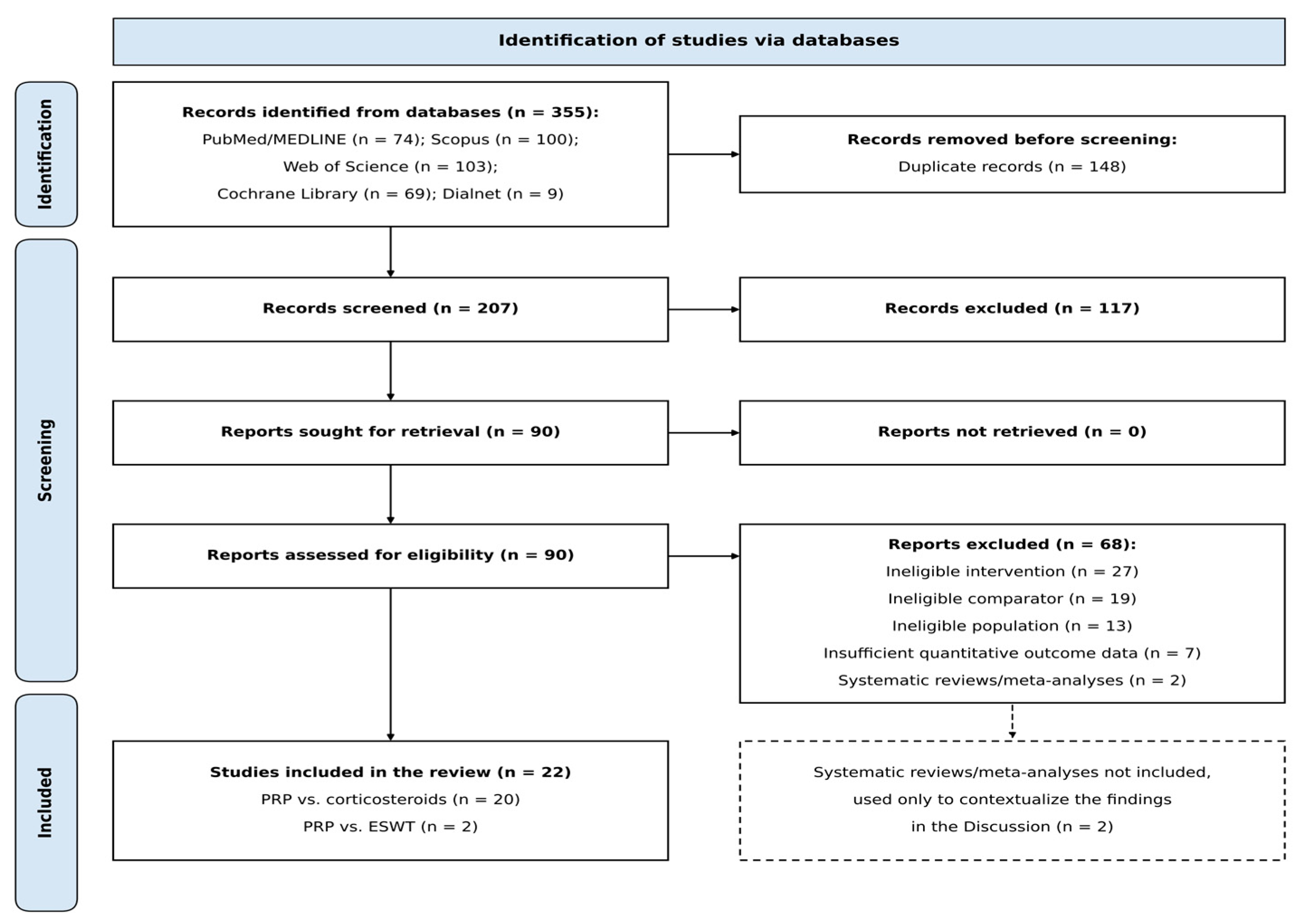
Records were imported into Zotero (ver. 10.0.6, Corporation for Digital Scholarship, Vienna, VA, USA) and deduplicated. Title and abstract screening was performed independently and in a blinded manner by two reviewers using Rayyan (web version: Rayyan System Inc. Cambridge, MA, USA) [28], after a calibration pilot on 50 randomly selected records. Records selected by either reviewer proceeded to full-text assessment; disagreements were resolved by discussion and, where necessary, by a third reviewer. The selection process is shown in Figure 1.
Figure 1.
PRISMA 2020 flow diagram of the study selection process.
A total of 355 records were identified: PubMed/MEDLINE (n = 74), Scopus (n = 100), Web of Science Core Collection (n = 103), Cochrane Library (n = 69) and Dialnet (n = 9). After removal of 148 duplicates, 207 records were screened by title and abstract, of which 117 were excluded. Ninety full-text articles were assessed for eligibility and 68 were excluded, including two systematic reviews with meta-analysis [25,26], which did not meet the predefined design criteria and are therefore considered only in Section 3.5. The remaining 66 reports were excluded because of an ineligible intervention (n = 27), an ineligible comparator (n = 19), an ineligible population (n = 13) or insufficient quantitative outcome data (n = 7). Finally, 22 primary studies were included (Figure 1).
2.5. Data Extraction and Synthesis
Data on study design, sample size, comparison, outcome measures, follow-up time points and results (means, standard deviations and p-values as reported by the original authors) were extracted using a standardized form. A quantitative meta-analysis was not considered appropriate because the studies used different outcome scales and follow-up time points, eight studies reported their results only in summary form without means and standard deviations, and PRP, CS and ESWT protocols varied substantially. Results were therefore synthesized narratively, grouped by comparison (PRP vs. CSs; PRP vs. ESWT), outcome (pain, function, PFT and adverse events) and follow-up period, defined as short term (≤6 weeks), medium term (>6 weeks to <6 months, typically 3 months) and long term (≥6 months).
2.6. Certainty of the Evidence
The certainty of the evidence was rated for each comparison, outcome and follow-up period using the Grading of Recommendations Assessment, Development and Evaluation (GRADE) approach [29], adapted for syntheses without a single pooled effect estimate [30]. Bodies of evidence consisting predominantly of RCTs started as high certainty and those based on non-randomized studies as low certainty, and were downgraded for risk of bias, inconsistency, indirectness, imprecision and publication bias. Certainty was classified as high, moderate, low or very low and is presented in a summary of findings table.
3. Results and Discussion
3.1. Characteristics of the Included Studies
The 22 included studies (Table 2) comprised 13 randomized controlled trials (RCTs) and 9 non-randomized comparative studies, with a total of 1915 patients (range 32–230 per study). Twenty studies (1788 patients) compared PRP with CSs and two (127 patients) compared PRP with ESWT. Pain was assessed with the visual analog scale (VAS) in all 22 studies; function with the American Orthopaedic Foot & Ankle Society (AOFAS) score in 13, the Foot Function Index (FFI) in four and the Foot and Ankle Ability Measure (FAAM) in three; and PFT in four. Follow-up ranged from 15 days to 2 years, and 19 of the 22 studies had a final follow-up of 6 months (24 weeks). Study selection was complete at the time of the search.
Table 2.
Characteristics of the included studies, in chronological order of publication.
Table 3 summarizes the symptom duration criteria and the intervention protocols reported by the included studies. Regarding the chronicity criterion (≥3 months), it was met in 12 studies [33,34,35,37,38,41,42,43,45,46,50,51]. Seven studies used shorter thresholds: heel pain of more than 4 weeks [32] or 6 weeks [39,47,48], symptoms of more than 2 months [36], or failure of at least 2 months [31] or 6 weeks [52] of conservative treatment; in the latter, 60–70% of patients had symptoms for more than 6 months. Three studies did not state a criterion [40,44,49], although in Batra et al. [44] the mean symptom duration was approximately 8 months. The PRP protocols differed in blood volume, centrifugation method (single, double or triple spin), volume obtained or injected (2–5 mL), number of injections (one or three) and image guidance (ultrasound or palpation guided), and three studies did not report the PRP preparation [42,44,49]. Only one study reported the platelet concentration relative to baseline [52], and none reported leukocyte content or activation. Most CS protocols used methylprednisolone, mostly at 40–80 mg, whereas one study used triamcinolone acetonide [45].
Table 3.
Symptom duration criteria and intervention protocols of the included studies.
3.2. PRP Versus CSs
3.2.1. Pain
Short-term (≤6 weeks) results were inconsistent (Table 4). Prakash and Kumar [37] found lower pain with CSs at 6 weeks (4.86 ± 1.02 vs. 7.05 ± 0.72) and 12 weeks (4.08 ± 0.72 vs. 6.34 ± 0.82) and Yadav et al. [52] at 6 weeks (1.50 ± 0.73 vs. 4.80 ± 0.93; 79.9% vs. 37.4% improvement from baseline) and 12 weeks (2.13 ± 1.04 vs. 3.33 ± 1.15). Sathyendra et al. [38] and Kumar K. et al. [41] found no difference at 15 days (7.03 ± 0.79 vs. 7.09 ± 0.98; p = 0.883, in the latter). In contrast, Khurana et al. [32] reported lower pain with PRP at 2 weeks (6.71 ± 0.99 vs. 7.30 ± 1.09) and 4 weeks (3.98 ± 1.03 vs. 4.93 ± 1.07), and Shah et al. [49] reported lower pain with PRP from the earliest assessments at 2 weeks (5.53 ± 0.776 vs. 6.47 ± 0.937; p < 0.001) and 4 weeks (4.70 ± 0.466 vs. 5.27 ± 0.868; p = 0.003).
Table 4.
Direction of the between-group differences in pain according to follow-up period.
From 1 month onward, most studies favored PRP. Kumar K. et al. [41] observed lower VAS scores with PRP at 1 month (6.26 ± 0.7 vs. 6.69 ± 0.87; p = 0.035), 3 months (5.37 ± 0.69 vs. 6.2 ± 0.72; p < 0.0001) and 6 months (3.71 ± 0.67 vs. 5.4 ± 0.65; p < 0.0001); Sathyendra et al. [38] reported significantly greater relief with PRP from 1 to 6 months; Shah et al. [49] found lower pain with PRP at 12 weeks (2.10 ± 0.712 vs. 2.77 ± 0.858; p = 0.002); and Kamala and Hanumantharaya [40] reported significant relief with PRP from 6 weeks, maintained at 3 and 6 months, with significantly greater improvements than with CSs.
At 6 months (24 weeks), VAS scores were lower with PRP in Khurana et al. [32] (0.52 ± 0.60 vs. 1.92 ± 1.03; p < 0.001), Prakash and Kumar [37] (2.9 ± 1.07 vs. 4.46 ± 0.96; p < 0.0001), Sharma R. et al. [39] (1.97 ± 1.13 vs. 2.71 ± 0.94), Yadav et al. [52] (1.43 ± 0.57 vs. 3.23 ± 1.01; 81.4% vs. 56.8% improvement, with some deterioration in the CS group between 12 and 24 weeks) and Priyam et al. [42] (1.9 ± 0.8 vs. 2.5 ± 0.9), in the largest sample of this subgroup (230 patients). Vellingiri et al. [34] also reported PRP superiority at 6 months. A further eight studies [43,44,45,46,47,48,50,51], reported only in summary form, described an early CS advantage followed by PRP superiority at medium-to-long term.
3.2.2. Function
Functional outcomes followed the same pattern. Khurana et al. [32] reported a 6-month AOFAS score of 95.09 ± 4.60 with PRP versus 85.96 ± 5.34 with CSs (p < 0.001). In Prakash and Kumar [37], PRP surpassed CSs from 12 weeks (82.38 ± 2.26 vs. 78.62 ± 2.48), and the difference widened at 6 months (86.64 ± 1.48 vs. 75.15 ± 2.02). Sharma R. et al. [39] found a 6-month AOFAS of 86.04 ± 7.45 versus 81.23 ± 9.60 and Shah et al. [49] a significantly higher FAAM with PRP at 12 weeks (91.03 ± 3.943 vs. 86.13 ± 6.388; p = 0.001).
3.2.3. Plantar Fascia Thickness
PFT decreased more markedly with PRP than with CSs in Prakash and Kumar [37] (from 5.76 ± 0.65 to 3.32 ± 0.44 mm vs. from 5.65 ± 0.62 to 3.73 ± 0.65 mm; p = 0.007) and was lower with PRP at 6 months in Sharma R. et al. [39] (3.53 ± 0.81 vs. 4.58 ± 1.02 mm).
3.2.4. Satisfaction and Adverse Events
Priyam et al. [42] reported 85% patient satisfaction with PRP versus 65% with CSs. Vellingiri et al. [34] observed complications in two patients treated with PRP (superficial infection) versus 10 treated with CSs (five superficial infections, three cases of skin depigmentation and two of fat-pad atrophy).
3.2.5. Discordant Results
Not all studies supported PRP superiority. Tabrizi et al. [31], in obese patients, found significantly better results in the CS group (p < 0.001), and Kumar N. [33] found no significant difference between treatments at any follow-up point.
3.3. PRP Versus ESWT
Pandey et al. [36], in an RCT of 72 patients, compared ESWT with PRP using the VAS, AOFAS and ultrasonographic PFT, with follow-up at days 15, 30 and 90. Baseline VAS scores were comparable (ESWT 6.44 ± 1.11 vs. PRP 6.78 ± 1.17; p = 0.237). At day 15, the ESWT group had lower pain (4.67 ± 1.45 vs. 6.67 ± 1.35; p < 0.001); at day 30 both groups were comparable (4.97 ± 1.46 vs. 4.69 ± 1.39; p = 0.391); and by day 90, PRP showed clearly lower pain (3.36 ± 0.96 vs. 5.47 ± 1.63; p < 0.001), with a similar pattern for PFT and AOFAS. Alessio-Mazzola et al. [35], in a retrospective study of 55 patients (24 athletes) with a minimum follow-up of 2 years, found no significant differences between ultrasound-guided PRP and focal ESWT in VAS and FFI, although the ESWT group had a higher, non-significant failure rate and required more follow-up visits, and athletes treated with PRP returned to sport significantly faster (p = 0.044).
3.4. Certainty of the Evidence
The certainty of the evidence according to GRADE is summarized in Table 5. For PRP versus CSs, it was low for long-term pain and for PFT, and very low for short-term and medium-term pain, function and adverse events. For PRP versus ESWT, the certainty of the evidence was very low.
Table 5.
Summary of findings and certainty of the evidence (GRADE).
Evidence from bodies of predominantly randomized studies started as high certainty, and that from non-randomized studies as low certainty. Risk of bias was rated serious owing to non-randomized and retrospective designs, small samples and results reported only in summary form. Imprecision was rated serious because no pooled estimate could be calculated and samples were small. Indirectness was not rated as serious, although the ≥3-month chronicity criterion could not be confirmed in several studies (Table 3). Publication bias was suspected because of the predominance of small positive studies. CSs, corticosteroids; ESWT, extracorporeal shockwave therapy; PFT, plantar fascia thickness; PRP, platelet-rich plasma; RCT, randomized controlled trial.
3.5. Interpretation of the Findings and Comparison with Previous Reviews
In the comparison between PRP and CSs, which is supported by 20 of the 22 included studies, a consistent time-dependent pattern was observed in the medium and long term: from approximately 3 months onward, most studies reported lower pain, better function and a greater reduction of PFT with PRP. In the short term, results were inconsistent: some studies favored CSs, two found no difference at 15 days and two favored PRP from 2 weeks. The evidence comparing PRP with ESWT is considerably more limited, as it relies on only two primary studies with mixed results. The two evidence bases are therefore not equivalent, and conclusions regarding PRP versus ESWT should be regarded as preliminary.
These findings may be related to the different mechanisms of action of each treatment, although these mechanisms were not assessed in the included studies. CSs act through anti-inflammatory mechanisms, which may explain the early analgesic effect observed in some studies; however, histopathological studies have not demonstrated a clear inflammatory process in chronic plantar fasciopathy, but rather degenerative changes [11], which may explain why their benefit is not sustained. PRP, in contrast, delivers growth factors that stimulate cell proliferation, collagen homeostasis and angiogenesis [18], which provides a biological rationale for a slower but more sustained effect. However, none of the included studies directly assessed tissue repair: the structural outcome measured was ultrasonographic PFT, and the greater reduction in PFT observed with PRP [37,39] should be interpreted as an imaging finding rather than as evidence of tissue repair.
These findings are broadly in agreement with those of previous systematic reviews and meta-analyses. Fei et al. [22] concluded that PRP was more effective than CSs in relieving pain and improving foot and ankle function at mid-term follow-up, without significant functional differences at short term. Seth et al. [24] reported better VAS scores with PRP than with CSs at 3 and 6 months, whereas differences in the AOFAS score appeared only at 6 months; they also found ESWT superior to CSs at 6 months. Aleid et al. [23], in 599 patients, concluded that PRP is promising for improving function and may provide greater pain relief than CSs, and that the functional benefits of CSs are not maintained over time. Peña-Martínez et al. [11], in 1109 patients, found that CS injections did not offer better results than other common interventions in reducing PFT or relieving pain.
Regarding ESWT, Pandey et al. [36] observed an early advantage of ESWT at day 15, which may reflect its analgesic effect [17], followed by an advantage of PRP at day 90, whereas the retrospective study by Alessio-Mazzola et al. [35] found no significant differences in its main overall outcome measures at a minimum of 2 years, although athletes treated with PRP returned to sport significantly earlier. Two meta-analyses of RCTs, which were not included in the present synthesis, provide additional context. Daher et al. [25] (6 RCTs; 214 PRP and 218 ESWT patients) found a statistically greater improvement with PRP in VAS (mean difference −0.67; 95% CI −1.16 to −0.18; p = 0.007) and PFT (mean difference −0.56; 95% CI −0.77 to −0.35; p < 0.001), although the pain difference did not reach clinical significance. Herber et al. [26] (21 RCTs; 1356 patients) found a significantly greater VAS improvement with PRP than with ESWT (standardized mean difference 0.86; 95% CI 0.30 to 1.41; p = 0.002); PRP also improved the AOFAS score more than CSs, but not more than ESWT, and no significant differences were found among PRP, CSs and ESWT in FFI or in the reduction of PFT. This last finding contrasts with the primary studies [37,39] and with Daher et al. [25], so PFT results should be interpreted with caution.
The discordant results also deserve comment. The CS advantage reported by Tabrizi et al. [31] was observed in obese patients, and a body mass index >25 kg/m2 is a recognized risk factor for plantar fasciopathy [6]; whether body weight modifies the response to PRP warrants specific investigation. In terms of safety, fewer local complications were reported with PRP than with CSs [34], consistent with the fat-pad atrophy and fascia rupture described after CS injection [11].
An important consideration is that PRP is not a uniform intervention. Platelet concentration, leukocyte content (leukocyte-rich or leukocyte-poor PRP), injected volume, activation method, number of and interval between injections, preparation method and the use of ultrasound guidance vary widely across protocols [18,19], and these characteristics were not analyzed in the present review. Consequently, the findings cannot be attributed to a specific PRP formulation or administration protocol, which limits their direct clinical applicability.
With these caveats, and given the low certainty of the evidence, PRP may be considered for patients with chronic plantar fasciopathy in whom conservative treatment has failed and who seek sustained medium- to long-term improvement. The position of ESWT relative to PRP cannot be established with the current evidence.
3.6. Limitations and Recommendations
This review has several limitations. The included studies differed in PRP preparation and administration, in the CS agent and dose and in ESWT parameters (density, frequency and number of sessions), and many did not report these characteristics in detail (Table 3); they were heterogeneous in sample size, outcome scales and follow-up time points, which precluded quantitative pooling; no formal study-level risk-of-bias assessment with a validated tool (RoB 2 or ROBINS-I) was performed, so risk of bias was considered only within the GRADE rating; the absence of pooling also precluded forest plots and statistical assessment of heterogeneity and publication bias; only two primary studies compared PRP with ESWT, one of them retrospective; eight studies were described only in summary form; the search was restricted to studies published from 2020 onward, so earlier trials were not included in the synthesis and the findings complement, rather than replace those of earlier reviews; the chronicity criterion (≥3 months) could not be confirmed in 10 of the 22 studies, as several used shorter thresholds (Table 3), although excluding them would not change the direction of the long-term findings for PRP versus CSs; the certainty of the evidence was low or very low (Table 5); and the protocol was registered retrospectively.
Several recommendations for future research derive from these limitations. First, adequately powered, multicenter RCTs with prospective registration, blinded outcome assessment and intention-to-treat analysis are needed, particularly for the comparison between PRP and ESWT. Second, studies should include only patients with symptoms for at least 3 months and report the mean symptom duration and previous treatments. Third, PRP should be described in detail—blood volume, centrifugation protocol, platelet concentration, leukocyte content, activation, injected volume, number of and interval between injections, and image guidance—together with the CS agent and dose and the ESWT device, energy flux density, number of pulses and sessions. Fourth, a core set of outcomes should be used, including the VAS, a validated foot function score and ultrasonographic PFT, with means and standard deviations reported at predefined time points (6 weeks, 3, 6 and 12 months) to allow future meta-analyses. Finally, subgroups that may respond differently, such as obese patients and athletes, deserve specific evaluation, as do cost-effectiveness and long-term recurrence.
For clinical practice, and considering the low certainty of the evidence, PRP may be considered in patients with chronic plantar fasciopathy who have not responded to conservative treatment and who seek medium- to long-term improvement, after informing them that short-term relief may be slower than with CSs in some cases. Clinicians are encouraged to use ultrasound for diagnosis and follow-up and to document the PRP preparation used, so that clinical experience can be compared across settings.
4. Conclusions
For PRP versus CSs (20 studies, 12 of them RCTs), PRP was associated with lower pain, better function and a greater reduction in ultrasonographic plantar fascia thickness from approximately 3 months onward, whereas short-term (≤6 weeks) results were inconsistent, with some studies favoring CSs. For PRP versus ESWT (two studies, one retrospective), the evidence is insufficient to draw conclusions. Because the certainty of the evidence was low for long-term pain and plantar fascia thickness and very low for the other outcomes, and PRP protocols varied widely, these findings should be interpreted with caution and confirmed in well-designed trials with standardized protocols and long-term follow-up.
Supplementary Materials
The following supporting information can be downloaded at: https://www.mdpi.com/article/10.3390/ijms27219550/s1.
Author Contributions
Conceptualization, M.O.-R. and E.P.-A.; methodology, M.O.-R., E.P.-A. and L.M.G.-F.; investigation, E.P.-A. and M.O.-R.; data curation, E.P.-A. and M.O.-R.; formal analysis, E.P.-A. and M.O.-R.; writing—original draft preparation, E.P.-A. and M.O.-R.; writing—review and editing, M.O.-R. and L.M.G.-F.; supervision, L.M.G.-F. All authors have read and agreed to the published version of the manuscript.
Funding
This research received no external funding.
Institutional Review Board Statement
Not applicable. This study is a systematic review of previously published data and did not involve new data collection from human or animal subjects.
Informed Consent Statement
Not applicable.
Data Availability Statement
The study protocol was registered retrospectively in OSF Registries (https://osf.io/hdcjb, accessed on 20 September 2026). Search results, screening decisions and the data extraction spreadsheet will be made available in the associated OSF project upon publication of this manuscript.
Acknowledgments
During the preparation of this manuscript, the authors used Claude Opus 5.5 (Anthropic, San Francisco, CA, USA) to assist with drafting and revising portions of the text, including alignment with the OSF-registered protocol. The authors have reviewed and edited the output and take full responsibility for the content of this publication.
Conflicts of Interest
The authors declare no conflicts of interest.
Abbreviations
The following abbreviations are used in this manuscript:
| AOFAS | American Orthopaedic Foot & Ankle Society score |
| CI | Confidence interval |
| CSs | Corticosteroids |
| EGF | Epidermal growth factor |
| ESWT | Extracorporeal shockwave therapy |
| FAAM | Foot and Ankle Ability Measure |
| FFI | Foot Function Index |
| FGF | Fibroblast growth factor |
| GRADE | Grading of Recommendations Assessment, Development and Evaluation |
| HGF | Hepatocyte growth factor |
| IGF-1 | Insulin-like growth factor 1 |
| MeSH | Medical Subject Headings |
| NR | Not reported |
| OSF | Open Science Framework |
| PDGF | Platelet-derived growth factor |
| PFT | Plantar fascia thickness |
| PICOS | Population, Intervention, Comparator, Outcomes and Study design |
| PRISMA | Preferred Reporting Items for Systematic Reviews and Meta-Analyses |
| PRP | Platelet-rich plasma |
| RCT | Randomized controlled trial |
| SDF-1α | Stromal cell-derived factor 1α |
| TGF-β | Transforming growth factor β |
| VAS | Visual analog scale |
References
- Flores, D.V.; Goes, P.K.; Damer, A.; Huang, B.K. The Heel Complex: Anatomy, Imaging, Pathologic Conditions, and Treatment. Radiographics 2024, 44, e230163. [Google Scholar] [CrossRef] [Scilit] [PubMed]
- Kelikian, A.S.; Sarrafian, S.K. Sarrafian’s Anatomy of the Foot and Ankle: Descriptive, Topographic, Functional, 3rd ed.; Lippincott Williams & Wilkins: Philadelphia, PA, USA, 2011. [Google Scholar]
- Stecco, C.; Corradin, M.; Macchi, V.; Morra, A.; Porzionato, A.; Biz, C.; De Caro, R. Plantar fascia anatomy and its relationship with Achilles tendon and paratenon. J. Anat. 2013, 223, 665–676. [Google Scholar] [CrossRef] [Scilit] [PubMed]
- Sichting, F.; Holowka, N.B.; Ebrecht, F.; Lieberman, D.E. Evolutionary anatomy of the plantar aponeurosis in primates, including humans. J. Anat. 2020, 237, 85–104. [Google Scholar] [CrossRef] [Scilit] [PubMed]
- Chen, D.W.; Li, B.; Aubeeluck, A.; Yang, Y.F.; Huang, Y.G.; Zhou, J.Q.; Yu, G.R. Anatomy and Biomechanical Properties of the Plantar Aponeurosis: A Cadaveric Study. PLoS ONE 2014, 9, e84347. [Google Scholar] [CrossRef] [Scilit] [PubMed]
- Buchanan, B.K.; Sina, R.E.; Kushner, D. Plantar Fasciitis. In StatPearls; StatPearls Publishing: Treasure Island, FL, USA, 2024. Available online: https://www.ncbi.nlm.nih.gov/books/NBK431073/ (accessed on 13 October 2026).
- McMillan, A.M.; Landorf, K.B.; Barrett, J.T.; Menz, H.B.; Bird, A.R. Diagnostic imaging for chronic plantar heel pain: A systematic review and meta-analysis. J. Foot Ankle Res. 2009, 2, 32. [Google Scholar] [CrossRef] [Scilit] [PubMed]
- Werber, B. Amniotic Tissues for the Treatment of Chronic Plantar Fasciosis and Achilles Tendinosis. J. Sports Med. 2015, 2015, 219896. [Google Scholar] [CrossRef] [Scilit] [PubMed]
- Hamstra-Wright, K.L.; Huxel Bliven, K.C.; Bay, R.C.; Aydemir, B. Risk Factors for Plantar Fasciitis in Physically Active Individuals: A Systematic Review and Meta-analysis. Sports Health 2021, 13, 296–303. [Google Scholar] [CrossRef] [Scilit] [PubMed]
- Schneider, H.P.; Knudson, J. Plantar Fasciitis. Clin. Podiatr. Med. Surg. 2020, 37, 605–613. [Google Scholar] [CrossRef] [Scilit] [PubMed]
- Peña-Martínez, V.M.; Acosta-Olivo, C.; Simental-Mendía, L.E.; Sánchez-García, A.; Jamialahmadi, T.; Sahebkar, A.; Vilchez-Cavazos, F.; Simental-Mendía, M. Effect of corticosteroids over plantar fascia thickness in plantar fasciitis: A systematic review and meta-analysis. Phys. Sportsmed. 2024, 52, 217–228. [Google Scholar] [CrossRef] [Scilit] [PubMed]
- Luffy, L.; Grosel, J.; Thomas, R.; So, E. Plantar fasciitis: A review of treatments. J. Am. Acad. Physician Assist. 2018, 31, 20–24. [Google Scholar]
- Petraglia, F.; Ramazzina, I.; Costantino, C. Plantar fasciitis in athletes: Diagnostic and treatment strategies. A systematic review. Muscles Ligaments Tendons J. 2017, 7, 107–118. [Google Scholar] [CrossRef] [Scilit] [PubMed]
- Trojian, T.; Tucker, A.K. Plantar Fasciitis. Am. Fam. Physician 2019, 99, 744–750. [Google Scholar] [CrossRef] [Scilit]
- Thompson, J.V.; Saini, S.S.; Reb, C.W.; Daniel, J.N. Diagnosis and Management of Plantar Fasciitis. J. Am. Osteopath. Assoc. 2014, 114, 900–906. [Google Scholar] [CrossRef] [Scilit] [PubMed]
- Akfirat, M.; Sen, C.; Gunes, T. Ultrasonographic appearance of the plantar fasciitis. Clin. Imaging 2003, 27, 353–357. [Google Scholar] [CrossRef] [Scilit] [PubMed]
- Moya, D.; Ramón, S.; Schaden, W.; Wang, C.J.; Guiloff, L.; Cheng, J.H. The Role of Extracorporeal Shockwave Treatment in Musculoskeletal Disorders. J. Bone Jt. Surg. Am. 2018, 100, 251–263. [Google Scholar] [CrossRef] [Scilit] [PubMed]
- Rath, M.; Spinnen, J.; Kuhrt, L.D.; Priglinger, E.; Seika, P.; Runge, D.; Schubring, S.; Laue, D.; Wickert, M.; Erdem, M.; et al. Platelet-Rich Plasma—A Comprehensive Review of Isolation, Activation, and Application. Acta Biomater. 2025, 204, 52–75. [Google Scholar] [CrossRef] [Scilit] [PubMed]
- Bagheri, K.; Krez, A.; Anastasio, A.T.; Adams, S.B. The use of platelet-rich plasma in pathologies of the foot and ankle: A comprehensive review of the recent literature. Foot Ankle Surg. 2023, 29, 551–559. [Google Scholar] [CrossRef] [Scilit] [PubMed]
- Lana, J.F.; Purita, J.; Paulus, C.; Huber, S.C.; Rodrigues, B.L.; Rodrigues, A.A.; Santana, M.H.; Madureira, J.L.; Luzo, Â.C.M.; Belangero, W.D.; et al. Contributions for classification of platelet rich plasma—Proposal of a new classification: MARSPILL. Regen. Med. 2017, 12, 565–574. [Google Scholar] [CrossRef] [Scilit] [PubMed]
- Vithran, D.T.A.; He, M.; Xie, W.; Essien, A.E.; Opoku, M.; Li, Y. Advances in the Clinical Application of Platelet-Rich Plasma in the Foot and Ankle: A Review. Front. Surg. 2023, 10, 1115598. [Google Scholar]
- Fei, X.; Lang, L.; Lingjiao, H.; Wei, C.; Zhou, X. Platelet-rich plasma has better mid-term clinical results than traditional steroid injection for plantar fasciitis: A systematic review and meta-analysis. Orthop. Traumatol. Surg. Res. 2021, 107, 103007. [Google Scholar] [CrossRef] [Scilit] [PubMed]
- Aleid, A.M.; Alyabis, N.A.; Aldanyowi, S.N.; Albinsaad, L.S.; AlAidarous, H.A.; Aleid, Z.M.; AlMutair, A.S. Efficacy of platelet-rich plasma versus corticosteroid injections in recovery from plantar fasciitis: A systematic review and meta-analysis. J. Taibah Univ. Med. Sci. 2025, 20, 120–128. [Google Scholar] [CrossRef] [Scilit] [PubMed]
- Seth, I.; Bulloch, G.; Seth, N.; Lower, K.; Rodwell, A.; Rastogi, A.; Rastogi, A.; Gibson, D.; Bedi, H. The role of corticosteroid injections in treating plantar fasciitis: A systematic review and meta-analysis. Foot 2023, 54, 101970. [Google Scholar] [CrossRef] [Scilit] [PubMed]
- Daher, M.; Covarrubias, O.; Herber, A.; Oh, I.; Gianakos, A.L. Platelet-Rich Plasma vs Extracorporeal Shock Wave Therapy in the Treatment of Plantar Fasciitis at 3–6 Months: A Systematic Review and Meta-analysis of Randomized Controlled Trials. Foot Ankle Int. 2024, 45, 796–803. [Google Scholar] [CrossRef] [Scilit] [PubMed]
- Herber, A.; Covarrubias, O.; Daher, M.; Tung, W.S.; Gianakos, A.L. Platelet rich plasma therapy versus other modalities for treatment of plantar fasciitis: A systematic review and meta-analysis. Foot Ankle Surg. 2024, 30, 285–293. [Google Scholar] [CrossRef] [Scilit] [PubMed]
- Page, M.J.; McKenzie, J.E.; Bossuyt, P.M.; Boutron, I.; Hoffmann, T.C.; Mulrow, C.D.; Shamseer, L.; Tetzlaff, J.M.; Akl, E.A.; Brennan, S.E.; et al. The PRISMA 2020 statement: An updated guideline for reporting systematic reviews. BMJ 2021, 372, n71. [Google Scholar] [CrossRef] [Scilit] [PubMed]
- Ouzzani, M.; Hammady, H.; Fedorowicz, Z.; Elmagarmid, A. Rayyan—A web and mobile app for systematic reviews. Syst. Rev. 2016, 5, 210. [Google Scholar] [CrossRef] [Scilit] [PubMed]
- Guyatt, G.H.; Oxman, A.D.; Vist, G.E.; Kunz, R.; Falck-Ytter, Y.; Alonso-Coello, P.; Schünemann, H.J. GRADE: An emerging consensus on rating quality of evidence and strength of recommendations. BMJ 2008, 336, 924–926. [Google Scholar] [CrossRef] [Scilit] [PubMed]
- Murad, M.H.; Mustafa, R.A.; Schünemann, H.J.; Sultan, S.; Santesso, N. Rating the certainty in evidence in the absence of a single estimate of effect. Evid. Based Med. 2017, 22, 85–87. [Google Scholar] [CrossRef] [Scilit] [PubMed]
- Tabrizi, A.; Dindarian, S.; Mohammadi, S. The Effect of Corticosteroid Local Injection Versus Platelet-Rich Plasma for the Treatment of Plantar Fasciitis in Obese Patients: A Single-Blind, Randomized Clinical Trial. J. Foot Ankle Surg. 2020, 59, 64–68. [Google Scholar] [CrossRef] [Scilit] [PubMed]
- Khurana, A.; Dhankhar, V.; Goel, N.; Jain, A.; Singh, S.K.; Marya, S.K.S. Comparison of midterm results of Platelet Rich Plasma (PRP) versus Steroid for plantar fasciitis: A randomized control trial of 118 patients. J. Clin. Orthop. Trauma 2021, 13, 9–14. [Google Scholar] [CrossRef] [Scilit] [PubMed]
- Kumar, N. A Comparison of the Effectiveness of Platelet-Rich Plasma Therapy and Corticosteroid Injection in the Treatment of Plantar Fasciitis. Int. J. Pharm. Clin. Res. 2021, 13, 178–183. [Google Scholar]
- Vellingiri, K.; Nagakumar, J.S.; Manohar, P.V.; Lourdu, J.P.; Andra Suryanarayana, M.S. A Prospective Study Comparing the Efficacy of Local Injection of Platelet-Rich Plasma (PRP) vs Methylprednisolone in Plantar Fasciitis. Cureus 2022, 14, e25523. [Google Scholar] [CrossRef] [Scilit] [PubMed]
- Alessio-Mazzola, M.; Stambazzi, C.; Ursino, C.; Tagliafico, A.; Trentini, R.; Formica, M. Ultrasound-Guided Autologous Platelet-Rich Plasma Injections Versus Focal Ultrasound-Guided Extracorporeal Shockwave Therapy for Plantar Fasciitis in Athletes and Nonathletes: A Retrospective Comparative Study with Minimum 2-Year Follow-Up. J. Foot Ankle Surg. 2023, 62, 417–421. [Google Scholar] [CrossRef] [Scilit] [PubMed]
- Pandey, S.; Kumar, N.; Kumar, A.; Biswas, A.; Sinha, U.; Pandey, J.; Ghosh, S.; Das, S.; A Johnson, R.; Kumar, R.; et al. Extracorporeal Shockwave Therapy Versus Platelet Rich Plasma Injection in Patients of Chronic Plantar Fasciitis: A Randomized Controlled Trial from a Tertiary Center of Eastern India. Cureus 2023, 15, e34430. [Google Scholar] [CrossRef] [Scilit] [PubMed]
- Prakash, S.; Kumar, M. Management of Chronic Plantar Fasciitis: A Comparative Study. Int. J. Curr. Pharm. Rev. Res. 2023, 15, 236–240. [Google Scholar]
- Sathyendra, K.G.; Dnyandeo Solankey, R.; Singh, M.; Venkatesh Kumar, S.; Singh, G.; Gupta, M.M. Comparative study of local injections of autologous platelet rich plasma versus corticosteroid in management of chronic plantar fasciitis. J. Clin. Orthop. Trauma 2023, 43, 102225. [Google Scholar] [CrossRef] [Scilit] [PubMed]
- Sharma, R.; Chaudhary, N.K.; Karki, M.; Sunuwar, D.R.; Singh, D.R.; Pradhan, P.M.S.; Gyawali, P.; Shrestha, S.K.D.; Bhandari, K.K. Effect of platelet-rich plasma versus steroid injection in plantar fasciitis: A randomized clinical trial. BMC Musculoskelet. Disord. 2023, 24, 172. [Google Scholar] [CrossRef] [Scilit] [PubMed]
- Kamala, G.R.; Hanumantharaya, G.H. Comparative Study of Corticosteroids v/s Platelet Rich Plasma for the Treatment of Plantar Fasciitis in a Teaching Hospital. J. Cardiovasc. Dis. Res. 2024, 15, 2154–2160. [Google Scholar]
- Kumar, K.; Rao, V.; Panda, A.; Sathyendra, K.G.; Buddhist, H. Comparison of Platelet-Rich Plasma and Corticosteroid Injections for Chronic Plantar Fasciitis: A Randomized Controlled Trial. Cureus 2024, 16, e59656. [Google Scholar] [CrossRef] [Scilit] [PubMed]
- Priyam, P.; Shikha; Kumar, V. Comparative Study of Platelet-Rich Plasma Vs Corticosteroid Injection for Plantar Fasciitis, Bihar, India. Int. J. Pharm. Clin. Res. 2024, 16, 1618–1621. [Google Scholar]
- Sharma, G.; Gupta, A.; Choudhary, A.S. Role of Corticosteroid (Methyl Prednisolone Injection) Versus Platelet Rich Plasma (PRP) in Treatment of Plantar Fasciitis. Int. J. Life Sci. Biotechnol. Pharma Res. 2024, 13, 64–69. [Google Scholar]
- Batra, A.; Dua, S.; Kumar, V.; Arora, J. Study of platelet rich plasma vrs corticosteroid injection in chronic plantar fasciitis: A teaching hospital based comparative study. Int. J. Med. Public Health 2025, 15, 313–316. [Google Scholar]
- Bucak, Ö.F.; Kalaoglu, E.; Atasoy, M.; Coskun, E. Comparative Effectiveness of Platelet-Rich Plasma and Steroid Injections in Chronic Plantar Fasciitis: Evaluating Pain Relief and Functional Recovery. Foot Ankle Int. 2025, 46, 987–995. [Google Scholar] [CrossRef] [Scilit] [PubMed]
- Chugh, L.; Makker, S.; Chugh, L. Comparing the Effects of Platelet-Rich Plasma and Corticosteroid Injections on Pain and Function in Plantar Fasciitis. Int. J. Life Sci. Biotechnol. Pharma Res. 2025, 14, 794–797. [Google Scholar]
- Gorantla, U.K.; Naresh, G.; Sahbaz, M.M. Comparative Study of Efficacy of Corticosteroid versus Analogues Platelet Rich Plasma Injection in the Management of Plantar Fasciitis. Int. J. Acad. Med. Pharm. 2025, 7, 372–375. [Google Scholar]
- Patil, J.; Umerjikar, S.; Biradar, N. Plantar Fasciitis Management with Platelet Rich Plasma versus Steroid Injection—A Comparative Study in North Karnataka Population. Int. J. Curr. Pharm. Rev. Res. 2025, 17, 153–159. [Google Scholar]
- Shah, H.; Prabhakaran, A.; Praveen, I.; Farook, U. A Comparative Study of Platelet-Rich Plasma V/S Corticosteroid Injection for Plantar Fasciitis. J. Pharm. Bioallied Sci. 2025, 17, S1689–S1691. [Google Scholar] [CrossRef] [Scilit] [PubMed]
- Sunil; Gulati, A. Randomized Evaluation of Platelet-Rich Plasma Versus Corticosteroid Injection in the Management of Chronic Plantar Fasciitis. Int. J. Life Sci. Biotechnol. Pharma Res. 2026, 15, 1327–1330. [Google Scholar]
- Vyshnavi, J.M.; Reddy, P.B.T.; Sneha, B.; Venkatesh, D.L. A Prospective Study of Platelet Rich Plasma Versus Corticosteroid Injection in Chronic Plantar Fasciitis. Int. J. Pharm. Res. Technol. 2026, 16, 4147–4153. [Google Scholar] [CrossRef] [Scilit] [PubMed]
- Yadav, A.; Meena, I.; Singh, A.; Gugale, S. Comparative Efficacy of Autologous Platelet-Rich Plasma versus Corticosteroid Injections in Chronic Plantar Fasciitis: A Prospective Randomized Study. Int. J. Med. Pharm. Res. 2026, 7, 880–885. [Google Scholar]

Disclaimer/Publisher’s Note: The statements, opinions and data contained in all publications are solely those of the individual author(s) and contributor(s) and not of MDPI and/or the editor(s). MDPI and/or the editor(s) disclaim responsibility for any injury to people or property resulting from any ideas, methods, instructions or products referred to in the content. |
© 2026 by the authors. Licensee MDPI, Basel, Switzerland. This article is an open access article distributed under the terms and conditions of the Creative Commons Attribution (CC BY) license.