


Abstract
Fundus neovascular diseases (FNDs) are vision-threatening ocular disorders featuring pathological neovascularization and pose a major global threat to vision health. Their pathogenesis is heterogeneous, involving multiple factors and pathways. Current clinical pharmacotherapies only target a limited range of molecular pathways via narrow mechanisms, leading to frequent suboptimal responses and disease recurrence, which restricts further therapeutic improvement for FNDs. Recent studies demonstrate that lysophospholipid signaling molecules selectively activate multiple cell-surface receptors, including G protein-coupled receptors, triggering multilayered downstream signaling cascades that govern neovascular initiation and progression. Aberrant activation of this endogenous signaling axis may underpin the limited efficacy of existing anti-angiogenic therapies, and its roles in pathological neovascularization have attracted intensive research attention. This review addresses three representative lysophospholipid mediators: lysophosphatidic acid, lysophosphatidylcholine, and sphingosine-1-phosphate. We systematically summarize their metabolic routes, receptor binding profiles, downstream signaling networks, and pro-angiogenic mechanisms. We also discuss the current research status and translational potential of targeting lysophospholipid axes as innovative therapies, aiming to provide theoretical and translational evidence for developing precise pathway-directed interventions.
1. Introduction
Fundus neovascular diseases (FNDs), a group of blinding ocular disorders characterized by pathological neovascularization, such as neovascular age-related macular degeneration (nAMD), proliferative diabetic retinopathy (PDR), and retinal vein occlusion (RVO), are a leading cause of irreversible vision loss worldwide [1,2,3,4]. Vascular endothelial growth factors (VEGFs), angiopoietins (Angs), dysregulated lipid metabolism, oxidative stress, and chronic inflammation, among other heterogeneous pathogenic factors, are recognized as key drivers of FNDs [4,5,6,7]. Currently, the therapeutic targets of first-line clinical interventions are primarily VEGFs and Ang-2. Although these approaches have demonstrated substantial in suppressing neovascular progression and maintaining central vision, their effectiveness remains constrained by the multifactorial and multipathway nature of FND pathogenesis. Approximately 25% of patients exhibit primary non-response or suboptimal response to initial treatment, and recurrence rates during long-term follow-up remain as high as 73.9%, highlighting the fundamental limitations of current therapeutic strategies in addressing the heterogeneous etiologies underlying FNDs [8,9].
Lysophospholipid signaling molecules, a class of endogenous bioactive lipids generated via the hydrolysis of membrane phospholipids by specific phospholipases, have emerged as increasingly important regulators in pathological angiogenesis. These molecules primarily include lysophosphatidic acid (LysoPA), lysophosphatidylcholine (LysoPC), and sphingosine-1-phosphate (S1P) [10,11], and are released by diverse cell types, including activated platelets, infiltrating inflammatory cells, vascular endothelial cells, and retinal pigment epithelial (RPE) cells. Accumulating evidence indicates that these molecules orchestrate multiple biological processes, including endothelial cell proliferation, migration, survival, and remodeling of the immune microenvironment, thereby exerting profound effects on the maintenance of vascular homeostasis and the modulation of pathological vascular remodeling [10,12]. Through autocrine and paracrine mechanisms, these bioactive lipids selectively engage key membrane receptors, such as G protein-coupled receptors (GPCRs) and Toll-like receptors (TLRs), thereby activating a broad spectrum of downstream signaling cascades, including Rho–ROCK, PLC–IP3–Ca2+, PI3K–Akt, MAPK–ERK, cAMP–protein kinase A (PKA), and Hippo–YAP pathways. These signaling networks collectively orchestrate pathological ocular angiogenesis and regulate the integrity of the blood–retinal barrier. Therefore, lysophospholipid signaling molecules may represent not only critical pathogenic mediators driving pathological angiogenesis, particularly in retinal neovascular disorders, but also central signaling hubs governing disease initiation and progression [13,14,15,16] (Figure 1).
Figure 1.
Interconnected lysophospholipid signaling networks contributing to fundus neovascular diseases. LysoPC, LysoPA, and S1P accumulate in the ocular microenvironment and form an interconnected signaling network; LysoPC is converted to LysoPA by autotaxin (ATX). Through distinct receptors and metabolic crosstalk, these mediators promote inflammation, endothelial cell proliferation and migration, vascular leakage, blood–retinal barrier disruption, and pathological neovascularization, driving nAMD, PDR, and RVO. ATX, autotaxin; TLR, Toll-like receptor; CD36, cluster of differentiation 36; GPR4, G protein-coupled receptor 4; LPAR, lysophosphatidic acid receptor; S1PR, sphingosine-1-phosphate receptor; nAMD, neovascular age-related macular degeneration; CNV, choroidal neovascularization; PDR, proliferative diabetic retinopathy; RVO, retinal vein occlusion.
This review focuses on key lysophospholipid signaling molecules, including LysoPA, LysoPC, and S1P, and systematically summarizes their metabolic pathways, receptor recognition mechanisms, downstream signaling cascades, pro-angiogenic functions, and translational potential as emerging therapeutic targets. By integrating current advances in this field, we aim to provide a theoretical framework and translational rationale for the development of precision intervention strategies targeting lysophospholipid signaling axes.
2. Metabolic Pathways of Lysophospholipid Signaling Molecules
2.1. Structural and Functional Diversity of Lysophospholipid Signaling Molecules
Based on the nature of their hydrophobic backbone structures, lysophospholipid signaling molecules can be broadly classified into two major categories: lysoglycerophospholipids (LysoGPLs), which are derived from a glycerol backbone and include LysoPA, LysoPC, Lysophosphatidylinositol (LysoPI), Lysophosphatidylethanolamine (LysoPE), and Lysophosphatidylserine (LysoPS); and lysosphingolipids (LysoSLs), which are derived from a sphingosine backbone and primarily comprise S1P and sphingosylphosphorylcholine (SPC).
Although LysoGPLs share a conserved glycerol backbone, they exhibit substantial structural heterogeneity in both their polar head groups and hydrophobic acyl chains [17,18]. The polar head group of LysoPA consists of a phosphate moiety, which undergoes ionization at physiological pH, conferring a net negative charge and acidic properties. In contrast, the choline head group of LysoPC becomes protonated at physiological pH, resulting in a positive charge and alkaline characteristics. At physiological pH, LysoPI, LysoPS, and LysoPE also display distinct and stable ionization profiles: LysoPE exhibits a zwitterionic ionization pattern, whereas LysoPS and LysoPI predominantly exist in an anionic form [19]. These charge-dependent differences contribute to the functional diversification of LysoGPLs, influencing their membrane interaction properties, receptor recognition specificity, and selective activation of downstream signaling pathways. The hydrophobic acyl chains of LysoGPLs further contribute to their remarkable structural diversity, which is primarily determined by variations in acyl chain attachment at the sn-1 or sn-2 positions of the glycerol backbone, carbon chain length (C12–C20), and degree of unsaturation (saturated, monounsaturated, or polyunsaturated). These structural features critically regulate the overall molecular conformation, membrane affinity, and binding affinity for specific target proteins, thereby enabling highly specialized regulation of physiological functions and pathological processes [18,20,21,22].
Unlike LysoGPLs, LysoSLs possess a distinct structural foundation derived from sphingosine, a long-chain amino alcohol. Their core structure consists of a long-chain sphingosine moiety and a phosphate group, giving rise to S1P. Further attachment of a choline group to the phosphate moiety generates SPC. This amphipathic architecture, established through ester-linked molecular interactions, enables LysoSLs to dynamically partition between polar aqueous environments and nonpolar lipid phases, thereby facilitating transmembrane signal transduction and intercellular communication [23,24].
2.2. Biosynthetic and Catabolic Pathways of Lysophospholipid Signaling Molecules
The biological activities of lysophospholipid signaling molecules are largely determined by the dynamic balance between their biosynthesis and catabolism, a balance tightly controlled by key metabolic enzymes (Figure 2). The biosynthesis of LysoPA occurs predominantly through two major routes. (1) Phospholipase D (PLD)-dependent pathway: In this pathway, PLD exhibits high substrate selectivity toward phosphatidylcholine (PC) and catalyzes its conversion into phosphatidic acid (PA), representing the major route of PA generation in mammalian cells [25]. PA is subsequently hydrolyzed by phospholipases A1/A2 (PLA1/2), which remove an acyl chain from the sn-1 or sn-2 position, thereby generating LysoPA [26]. (2) ATX-dependent pathway: This pathway is coupled to LysoPC catabolism, whereby ATX efficiently hydrolyzes circulating LysoPC to produce LysoPA [27]. This reaction represents the predominant extracellular source of LysoPA generation [28,29,30,31]. The catabolism of LysoPA involves several distinct pathways, including: (1) Lipid phosphate phosphatase (LPP)-mediated dephosphorylation: LPPs are a family of integral plasma membrane proteins with extracellularly oriented catalytic domains. They directly hydrolyze extracellular LysoPA, converting it into monoacylglycerol (MAG) through dephosphorylation [27]. (2) Lysophosphatidic acid acyltransferase (LPAAT)-mediated reacylation [32] and (3) soluble epoxide hydrolase (sEH)-mediated hydrolysis [33].
Figure 2.
Metabolic pathways of lysophospholipid signaling molecules. lysoglycerophospholipid (LysoGPL)-derived signaling molecules share conserved structural features. Their metabolic pathways also exhibit extensive similarities at both structural and enzymatic levels. These pathways are also functionally interconnected with those of lysosphingolipids (LysoSLs), thereby collectively forming an integrated regulatory network of lysophospholipid signaling governed by common enzymatic mechanisms. Abbreviations: PC, phosphatidylcholine; PE, phosphatidylethanolamine; PS, phosphatidylserine; PI, phosphatidylinositol; PA, phosphatidic acid; MAG, monoacylglycerol; GPC, glycerophosphocholine; GPE, glycerophosphoethanolamine; GPS, glycerophosphoserine; CoA, coenzyme A.
The biosynthesis of LysoPC primarily occurs through the following pathways: (1) PLA1/2-mediated hydrolysis of PC: Based on their biochemical properties, subcellular localization, and activation mechanisms, PLA1/2 can be broadly classified into secreted PLA1/2 (sPLA1/2), cytosolic PLA1/2 (cPLA1/2), calcium-independent PLA2 (iPLA2), and membrane-bound PLA1 (mbPLA1). These enzymes selectively cleave the acyl chains at the sn-1 or sn-2 positions of membrane phosphatidylcholine (PC) molecules, thereby generating LysoPC. In addition, oxidized PC (ox-PC) can be selectively hydrolyzed by specific PLA2 isoforms, such as PLA2G6, resulting in the release of oxidized free fatty acids (ox-FFA) and the generation of LysoPC [34,35]. In oxidized low-density lipoprotein (oxLDL), ox-PC is hydrolyzed by lipoprotein-associated phospholipase A2 (Lp-PLA2), releasing sn-2-specific oxidized free fatty acids (ox-FFA) and generating LysoPC. This process represents a critical molecular link connecting oxidative stress, lipid metabolism, and vascular inflammation [36,37,38,39,40,41,42]. (2) Lecithin–cholesterol acyltransferase (LCAT)-mediated pathway: LCAT specifically catalyzes the transfer of fatty acids from the sn-2 position of PC on the surface of high-density lipoproteins (HDL) to free cholesterol, resulting in the formation of cholesteryl esters and LysoPC [43]. (3) Oxidative stress-driven modification pathway: Within oxidative stress-enriched microenvironments, reactive oxygen species (ROS) attack unsaturated fatty acyl chains within PC molecules, leading to the generation of ox-PC. In addition to PLA2-mediated hydrolysis, oxidatively modified acyl chains of ox-PC can undergo spontaneous non-enzymatic fragmentation, producing short-chain aldehydes and LysoPC [34,35]. The catabolism of LysoPC occurs through several major pathways, including: (1) Lysophospholipase (LPL)-mediated deacylation: LPL hydrolyzes LysoPC to generate glycerophosphocholine (GPC) and free fatty acids (FFA). (2) ATX-dependent pathway: This pathway is coupled to LysoPA biosynthesis and contributes to the metabolic interconversion between lysophospholipid species. (3) Acyltransferase-mediated reacylation: Members of the lysophosphatidylcholine acyltransferase (LPCAT) family utilize acyl-CoA donors to reacylate LysoPC, thereby regenerating PC [34,35].
As important members of the LysoGPL family, LysoPI, LysoPE, and LysoPS share the characteristic monoacyl glycerophospholipid architecture of LysoPC. Accordingly, their metabolic pathways exhibit substantial structural and enzymatic similarities to those of LysoPC. However, differences in their polar head groups confer distinct metabolic fates and biological functions. During biosynthesis, LysoPI, LysoPE, and LysoPS are primarily generated through PLA1/2-mediated hydrolysis of the acyl chains at sn-1 or sn-2 positions of phosphatidylinositol (PI), phosphatidylethanolamine (PE), and phosphatidylserine (PS), respectively, resulting in the removal of one fatty acyl group and the formation of the corresponding lysophospholipids [44,45]. In addition to PLA1/2-mediated metabolism, LysoPI, LysoPE, and LysoPS undergo distinct enzyme-specific metabolic processes. For example, the PLA1 family member DDHD1 exhibits a strong substrate preference for PI species containing polyunsaturated fatty acids (PUFAs) at the sn-2 position, such as arachidonic acid (C20:4) and eicosapentaenoic acid (C20:5). DDHD1 preferentially hydrolyzes the sn-1 acyl chain of these PI molecules, thereby generating PUFA-enriched LysoPI species [46]; moreover, α/β-hydrolase domain-containing protein 16A (ABHD16A) serves as a key biosynthetic enzyme for LysoPS in the brain. Primarily localized to the endoplasmic reticulum, ABHD16A specifically hydrolyzes the sn-1 acyl chain of phosphatidylserine (PS), resulting in the production of LysoPS [47,48]. Regarding their catabolism, LysoPI, LysoPE, and LysoPS can be metabolized through multiple routes. These include lysophospholipase D (LysoPLD)-mediated hydrolysis of the phosphodiester bond to generate LysoPA, as well as lysophospholipase A1/A2 (LysoPLA1/2)-driven deacylation, which produces glycerophosphoinositol (GPI), glycerophosphoethanolamine (GPE), and glycerophosphoserine (GPS), respectively.
S1P biosynthesis represents a critical step in sphingolipid metabolism. The canonical biosynthetic pathway involves the deacylation of ceramide to generate sphingosine, followed by phosphorylation of sphingosine by sphingosine kinases (SphKs) to produce S1P. Two major SphK isoforms have been identified: SphK1 and SphK2 [49,50]. SphK1 is predominantly localized in the cytoplasm and can translocate to the plasma membrane in response to extracellular stimuli, including growth factors and inflammatory mediators, thereby promoting localized S1P production and secretion. Extracellular S1P subsequently functions as an autocrine or paracrine signaling molecule. In contrast, SphK2 is primarily localized in the nucleus and endoplasmic reticulum, where S1P generated by SphK2 mainly regulates intracellular signaling processes. Various cytokines, including tumor necrosis factor-α (TNF-α), interleukin-1β (IL-1β), and VEGF, can selectively activate SphKs and consequently promote S1P generation [51]. S1P degradation occurs mainly through two distinct pathways: (1) irreversible cleavage by S1P lyase (SPL), which converts S1P into phosphoethanolamine and trans-2-hexadecenal; and (2) dephosphorylation by S1P phosphatases (SPPs), which regenerates sphingosine and enables sphingolipid recycling [52].
Notably, LPPs function as shared regulatory enzymes within lysophospholipid signaling networks. In addition to catalyzing the dephosphorylation of LysoPA and directly terminating LysoPA-mediated signal transduction, LPPs also dephosphorylate S1P, thereby attenuating S1P-dependent signaling outputs [53]. This enzymatic convergence establishes a functional coupling between the LysoPA and S1P metabolic pathways, creating an integrated lysophospholipid signaling system governed by a common enzymatic framework. Furthermore, LysoPC can be efficiently converted into LysoPA through the “LysoPC-ATX-LysoPA” metabolic axis, whereas LysoPI, LysoPE, and LysoPS can also serve as alternative substrates for LysoPLD-mediated hydrolysis to generate LysoPA [54,55]. These multi-source LysoPA biosynthetic routes not only enhance the abundance and persistence of LysoPA signaling within local microenvironments but also molecularly integrate glycerophospholipid-derived LysoGPLs with sphingolipid-derived LysoSLs, thereby establishing a structurally interconnected and functionally coordinated lysophospholipid signaling network.
2.3. Receptors and Signaling Pathways of Lysophospholipid Signaling Molecules
2.3.1. Biological Receptors and Signaling Pathways of LysoPA
Among the LysoGPL family members, LysoPA possesses the simplest chemical structure. Through receptor-mediated signal transduction, LysoPA activates a diverse array of downstream pathways, primarily including four major signaling axes: (1) canonical G protein-coupled receptor (GPCR)-mediated signaling; (2) β-arrestin-dependent signaling; (3) receptor kinase transactivation-dependent signaling; and (4) nuclear receptor peroxisome proliferator-activated receptor-γ (PPAR-γ)-mediated signaling. Together, these pathways constitute an intricate and integrated regulatory network. LysoPA receptors (LPARs) belong to the classical seven-transmembrane GPCR superfamily. To date, six specific LPAR subtypes have been identified, each exhibiting distinct tissue distribution patterns, signaling properties, and physiological functions [56]. LPARs can couple with distinct G protein subtypes, including Gαi/o, Gαq/11, Gα12/13, and Gαs, thereby activating multiple downstream signaling networks, such as Rho–ROCK, PLC–IP3–Ca2+, PI3K–Akt, MAPK–ERK, cAMP–PKA, and Hippo–YAP pathways. Through these signaling cascades, LPAR activation regulates a broad spectrum of cellular processes, including vascular endothelial cell proliferation and migration, vascular tone regulation, and inflammatory responses [57]. Moreover, individual LPAR subtypes exhibit substantial heterogeneity in their ligand-binding affinity and functional activation preferences toward LysoPA. This receptor selectivity is largely influenced by the acyl chain length and saturation status of LysoPA species [58]. Consequently, distinct LysoPA molecular species exert subtype-specific effects on diverse pathophysiological processes, including cell migration, proliferation, survival, morphological remodeling, inflammatory responses, and tissue fibrosis [59,60].
The β-arrestin-dependent pathway represents a non-canonical signaling mechanism that not only mediates LPAR1 desensitization and internalization but also initiates downstream signaling cascades independently of G protein activation. Among β-arrestin isoforms, β-arrestin 2 plays a predominant role by regulating NF-κB activation and IL-6 expression, thereby directly contributing to chronic vascular inflammation and smooth muscle cell activation [61]. Distinct from the canonical Gαi/RhoA/ROCK/NF-κB inflammatory pathway, β-arrestin-mediated signaling promotes G protein-independent NF-κB activation and inflammatory responses, representing an additional mechanism through which LysoPA drives vascular inflammation [62].
Regarding receptor kinase transactivation-dependent signaling, different LPAR subtypes exhibit distinct transactivation profiles toward downstream receptor targets. Without requiring de novo ligand synthesis, LPAR1/3 can transactivate receptor tyrosine kinases, including epidermal growth factor receptor (EGFR) and platelet-derived growth factor receptor (PDGFR) [63]. In contrast, LPAR5 can transactivate serine/threonine kinase receptors, such as transforming growth factor-β type I receptor (TGFBR1) [64]. These transactivation events amplify signaling programs involved in vascular smooth muscle cell proliferation, migration, and vascular remodeling.
In addition, LysoPA functions as an endogenous ligand for the nuclear transcription factor PPAR-γ, directly regulating gene transcription within the nucleus. Notably, LysoPA exhibits context-dependent and bidirectional effects in vascular pathology: on the one hand, it suppresses the expression of inflammatory mediators, including IL-1, IL-6, and TNF-α, thereby alleviating vascular inflammation; on the other hand, it promotes vascular smooth muscle cell proliferation and migration, contributing to vascular wall remodeling and neointimal formation during pathological angiogenesis [65].
Collectively, the coordinated activation of these four signaling modalities enables LysoPA to exert multilayered and multitarget regulation across vascular endothelial cells, smooth muscle cells, immune cells, and platelets, thereby positioning LysoPA as a critical regulator of both physiological and pathological angiogenesis. Beyond GPCR-mediated signaling, LysoPA can also engage non-GPCRs, including transient receptor potential vanilloid 1 (TRPV1) and the receptor for advanced glycation end products (RAGE), indirectly activating downstream pathways such as ROS, NF-κB, and signal transducer and activator of transcription (STAT) signaling. These additional signaling routes further expand the physiological and pathological regulatory repertoire of LysoPA [66].
2.3.2. Biological Receptors and Signaling Pathways of LysoPC
LysoPC is the most abundant lysophospholipid species in plasma and represents a key endogenous bioactive lipid mediator with remarkable context-dependent functions. Depending on the cellular and microenvironmental context, LysoPC can act either as a pro-inflammatory mediator or as an anti-inflammatory and protective factor, highlighting its complex and central regulatory roles in diverse physiological and pathological processes [67]. The biological activities of LysoPC are primarily mediated through the activation or modulation of multiple cell-surface and intracellular receptors, including GPCRs, Toll-like receptors (TLRs), ion channel receptors, and nuclear receptors. Collectively, these receptors form a complex regulatory network that plays essential roles in a broad spectrum of pathophysiological processes, including lipotoxic inflammation, pathological angiogenesis, cellular proliferation, migration, apoptosis, and immune regulation [68,69,70,71].
Among the receptor systems involved in LysoPC signaling, GPCRs serve as the principal mediators responsible for transducing its biological effects. Accumulating evidence has established that LysoPC functions as an endogenous ligand for GPR4, activating Gαq/11 and Gαi/o proteins to initiate downstream signaling cascades. On one hand, GPR4 activation promotes intracellular calcium (Ca2+) mobilization and protein kinase C (PKC) activation, thereby inducing the expression of endothelial adhesion molecules, including intercellular adhesion molecule-1 (ICAM-1) and vascular cell adhesion molecule-1 (VCAM-1), which facilitates monocyte adhesion and vascular inflammation. On the other hand, GPR4 signaling suppresses adenylate cyclase (AC) activity and reduces cyclic adenosine monophosphate (cAMP) levels, thereby regulating vascular smooth muscle cell contraction and proliferation and contributing to the initiation and progression of atherosclerosis [57]. LysoPC can also activate other GPCRs, including GPR119, GPR55, and free fatty acid receptor 1 (FFAR1/GPR40). Together with GPR4, these receptors constitute a more intricate metabolic–inflammatory regulatory network. Through coordinated regulation of key physiological processes, such as intracellular calcium homeostasis, glucose metabolism, and insulin secretion, this network is deeply involved in the pathogenesis and progression of metabolic disorders, further highlighting the pivotal role of LysoPC in orchestrating lipotoxic inflammation and metabolic dysregulation.
As a major phospholipid component of oxLDL, LysoPC plays a central role in orchestrating the crosstalk between innate immunity and lipid metabolism. Studies have demonstrated that, in the absence of classical ligands such as lipopolysaccharide (LPS), LysoPC can directly activate TLR signaling pathways, particularly through TLR4 and the TLR2/1 heterodimer, leading to NF-κB nuclear translocation and induction of interleukin-8 (IL-8) production. In addition, LysoPC activates the p38 MAPK and c-Jun N-terminal kinase (JNK) pathways, but not extracellular signal-regulated kinase (ERK), thereby further potentiating its pro-inflammatory effects [3]. However, in the presence of classical ligands such as LPS, LysoPC exhibits pronounced context-dependent and bidirectional regulatory effects. On one hand, LysoPC competitively inhibits the interaction between LPS and TLR4, thereby preventing NF-κB nuclear translocation, nitric oxide (NO) production, and inducible nitric oxide synthase (iNOS) expression. Meanwhile, LysoPC suppresses LPS-induced ERK phosphorylation, leading to reduced inflammatory mediator release and attenuated inflammatory injury and endotoxemia. On the other hand, LysoPC enhances the expression of LPCAT, which remodels membrane phospholipid composition and promotes TLR4 translocation into lipid raft domains, thereby increasing the binding efficiency between LPS and TLR4 and amplifying inflammatory responses. These findings highlight the critical roles of LysoPC and its metabolic enzymes in regulating TLR4 signaling sensitivity. This dual functionality positions LysoPC as a key regulatory molecule across diverse disease models [72,73,74].
CD36, a class B scavenger receptor, is widely expressed in macrophages, endothelial cells, and adipocytes and serves as a critical receptor for the recognition and internalization of LysoPC. It plays a pivotal role in the interplay between lipid metabolism and inflammatory signaling amplification. LysoPC is recognized by CD36 and internalized into cells through CD36-mediated lipid uptake pathways, thereby activating a cascade of downstream protein kinase-dependent signaling pathways, including those mediated by PKA, protein kinase B (PKB), and PKC [75,76,77]. Activated protein kinases subsequently promote the activation of the nuclear transcription factor PPAR-γ through phosphorylation. Activated PPAR-γ, in turn, transcriptionally upregulates CD36 expression, establishing a positive feedback loop characterized by “CD36-mediated LysoPC internalization → PPAR-γ activation → CD36 upregulation → enhanced LysoPC uptake.” This self-amplifying circuit results in excessive intracellular accumulation of LysoPC, ultimately promoting the secretion of pro-inflammatory cytokines and the generation of ROS, thereby exacerbating oxidative stress and inflammatory injury. Such mechanisms critically contribute to the pathogenesis of lipid toxicity-associated disorders, including atherosclerosis and non-alcoholic fatty liver disease (NAFLD) [78,79].
Furthermore, transient receptor potential (TRP) channels represent a class of lipid-sensitive ion channels that are highly responsive to LysoPC-mediated signaling. LysoPC can modulate TRP channel activity by altering plasma membrane capacitance and fluidity or by directly interacting with the intracellular N-terminal and transmembrane domains of transient receptor potential ankyrin 1 (TRPA1), transient receptor potential vanilloid 2 (TRPV2), and transient receptor potential canonical (TRPC) channels, thereby inducing the formation of functional calcium-permeable channels. This process results in Ca2+ influx and subsequent activation of downstream nuclear factor of activated T cells (NFAT) and p38 MAPK signaling pathways, ultimately promoting oxidative stress and inflammatory responses [78].
Collectively, LysoPC functions as an endogenous lipid mediator with dual pro-inflammatory and anti-inflammatory activities. Through coordinated signaling via multiple receptor systems, including GPCRs, TLRs, CD36, and TRP channels, LysoPC establishes a complex regulatory network encompassing calcium signaling, inflammatory pathways, lipid metabolism, and apoptosis. The biological outcomes of LysoPC signaling are determined by the integrated effects of its local concentration, cellular microenvironment, and receptor expression profile. Under physiological conditions, LysoPC contributes to the maintenance of lipid homeostasis, immune equilibrium, and vascular function. However, under pathological conditions, dysregulated LysoPC metabolism or aberrant activation of LysoPC-responsive receptors can drive the progression of inflammation-associated disorders, metabolic diseases, atherosclerosis, and cancer.
2.3.3. Biological Receptors and Signaling Pathways of S1P
Most S1P-mediated biological functions are transduced through a family of specific GPCRs, namely S1P receptor1-5 (S1PR1–5). Upon ligand binding, these receptors activate heterotrimeric G proteins, predominantly Gαi/o, Gαq/11, and Gα12/13, thereby initiating downstream signaling cascades [13]. Individual S1PR subtypes exhibit substantial diversity in their tissue distribution, G protein coupling preferences, and downstream signaling outputs, which underlies their distinct and multifaceted roles in regulating physiological and pathological processes, including cell migration, proliferation, survival, vascular stabilization, and immune cell trafficking [13]. Notably, LPAR1–3 and S1PRs belong to the same endothelial differentiation gene (EDG) subfamily of GPCRs. Their ligand-binding pockets may exhibit partial structural overlap, potentially resulting in receptor cross-activation [53,80].
In ocular tissues, S1PRs exhibit highly cell-type-specific expression patterns. RPE cells constitutively express S1PR1–3, retinal vascular endothelial cells predominantly express S1PR2/3, whereas choroidal endothelial cells mainly express S1PR1 [81,82,83]. Based on existing evidence, S1PR1 is generally regarded as a key protective receptor in pathological angiogenesis. Through Gαi/o-mediated activation of the Akt/eNOS signaling axis and suppression of cAMP production, S1PR1 enhances endothelial barrier integrity and stabilizes intercellular junctions, thereby restraining aberrant vascular sprouting, inflammatory mediator release, and vascular leakage [84,85]. In contrast, S1PR2 is considered a major pathogenic receptor. Activation of S1PR2 through Gα12/13 stimulates the Rho/ROCK–COX-2 pathway, promoting inflammatory responses and disrupting endothelial junctional integrity. Meanwhile, Gαq-dependent signaling induces VEGF secretion, thereby driving pathological angiogenesis under hypoxic conditions. In addition, S1PR2 markedly increases the permeability of RPE cells and vascular endothelial cells, contributing to macular edema and hemorrhagic complications. S1PR3 is preferentially expressed in retinal vascular endothelial cells and is also generally regarded as a pathogenic receptor. It primarily regulates cytoskeletal remodeling through Gα12/13-dependent signaling and activates the phospholipase C (PLC) pathway via Gαq signaling [86,87,88]. Beyond the canonical plasma membrane receptor-mediated signaling, S1P also exerts receptor-independent intracellular functions. Intracellular S1P can serve as a secondary messenger by directly interacting with specific target proteins. Among these noncanonical pathways, TRAF2 (tumor necrosis factor receptor-associated factor 2)-mediated activation of the NF-κB pathway and amplification of S1P biosynthesis through the PLD–SK1 positive feedback axis represent two particularly important mechanisms, contributing substantially to cell survival, inflammatory amplification, and stress responses [89,90].
Collectively, through an integrated signaling network composed of receptor-dependent and receptor-independent pathways, S1P enables precise regulation of multiple ocular physiological and pathological processes, including angiogenesis, vascular barrier function, and inflammatory responses (Figure 3).
Figure 3.
Signaling pathways mediated by LysoPA, LysoPC, and S1P. LysoPA, LysoPC, and S1P exert their regulatory functions through both GPCR-dependent mechanisms and G protein-independent receptor-mediated pathways. Upon activation, S1PRs and LPARs engage their preferential G protein partners to initiate diverse downstream signaling cascades, including the activation of Rho family small GTPases, PLC, PI3K, and AC. These interconnected signaling networks centrally regulate a broad spectrum of pathophysiological processes, including lipotoxic inflammation, pathological angiogenesis, cell proliferation, migration, apoptosis, and immune modulation. AC, adenylate cyclase; PLC, phospholipase C; ROCK, Rho-associated coiled-coil-containing protein kinase; SRF, serum response factor; IP3, inositol 1,4,5-trisphosphate; DAG, diacylglycerol; PKC, protein kinase C; cAMP, cyclic adenosine monophosphate.
3. Pro-Angiogenic Regulatory Effects of Lysophospholipid Signaling Molecules
As an important class of signaling molecules, lysophospholipids exert diverse biological functions through multiple signaling pathways. In FND-associated pathological processes, lysophospholipid-mediated regulation of pathological neovascularization exhibits a remarkable duality, with these molecules capable of both suppressing and promoting aberrant vascular growth. However, under typical pathological microenvironments characterized by persistent chronic inflammation, oxidative stress, and dysregulated lipid metabolism, lysophospholipid signaling predominantly exerts pro-angiogenic effects, thereby driving the initiation and progression of pathological neovascularization.
3.1. Changes in the Levels of Lysophospholipid Signaling Molecules in FND Patients
Recent metabolomic and lipidomic studies analyzing vitreous fluid, aqueous humor, and peripheral blood samples from patients with diabetic retinopathy (DR) and nAMD have revealed characteristic dysregulation of lysophospholipid signaling molecules, which is strongly associated with pathological angiogenesis, vascular leakage, and disruption of the local inflammatory microenvironment [91,92]. Among these alterations, LysoPC exhibits a pronounced overall elevation, with LysoPC 16:0, LysoPC 18:0, LysoPC 18:1, and LysoPC 20:4 representing the most significantly altered species. The abundance of these LysoPC molecular species positively correlates with VEGF levels, the extent of vascular leakage, and retinal vascular density [93,94]. Moreover, LysoPC elevation is accompanied by coordinated upregulation of VEGF-A, Ang-2, and LysoPA expression [95,96]. In retinal tissues from patients with reticular pseudodrusen and early-stage DR, substantial accumulation of LysoPC as well as marked increases in S1P levels has been detected, suggesting that bidirectional crosstalk between glycerophospholipid and sphingolipid metabolic pathways plays a critical role in FND pathogenesis [39,97,98,99]. Compared with non-diabetic controls, vitreous samples from patients with PDR exhibit significantly elevated local expression levels of PA, LysoPA, PLA1, S1PR1, and sphingosine phosphate lyase 1 (SGPL1). These findings suggest that activation of the PLA1A/AGK signaling axis may enhance PA production and promote abnormal LysoPA accumulation, thereby contributing to the development of pathological neovascularization [100]. Building upon the upstream–downstream relationship between LysoPC and LysoPA within the lipid metabolic network, the LysoPC–ATX–LysoPA–VEGF signaling axis has been proposed as a critical driver of pathological angiogenesis. Furthermore, serum levels of LysoPA 18:2 are significantly elevated in patients with polypoidal choroidal vasculopathy (PCV), demonstrating potential diagnostic value in distinguishing affected individuals from healthy controls [101]. In the microenvironment of PDR, the SphK1/S1P signaling pathway is aberrantly activated and cooperatively enhances VEGF expression, thereby promoting retinal endothelial cell proliferation and migration and exacerbating pathological neovascularization [102].
Collectively, these findings indicate that lysophospholipid signaling molecules tend to accumulate in the ocular microenvironment of patients with FNDs. Through their capacity to regulate multiple signaling cascades, together with the synergistic interactions among LysoPC, LysoPA, and S1P, these lipid mediators exert complex regulatory functions in the central pathological process of ocular angiogenesis. Therefore, targeting lysophospholipid signaling molecules, their metabolic enzymes (such as LPCAT and ATX), or downstream signaling nodes may represent a promising therapeutic strategy for the treatment of neovascular fundus diseases.
3.2. Mechanistic Insights into Lysophospholipid Signaling
Studies have demonstrated that LysoPC can be hydrolyzed into LysoPA by ATX, thereby activating LPAR1–3 receptors and initiating downstream VEGF-associated signaling cascades, ultimately establishing the LysoPC–ATX–LysoPA–VEGF signaling axis [103]. This signaling axis has been identified by integrated lipidomic and mechanistic studies of ocular diseases as a central regulatory pathway governing retinal neovascular disorders, including PDR and nAMD [16,104,105]. The catalytic activity of ATX is highly context-dependent and jointly regulated by local LysoPC substrate availability, extracellular pH, and hypoxic conditions. Under pathological microenvironments characterized by retinal ischemia or hyperglycemia, retinal endothelial cells and microglia aberrantly upregulate ATX expression, markedly enhancing LysoPA biosynthesis. This process leads to excessive accumulation of intraocular LysoPA and sustained activation of pro-angiogenic signaling pathways.
At the molecular level, LysoPA, the structurally simplest member of the LysoGPL family, exerts multifaceted regulatory effects on retinal endothelial cell behaviors through coordinated signaling mechanisms. Under physiological conditions, LysoPA activates LPAR4/6 receptors in endothelial cells, triggering the Gα12/13–Rho–ROCK signaling cascade, which subsequently promotes activation of the YAP/TAZ transcriptional complex. This process suppresses the expression of the Notch ligand Delta-like ligand 4 (DLL4), thereby moderately facilitating endothelial sprouting and vascular network remodeling during embryonic development and normal retinal angiogenesis, while maintaining vascular developmental homeostasis [16]. However, within pathological microenvironments such as PDR and nAMD, the biological functions of LysoPA undergo a disease-associated shift, predominantly exerting pro-angiogenic and pro-inflammatory effects through LPAR1/2/3-mediated signaling. On one hand, LysoPA activates the NF-κB inflammatory pathway, inducing the release of pro-inflammatory cytokines including IL-6 and IL-8, while synergistically upregulating VEGF-A and VEGF-C expression, thereby exacerbating retinal inflammatory infiltration and pathological neovascularization [16]; on the other hand, by modulating the expression of vascular endothelial cadherin (VE-cadherin), LysoPA remodels endothelial junctional architecture and participates in regulating vascular permeability, ultimately contributing to retinal vascular leakage and barrier disruption [16]. Previous cancer-related studies have suggested that LysoPA may exert vascular normalization effects through LPAR4 signaling. Nevertheless, this mechanism does not appear to represent the predominant response within the retinal pathological microenvironment. Instead, in ocular fundus disorders, LysoPA primarily functions as a promoter of pathological angiogenesis and disease progression [16,106,107]. Furthermore, LysoPA enhances endothelial cell proliferation and survival by activating the PI3K/Akt and MAPK/ERK signaling pathways while also stimulating the Rho/ROCK pathway to reinforce endothelial migration and invasive capacity. Through these multifaceted mechanisms, LysoPA promotes multiple stages of pathological angiogenesis, including vascular sprouting, extension, and remodeling. More importantly, ocular neovascular disorders are characterized by a pathological VEGF–ATX–LysoPA positive feedback loop. Elevated VEGF-A expression can further induce retinal vascular endothelial cells to upregulate ATX and LPAR1 expression, thereby continuously amplifying LysoPA biosynthesis and downstream signaling activation. This self-reinforcing cascade establishes a vicious cycle that accelerates pathological neovascularization, aggravates vascular leakage, and disrupts the inflammatory microenvironment. Such a mechanism may represent a critical molecular basis underlying the persistent progression and therapeutic refractoriness of retinal neovascular diseases [108].
Microglial activation represents a critical driving mechanism underlying choroidal neovascularization (CNV) in advanced nAMD. Microglia are extensively enriched throughout the course of nAMD lesions and serve not only as a major intraocular source of VEGF but also as active regulators of pathological retinal–choroidal vascular remodeling [109]. Animal studies have demonstrated that LysoPC potently induces microglial activation, promoting the transition of microglia from a resting state toward an amoeboid-activated phenotype. This process is accompanied by a marked upregulation of the phagocytic marker CD68 and selective activation of the NLRP3 inflammasome. Subsequently, through coordinated regulation of the NLRP3/GSDMD/IL-1β pyroptotic axis, together with the canonical inflammatory signaling pathways ERK1/2 and NF-κB, LysoPC drives programmed pyroptotic cell death in microglia [110]. During pyroptosis, GSDMD-mediated pore formation in the plasma membrane facilitates the extensive release of damage-associated molecular patterns (DAMPs) from the cytoplasm. These signals subsequently recruit and activate peripheral immune cells infiltrating the retina, thereby amplifying local inflammatory responses and ultimately establishing a self-perpetuating vicious cycle of chronic retinal inflammation [111,112,113]. Furthermore, LysoPC functions as an endogenous ligand for TLR2/4 on monocytes/macrophages [114,115]. Through coordinated activation of downstream mediators, including prostaglandin E2 (PGE2) and adenosine, LysoPC facilitates the polarization of macrophages toward a pro-angiogenic M2d-like phenotype, thereby further exacerbating pathological neovascularization through the paracrine release of angiogenic factors such as VEGF [114,116,117]. Meanwhile, as a key lipid metabolic mediator, LysoPC has been hypothesized to participate in the regulation of the ocular inflammatory microenvironment through the gut–retina axis, amplifying local pro-inflammatory and pro-angiogenic responses and synergistically accelerating nAMD progression [118]. In vitro studies have demonstrated that LysoPC exhibits concentration-dependent effects on endothelial angiogenic responses: low concentrations of LysoPC promote endothelial cell proliferation and angiogenesis, whereas excessive LysoPC exposure induces endothelial apoptosis and suppresses angiogenic activity [119,120]. This seemingly contradictory biological duality represents the major reason underlying the ongoing controversy regarding the functional roles of LysoPC. To date, a unified concentration threshold that distinguishes these opposing effects has not been clearly established, and the critical range appears to vary depending on cellular context and LysoPC acyl chain composition. Importantly, metabolomic studies have demonstrated that LysoPC levels are markedly dysregulated in patients with pathological retinal neovascular disorders, including PDR and nAMD. Collectively, these findings suggest that the concentration-dependent bidirectional regulatory mechanism mediated by LysoPC may contribute to the pathogenesis of retinal neovascular diseases while also providing an important theoretical foundation for targeting lipid metabolic pathways as therapeutic strategies against fundus neovascular disorders [91,121,122].
The molecular mechanisms by which S1P regulates pathological angiogenesis are primarily mediated through the coordinated actions of distinct receptor subtypes, including S1PR1, S1PR2, and S1PR3, which collectively contribute to endothelial barrier disruption and aberrant activation of pro-angiogenic signaling pathways. First, under pathological microenvironments characterized by hypoxia, hyperglycemia, and inflammation, S1PR1 expression is downregulated, resulting in reduced expression of the tight junction protein zonula occludens-1 (ZO-1). Meanwhile, aberrant activation of S1PR3 similarly downregulates ZO-1 expression, thereby compromising endothelial intercellular tight junction integrity [123,124,125]. In addition, excessive S1PR3 activation markedly enhances VEGF-A expression, ultimately cooperating with other angiogenic signals to drive pathological neovascular lumen formation [124,125]. Second, impairment of ZO-1 function disrupts endothelial barrier integrity, directly increasing vascular permeability. Notably, S1P secreted by pericytes serves as a critical regulator of blood–retinal barrier (BRB) homeostasis. Dysregulation of this signaling axis not only attenuates barrier-protective effects but also triggers the activation of angiogenic mediators, including VEGF and transforming growth factor-β (TGF-β), together with their downstream signaling cascades [126]. Furthermore, S1P can suppress the expression of vascular endothelial cadherin (VE-cadherin) through S1PR2 activation, disrupting the assembly of endothelial adherens junctions. This process further aggravates barrier dysfunction and promotes aberrant endothelial cell proliferation and migration [127].
LysoPC, LysoPA, and S1P represent three central lysophospholipid messengers that mediate extracellular lipid signal transduction. Through metabolic interplay, enzymatic interconversion, and coordinated receptor signaling, these molecules collectively establish a dynamic and highly adaptable lipid signaling network. Among them, LysoPA and S1P exhibit substantial evolutionary and functional homology across multiple dimensions, including molecular structure, metabolic origin, receptor recognition, and biological activity. From a chemical perspective, both molecules are negatively charged lysophospholipids [80]. Metabolically, they are generated through enzymatic remodeling of membrane phospholipids under conditions of cellular stress or membrane restructuring and function as extracellular lipid messengers to mediate intercellular communication. Moreover, both of them share a common degradation pathway mediated by LPPs. At the receptor level, the ligand-binding pockets of their respective G protein-coupled receptors (LPARs and S1PRs) display a high degree of structural conservation [128]. Cryo-electron microscopy (cryo-EM) analyses have further revealed highly similar ligand recognition conformations between these receptors, providing a structural basis for extensive downstream signaling crosstalk [129]. At the functional level, both lipid mediators can cooperatively activate the Rho–ROCK pathway to regulate cytoskeletal dynamics and engage the PI3K–Akt axis to promote cellular proliferation and resistance to apoptosis [80,130,131]. At the biological level, they exert coordinated effects on diverse cellular processes, including migration, proliferation, survival, morphological remodeling, and intercellular adhesion. At the tissue and systemic levels, they jointly contribute to vascular development, maintenance of barrier integrity, inflammatory responses, and immune cell trafficking [80,130,131]. In contrast, although LysoPC does not directly activate classical EDG family lysophospholipid receptors [80], it participates in signal regulation through two non-canonical mechanisms. First, as a major endogenous substrate of ATX, LysoPC is efficiently converted into LysoPA through the PLD activity of ATX, thereby feeding into the LysoPA signaling axis [132]. Second, LysoPC can selectively engage atypical GPCR pathways, such as GPR119-mediated signaling, to induce indirect signal transduction [133]. Thus, through enzymatic conversion and receptor crosstalk, LysoPC forms a tightly interconnected “precursor-effector” functional module with LysoPA and S1P. This integrated signaling network not only establishes LysoPC-LysoPA coupling and S1P as therapeutic target systems supported by well-defined pathogenic mechanisms but also highlights their potential utility as circulating biomarkers for clinical assessment [80,134,135].
Collectively, LysoPC, LysoPA, and S1P, as three pivotal lysophospholipid signaling mediators, may collectively contribute to the initiation, progression, and recurrence of ocular neovascular disorders, including PDR and nAMD, through intricate metabolic networks and interconnected signaling cascades. This integrated lysophospholipid signaling dysregulation may represent a fundamental pathogenic mechanism underlying pathological vascular remodeling and inflammatory imbalance in retinal diseases (Figure 4B,C).
Figure 4.
Pro-angiogenic regulatory effects and clinical translation of lysophospholipid signaling molecules. LysoPC, LysoPA, and S1P promote pathological angiogenesis via interconnected signaling networks, collectively driving chronic inflammation, blood–retinal barrier disruption, and pathological neovascularization. These mediators promote immune cell activation, vascular permeability and leakage, and endothelial proliferation and migration, ultimately contributing to retinal and choroidal neovascular diseases. Therapeutic strategies targeting lysophospholipid signaling include small-molecule inhibitors (GLPG1690, HA130, JTE-013, and AM095), a lipid analogue (BrP-LysoPA), a fusion protein (ApoM-Fc), and a monoclonal antibody (sonepcizumab), highlighting the translational potential of targeting these pathways in ocular neovascular diseases. GLPG1690 and HA130, autotaxin inhibitors; JTE-013, sphingosine-1-phosphate receptor 2 antagonist; AM095, lysophosphatidic acid receptor 1 antagonist; BrP-LysoPA, bromophosphonate-LysoPA; ApoM-Fc, apolipoprotein M–Fc fusion protein.
4. Clinical Translational Potential of Lysophospholipid Signaling Molecules as Therapeutic Targets for FNDs
As critical risk-associated mediators in FNDs, lysophospholipid signaling molecules have attracted increasing attention in recent years, with accumulating evidence demonstrating their substantial potential for clinical translation (Figure 4A).
Therapeutic strategies targeting the LysoPC/ATX/LysoPA signaling axis: Among these approaches, the development of ATX inhibitors has emerged as a particularly prominent area of investigation. Structure-guided design of V-type ATX inhibitors (e.g., GLPG1690 derivatives), based on the crystallographic characterization of the ATX catalytic pocket, enables specific coordination with the catalytic zinc ion within the active site, thereby markedly enhancing ATX-binding affinity (with Ki values in the nanomolar range). In vitro studies have demonstrated that these inhibitors effectively suppress ATX-mediated LysoPA production and attenuate downstream ERK1/2 and Akt phosphorylation. Consequently, they significantly reduce VEGF expression and impair LysoPA-induced tube formation and migratory capacity in retinal endothelial cells [136]. Furthermore, the ATX-selective inhibitor HA130 effectively suppresses the aberrant upregulation of ATX expression and enzymatic hyperactivity following cerebral ischemia–reperfusion injury, leading to a marked reduction in the pathologically elevated levels of multiple LysoPA species in both plasma and brain tissues. In parallel, HA130 preserves the expression and spatial distribution of tight junction and adherens junction proteins, thereby significantly attenuating blood–brain barrier (BBB) hyperpermeability, reducing infarct volume, and ultimately maintaining cerebrovascular structural integrity and microenvironmental homeostasis. Given that ATX is not indispensable for adult physiological functions, ATX-selective inhibitors such as HA130 exhibit favorable safety profiles and considerable therapeutic potential. These compounds therefore represent promising candidates for targeting the LysoPC/ATX/LysoPA signaling axis, protecting BBB integrity, maintaining cerebrovascular homeostasis, and preventing ischemic stroke [137]. Importantly, ATX-targeted intervention strategies provide a novel therapeutic avenue to overcome the efficacy limitations associated with current anti-VEGF and anti-Ang-2 treatments [138].
Therapeutic strategies targeting the LysoPA–LPAR signaling axis: In the regulation of cerebrovascular function within the central nervous system (CNS), LysoPA-mediated signaling through its receptor LPAR1 has been established as a key mechanism governing the directional migration of vascular pericytes. This pathway not only provides critical insights into the maintenance of cerebrovascular homeostasis but also offers a cross-tissue conserved molecular basis for understanding retinal microvascular disorders [137,139]. In vitro functional studies using both murine and human primary brain vascular pericytes consistently demonstrate that exogenous LysoPA markedly enhances pericyte migratory capacity [140]. This effect is completely abolished by the selective LPAR1 antagonist AM095 and the pan-LPAR antagonist BrP-LysoPA [141], indicating that the LysoPA–LPAR1 axis plays a pivotal role in driving pericyte motility. Aberrant pericyte migration represents not only a major event contributing to blood–brain barrier disruption but also a shared pathological hub linking injury-induced vascular remodeling and fibrotic progression [140,142]. Therefore, pharmacological inhibition of LPAR1 signaling may represent a promising therapeutic strategy for CNS disorders characterized by BBB impairment, with the potential to stabilize vascular architecture, suppress pathological neovascularization, and limit scar formation [140,142]. As an embryologically homologous extension of the CNS, the retina retains highly conserved mechanisms of LysoPA-dependent vascular remodeling regulation within its microvascular network. Both retinal pericytes and endothelial cells functionally express LPAR1, through which LysoPA activates pro-angiogenic and pro-inflammatory signaling programs by engaging surface LPAR1 receptors on these vascular cells. This signaling cascade disrupts BRB homeostasis, promotes pathological neovascular sprouting, and drives excessive fibrovascular membrane proliferation, collectively representing a critical pathogenic process underlying the development and progression of FNDs, including nAMD and PDR [140,143]. Therefore, pharmacological blockade of the LysoPA–LPAR1 pathway may simultaneously suppress aberrant retinal pericyte migration, preserve BRB integrity, and restrain pathological neovascular expansion. LPAR1-selective antagonists consequently hold considerable translational promise as a novel lipid-targeted therapeutic strategy for FNDs.
Targeting S1P signaling molecules and therapeutic intervention strategies: In recent years, extensive efforts have been devoted to the development of pharmacological approaches targeting S1P signaling molecules and their downstream pathways. Studies have demonstrated that intravitreal administration of the humanized anti-S1P monoclonal antibody sonepcizumab markedly suppresses retinal neovascularization and macrophage infiltration in the oxygen-induced retinopathy (OIR) mouse model, while also reducing the lesion area of laser-induced CNV [15,16,144].
Furthermore, the biological effects mediated by the S1P signaling pathway exhibit pronounced receptor subtype specificity. Among the S1P receptors, S1PR1 and S1PR2 often exert opposing effects in the regulation of vascular endothelial function. Endothelial cell-specific activation or overexpression of S1PR1 markedly suppresses laser-induced CNV formation and attenuates vascular leakage in mice, whereas endothelial cell-specific deletion of S1pr1 gene significantly exacerbates CNV lesion development [81]. Apolipoprotein M (ApoM), the major carrier protein for S1P, not only determines the circulatory half-life of S1P but also facilitates the selective activation of S1PR1 by S1P [81,145]. Therefore, potentiation of the ApoM–S1P/S1PR1 axis has emerged as a promising therapeutic strategy. Notably, systemic administration of an engineered ApoM-Fc fusion protein not only achieves CNV suppression comparable to that observed with anti-VEGF antibody therapy but also exhibits potent efficacy in limiting pathological vascular leakage [81]. In contrast, activation of S1PR2 is predominantly associated with pro-inflammatory and pro-angiogenic effects. For example, the selective S1PR2 antagonist JTE-013 has been shown to significantly reduce laser-induced CNV lesion size [144]. These findings provide new mechanistic insights and experimental evidence for the development of therapeutic approaches beyond VEGF blockade, with the dual capacity to inhibit pathological angiogenesis and restore vascular barrier integrity. Collectively, the S1P–S1PR signaling axis represents a highly promising therapeutic target.
In summary, although investigations targeting lysophospholipid signaling molecules in FNDs remain at an early exploratory stage, with substantial evidence derived primarily from in vitro models and studies of systemic diseases such as cancer and fibrosis, their central roles in regulating angiogenesis, inflammatory responses, and cellular migration have been increasingly recognized. As our understanding of the lysophospholipid signaling network continues to advance, this emerging field may provide novel avenues for precision therapeutic intervention in FNDs.
5. Conclusions and Future Perspectives
Collectively, lysophospholipid signaling molecules, including LysoPA, LysoPC, and S1P, orchestrate the pathological progression of FNDs through their receptor-specific signaling networks, providing a novel conceptual framework and promising therapeutic targets for disease intervention. Nevertheless, several critical challenges remain unresolved. First, systematic investigations into the spatiotemporal expression dynamics, cell type-specific functions, and signaling network interactions of lysophospholipid molecules in specific FND models are still limited. Second, the effects of these lipid mediators on angiogenesis are highly dependent on the receptor expression landscape within specific tissue microenvironments and on local concentration gradients of bioactive metabolites. Moreover, the complexity of receptor functional specialization remains incompletely understood, and the mechanisms governing receptor homeostasis and dynamic equilibrium during FND progression continue to be largely undefined. Third, cell type-specific mechanisms remain insufficiently characterized. The response profiles of distinct retinal cell populations (e.g., RPE cells, endothelial cells, and microglia) to lysophospholipid signaling have yet to be systematically delineated at single-cell resolution. Fourth, clinical translation remains a major challenge. Currently, comprehensive and well-controlled clinical trials evaluating therapeutic strategies targeting bioactive lipid metabolites in FNDs are lacking, highlighting the urgent need for the development of more precise and effective lipid-targeted interventions. Fifth, current studies mostly treat individual lysophospholipid species as independent signaling molecules, while largely overlooking the functional consequences of structural diversity arising from variations in hydrophobic acyl chains during disease pathogenesis.
Future efforts to overcome these limitations may focus on four major directions. First, the integration of single-cell and spatially resolved lipidomics approaches will enable precise characterization of lipid signaling response profiles and spatial distribution patterns across distinct retinal cell populations. Second, the application of genome-editing technologies to selectively manipulate key lipid metabolic enzymes or receptors will facilitate the elucidation of dynamic mechanisms underlying receptor functional specialization, the establishment of causal relationships between lipid signaling and FND pathogenesis, and the identification of potential therapeutic targets. Third, the establishment of precise relationships between lipid molecular subtypes and their biological functions, together with the development of quantitative structure–activity relationship models, will clarify the metabolic conversion pathways and disease-relevant functions of specific lipid species in ocular tissues. Fourth, innovative therapeutic combination strategies integrating lipid signaling pathway modulation should be developed and evaluated through multicenter, large-scale clinical cohort studies. Comprehensive longitudinal profiling of lysophospholipid signaling molecules and their molecular subtypes in aqueous humor, vitreous fluid, and serum from patients with FNDs may facilitate the establishment of predictive models linking lipid signatures with disease progression and poor responses to anti-VEGF therapy, thereby accelerating clinical translation.
LysoPA, LysoPC, and S1P, as key members of the lysophospholipid signaling family, play complex and pivotal roles in the pathological processes underlying FNDs. Although current studies have begun to delineate their pro-angiogenic functions, the sophisticated regulatory mechanisms governing lipid subtype diversity, cell type-specific responses, and receptor functional specialization remain largely unresolved. Future investigations should integrate multi-omics approaches, genome-editing technologies, and next-generation pharmacological strategies to establish a comprehensive evidence framework bridging molecular mechanisms and clinical translation. Precise targeting of lipid metabolic networks may open an entirely new therapeutic dimension for neovascular retinal diseases. A deeper understanding of the fine-tuned regulatory mechanisms within bioactive lipid signaling networks will not only provide critical theoretical insights into the fundamental pathogenesis of FNDs but also facilitate the development of precision intervention strategies based on receptor subtype selectivity and carrier-targeted delivery, thereby offering substantial scientific value and clinical implications.
Author Contributions
Conceptualization: X.Z., S.C., C.F. and Q.L.; Writing—original draft: X.Z. and S.C.; Visualization: X.Z.; Writing—review and editing: X.Z., S.C., J.L., J.C., H.H., W.K.C., C.F. and Q.L.; Supervision: C.F. and Q.L.; Project administration: C.F. and Q.L.; Funding acquisition: C.F. and Q.L. All authors have read and agreed to the published version of the manuscript.
Funding
Supported by the Special Projects in Key Fields of Colleges and Universities in Guangdong Province (2021ZDZX2031); Guangdong Basic and Applied Basic Research Foundation (2022A1515011646); Guangdong Basic and Applied Basic Research Fund Provincial Enterprise Joint Fund (2023A1515220249); Shantou Medical Health, Science and Technology Project Foundation (220516106492037); Shantou University Medical College Research Incubation Program; Key Research Platforms and Projects of Guangdong Provincial Universities (2022LSYS006, 2023KCXTD013).
Institutional Review Board Statement
Not applicable.
Informed Consent Statement
Not applicable.
Data Availability Statement
No new data were created or analyzed in this study. Data sharing is not applicable to this article.
Acknowledgments
The authors thank their lab members for helpful discussions.
Conflicts of Interest
The authors declare no conflicts of interest.
Abbreviations
The following abbreviations are used in this manuscript:
| FNDs | Fundus neovascular diseases |
| nAMD | Neovascular age-related macular degeneration |
| PDR | Proliferative diabetic retinopathy |
| RVO | Retinal vein occlusion |
| VEGF | Vascular endothelial growth factor |
| Angs | Angiopoietins |
| Ang-2 | Angiopoietin-2 |
| RPE | Retinal pigment epithelium |
| GPCRs | G protein-coupled receptors |
| TLRs | Toll-like receptors |
| Rho-ROCK | Rho-Rho-associated coiled-coil-containing protein kinase |
| PLC-IP3-Ca2+ | Phospholipase C-inositol 1,4,5-trisphosphate-calcium ion |
| PI3K-Akt | Phosphatidylinositol 3-kinase-protein kinase B |
| MAPK-ERK | Mitogen-activated protein kinase-extracellular signal-regulated kinase |
| cAMP-PKA | Cyclic adenosine monophosphate-protein kinase A |
| Hippo-YAP | Hippo-yes-associated protein |
| LysoGPLs | Lysoglycerophospholipids |
| LysoPA/LPA | Lysophosphatidic acid |
| LysoPC/LPC | Lysophosphatidylcholine |
| LysoPI | Lysophosphatidylinositol |
| LysoPE | Lysophosphatidylethanolamine |
| LysoPS | Lysophosphatidylserine |
| LysoSLs | Lysosphingolipids |
| S1P | Sphingosine-1-phosphate |
| SPC | Sphingosylphosphorylcholine |
| PLD | Phospholipase D |
| PA | Phosphatidic acid |
| PLA1/2 | Phospholipases A1/A2 |
| ATX | Autotaxin |
| LPP | Lipid phosphate phosphatase |
| MAG | Monoacylglycerol |
| LPAAT | Lysophosphatidic acid acyltransferase |
| sEH | Soluble epoxide hydrolase |
| PC | Phosphatidylcholine |
| sPLA1/2 | Secreted PLA1/2 |
| cPLA1/2 | Cytosolic PLA1/2 |
| iPLA2 | Calcium-independent PLA2 |
| mbPLA1 | Membrane-bound PLA1 |
| ox-PC | Oxidized PC |
| ox-FFA | Oxidized free fatty acids |
| oxLDL | Oxidized low-density lipoprotein |
| Lp-PLA2 | Lipoprotein-associated phospholipase A2 |
| LCAT | Lecithin–cholesterol acyltransferase |
| HDL | High-density lipoproteins |
| ROS | Reactive oxygen species |
| LPL | Lysophospholipase |
| GPC | Glycerophosphocholine |
| FFA | Free fatty acids |
| LPCAT | Lysophosphatidylcholine acyltransferase |
| PI | Phosphatidylinositol |
| PE | Phosphatidylethanolamine |
| PS | Phosphatidylserine |
| PUFAs | Polyunsaturated fatty acids |
| ABHD16A | α/β-hydrolase domain-containing protein 16A |
| LysoPLD | Lysophospholipase D |
| LysoPLA1/2 | Lysophospholipase A1/A2 |
| GPI | Glycerophosphoinositol |
| GPE | Glycerophosphoethanolamine |
| GPS | Glycerophosphoserine |
| SphK | Sphingosine kinase |
| SPL | S1P lyase |
| SPP | S1P phosphatases |
| TNF-α | Tumor necrosis factor-α |
| IL-1β | Interleukin-1β |
| LPARs | LysoPA receptors |
| PPAR-γ | Peroxisome proliferator-activated receptor-γ |
| NF-κB | Nuclear factor-κB |
| IL-6 | Interleukin-6 |
| EGFR | Epidermal growth factor receptor |
| PDGFR | Platelet-derived growth factor receptor |
| TGFBR1 | Transforming growth factor-β type I receptor |
| TGF-β | Transforming growth factor-β |
| TRPV1 | Transient receptor potential vanilloid 1 |
| RAGE | Receptor for advanced glycation end products |
| STAT | Signal transducer and activator of transcription |
| PKC | Protein kinase C |
| ICAM-1 | Intercellular adhesion molecule-1 |
| VCAM-1 | Vascular cell adhesion molecule-1 |
| AC | Adenylate cyclase |
| LPS | Lipopolysaccharide |
| IL-8 | Interleukin-8 |
| JNK | c-Jun N-terminal kinase |
| NO | Nitric oxide |
| iNOS | Inducible nitric oxide synthase |
| CD36 | Class B scavenger receptor CD36 |
| TRP | Transient receptor potential |
| TRPA1 | Transient receptor potential ankyrin 1 |
| TRPV2 | Transient receptor potential vanilloid 2 |
| TRPC | Transient receptor potential canonical |
| NFAT | Nuclear factor of activated T cells |
| S1PR1-5 | S1P receptor1-5 |
| EDG | Endothelial differentiation gene |
| ZO-1 | Zonula occludens-1 |
| TRAF2 | Tumor necrosis factor receptor-associated factor 2 |
| DR | Diabetic retinopathy |
| PCV | Polypoidal choroidal vasculopathy |
| SGPL1 | Sphingosine phosphate lyase 1 |
| DLL4 | Delta-like ligand 4 |
| VE-cadherin | Vascular endothelial cadherin |
| CNV | Choroidal neovascularization |
| NLRP3 | NOD-like receptor thermal protein domain associated protein 3 |
| GSDMD | Gasdermin D |
| DAMPs | Damage–associated molecular patterns |
| PGE2 | Prostaglandin E2 |
| BRB | Blood–retina barrier |
| BBB | Blood–brain barrier |
| CNS | Central nervous system |
| OIR | Oxygen–induced retinopathy |
| ApoM | Apolipoprotein M |
References
- Mounirou, B.A.M.; Adam, N.D.; Yakoura, A.K.H.; Aminou, M.S.M.; Liu, Y.T.; Tan, L.Y. Diabetic Retinopathy: An Overview of Treatments. Indian J. Endocrinol. Metab. 2022, 26, 111–118. [Google Scholar] [CrossRef] [Scilit] [PubMed]
- Campochiaro, P.A. Retinal and Choroidal Vascular Diseases: Past, Present, and Future: The 2021 Proctor Lecture. Investig. Ophthalmol. Vis. Sci. 2021, 62, 26. [Google Scholar] [CrossRef] [Scilit] [PubMed]
- Zhang, Y.; Wang, Y.; Zhou, J.; Wang, Q.; Yan, Y.; Yang, X.; Yang, J.; Zhou, W.; Wang, P.; Shen, C.; et al. The Association Between Diabetic Retinopathy and the Prevalence of Age-Related Macular Degeneration-The Kailuan Eye Study. Front. Public Health 2022, 10, 922289. [Google Scholar] [CrossRef] [Scilit] [PubMed]
- Mitchell, P.; Liew, G.; Gopinath, B.; Wong, T.Y. Age-related macular degeneration. Lancet 2018, 392, 1147–1159. [Google Scholar] [CrossRef] [Scilit] [PubMed]
- Zhao, Y.; Yu, B.; Wang, Y.; Tan, S.; Xu, Q.; Wang, Z.; Zhou, K.; Liu, H.; Ren, Z.; Jiang, Z. Ang-1 and VEGF: Central regulators of angiogenesis. Mol. Cell. Biochem. 2025, 480, 621–637. [Google Scholar] [CrossRef] [Scilit] [PubMed]
- Wu, Y.; Lao, X.; Su, X.; Chen, H.; Fu, C.; Liu, Q. Progress in the Cross-Organ Biomarker oxLDL in Promoting Pathological Neovascular Diseases. Antioxidants 2026, 15, 182. [Google Scholar] [CrossRef] [Scilit] [PubMed]
- Liu, S.D.; Chen, C.M.; Ning, J.Y.; Ding, P.; Chen, Y.X.; Xie, T.K.; Yang, H.Y.; Yan, X.L.; Han, J. Association between early inflammation and neurodegeneration in diabetic retinopathy: A narrative review. Vis. Neurosci. 2025, 42, e012. [Google Scholar] [CrossRef] [Scilit]
- Miyata, M.; Ooto, S.; Yamashiro, K.; Tamura, H.; Uji, A.; Miyake, M.; Muraoka, Y.; Takahashi, A.; Tsujikawa, A. Influence of vitreomacular interface score on treatment outcomes of anti-VEGF therapy for neovascular age-related macular degeneration. Int. J. Retin. Vitr. 2021, 7, 77. [Google Scholar] [CrossRef] [Scilit] [PubMed]
- Artiaga, J.C.M.; Wong, S.W.; Menon, D.; Kumar, S.; Dhoble, P.; Thottarath, S.; Nicholson, L. Reactivation of stable neovascular age-related macular degeneration following treat-and-extend regimen discontinuation. Clin. Exp. Ophthalmol. 2025, 53, 76–83. [Google Scholar] [CrossRef] [Scilit] [PubMed]
- Grzelczyk, A.; Gendaszewska-Darmach, E. Novel bioactive glycerol-based lysophospholipids: New data—New insight into their function. Biochimie 2013, 95, 667–679. [Google Scholar] [CrossRef] [Scilit] [PubMed]
- Liebisch, G.; Scherer, M. Quantification of bioactive sphingo- and glycerophospholipid species by electrospray ionization tandem mass spectrometry in blood. J. Chromatogr. B Anal. Technol. Biomed. Life Sci. 2012, 883–884, 141–146. [Google Scholar] [CrossRef] [Scilit] [PubMed]
- Karliner, J.S. Lysophospholipids and the cardiovascular system. Biochim. Biophys. Acta 2002, 1582, 216–221. [Google Scholar] [CrossRef] [Scilit] [PubMed]
- Alshaikh, R.A.; Ryan, K.B.; Waeber, C. Sphingosine 1-phosphate, a potential target in neovascular retinal disease. Br. J. Ophthalmol. 2022, 106, 1187–1195. [Google Scholar] [CrossRef] [Scilit] [PubMed]
- Chen, L.; Zhang, H.; Zhang, Y.; Li, X.; Wang, M.; Shen, Y.; Cao, Y.; Xu, Y.; Yao, J. Ganglion cell-derived LysoPS induces retinal neovascularisation by activating the microglial GPR34-PI3K-AKT-NINJ1 axis. J. Neuroinflamm. 2024, 21, 278. [Google Scholar] [CrossRef] [Scilit] [PubMed]
- Terao, R.; Honjo, M.; Totsuka, K.; Miwa, Y.; Kurihara, T.; Aihara, M. The role of sphingosine 1-phosphate receptors on retinal pigment epithelial cells barrier function and angiogenic effects. Prostaglandins Other Lipid Mediat. 2019, 145, 106365. [Google Scholar] [CrossRef] [Scilit] [PubMed]
- Terao, R.; Kaneko, H. Lipid Signaling in Ocular Neovascularization. Int. J. Mol. Sci. 2020, 21, 4758. [Google Scholar] [CrossRef] [Scilit] [PubMed]
- Chai, Y.C.; Binion, D.G.; Chisolm, G.M. Relationship of molecular structure to the mechanism of lysophospholipid-induced smooth muscle cell proliferation. Am. J. Physiol. Heart Circ. Physiol. 2000, 279, H1830–H1838. [Google Scholar] [CrossRef] [Scilit] [PubMed]
- Yanagida, K.; Shimizu, T. Lysophosphatidic acid, a simple phospholipid with myriad functions. Pharmacol. Ther. 2023, 246, 108421. [Google Scholar] [CrossRef] [Scilit] [PubMed]
- Okudaira, M.; Inoue, A.; Shuto, A.; Nakanaga, K.; Kano, K.; Makide, K.; Saigusa, D.; Tomioka, Y.; Aoki, J. Separation and quantification of 2-acyl-1-lysophospholipids and 1-acyl-2-lysophospholipids in biological samples by LC-MS/MS. J. Lipid Res. 2014, 55, 2178–2192. [Google Scholar] [CrossRef] [Scilit] [PubMed]
- Sando, J.J.; Chertihin, O.I. Activation of protein kinase C by lysophosphatidic acid: Dependence on composition of phospholipid vesicles. Biochem. J. 1996, 317, 583–588. [Google Scholar] [CrossRef] [Scilit] [PubMed]
- Yang, F.; Chen, G.X. Production of extracellular lysophosphatidic acid in the regulation of adipocyte functions and liver fibrosis. World J. Gastroenterol. 2018, 24, 4132–4151. [Google Scholar] [CrossRef] [Scilit] [PubMed]
- Zhang, C.; Feng, Y.; Calderin, J.D.; Balutowski, A.; Ahmed, R.; Knapp, C.; Fratti, R.A. Lysophospholipid headgroup size, and acyl chain length and saturation differentially affect vacuole acidification, Ca (2+) transport, and fusion. bioRxiv 2024. [Google Scholar] [CrossRef] [Scilit] [PubMed]
- van Echten-Deckert, G. The role of sphingosine 1-phosphate metabolism in brain health and disease. Pharmacol. Ther. 2023, 244, 108381. [Google Scholar] [CrossRef] [Scilit] [PubMed]
- Chrencik, J.E.; Roth, C.B.; Terakado, M.; Kurata, H.; Omi, R.; Kihara, Y.; Warshaviak, D.; Nakade, S.; Asmar-Rovira, G.; Mileni, M.; et al. Crystal Structure of Antagonist Bound Human Lysophosphatidic Acid Receptor 1. Cell 2015, 161, 1633–1643. [Google Scholar] [CrossRef] [Scilit] [PubMed]
- Onono, F.O.; Morris, A.J. Phospholipase D and Choline Metabolism. Handb. Exp. Pharmacol. 2020, 259, 205–218. [Google Scholar] [CrossRef] [Scilit]
- Aoki, J.; Inoue, A.; Okudaira, S. Two pathways for lysophosphatidic acid production. Biochim. Biophys. Acta 2008, 1781, 513–518. [Google Scholar] [CrossRef] [Scilit] [PubMed]
- Jose, A.; Kienesberger, P.C. Autotaxin-LPA-LPP3 Axis in Energy Metabolism and Metabolic Disease. Int. J. Mol. Sci. 2021, 22, 9575. [Google Scholar] [CrossRef] [Scilit] [PubMed]
- Zhang, X.; Li, M.; Yin, N.; Zhang, J. The Expression Regulation and Biological Function of Autotaxin. Cells 2021, 10, 939. [Google Scholar] [CrossRef] [Scilit] [PubMed]
- Xie, Y.; Ella, K.M.; Gibbs, T.C.; Yohannan, M.E.; Knoepp, S.M.; Balijepalli, P.; Meier, G.P.; Meier, K.E. Characterization of Lysophospholipase D Activity in Mammalian Cell Membranes. Cells 2024, 13, 520. [Google Scholar] [CrossRef] [Scilit] [PubMed]
- Duan, Y.; Xu, Z.; Hao, B.; Zhang, A.; Guo, C.; He, Y. Molecular mechanism of ligand recognition and activation of lysophosphatidic acid receptor LPAR6. Proc. Natl. Acad. Sci. USA 2025, 122, e2415426122. [Google Scholar] [CrossRef] [Scilit] [PubMed]
- Khiar-Fernández, N.; Zian, D.; Vázquez-Villa, H.; Martínez, R.F.; Escobar-Peña, A.; Foronda-Sainz, R.; Ray, M.; Puigdomenech-Poch, M.; Cincilla, G.; Sánchez-Martínez, M.; et al. Novel Antagonist of the Type 2 Lysophosphatidic Acid Receptor (LPA(2)), UCM-14216, Ameliorates Spinal Cord Injury in Mice. J. Med. Chem. 2022, 65, 10956–10974. [Google Scholar] [CrossRef] [Scilit] [PubMed]
- Valentine, W.J.; Hashidate-Yoshida, T.; Yamamoto, S.; Shindou, H. Biosynthetic Enzymes of Membrane Glycerophospholipid Diversity as Therapeutic Targets for Drug Development. Adv. Exp. Med. Biol. 2020, 1274, 5–27. [Google Scholar] [CrossRef] [Scilit] [PubMed]
- Matsumura, S.; Shida, A.; Tsuchii, M.; Wada, M.; Charneau, J.; Tsuji, M.; Hasumi, K.; Suzuki, E. Intradomain Allosteric Regulation of Soluble Epoxide Hydrolase by Its Substrates. Int. J. Mol. Sci. 2024, 25, 13496. [Google Scholar] [CrossRef] [Scilit] [PubMed]
- Li, D.; Qian, Y.; Wan, L.; Zhang, K.; Song, L.; Zhang, X.; Yang, X. Lipoprotein-associated phospholipase A2 (Lp-PLA2): A key hub linking lipid metabolism and immune inflammation. Front. Immunol. 2026, 17, 1705738. [Google Scholar] [CrossRef] [Scilit] [PubMed]
- Mannheim, D.; Herrmann, J.; Versari, D.; Gössl, M.; Meyer, F.B.; McConnell, J.P.; Lerman, L.O.; Lerman, A. Enhanced expression of Lp-PLA2 and lysophosphatidylcholine in symptomatic carotid atherosclerotic plaques. Stroke 2008, 39, 1448–1455. [Google Scholar] [CrossRef] [Scilit] [PubMed]
- Shcherbinina, E.; Abakumova, T.; Bobrovskiy, D.; Kurochkin, I.; Deinichenko, K.; Stekolshchikova, E.; Anikanov, N.; Ziganshin, R.; Melnikov, P.; Khrameeva, E.; et al. Murine Falcor/LL35 lncRNA Contributes to Glucose and Lipid Metabolism In Vitro and In Vivo. Biomedicines 2022, 10, 1397. [Google Scholar] [CrossRef] [Scilit] [PubMed]
- Kline, I.; Mokry, R.L.; Xi, Y.; Passols Manzano, M.; Layesa, S.; Sohrab Ali, N.; Moy, M.A.; Goodrum, F.D.; Purdy, J.G. Human cytomegalovirus promotes de novo PC synthesis during early virus replication. J. Virol. 2025, 99, e0057925. [Google Scholar] [CrossRef] [Scilit] [PubMed]
- Xu, J.; Yang, Y. Identification of candidate biomarkers and mechanisms in foam cell formation from heterogeneous cellular origins via integrated transcriptome analysis. Ann. Transl. Med. 2023, 11, 189. [Google Scholar] [CrossRef] [Scilit] [PubMed]
- Zou, J.; Shen, Y.; Chen, M.; Zhang, Z.; Xiao, S.; Liu, C.; Wan, Y.; Yang, L.; Jiang, S.; Shang, E.; et al. Lizhong decoction ameliorates ulcerative colitis in mice via modulating gut microbiota and its metabolites. Appl. Microbiol. Biotechnol. 2020, 104, 5999–6012. [Google Scholar] [CrossRef] [Scilit] [PubMed]
- Anderson, D.M.G.; Kotnala, A.; Migas, L.G.; Patterson, N.H.; Tideman, L.; Cao, D.; Adhikari, B.; Messinger, J.D.; Ach, T.; Tortorella, S.; et al. Lysolipids are prominent in subretinal drusenoid deposits, a high-risk phenotype in age-related macular degeneration. Front. Ophthalmol. 2023, 3, 1258734. [Google Scholar] [CrossRef] [Scilit] [PubMed]
- Shen, L.; Yamamoto, T.; Tan, X.W.; Ogata, K.; Ando, E.; Ozeki, E.; Matsuura, E. Identification and visualization of oxidized lipids in atherosclerotic plaques by microscopic imaging mass spectrometry-based metabolomics. Atherosclerosis 2020, 311, 1–12. [Google Scholar] [CrossRef] [Scilit] [PubMed]
- Hamsanathan, S.; Gurkar, A.U. Lipids as Regulators of Cellular Senescence. Front. Physiol. 2022, 13, 796850. [Google Scholar] [CrossRef] [Scilit] [PubMed]
- Jonas, A. Lecithin cholesterol acyltransferase. Biochim. Biophys. Acta 2000, 1529, 245–256. [Google Scholar] [CrossRef] [Scilit] [PubMed]
- Omi, J.; Kano, K.; Aoki, J. Current Knowledge on the Biology of Lysophosphatidylserine as an Emerging Bioactive Lipid. Cell Biochem. Biophys. 2021, 79, 497–508. [Google Scholar] [CrossRef] [Scilit] [PubMed]
- Milbeck, S.M.; Bhattacharya, S.K. Alteration in Lysophospholipids and Converting Enzymes in Glaucomatous Optic Nerves. Investig. Ophthalmol. Vis. Sci. 2020, 61, 60. [Google Scholar] [CrossRef] [Scilit] [PubMed]
- Inloes, J.M.; Jing, H.; Cravatt, B.F. The Spastic Paraplegia-Associated Phospholipase DDHD1 Is a Primary Brain Phosphatidylinositol Lipase. Biochemistry 2018, 57, 5759–5767. [Google Scholar] [CrossRef] [Scilit] [PubMed]
- Lemire, G.; Ito, Y.A.; Marshall, A.E.; Chrestian, N.; Stanley, V.; Brady, L.; Tarnopolsky, M.; Curry, C.J.; Hartley, T.; Mears, W.; et al. ABHD16A deficiency causes a complicated form of hereditary spastic paraplegia associated with intellectual disability and cerebral anomalies. Am. J. Hum. Genet. 2021, 108, 2017–2023. [Google Scholar] [CrossRef] [Scilit] [PubMed]
- Yahia, A.; Elsayed, L.E.O.; Valter, R.; Hamed, A.A.A.; Mohammed, I.N.; Elseed, M.A.; Salih, M.A.; Esteves, T.; Auger, N.; Abubaker, R.; et al. Pathogenic Variants in ABHD16A Cause a Novel Psychomotor Developmental Disorder with Spastic Paraplegia. Front. Neurol. 2021, 12, 720201. [Google Scholar] [CrossRef] [Scilit] [PubMed]
- Rufail, M.L.; Bassi, R.; Giussani, P. Sphingosine-1-Phosphate Metabolic Pathway in Cancer: Implications for Therapeutic Targets. Int. J. Mol. Sci. 2025, 26, 1056. [Google Scholar] [CrossRef] [Scilit] [PubMed]
- Fathallah, S.; Abdellatif, A.; Saadeldin, M.K. Unleashing nature’s potential and limitations: Exploring molecular targeted pathways and safe alternatives for the treatment of multiple sclerosis (Review). Med. Int. 2023, 3, 42. [Google Scholar] [CrossRef] [Scilit] [PubMed]
- Qin, H.; Chen, R.; Xie, Y.; Liu, H.; Xiao, Y.; Li, L. Kummerowia striata extract protects paracetamol-induced liver injury by modulating the S1P/Nrf2/Keap1 pathway. PLoS ONE 2025, 20, e0329837, Erratum in PLoS ONE 2026, 21, e0356628. [Google Scholar] [CrossRef] [Scilit] [PubMed]
- Qian, X.; Ge, R.; Chu, Y.; Kuang, T.; Zhang, X.; Bennewitz, K.; Lou, B.; Hao, W.; Ast, V.; Klinke, G.; et al. Sphingosine-1-Phosphate-derived 2-Hexadecenal is a central mediator of ocular neovascularization by inhibiting Sphingosine-1-Phosphate receptor 5. Nat. Commun. 2026, 17, 3488. [Google Scholar] [CrossRef] [Scilit] [PubMed]
- Su, W.; Raza, A.; Gao, A.; Zeng, L.; Lv, Y.; Ding, X.; Cheng, Y.; Zou, X. Plant lipid phosphate phosphatases: Current advances and future outlooks. Crit. Rev. Biotechnol. 2023, 43, 384–392. [Google Scholar] [CrossRef] [Scilit] [PubMed]
- Garcia-de Soto, J.; Castro-Mosquera, M.; Pouso-Diz, J.M.; Fernandez-Cabrera, A.; Rodriguez-Arrizabalaga, M.; Debasa-Mouce, M.; Camino-Castineiras, J.; Minguillon Pereiro, A.M.; Aramburu-Nunez, M.; Romaus-Sanjurjo, D.; et al. Unraveling the Role of Autotaxin and Lysophosphatidic Acid in Alzheimer’s Disease: From Molecular Mechanisms to Therapeutic Potential. Int. J. Mol. Sci. 2025, 26, 7068. [Google Scholar] [CrossRef] [Scilit] [PubMed]
- Taketomi, Y.; Murakami, M. Regulatory Roles of Phospholipase A(2) Enzymes and Bioactive Lipids in Mast Cell Biology. Front. Immunol. 2022, 13, 923265. [Google Scholar] [CrossRef] [Scilit] [PubMed]
- Birgbauer, E. Lysophosphatidic Acid Signalling in Nervous System Development and Function. Neuromol. Med. 2021, 23, 68–85. [Google Scholar] [CrossRef] [Scilit] [PubMed]
- Kihara, Y.; Maceyka, M.; Spiegel, S.; Chun, J. Lysophospholipid receptor nomenclature review: IUPHAR Review 8. Br. J. Pharmacol. 2014, 171, 3575–3594. [Google Scholar] [CrossRef] [Scilit] [PubMed]
- Tobita, H.; Sakai, H.; Yamaguchi, A.; Notsu, Y.; Kataoka, M.; Yazaki, T.; Nabika, T.; Ishihara, S.; Kobayashi, H. Association of lysophosphatidic acid molecules with liver fibrosis: Different roles indicated. J. Clin. Biochem. Nutr. 2023, 73, 255–261. [Google Scholar] [CrossRef] [Scilit] [PubMed]
- Li, J.; Wu, B.; Fan, G.; Huang, J.; Li, Z.; Cao, F. Lc-ms-based untargeted metabolomics reveals potential mechanisms of histologic chronic inflammation promoting prostate hyperplasia. PLoS ONE 2024, 19, e0314599. [Google Scholar] [CrossRef] [Scilit] [PubMed]
- Dacheux, M.A.; Norman, D.D.; Tigyi, G.J.; Lee, S.C. Emerging roles of lysophosphatidic acid receptor subtype 5 (LPAR5) in inflammatory diseases and cancer. Pharmacol. Ther. 2023, 245, 108414. [Google Scholar] [CrossRef] [Scilit] [PubMed]
- Sun, J.; Lin, X. Beta-arrestin 2 is required for lysophosphatidic acid-induced NF-kappaB activation. Proc. Natl. Acad. Sci. USA 2008, 105, 17085–17090. [Google Scholar] [CrossRef] [Scilit] [PubMed]
- Nadhan, R.; Nath, K.; Basu, S.; Isidoro, C.; Song, Y.S.; Dhanasekaran, D.N. Decoding lysophosphatidic acid signaling in physiology and disease: Mapping the multimodal and multinodal signaling networks. Signal Transduct. Target. Ther. 2025, 10, 337. [Google Scholar] [CrossRef] [Scilit] [PubMed]
- Ntatsoulis, K.; Karampitsakos, T.; Tsitoura, E.; Stylianaki, E.A.; Matralis, A.N.; Tzouvelekis, A.; Antoniou, K.; Aidinis, V. Commonalities Between ARDS, Pulmonary Fibrosis and COVID-19: The Potential of Autotaxin as a Therapeutic Target. Front. Immunol. 2021, 12, 687397. [Google Scholar] [CrossRef] [Scilit] [PubMed]
- Zhou, Y.; Little, P.J.; Cao, Y.; Ta, H.T.; Kamato, D. Lysophosphatidic acid receptor 5 transactivation of TGFBR1 stimulates the mRNA expression of proteoglycan synthesizing genes XYLT1 and CHST3. Biochim. Biophys. Acta Mol. Cell Res. 2020, 1867, 118848. [Google Scholar] [CrossRef] [Scilit] [PubMed]
- Zhou, Y.; Little, P.J.; Ta, H.T.; Xu, S.; Kamato, D. Lysophosphatidic acid and its receptors: Pharmacology and therapeutic potential in atherosclerosis and vascular disease. Pharmacol. Ther. 2019, 204, 107404. [Google Scholar] [CrossRef] [Scilit] [PubMed]
- Lee, J.H.; Kim, D.; Oh, Y.S.; Jun, H.S. Lysophosphatidic Acid Signaling in Diabetic Nephropathy. Int. J. Mol. Sci. 2019, 20, 2850. [Google Scholar] [CrossRef] [Scilit] [PubMed]
- Law, S.H.; Chan, M.L.; Marathe, G.K.; Parveen, F.; Chen, C.H.; Ke, L.Y. An Updated Review of Lysophosphatidylcholine Metabolism in Human Diseases. Int. J. Mol. Sci. 2019, 20, 1149. [Google Scholar] [CrossRef] [Scilit] [PubMed]
- Cao, X.; van Putten, J.P.M.; Wösten, M. Biological functions of bacterial lysophospholipids. Adv. Microb. Physiol. 2023, 82, 129–154. [Google Scholar] [CrossRef] [Scilit] [PubMed]
- Cui, X.; Xing, R.; Tian, Y.; Wang, M.; Sun, Y.; Xu, Y.; Yang, Y.; Zhao, Y.; Xie, L.; Xiao, Y.; et al. The G2A Receptor Deficiency Aggravates Atherosclerosis in Rats by Regulating Macrophages and Lipid Metabolism. Front. Physiol. 2021, 12, 659211. [Google Scholar] [CrossRef] [Scilit] [PubMed]
- Hui, X.; Xue, M.; Ren, Y.; Chen, Y.; Chen, X.; Farooq, M.A.; Ji, Y.; Zhan, W.; Huang, Y.; Du, B.; et al. GPR132 regulates the function of NK cells through the Gαs/CSK/ZAP70/NF-κB signaling pathway as a potential immune checkpoint. Sci. Adv. 2025, 11, eadr9395. [Google Scholar] [CrossRef] [Scilit] [PubMed]
- Dong, W.; Zhao, Y.; Hao, Y.; Sun, G.; Huo, J.; Wang, W. Integrated molecular biology and metabonomics approach to understand the mechanism underlying reduction of insulin resistance by corn silk decoction. J. Ethnopharmacol. 2022, 284, 114756. [Google Scholar] [CrossRef] [Scilit] [PubMed]
- Ren, J.; Lin, J.; Yu, L.; Yan, M. Lysophosphatidylcholine: Potential Target for the Treatment of Chronic Pain. Int. J. Mol. Sci. 2022, 23, 8274. [Google Scholar] [CrossRef] [Scilit] [PubMed]
- Abate, W.; Alrammah, H.; Kiernan, M.; Tonks, A.J.; Jackson, S.K. Lysophosphatidylcholine acyltransferase 2 (LPCAT2) co-localises with TLR4 and regulates macrophage inflammatory gene expression in response to LPS. Sci. Rep. 2020, 10, 10355. [Google Scholar] [CrossRef] [Scilit] [PubMed]
- Knuplez, E.; Marsche, G. An Updated Review of Pro- and Anti-Inflammatory Properties of Plasma Lysophosphatidylcholines in the Vascular System. Int. J. Mol. Sci. 2020, 21, 4501. [Google Scholar] [CrossRef] [Scilit] [PubMed]
- Terry, A.R.; Nogueira, V.; Rho, H.; Ramakrishnan, G.; Li, J.; Kang, S.; Pathmasiri, K.C.; Bhat, S.A.; Jiang, L.; Kuchay, S.; et al. CD36 maintains lipid homeostasis via selective uptake of monounsaturated fatty acids during matrix detachment and tumor progression. Cell Metab. 2023, 35, 2060–2076.e2069. [Google Scholar] [CrossRef] [Scilit] [PubMed]
- Peche, V.S.; Pietka, T.A.; Jacome-Sosa, M.; Samovski, D.; Palacios, H.; Chatterjee-Basu, G.; Dudley, A.C.; Beatty, W.; Meyer, G.A.; Goldberg, I.J.; et al. Endothelial cell CD36 regulates membrane ceramide formation, exosome fatty acid transfer and circulating fatty acid levels. Nat. Commun. 2023, 14, 4029. [Google Scholar] [CrossRef] [Scilit] [PubMed]
- Glatz, J.F.C.; Heather, L.C.; Luiken, J. CD36 as a gatekeeper of myocardial lipid metabolism and therapeutic target for metabolic disease. Physiol. Rev. 2024, 104, 727–764. [Google Scholar] [CrossRef] [Scilit] [PubMed]
- Putta, P.; Chaudhuri, P.; Guardia-Wolff, R.; Rosenbaum, M.A.; Graham, L.M. iPLA2 inhibition blocks LysoPC-induced TRPC6 externalization and promotes Re-endothelialization of carotid injuries in hypercholesterolemic mice. Cell Calcium 2023, 112, 102734. [Google Scholar] [CrossRef] [Scilit] [PubMed]
- Zhan, J.; Jin, K.; Ding, N.; Zhou, Y.; Hu, G.; Yuan, S.; Xie, R.; Wen, Z.; Chen, C.; Li, H.; et al. Positive feedback loop of miR-320 and CD36 regulates the hyperglycemic memory-induced diabetic diastolic cardiac dysfunction. Mol. Ther. Nucleic Acids 2023, 31, 122–138. [Google Scholar] [CrossRef] [Scilit] [PubMed]
- Kano, K.; Aoki, J.; Hla, T. Lysophospholipid Mediators in Health and Disease. Annu. Rev. Pathol. 2022, 17, 459–483. [Google Scholar] [CrossRef] [Scilit] [PubMed]
- Jung, B.; Yagi, H.; Kuo, A.; Dorweiler, T.F.; Aikawa, M.; Kasai, T.; Singh, S.A.; Dannenberg, A.J.; Fu, Z.; Niaudet, C.; et al. ApoM-bound S1P acts via endothelial S1PR1 to suppress choroidal neovascularization and vascular leakage. Angiogenesis 2025, 28, 24. [Google Scholar] [CrossRef] [Scilit] [PubMed]
- Taylor, O.; Kelly, L.E.; El-Hodiri, H.M.; Fischer, A.J. Sphingosine-1-Phosphate Signaling through Müller Glia Regulates Neuroprotection, Accumulation of Immune Cells, and Neuronal Regeneration in the Rodent Retina. J. Neurosci. 2025, 45, e0150252025. [Google Scholar] [CrossRef] [Scilit] [PubMed]
- Yanagida, K.; Engelbrecht, E.; Niaudet, C.; Jung, B.; Gaengel, K.; Holton, K.; Swendeman, S.; Liu, C.H.; Levesque, M.V.; Kuo, A.; et al. Sphingosine 1-Phosphate Receptor Signaling Establishes AP-1 Gradients to Allow for Retinal Endothelial Cell Specialization. Dev. Cell 2020, 52, 779–793.e777. [Google Scholar] [CrossRef] [Scilit] [PubMed]
- Lee, J.F.; Gordon, S.; Estrada, R.; Wang, L.; Siow, D.L.; Wattenberg, B.W.; Lominadze, D.; Lee, M.J. Balance of S1P1 and S1P2 signaling regulates peripheral microvascular permeability in rat cremaster muscle vasculature. Am. J. Physiol. Heart Circ. Physiol. 2009, 296, H33–H42. [Google Scholar] [CrossRef] [Scilit] [PubMed]
- Xin, C.; Ren, S.; Pfeilschifter, J.; Huwiler, A. Heterologous desensitization of the sphingosine-1-phosphate receptors by purinoceptor activation in renal mesangial cells. Br. J. Pharmacol. 2004, 143, 581–589. [Google Scholar] [CrossRef] [Scilit] [PubMed]
- Yan, Z.; Yang, T.; Li, X.; Jiang, Z.; Jia, W.; Zhou, J.; Fang, H. Apelin-13: A novel approach to suppressing renin production in RVHT. Am. J. Physiol. Cell Physiol. 2024, 326, C1683–C1696. [Google Scholar] [CrossRef] [Scilit] [PubMed]
- Liu, W.; Yu, Q.; Liu, X.; Zhang, F.; Jiang, Q.; Tang, W.; Dong, L. GPR34 inhibition reprograms tumor-associated macrophages and enhances the sensitivity of anti-PD-1 therapy in hepatocellular carcinoma. Cancer Cell Int. 2025, 25, 419. [Google Scholar] [CrossRef] [Scilit] [PubMed]
- Wang, C.C.; Kuo, J.R.; Wang, S.J. Fingolimod inhibits glutamate release through activation of S1P1 receptors and the G protein βγ subunit-dependent pathway in rat cerebrocortical nerve terminals. Neuropharmacology 2021, 185, 108451. [Google Scholar] [CrossRef] [Scilit] [PubMed]
- Long, J.; Yao, Z.; Sui, Y.; Fang, S. SphK1 Promotes Cancer Progression through Activating JAK/STAT Pathway and Up-Regulating S1PR1 Expression in Colon Cancer Cells. Anticancer Agents Med. Chem. 2022, 22, 254–260. [Google Scholar] [CrossRef] [Scilit] [PubMed]
- Fei, X.; Huang, J.; Li, F.; Wang, Y.; Shao, Z.; Dong, L.; Wu, Y.; Li, B.; Zhang, X.; Lv, B.; et al. The Scap-SREBP1-S1P/S2P lipogenesis signal orchestrates the homeostasis and spatiotemporal activation of NF-kappaB. Cell Rep. 2023, 42, 112586. [Google Scholar] [CrossRef] [Scilit] [PubMed]
- Qu, Y.; Liu, Y.; Fang, J.; Chen, C.; Cheng, L.; Xu, X.; Jin, J.; Chen, X.; Niu, T.; Wang, H.; et al. Dysregulated serum lipid profiles in neovascular age-related macular degeneration revealed by UPLC–MS/MS lipidomics. Lipids Health Dis. 2025, 24, 302. [Google Scholar] [CrossRef] [Scilit] [PubMed]
- Li, X.; Cai, S.; He, Z.; Reilly, J.; Zeng, Z.; Strang, N.; Shu, X. Metabolomics in Retinal Diseases: An Update. Biology 2021, 10, 944. [Google Scholar] [CrossRef] [Scilit] [PubMed]
- Wang, X.; Ma, X.; Song, J.; Liu, B.; Wang, J. Analysis aqueous humor lipid profile of neovascular glaucoma secondary to diabetic retinopathy and lipidomic alteration response to anti-VEGF treatment. Exp. Eye Res. 2024, 242, 109878. [Google Scholar] [CrossRef] [Scilit] [PubMed]
- He, F.; Chen, Q.; Gu, P.; Liu, X.; Chen, Y.; Liu, T.; Li, C. Exploring the Causal Relationships between Lipid Biomarkers and Anti-VEGF Treatment Response in Patients with Neovascular Age-related Macular Degeneration. Ophthalmol. Sci. 2025, 5, 100711. [Google Scholar] [CrossRef] [Scilit] [PubMed]
- Guo, C.; Jiang, D.; Xu, Y.; Peng, F.; Zhao, S.; Li, H.; Jin, D.; Xu, X.; Xia, Z.; Che, M.; et al. High-Coverage Serum Metabolomics Reveals Metabolic Pathway Dysregulation in Diabetic Retinopathy: A Propensity Score-Matched Study. Front. Mol. Biosci. 2022, 9, 822647. [Google Scholar] [CrossRef] [Scilit] [PubMed]
- Tomita, Y.; Cagnone, G.; Fu, Z.; Cakir, B.; Kotoda, Y.; Asakage, M.; Wakabayashi, Y.; Hellström, A.; Joyal, J.-S.; Talukdar, S.; et al. Vitreous metabolomics profiling of proliferative diabetic retinopathy. Diabetologia 2021, 64, 70–82. [Google Scholar] [CrossRef] [Scilit] [PubMed]
- Gao, Y.; Teo, Y.C.K.; Beuerman, R.W.; Wong, T.Y.; Zhou, L.; Cheung, C.M.G. A serum metabolomics study of patients with nAMD in response to anti-VEGF therapy. Sci. Rep. 2020, 10, 1341. [Google Scholar] [CrossRef] [Scilit] [PubMed]
- Shinojima, A.; Lee, D.; Tsubota, K.; Negishi, K.; Kurihara, T. Retinal Diseases Regulated by Hypoxia-Basic and Clinical Perspectives: A Comprehensive Review. J. Clin. Med. 2021, 10, 5496. [Google Scholar] [CrossRef] [Scilit] [PubMed]
- Zhu, X.R.; Yang, F.Y.; Lu, J.; Zhang, H.R.; Sun, R.; Zhou, J.B.; Yang, J.K. Plasma metabolomic profiling of proliferative diabetic retinopathy. Nutr. Metab. 2019, 16, 37. [Google Scholar] [CrossRef] [Scilit] [PubMed]
- Fujii, Y.; Ueda, Y.; Ohtake, H.; Ono, N.; Takayama, T.; Nakazawa, K.; Igarashi, Y.; Goitsuka, R. Blocking S1P interaction with S1P(1) receptor by a novel competitive S1P(1)-selective antagonist inhibits angiogenesis. Biochem. Biophys. Res. Commun. 2012, 419, 754–760. [Google Scholar] [CrossRef] [Scilit] [PubMed]
- Li, M.; Zhang, X.; Liao, N.; Ye, B.; Peng, Y.; Ji, Y.; Wen, F. Analysis of the Serum Lipid Profile in Polypoidal Choroidal Vasculopathy. Sci. Rep. 2016, 6, 38342. [Google Scholar] [CrossRef] [Scilit] [PubMed]
- Song, Y.; Li, H.; Feng, L.; Liu, G.; Wang, F.; Liao, X.; Li, M. Macrophage extracellular traps amplify retinal endothelial anoikis via the S1P–S1PR axis in diabetic retinopathy. J. Transl. Med. 2026, 24, 327. [Google Scholar] [CrossRef] [Scilit] [PubMed]
- Fujiwara, Y.; Sebok, A.; Meakin, S.; Kobayashi, T.; Murakami-Murofushi, K.; Tigyi, G. Cyclic phosphatidic acid elicits neurotrophin-like actions in embryonic hippocampal neurons. J. Neurochem. 2003, 87, 1272–1283. [Google Scholar] [CrossRef] [Scilit] [PubMed]
- Yokota, H.; Hayashi, H.; Nakagami, H.; Kushiyama, A.; Hanaguri, J.; Honjo, M.; Aihara, M.; Kurano, M.; Igarashi, K.; Kushiyam, S.; et al. Evidence Supporting Autotaxin as a Potential New Drug Treatment Target in Patients With Advanced Diabetic Retinopathy. Investig. Ophthalmol. Vis. Sci. 2025, 66, 49. [Google Scholar] [CrossRef] [Scilit] [PubMed]
- Chen, W.; Chen, X.; Wang, Y.; Guo, C.; Yu, S.; Zhao, W. Correlation between serum autotaxin levels and optic neuropathy in early diabetic retinopathy: A case-control study. Front. Med. 2025, 12, 1553136. [Google Scholar] [CrossRef] [Scilit] [PubMed]
- Zhao, Y.; Han, Z.; Zou, Y.; Li, Y.; Xu, T.; Liu, L.; Cheng, H. Effect of lysophosphatidic acid on hepatoma cells and related mechanism. J. Clin. Hepatol. 2023, 39, 2623–2628. [Google Scholar] [CrossRef]
- Wang, Y.; Bai, M.; Peng, Q.; Li, L.; Tian, F.; Guo, Y.; Jing, C. Angiogenesis, a key point in the association of gut microbiota and its metabolites with disease. Eur. J. Med. Res. 2024, 29, 614. [Google Scholar] [CrossRef] [Scilit] [PubMed]
- Zhu, W.; Gliddon, B.L.; Jarman, K.E.; Moretti, P.A.B.; Tin, T.; Parise, L.V.; Woodcock, J.M.; Powell, J.A.; Ruszkiewicz, A.; Pitman, M.R.; et al. CIB1 contributes to oncogenic signalling by Ras via modulating the subcellular localisation of sphingosine kinase 1. Oncogene 2017, 36, 2619–2627. [Google Scholar] [CrossRef] [Scilit] [PubMed]
- Ma, K.; Nakajima, H.; Basak, N.; Barman, A.; Ratnapriya, R. Integrating explainable machine learning and transcriptomics data reveals cell-type specific immune signatures underlying macular degeneration. npj Genom. Med. 2025, 10, 48. [Google Scholar] [CrossRef] [Scilit] [PubMed]
- Xie, Y.; Chen, X.; Li, Y.; Chen, S.; Liu, S.; Yu, Z.; Wang, W. Transforming growth factor-β1 protects against LPC-induced cognitive deficit by attenuating pyroptosis of microglia via NF-κB/ERK1/2 pathways. J. Neuroinflamm. 2022, 19, 194. [Google Scholar] [CrossRef] [Scilit] [PubMed]
- Wright, S.S.; Kumari, P.; Fraile-Agreda, V.; Wang, C.; Shivcharan, S.; Kappelhoff, S.; Margheritis, E.G.; Matz, A.; Vasudevan, S.O.; Rubio, I.; et al. Transplantation of gasdermin pores by extracellular vesicles propagates pyroptosis to bystander cells. Cell 2025, 188, 280–291.e17. [Google Scholar] [CrossRef] [Scilit] [PubMed]
- Zhang, Y.; Chen, X.; Gueydan, C.; Han, J. Plasma membrane changes during programmed cell deaths. Cell Res. 2018, 28, 9–21. [Google Scholar] [CrossRef] [Scilit] [PubMed]
- Aglietti, R.A.; Estevez, A.; Gupta, A.; Ramirez, M.G.; Liu, P.S.; Kayagaki, N.; Ciferri, C.; Dixit, V.M.; Dueber, E.C. GsdmD p30 elicited by caspase-11 during pyroptosis forms pores in membranes. Proc. Natl. Acad. Sci. USA 2016, 113, 7858–7863. [Google Scholar] [CrossRef] [Scilit] [PubMed]
- Carneiro, A.B.; Iaciura, B.M.; Nohara, L.L.; Lopes, C.D.; Veas, E.M.; Mariano, V.S.; Bozza, P.T.; Lopes, U.G.; Atella, G.C.; Almeida, I.C.; et al. Lysophosphatidylcholine triggers TLR2- and TLR4-mediated signaling pathways but counteracts LPS-induced NO synthesis in peritoneal macrophages by inhibiting NF-κB translocation and MAPK/ERK phosphorylation. PLoS ONE 2013, 8, e76233. [Google Scholar] [CrossRef] [Scilit] [PubMed]
- Zhao, H.; He, Y. Lysophosphatidylcholine Offsets the Protective Effects of Bone Marrow Mesenchymal Stem Cells on Inflammatory Response and Oxidative Stress Injury of Retinal Endothelial Cells via TLR4/NF-κB Signaling. J. Immunol. Res. 2021, 2021, 2389029. [Google Scholar] [CrossRef] [Scilit] [PubMed]
- Pinhal-Enfield, G.; Ramanathan, M.; Hasko, G.; Vogel, S.N.; Salzman, A.L.; Boons, G.J.; Leibovich, S.J. An angiogenic switch in macrophages involving synergy between Toll-like receptors 2, 4, 7, and 9 and adenosine A(2A) receptors. Am. J. Pathol. 2003, 163, 711–721. [Google Scholar] [CrossRef] [Scilit] [PubMed]
- Deng, X.; Huang, S. Microbiome-macrophage crosstalk in the tumor microenvironment: Implications for oral squamous cell carcinoma progression and therapy. Front. Immunol. 2025, 16, 1651837. [Google Scholar] [CrossRef] [Scilit] [PubMed]
- Wang, N.; Luo, L.; Yang, X. The gut-eye axis in age-related macular degeneration: From microbial dysbiosis to targeted intervention strategies. Exp. Biol. Med. 2026, 251, 10876. [Google Scholar] [CrossRef] [Scilit] [PubMed]
- Chang, M.C.; Lee, J.J.; Chen, Y.J.; Lin, S.I.; Lin, L.D.; Jein-Wen Liou, E.; Huang, W.L.; Chan, C.P.; Huang, C.C.; Jeng, J.H. Lysophosphatidylcholine induces cytotoxicity/apoptosis and IL-8 production of human endothelial cells: Related mechanisms. Oncotarget 2017, 8, 106177–106189. [Google Scholar] [CrossRef] [Scilit] [PubMed]
- Zhao, H.; He, Y. The Inhibitory Effect of Lysophosphatidylcholine on Proangiogenesis of Human CD34(+) Cells Derived Endothelial Progenitor Cells. Front. Mol. Biosci. 2021, 8, 682367. [Google Scholar] [CrossRef] [Scilit] [PubMed]
- Zhou, C.; Kuang, Y.; Li, Q.; Duan, Y.; Liu, X.; Yue, J.; Chen, X.; Liu, J.; Zhang, Y.; Zhang, L. Endothelial S1pr2 regulates post-ischemic angiogenesis via AKT/eNOS signaling pathway. Theranostics 2022, 12, 5172–5188. [Google Scholar] [CrossRef] [Scilit] [PubMed]
- Samson, F.P.; Fabunmi, T.E.; Patrick, A.T.; Jee, D.; Gutsaeva, D.R.; Jahng, W.J. Fatty Acid Composition and Stoichiometry Determine the Angiogenesis Microenvironment. ACS Omega 2021, 6, 5953–5961. [Google Scholar] [CrossRef] [Scilit] [PubMed]
- Thompson, J.; Yang, Y.; Duval, K.; Griego, M.; Chen, H.; SantaCruz, K.; Deng, H.; Perez, C.; Maez, S.; Hobson, S.; et al. Progressive Disruption of Sphingosine-1-Phosphate Receptor 1 Correlates with Blood-Brain Barrier Leakage in A Rat Model of Chronic Hypoxic Hypoperfusion. Aging Dis. 2024, 16, 1099–1119. [Google Scholar] [CrossRef] [Scilit] [PubMed]
- Alshaikh, R.A.; Zaki, R.G.E.; El Din, R.A.S.; Ryan, K.B.; Waeber, C. Siponimod As a Novel Inhibitor of Retinal Angiogenesis: In Vitro and In Vivo Evidence of Therapeutic Efficacy. J. Pharmacol. Exp. Ther. 2023, 386, 224–241. [Google Scholar] [CrossRef] [Scilit] [PubMed]
- Weigel, C.; Bellaci, J.; Spiegel, S. Sphingosine-1-phosphate and its receptors in vascular endothelial and lymphatic barrier function. J. Biol. Chem. 2023, 299, 104775. [Google Scholar] [CrossRef] [Scilit] [PubMed]
- McGuire, P.G.; Rangasamy, S.; Maestas, J.; Das, A. Pericyte-derived sphingosine 1-phosphate induces the expression of adhesion proteins and modulates the retinal endothelial cell barrier. Arterioscler. Thromb. Vasc. Biol. 2011, 31, e107–e115. [Google Scholar] [CrossRef] [Scilit] [PubMed]
- Ouyang, J.; Long, H.; Chen, S.; Xiang, H.; Shu, Z.; Wang, X.; Zhang, J.; Liu, H.; Tang, B.; Xiao, J.; et al. S1PR2 Mediates Smooth Muscle Cell Proliferation and Endothelial Cell Permeability via Akt/mTOR and RhoA/ROCK1 in Atherosclerosis. J. Biochem. Mol. Toxicol. 2025, 39, e70351. [Google Scholar] [CrossRef] [Scilit] [PubMed]
- Chun, J.; Hla, T.; Lynch, K.R.; Spiegel, S.; Moolenaar, W.H. International Union of Basic and Clinical Pharmacology. LXXVIII. Lysophospholipid receptor nomenclature. Pharmacol. Rev. 2010, 62, 579–587. [Google Scholar] [CrossRef] [Scilit] [PubMed]
- Liu, S.; Paknejad, N.; Zhu, L.; Kihara, Y.; Ray, M.; Chun, J.; Liu, W.; Hite, R.K.; Huang, X.Y. Differential activation mechanisms of lipid GPCRs by lysophosphatidic acid and sphingosine 1-phosphate. Nat. Commun. 2022, 13, 731. [Google Scholar] [CrossRef] [Scilit] [PubMed]
- Kapolka, N.J.; Taghon, G.J.; Rowe, J.B.; Morgan, W.M.; Enten, J.F.; Lambert, N.A.; Isom, D.G. DCyFIR: A high-throughput CRISPR platform for multiplexed G protein-coupled receptor profiling and ligand discovery. Proc. Natl. Acad. Sci. USA 2020, 117, 13117–13126. [Google Scholar] [CrossRef] [Scilit] [PubMed]
- Hisano, Y.; Kono, M.; Cartier, A.; Engelbrecht, E.; Kano, K.; Kawakami, K.; Xiong, Y.; Piao, W.; Galvani, S.; Yanagida, K.; et al. Lysolipid receptor cross-talk regulates lymphatic endothelial junctions in lymph nodes. J. Exp. Med. 2019, 216, 1582–1598. [Google Scholar] [CrossRef] [Scilit] [PubMed]
- Perrakis, A.; Moolenaar, W.H. Autotaxin: Structure-function and signaling. J. Lipid Res. 2014, 55, 1010–1018. [Google Scholar] [CrossRef] [Scilit] [PubMed]
- Xu, P.; Huang, S.; Guo, S.; Yun, Y.; Cheng, X.; He, X.; Cai, P.; Lan, Y.; Zhou, H.; Jiang, H.; et al. Structural identification of lysophosphatidylcholines as activating ligands for orphan receptor GPR119. Nat. Struct. Mol. Biol. 2022, 29, 863–870. [Google Scholar] [CrossRef] [Scilit] [PubMed]
- Tanaka, T.; Koyama, K.; Takahashi, N.; Morito, K.; Ali, H.; Azuma, M.; Kagawa, K.; Kawano, H.; Has, R.Y.; Aihara, M.; et al. Lysophosphatidic acid, ceramide 1-phosphate and sphingosine 1-phosphate in peripheral blood of patients with idiopathic pulmonary fibrosis. J. Med. Investig. 2022, 69, 196–203. [Google Scholar] [CrossRef] [Scilit] [PubMed]
- Yanagida, K.; Valentine, W.J. Druggable Lysophospholipid Signaling Pathways. Adv. Exp. Med. Biol. 2020, 1274, 137–176. [Google Scholar] [CrossRef] [Scilit] [PubMed]
- Thomson, C.G.; Le Grand, D.; Dowling, M.; Brocklehurst, C.E.; Chinn, C.; Elphick, L.; Faller, M.; Freeman, M.; Furminger, V.; Gasser, C.; et al. Development of autotaxin inhibitors: A series of zinc binding triazoles. Bioorg. Med. Chem. Lett. 2018, 28, 2279–2284. [Google Scholar] [CrossRef] [Scilit] [PubMed]
- Bhattarai, S.; Sharma, S.; Ara, H.; Subedi, U.; Sun, G.; Li, C.; Bhuiyan, M.S.; Kevil, C.; Armstrong, W.P.; Minvielle, M.T.; et al. Disrupted Blood-Brain Barrier and Mitochondrial Impairment by Autotaxin-Lysophosphatidic Acid Axis in Postischemic Stroke. J. Am. Heart Assoc. 2021, 10, e021511, Erratum in J. Am. Heart Assoc. 2023, 12, e020847. [Google Scholar] [CrossRef] [Scilit] [PubMed]
- Niaudet, C.; Jung, B.; Kuo, A.; Swendeman, S.; Bull, E.; Seno, T.; Crocker, R.; Fu, Z.; Smith, L.E.H.; Hla, T. Therapeutic activation of endothelial sphingosine-1-phosphate receptor 1 by chaperone-bound S1P suppresses proliferative retinal neovascularization. EMBO Mol. Med. 2023, 15, e16645. [Google Scholar] [CrossRef] [Scilit] [PubMed]
- Xiao, D.; Su, X.; Gao, H.; Li, X.; Qu, Y. The Roles of Lpar1 in Central Nervous System Disorders and Diseases. Front. Neurosci. 2021, 15, 710473. [Google Scholar] [CrossRef] [Scilit] [PubMed]
- Yonezu, Y.; Tanabe, S.; Misawa, H.; Muramatsu, R. Lysophosphatidic acid stimulates pericyte migration via LPA receptor 1. Biochem. Biophys. Res. Commun. 2022, 618, 61–66. [Google Scholar] [CrossRef] [Scilit] [PubMed]
- Zhang, H.; Xu, X.; Gajewiak, J.; Tsukahara, R.; Fujiwara, Y.; Liu, J.; Fells, J.I.; Perygin, D.; Parrill, A.L.; Tigyi, G.; et al. Dual activity lysophosphatidic acid receptor pan-antagonist/autotaxin inhibitor reduces breast cancer cell migration in vitro and causes tumor regression in vivo. Cancer Res. 2009, 69, 5441–5449. [Google Scholar] [CrossRef] [Scilit] [PubMed]
- Swaney, J.S.; Chapman, C.; Correa, L.D.; Stebbins, K.J.; Broadhead, A.R.; Bain, G.; Santini, A.M.; Darlington, J.; King, C.D.; Baccei, C.S.; et al. Pharmacokinetic and pharmacodynamic characterization of an oral lysophosphatidic acid type 1 receptor-selective antagonist. J. Pharmacol. Exp. Ther. 2011, 336, 693–700. [Google Scholar] [CrossRef] [Scilit] [PubMed]
- Zheng, Q.; Dai, X.; Fang, W.; Zheng, Y.; Zhang, J.; Liu, Y.; Gu, D. Overexpression of microRNA-367 inhibits angiogenesis in ovarian cancer by downregulating the expression of LPA1. Cancer Cell Int. 2020, 20, 476. [Google Scholar] [CrossRef] [Scilit] [PubMed]
- Xie, B.; Shen, J.; Dong, A.; Rashid, A.; Stoller, G.; Campochiaro, P.A. Blockade of sphingosine-1-phosphate reduces macrophage influx and retinal and choroidal neovascularization. J. Cell. Physiol. 2009, 218, 192–198. [Google Scholar] [CrossRef] [Scilit] [PubMed]
- Yatomi, Y.; Kurano, M.; Ikeda, H.; Igarashi, K.; Kano, K.; Aoki, J. Lysophospholipids in laboratory medicine. Proc. Jpn. Acad. Ser. B Phys. Biol. Sci. 2018, 94, 373–389. [Google Scholar] [CrossRef] [Scilit] [PubMed]
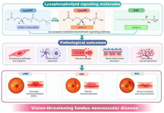
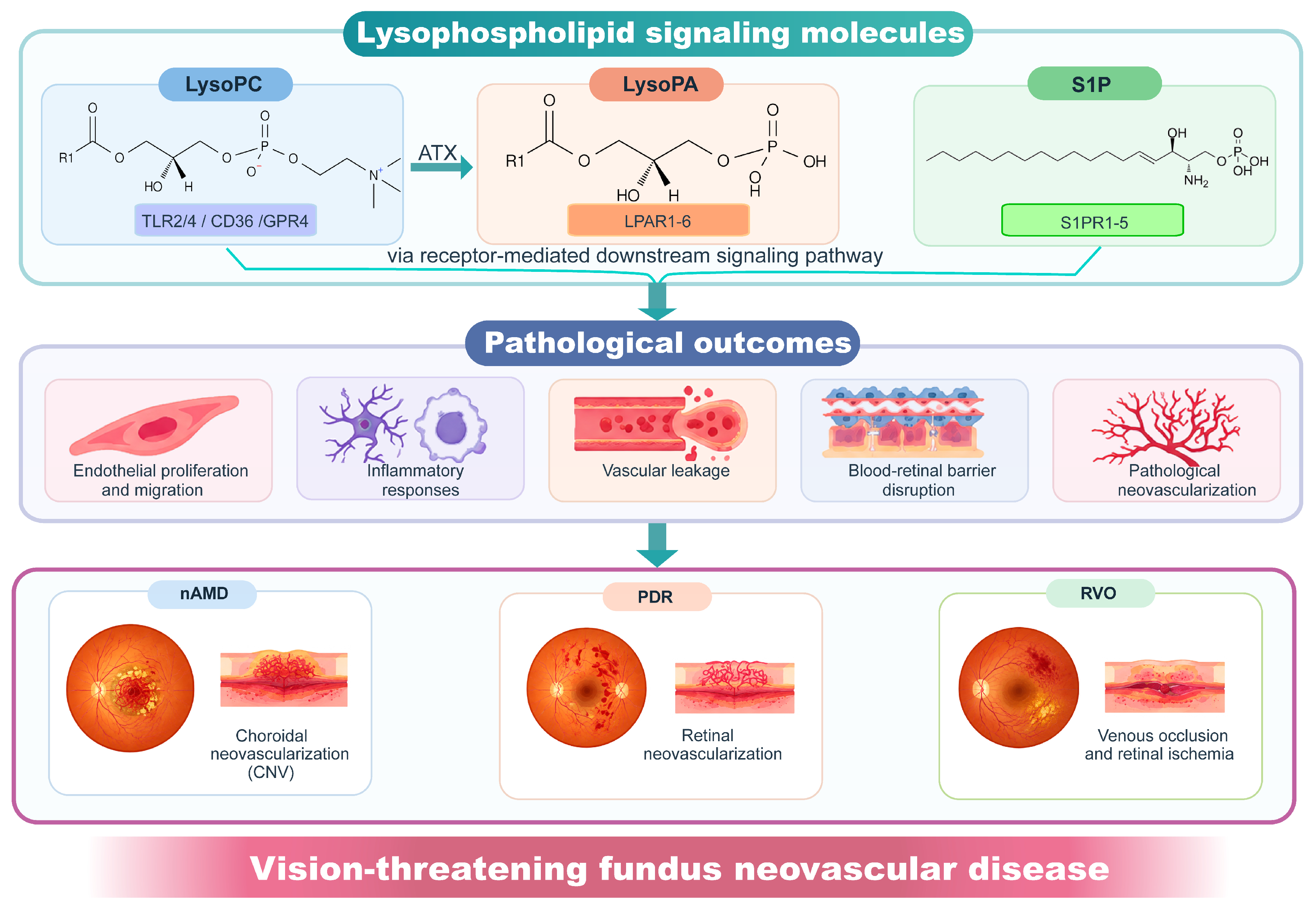
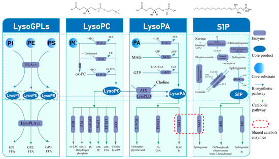
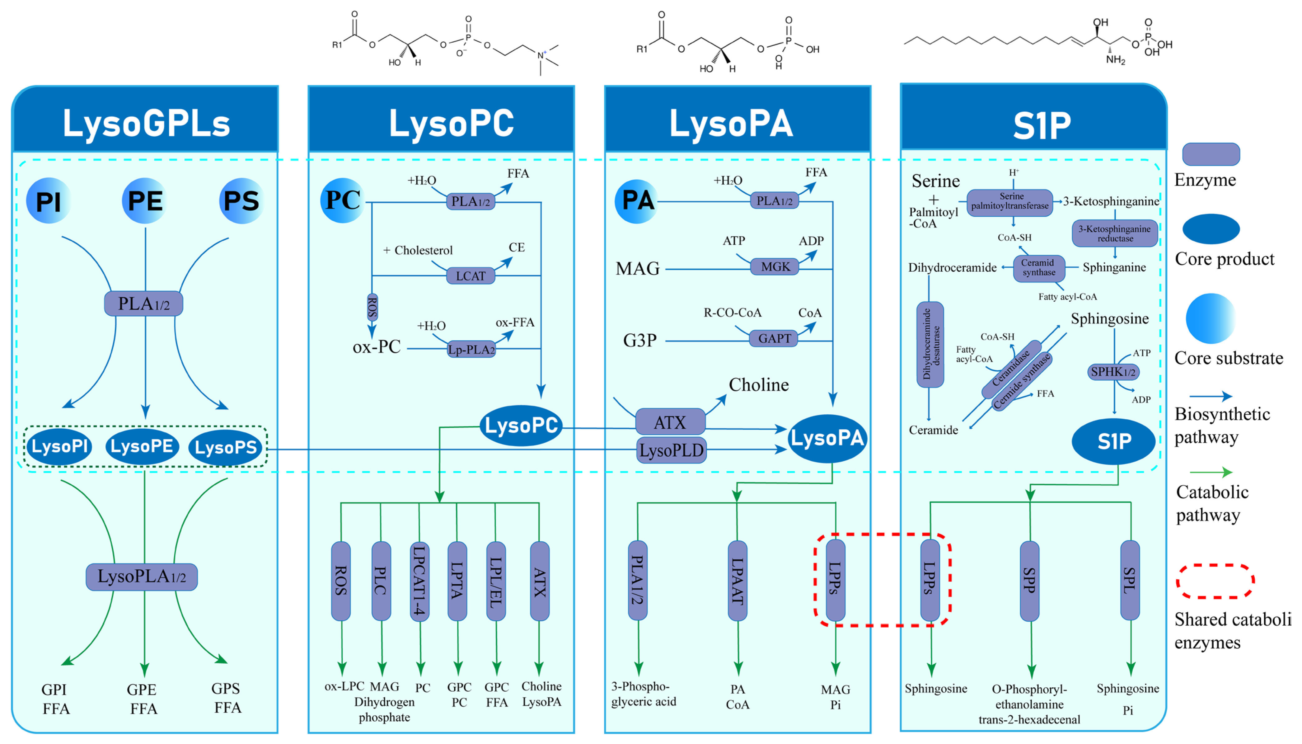
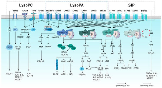
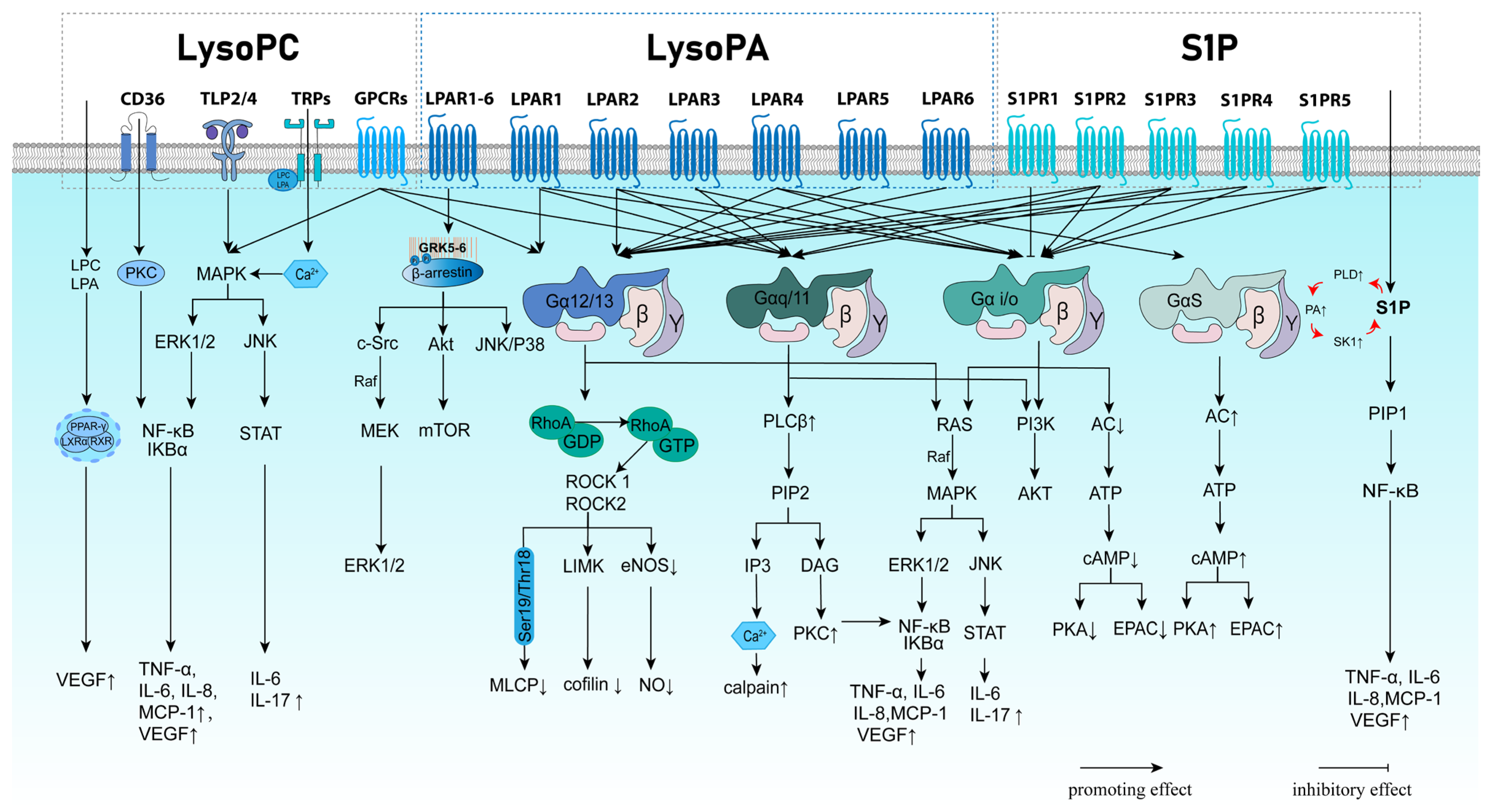
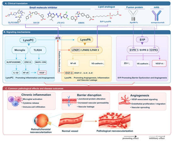
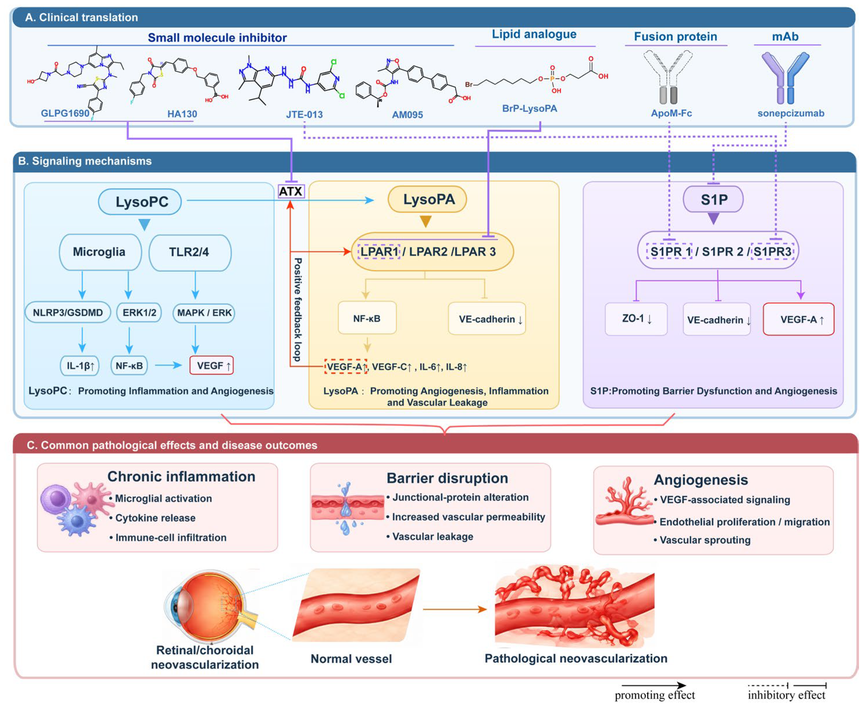
Disclaimer/Publisher’s Note: The statements, opinions and data contained in all publications are solely those of the individual author(s) and contributor(s) and not of MDPI and/or the editor(s). MDPI and/or the editor(s) disclaim responsibility for any injury to people or property resulting from any ideas, methods, instructions or products referred to in the content. |
© 2026 by the authors. Licensee MDPI, Basel, Switzerland. This article is an open access article distributed under the terms and conditions of the Creative Commons Attribution (CC BY) license.