


Abstract
Digestive malignancies remain a major cause of cancer-related mortality, with tumor progression (TP) being strongly influenced by the complex interactions between tumor cells (TCs) and their microenvironment. This review summarizes recently described mechanisms of interactions between tumor cells, macrophages and CD8+ T lymphocytes in colorectal and gastric cancer. PubMed and Web of Science were searched for studies published between 2020 and 2026, with the final search being conducted on 8 July 2026. Experimental and clinical studies, together with relevant reviews, were considered to examine these interactions and their therapeutic implications. The evidence shows a bilateral crosstalk in which tumor cells influence immune cell phenotype and function, while immune cells can further support or restrict tumor progression. These interactions involve receptor-ligand systems and paracrine signaling, followed by changes in intracellular pathways regulating proliferation, invasion, macrophage polarization and CD8+ T-cell exhaustion. Considerable methodological heterogeneity among studies limits a direct comparison of the reported mechanisms. Several therapeutic approaches targeting these interactions are currently being investigated in early-phase clinical trials. However, many components of tumor–immune crosstalk and their connections remain incompletely understood. A further characterization of these mechanisms may help identify therapeutic strategies able to interfere with the immunosuppressive microenvironment of colorectal and gastric cancers.
1. Introduction
1.1. Current Global Incidence and Future Projections
Cancer is a disease with a severe medical, social, and psychological impact, with the lifetime probability of developing a form of cancer ranging from 39% to 39.9% [1]. Colorectal cancer (CRC) is the third most frequent cancer type (10% of cancer diagnoses) and the second leading cause of cancer-related death, while gastric cancer (GC) ranks among the top three cancers in 19 countries [2,3]. In 2022, the incidence of CRC (reported as new cases per 100,000 people) was 18.4 worldwide, with the lowest rate of 5.5 in Middle Africa and the highest rate of 35.33 in Australia-New Zealand [4]. GC incidence appears to be lower, with the highest rate of 30/100,000 reported in East Asia in 2020 [2]. Moreover, global trend predictions are not optimistic. CRC incidence is expected to increase by 70.53% by 2045, including a peak increase of 129.64% in East Africa [4]. GC incidence is projected to increase by 62% by 2040 [2].
1.2. Tumor Cells and Neighboring Microenvironment
The tumor microenvironment (TME) is described as an extensive, diverse and dynamic ecosystem that continuously interacts with tumor cells which arise, survive and progress within it [5]. From a historical perspective, the first descriptions of TME cells assigned them a bystander role. However, later experiments and observations described key functions in tumor progression, immunosuppression, and therapy resistance [5,6]. TME can be divided into numerous components. The first and most representative are TCs, which constitute between 5% and 100% of the TME volume [7]. Immune cells are located in the vicinity of TCs and constitute the inflammatory tumor microenvironment (iTME), with some of them having a significant impact on TP. The other cell populations identified within the TME are non-tumor, non-immune cells: endothelial cells, pericytes, fibroblasts, adipocytes, mesenchymal stem cells, and neuronal cells. Within the TME, molecules such as cytokines, involved in paracrine interactions, and extracellular matrix components, are also identified [5,6,8,9,10]. Another characteristic of the TME is hypoxia [6]. Higher hypoxia-related scores and molecular signatures are associated with shorter overall survival (OS) [11,12]. Numerous and complex interactions between TCs, tumor-associated macrophages (TAMs), and T lymphocytes have been described within the TME, and these regulate TP in a symbiotic manner. Throughout this review, molecular or histological subtypes are specified where reported in the primary studies. When subtype information is unavailable, the findings should not be interpreted as uniformly applicable across all CRC or GC subtypes.
1.3. Current Treatment Strategies and Their Outcomes
Although colonoscopy screening has decreased CRC incidence, mortality has not been affected [13]. As a treatment option, surgery remains an important part of digestive cancer management, and 5-year overall survival could increase to 84.1% [14]. Standard chemotherapy protocols (alone or complementary to surgical resection) are combined with various molecules that target intracellular pathways or surface receptors of tumor cells, with improved outcomes being observed. As an example, a phase III clinical trial has reported twofold longer OS in the mFOLFOX6 + anti-EGFR + anti-BRAF arm compared with the standard-of-care arm (NCT04607421 [15]). Immunotherapy is beginning to show advantages over conventional therapies in clinical trials, as Wang et al. noted in their meta-analysis [16]. However, the projection of cancer-related deaths for 2045 shows an increase of 83.47% compared with 2022 [4]. Moreover, 1.27 million deaths among GC patients are expected by 2040 [2].
All of the above shows the promising potential of immunotherapy and raises considerable interest in the manipulation of various interaction mechanisms between tumor cells and immune cells in order to improve the outcomes of patients with gastrointestinal cancers.
1.4. Scope of This Review
The importance of studying these interactions stems from the epidemiological data on malignant digestive cancers and the current outcomes of widely used treatments. Despite the fact that the TME is a highly complex system with a variety of interactions between its components, we aim to focus in this review on those recently described in the literature: (i) ligand-receptor or paracrine signaling between tumor cells and CD8+ T-cells and the intracellular pathways involved, (ii) ligand-receptor or paracrine signaling between tumor cells and macrophages and the intracellular pathways involved, and (iii) current treatment strategy outcomes and clinical trials based on the mechanisms described in the first two points. This review and its search strategy are based on two questions: “What has been discovered in recent years about these interactions and their impact on tumor progression?” and “What is the current state of therapies targeting these mechanisms?”
2. Materials and Methods
2.1. Databases and Search Strategy
For this literature review, the PubMed and Web of Science databases were analyzed and were last accessed on 8 July 2026. The search strategy was a general-to-specific one, starting with search terms such as: “Colorectal cancer tumor cell and macrophage”, “Gastric cancer tumor cell and macrophage”, “Colorectal cancer tumor cell and CD8+ T lymphocyte”, and “Gastric cancer tumor cell and CD8+ T lymphocyte”, grouped yearly within the 2020–2026 time frame. The next step was to use key molecules/mechanisms identified in the previous step as a new foundation for the search strategy (e.g., “Galectin-9 and colorectal cancer and CD8+ T-cell exhaustion”) to find results that supported or contradicted the previous ones. Duplicates were manually identified and removed. At the end of the search period, an updated search was conducted to identify studies published during the time elapsed since the initial search. Depending on the search terms used, the number of retrieved records ranged from 3 (“colorectal cancer” and “macrophages” and “ADAR1”) to 2578 (“colorectal cancer tumor cell” and “macrophages”). During study-selection process, A.P. screened titles and abstracts according to inclusion and exclusion criteria. Further, the full texts of potentially eligible studies or studies for which eligibility could not be determined from the abstract alone were assessed to reach a final inclusion decision. The final selection of the included studies was subsequently cross-checked by V.I. and M.D.
2.2. Inclusion and Exclusion Criteria
To meet the inclusion criteria, a study had to focus on molecular mechanisms that govern tumor cell-CD8+ T lymphocyte/macrophage interactions within the microenvironment of digestive malignancies. Only studies published in English and available in full text were included. For this purpose, research articles that were published within the mentioned time frame and primarily focused on the mechanism of interest were selected. To answer the review questions, studies using various methodological approaches were included, based to some extent on online biodatabases, cell lines, and animal or human tissue-based methods (see Section 6). Secondarily, 32 studies published before the 2020–2026 time frame were included for their enduring value and the relevance of the information they provided. Among these studies, 11 were included in Section 6, “Current therapeutic strategies and clinical trials,” and 21 landmark references were scattered throughout the rest of the text. For Section 6, conference abstracts reporting results from the included clinical trials were also considered when these results were not available in full-length publications at the time of the search. Twenty-five studies focusing on non-tumor subjects were included for their highly valuable information about immune cell response. Knowledge gaps such as incompletely described mechanisms for gastric or colorectal cancers, unknown related mechanisms, or controversies were addressed by including 22 articles focusing on non-digestive tumors, which provided additional support.
Studies were excluded if they did not address this review’s questions or mechanisms of interest. Studies investigating non-digestive malignancies or published before 2020 were excluded from the primary analysis. However, some studies focusing on other cancers were considered to provide complementary mechanistic evidence relevant to this review. Of particular importance, this review did not include articles that mentioned mechanisms of interest as a matter of fact, without further contextualization, or articles without full-text access.
3. Tumor Cell–Lymphocyte Interactions
CD8+ T-cells are a highly effective component of the immune system involved in tumor cell eradication, and they represent a priority target of immunotherapy [17,18] because of their recognition and elimination capabilities through intracellular signals and pathways mediated by MHC I molecules.
Physiologically, when CD8+ T-cells are stimulated by an antigen and receive co-stimulatory signals, they enter a proliferation phase and differentiate into effector or memory CD8+ T lymphocytes. If these CD8+ T-cells are exposed to continuous stimuli, inflammatory or tumoral in nature, molecular and functional changes may develop, driving CD8+ cells to a dysfunctional state [18]. Lymphocytes continuously communicate with TCs, and the number of CD8+ T lymphocytes is highly correlated with tumor progression [19]. The density and functional competence of T lymphocytes within the TME represent crucial determinants of immunotherapy success and an efficient antitumor immune response.
The number of T-cells in CRC is lower compared to normal mucosa [19], with the number and density of T-cells decreasing in late stages [20,21,22]. The density of T-cells in the tumor center and invasive front is highly variable; some authors have observed that right-sided CRCs feature fewer T-cells [20], with the invasive front presenting a higher density compared to the tumor center [21], while others suggest that T-cells are frequently excluded from close proximity to tumor cells at the tumor-stroma interface, in contrast with macrophages, and exhibit a lower immune response mediated by decreased cytokine production [23].
Lymphocyte populations are characterized by substantial heterogeneity within the TME; two examples from GC are CD8+ T-cells expressing CD226, associated with better survival [24], or CD39, characterized by an exhausted state [25]. A rare subset comprises T γδ cells, further divided into Vδ1, Vδ2, and Vδ3 subtypes, with the first two showing an association with a cytotoxic effect but being rarely identified within the CRC TME [26]. The CD8+ T-cell compartment comprises functionally active cells, precursor exhausted CD8+ T-cells (Tpex), and exhausted CD8+ T-cells (Tex), with their relative proportions changing dynamically [27]. This ratio was estimated and named PETER (Progenitor Exhaustion-to-Terminal Exhaustion Ratio); low values, corresponding to a higher Tex cell count, indicated a worse immunotherapy response [28].
Early stages of CRC show a significant density of the cytotoxic T-cell population transitioning to an exhausted phenotype with tumor progression [20]. In particular, Tex are observed in tumor tissue but not within non-tumor colorectal mucosa [19]. In contrast, Tpex cells can be identified in both tumor and non-tumor tissue [24]. The pathological mechanisms of exhaustion are multiple and incompletely understood. Some evidence indicates prolonged antigen exposure as a predominant factor because the TCR is continuously engaged and initiates the exhaustion process [29]. Other findings suggest strong communication between Tex and TCs through CD99-CD99, Migration Inhibitory Factor (MIF)-(CD74-CXCR4), or MIF-(CD74-CD44) with possible roles in the exhaustion process [19].
Lymphocytes identified as terminally exhausted exhibit reduced function, characterized by a low proliferative rate, cytokine synthesis and secretion and, more importantly for antitumor function, low cytolytic activity [27]. Throughout this process, CD8+ T-cells can be found in an intermediate state referred to asTpex [18] or as a progenitor exhausted by other authors [30]. CD8+ T-cells can be differentiated into stages of exhaustion based on molecular expression patterns. Tpex cells are characterized by the expression of TCF1, PD-1, CD8, activation markers (CD69, CD107), inhibitory immune checkpoint molecules (LAG3, CD200) and transcription factors (ID3, EOMES, T-bet) [17,18,31]. These cells share common characteristics with early memory T lymphocyte surface markers such as CCR7, IL7R and CD26L [27].
Tex lymphocytes are characterized by the expression of CD39, CD244 and TIM-3 [27]. Transcriptional changes can also be identified, with TOX and NR4A being highly expressed [17,32,33], as well as epigenetic changes manifested as a decrease in chromatin accessibility for some effector genes and an increase in chromatin accessibility for some exhaustion-associated genes [33].
Favorable results of anti-PD-1 therapy are immunologically dependent on CD8+ T-cell function, reestablishment of cytolytic function and the capacity to counter the immunosuppressive effect of the TME [17,18], with a higher number of CD8+TCF1+ (Tpex) cells being associated with better therapeutic outcomes. One highly significant fact is that Tpex lymphocyte proliferation can be induced, with these cells being able to repopulate and maintain the T-cell response against tumor cells. This capacity is observed among TCF1+ T-cells but not among TCF1- T-cells (Tex) [31]. As a result, one hypothesis is that the low number of patients responding to anti-PD-1 therapy can be explained by the inability to activate dysfunctional Tex cells [18].
In the TME, a lymphocyte subset called “bystander T cells” can be identified, comprising non-specifically activated lymphocytes that happen to be present during the development of the immune response [27,34]. These bystander lymphocytes express a variety of markers such as memory T-cell markers (CCR7, CD27, CD28) and exhaustion markers (PD-1, TIGIT). This particular situation makes the distinction between Tpex and Tex difficult because of their shared molecular characteristics [27]. CD83 is a molecule that can differentiate between these two cell subpopulations because T lymphocytes express CD83 only after antigen stimulation and its expression decreases with the development of the Tex state. A marker that can differentiate bystander lymphocytes from Tpex lymphocytes is highly useful because of the common marker expression of these cells [27].
The interaction between T-cells and TCs is not the only determinant. Various cells that form the iTME and have immunosuppressive effects, such as tumor-associated fibroblasts (TAFs), tumor-associated macrophages, myeloid-derived suppressor cells (MDSCs) or regulatory T-cells (Tregs), can initiate and facilitate the exhaustion process [35].
Another crucial perspective is the metabolic one, dominated by hypoxia, which determines mitochondrial dysfunction and stimulates reactive oxygen species (ROS) synthesis, leading to T-cell exhaustion [32,33,36]. Detailed analysis of the influence of hypoxia on the immune cell populations within the TME revealed an increase in inhibitory subsets (including macrophages) and a decrease in stimulatory subsets (for example, central memory T-cells) [36]. The cumulative effect is a significant number of inflammatory cells with a reduced cytotoxic capacity, with hypoxia-related pathways showing the greatest upregulation in CMS4 CRC subtype [37]. Hypoxia directly influences the antitumor response via HIF-1α expression in TCs associated with follistatin-like 3 (FSTL3) overexpression [38], which favors an increase in PD-L1 expression by TCs [39] and epithelial-mesenchymal transition (EMT) through various pathways [40,41]. FSTL3 expression is higher in MSI-H CRC tumors [39]. Nutrient competition and metabolite accumulation, including lactate, in TME can initiate or sustain T-cell exhaustion [35].
The PI3K/AKT pathway represents one of the most important and widely studied signaling pathways in cancer and is involved in proliferation, apoptosis, neoangiogenesis, autophagy, EMT and chemoresistance [42]. The first step is PI3K activation and the formation of phosphatidylinositol 3,4,5-trisphosphate (PIP3), which further activates AKT [43]. The biological functions of this pathway highlight its crucial role in tumor initiation and progression [44,45] and treatment resistance [46,47].
In TCs, this axis is stimulated by various molecules (Figure 1), for example, HECW1—overexpressed in GC [48] and mediated by HIPK2 [49] or YWHAH, a signaling and intercellular interaction protein—via FRA1/HMGA1 stimulation [50]. A TME with overexpression of these molecules is characterized by few Tex cells and a high density of TAMs as a result of Hippo pathway and IFN-α/TNF-α downregulation [51] and increased expression of PD-1/TIM-3 on TCs. In addition, YWHAH also activates the MAPK/ERK pathway. Its precise mechanism of direct interaction with lymphocytes is unknown. Although YWHAH was associated with MSI status, based on a TCGA-TIDE score, clinical MSI-H/MSS status was not reported [52].
Figure 1.
Main molecular interactions between tumor cells (left) and CD8+ T lymphocytes (right) in colorectal and gastric cancer. The figure focuses on the principal mechanisms mediating direct interactions between cells of interest. Green arrows indicate stimulatory effects; red arrows indicate inhibitory effects; black arrows connect successive steps of signaling pathways or indicate the transfer route of molecules. The illustrated mechanisms are not assigned to a particular molecular subtype. Additional components are presented in Supplementary Figure S1. Created in BioRender. Andrei, P. (2026) https://BioRender.com/8yqghug.
This pathway can be activated at the AKT level by FSTL3, a glycoprotein detected in numerous cells and related to cell growth, metabolism and various cancers, including CRC [39,53]. Cancer-associated fibroblast-derived FSTL3 binds to TfR1 on TCs and activates the AKT/mTOR pathway [54]. This molecule has multiple targets, activating c-Myc to increase PD-L1 levels [39] or HIF-1α [38,55], and its expression is reduced by ZNF454 [55]. Apart from the FSTL3-MSI association described above [39], molecular subtype information was not reported in these mechanistic studies, limiting the assessment of subtype-dependent effects.
The PI3K/AKT pathway can be suppressed within CD8+ T-cells as a result of crosstalk with CD155-expressing TCs. CD155 is a receptor first described as the poliovirus receptor and now frequently identified on malignant TCs [56,57,58], with known inhibitory effects on immune cells through crosstalk with various surface molecules [59]. A highly CD155-expressing TME is immunosuppressed [60] and characterized by few Tex cells, thereby indicating a poor prognosis associated with the CD155-TIGIT axis [56,61]. The CD155 level is also upregulated by CEBPB in GC cells [62].
This molecule interacts with TIGIT [57,63], whose expression is higher in MSI and BRAF-mutant CRC [60], located on the surface of CD8+ T-cells, intracellularly inhibiting the PI3K/AKT pathway and subsequently NF-kB [56], but other pathways can be involved, such as mTOR in triple-negative breast cancer [63], although these have still not been researched or described in digestive malignancies. In contrast to previous results, He et al. reported an increased level of AKT phosphorylation in GC, while CD155 was downregulated [64].
The activity of the PI3K/AKT and ERK pathways is downregulated in CD8+ T-cells by the KLRB1 receptor as a result of binding to CLEC2B, a ligand identified on the membrane of CRC TCs, as demonstrated primarily in MSS CRC models [65]. Some results indicate increased cytotoxic activity of CD8+ T-cells and low PD-1 levels as a result of PI3K/AKT/mTOR axis activation by interferon-induced protein 35 (IFI35), highly expressed in CMS1 CRC and secreted by TCs in response to IFN-γ exposure [66].
The PD-1 level in TCs is positively influenced by lipin 3 (LPIN3) and YTH N6-methyladenosine RNA-binding protein 1 (YTHDF1). LPIN3, whose expression correlates with MSI in TCGA data, plays physiological roles in lipid metabolism but also promotes TP in CRC via β-catenin activation. Its inhibition raises IFN-γ levels, enhancing the cytotoxic CD8+ T-cell response [67]. However, prolonged exposure to IFN-γ could initiate T-cell exhaustion [68].
Similar to LPIN3, YTHDF1 is known for its involvement in TP and metabolism. It is highly expressed by GC TCs, with lower expression in the genomically stable (GS) subtype, and correlates with the mutant p53 state, a low number of inflammatory cells and their exhausted state [69], and the risk of peritoneal metastasis [70]. The changes noticed after YTHDF1 knockdown reveal a reshaped antitumor TME [71,72].
Moreover, the PD-1 level is increased in CD8+ T-cells as part of the PKA/CREB pathway activity, with a similar tendency being observed for TIM-3 levels as a result of thyroid-stimulating hormone receptor (TSHR) activation. TSHR is highly expressed in tumor and stromal cells of CRC as well as in CD8+ T-cells. TCs employ extracellular vesicles (EVs) to deliver TSHR to lymphocytes. Its high expression is associated with MSS status and immune checkpoint blockade failure [73]. A similar mechanism has not yet been identified in GC.
Intracellular domains of PD-1 and TIM-3, two well-known molecules with inhibitory effects in digestive malignancies [74,75], are crosslinked by Galectin-9 [76], a β-galactoside-binding lectin identified in numerous immune cells and associated with physiological roles such as cell growth or apoptosis [77]. In GC, galectin-9 is associated with TP [77], and in CRC, the TIM-3/Gal-9 axis supports an immunosuppressive TME [78,79] characterized by high LAG3 and TGF-β levels with a known inhibitory impact on CD8+ T-cell function [79]. TIM-3is also expressed by various immune cells, with its level in CD8+ T-cells being correlated with a better prognosis when it is coexpressed with PD-1 [80], and M2-like macrophages raise TIM-3 levels as a result of TGF-β stimulation. This axis is reported in MSI, CMS1 and CMS4 TME [81]. Some authors consider Galectin-9 a ligand for the TIM-3 receptor with protumoral effects [82]. However, TIM-3 inhibition reveals better outcomes of anti-PD-L1 treatment in predominantly pMMR experimental settings [78].
Another mode of interaction between TCs and CD8+ T-cells is mediated by various cytokines, such as IFN-α [83], TGF-β [30], IL-10, IL-6, IL-35 or IL-27, that initiate and sustain the T-cell dysfunction process [32].
TGF-β is secreted by various T-cell subtypes [26,84] and even by CRC TCs [85], with known roles in CD8+ T-cell exhaustion [86]; increased infiltration of cytotoxic CD8+ T-cells has been observed as a result of TGF-β and PD-L1 co-inhibition in a CMS4-like mouse model [87]. TGF-β has various roles within TCs, with the TGF-β/Smad axis being activated by multiple factors: Forkhead box protein C1 (FOXC1) [88], ADAM17 [89], SNHG12 [85], Prkci [90] or ARHGAP4. This pathway stimulates colorectal tumor cell proliferation, migration, epithelial-mesenchymal transition and invasion [91].
One essential cytokine in the immune response is IL-6 [92]. Within the TME, it can be released by CD3+ cells [93] or TCs [85,94,95] and promotes TP via STAT3 activation [93,96], either directly, by VEGF upregulation [96], or as a promoter of the metastatic process [97]. The IL6/JAK/STAT3 pathway is also activated by HOXC6, which is highly expressed in MSI-H CRC TCs [98]. In contrast, Pennel et al. observed that high stromal IL-6 expression is associated with poor prognosis but not with its expression in TCs [95]. The level of IL-6 modulates immune populations, with its overexpression being correlated with an immunosuppressed TME. ASCL2 is one key factor that directly influences IL-6 levels and, consequently, T-cell numbers, as described primarily in MSS CRC [99]. Other cytokines, such as IL-12, increase the numbers of Tpex cells and M1-like macrophages [100], and IL-2 stimulates TME infiltration by M2-like macrophages and CD8+ T-cells [92].
CRC TCs frequently exhibit urocortin (UCN), a member of the corticotropin-releasing hormone family involved in inflammation and tumorigenesis. UCN facilitates TP by CCL23 upregulation, a T-cell exhaustion-promoting chemokine [101], or by p53 inhibition by UCN-1 [102]. Increased levels of T-cell exhaustion markers induced by CCL23 in non-digestive cancers were reported as part of the effect of GSK3β activation [103], a substrate of the PI3K/AKT pathway [104], with no evidence of the CCL23/GSK3β axis being involved in TP in digestive cancers.
Major facilitator superfamily domain-containing 2 (MFSD2A) is a molecule identified in multiple tissues with various actions (TP, hepatic regeneration) [105], with its expression decreasing from normal mucosa to tumor tissue [106,107]. MFSD2A overexpression reduces TAM abundance and promotes T-cell infiltration and function through COX2-mediated reduction in TGF-β1. The results are based on a human GC cohort predominantly MSS/MSI-L (96,3%), EBV-negative (59,3%) [108]. These results are consistent with findings showing the antitumor effect of MFSD2A in CRC via the activation of S100A14 and secondary inhibition of STAT3 [106], or inhibition of the ERK/MAPK pathway with a secondary decrease in PD-L1 levels and a better cytotoxic T-cell response [109].
The desired effect of the interaction between a CD8+ T-cell and a TC is the programmed cell death of the latter—for example, ferroptosis or pyroptosis—with a mechanism based on glutathione peroxidase 4 (GPX4) and GPX8. Their overexpression is observed in multiple cancers, including GC, where it correlates with prognosis and the extent of immune cell infiltration [110,111]. GPX4 activity is based on SLC7A11, but conversely, its overexpression results in decreased ferroptosis [112] and high TRIB3 expression [113]. A TME with SLC7A11 overexpression presents low CXCR3 and CXCL14 levels [112]. CXCR3 does not modulate CD8+ T-cell numbers [114], but CXCL14 can stimulate the infiltration of immune cells and antitumor action [115,116]. As part of the pyroptosis mechanism, DFNA5 (GSDME) exhibits low expression in Epstein–Barr virus-associated GC [117] and is correlated with exhaustion markers and the number of M2-like macrophages [118].
4. Tumor Cell–Macrophage Interactions
Complex and reciprocal interactions between TCs and TAMs have been described that lead to immunosuppression, chemoresistance and TP, with the density of the latter being directly linked to prognosis [119,120]. Macrophages represent the most frequently identified cells in the iTME, accounting for up to 40% of immune cells [17,28,121]. Right-sided CRCs tend to exhibit an anti-inflammatory SPP1+ phenotype of TAMs [20] that is associated with a worse prognosis [122,123,124]. However, regardless of side, macrophages tend to start with a pro-inflammatory state and end with an anti-inflammatory one [20].
Macrophage activation has traditionally been described using an M1/M2 polarization framework that plays major roles within the TME. The M1/M2 framework developed from earlier studies demonstrating differential regulation of macrophage nitric oxide synthase and arginase activity by Th1/Th2 cytokines [125] and was subsequently popularized by Mills et al. in 2000 [126].
However, this binary classification represents a simplified representation of a broader and highly plastic spectrum of macrophage activation states, a complexity further revealed by single-cell RNA sequencing, which has identified macrophage phenotypes such as SPP1+, C1QC+, INHBA+, ISG15+, LNRP3+ or LYVE1+, the first two being well known for their influence on the immune response within the TME [127].
Spatially, macrophages tend to be located at the TC-stromal interface, at the invasive front [128], where they assemble a barrier-like structure that promotes local immunosuppression. Moreover, macrophages are located closer to TCs than T lymphocytes [23]. SPP1+ macrophages, a specific subtype, are identified in digestive malignancies in the following order: tumor core > invasive margins > non-tumor mucosa [20,129,130,131,132]. In contrast, one study identified the highest density of SPP1+ macrophages at the invasive front [133]. Moreover, the number of SPP1+ macrophages is higher in metastases compared with the primary tumor [124,129]. The SPP1+ TAM signature is enriched in MSI-H CRC TME [129].
Osteopontin, present in intracellular or extracellular secreted forms [120], is the glycophosphoprotein encoded by the SPP1 gene and fulfills various roles in the TME, showing a significant impact on M2-like macrophage polarization (Figure 2) [134]. SPP1 is associated with a negative prognosis because of its pro-metastatic activity in digestive malignancies and the co-synthesis of PD-1, PD-L1, VEGF and MMP2, collectively promoting TP [129,131,135,136]. However, SPP1+ TCs do not correlate with migratory or invasive capacity [134]. Taken together, these findings support SPP1+ macrophages as relevant biomarkers of TP. At the opposite end of the spectrum are CICQC+ macrophages that exert cytophagic and MHC-II antigen-presenting capacities [129,136].
Figure 2.
Main molecular interactions between tumor cells (left) and SPP1+ macrophages (right) in colorectal and gastric cancer. The figure focuses on the principal mechanisms mediating direct interactions between these cells, with particular emphasis on osteopontin-associated signaling. Green arrows indicate stimulatory effects; black arrows connect successive steps of signaling pathways. Molecular subtypes were not consistently specified in the underlying studies; therefore, the illustrated mechanisms are not assigned to a particular molecular subtype. Additional components are presented in Supplementary Figure S2. Created in BioRender. Andrei, P. (2026) https://BioRender.com/2cit6np.
Numerous interactions between TCs and macrophages center on osteopontin. TCs can promote the SPP1+ phenotype of macrophages through intracellular RING finger 32 (RNF32), which activates the Wnt/β-catenin pathway, firstly promoting TP and secondly increasing CCL2 levels. This chemokine interacts with the CCR2 surface receptor on macrophages and promotes SPP1 expression [137]. Another molecule delivered by TCs is migration inhibitory factor (MIF), which binds to the CD44 receptor. The subsequently activated pathways lead to BCL9L stimulation and the SPP1+ phenotype [138].
Hypoxia plays an important role in the development of the SPP1+ state, which is enriched in CMS4 tumors [139], with these cells exhibiting hypoxia-associated [129] or angiogenesis-associated genes, such as VCAN [140], leading to increased angiogenic capacity of SPP1+ macrophages [124,136], and with a high number of these cells within the TME being correlated with lymphovascular tumor invasion [123,136]. High levels of HIF-1α, increased by hypoxia, interact with STAT3 and, finally, promote the development of the SPP1+ phenotype [135]. TC-derived osteopontin influences macrophages by upregulating PI3K/AKT within these immune cells and increasing CSF1 levels, a known factor for macrophage recruitment and the promotion of the M2-like phenotype [134].
Osteopontin binds to the CD44 receptor located on the surface membrane of TCs, thereby activating the Wnt/BTF3 pathway and promoting TP [141,142]. Moreover, SPP1+ macrophages activate JAK/STAT3 in MSS CRC TCs [143]. In diffuse-type GC, SPP1+ macrophages are promoters of TP, a worse prognosis and macrophage recruitment [131]. Osteopontin interacts with TCs directly through αvβ3 integrin [135] or indirectly through PDGFB upregulation, which, as a result of binding to a TC receptor and crosstalk with FADS1, activates the mTOR pathway [144].
Collectively, these observations describe TP as a result of direct interactions between SPP1+ macrophages and TCs. However, TP can also occur as a result of multiple interactions with CD8+ T-cells and their exhaustion. TAMs possess a high phagocytic capacity and the ability to process tumor antigens; however, they are unable to initiate or sustain the T-cell activation process [17].
SPP1+ TAMs interact directly with PD-1+ CD8+ T lymphocytes, an interaction mediated by SLC2A1 [145], a molecule upregulated by YAP1—a promoter of invasive behavior through the Wnt/β-catenin pathway [146]. Another result is the downregulation of the LCK-Zap70-LAT axis that transfers the TCR signal to the nucleus [132]. This macrophage subset produces CXCL16, CXCL9, and IL-10, which may influence CD8+ T-cell activity and trafficking. Collectively, these findings suggest that macrophages may contribute substantially to T-cell exhaustion.
Initially characterized for its role in metabolism and phagocytosis, studied frequently in neuropathology [147], and reported to have anti-inflammatory roles in non-alcoholic liver steatosis [148], triggering receptor expressed on myeloid cells 2 (TREM2)—a type I transmembrane immunoglobulin receptor expressed by macrophages [149,150]—is now recognized for its role in multiple cancers [151]. Upon ligand binding, signaling through the adaptor proteins DAP10 and DAP12 leads to the activation of the PI3K/AKT pathway [150]. TREM2 expression is identified in TCs and macrophages, with both digestive malignancies showing its overexpression [151,152,153]. In GC, TREM2 expression was inversely associated with HER2 expression, with no significant differences being observed between histological types [153]. In TCs, it acts as a Wnt/β-catenin/TCF4 pathway inhibitor, decreasing TP [154], with a high impact on 5FU treatment resistance in GC when it is underexpressed [155]. TREM2 knockdown significantly reduced the proliferative capacity of TCs and their invasion ability and increased E-cadherin expression levels, with a known role in EMT [152]. In lung adenocarcinoma, the transfer of circ_0001715 from TCs to macrophages via extracellular vesicles was observed to increase TREM2 levels and facilitate TP [156]. At present, there is no evidence in the literature of such a mechanism occurring in digestive malignancies.
TREM2 activity within TAMs is highly correlated with STAT1 phosphorylation, leading to high CCL8 and PD-L1 levels [157], which accentuate CD8+ T-cell exhaustion. Moreover, the TREM2+ signature was associated with the EBV molecular subtype of GC [158]. As expected, anti-PD-L1 treatment outcomes can be improved by targeting this molecule [157]. A hypothetical interaction with APOC1 exhibited by TAMs can elevate CCL2/5 levels within the TME [159,160]. TREM2+ macrophage density can increase under glucocorticoid exposure [161].
The Wnt/β-catenin signaling pathway is well known for its impact on the progression of digestive malignancies [162,163], with macrophages activating this pathway in TCs by TGF-β secretion and using tribbles pseudokinase 3 (TRIB3) as a mediator [164]. TRIB3 is known to function as a key factor in physiological cellular proliferation, differentiation and also TP, being overexpressed in CRC [113,165,166].
In TCs, HIF-1α upregulates TRIB3 [164,167], which interacts with COP1 to increase β-catenin levels [167].
Another pathway influenced by TRIB3 is centered on STAT3. Furthermore, this enhances VEGFA expression and supports TP through neoangiogenesis [165]. Another promoter of the TRIB3/VEGFA axis in digestive malignancies is TEAD4 [113,168], with its overexpression being correlated with high ferroptosis resistance in TCs through activation of the MEK/ERK axis and increased GPX4 and SLC7A11 levels [113,169]. An inhibitor of this pathway is METTL14 [169].
Without strong evidence, TRIB3 could activate the PI3K/AKT pathway under high-glucose conditions in diabetic patients [170]. In contrast to previous studies, Liu et al. identified opposite results characterized by a decrease in TRIB3 in GC with TP. Notably, a TME with high TRIB3 expression is associated with an increased number of M1-like macrophages and better survival [171]. As a member of the same family, TRIB2 plays a similar tumor-suppressor role in GC [166].
The Wnt/β-catenin axis is also stimulated in TCs by ankyrin repeat domain-containing protein 22 (ANKRD22), physiologically identified in normal gastric epithelium and activated macrophages but overexpressed in CRC TCs [172,173]. Furthermore, FOSL1 gene transcription is promoted [173], neoangiogenesis is increased by FOSL1/TIMP1/VEGF [174], and TCs become more resistant to 5FU [175]. There is a limited number of studies in the literature on ANKRD22 and intercellular interactions in digestive malignancies. Similar tendencies in TP are described for other tumors such as lung adenocarcinoma [176,177], breast cancer [178] and papillary thyroid carcinoma [179]. In contrast to previous studies, some authors reported an M1-like-promoting effect of ANKRD22 in CRC [180], with similar tendencies in sepsis-induced ARDS [181] or gastric mucosal injury, in which ANKRD22 knockdown decreased M1-like polarization without an impact on M2-like macrophages [182].
Macrophages interact with and influence TCs through the secretion of matrix metalloproteinases (MMPs), members of the metzincin family with a well-established role in extracellular matrix proteolysis, with a subset of them (MMP1, 3, 7, 9–12, 14) being markedly increased in CRC [183]. MMP1 is expressed by TCs, T lymphocytes and TAMs [184], and MMP1+ TCs represent a pre-metastatic population [184]. Tumor cells stimulate MMP1 secretion by macrophages, mediated by GM-CSF as a result of stimulation of the PI3K/AKT pathway in these immune cells [185]. Conversely, M2-like macrophages can promote TC invasion through cytokine synthesis, such as TNF-α, which stimulates MMP9 secretion by TCs [186], or TGF-β, which facilitates MMP2 secretion [187].
MMP1 secreted by TAMs binds to the TC receptor PAR1, which favors EMT via the MAPK/Erk axis [188,189,190]. This receptor may contribute to TP via the 3-phosphoinositide-dependent kinase 1 (PDK1)/AKT pathway in KRAS mutant CRC [189]. TP is also positively influenced by MMP1 expression in TCs as a result of its intra- and/or extracellular roles. MMP1 synthesis by TCs is enhanced by numerous factors: CD26/CAV1 axis activation [191], HIF-1α [192], zinc finger protein 3 (ZNF3) [193] or calmodulin-regulated spectrin-associated protein 2 (CAMSAP2) [194]. In contrast to these results, Ito et al. reported decreased TC proliferation in line with higher MMP1 expression in GC TCs [192]. From another perspective, MMP1 interacts with and exhausts CD8+ T-cells through the CXCL16-CXCR6 axis [184]. Tissue inhibitor of metalloproteinase-2 (TIMP2) reduces the proteolytic capacity of metalloproteinases (particularly MMP2). TIMP2 has certain roles in digestive malignancies but not as prominent as in other cancers. mRNA levels reveal decreased expression in CRC but increased in GC, subtypes not reported in a primary study. TIMP2 expression in both digestive cancers correlates with TAMs, the number of CD8+ T-cells and their exhaustion status within the TME [195].
On the other hand, TCs can influence macrophage behavior using various molecules (Figure 3) such as ADAR1, an RNA-editing enzyme highly expressed in CRC [196] with known roles in TP [197]. TCs use extracellular vesicles to transfer this molecule to macrophages [196] through four known mechanisms: increasing AZIN1 levels to promote an M2-like phenotype [196], activating the PKR/EIF2α pathway to decrease TIM-3 levels, with a well-known impact on T-cell cytotoxic function [198], favoring SPP1 secretion, which activates the NF-κB axis in TCs to increase resistance to oxaliplatin treatment [196], or activating the Wnt/β-catenin/CALR pathway (in gastric cancer cells) [199]. The pro-M2-like effect of ADAR1 is also reported in non-tumor diseases [200].
Figure 3.
Main molecular interactions between tumor cell (left) and macrophages (right) in colorectal and gastric cancer. The figure focuses on the mechanisms mediating direct interactions between these cells, including EVs-mediated communication. Green arrows indicate stimulatory effects; red arrows indicate inhibitory effects; black arrows connect successive steps of signaling pathways or indicate the transfer route of molecules. The illustrated mechanisms are not assigned to a particular molecular subtype. Additional intracellular modulators are presented in Supplementary Figure S3. Created in BioRender. Andrei, P. (2026) https://BioRender.com/aavlvgp.
TCs secrete chemokines belonging to the CC or CXC families, with minimal structural differences [201], to influence macrophage behavior. CXC motif chemokine receptor 2 (CXCR2) is a chemokine receptor identified on several immune cells that interacts with seven ligands differentially expressed by various tumors. Stimulation of this receptor on macrophages leads to activation of the PI3K pathway. CRC TCs secrete CXCL1–3, 5, 8, while GC TCs secrete CXCL1, 3, 5, 6, 8 [202], with a subset of them (CXCL3, 4, 14) being correlated with macrophage numbers within the TME [203]. CXCL1 is overexpressed by CRC TCs [204]; however, the mechanism can be bidirectional, as macrophages secrete CXCL1 within the TME and promote TC invasion [205]. CXCL1 and CXCL3 are overexpressed in CRC TCs as a consequence of the activation of the NF-κB pathway by SUB1 [206]. CXCL8 is a chemokine secreted by CRC TCs that increases TAM numbers and promotes M2-like polarization, with these effects being mediated by STAT3 [207]. A further decrease in the number and function of CD8+ T-cells is modulated by M2-like macrophages [207] or via NETs (neutrophil extracellular traps) that are stimulated by CXCL8 [208,209]. Conversely, Chen et al. reported a positive gradient between CXCL8 and the number of immune cells/cytotoxic T-cells within the TME [203], while Korbecki et al. reported that CXCL8 secretion in CRC is associated with macrophage infiltration without M2 characteristics [202]. TAMs secrete CXCL9/10, which binds to CXCR3 on TCs, promoting EMT, increasing PD-L1 expression and T-cell exhaustion. Macrophages use this mechanism through STAT3 activity and decreased expression of NF-kappa B activator 1 (Act1) [210].
CCL2 (also known as monocyte chemoattractant protein-1) is expressed by TCs in digestive malignancies and has a major impact on macrophage recruitment and M2 phenotype polarization [211,212,213]. Numerous factors that positively modulate its level within TCs are described in the literature: STAT3 [214] (which is stimulated by PIPKIγ [215], ETV5 [214], VSIG4 [216], HMGA2 [213]), GAL3ST4 (in KRAS mutated CRC) [217], TCF4 [218], RUNX1 [219], HOXC6 (in MSI-H CRC) [98], HOXD13 (via TGF-β1-p-Smad in CMS4/MSS CRC [220]) and SLITRK4 (via PI3K/AKT/NFκB) [221]. This chemokine binds to the CCR2 receptor and stimulates multiple intracellular signaling pathways [212], including the Hedgehog pathway in macrophages, to facilitate M2-like polarization [219].
Conversely, M2-like TAMs secrete CCL2 within the TME via the Wnt5a/CaMKII/ERK pathway, enhancing macrophage recruitment [222]. CCL2 levels can be decreased in GC, for example by CDK5RAP3, leading to M1-like macrophage polarization [223]. In addition to CCL2, TCs can promote macrophage recruitment via interleukins such as IL-6 [128] or CSF1, which is directly correlated with GPSM1 expression and T-cell exhaustion; targeting this pathway reduces anti-PD-1 treatment resistance [224].
TCs promote the development of the SPP1+ phenotype in macrophages [130], which are characterized by a genetic profile similar to that of M2-like macrophages [131]. Within the MSI-H CRC TME, their presence is strongly correlated with SPP1 expression [129]. TCs exhibit ITGB1 and CD44 receptors, with the latter being well known for its interaction with SPP1 [131].
Another described mechanism through which TCs induce SPP1+ macrophage activation is centered on extracellular vesicles (EVs) [225]. Extracellular vesicles are particles released by cells, enclosed by a lipid bilayer, that play roles in intercellular communication through various molecules [226,227]. A large number of EVs have been observed and studied to date, with their main characteristics being summarized in Table 1.
Table 1.
Detailed interactions through EVs between tumor cells and macrophages in GC and CRC; M1P/M2P—M1-like /M2-like polarization; EMT—epithelial-mesenchymal transition; ANG—angiogenesis; TG—tumor growth; CTR—chemotherapy resistance; PM—metastasis promotion; NGFR—nerve growth factor receptor.
Numerous interaction mechanisms between TCs and macrophages within the digestive TME are described in the literature; however, they remain poorly studied to date. These mechanisms can be grouped as promoting the M2 phenotype: ArfGAP with dual PH domains 1 (ADAP1) [254], SerpinE2 [255], TIPE [256], or the M1 phenotype: USP48 (ubiquitin-specific peptidase 48) [257], MCAM (Melanoma Cell Adhesion Molecule) [258], TRIM26 (Tripartite motif-containing protein 26)—through loss of expression [259], or ZFAND2A [260], described in a genetic prognostic score in GC [261]. An uncommon influence is exhibited by myeloid cell leukemia 1 (MCL-1), part of the BCL-2 cluster, which has an anti-apoptotic role and is involved in the pathogenesis of a number of cancers [262]. CRCs with high MCL-1 expression have an increased TME density of M1-like and M2-like macrophages and increased PD-L1 expression [263]. On the other hand, M2-like macrophages highly express AKR1B1, with a currently unknown impact on TCs [264]. High MCL-1 and AKR1B1 expression is identified predominantly in CMS4 and CMS1 subtypes of CRC [263,264].
The main molecular interactions between TCs and macrophages or CD8+ T-cells, along with their key characteristics, are summarized in Table 2.
Table 2.
Main molecular mechanisms mediating interactions between TCs, TAMs and CD8+ T-cells in CRC and GC. Mechanistic effects were not systematically compared across molecular subtypes; therefore, their transferability between subtypes remains unestablished.
5. Molecular Subtypes and Their Impact on the Tumor Microenvironment
The associations discussed below reflect the molecular or histological context reported in the cited studies and should not be interpreted as evidence that all the described mechanisms are specific to, or uniformly active within, a given subtype.
CRC and GC comprise distinct molecular subtypes with immune and stromal characteristics that are important for understanding tumor–immune interactions. In CRC, the consensus molecular subtypes (CMS) classification distinguishes several subtypes: CMS1-4, according to transcriptomic features [265].
CMS1 CRC (MSI-immune) is characterized by hypermutation and immune activation [265] and shows a TME subtype with both antitumor immunity and inhibitory influence. Tumor cells express high IFI35 that promotes CD8+ T-cell proliferation and cytotoxicity through PI3K/AKT/mTOR signaling [66]. Increased CXCL9/STAT1 expression and stronger Tpex-TAM association highlight coordinated lymphocyte-macrophage responses [18].
CMS2 (canonical) and CMS3 CRC (metabolic), characterized by Wnt/MYC signaling and metabolic dysregulation, respectively [265]. The studies included in this review provide insufficient evidence for these subtypes in order to establish a link with a mechanism.
CMS4 CRC (mesenchymal), associated with TGF-β signaling, stromal infiltration and angiogenesis [265] and the TME shows high TIM-3 expression and M2-like macrophage signatures [81] with a well-known impact on TP and immunosuppression by TIM-3/Gal-9 axis [78,79]. Single-cell and spatial analyses identify a FAP+ fibroblast/SPP1+ macrophage niche associated with immune exclusion [139]. Another crucial characteristic of CMS4 CRC TME is hypoxia [37]. By means of HOXD13, TCs secrete CCL2 [220] and stimulate macrophage recruitment [222] and M2-like polarization [219]. TCs express MCL-1, which promotes a high macrophage density and an increased PD-L1 level [263]. Macrophages exhibit high AKR1B1 levels, with an unknown impact on the TME [264].
MSI-H/dMMR CRC shows high TIGIT expression [60] and TGF-β/TIM-3/macrophage signatures can also be elevated [81]. Another characteristic is a higher level of HOXC6 and FSTL3. FSTL3 binds to TfR1 receptor on TC and activates AKT/mTOR signaling [54], while c-Myc activation increases PD-L1 expression [39]. HOXC6 expression is associated with M2-like macrophage infiltration [98].
MSS/pMMR CRC frequently shows high ASCL2 levels, which promote fibroblast activation and IL-6 secretion, contributing to CD8+ T-cell exclusion [99]. Furthermore, CLEC2B-KLRB1 interaction and PI3K/AKT and ERK subsequent signaling promote T-cell exhaustion [65]. SPP1+ TAM enrichment of TME can be associated with MSI or MSS tumors [129,143], with these suggesting that multiple underlying mechanisms are involved. Although tumor laterality has been investigated in some CRC studies, the evidence reviewed here does not establish its reliability as a surrogate for molecular subtype classification.
In GC, the TCGA classification identifies EBV-positive, MSI, GS and chromosomal instability (CIN) subtypes [266].
EBV-positive GC exhibits high HAVCR2/TIM-3 expression [58]. TREM2 suppresses CD8+ T-cell function through PD-L1 and arginase-1, contributing to an immunosuppressive TME [158]. The YTHDF1-TSC22D1 axis is involved in TP [72]. YTHDF1 findings are context-dependent: one study has reported relatively high expression in EBV-associated tumors [69], whereas experimental EBV infection reduced its expression [72].
MSI GC shows frequent immune infiltration and checkpoint expression [267]. The studies included in this review provide insufficient evidence for these subtypes.
GS GC is characterized by low YTHDF1 expression [69]. The YTHDF1 impact on GC TME was highlighted in EBV-positive tumors.
CIN GC, in comparison with the MSI subtype, shows higher TRIB3 expression but lower TRIB2 expression [166]. The included articles do not provide enough mechanistic information for further characterization.
The studies included in this review did not establish consistent associations between Laurén histological subtypes and specific tumor–immune interaction mechanisms. Several molecules, such as GPX8 [111] or TIMP2 [195], were assessed in relation to histological type, neither showing predominance.
6. Current Therapeutic Strategies and Clinical Trials
Recent advances in the treatment of GC and CRC emphasize a multidisciplinary approach and personalized care. Among treatment options, surgery remains an important part of digestive cancer management. The OS of CRC patients treated by surgical resection after 5 years of follow-up ranges from 79.5% to 84.1% (NCT00995202) [14] and drops to 61.1% for pT4bN0M0 cases that undergo multivisceral resection (NCT06115837 [268]).
Currently, conventional chemotherapy protocols (with or without surgical resection) are increasingly combined with targeted agents directed against intracellular signaling pathways or tumor cell surface receptors, resulting in improved clinical outcomes. As an example, FOLFIRI (5-fluorouracil and irinotecan) alone improved OS to 13.6 months, but when combined with bevacizumab or epidermal growth factor receptor inhibitors, it increased OS to 18.3 months [269]. Similar trends were observed with the standard care protocol vs. mFOLFOX6 + encorafenib (BRAF inhibitor) + cetuximab (anti-EGFR antibody): 15.1 vs. 30.3 months (NCT04607421 [15]). The addition of immunotherapy improves OS, and this strategy represents a real hope for future cancer therapy. One trial (NCT02563002) reported twice the progression-free survival (16.5 vs. 8.2 months) in the pembrolizumab group compared with chemotherapy [270], and another (NCT02912559) reported better 3-year OS for atezolizumab (PD-L1 inhibitor) + mFOLFOX6 compared with chemotherapy alone (86.3% vs. 76.2%) in dMMR/MSI CRC patients [271], with these results being supported by a meta-analysis that confirmed better outcomes with PD-1/PD-L1 therapy compared with standard chemotherapy [16]. Similar outcomes were described in a gastric cancer trial (NCT02872116 [272]). pMMR/MSS CRC patients exhibit worse outcomes with immunotherapy, with a median progression-free survival of 9.6 months for untreated patients in one trial [273], while another clinical trial reported a median progression-free survival of 1.5 months for patients previously unsuccessfully treated for metastatic CRC and subsequently treated with checkpoint inhibitors [274]. However, a clinical trial concluded that PD-1 blockade shows similar pathologic response rates in pMMR/MSS and dMMR/MSI CRC patients [275].
Targeting molecules that are part of the complex communication network within the TME can increase the cytotoxic response of CD8+ T-cells to tumor cells and improve patient outcomes. Positive results have been observed in cell culture or animal model experiments. However, these are not equivalent to the complex in vivo microenvironment of patients suffering from gastric or colorectal cancer. Current human clinical trials are in early phases and limited in number, mostly focused on intracellular pathways rather than surface molecules (Table 3). The optimistic aspect is that a large number of experiments are being conducted and new information may be obtained from future research.
Table 3.
Clinical trials focused on tumor cell–immune cell interaction mechanisms and their main characteristics; RNF—results not found; A—active; R—recruiting; NYR—not yet recruiting; T—terminated; NBSS—no biomarker-selected subgroup; WT—wild type; for other abbreviations, see the “Abbreviations” section.
7. Discussion and Future Perspectives
Intercellular crosstalk within the tumor microenvironment of digestive cancers is highly dynamic, biologically significant and represents a major driver of tumor progression. Tumor cells establish complex interactions with various immune and non-immune cells—with this review focusing on CD8+ T lymphocytes and macrophages—shaping an immunosuppressive niche that supports tumor progression.
Although CD8+ T-cells are the main effectors of antitumor immunity, sustained exposure to the tumor microenvironment gradually induces an exhausted phenotype characterized by impaired cytotoxic function and reduced proliferative capacity. This process is the result of multiple mechanisms, including ligand-receptor interactions (PD-1/PD-L1, CD155/TIGIT or Galectin-9/TIM-3), regulators of intracellular pathways or soluble molecules such as TGF-β, IL-6 or CCL23. These mechanisms are extensively studied and represent promising therapeutic strategies for multiple non-digestive malignancies [296,297,298,299,300].
Concurrently, tumor cells recruit and polarize macrophages toward the M2 phenotype (known for its immunosuppressive influence) through receptor-mediated interactions, cytokines, chemokines or EV-derived molecules. These interactions have already been described and studied as potential therapeutic targets in various cancers [301,302,303].
This review includes research studies on recently described mechanisms involved in tumor cell–immune cell interactions within the tumor microenvironment of digestive malignancies. These experiments are based on preclinical methods or use human tumor tissue that highlight tumor microenvironment characteristics at a single time point during tumor progression, rather than dynamic changes resulting from tumor progression or a therapeutic intervention. The methodological aspects of the original studies included in this review are summarized in Table 4. It should be noted that reviews or original articles used to provide general information (e.g., “The density of T cells in the tumor center and invasive front is highly variable…”), studies of non-digestive malignancies or treatment-based studies were not included in this methodological comparison.
Table 4.
Main methodological differences between gastric and colorectal cancer TME studies, expressed as the total number and proportion of studies reporting each methodology. Categories are not mutually exclusive, as individual studies may employ more than one implantation model or metastatic site. Percentages were calculated using the total number of studies for each cancer type (CRC, n = 132; GC, n = 45), except for tumor implantation sites and metastatic niches, for which the denominators were the numbers of studies using mouse models (CRC, n = 80; GC, n = 25).
A total of 132 CRC and 45 GC original articles were included in Table 4 for comparison. The larger number of CRC studies included in our methodological analysis compared with GC studies highlights an imbalance in the available evidence. Several mechanisms are extensively characterized in CRC and their applicability to GC should not be assumed without direct validation. This imbalance also highlights opportunities for future research to determine whether mechanisms characterized in CRC operate in GC and to identify GC-specific pathways of tumor–immune crosstalk.
Around half of each cancer group used online biodatabases (e.g., Gene Expression Omnibus—GEO; The Cancer Genome Atlas—TCGA) to identify molecules or mechanisms involved in the described crosstalk, with further in vivo/in vitro experiments. Cell lines were used in 73% of studies, with HCTs (human colon cancer cell lines) and MKNs (human gastric adenocarcinoma cell lines) being the most frequently used. Experiments based on cell lines were grouped into multiple categories based on their main purpose. The molecular analysis category includes experiments such as RT-qPCR, immunofluorescence (IF), Western Blot (WB) and others, with most (up to 68.9%) cell line-based experiments using such techniques. Genetic engineering, the second most commonly used approach, refers to transfection or gene knockdown-based experiments. Cellular functional assays were used in almost half of the studies and included assays based on cell viability, apoptosis, proliferation, migration or invasive capacity. No more than 9% of studies used co-cultures of human cells and cell lines. Human tissue-based experiments were used in up to 80% of studies, with various technical approaches and with the most frequently reported being immunohistochemistry (IHC) in both malignancies.
Beyond methodological heterogeneity, the limited molecular and clinicopathological characterization of the investigated populations represents an important finding of this review. Among CRC studies, MMR/MSI status and CMS assignment were reported in only 18.9% and 8.3%, respectively, whereas EBV status and Laurén type were reported in 17.8% and 11.1% of GC studies. Given the distinct immune features associated with CRC CMS and GC molecular and histological classifications, such limited reporting restricts the assessment of subtype-specific tumor–immune interactions. Consequently, the extent to which the described mechanisms are conserved across distinct tumor subtypes remains uncertain. These findings highlight a gap in the primary literature rather than a limitation inherent to the present review.
The substantial methodological heterogeneity of the included studies, together with their limited molecular and clinicopathological stratification, represents a major challenge in interpreting and comparing the available evidence.
The included studies are based on considerably different experimental approaches, with a predominance of in vitro ones. Moreover, the studies included cannot be directly compared, representing a possible source of inconsistency in the reported findings. Another important aspect is the limited representativeness of the cell lines used, with HCT and SWs cell lines being predominantly used for CRC and AGS, MKNs and HGC27 for GC. An important advantage of this methodological approach is the homogeneity and better direct comparison of results but, on the other hand, this reduces the ability to cover the biological diversity of the patients.
CRC experimental models differ in their molecular characteristics and immunogenicity. MC38 is generally considered a relatively immunogenic, checkpoint-responsive murine model, whereas CT26 has a distinct immune phenotype [304]. Human CRC cell lines, including LoVo, RKO, DLD-1, and HCT116, differ in their molecular backgrounds and mismatch-repair status [305]. In GC, AGS and MKN45/MKN28 have been associated with particular molecular subtype features, but such assignments do not establish subtype-specific immune behavior [306]. Therefore, tumor–immune interactions must be interpreted in the context of the selected cell line, experimental model, and host immune competence. Conventional and widely used validation techniques on human samples (IHC, RT-qPCR, WB, IF) suggest that most studies focused on validating candidate biomarkers rather than conducting comprehensive or systems-level experiments. Furthermore, molecular analyses based on public databases were frequently employed, with the results being dependent on certain variables such as the quality of the original datasets or cohort composition, which represents a risk of bias.
Our analysis revealed a predominance of subcutaneous over orthotopic models in both CRC and GC (Table 4). Subcutaneous implantation does not fully reproduce the immune microenvironment of the colonic or gastric mucosa. Because the efficacy of immune checkpoint inhibition depends partly on the spatial distribution of immune cells within tumors, therapeutic responses observed in subcutaneous models may not reliably predict responses in orthotopic tumors or clinical settings [304]. Although these models remain valuable for mechanistic research, complementary validation in orthotopic models and relevant metastatic niches is needed to strengthen translational relevance. Findings involving extracellular vesicles and peritoneal dissemination should likewise be interpreted in the context of the tumor models and anatomical sites investigated, rather than generalized to all GC subtypes.
These methodological approaches cannot fully recapitulate the highly complex in vivo tumor microenvironment, directly influenced by various extratumoral factors (e.g., hormones, cytokines), the absence of a human systemic immune response, and low cellular heterogeneity as a result of cell cultures composed of a single cell type. Mouse experiments overcome some of these limitations but cannot fully replace the human microenvironment.
The limitations of the review process are highlighted by the necessity of excluding some of the research papers. The available pool of studies is extensive for such a complex subject, and we included studies focused mostly on molecules/mechanisms of interest. Because several clinical trials enrolled biomarker-selected populations, their findings may not be generalizable to unselected CRC or GC populations.
Despite the above, the results obtained and published in recent years represent a solid foundation for further studies and in-depth analysis of crosstalk mechanisms between tumor cells and immune cells for their subsequent exploitation. The first steps toward this aim have already been taken in early-phase clinical trials presented in Section 6, and these could represent the future of targeted, highly efficient immunotherapy strategies.
There are numerous gaps in the knowledge of molecular interactions between tumor cells and immune cells within the microenvironment of digestive malignancies. Some of the recently identified molecules involved in tumor progression but with unknown roles are already specified in the last paragraph of Section 5.
The literature describes numerous molecules as important contributors to tumor progression, but the significant impact comes from their coordinated or combined action, not from individual effects. A subset of them has an established impact on tumor microenvironment shaping, but the precise molecular mechanisms underlying their interactions with other cells are unknown. Moreover, a subset of these molecules or mechanisms is extensively studied in one cancer but with little to no evidence for the other one. Although the results are expected to be similar, they may be unpredictable.
The tumor cell–macrophage interactions described in this review revealed some gaps in knowledge. In gastric cancer, further research needs to elucidate the role of the RNF32/SPP1 (osteopontin) axis, including its regulators in both cancers and the intracellular signaling cascades influenced by osteopontin, which remain remarkably less characterized than in colorectal cancer. Given that ADAR1 has been reported to regulate osteopontin and the PKR/EIF2α signaling pathway, it is also important to determine whether these mechanisms are interconnected. Moreover, the impact of ANKRD22 on gastric cancer progression remains to be established, as the current evidence is largely focused on CRC. Further investigations need to focus on the MMP1/PAR1 interaction in GC and on a more comprehensive characterization of its role in CRC. Moreover, the mechanism based on EV-mediated TREM2 transfer between tumor cells and macrophages should be investigated in digestive cancers, as similar results were reported in lung adenocarcinoma. Finally, the regulatory network involving TEAD4, TRIB3 and VEGFA requires further clarification. Since TEAD4 increases TRIB3 levels and increased TRIB3 levels are associated with high VEGFA levels, it remains to be determined whether TEAD4 may directly regulate VEGFA levels.
The tumor cell-CD8+ T-cell interactions described in this review also highlight some knowledge gaps. In gastric cancer, there are knowledge gaps regarding urocortin’s role and its downstream effector pathways, for example, UCN/CCL23, described in colorectal cancer. Furthermore, the relevance of the CCL23/GSK3β axis to tumor progression in digestive cancers should be evaluated. The FSTL3-HIF-1α pathway and the capacity of YWHAH to promote tumor progression via the MAPK/ERK pathway represent future research topics. LPIN3 shows possible roles in digestive cancers, but there are gaps in the literature regarding its modulators and downstream pathways. The transfer of TSHR by EVs to CD8+ T lymphocytes in CRC should also be investigated in GC. Receptor-ligand interactions such as KLRB1-CLEC2B or CD155-TIGIT need to be described in GC and compared with the results already published for CRC. Moreover, the latter can use the PI3K/AKT/mTOR axis to induce T-cell exhaustion, as noted in non-digestive malignancies. In addition, the impact of HECW1 on tumor progression in CRC should be defined.
Finally, various molecules have a similar influence on the tumor microenvironment (for example, M2-like macrophage enrichment, T-cell exhaustion), suggesting a shared intracellular pathway or gene activator that “switches on” all these molecules that drive tumor progression. These shared roots and precise interactions need to be further studied intensively because the tumor microenvironment remains the most important factor that promotes and drives tumor progression and possesses the highest potential for therapeutic success in malignant digestive tumors.
The central evidence emerging from the current literature is that tumor cell–immune cell interactions in colorectal and gastric cancers are governed by a dynamic and highly interconnected ecosystem, rather than by the isolated effects of individual immune cell populations. Tumor cells actively shape the tumor microenvironment by promoting CD8+ T-cell dysfunction while favoring the macrophage M2 phenotype. Moreover, immune cells are highly plastic compartments capable of dynamically adapting to the tumor microenvironment, engaging feedback mechanisms that may progressively favor immune evasion and tumor progression. These observations support a therapeutic strategy of simultaneous modulation of TAMs and CD8+ T-cells rather than targeting either compartment alone, as reflected in most current therapeutic strategies.
Supplementary Materials
The following supporting information can be downloaded at: https://www.mdpi.com/article/10.3390/ijms27209346/s1.
Author Contributions
A.P., V.I., M.D. wrote the manuscript. A.P. and V.I. prepared the figures. M.D. revised the manuscript. All authors have read and agreed to the published version of the manuscript.
Funding
This research was funded by The Health Programme (PS) 2021–2027, Policy Objective 4, Priority 3, project title “Support for Enhancing Performance and Innovation in Excellent Medical Doctoral Research”, acronym INNODOC-MED, SMIS code 351058.
Institutional Review Board Statement
Not applicable.
Informed Consent Statement
Not applicable.
Data Availability Statement
No new data were created or analyzed in this study. Data sharing is not applicable to this article.
Acknowledgments
Figures were created using BioRender.com.
Conflicts of Interest
The authors declare no conflicts of interest.
Abbreviations
The following abbreviations are used in this manuscript:
| Abbreviation | Definition |
| A | Active |
| Act1 | NF-kappa B activator 1 |
| ADAP1 | ArfGAP with dual PH domains 1 |
| AE | Adverse event |
| ANG | Angiogenesis |
| ANKRD22 | Ankyrin repeat domain-containing protein 22 |
| CAMSAP2 | Calmodulin-regulated spectrin-associated protein 2 |
| CR | Complete response |
| CRC | Colorectal cancer |
| CTR | Chemotherapy resistance |
| CXCR/L | CXC motif chemokine receptor/ligand |
| DCR | Disease control rate (SD + CR + PR) |
| DLT | Dose-limiting toxicity |
| ELISA | Enzyme-linked immunosorbent assay |
| EMT | Epithelial-mesenchymal transition |
| EVs | Extracellular vesicles |
| FC | Flow cytometry |
| FOXC1 | Forkhead box protein C1 |
| FSTL3 | Follistatin-like 3 |
| GC | Gastric cancer |
| GPX4 | Glutathione peroxidase 4 |
| HIF-1α | Hypoxia-inducible factor 1-alpha |
| IB | Immunoblotting |
| IF | Immunofluorescence |
| IFI35 | Interferon-induced protein 35 |
| IFN-α/γ | Interferon alpha/gamma |
| IHC | Immunohistochemistry |
| IL | Interleukin |
| LPIN3 | Lipin 3 |
| M1P/M2P | M1-like/M2-like polarization |
| MCAM | Melanoma cell adhesion molecule |
| MCL-1 | Myeloid cell leukemia 1 |
| MDSCs | Myeloid-derived suppressor cells |
| MFSD2A | Major facilitator superfamily domain-containing 2 |
| MIF | Macrophage migration inhibitory factor |
| MMPs | Matrix metalloproteinases |
| MTD | Maximum tolerated dose |
| NETs | Neutrophil extracellular traps |
| NGFR | Nerve growth factor receptor |
| NYR | Not yet recruiting |
| ORR | Overall response rate |
| OS | Overall survival |
| PD | Progressive disease |
| PFS | Progression-free survival |
| PK | Pharmacokinetics |
| PM | Promotes metastasis |
| PR | Partial response |
| R | Recruiting |
| RNA-seq | RNA sequencing |
| RNF32 | RING finger protein 32 |
| RNF | Results not found |
| ROS | Reactive oxygen species |
| RR | Response rate |
| RT-qPCR | Reverse transcription quantitative PCR |
| SAE | Serious adverse event |
| SD | Stable disease |
| ST | Safety and tolerability |
| T | Terminated |
| Tpex | Precursor exhausted CD8+ T-cell |
| Tex | Exhausted CD8+ T-cells |
| TAFs | Tumor-associated fibroblasts |
| TAMs | Tumor-associated macrophages |
| TCs | Tumor cells |
| TG | Tumor growth |
| TIMP2 | Tissue inhibitor of metalloproteinase-2 |
| TME | Tumor microenvironment |
| TNF-α | Tumor necrosis factor alpha |
| TP | Tumor progression |
| Tregs | Regulatory T-cells |
| TREM2 | Triggering receptor expressed on myeloid cells 2 |
| TRIB3 | Tribbles pseudokinase 3 |
| TRIM26 | Tripartite motif-containing protein 26 |
| TSHR | Thyroid-stimulating hormone receptor |
| UCN | Urocortin |
| USP48 | Ubiquitin-specific peptidase 48 |
| VEGF | Vascular endothelial growth factor |
| WB | Western blot |
| YTHDF1 | YTH N6-methyladenosine RNA-binding protein 1 |
| ZNF3 | Zinc finger protein 3 |
References
- Siegel, R.L.; Kratzer, T.B.; Giaquinto, A.N.; Sung, H.; Jemal, A. Cancer statistics, 2025. CA Cancer J. Clin. 2025, 75, 10–45. [Google Scholar] [CrossRef] [Scilit] [PubMed]
- Morgan, E.; Arnold, M.; Camargo, M.C.; Gini, A.; Kunzmann, A.T.; Matsuda, T.; Meheus, F.; Verhoeven, R.H.; Vignat, J.; Laversanne, M.; et al. The current and future incidence and mortality of gastric cancer in 185 countries, 2020–2040: A population-based modelling study. eClinicalMedicine 2022, 47, 101404. [Google Scholar] [CrossRef] [Scilit] [PubMed]
- Matsuda, T.; Fujimoto, A.; Igarashi, Y. Colorectal Cancer: Epidemiology, Risk Factors, and Public Health Strategies. Digestion 2025, 106, 91–99. [Google Scholar] [CrossRef] [Scilit] [PubMed]
- Tu, J.; Zheng, L.; Yan, L.; Shi, L.; Kuang, Z. The Global Landscape of Colorectal cancer Incidence and Mortality in 2022 and Projections to 2045: New Estimates from GLOBOCAN 2022. J. Gastrointest. Cancer 2026, 57, 88. [Google Scholar] [CrossRef] [Scilit] [PubMed]
- de Visser, K.E.; Joyce, J.A. The evolving tumor microenvironment: From cancer initiation to metastatic outgrowth. Cancer Cell 2023, 41, 374–403. [Google Scholar] [CrossRef] [Scilit] [PubMed]
- Khalaf, K.; Hana, D.; Chou, J.T.T.; Singh, C.; Mackiewicz, A.; Kaczmarek, M. Aspects of the Tumor Microenvironment Involved in Immune Resistance and Drug Resistance. Front. Immunol. 2021, 12, 656364. [Google Scholar] [CrossRef] [Scilit] [PubMed]
- Xiao, Y.; Hassani, M.; Moghaddam, M.B.; Fazilat, A.; Ojarudi, M.; Valilo, M. Contribution of tumor microenvironment (TME) to tumor apoptosis, angiogenesis, metastasis, and drug resistance. Med. Oncol. 2025, 42, 108. [Google Scholar] [CrossRef] [Scilit] [PubMed]
- Daveri, E.; Vergani, B.; Lalli, L.; Ferrero, G.; Casiraghi, E.; Cova, A.; Zorza, M.; Huber, V.; Gariboldi, M.; Pasanisi, P.; et al. Cancer-associated foam cells hamper protective T cell immunity and favor tumor progression in human colon carcinogenesis. J. Immunother. Cancer 2024, 12, e009720. [Google Scholar] [CrossRef] [Scilit] [PubMed]
- Rostamizadeh, L.; Ramezani, M.; Monirinasab, H.; Rostamizadeh, K.; Sabzichi, M.; Bahavarnia, S.R.; Osouli-Bostanabad, K.; Ramezani, F.; Molavi, O. Modulating the tumor microenvironment in a mouse model of colon cancer using a combination of HIF-1α inhibitors and Toll-Like Receptor 7 agonists. Naunyn Schmiedebergs Arch. Pharmacol. 2024, 398, 5867–5880. [Google Scholar] [CrossRef] [Scilit] [PubMed]
- Wu, H.; Zhang, W.; Chang, J.; Wu, J.; Zhang, X.; Jia, F.; Li, L.; Liu, M.; Zhu, J. Comprehensive analysis of mitochondrial-related gene signature for prognosis, tumor immune microenvironment evaluation, and candidate drug development in colon cancer. Sci. Rep. 2025, 15, 6137, Correction in Sci. Rep. 2025, 15, 12027. https://doi.org/10.1038/s41598-025-97128-7. [Google Scholar] [CrossRef] [Scilit] [PubMed]
- Zhong, X.; He, X.; Wang, Y.; Hu, Z.; Yu, D.; Huang, H.; Zhao, S.; Wei, P.; Li, D. A Hypoxia-Related lncRNA Signature Correlates with Survival and Tumor Microenvironment in Colorectal Cancer. J. Immunol. Res. 2022, 2022, 9935705. [Google Scholar] [CrossRef] [Scilit] [PubMed]
- Zhang, R.; Gao, Y.; Li, C.; Tao, R.; Mao, G.; Song, T.; Nie, W.; Liu, S.; Tao, K.; Li, W. Hypoxia reconstructed colorectal tumor microenvironment weakening anti-tumor immunity: Construction of a new prognosis predicting model through transcriptome analysis. Front. Immunol. 2024, 15, 1425687. [Google Scholar] [CrossRef] [Scilit] [PubMed]
- Kaminski, M.F.; Kalager, M.; Løberg, M.; Emilsson, L.; Macios, A.; Samy, F.; Shi, J.; Fielding, S.; Hernán, M.A.; Garborg, K.; et al. Long-term effects of colonoscopy screening on colorectal cancer incidence and mortality: A multicountry, population-based randomised controlled trial. Lancet 2026, 407, 1787–1795, Correction in Lancet 2026, 407, 10543. https://doi.org/10.1016/S0140-6736(26)00964-5. [Google Scholar] [CrossRef] [Scilit] [PubMed]
- Lepage, C.; Phelip, J.M.; Cany, L.; Barbier, E.; Manfredi, S.; Deguiral, P.; Laly, M.; Baconnier, M.; Jary, M.; Latrive, J.-P.; et al. Effect of 5 years of CT-scan and CEA follow-up on survival endpoints in patients with colorectal cancer: The PRODIGE-13 FFCD phase III trial. Ann. Oncol. 2025, 36, 1468–1479. [Google Scholar] [CrossRef] [Scilit] [PubMed]
- Elez, E.; Yoshino, T.; Shen, L.; Lonardi, S.; Van Cutsem, E.; Eng, C.; Kim, T.W.; Wasan, H.S.; Desai, J.; Ciardiello, F.; et al. Encorafenib, Cetuximab, and mFOLFOX6 in BRAF-Mutated Colorectal Cancer. N. Engl. J. Med. 2025, 392, 2425–2437. [Google Scholar] [CrossRef] [Scilit] [PubMed]
- Wang, S.; Gu, R.; Lu, J.; Wang, S.; Tao, S.; Zhang, X.; Chen, L.; An, Y.; Gu, X.; Li, Z.; et al. Efficacy and risk analysis of monotherapy and dual immunotherapy in dMMR/MSI-H metastatic colorectal cancer: A meta-analysis based on randomized controlled trials. BMC Cancer 2026, 26, 663. [Google Scholar] [CrossRef] [Scilit] [PubMed]
- Kersten, K.; Hu, K.H.; Combes, A.J.; Samad, B.; Harwin, T.; Ray, A.; Rao, A.A.; Cai, E.; Marchuk, K.; Artichoker, J.; et al. Spatiotemporal co-dependency between macrophages and exhausted CD8+ T cells in cancer. Cancer Cell 2022, 40, 624–638.e9. [Google Scholar] [CrossRef] [Scilit] [PubMed]
- Huang, H.; Ge, J.; Fang, Z.; Wu, S.; Jiang, H.; Lang, Y.; Chen, J.; Xiao, W.; Xu, B.; Liu, Y.; et al. Precursor exhausted CD8+T cells in colorectal cancer tissues associated with patient’s survival and immunotherapy responsiveness. Front. Immunol. 2024, 15, 1362140. [Google Scholar] [CrossRef] [Scilit] [PubMed]
- Ling, X.H.; Chen, G.; Liu, N.N.; Xu, W.X.; Ding, M.F. Integration of Single-Cell RNA Sequencing Data and Bulk Sequencing Data to Characterise the CD8+ T-Cell Exhaustion Mediated Immune Microenvironment in CRC. J. Cell. Mol. Med. 2025, 29, e70556. [Google Scholar] [CrossRef] [Scilit] [PubMed]
- Hu, C.; Huang, X.; Chen, J.; Liang, W.; Yang, K.; Jiang, H.; Yang, K.; Ou, Q.; Li, X.; Zhang, Y. Dissecting the cellular reprogramming and tumor microenvironment in left- and right-sided Colorectal Cancer by single cell RNA sequencing. Transl. Res. 2025, 276, 22–37. [Google Scholar] [CrossRef] [Scilit] [PubMed]
- Markowski, A.R.; Markowska, A.J.; Ustymowicz, W.; Pryczynicz, A.; Guzińska-Ustymowicz, K. Simultaneous analysis of tumor-infiltrating immune cells density, tumor budding status, and presence of lymphoid follicles in CRC tissue. Sci. Rep. 2022, 12, 21732. [Google Scholar] [CrossRef] [Scilit] [PubMed]
- Efil, S.C.; Guner, G.; Guven, D.C.; Celikten, B.; Celebiyev, E.; Taban, H.; Akyol, A.; Isik, A.; Kilickap, S.; Yalcin, S.; et al. Prognostic and predictive value of tumor infiltrating lymphocytes in combination with systemic inflammatory markers in colon cancer. Clin. Res. Hepatol. Gastroenterol. 2023, 47, 102171. [Google Scholar] [CrossRef] [Scilit] [PubMed]
- Zheng, X.; Mund, A.; Mann, M. Deciphering functional tumor-immune crosstalk through highly multiplexed imaging and deep visual proteomics. Mol. Cell 2025, 85, 1008–1023.e7. [Google Scholar] [CrossRef] [Scilit] [PubMed]
- Huang, H.; Huang, Z.; Ge, J.; Yang, J.; Chen, J.; Xu, B.; Wu, S.; Zheng, X.; Chen, L.; Zhang, X.; et al. CD226 identifies functional CD8+T cells in the tumor microenvironment and predicts a better outcome for human gastric cancer. Front. Immunol. 2023, 14, 1150803. [Google Scholar] [CrossRef] [Scilit] [PubMed]
- Shen, Y.; Qiu, Y.; Duan, Z.-Q.; Li, Y.-X.; Wang, Y.; Zhang, Y.-Y.; Zhu, B.-H.; Yu, X.-H.; Tan, X.-L.; Chen, W.; et al. CD39hi identifies an exhausted tumor-reactive CD8+ T cell population associated with tumor progression in human gastric cancer. Pharmacol. Res. 2024, 202, 107122. [Google Scholar] [CrossRef] [Scilit] [PubMed]
- Rodin, W.; Szeponik, L.; Rangelova, T.; Tamiru Kebede, F.; Österlund, T.; Sundström, P.; Hogg, S.; Wettergren, Y.; Cosma, A.; Ståhlberg, A.; et al. Γδ T cells in human colon adenocarcinomas comprise mainly Vδ1, Vδ2, and Vδ3 cells with distinct phenotype and function. Cancer Immunol. Immunother. 2024, 73, 174. [Google Scholar] [CrossRef] [Scilit] [PubMed]
- Wu, Z.; Yoshikawa, T.; Inoue, S.; Ito, Y.; Kasuya, H.; Nakashima, T.; Zhang, H.; Kotaka, S.; Hosoda, W.; Suzuki, S.; et al. CD83 expression characterizes precursor exhausted T cell population. Commun. Biol. 2023, 6, 258. [Google Scholar] [CrossRef] [Scilit] [PubMed]
- Waibl Polania, J.; Hoyt-Miggelbrink, A.; Tomaszewski, W.H.; Wachsmuth, L.P.; Lorrey, S.J.; Wilkinson, D.S.; Lerner, E.; Woroniecka, K.; Finlay, J.B.; Ayasoufi, K.; et al. Antigen presentation by tumor-associated macrophages drives T cells from a progenitor exhaustion state to terminal exhaustion. Immunity 2025, 58, 232–246.e6. [Google Scholar] [CrossRef] [Scilit] [PubMed]
- Dolina, J.S.; Van Braeckel-Budimir, N.; Thomas, G.D.; Salek-Ardakani, S. CD8+ T Cell Exhaustion in Cancer. Front. Immunol. 2021, 12, 715234. [Google Scholar] [CrossRef] [Scilit] [PubMed]
- Zhang, B.; Liu, J.; Mo, Y.; Zhang, K.; Huang, B.; Shang, D. CD8+ T cell exhaustion and its regulatory mechanisms in the tumor microenvironment: Key to the success of immunotherapy. Front. Immunol. 2024, 15, 1476904. [Google Scholar] [CrossRef] [Scilit] [PubMed]
- Kallies, A.; Zehn, D.; Utzschneider, D.T. Precursor exhausted T cells: Key to successful immunotherapy? Nat. Rev. Immunol. 2019, 20, 128–136. [Google Scholar] [CrossRef] [Scilit] [PubMed]
- Baessler, A.; Vignali, D.A.A. T Cell Exhaustion. Annu Rev. Immunol. 2024, 42, 179–206. [Google Scholar] [CrossRef] [PubMed]
- Chow, A.; Perica, K.; Klebanoff, C.A.; Wolchok, J.D. Clinical implications of T cell exhaustion for cancer immunotherapy. Nat. Rev. Clin. Oncol. 2022, 19, 775–790. [Google Scholar] [CrossRef] [Scilit] [PubMed]
- Yosri, M.; Dokhan, M.; Aboagye, E.; Al Moussawy, M.; Abdelsamed, H.A. Mechanisms governing bystander activation of T cells. Front. Immunol. 2024, 15, 1465889. [Google Scholar] [CrossRef] [Scilit] [PubMed]
- Wang, Q.; Qin, Y.; Li, B. CD8+ T cell exhaustion and cancer immunotherapy. Cancer Lett. 2023, 559, 216043. [Google Scholar] [CrossRef] [Scilit] [PubMed]
- Xun, Z.; Zhou, H.; Shen, M.; Liu, Y.; Sun, C.; Du, Y.; Jiang, Z.; Yang, L.; Zhang, Q.; Lin, C.; et al. Identification of Hypoxia-ALCAMhigh Macrophage- Exhausted T Cell Axis in Tumor Microenvironment Remodeling for Immunotherapy Resistance. Adv. Sci. 2024, 11, 2309885. [Google Scholar] [CrossRef] [Scilit] [PubMed]
- Huang, A.; Sun, Z.; Hong, H.; Yang, Y.; Chen, J.; Gao, Z.; Gu, J. Novel hypoxia- and lactate metabolism-related molecular subtyping and prognostic signature for colorectal cancer. J. Transl. Med. 2024, 22, 587. [Google Scholar] [CrossRef] [Scilit] [PubMed]
- Luo, X.R.; Huang, L.Z.; Yin, J.; Xiong, Z.M.; Li, W.X.; Liao, C.; Lin, M.-L.; Huang, W.; Zhang, S. FSTL3 promotes colorectal cancer by activating the HIF1 pathway. Gene 2025, 954, 149435. [Google Scholar] [CrossRef] [Scilit] [PubMed]
- Li, H.; Zheng, N.; Guo, A.; Tang, W.; Li, M.; Cao, Y.; Ma, X.; Cao, H.; Ma, Y.; Wang, H.; et al. FSTL3 promotes tumor immune evasion and attenuates response to anti-PD1 therapy by stabilizing c-Myc in colorectal cancer. Cell Death Dis. 2024, 15, 107. [Google Scholar] [CrossRef] [Scilit] [PubMed]
- Li, Y.; Tian, M.; Liu, W.; Wang, D.; Zhou, Z.; Pei, Q.; Huang, Y.; Tan, F.; Güngör, C. Follistatin-Like 3 Enhances Invasion and Metastasis via β-Catenin-Mediated EMT and Aerobic Glycolysis in Colorectal Cancer. Front. Cell Dev. Biol. 2021, 9, 660159. [Google Scholar] [CrossRef] [Scilit] [PubMed]
- Li, C.H.; Fang, C.Y.; Chan, M.H.; Chen, C.L.; Chang, Y.C.; Hsiao, M. The cytoplasmic expression of FSTL3 correlates with colorectal cancer progression, metastasis status and prognosis. J. Cell. Mol. Med. 2023, 27, 672–686. [Google Scholar] [CrossRef] [Scilit] [PubMed]
- Peng, Y.; Wang, Y.; Zhou, C.; Mei, W.; Zeng, C. PI3K/Akt/mTOR Pathway and Its Role in Cancer Therapeutics: Are We Making Headway? Front. Oncol. 2022, 12, 819128. [Google Scholar] [CrossRef] [Scilit] [PubMed]
- Li, P.; Huang, D.; Gu, X. Exploring the dual role of circRNA and PI3K/AKT pathway in tumors of the digestive system. Biomed. Pharmacother. 2023, 168, 115694. [Google Scholar] [CrossRef] [Scilit] [PubMed]
- Leiphrakpam, P.D.; Are, C. PI3K/Akt/mTOR Signaling Pathway as a Target for Colorectal Cancer Treatment. Int. J. Mol. Sci. 2024, 25, 3178. [Google Scholar] [CrossRef] [Scilit] [PubMed]
- Stefani, C.; Miricescu, D.; Stanescu-Spinu, I.I.; Nica, R.I.; Greabu, M.; Totan, A.R.; Jinga, M. Growth factors, pi3k/akt/mtor and mapk signaling pathways in colorectal cancer pathogenesis: Where are we now? Int. J. Mol. Sci. 2021, 22, 10260. [Google Scholar] [CrossRef] [Scilit] [PubMed]
- Dong, S.; Liang, S.; Cheng, Z.; Zhang, X.; Luo, L.; Li, L.; Zhang, W.; Li, S.; Xu, Q.; Zhong, M.; et al. ROS/PI3K/Akt and Wnt/β-catenin signalings activate HIF-1α-induced metabolic reprogramming to impart 5-fluorouracil resistance in colorectal cancer. J. Exp. Clin. Cancer Res. 2022, 41, 15. [Google Scholar] [CrossRef] [Scilit] [PubMed]
- Tufail, M.; Wan, W.D.; Jiang, C.; Li, N. Targeting PI3K/AKT/mTOR signaling to overcome drug resistance in cancer. Chem. Biol. Interact. 2024, 396, 111055. [Google Scholar] [CrossRef] [Scilit] [PubMed]
- Wang, J.; Wang, B.; Yin, Y.; Tian, N.; Li, T. HECW1 Gene’s Role in Gastric Cancer Prognosis and Its Suppressive Effect on Cell Progression after Knockdown. Crit. Rev. Eukaryot. Gene Expr. 2025, 35, 1–16. [Google Scholar] [CrossRef] [Scilit] [PubMed]
- Zhang, G.; Qu, W.; Huang, X.; Yi, J.; Gao, H.; He, J.; Xue, W. HECW1-Mediated Ubiquitination of HIPK2 Drives Metastasis in Gastric Cancer Through the AKT Signaling Pathway. Lab. Investig. 2025, 105, 102202. [Google Scholar] [CrossRef] [Scilit] [PubMed]
- He, J.; Zeng, F.; Jin, X.; Liang, L.; Gao, M.; Li, W.; Li, G.; Zhou, Y. YWHAH activates the HMGA1/PI3K/AKT/mTOR signaling pathway by positively regulating Fra-1 to affect the proliferation of gastric cancer cells. Oncol. Res. 2023, 31, 615. [Google Scholar] [CrossRef] [Scilit] [PubMed]
- Yang, Z.; Zhou, P.; Wang, L.; Yang, X.; Yang, M.; Xia, J. High expression of HECW1 is associated with the poor prognosis and cancer progression of gastric cancer. World J. Surg. Oncol. 2025, 23, 204. [Google Scholar] [CrossRef] [Scilit] [PubMed]
- Li, Q.; Yuan, Z.; Wang, Y.; Zhai, P.; Wang, J.; Zhang, C.; Shao, Z.; Xing, C. Unveiling YWHAH: A potential therapeutic target for overcoming CD8+ T cell exhaustion in colorectal cancer. Int. Immunopharmacol. 2024, 135, 112317. [Google Scholar] [CrossRef] [Scilit] [PubMed]
- Liu, Y.J.; Li, J.P.; Zhang, Y.; Nie, M.J.; Zhang, Y.H.; Liu, S.L.; Zou, X. Fstl3 is a prognostic biomarker in gastric cancer and is correlated with m2 macrophage infiltration. Onco Targets Ther. 2021, 14, 4099–4117. [Google Scholar] [CrossRef] [Scilit] [PubMed]
- Ying, L.; Zhu, Y.; Zhang, L.; Ji, M.; Wang, M.; Dong, L.; Yun, Z.; Chen, Y.; Zhou, J.; Huang, C.; et al. Cancer-associated fibroblasts expressing FSTL3 promote vasculogenic mimicry formation and drive colon cancer malignancy. Cell Death Dis. 2025, 16, 706. [Google Scholar] [CrossRef] [Scilit] [PubMed]
- Tang, P.; Wu, Y.; Tan, L.; Zhang, C.; Qin, L.; Wu, D. ZNF454-FSTL3 axis inhibits colorectal cancer progression by inhibiting HIF-1α-mediated glycolysis in hypoxia. J. Gastrointest. Oncol. 2025, 16, 2703–2718. [Google Scholar] [CrossRef] [Scilit] [PubMed]
- Liang, R.; Liu, L.; Ding, D.; Li, Y.; Ren, J.; Wei, B. CD155 promotes the progression of colorectal cancer by restraining CD8+ T cells via the PI3K/AKT/NF-κB pathway. Cancer Immunol. Immunother. 2025, 74, 94. [Google Scholar] [CrossRef] [Scilit] [PubMed]
- Ducoin, K.; Bilonda-Mutala, L.; Deleine, C.; Oger, R.; Duchalais, E.; Jouand, N.; Bossard, C.; Jarry, A.; Gervois-Segain, N. Defining the Immune Checkpoint Landscape in Human Colorectal Cancer Highlights the Relevance of the TIGIT/CD155 Axis for Optimizing Immunotherapy. Cancers 2022, 14, 4261. [Google Scholar] [CrossRef] [Scilit] [PubMed]
- Salnikov, M.; Prusinkiewicz, M.A.; Lin, S.; Ghasemi, F.; Cecchini, M.J.; Mymryk, J.S. Tumor-Infiltrating T Cells in EBV-Associated Gastric Carcinomas Exhibit High Levels of Multiple Markers of Activation, Effector Gene Expression, and Exhaustion. Viruses 2023, 15, 176. [Google Scholar] [CrossRef] [Scilit] [PubMed]
- Zhou, R.; Chen, S.; Wu, Q.; Liu, L.; Wang, Y.; Mo, Y.; Zeng, Z.; Zu, X.; Xiong, W.; Wang, F. CD155 and its receptors in cancer immune escape and immunotherapy. Cancer Lett. 2023, 573, 216381. [Google Scholar] [CrossRef] [Scilit] [PubMed]
- Ochman, B.; Limanówka, P.; Mielcarska, S.; Kula, A.; Dawidowicz, M.; Hudy, D.; Szrot, M.; Piecuch, J.; Czuba, Z.; Waniczek, D.; et al. Association of TIGIT and CD155 with KRAS, NRAS, BRAF, PIK3CA, and AKT Gene Mutations, MSI Status, and Cytokine Profiles in Colorectal Cancer. Int. J. Mol. Sci. 2026, 27, 937. [Google Scholar] [CrossRef] [Scilit] [PubMed]
- Murakami, D.; Matsuda, K.; Iwamoto, H.; Mitani, Y.; Mizumoto, Y.; Nakamura, Y.; Matsuzaki, I.; Iwamoto, R.; Takahashi, Y.; Kojima, F.; et al. Prognostic value of CD155/TIGIT expression in patients with colorectal cancer. PLoS ONE 2022, 17, e0265908. [Google Scholar] [CrossRef] [Scilit] [PubMed]
- Zuo, Z.; Liang, J.; He, L.N.; Xu, K.; Hu, M.; Zhang, K.; Gao, W.; Yin, J.; Zhang, L.; Ma, B.; et al. CEBPB-Regulated Gastric Cell Plasticity Promotes Liver Metastasis of Gastric Cancer. Cancer Commun. 2026, 46, 0016. [Google Scholar] [CrossRef] [Scilit] [PubMed]
- Huang, M.; Yu, X.; Wang, Q.; Jiang, Z.; Li, X.; Chen, W.; Song, C. The immune checkpoint TIGIT/CD155 promotes the exhaustion of CD8 + T cells in TNBC through glucose metabolic reprogramming mediated by PI3K/AKT/mTOR signaling. Cell Commun. Signal. 2024, 22, 35. [Google Scholar] [CrossRef] [Scilit] [PubMed]
- He, W.; Zhang, H.; Han, F.; Chen, X.; Lin, R.; Wang, W.; Qiu, H.; Zhuang, Z.; Liao, Q.; Zhang, W.; et al. CD155T/TIGIT signaling regulates CD8+ T-cell metabolism and promotes tumor progression in human gastric cancer. Cancer Res. 2017, 77, 6375–6388. [Google Scholar] [CrossRef] [Scilit] [PubMed]
- Cheng, J.; Lu, J.; Gao, Z.; Xiao, Y.; Chen, P.; Zhou, M.; Huang, D.; Zhao, Z.; Zhang, X.; Zhao, Y.; et al. CLEC2B-KLRB1 axis acts as an immune checkpoint, governing the exhaustion of CD8+ T cells and their resistance to immune checkpoint blockade. J. Immunother. Cancer 2026, 14, 13637. [Google Scholar] [CrossRef] [Scilit] [PubMed]
- Li, P.; Zhou, D.; Chen, D.; Cheng, Y.; Chen, Y.; Lin, Z.; Zhang, X.; Huang, Z.; Cai, J.; Huang, W.; et al. Tumor-secreted IFI35 promotes proliferation and cytotoxic activity of CD8+ T cells through PI3K/AKT/mTOR signaling pathway in colorectal cancer. J. Biomed. Sci. 2023, 30, 47. [Google Scholar] [CrossRef] [Scilit] [PubMed]
- Zhang, X.; Fang, H.; Wu, W.; Jiang, C.; Wang, H.; Shi, Y. LPIN3 promotes colorectal cancer growth by dampening intratumoral CD8+ T cell effector function. Cancer Immunol. Immunother. 2025, 74, 135. [Google Scholar] [CrossRef] [Scilit] [PubMed]
- Farhood, B.; Najafi, M.; Mortezaee, K. CD8+ cytotoxic T lymphocytes in cancer immunotherapy: A review. J. Cell Physiol. 2019, 234, 8509–8521. [Google Scholar] [CrossRef] [Scilit] [PubMed]
- Liao, Q.; Xiong, J. YTHDF1 regulates immune cell infiltration in gastric cancer via interaction with p53. Exp. Ther. Med. 2024, 27, 255. [Google Scholar] [CrossRef] [Scilit] [PubMed]
- Li, S.; Zhou, J.; Wang, S.; Yang, Q.; Nie, S.; Ji, C.; Zhang, X.; Li, S.; Zhou, X.; Chu, J.; et al. N6-methyladenosine-regulated exosome biogenesis orchestrates an immunosuppressive pre-metastatic niche in gastric cancer peritoneal metastasis. Cancer Commun. 2025, 45, 941–965. [Google Scholar] [CrossRef] [Scilit] [PubMed]
- Bai, X.; Wong, C.C.; Pan, Y.; Chen, H.; Liu, W.; Zhai, J.; Kang, W.; Shi, Y.; Yamamoto, M.; Tsukamoto, T.; et al. Loss of YTHDF1 in gastric tumors restores sensitivity to antitumor immunity by recruiting mature dendritic cells. J. Immunother. Cancer 2022, 10, e003663. [Google Scholar] [CrossRef] [Scilit] [PubMed]
- An, Y.R.; Jung, J.; Kwon, K.M.; Kim, J.Y.; Lee, M.H.; Lee, J.Y.; Lee, M.; Lee, S.K. Epstein–Barr Virus Promotes Gastric Cancer Progression by Modulating m6A-Dependent YTHDF1–TSC22D1 Axis. Microorganisms 2025, 13, 2820. [Google Scholar] [CrossRef] [Scilit] [PubMed]
- Zeng, S.; Hu, H.; Li, Z.; Hu, Q.; Shen, R.; Li, M.; Liang, Y.; Mao, Z.; Zhang, Y.; Zhan, W.; et al. Local TSH/TSHR signaling promotes CD8+ T cell exhaustion and immune evasion in colorectal carcinoma. Cancer Commun. 2024, 44, 1287–1310. [Google Scholar] [CrossRef] [Scilit] [PubMed]
- Yaghoubi, N.; Soltani, A.; Ghazvini, K.; Hassanian, S.M.; Hashemy, S.I. PD-1/PD-L1 blockade as a novel treatment for colorectal cancer. Biomed. Pharmacother. 2019, 110, 312–318. [Google Scholar] [CrossRef] [Scilit] [PubMed]
- Chen, K.; Gu, Y.; Cao, Y.; Fang, H.; Lv, K.; Liu, X.; He, X.; Wang, J.; Lin, C.; Liu, H.; et al. TIM3+ cells in gastric cancer: Clinical correlates and association with immune context. Br. J. Cancer 2021, 126, 100–108. [Google Scholar] [CrossRef] [Scilit] [PubMed]
- Yang, R.; Sun, L.; Li, C.F.; Wang, Y.H.; Yao, J.; Li, H.; Yan, M.; Chang, W.; Hsu, J.; Cha, J.; et al. Galectin-9 interacts with PD-1 and TIM-3 to regulate T cell death and is a target for cancer immunotherapy. Nat. Commun. 2021, 12, 832. [Google Scholar] [CrossRef] [Scilit] [PubMed]
- Huang, W.; Zheng, W.; Zhu, D.; Sun, Z.; Lu, W.; Tao, J.; Liu, C.; Li, L.; Zhou, Y.; Fan, H.; et al. The LGALS9/miR-491-5p Axis Regulates CD8 + T Cell Function and Inhibits the Progression of Gastric Cancer. Cancer Sci. 2025, 117, 50. [Google Scholar] [CrossRef] [Scilit] [PubMed]
- Wang, X.X.; Zhu, C.; Sun, Y.; Xie, X.; Wang, M.; Jin, Y.; Gu, Y.; Qian, L.; Shi, L.; Xu, J.; et al. Modulating the tumor immune phenotypes by radiotherapy: Formulating and validating the combination therapy of radiation, PD-L1, and TIM-3 blockade in colorectal cancer. J. Immunother. Cancer 2026, 14, e013656. [Google Scholar] [CrossRef] [Scilit] [PubMed]
- Huang, Y.H.; Wu, S.Y.; Lee, C.T.; Chung, B.S.; Lin, P.C.; Chan, R.H.; Chen, P.; Lin, B.; Shen, M.; Chen, S.; et al. Clinical and immunological implications of lymphocyte-activation gene 3 expression in metastatic colorectal cancer. Cancer Immunol. Immunother. 2026, 75, 93. [Google Scholar] [CrossRef] [Scilit] [PubMed]
- Meyiah, A.; Mahmoodi Chalbatani, G.; Al-Mterin, M.A.; Malekraeisi, M.A.; Murshed, K.; Elkord, E. Co-expression of PD-1 with TIGIT or PD-1 with TIM-3 on tumor-infiltrating CD8+ T cells showed synergistic effects on improved disease-free survival in treatment-naïve CRC patients. Int. Immunopharmacol. 2023, 119, 110207. [Google Scholar] [CrossRef] [Scilit] [PubMed]
- Katagata, M.; Okayama, H.; Nakajima, S.; Saito, K.; Sato, T.; Sakuma, M.; Fukai, S.; Endo, E.; Sakamoto, W.; Saito, M.; et al. TIM-3 Expression and M2 Polarization of Macrophages in the TGFβ-Activated Tumor Microenvironment in Colorectal Cancer. Cancers 2023, 15, 4943. [Google Scholar] [CrossRef] [Scilit] [PubMed]
- Huang, Y.; Huang, X.; Zhu, Z.; Wulamu, W.; Huang, K.; Tang, D.; Yu, J. CD137 Protein Expression Pattern Determines the Functional Role of Galectin-9 in Colorectal Cancer. Mol. Carcinog. 2024, 64, 226–243. [Google Scholar] [CrossRef] [Scilit] [PubMed]
- Notaro, M.; Borghetti, M.; Bresesti, C.; Giacca, G.; Kerzel, T.; Mercado, C.M.; Beretta, S.; Monti, M.; Merelli, I.; Iaia, S.; et al. In vivo armed macrophages curb liver metastasis through tumor-reactive T-cell rejuvenation. Nat. Commun. 2025, 16, 3471. [Google Scholar] [CrossRef] [Scilit] [PubMed]
- Poschel, D.B.; Klement, J.D.; Merting, A.D.; Lu, C.; Zhao, Y.; Yang, D.; Xiao, W.; Zhu, H.; Rajeshwari, P.; Toscano, M.; et al. PD-L1 restrains PD-1+Nrp1lo Treg cells to suppress inflammation-driven colorectal tumorigenesis. Cell Rep. 2024, 43, 114819. [Google Scholar] [CrossRef] [Scilit] [PubMed]
- Gneo, L.; Rizkalla, N.; Hejmadi, R.; Mussai, F.; de Santo, C.; Middleton, G. TGF-β orchestrates the phenotype and function of monocytic myeloid-derived suppressor cells in colorectal cancer. Cancer Immunol. Immunother. 2021, 71, 1583–1596. [Google Scholar] [CrossRef] [Scilit] [PubMed]
- Gunderson, A.J.; Jordan, K.; Yamazaki, T.; Raué, H.P.; Kramer, G.; Fox, N.; Slifka, M.K.; Gough, M.J.; Crittenden, M.R.; Young, K.H. TGFβ limits proximal CD8+ TCR signaling via PTPN22 following strong and moderate agonism. J. Immunol. 2025, 214, 3647–3657. [Google Scholar] [CrossRef] [Scilit] [PubMed]
- Nair, R.; Lannagan, T.R.M.; Jackstadt, R.; Andrusaite, A.; Cole, J.; Boyne, C.; Nibbs, R.J.B.; Sansom, O.J.; Milling, S. Co-inhibition of TGF-β and PD-L1 pathways in a metastatic colorectal cancer mouse model triggers interferon responses, innate cells and T cells, alongside metabolic changes and tumor resistance. Oncoimmunology 2024, 13, 2330194. [Google Scholar] [CrossRef] [Scilit] [PubMed]
- Zhang, H.; Nie, J.; Bao, Z.; Shi, Y.; Gong, J.; Li, H. FOXC1 promotes EMT and colorectal cancer progression by attracting M2 macrophages via the TGF-β/Smad2/3/snail pathway. Cell. Signal. 2025, 130, 111680. [Google Scholar] [CrossRef] [Scilit] [PubMed]
- Li, J.; Zheng, T.; Xu, Y.; Tian, M.; Shi, L.; Chen, J.; Li, T.; Zhang, Z. ADAM17 promotes colorectal cancer migration and invasion by regulating the TGF-β/Smad signaling pathway. Tissue Cell 2025, 93, 102648. [Google Scholar] [CrossRef] [Scilit] [PubMed]
- Li, P.; Liu, G.; Zhang, W.; Li, T. Prkci promotes colorectal cancer metastasis by phosphorylating and stabilizing Tgfbr1 to activate TGF-β signaling. Cell Commun. Signal. 2025, 23, 230. [Google Scholar] [CrossRef] [Scilit] [PubMed]
- Jiang, S.; Tang, Y.; Wang, X.; Guo, H.; Chen, L.; Hu, G.; Cui, Y.; Liang, S.; Zuo, J.; Luo, Z.; et al. ARHGAP4 promotes colon cancer metastasis through the TGF-β signaling pathway and may be associated with T cell exhaustion. Biochem. Biophys. Res. Commun. 2024, 722, 150172. [Google Scholar] [CrossRef] [Scilit] [PubMed]
- Huang, J.; Xiao, R.; Shi, S.; Li, Q.; Li, M.; Xiao, M.; Wang, Y.; Yang, Y.; Li, W.; Tang, Y. Circulating IL6 is involved in the infiltration of M2 macrophages and CD8+ T cells. Sci. Rep. 2025, 15, 8681. [Google Scholar] [CrossRef] [Scilit] [PubMed]
- De Simone, V.; Franzè, E.; Ronchetti, G.; Colantoni, A.; Fantini, M.C.; Di Fusco, D.; Sica, G.S.; Sileri, P.; MacDonald, T.T.; Pallone, F.; et al. Th17-type cytokines, IL-6 and TNF-α synergistically activate STAT3 and NF-kB to promote colorectal cancer cell growth. Oncogene 2015, 34, 3493–3503. [Google Scholar] [CrossRef] [Scilit] [PubMed]
- Todorović, S.; Ćeranić, M.S.; Tošković, B.; Diklić, M.; Mitrović Ajtić, O.; Subotički, T.; Vukotić, M.; Dragojević, T.; Živković, E.; Oprić, S.; et al. Proinflammatory Microenvironment in Adenocarcinoma Tissue of Colorectal Carcinoma. Int. J. Mol. Sci. 2024, 25, 10062. [Google Scholar] [CrossRef] [Scilit] [PubMed]
- Pennel, K.A.F.; Kurniawan, A.; Samir Foad Al-Badran, S.; Schubert Santana, L.; Quinn, J.; Nixon, C.; Hatthakarnkul, P.; Maka, N.; Roxburgh, C.; McMillan, D.; et al. IL6 and IL6R as Prognostic Biomarkers in Colorectal Cancer. Biomolecules 2024, 14, 1629. [Google Scholar] [CrossRef] [Scilit] [PubMed]
- Li, X.M.; Yang, Y.; Jiang, F.Q.; Hu, G.; Wan, S.; Yan, W.Y.; He, X.; Xiao, F.; Yang, X.; Guo, X.; et al. Histone lactylation inhibits RARγ expression in macrophages to promote colorectal tumorigenesis through activation of TRAF6-IL-6-STAT3 signaling. Cell Rep. 2024, 43, 113688. [Google Scholar] [CrossRef] [Scilit] [PubMed]
- Zhao, S.; Mi, Y.; Zheng, B.; Wei, P.; Gu, Y.; Zhang, Z.; Xu, Y.; Cai, S.; Li, X.; Li, D. Highly-metastatic colorectal cancer cell released miR-181a-5p-rich extracellular vesicles promote liver metastasis by activating hepatic stellate cells and remodelling the tumour microenvironment. J. Extracell. Vesicles 2022, 11, e12186. [Google Scholar] [CrossRef] [Scilit] [PubMed]
- Qi, L.; Zhou, B.; Chen, J.; Xu, K.; Wang, K.; Zheng, S.; Hu, W.; Yang, Y. HOXC6 promotes the metastasis of MSI-H CRC by interacting with M2 macrophages and inducing effector T cell exhaustion. Cell Commun. Signal. 2025, 23, 168. [Google Scholar] [CrossRef] [Scilit] [PubMed]
- Zhang, D.; Ni, Q.Q.; Liang, Q.Y.; He, L.L.; Qiu, B.W.; Zhang, L.J.; Mou, T.; Le, C.; Huang, Y.; Li, T.; et al. ASCL2 induces an immune excluded microenvironment by activating cancer-associated fibroblasts in microsatellite stable colorectal cancer. Oncogene 2023, 42, 2841–2853. [Google Scholar] [CrossRef] [Scilit] [PubMed]
- Nash, A.; Debonis, J.; Murungi, D.; Castillo, B.; Kim, B.; Hu, F.; Chambers, C.; Nguyen, A.; Hernandez, A.; Wang, Z.; et al. IL-12-producing cytokine factories induce precursor exhausted T cells and elimination of primary and metastatic tumors. J. Immunother. Cancer 2025, 13, e010685. [Google Scholar] [CrossRef] [Scilit] [PubMed]
- Xiong, Z.; Hu, L.; Peng, H.; Jiang, X.; Wang, X.; Tu, L.; Xu, S. UCN expresses differently in left-sided and right-sided colon cancer contributing to distinct immune microenvironment via regulating CCL23. Hum. Immunol. 2025, 86, 111308. [Google Scholar] [CrossRef] [Scilit] [PubMed]
- Guo, X.; Li, Y.; Chen, X.; Sun, B.; Guo, X. Urocortin-1 promotes colorectal cancer cell migration and proliferation and inhibits apoptosis via inhibition of the p53 signaling pathway. J. Cancer Res. Clin. Oncol. 2024, 150, 163. [Google Scholar] [CrossRef] [Scilit] [PubMed]
- Kamat, K.; Krishnan, V.; Dorigo, O. Macrophage-derived CCL23 upregulates expression of T-cell exhaustion markers in ovarian cancer. Br. J. Cancer 2022, 127, 1026–1033. [Google Scholar] [CrossRef] [Scilit] [PubMed]
- Yang, K.; Chen, Z.; Gao, J.; Shi, W.; Li, L.; Jiang, S.; Hu, H.; Liu, Z.; Xu, D.; Wu, L. The Key Roles of GSK-3β in Regulating Mitochondrial Activity. Cell. Physiol. Biochem. 2017, 44, 1445–1459. [Google Scholar] [CrossRef] [Scilit] [PubMed]
- Xiao, C.; Zhao, X.; Hu, Z.; Long, G. MFSD2A Overexpression Inhibits Hepatocellular Carcinoma Through TGF-β/Smad Signaling. Mol. Carcinog. 2025, 64, 597–611. [Google Scholar] [CrossRef] [Scilit] [PubMed]
- Sun, L.; Li, X.; Xiao, Y.; Yu, W.; Chen, X.; Wang, Z.; Xia, N.; Chen, X.; Chen, M.; Zhu, H.; et al. Mfsd2a suppresses colorectal cancer progression and liver metastasis via the S100A14/STAT3 axis. J. Transl. Med. 2025, 23, 59. [Google Scholar] [CrossRef] [Scilit] [PubMed]
- Shi, X.; Huang, Y.; Wang, H.; Zheng, W.; Chen, S. MFSD2A expression predicts better prognosis in gastric cancer. Biochem. Biophys. Res. Commun. 2018, 505, 699–704. [Google Scholar] [CrossRef] [Scilit] [PubMed]
- Zhang, B.; Wang, C.M.; Wu, H.X.; Wang, F.; Chai, Y.Y.; Hu, Y.; Wang, B.; Yu, Z.; Xia, R.; Xu, R.; et al. MFSD2A potentiates gastric cancer response to anti-PD-1 immunotherapy by reprogramming the tumor microenvironment to activate T cell response. Cancer Commun. 2023, 43, 1097–1116. [Google Scholar] [CrossRef] [Scilit] [PubMed]
- Feng, S.J.; Zhou, Y.F.; Yang, J.F.; Mao, Y.S.; Ruan, W.L.; Zhang, X.F. MFSD2A suppresses CD8+ T cell exhaustion in colorectal cancer by inhibiting ERK/p38 MAPK-PD-L1 signaling. World J. Gastroenterol. 2026, 32, 115336. [Google Scholar] [CrossRef] [Scilit]
- Zou, Y.W.; Cheng, Y.; Liu, Z.L.; Wang, H.T. GPX8 as a potential prognostic marker in gastric and colorectal cancer. Medicine 2025, 104, e39815. [Google Scholar] [CrossRef] [Scilit] [PubMed]
- Zhang, X.; Xu, H.; Zhang, Y.; Sun, C.; Li, Z.; Hu, C.; Zhao, D.; Guo, C. Immunohistochemistry and Bioinformatics Identify GPX8 as a Potential Prognostic Biomarker and Target in Human Gastric Cancer. Front. Oncol. 2022, 12, 878546. [Google Scholar] [CrossRef] [Scilit] [PubMed]
- Cheng, X.; Wang, Y.; Liu, L.; Lv, C.; Liu, C.; Xu, J. SLC7A11, a Potential Therapeutic Target Through Induced Ferroptosis in Colon Adenocarcinoma. Front. Mol. Biosci. 2022, 9, 889688. [Google Scholar] [CrossRef] [Scilit] [PubMed]
- Wang, J.; Wu, X. TEAD4 Transcriptionally Activates TRIB3 to Induce Ferroptosis Resistance through the MEK/ERK Signaling Pathway in Colorectal Cancer. Gut Liver 2025, 15, 579–591. [Google Scholar] [CrossRef] [Scilit] [PubMed]
- Chow, M.T.; Ozga, A.J.; Servis, R.L.; Frederick, D.T.; Lo, J.A.; Fisher, D.E.; Freeman, G.J.; Boland, G.M.; Luster, A.D. Intratumoral Activity of the CXCR3 Chemokine System Is Required for the Efficacy of Anti-PD-1 Therapy. Immunity 2019, 50, 1498–1512.e5. [Google Scholar] [CrossRef] [Scilit] [PubMed]
- Zhang, Y.; Wang, S.; Niu, Y.; Wang, Y.; Ma, B.; Li, J. CXCL14 Inhibits Colon Cancer Progression by Modulating Tumor Cell Invasion and Immune Microenvironment. Cells 2026, 15, 860. [Google Scholar] [CrossRef] [Scilit] [PubMed]
- Zhou, L.; Zhang, Y.; Wei, M.; Du, K.; Lin, J.; Wei, L. Comprehensive analysis of CXCL14 uncovers its role during liver metastasis in colon cancer. BMC Gastroenterol. 2023, 23, 273. [Google Scholar] [CrossRef] [Scilit] [PubMed]
- Lee, M.H.; Lee, J.Y.; Kim, J.Y.; An, Y.R.; Lee, S.K. Epstein–Barr Virus Silences GSDME and Pyroptosis in Gastric Cancer. Microorganisms 2025, 13, 2704. [Google Scholar] [CrossRef] [Scilit] [PubMed]
- Hu, J.; Pei, W.; Jiang, M.; Huang, Y.; Dong, F.; Jiang, Z.; Xu, Y.; Li, Z. DFNA5 regulates immune cells infiltration and exhaustion. Cancer Cell Int. 2022, 22, 107. [Google Scholar] [CrossRef] [Scilit] [PubMed]
- Trahearn, N.; Sakr, C.; Banerjee, A.; Lee, S.H.; Baker, A.M.; Kocher, H.M.; Angerilli, V.; Morano, F.; Bergamo, F.; Maddalena, G.; et al. Computational pathology applied to clinical colorectal cancer cohorts identifies immune and endothelial cell spatial patterns predictive of outcome. J. Pathol. 2025, 265, 198–210. [Google Scholar] [CrossRef] [Scilit] [PubMed]
- Palma, A. The Landscape of SPP1+ Macrophages Across Tissues and Diseases: A Comprehensive Review. Immunology 2025, 176, 179–196. [Google Scholar] [CrossRef] [Scilit] [PubMed]
- Su, A.; Lee, H.J.; Tran, M.; Dela Cruz, R.C.; Sathe, A.; Bai, X.; Wichmann, I.; Pflieger, L.; Moulton, B.; Barker, T.; et al. The single-cell spatial landscape of stage III colorectal cancers. npj Precis Oncol. 2025, 9, 101. [Google Scholar] [CrossRef] [Scilit] [PubMed]
- Lin, D.; Zheng, T.; Huang, S.; Liu, R.; Guan, S.; Zhang, Z. Identification of a novel macrophage-related prognostic signature in colorectal cancer. Sci. Rep. 2024, 14, 2767. [Google Scholar] [CrossRef] [Scilit] [PubMed]
- Kazakova, E.; Rakina, M.; Sudarskikh, T.; Iamshchikov, P.; Tarasova, A.; Tashireva, L.; Afanasiev, S.; Dobrodeev, A.; Zhuikova, L.; Cherdyntseva, N.; et al. Angiogenesis regulators S100A4, SPARC and SPP1 correlate with macrophage infiltration and are prognostic biomarkers in colon and rectal cancers. Front. Oncol. 2023, 13, 1058337. [Google Scholar] [CrossRef] [Scilit] [PubMed]
- Liu, Y.; Zhang, Q.; Xing, B.; Luo, N.; Gao, R.; Yu, K.; Hu, X.; Bu, Z.; Peng, J.; Ren, X.; et al. Immune phenotypic linkage between colorectal cancer and liver metastasis. Cancer Cell 2022, 40, 424–437.e5. [Google Scholar] [CrossRef] [Scilit] [PubMed]
- Modolell, M.; Corraliza, I.M.; Link, F.; Soler, G.; Eichman, K. Reciprocal regulation of the nitric oxide synthase/arginase balance in mouse bone marrow-derived macrophages by TH1 and TH2 cytokines. Eur. J. Immunol. 1995, 25, 1101–1104. [Google Scholar] [CrossRef] [Scilit] [PubMed]
- Mills, C.D.; Kincaid, K.; Alt, J.M.; Heilman, M.J.; Hill, A.M. M-1/M-2 Macrophages and the Th1/Th2 Paradigm. J. Immunol. 2000, 164, 6166–6173. [Google Scholar] [CrossRef] [Scilit] [PubMed]
- Ma, R.Y.; Black, A.; Qian, B.Z. Macrophage diversity in cancer revisited in the era of single-cell omics. Trends Immunol. 2022, 43, 546–563. [Google Scholar] [CrossRef] [Scilit] [PubMed]
- Wei, C.; Yang, C.; Wang, S.; Shi, D.; Zhang, C.; Lin, X.; Liu, Q.; Dou, R.; Xiong, B. Crosstalk between cancer cells and tumor associated macrophages is required for mesenchymal circulating tumor cell-mediated colorectal cancer metastasis. Mol. Cancer 2019, 18, 64. [Google Scholar] [CrossRef] [Scilit] [PubMed]
- Xie, Z.; Zheng, G.; Niu, L.; Du, K.; Li, R.; Dan, H.; Duan, L.; Wu, H.; Ren, G.; Dou, X.; et al. SPP1+ macrophages in colorectal cancer: Markers of malignancy and promising therapeutic targets. Genes Dis. 2025, 12, 101340. [Google Scholar] [CrossRef] [Scilit] [PubMed]
- Li, N.; Zhu, Q.; Tian, Y.; Ahn, K.J.; Wang, X.; Cramer, Z.; Jou, J.; Folkert, I.W.; Yu, P.; Adams-Tzivelekidis, S.; et al. Mapping and modeling human colorectal carcinoma interactions with the tumor microenvironment. Nat. Commun. 2023, 14, 7915. [Google Scholar] [CrossRef] [Scilit] [PubMed]
- Xie, W.; Cheng, J.; Hong, Z.; Cai, W.; Zhuo, H.; Hou, J.; Lin, L.; Wei, X.; Wang, K.; Chen, X.; et al. Multi-Transcriptomic Analysis Reveals the Heterogeneity and Tumor-Promoting Role of SPP1/CD44-Mediated Intratumoral Crosstalk in Gastric Cancer. Cancers 2023, 15, 164. [Google Scholar] [CrossRef] [Scilit] [PubMed]
- Hao, C.F.; Sun, Z.N.; Chen, G.S.; Zhang, B.T.; Xiong, W.H.; Zhang, X.B.; Qi, F. Experimental study of tumor-associated macrophage-derived SPP1 inhibit CD8+ T cells to promote Colorectal cancer progression. Neoplasia 2026, 73, 101276. [Google Scholar] [CrossRef] [Scilit] [PubMed]
- Ozato, Y.; Kojima, Y.; Kobayashi, Y.; Hisamatsu, Y.; Toshima, T.; Yonemura, Y.; Masuda, T.; Kagawa, K.; Goto, Y.; Utou, M.; et al. Spatial and single-cell transcriptomics decipher the cellular environment containing HLA-G+ cancer cells and SPP1+ macrophages in colorectal cancer. Cell Rep. 2023, 42, 111929. [Google Scholar] [CrossRef] [Scilit] [PubMed]
- Liang, X.; Qin, F.; Yuan, Z.; Wu, M.; Zhang, J.; Liu, X.; Chen, D. Colorectal cancer-derived osteopontin rewires macrophages into a pro-metastatic M2 state via the PI3K/AKT/CSF1-CSF1R axis. Cell Death Discov. 2026, 12, 92. [Google Scholar] [CrossRef] [Scilit] [PubMed]
- Qu, Y.; Wang, X.; Li, J.; Luo, H.; Liu, H.; Wang, T.; Han, X. TAMs-derived SPP1, regulated by HIF-1α/STAT3 signaling pathway, influences colorectal cancer malignant progression by activation of EMT via integrin αvβ3. Int. Immunopharmacol. 2025, 159, 114947. [Google Scholar] [CrossRef] [Scilit] [PubMed]
- Wang, L.; Ma, L.; Song, Z.; Zhou, L.; Chen, K.; Wang, X.; Liu, Z.; Wang, B.; Shen, C.; Guo, X.; et al. Single-cell transcriptome analysis profiling lymphatic invasion-related TME in colorectal cancer. Sci. Rep. 2024, 14, 8911. [Google Scholar] [CrossRef] [Scilit] [PubMed]
- Wang, H.; Dai, S.; Xie, Y.; Chen, P.; Chai, Y.; Wang, C.; Huang, X.; Dong, X.; Shi, J.; Xia, Y.; et al. Colorectal Cancer Cell’s Weapon: RNF32 Engages SPP1+ Macrophages to Foster Liver Metastasis, Targeted by Indole-3-Acetic Acid. Adv. Sci. 2025, 13, e19735. [Google Scholar] [CrossRef] [Scilit] [PubMed]
- Zhang, F.; Zhao, B.; Fan, X.; Shi, Z.; Guo, P.; Zhang, H.; Kwan, H.; Liu, Z.; Su, T. CD44/POU2F2/BCL9L axis mediates MIF-driven SPP1+TAM activation in colorectal cancer metastasis. Int. J. Biol. Sci. 2026, 22, 2492–2511. [Google Scholar] [CrossRef] [Scilit] [PubMed]
- Qi, J.; Sun, H.; Zhang, Y.; Wang, Z.; Xun, Z.; Li, Z.; Ding, X.; Bao, R.; Hong, L.; Jia, W.; et al. Single-cell and spatial analysis reveal interaction of FAP+ fibroblasts and SPP1+ macrophages in colorectal cancer. Nat. Commun. 2022, 13, 1742. [Google Scholar] [CrossRef] [Scilit] [PubMed]
- Su, Z.; He, Y.; You, L.; Chen, J.; Zhang, G.; Liu, Z. SPP1+ macrophages and FAP+ fibroblasts promote the progression of pMMR gastric cancer. Sci. Rep. 2024, 14, 26221. [Google Scholar] [CrossRef] [Scilit] [PubMed]
- Yan, F.; Shi, Y.; Wang, H.; Chen, B.; Sheng, K.; Wu, J.; Zhang, W.; Wang, H.; Li, X. IGLC3- tumor cells drive chemoresistance in colorectal cancer by polarizing SPP1+ macrophages via the CD44-Wnt-BTF3 axis. Front. Immunol. 2026, 17, 1731216. [Google Scholar] [CrossRef] [Scilit] [PubMed]
- Zhou, W.; Yun, Z.; Wang, T.; Li, C.; Zhang, J. BTF3-mediated regulation of BMI1 promotes colorectal cancer through influencing epithelial-mesenchymal transition and stem cell-like traits. Int. J. Biol. Macromol. 2021, 187, 800–810. [Google Scholar] [CrossRef] [Scilit] [PubMed]
- Yang, S.; Ma, C.; Zhao, Y. SPP1+ macrophages promote colorectal cancer progression by activating JAK2/STAT3 signaling pathway. Sci. Rep. 2025, 15, 37502. [Google Scholar] [CrossRef] [Scilit] [PubMed]
- Zhao, P.; Pei, F.; Liu, Y.; Jiang, Y.; Fang, X.; Shu, L.; Hu, X.; Chen, F.; Feng, M.; Li, X. SPP1+ macrophage-FADS1+ tumor cell crosstalk via the PDGFB-PDGFRB axis drives liver metastasis in colorectal cancer. Transl. Oncol. 2026, 66, 102696. [Google Scholar] [CrossRef] [Scilit] [PubMed]
- Yang, S.; Qian, L.; Li, Z.; Li, Y.; Bai, J.; Zheng, B.; Chen, K.; Qiu, X.; Cai, G.; Wang, S.; et al. Integrated Multi-Omics Landscape of Liver Metastases. Gastroenterology 2023, 164, 407–423.e17. [Google Scholar] [CrossRef] [Scilit] [PubMed]
- Li, K.; Dai, Y.J.; Zhang, H.; Zhang, Z. YAP1 activates SLC2A1 transcription and augments the malignant behavior of colorectal cancer cells by activating the Wnt/β-catenin signaling pathway. Cell Div. 2025, 20, 8. [Google Scholar] [CrossRef] [Scilit] [PubMed]
- Shi, Q.; Gutierrez, R.A.; Bhat, M.A. Microglia, Trem2, and Neurodegeneration. Neuroscientist 2025, 31, 159–176. [Google Scholar] [CrossRef] [Scilit] [PubMed]
- Liebold, I.; Meyer, S.; Heine, M.; Kuhl, A.; Witt, J.; Eissing, L.; Fischer, A.; Koop, A.; Kluwe, J.; Schulze zur Wiesch, J.; et al. TREM2 Regulates the Removal of Apoptotic Cells and Inflammatory Processes during the Progression of NAFLD. Cells 2023, 12, 341. [Google Scholar] [CrossRef] [Scilit] [PubMed]
- Zheng, P.; Tan, Y.; Liu, Q.; Wu, C.; Kang, J.; Liang, S.; Zhu, L.; Yan, K.; Zeng, L.; Chen, B. Deciphering the molecular and clinical characteristics of TREM2, HCST, and TYROBP in cancer immunity: A comprehensive pan-cancer study. Heliyon 2024, 10, e26993. [Google Scholar] [CrossRef] [Scilit] [PubMed]
- Peshoff, M.M.; Gupta, P.; Oberai, S.; Trivedi, R.; Katayama, H.; Chakrapani, P.; Dang, M.; Migliozzi, S.; Gumin, J.; Kadri, D.B.; et al. Triggering receptor expressed on myeloid cells 2 (TREM2) regulates phagocytosis in glioblastoma. Neuro Oncol. 2024, 26, 826–839. [Google Scholar] [CrossRef] [Scilit] [PubMed]
- Lei, X.; Gou, Y.N.; Hao, J.Y.; Huang, X.J. Mechanisms of TREM2 mediated immunosuppression and regulation of cancer progression. Front. Oncol. 2024, 14, 1375729. [Google Scholar] [CrossRef] [Scilit] [PubMed]
- Li, C.; Hou, X.; Yuan, S.; Zhang, Y.; Yuan, W.; Liu, X.; Li, J.; Wang, Y.; Guan, Q.; Zhou, Y. High expression of TREM2 promotes EMT via the PI3K/AKT pathway in gastric cancer: Bioinformatics analysis and experimental verification. J. Cancer 2021, 12, 3277–3290. [Google Scholar] [CrossRef] [Scilit] [PubMed]
- Zhang, X.; Wang, W.; Li, P.; Wang, X.; Ni, K. High TREM2 expression correlates with poor prognosis in gastric cancer. Hum. Pathol. 2018, 72, 91–99. [Google Scholar] [CrossRef] [Scilit] [PubMed]
- Kim, S.M.; Kim, E.M.; Ji, K.Y.; Lee, H.Y.; Yee, S.M.; Woo, S.M.; Yi, J.; Yun, C.; Choi, H.; Kang, H. TREM2 Acts as a Tumor Suppressor in Colorectal Carcinoma through Wnt1/β-catenin and Erk Signaling. Cancers 2019, 11, 1315. [Google Scholar] [CrossRef] [Scilit] [PubMed]
- Wang, X.-Y.; Zhou, Y.-C.; Wang, Y.; Liu, Y.-Y.; Wang, Y.-X.; Chen, D.-D.; Fan, Y. MiR-149 contributes to resistance of 5-FU in gastric cancer via targeting TREM2 and regulating β-catenin pathway. Biochem. Biophys. Res. Commun. 2020, 532, 329–335. [Google Scholar] [CrossRef] [Scilit] [PubMed]
- Chen, M.; Cao, C.; Ma, J. Tumor-related exosomal circ_0001715 promotes lung adenocarcinoma cell proliferation and metastasis via enhancing M2 macrophage polarization by regulating triggering receptor expressed on myeloid cells-2. Thorac. Cancer 2023, 15, 227. [Google Scholar] [CrossRef] [Scilit] [PubMed]
- Zhang, Z.; Yu, K.; Cao, Y.; Xie, P.; Wang, L.; Shen, Z.; Qin, J. TREM2 facilitates gastric cancer progression and immune evasion via inhibiting TRIM21-mediated STAT1 degradation in tumor-associated macrophages. Cell Death Dis. 2025, 16, 845. [Google Scholar] [CrossRef] [Scilit] [PubMed]
- Xu, T.; Xue, A.; Lin, C. Targeting TREM2+tumor-associated macrophages reactivates antitumor immunity and improves anti-PD-1 efficacy in gastric cancer. Semin. Oncol. 2026, 53, 152495. [Google Scholar] [CrossRef] [Scilit] [PubMed]
- Tang, W.; Liu, H.; Li, X.; Ooi, T.C.; Rajab, N.F.; Cao, H.; Sharif, R. Upregulation of APOC1 Promotes Colorectal Cancer Progression and Serves as a Potential Therapeutic Target Based on Bioinformatics Analysis. J. Oncol. 2023, 2023, 2611105. [Google Scholar] [CrossRef] [Scilit]
- Qin, F.; Zheng, H.; Wu, J.; Liu, Z.; Zheng, Y.; Yang, X.; Chen, J.; Deng, W.; Luo, Z.; Tan, J.; et al. APOC1 expressed in macrophages promotes the pulmonary metastasis of colorectal cancer via CCL2/CCL5. Int. Immunopharmacol. 2025, 154, 114611. [Google Scholar] [CrossRef] [Scilit] [PubMed]
- Kleeman, S.O.; Thakir, T.M.; Demestichas, B.; Mourikis, N.; Loiero, D.; Ferrer, M.; Bankier, S.; Riazat-Kesh, Y.J.; Lee, H.; Chantzichristos, D.; et al. Cystatin C is glucocorticoid responsive, directs recruitment of Trem2+ macrophages, and predicts failure of cancer immunotherapy. Cell Genom. 2023, 3, 100347. [Google Scholar] [CrossRef] [Scilit] [PubMed]
- Liu, J.; Xiao, Q.; Xiao, J.; Niu, C.; Li, Y.; Zhang, X.; Zhou, Z.; Shu, G.; Yin, G. Wnt/β-catenin signalling: Function, biological mechanisms, and therapeutic opportunities. Signal Transduct. Target Ther. 2022, 7, 3. [Google Scholar] [CrossRef] [Scilit] [PubMed]
- Song, P.; Gao, Z.; Bao, Y.; Chen, L.; Huang, Y.; Liu, Y.; Dong, Q.; Wei, X. Wnt/β-catenin signaling pathway in carcinogenesis and cancer therapy. J. Hematol. Oncol. 2024, 17, 46. [Google Scholar] [CrossRef] [Scilit] [PubMed]
- Liu, C.; Zhang, W.; Wang, J.; Si, T.; Xing, W. Tumor-associated macrophage-derived transforming growth factor-β promotes colorectal cancer progression through HIF1-TRIB3 signaling. Cancer Sci. 2021, 112, 4198–4207. [Google Scholar] [CrossRef] [Scilit] [PubMed]
- Chen, Q.; Chen, Y.; Li, X.; Liu, H.; Sun, X.-L. TRIB3 Interacts with STAT3 to Promote Cancer Angiogenesis. Curr. Med. Sci. 2022, 42, 932–940. [Google Scholar] [CrossRef] [Scilit] [PubMed]
- Foscarini, A.; Tricarico, R.; Gentile, F.; Satam, S.; Mohr, H.; Kiss-Toth, E.; Ranzani, G.N.; Pellegata, N.S. Tribbles Genes in Gastric Cancer: A Tumor-Suppressive Role for TRIB2. Genes 2024, 15, 26. [Google Scholar] [CrossRef] [Scilit] [PubMed]
- Zahid, S.; Basharat, S.; Fakhar, M.; Rashid, S. Molecular dynamics and structural analysis of the binding of COP1 E3 ubiquitin ligase to β-catenin and TRIB pseudokinases. Proteins Struct. Funct. Bioinform. 2022, 90, 993–1004. [Google Scholar] [CrossRef] [Scilit] [PubMed]
- Lei, X.; Zheng, B.; Peng, Y.; Zhang, G.; Cheng, X.; Li, W.; He, J.; Li, F.; Ling, R.; Fu, Z.; et al. Targeting the NAT10/XIST/YAP1 Axis-Mediated Vascular Abnormalization Enhances Immune Checkpoint Blockade in Gastric Cancer. Int. J. Biol. Sci. 2025, 2025, 4997–5014. [Google Scholar] [CrossRef] [Scilit] [PubMed]
- Zhang, D.; Liu, X.; Luo, B.; Zhang, X.; Teng, Q.; Xia, X.; Liao, B. METTL14 induces ferroptosis to inhibit colorectal cancer progression by inhibiting TRIB3 via an m6A-YTHDF2-dependent manner. J. Mol. Histol. 2025, 56, 233. [Google Scholar] [CrossRef] [Scilit] [PubMed]
- Bai, Y.; Huang, X.; An, G.; Ge, Y.; Yan, R.; Yu, D. Glucosedriven TRIB3 enhances the tumorigenic potential of colon cancer via the PI3K/AKT pathway. Mol. Med. Rep. 2026, 33, 13842. [Google Scholar] [CrossRef] [Scilit] [PubMed]
- Liu, S.; Ni, C.; Li, Y.; Yin, H.; Xing, C.; Yuan, Y.; Gong, Y. The Involvement of TRIB3 and FABP1 and Their Potential Functions in the Dynamic Process of Gastric Cancer. Front. Mol. Biosci. 2021, 8, 790433. [Google Scholar] [CrossRef] [Scilit] [PubMed]
- Pan, T.; Liu, J.; Xu, S.; Yu, Q.; Wang, H.; Sun, H.; Wu, J.; Zhu, Y.; Zhou, J.; Zhu, Y. ANKRD22, a novel tumor microenvironment-induced mitochondrial protein promotes metabolic reprogramming of colorectal cancer cells. Theranostics 2020, 10, 516–536. [Google Scholar] [CrossRef] [Scilit] [PubMed]
- Zhang, X.; Zhao, Y.; Xu, Q.; Li, S.; Shen, P.; Xu, Y. FOSL1-ANKRD22 Transcriptional Axis Regulates Mitochondrial Homeostasis to Drive Colorectal Cancer. Discov. Med. 2025, 37, 2786–2802. [Google Scholar] [CrossRef] [Scilit]
- Wang, M.; Wang, X.; Zhang, Y.; Gu, J.; Zhang, J.; Wen, X. Transcription Factor FOSL1 Promotes Angiogenesis of Colon Carcinoma by Regulating the VEGF Pathway Through Activating TIMP1. Biochem. Genet. 2023, 62, 3389–3402. [Google Scholar] [CrossRef] [Scilit] [PubMed]
- Liu, G.; Wang, H.; Ran, R.; Wang, Y.; Li, Y. FOSL1 transcriptionally regulates PHLDA2 to promote 5-FU resistance in colon cancer cells. Pathol. Res. Pract. 2023, 246, 154496. [Google Scholar] [CrossRef] [Scilit] [PubMed]
- Zhou, L.; Ma, D.; Li, X.; Jin, J.; Zheng, T.; Zhang, N. Macrophage M2 polarization induced by ANKRD22 in lung adenocarcinoma facilitates tumor angiogenesis. Cent. Eur. J. Immunol. 2025, 50, 38–51. [Google Scholar] [CrossRef] [Scilit] [PubMed]
- Cao, J.; Zhang, H.; Wei, X.; Zhou, H. ANKRD22 promotes M2 polarization in lung adenocarcinoma macrophages via the glycolytic pathway. Chem. Biol. Drug Des. 2024, 103, e14445. [Google Scholar] [CrossRef] [Scilit] [PubMed]
- Wu, Y.; Liu, H.; Gong, Y.; Zhang, B.; Chen, W. ANKRD22 enhances breast cancer cell malignancy by activating the Wnt/β-catenin pathway via modulating NuSAP1 expression. Biomol. Biomed. 2021, 21, 294–304. [Google Scholar] [CrossRef] [Scilit] [PubMed]
- Wu, Y.; Chen, W.X.; Zhang, B.; Liu, H.X. ANKRD22 knockdown suppresses papillary thyroid cell carcinoma growth and migration and modulates the Wnt/β-catenin signaling pathway. Tissue Cell 2023, 84, 102193. [Google Scholar] [CrossRef] [Scilit] [PubMed]
- Wang, X.; Yang, K.; Yang, B.; Wang, R.; Zhu, Y.; Pan, T. ANKRD22 participates in the proinflammatory activities of macrophages in the colon cancer tumor microenvironment. Cancer Immunol. Immunother. 2025, 74, 86. [Google Scholar] [CrossRef] [Scilit] [PubMed]
- Zhang, S.; Liu, Y.; Zhang, X.L.; Sun, Y.; Lu, Z.H. ANKRD22 aggravates sepsis-induced ARDS and promotes pulmonary M1 macrophage polarization. J. Transl. Autoimmun. 2024, 8, 100228. [Google Scholar] [CrossRef] [Scilit] [PubMed]
- Liu, J.; Wu, J.; Wang, R.; Zhong, D.; Qiu, Y.; Wang, H.; Song, Z.; Zhu, Y. ANKRD22 Drives Rapid Proliferation of Lgr5+ Cells and Acts as a Promising Therapeutic Target in Gastric Mucosal Injury. Cell. Mol. Gastroenterol. Hepatol. 2021, 12, 1433–1455. [Google Scholar] [CrossRef] [Scilit] [PubMed]
- Yu, J.; He, Z.; He, X.; Luo, Z.; Lian, L.; Wu, B.; Lan, P.; Chen, H. Comprehensive Analysis of the Expression and Prognosis for MMPs in Human Colorectal Cancer. Front. Oncol. 2021, 11, 771099. [Google Scholar] [CrossRef] [Scilit] [PubMed]
- Xu, D.M.; Chen, L.X.; Xue, T.; Zhuang, X.Y.; Wei, L.C.; Han, H.; Mo, M. Decoding the impact of MMP1+ malignant subsets on tumor-immune interactions: Insights from single-cell and spatial transcriptomics. Cell Death Discov. 2025, 11, 244. [Google Scholar] [CrossRef] [Scilit] [PubMed]
- Zhang, S.; Ren, D.; Hou, H.; Yao, L.; Yuan, H. M-CSF secreted by gastric cancer cells exacerbates the progression of gastric cancer by increasing the expression of SHP2 in tumor-associated macrophages. Aging 2023, 15, 15525–15534. [Google Scholar] [CrossRef] [Scilit] [PubMed]
- Vinnakota, K.; Zhang, Y.; Selvanesan, B.C.; Topi, G.; Salim, T.; Sand-Dejmek, J.; Jönsson, G.; Sjölander, A. M2-like macrophages induce colon cancer cell invasion via matrix metalloproteinases. J. Cell. Physiol. 2017, 232, 3468–3480. [Google Scholar] [CrossRef] [Scilit] [PubMed]
- Oshima, H.; Nakayama, M.; Han, T.S.; Naoi, K.; Ju, X.; Maeda, Y.; Robine, S.; Tsuchiya, K.; Sato, T.; Sato, H.; et al. Suppressing TGFβ Signaling in Regenerating Epithelia in an Inflammatory Microenvironment Is Sufficient to Cause Invasive Intestinal Cancer. Cancer Res. 2015, 75, 766–776. [Google Scholar] [CrossRef] [Scilit] [PubMed]
- Yu, J.; Xu, Z.; Guo, J.; Yang, K.; Zheng, J.; Sun, X. Tumor-associated macrophages (TAMs) depend on MMP1 for their cancer-promoting role. Cell Death Discov. 2021, 7, 343. [Google Scholar] [CrossRef] [Scilit] [PubMed]
- Wu, K.; Wu, B.; Yan, K.; Ding, Q.; Miao, Z. KLK10 promotes the progression of KRAS mutant colorectal cancer via PAR1-PDK1-AKT signaling pathway. Cell Biol. Int. 2024, 48, 440–449. [Google Scholar] [CrossRef] [Scilit] [PubMed]
- Hua, Q.; Sun, Z.; Liu, Y.; Shen, X.; Zhao, W.; Zhu, X.; Xu, P. KLK8 promotes the proliferation and metastasis of colorectal cancer via the activation of EMT associated with PAR1. Cell Death Dis. 2021, 12, 860. [Google Scholar] [CrossRef] [Scilit] [PubMed]
- Ng, L.; Wong, S.K.M.; Huang, Z.; Lam, C.S.C.; Chow, A.K.M.; Foo, D.C.C.; Lo, O.S.; Pang, R.W.; Law, W. CD26 Induces Colorectal Cancer Angiogenesis and Metastasis through CAV1/MMP1 Signaling. Int. J. Mol. Sci. 2022, 23, 1181. [Google Scholar] [CrossRef] [Scilit] [PubMed]
- Ito, K.; Kitajima, Y.; Kai, K.; Matsufuji, S.; Yamada, K.; Egawa, N.; Kitagawa, H.; Okuyama, K.; Tanaka, T.; Noshiro, H. Matrix metalloproteinase1 expression is regulated by HIF1dependent and epigenetic mechanisms and serves a tumorsuppressive role in gastric cancer progression. Int. J. Oncol. 2021, 59, 102. [Google Scholar] [CrossRef] [Scilit] [PubMed]
- Du, L.; Liu, N.; Jin, J.; Cao, M.; Sun, Y.; Gao, X.; Ruan, B.; Yang, S.; Ge, D.; Ye, Y.; et al. ZNF3 regulates proliferation, migration and invasion through MMP1 and TWIST in colorectal cancer. Acta Biochim. Biophys. Sin. 2022, 54, 1889–1896. [Google Scholar] [CrossRef] [Scilit] [PubMed]
- Wang, X.; Liu, Y.; Ding, Y.; Feng, G. CAMSAP2 promotes colorectal cancer cell migration and invasion through activation of JNK/c-Jun/MMP-1 signaling pathway. Sci. Rep. 2022, 12, 16899. [Google Scholar] [CrossRef] [Scilit] [PubMed]
- Jian, F.; Yanhong, J.; Limeng, W.; Guoping, N.; Yiqing, T.; Hao, L.; Zhaoji, P. TIMP2 is associated with prognosis and immune infiltrates of gastric and colon cancer. Int. Immunopharmacol. 2022, 110, 109008. [Google Scholar] [CrossRef] [Scilit] [PubMed]
- Umeda, H.; Shigeyasu, K.; Takahashi, T.; Moriwake, K.; Kondo, Y.; Yoshida, K.; Takeda, S.; Yano, S.; Matsumi, Y.; Kishimoto, H.; et al. ADAR1-high tumor-associated macrophages induce drug resistance and are therapeutic targets in colorectal cancer. Mol. Cancer 2025, 24, 116. [Google Scholar] [CrossRef] [Scilit] [PubMed]
- Deng, Z.; Jin, X.; Liu, B.; Zhen, H.; Wang, X. Unveiling the prognostic significance of RNA editing-related genes in colon cancer: Evidence from bioinformatics and experiment. Eur. J. Med. Res. 2025, 30, 94. [Google Scholar] [CrossRef] [Scilit] [PubMed]
- Lin, W.; Luo, Y.; Wu, J.; Zhang, H.; Jin, G.; Guo, C.; Zhou, H.; Liang, H.; Xu, X. Loss of ADAR1 in macrophages in combination with interferon gamma suppresses tumor growth by remodeling the tumor microenvironment. J. Immunother. Cancer 2023, 11, e007402. [Google Scholar] [CrossRef] [Scilit] [PubMed]
- Li, Z.; Huang, Y.; Xu, Y.; Wang, X.; Wang, H.; Zhao, S.; Liu, H.; Yu, G.; Che, X. Targeting ADAR1 suppresses progression and peritoneal metastasis of gastric cancer through Wnt / β-catenin pathway. J. Cancer 2021, 12, 7334–7348. [Google Scholar] [CrossRef] [Scilit] [PubMed]
- Wang, L.; Duan, C.; Wu, X.; Xie, J.; Zhao, X.; Si, Y.; Wu, D.; Wang, Y.; Zhao, P.; Chen, J.; et al. ADAR1 regulates macrophage polarization and is protective against liver ischemia and reperfusion injury. Immunobiology 2024, 229, 152777. [Google Scholar] [CrossRef] [Scilit] [PubMed]
- Miller, M.C.; Mayo, K.H. Chemokines from a Structural Perspective. Int. J. Mol. Sci. 2017, 18, 2088. [Google Scholar] [CrossRef] [Scilit] [PubMed]
- Korbecki, J.; Bosiacki, M.; Chlubek, D.; Baranowska-Bosiacka, I. Bioinformatic Analysis of the CXCR2 Ligands in Cancer Processes. Int. J. Mol. Sci. 2023, 24, 13287. [Google Scholar] [CrossRef] [Scilit] [PubMed]
- Chen, X.; Chen, R.; Jin, R.; Huang, Z. The role of CXCL chemokine family in the development and progression of gastric cancer. Int. J. Clin. Exp. Pathol. 2020, 13, 484. [Google Scholar] [PubMed]
- Xia, C.; He, L.; Sun, Y. Expression and Prognostic Role of CXCL1 Gene in Colorectal Adenocarcinoma. Comput. Intell. Neurosci. 2022, 2022, 5504731. [Google Scholar] [CrossRef] [Scilit] [PubMed]
- Wang, L.; Chen, W.; Jin, H.; Tan, Y.; Guo, C.; Fu, W.; Wu, Z.; Cui, K.; Wang, Y.; Qiu, Z.; et al. CXCL1/IGHG1 signaling enhances crosstalk between tumor cells and tumor-associated macrophages to promote MC-LR-induced colorectal cancer progression. Environ. Pollut. 2024, 351, 124081. [Google Scholar] [CrossRef] [Scilit] [PubMed]
- Wang, H.; Chen, W.; Wang, Y.; Gao, Y.; Zhang, Z.; Mi, S.; Wang, L.; Xue, M. SUB1 promotes colorectal cancer metastasis by activating NF-κB signaling via UBR5-mediated ubiquitination of UBXN1. Sci. China Life Sci. 2024, 67, 1199–1211. [Google Scholar] [CrossRef] [Scilit] [PubMed]
- Shao, Y.; Lan, Y.; Chai, X.; Gao, S.; Zheng, J.; Huang, R.; Shi, Y.; Xiang, Y.; Guo, H.; Xi, Y.; et al. CXCL8 induces M2 macrophage polarization and inhibits CD8+ T cell infiltration to generate an immunosuppressive microenvironment in colorectal cancer. FASEB J. 2023, 37, e23173. [Google Scholar] [CrossRef] [Scilit] [PubMed]
- Zhang, Z.; Zhu, Q.; Wang, S.; Shi, C. Epigallocatechin-3-gallate inhibits the formation of neutrophil extracellular traps and suppresses the migration and invasion of colon cancer cells by regulating STAT3/CXCL8 pathway. Mol. Cell. Biochem. 2023, 478, 887–898. [Google Scholar] [CrossRef] [Scilit] [PubMed]
- Gao, J.; Liu, J.; Lu, J.; Zhang, X.; Zhang, W.; Li, Q.; Cai, J.; Li, M.; Gan, Y.; Tang, Y.; et al. SKAP1 Expression in Cancer Cells Enhances Colon Tumor Growth and Impairs Cytotoxic Immunity by Promoting Neutrophil Extracellular Trap Formation via the NFATc1/CXCL8 Axis. Adv. Sci. 2024, 11, 2403430. [Google Scholar] [CrossRef] [Scilit] [PubMed]
- Wang, S.; Kuai, Y.; Lin, S.; Li, L.; Gu, Q.; Zhang, X.; Li, X.; He, Y.; Chen, S.; Xia, X.; et al. NF-κB Activator 1 downregulation in macrophages activates STAT3 to promote adenoma-adenocarcinoma transition and immunosuppression in colorectal cancer. BMC Med. 2023, 21, 115. [Google Scholar] [CrossRef] [Scilit] [PubMed]
- Xu, M.; Wang, Y.; Xia, R.; Wei, Y.; Wei, X. Role of the CCL2-CCR2 signalling axis in cancer: Mechanisms and therapeutic targeting. Cell Prolif. 2021, 54, e13115. [Google Scholar] [CrossRef] [Scilit] [PubMed]
- Kadomoto, S.; Izumi, K.; Mizokami, A. Roles of CCL2-CCR2 Axis in the Tumor Microenvironment. Int. J. Mol. Sci. 2021, 22, 8530. [Google Scholar] [CrossRef] [Scilit] [PubMed]
- Wang, X.; Wang, J.; Zhao, J.; Wang, H.; Chen, J.; Wu, J. HMGA2 facilitates colorectal cancer progression via STAT3-mediated tumor-associated macrophage recruitment. Theranostics 2022, 12, 963–975. [Google Scholar] [CrossRef] [Scilit] [PubMed]
- Feng, H.; Liu, K.; Shen, X.; Liang, J.; Wang, C.; Qiu, W.; Cheng, X.; Zhao, R. Targeting tumor cell-derived CCL2 as a strategy to overcome Bevacizumab resistance in ETV5+ colorectal cancer. Cell Death Dis. 2020, 11, 916, Correction in Cell Death Dis. 2020, 11, 1006. https://doi.org/10.1038/s41419-020-03208-z. [Google Scholar] [CrossRef] [Scilit] [PubMed]
- Xue, J.; Ge, X.; Zhao, W.; Xue, L.; Dai, C.; Lin, F.; Peng, W. PIPKIγ Regulates CCL2 Expression in Colorectal Cancer by Activating AKT-STAT3 Signaling. J. Immunol. Res. 2019, 2019, 3690561, Correction in J. Immunol. Res. 2020, 2020, 1786505. https://doi.org/10.1155/2020/1786505. [Google Scholar] [CrossRef] [Scilit] [PubMed]
- Liu, J.; Zhang, W.; Chen, L.; Wang, X.; Mao, X.; Wu, Z.; Shi, H.; Qi, H.; Chen, L.; Huang, Y.; et al. VSIG4 Promotes Tumour-Associated Macrophage M2 Polarization and Immune Escape in Colorectal Cancer via Fatty Acid Oxidation Pathway. Clin. Transl. Med. 2025, 15, e70340. [Google Scholar] [CrossRef] [Scilit] [PubMed]
- Liu, H.; Zhang, C.; Peng, S.; Yin, Y.; Xu, Y.; Wu, S.; Wang, L.; Fu, Y. Prognostic models of immune-related cell death and stress unveil mechanisms driving macrophage phenotypic evolution in colorectal cancer. J. Transl. Med. 2025, 23, 127. [Google Scholar] [CrossRef] [Scilit] [PubMed]
- Tu, W.; Gong, J.; Zhou, Z.; Tian, D.; Wang, Z. TCF4 enhances hepatic metastasis of colorectal cancer by regulating tumor-associated macrophage via CCL2/CCR2 signaling. Cell Death Dis. 2021, 12, 882. [Google Scholar] [CrossRef] [Scilit] [PubMed]
- Guo, X.; Zhang, H.; He, C.; Qin, K.; Lai, Q.; Fang, Y.; Chen, Q.; Li, W.; Wang, Y.; Wang, X.; et al. RUNX1 promotes angiogenesis in colorectal cancer by regulating the crosstalk between tumor cells and tumor associated macrophages. Biomark. Res. 2024, 12, 29. [Google Scholar] [CrossRef] [Scilit] [PubMed]
- Chen, X.; Chen, J.; Yin, Y.; Sun, M.; Li, S.; Zhang, J.; Wu, Z.; Jiang, J.; Hu, D.; Fan, D.; et al. HOXD13-mediated immune crosstalk between cancer cells and tumor-associated macrophages drives colorectal cancer progression. J. Immunother. Cancer 2026, 14, e014028. [Google Scholar] [CrossRef] [Scilit] [PubMed]
- Sun, X.; Zhang, J.; Dong, B.; Xiong, Q.; Wang, X.; Gu, Y.; Wang, Z.; Liu, H.; Zhang, J.; He, X.; et al. Targeting SLITRK4 Restrains Proliferation and Liver Metastasis in Colorectal Cancer via Regulating PI3K/AKT/NFκB Pathway and Tumor-Associated Macrophage. Adv. Sci. 2024, 12, 2400367. [Google Scholar] [CrossRef] [Scilit] [PubMed]
- Liu, Q.; Song, J.; Pan, Y.; Shi, D.; Yang, C.; Wang, S.; Xiong, B. Wnt5a/caMKII/ERK/CCL2 axis is required for tumor-associated macrophages to promote colorectal cancer progression. Int. J. Biol. Sci. 2020, 16, 1023–1034. [Google Scholar] [CrossRef] [Scilit] [PubMed]
- Wang, J.B.; Gao, Y.X.; Ye, Y.H.; Lin, T.X.; Li, P.; Lin, J.X.; Chen, Q.; Cao, L.; Lin, M.; Tu, R.; et al. CDK5RAP3 acts as a tumour suppressor in gastric cancer through the infiltration and polarization of tumour-associated macrophages. Cancer Gene Ther. 2022, 30, 22–37. [Google Scholar] [CrossRef] [Scilit] [PubMed]
- Chen, Y.; Jia, H.; Zhang, X.; Zhao, H.; Xiao, Y.; Li, N.; Yao, Y.; Xing, X. Disruption of GPSM1/CSF1 signaling reprograms tumor-associated macrophages to overcome anti-PD-1 resistance in colorectal cancer. J. Immunother. Cancer 2025, 13, e010826. [Google Scholar] [CrossRef] [Scilit] [PubMed]
- Zhou, J.; Song, Q.; Li, H.; Han, Y.; Pu, Y.; Li, L.; Rong, W.; Liu, X.; Wang, Z.; Sun, J.; et al. Targeting circ-0034880-enriched tumor extracellular vesicles to impede SPP1highCD206+ pro-tumor macrophages mediated pre-metastatic niche formation in colorectal cancer liver metastasis. Mol. Cancer 2024, 23, 168. [Google Scholar] [CrossRef] [Scilit] [PubMed]
- Li, A.; Hong, J.; Ma, X.; Huang, Y.; Jiang, Q.; Zhang, C.; Wang, Y.; Huang, Y. Cancer-Derived Exosomal LINC01615 Induces M2 Polarization of Tumor-Associated Macrophages via RBMX-EZH2 Axis to Promote Colorectal Cancer Progression. Int. J. Nanomed. 2025, 20, 7343–7358. [Google Scholar] [CrossRef] [Scilit] [PubMed]
- Li, S.J.; Fu, X.; Ning, D.; Liu, Q.M.; Zhao, J.F.; Cheng, Q.; Chen, X.; Jiang, L. Colon cancer exosome-associated HSP90B1 initiates pre-metastatic niche formation in the liver by polarizing M1 macrophage into M2 phenotype. Biol. Direct 2025, 20, 52. [Google Scholar] [CrossRef] [Scilit] [PubMed]
- Wang, D.; Wang, X.; Si, M.; Yang, J.; Sun, S.; Wu, H.; Cui, S.; Qu, X.; Yu, X. Exosome-encapsulated miRNAs contribute to CXCL12/CXCR4-induced liver metastasis of colorectal cancer by enhancing M2 polarization of macrophages. Cancer Lett. 2020, 474, 36–52, Correction in Cancer Lett. 2022, 525, 200–202. https://doi.org/10.1016/j.canlet.2021.11.010. [Google Scholar] [CrossRef] [Scilit] [PubMed]
- Tang, Y.; Hu, S.; Li, T.; Qiu, X. Tumor cells-derived exosomal circVCP promoted the progression of colorectal cancer by regulating macrophage M1/M2 polarization. Gene 2023, 870, 147413. [Google Scholar] [CrossRef] [Scilit] [PubMed]
- Pei, W.; Wei, K.; Wu, Y.; Qiu, Q.; Zhu, H.; Mao, L.; Shi, X.; Zhang, S.; Shi, Y.; Tao, S.; et al. Colorectal cancer tumor cell-derived exosomal miR-203a-3p promotes CRC metastasis by targeting PTEN-induced macrophage polarization. Gene 2023, 885, 147692. [Google Scholar] [CrossRef] [Scilit] [PubMed]
- Li, X.; Lan, Q.; Lai, W.; Wu, H.; Xu, H.; Fang, K.; Chu, Z.; Zeng, Y. Exosome-derived lnc-HOXB8-1:2 induces tumor-associated macrophage infiltration to promote neuroendocrine differentiated colorectal cancer progression by sponging hsa-miR-6825-5p. BMC Cancer 2022, 22, 928. [Google Scholar] [CrossRef] [Scilit] [PubMed]
- Wang, Y.; Song, J.; Song, S.; Zheng, S.; Li, Y.; Zhang, L.; Wang, B.; Wang, S. The notch-miR-188-5p-TIMP2/3 axis orchestrates exosome-driven pre-metastatic niche formation in colorectal cancer. Cancer Cell Int. 2026, 26, 79. [Google Scholar] [CrossRef] [Scilit] [PubMed]
- Wu, Y.; Xiao, Y.; Ding, Y.; Ran, R.; Wei, K.; Tao, S.; Mao, H.; Wang, J.; Pang, S.; Shi, J.; et al. Colorectal cancer cell-derived exosomal miRNA-372-5p induces immune escape from colorectal cancer via PTEN/AKT/NF-κB/PD-L1 pathway. Int. Immunopharmacol. 2024, 143, 113261. [Google Scholar] [CrossRef] [Scilit] [PubMed]
- Shi, X.; Wei, K.; Wu, Y.; Mao, L.; Pei, W.; Zhu, H.; Shi, Y.; Zhang, S.; Tao, S.; Wang, J.; et al. Exosome-derived miR-372-5p promotes stemness and metastatic ability of CRC cells by inducing macrophage polarization. Cell. Signal. 2023, 111, 110884. [Google Scholar] [CrossRef] [Scilit] [PubMed]
- Yang, C.; Dou, R.; Wei, C.; Liu, K.; Shi, D.; Zhang, C.; Liu, Q.; Wang, S.; Xiong, B. Tumor-derived exosomal microRNA-106b-5p activates EMT-cancer cell and M2-subtype TAM interaction to facilitate CRC metastasis. Mol. Ther. 2021, 29, 2088–2107. [Google Scholar] [CrossRef] [Scilit] [PubMed]
- Sun, Z.; Xu, Y.; Shao, B.; Dang, P.; Hu, S.; Sun, H.; Chen, C.; Wang, C.; Liu, J.; Liu, Y.; et al. Exosomal circPOLQ promotes macrophage M2 polarization via activating IL-10/STAT3 axis in a colorectal cancer model. J. Immunother. Cancer 2024, 12, e008491. [Google Scholar] [CrossRef] [Scilit] [PubMed]
- Lan, Q.; Xu, H.; Zeng, Y.; Liu, L.; Hu, X.; Yang, Q.; Zhang, Y.; Liu, W.; Wu, J.; Weng, J.; et al. PRL-3 up-regulates exosomal ITGαvβ5 expression to promote liver pre-metastatic niche formation and colon cancer liver metastasis. Exp. Hematol. Oncol. 2026, 15, 6. [Google Scholar] [CrossRef] [Scilit] [PubMed]
- Cui, Z.; Li, W.; Wang, Y.; Zhao, M.; Liu, K.; Yang, Y.; Teng, S.; Zhang, N.; Min, L.; Li, P.; et al. M2 Macrophage-Derived Exosomal Ferritin Heavy Chain Promotes Colon Cancer Cell Proliferation. Biol. Trace Elem. Res. 2023, 201, 3717–3728. [Google Scholar] [CrossRef] [Scilit] [PubMed]
- Liu, Y.; Lv, H.; Liu, X.; Xu, L.; Li, T.; Zhou, H.; Zhu, H.; Hao, C.; Lin, C.; Zhang, Y. The RP11-417E7.1/THBS2 signaling pathway promotes colorectal cancer metastasis by activating the Wnt/β-catenin pathway and facilitating exosome-mediated M2 macrophage polarization. J. Exp. Clin. Cancer Res. 2024, 43, 195. [Google Scholar] [CrossRef] [Scilit] [PubMed]
- Tian, J.; Zhang, L.; La, X.; Fan, X.; Li, Z. MiR-769-5p of macrophage exosomes induced by GRP78 promotes stemness and chemoresistance in colorectal cancer. Cell Death Dis. 2025, 16, 156. [Google Scholar] [CrossRef] [Scilit] [PubMed]
- Isik, M.; Akkulak, M.; Derkus, B. M2 macrophage–derived extracellular vesicles induce EMT-like transcriptional reprogramming in colorectal cancer cells via upregulation of FAM83A. Sci. Rep. 2026, 16, 9829. [Google Scholar] [CrossRef] [Scilit] [PubMed]
- Teng, S.; Ge, J.; Yang, Y.; Cui, Z.; Min, L.; Li, W.; Yang, G.; Liu, K.; Wu, J. M1 macrophages deliver CASC19 via exosomes to inhibit the proliferation and migration of colon cancer cells. Med. Oncol. 2024, 41, 286. [Google Scholar] [CrossRef] [Scilit] [PubMed]
- Gu, J.; Chu, X.; Huo, Y.; Liu, C.; Chen, Q.; Hu, S.; Pei, Y.; Ding, P.; Pang, S.; Wang, M. Gastric cancer-derived exosomes facilitate pulmonary metastasis by activating ERK-mediated immunosuppressive macrophage polarization. J. Cell. Biochem. 2023, 124, 557–572. [Google Scholar] [CrossRef] [Scilit] [PubMed]
- Xiao, H.; Fu, J.; Liu, R.; Yan, L.; Zhou, Z.; Yuan, J. Gastric cancer cell-derived exosomal miR-541-5p induces M2 macrophage polarization through DUSP3/JAK2/STAT3 pathway. BMC Cancer 2024, 24, 957. [Google Scholar] [CrossRef] [Scilit] [PubMed]
- Deng, C.; Huo, M.; Chu, H.; Zhuang, X.; Deng, G.; Li, W.; Wei, H.; Zeng, L.; He, Y.; Liu, H.; et al. Exosome circATP8A1 induces macrophage M2 polarization by regulating the miR-1-3p/STAT6 axis to promote gastric cancer progression. Mol. Cancer 2024, 23, 49. [Google Scholar] [CrossRef] [Scilit] [PubMed]
- Qiu, S.; Xie, L.; Lu, C.; Gu, C.; Xia, Y.; Lv, J.; Xuan, Z.; Fang, L.; Yang, J.; Zhang, L.; et al. Gastric cancer-derived exosomal miR-519a-3p promotes liver metastasis by inducing intrahepatic M2-like macrophage-mediated angiogenesis. J. Exp. Clin. Cancer Res. 2022, 41, 296. [Google Scholar] [CrossRef] [Scilit] [PubMed]
- Ye, Z.; Yi, J.; Jiang, X.; Shi, W.; Xu, H.; Cao, H.; Qin, L.; Liu, L.; Wang, T.; Ma, Z.; et al. Gastric cancer-derived exosomal let-7 g-5p mediated by SERPINE1 promotes macrophage M2 polarization and gastric cancer progression. J. Exp. Clin. Cancer Res. 2025, 44, 2. [Google Scholar] [CrossRef] [Scilit] [PubMed]
- Liu, K.; Wang, H.; Zhou, J.; Zhu, S.; Ma, M.; Xiao, H.; Ding, Y. HMGB1 in exosomes derived from gastric cancer cells induces M2-like macrophage polarization by inhibiting the NF-κB signaling pathway. Cell Biol. Int. 2024, 48, 334–346. [Google Scholar] [CrossRef] [Scilit] [PubMed]
- Yang, H.; Zhang, Z.; Wang, L.; Liu, Z.; Chen, Q.; Li, Y.; Xue, H.; Jin, H. Exosomal NGFR derived from gastric cancer drives PANoptosis of M2 macrophages by activating RIPK1 via phosphorylation. J. Radiat. Res. Appl. Sci. 2026, 19, 102348. [Google Scholar] [CrossRef] [Scilit]
- Zhou, P.; Qu, H.; Tang, Y.; Shi, K.; Zhuang, Z.; Qiu, C.; Zhao, Y.; Han, Y.; Yang, Z.; Ding, Y.; et al. Gastric cancer cells-derived exosomal miR-151a-5p induces an immunosuppressive microenvironment through promoting LAG3+TAMs infiltration. J. Exp. Clin. Cancer Res. 2026, 45, 115. [Google Scholar] [CrossRef] [Scilit] [PubMed]
- Wang, Y.; Zhang, J.; Shi, H.; Wang, M.; Yu, D.; Fu, M.; Qian, Y.; Zhang, X.; Ji, R.; Wang, S.; et al. M2 Tumor-Associated Macrophages-Derived Exosomal MALAT1 Promotes Glycolysis and Gastric Cancer Progression. Adv. Sci. 2024, 11, e2309298. [Google Scholar] [CrossRef] [Scilit] [PubMed]
- Ma, B.; Wang, J.; Yusufu, P. Tumor-derived exosome ElNF1-AS1 affects the progression of gastric cancer by promoting M2 polarization of macrophages. Environ. Toxicol. 2023, 38, 2228–2239. [Google Scholar] [CrossRef] [Scilit] [PubMed]
- Yang, X.; Cai, S.; Shu, Y.; Deng, X.; Zhang, Y.; He, N.; Wan, L.; Chen, X.; Qu, Y.; Yu, S. Exosomal miR-487a derived from m2 macrophage promotes the progression of gastric cancer. Cell Cycle 2021, 20, 434–444. [Google Scholar] [CrossRef] [Scilit] [PubMed]
- Zhu, S.; Zhang, Y.; Zhu, W.; Feng, Y.; Liu, H. ADAP1 promotes colorectal cancer progression and macrophage M2 polarization via ERK1/2-CCL2 signaling. J. Radiat. Res. Appl. Sci. 2026, 19, 102234. [Google Scholar] [CrossRef] [Scilit]
- Liu, H.L.; Gao, X.S.; Yang, W.F.; Li, W.L. SerpinE2 promotes M2 polarization in macrophage to accelerate colorectal cancer progression. Front. Oncol. 2025, 15, 1585935. [Google Scholar] [CrossRef] [Scilit] [PubMed]
- Zhu, Q.; Zhang, S.; Yan, C.; Lin, Z.; Zhang, S.; Li, H.; Ye, Y.; Liu, Z.; Zhuang, G.; Zhang, K. TIPE regulates TGFB2 expression and induces extracellular M2 polarization in CRC. J. Leukoc. Biol. 2025, 117, qiaf066. [Google Scholar] [CrossRef] [Scilit] [PubMed]
- Zhang, X.; Zhao, J.; Qi, G.; Chen, Y.; Guo, X.; Zhang, J.; Chen, S.; Xu, X.; Feng, J.; Zhang, Q.; et al. USP48 inhibits colorectal cancer progression and promotes M1-like macrophage polarization by stabilizing TAK1. Exp. Cell Res. 2025, 446, 114469. [Google Scholar] [CrossRef] [Scilit] [PubMed]
- Zhou, J.; Liu, J.; Yu, Y.; Nie, H.; Hong, Y.; Ning, Y.; Yang, C.; Lai, J.; Wang, H.; Tang, X.; et al. Melanoma Cell Adhesion Molecule Plays a Pivotal Role in Proliferation, Migration, Tumor Immune Microenvironment, and Immunotherapy in Colorectal Cancer. Cancer Med. 2025, 14, e70740. [Google Scholar] [CrossRef] [Scilit] [PubMed]
- Zhong, W.; Zhang, Y.; Wang, W.; Shao, Z.; Xu, Z.; Zhang, G.; Gao, Z.; Zheng, Z.; Zhang, Y.; Chen, H.; et al. TRIM26 deficiency potentially suppresses colorectal cancer liver metastasis through NF-κB-mediated M1-like tumor-associated macrophage polarization. Br. J. Cancer 2025, 133, 435–447. [Google Scholar] [CrossRef] [Scilit] [PubMed]
- Shi, J.; Zheng, P.; Ouyang, L.; Cui, F. Single-Cell RNA-Seq Recognized Key Genes for Metastasis and Macrophage Infiltration in Colorectal Cancer. Hum. Mutat. 2025, 2025, 9488531. [Google Scholar] [CrossRef] [Scilit] [PubMed]
- Ma, Q.; Gao, J.; Hui, Y.; Zhang, Z.M.; Qiao, Y.J.; Yang, B.F.; Gong, T.; Zhao, D.; Huang, B. Single-cell RNA-sequencing and genome-wide Mendelian randomisation along with abundant machine learning methods identify a novel B cells signature in gastric cancer. Discov. Oncol. 2025, 16, 11. [Google Scholar] [CrossRef] [Scilit] [PubMed]
- Mittal, P.; Singh, S.; Sinha, R.; Shrivastava, A.; Singh, A.; Singh, I.K. Myeloid cell leukemia 1 (MCL-1): Structural characteristics and application in cancer therapy. Int. J. Biol. Macromol. 2021, 187, 999–1018. [Google Scholar] [CrossRef] [Scilit] [PubMed]
- Mittal, P.; Battaglin, F.; Baca, Y.; Xiu, J.; Farrell, A.; Soni, S.; Lo, J.H.; Torres-Gonzalez, L.; Algaze, S.; Jayachandran, P.; et al. Comprehensive characterization of MCL-1 in patients with colorectal cancer: Expression, molecular profiles, and outcomes. Int. J. Cancer 2025, 156, 1583–1593. [Google Scholar] [CrossRef] [Scilit] [PubMed]
- Demirkol Canlı, S.; Güner, G.; Işık, A.; Sosic-Jurjevi, B.; Rom, A.D.; Seza, E.G.; Dizdar, Ö.; Dragicevic, S.; Nikolic, A.; Akyol, A.; et al. AKR1B1 Expression in the Colorectal Tumor Microenvironment Contributes Towards Its Prognostic Significance. Cancer Med. 2025, 14, e70974. [Google Scholar] [CrossRef] [Scilit] [PubMed]
- Guinney, J.; Dienstmann, R.; Wang, X.; de Reynies, A.; Schlicker, A.; Soneson, C.; Marisa, L.; Roepman, P.; Nyamundanda, G.; Angelino, P.; et al. The consensus molecular subtypes of colorectal cancer. Nat. Med. 2015, 21, 1350–1356. [Google Scholar] [CrossRef] [Scilit] [PubMed]
- The Cancer Genome Atlas Research Network. Comprehensive molecular characterization of gastric adenocarcinoma. Nature 2014, 513, 202–209. [Google Scholar] [CrossRef] [Scilit] [PubMed]
- Cho, J.; Chang, Y.H.; Heo, Y.J.; Kim, S.; Kim, N.K.D.; Park, J.O.; Kang, W.K.; Lee, J.; Kim, K. Four distinct immune microenvironment subtypes in gastric adenocarcinoma with special reference to microsatellite instability. ESMO Open 2018, 3, e000326. [Google Scholar] [CrossRef] [Scilit] [PubMed]
- Quan, J.; Zuo, K.; Li, G.; Liu, J.; Mei, S.; Hu, G.; Qiu, W.; Zhuang, M.; Meng, L.; Wang, X.; et al. Prognostic stratification of patients with pT4bN0M0 colorectal cancer following multivisceral resection: A multi-institutional case series analysis. Int. J. Surg. 2024, 110, 5323. [Google Scholar] [CrossRef] [Scilit] [PubMed]
- Wilson, B.E.; Booth, C.M.; Patel, S.; Berry, S.; Kong, W.; Merchant, S.J. First-line Palliative Chemotherapy for Colorectal Cancer: A Population-based Analysis of Delivery and Outcomes in a Single-payer Health System. Clin. Oncol. 2024, 36, 211–220. [Google Scholar] [CrossRef] [Scilit] [PubMed]
- André, T.; Shiu, K.K.; Kim, T.W.; Jensen, B.V.; Jensen, L.H.; Punt, C.; Smith, D.; Garcia-Carbonero, R.; Benavides, M.; Gibbs, P.; et al. Pembrolizumab in Microsatellite-Instability–High Advanced Colorectal Cancer. N. Engl. J. Med. 2020, 383, 2207–2218. [Google Scholar] [CrossRef] [Scilit] [PubMed]
- Sinicrope, F.A.; Ou, F.S.; Arnold, D.; Peters, W.R.; Behrens, R.J.; Lieu, C.H.; Matin, K.; Cohen, D.J.; Potter, S.L.; Nixon, A.B.; et al. Atezolizumab plus FOLFOX for Stage III Mismatch Repair-Deficient Colon Cancer. N. Engl. J. Med. 2026, 394, 1155–1166. [Google Scholar] [CrossRef] [Scilit] [PubMed]
- Janjigian, Y.Y.; Shitara, K.; Moehler, M.; Garrido, M.; Salman, P.; Shen, L.; Wyrwicz, L.; Yamaguchi, K.; Skoczylas, T.; Campos Bragagnoli, A.; et al. Nivolumab plus chemotherapy versus chemotherapy as first-line treatment for advanced gastric cancer/gastroesophageal junction cancer/oesophageal adenocarcinoma (CheckMate 649): A multicentre, randomised, open-label, phase 3 trial. Lancet 2021, 398, 27. [Google Scholar] [CrossRef] [Scilit] [PubMed]
- Liu, X.; Ou, K.; Ma, X.; Yang, W.; Cao, B.; Lu, J.; Zhang, L.; Gao, L.; Wang, Q.; Zhang, W.; et al. Tislelizumab plus bevacizumab and XELOX as first-line treatment of MSS/pMMR RAS-mutant metastatic colorectal cancer (TACTIC CRC-02): A phase II study. ESMO Open 2026, 11, 106056. [Google Scholar] [CrossRef] [Scilit] [PubMed]
- Overman, M.J.; McCraith, S.; He, B.; Jin, L.; Lei, M.; Yao, J.; Morse, M. Nivolumab plus ipilimumab and cobimetinib in previously treated microsatellite-stable/DNA mismatch repair-proficient metastatic colorectal cancer: The phase II CheckMate 142 trial. ESMO Gastrointest. Oncol. 2026, 12, 100334. [Google Scholar] [CrossRef] [Scilit] [PubMed]
- Pei, F.; He, W.; Duan, Y.; Yao, Q.; Zhao, Y.; Fan, X.; Liu, S.; Chen, H.; He, F.; Liu, T.; et al. PD-1 blockade enhances the effect of targeted chemotherapy on locally advanced pMMR/MSS colorectal cancer. Cancer Med. 2024, 13, e7224. [Google Scholar] [CrossRef] [Scilit] [PubMed]
- Sharma, M.; Noonan, A.M.; Vaena, D.A.; Dumbrava, E.E.; Wang, J.S.; Rasco, D.W.; Papadopoulos, K.P.; Shreenivas, A.V.; Ferre, P.; Ophir, E.; et al. Preliminary antitumor activity of COM701 in combination with COM902 and pembrolizumab in patients with MSS-CRC and liver metastases. J. Clin. Oncol. 2024, 42, 3597. [Google Scholar] [CrossRef] [Scilit]
- Tang, W.; Wu, A.; Lan, P.; Ding, K.; Wang, Z.; Wang, J.; Chen, N.; Wu, X.; Jiao, Y.; Liu, F.; et al. Randomized Parallel-Group Phase II Study (NEOTERIC) of Atezolizumab with or Without Tiragolumab After Neoadjuvant Chemoradiotherapy in Locally Advanced Rectal Cancer. J. Clin. Oncol. 2026, 44, 565. [Google Scholar] [CrossRef] [Scilit] [PubMed]
- Kasper, S.; Elez, E.; Neumann, U.; Lang, S.; Trampe, A.K.; Salva, F.; Dopazo, C.; Virchow, I.; Hartmann, S.; Herrmann, K.; et al. Preoperative immunotherapy with atezolizumab and tiragolumab in patients with colorectal liver metastases—The PURPLE trial. ESMO Gastrointest. Oncol. 2025, 9, 100226. [Google Scholar] [CrossRef] [Scilit] [PubMed]
- Janjigian, Y.Y.; Oh, D.Y.; Pelster, M.; Wainberg, Z.A.; Prusty, S.; Nelson, S.; DuPage, A.; Thompson, A.; Koralek, D.O.; Sison, E.A.R.; et al. Domvanalimab and zimberelimab in advanced gastric, gastroesophageal junction or esophageal cancer: A phase 2 trial. Nat. Med. 2025, 31, 4274. [Google Scholar] [CrossRef] [Scilit] [PubMed]
- Falchook, G.; Rasco, D.; Papadopoulos, K.; Parikh, A.; Mahipal, A.; Messersmith, W.; Manji, G.; Fu, S.; Sherman, E.; Fein, D.; et al. 128P A phase I dose escalation and expansion trial of LYT-200: A galectin-9 antibody +/- tislelizumab. Immuno-Oncol. Technol. 2023, 20, 100600. [Google Scholar] [CrossRef] [Scilit]
- Schram, A.M.; Gandhi, L.; Mita, M.M.; Damstrup, L.; Campana, F.; Hidalgo, M.; Grande, E.; Hyman, D.M.; Heist, R.S. A phase Ib dose-escalation and expansion study of the oral MEK inhibitor pimasertib and PI3K/MTOR inhibitor voxtalisib in patients with advanced solid tumours. Br. J. Cancer 2018, 119, 1471. [Google Scholar] [CrossRef] [Scilit] [PubMed]
- Capano, S.; Merlino, G.; Bigioni, M.; Tunici, P.; Cottino, F.; Zanella, E.; Vurchio, V.; Bertotti, A.; Trusolino, L.; Laurent, D.; et al. 489P MEN1611 in combination with cetuximab: Targeting PIK3CA mutations in RAS-wild-type patient-derived colorectal cancer xenografts. Ann. Oncol. 2021, 32, S573. [Google Scholar] [CrossRef] [Scilit]
- Bowles, D.W.; Kochenderfer, M.; Cohn, A.; Sideris, L.; Nguyen, N.; Cline-Burkhardt, V.; Schnadig, I.; Choi, M.; Nabell, L.; Chaudhry, A.; et al. A Randomized, Phase II Trial of Cetuximab with or Without PX-866, an Irreversible Oral Phosphatidylinositol 3-Kinase Inhibitor, in Patients with Metastatic Colorectal Carcinoma. Clin. Colorectal Cancer 2016, 15, 337–344.e2. [Google Scholar] [CrossRef] [Scilit] [PubMed]
- Van Geel, R.M.J.M.; Tabernero, J.; Elez, E.; Bendell, J.C.; Spreafico, A.; Schuler, M.; Yoshino, T.; Delord, J.; Yamada, Y.; Lolkema, M.P.; et al. A Phase 1b Dose-Escalation Study of Encorafenib (LGX818) and Cetuximab with or Without Alpelisib in Metastatic BRAF-Mutant Colorectal Cancer. Cancer Discov. 2017, 7, 610. [Google Scholar] [CrossRef] [Scilit] [PubMed]
- Tsao, A.S.; Song, Z.; Ho, A.L.; Mehnert, J.M.; Ivy, P.; Gray, R.J.; Wang, V.; McShane, L.M.; Rubinstein, L.V.; Patton, D.R.; et al. Phase II Study of Vismodegib in Patients with SMO- or PTCH1-Mutated Tumors: Results from the National Cancer Institute Molecular Analysis for Therapy Choice Eastern Cooperative Oncology Group-American College of Radiology Imaging Network Trial (EAY131) Subprotocol T. JCO Precis Oncol. 2025, 9, e2500119. [Google Scholar] [CrossRef] [Scilit] [PubMed]
- Ramanathan, R.K.; McDonough, S.L.; Kennecke, H.F.; Iqbal, S.; Baranda, J.C.; Seery, T.E.; Lim, H.J.; Hezel, A.F.; Vaccaro, G.M.; Blanke, C.D. Phase 2 study of MK-2206, an allosteric inhibitor of AKT, as second-line therapy for advanced gastric and gastroesophageal junction cancer: A SWOG cooperative group trial (S1005). Cancer 2015, 121, 2193–2197. [Google Scholar] [CrossRef] [Scilit] [PubMed]
- Bang, Y.J.; Kang, Y.K.; Ng, M.; Chung, H.C.; Wainberg, Z.A.; Gendreau, S.; Chan, W.; Xu, N.; Maslyar, D.; Meng, R.; et al. A phase II, randomised study of mFOLFOX6 with or without the Akt inhibitor ipatasertib in patients with locally advanced or metastatic gastric or gastroesophageal junction cancer. Eur. J. Cancer 2019, 108, 17–24. [Google Scholar] [CrossRef] [Scilit] [PubMed]
- Dasari, A.; Overman, M.J.; Fogelman, D.R.; Kee, B.K.; Menter, D.; Raghav, K.P.S.; Morris, V.K.; Oh, J.; Wu, J.; Jiang, Z.; et al. A phase II and co-clinical study of an AKT inhibitor in patients (pts) with biomarker-enriched, previously treated metastatic colorectal cancer (mCRC). J. Clin. Oncol. 2016, 34, 3563. [Google Scholar] [CrossRef] [Scilit]
- Bendell, J.C.; Jones, S.F.; Hart, L.; Spigel, D.R.; Lane, C.M.; Earwood, C.; Infante, J.R.; Barton, J.; Burris, H.A. A phase Ib study of linsitinib (OSI-906), a dual inhibitor of IGF-1R and IR tyrosine kinase, in combination with everolimus as treatment for patients with refractory metastatic colorectal cancer. Investig. New Drugs 2015, 33, 187–193. [Google Scholar] [CrossRef] [Scilit] [PubMed]
- Weldon Gilcrease, G.; Stenehjem, D.D.; Wade, M.L.; Weis, J.; McGregor, K.; Whisenant, J.; Boucher, K.M.; Thorne, K.; Orgain, N.; Garrido-Laguna, I.; et al. Phase I/II study of everolimus combined with mFOLFOX-6 and bevacizumab for first-line treatment of metastatic colorectal cancer. Investig. New Drugs 2019, 37, 482–489. [Google Scholar] [CrossRef] [Scilit] [PubMed]
- Wainberg, Z.A.; Soares, H.P.; Patel, R.; Dicarlo, B.; Park, D.J.; Liem, A.; Wang, H.; Yonemoto, L.; Martinez, D.; Laux, I.; et al. Phase II trial of everolimus in patients with refractory metastatic adenocarcinoma of the esophagus, gastroesophageal junction and stomach: Possible role for predictive biomarkers. Cancer Chemother. Pharmacol. 2015, 76, 61–67. [Google Scholar] [CrossRef] [Scilit] [PubMed]
- Vetere, G.; Sun, R.; Morris, V.K.; Dasari, A.; Kee, B.K.; Shen, J.P.Y.C.; Willis, J.; Morelli, M.P.; Sorokin, A.; Kanikarla Marie, P.; et al. Open-label phase Ib/II study of cetuximab (CET) plus LY3214996 with or without abemaciclib in patients (pts) with anti-EGFR-refractory metastatic colorectal cancer (mCRC). J. Clin. Oncol. 2025, 43, 3534. [Google Scholar] [CrossRef] [Scilit]
- Bhagwat, S.V.; McMillen, W.T.; Cai, S.; Zhao, B.; Whitesell, M.; Shen, W.; Kindler, L.; Flack, R.S.; Wu, W.; Anderson, B.; et al. ERK inhibitor LY3214996 targets ERK pathway–driven cancers: A therapeutic approach toward precision medicine. Mol. Cancer Ther. 2020, 19, 325–336. [Google Scholar] [CrossRef] [Scilit] [PubMed]
- Regenbogen, T.; Chen, L.; Trinkaus, K.; Wang-Gillam, A.; Tan, B.R.; Amin, M.; Pedersen, K.S.; Park, H.; Suresh, R.; Lim, K.; et al. Pacritinib to inhibit JAK/STAT signaling in refractory metastatic colon and rectal cancer. J. Gastrointest. Oncol. 2017, 8, 985. [Google Scholar] [CrossRef] [Scilit] [PubMed]
- Fogelman, D.; Cubillo, A.; García-Alfonso, P.; Mirón, M.L.L.; Nemunaitis, J.; Flora, D.; Borg, C.; Mineur, L.; Vieitez, J.M.; Cohn, A.; et al. Randomized, double-blind, phase two study of ruxolitinib plus regorafenib in patients with relapsed/refractory metastatic colorectal cancer. Cancer Med. 2018, 7, 5382. [Google Scholar] [CrossRef] [Scilit] [PubMed]
- Wang, S.; Zhang, X.; Chen, Q.; Wu, H.; Cao, S.; Zhao, S.; Li, G.; Wang, J.; Gong, Y.; Wang, X.; et al. FTO activates PD-L1 promotes immunosuppression in breast cancer via the m6A/YTHDF3/PDK1 axis under hypoxic conditions. J. Adv. Res. 2025, 76, 191–206. [Google Scholar] [CrossRef] [Scilit] [PubMed]
- Cheng, W.; Kang, K.; Zhao, A.; Wu, Y. Dual blockade immunotherapy targeting PD-1/PD-L1 and CTLA-4 in lung cancer. J. Hematol. Oncol. 2024, 17, 54. [Google Scholar] [CrossRef] [Scilit] [PubMed]
- Ge, Y.; Zhou, Q.; Pan, F.; Wang, R. Utilizing Nanoparticles to Overcome Anti-PD-1/PD-L1 Immunotherapy Resistance in Non-Small Cell Lung cancer: A Potential Strategy. Int. J. Nanomed. 2025, 20, 2371–2394. [Google Scholar] [CrossRef] [Scilit] [PubMed]
- Ran, R.; Chen, X.; Yang, J.; Xu, B. Immunotherapy in breast cancer: Current landscape and emerging trends. Exp. Hematol. Oncol. 2025, 14, 77. [Google Scholar] [CrossRef] [Scilit] [PubMed]
- Lu, Q.; Kou, D.; Lou, S.; Ashrafizadeh, M.; Aref, A.R.; Canadas, I.; Tian, Y.; Niu, X.; Wang, Y.; Torabian, P.; et al. Nanoparticles in tumor microenvironment remodeling and cancer immunotherapy. J. Hematol. Oncol. 2024, 17, 16. [Google Scholar] [CrossRef] [Scilit] [PubMed]
- Zhang, Q.; Sioud, M. Tumor-Associated Macrophage Subsets: Shaping Polarization and Targeting. Int. J. Mol. Sci. 2023, 24, 7493. [Google Scholar] [CrossRef] [Scilit] [PubMed]
- Zhang, H.; Liu, L.; Liu, J.; Dang, P.; Hu, S.; Yuan, W.; Sun, Z.; Liu, Y.; Wang, C. Roles of tumor-associated macrophages in anti-PD-1/PD-L1 immunotherapy for solid cancers. Mol. Cancer 2023, 22, 58. [Google Scholar] [CrossRef] [Scilit] [PubMed]
- Jumaniyazova, E.; Lokhonina, A.; Dzhalilova, D.; Miroshnichenko, E.; Kosyreva, A.; Fatkhudinov, T. The Role of Macrophages in Various Types of Tumors and the Possibility of Their Use as Targets for Antitumor Therapy. Cancers 2025, 17, 342. [Google Scholar] [CrossRef] [Scilit] [PubMed]
- Thomas, E.M.; Wright, J.A.; Blake, S.J.; Page, A.J.; Worthley, D.L.; Woods, S.L. Advancing translational research for colorectal immuno-oncology. Br. J. Cancer 2023, 129, 1442–1450. [Google Scholar] [CrossRef] [Scilit] [PubMed]
- Ahmed, D.; Eide, P.W.; Eilertsen, I.A.; Danielsen, S.A.; Eknæs, M.; Hektoen, M.; Lind, G.E.; Lothe, R.A. Epigenetic and genetic features of 24 colon cancer cell lines. Oncogenesis 2013, 2, e71. [Google Scholar] [CrossRef] [Scilit] [PubMed]
- Busuttil, R.A.; Liu, D.S.; Di Costanzo, N.; Schröder, J.; Mitchell, C.; Boussioutas, A. An orthotopic mouse model of gastric cancer invasion and metastasis. Sci. Rep. 2018, 8, 825. [Google Scholar] [CrossRef] [Scilit] [PubMed]
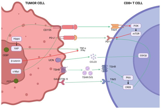
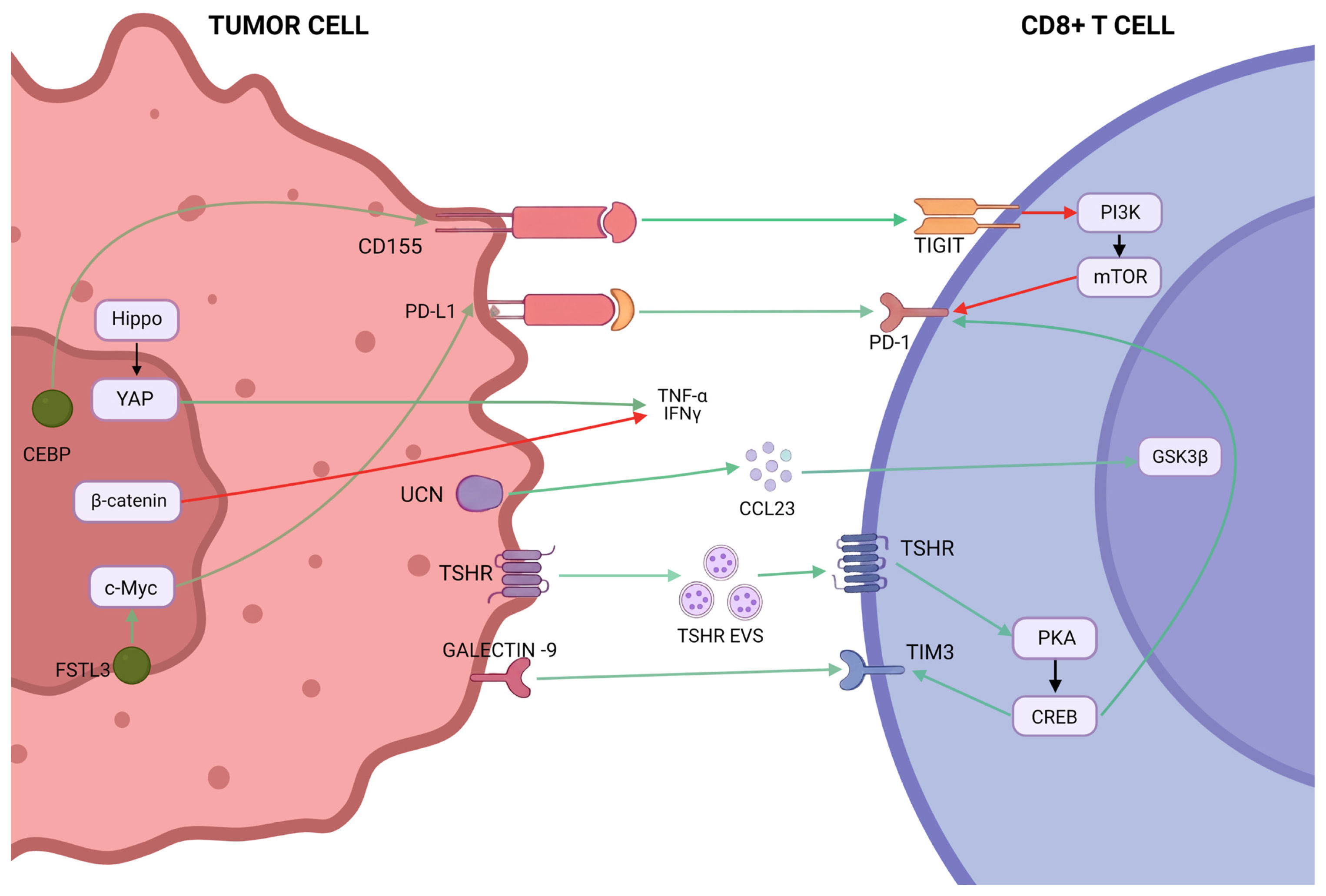
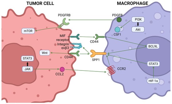
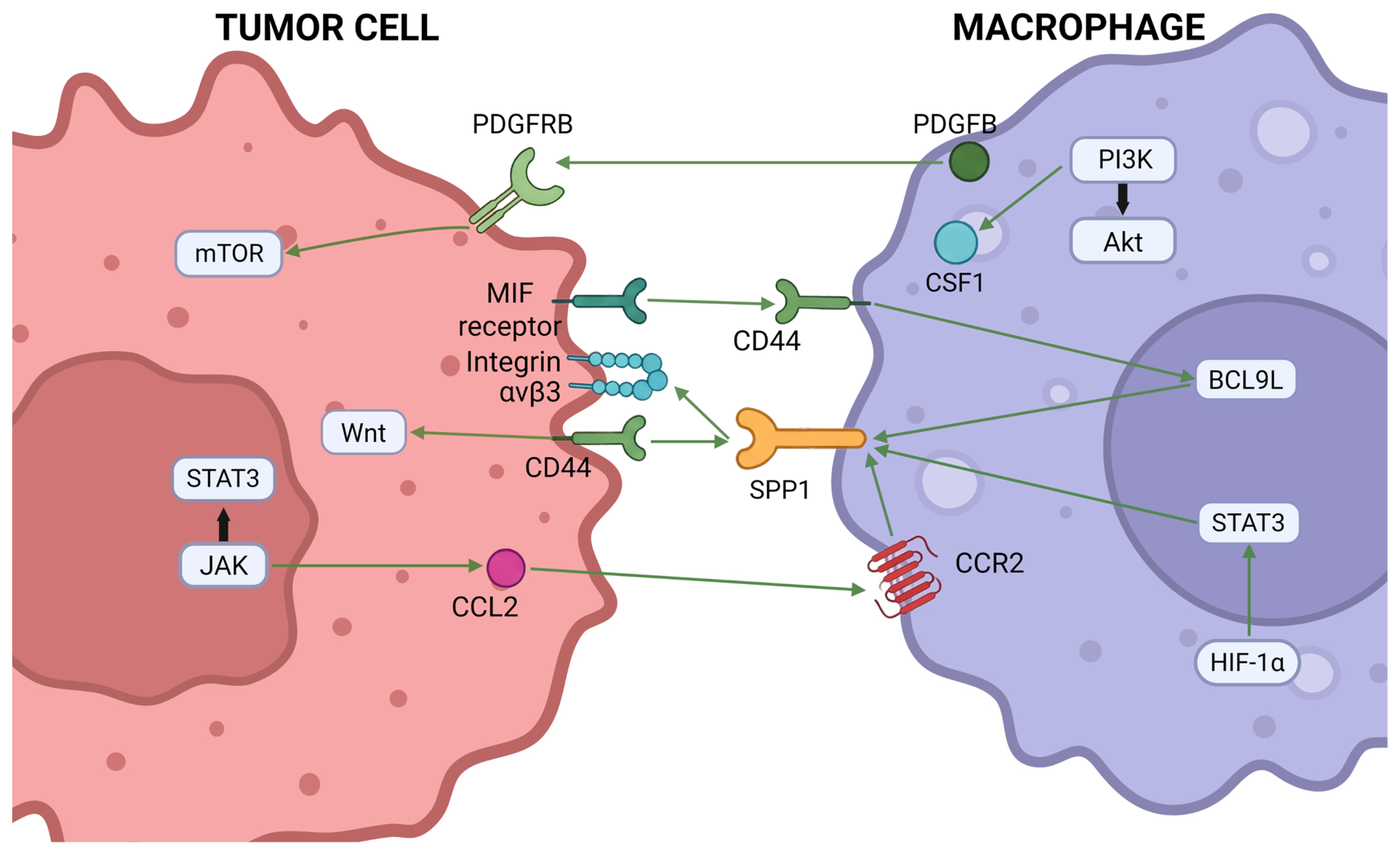
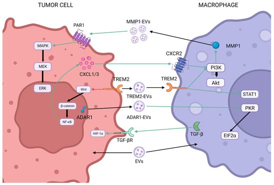
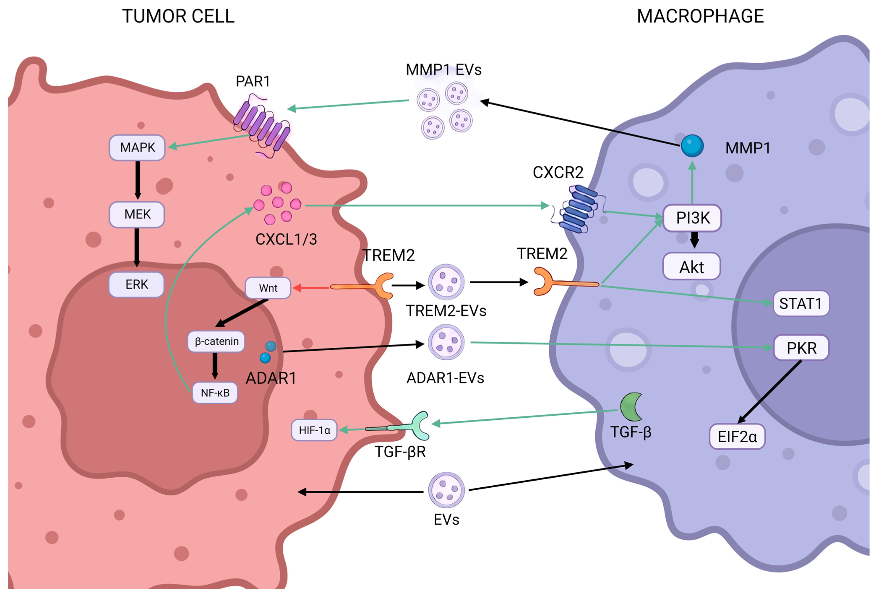

Disclaimer/Publisher’s Note: The statements, opinions and data contained in all publications are solely those of the individual author(s) and contributor(s) and not of MDPI and/or the editor(s). MDPI and/or the editor(s) disclaim responsibility for any injury to people or property resulting from any ideas, methods, instructions or products referred to in the content. |
© 2026 by the authors. Licensee MDPI, Basel, Switzerland. This article is an open access article distributed under the terms and conditions of the Creative Commons Attribution (CC BY) license.