


Abstract
The peripheral nerves that innervate solid tumors communicate with the malignant cells through a restricted set of molecular signaling systems: adrenergic and muscarinic receptors, sensory neuropeptides, neurotrophins and their tyrosine kinase receptors, axon guidance molecules, and nociceptor ion channels. Each has been proposed separately as a therapeutic target but never quantitatively compared with the others. The question is whether one of them dominates—or whether the real target is the axis as a system. We systematically searched PubMed, Scopus, and Web of Science, with four query modules per database, without year or language restriction. We included in vivo studies in mammals, on extracranial solid cancers, with an intervention that modulated tumor innervation, peripheral nerve activity, or neural signaling, and with at least one tumor burden outcome. Of 16,649 unique records, 362 were included, with 1291 experiments and 1437 effects. Blockade of the peripheral nerve–tumor axis reduced tumor burden by −44.8%. In the prespecified primary analysis, on established tumors, the reduction was −52.0% with no significant difference from preventive interventions (p = 0.17). All eight molecular signaling systems responded to blockade, with estimates ranging from −34.3% for TRP channels to −70.2% for glutamatergic signaling. Interventions that activate the axis produced the opposite effect, with a 52.1% increase (95% CI: 32.1 to 75.1) across 253 effects, and bidirectionality was demonstrated within the same molecular system for the adrenergic compartment (−42.8% for blockade vs. 58.1% for activation), axon guidance molecules, and TRP channels. Egger’s test indicated marked small-study effects (slope −1.12; p < 0.001); the estimate remained stable when low-confidence digitized values were excluded (−42.6%). Eight distinct molecular signaling systems, disrupted by methods with nothing in common, produce reductions of a similar magnitude in tumor burden, and stimulation of three of them produces the opposite effect.
1. Introduction
The idea that solid tumors build their own innervation and that this innervation serves their growth has moved from histological observation to a therapeutic program in the last fifteen years. Intratumoral nerve fiber density correlates with aggressiveness and survival in prostate, pancreatic, stomach, and colorectal cancers [1], and tumor axonogenesis is an active process, dependent on neurotrophins secreted by malignant cells and stromal cells [1,2]. The mechanisms described so far cover all three compartments of the peripheral nervous system. Sympathetic fibers, through β-adrenergic receptors, promote angiogenic switching and immunosuppressive remodeling of the microenvironment [3,4,5]. Parasympathetic fibers, through muscarinic receptors, support the expansion of tumor stem cells in the stomach and colon [6,7,8]. Sensory fibers release neuropeptides—calcitonin gene-related peptide (CGRP), substance P, and vasoactive intestinal peptide (VIP)—that modulate both proliferation and lymphocyte infiltration [9,10,11,12,13]. Perineural invasion, long considered a prognostic marker, has been shown to be a bidirectional interaction, in which tumor cells and Schwann cells reprogram each other [14,15,16,17]. The clinical correlates of this axis have also been documented in human cohorts. In colorectal cancer, nerve fiber density and synaptophysin expression are associated with survival [18,19], and the expression of the sensory peptides—substance P and CGRP, along with their calcitonin receptor-like receptor (CRLR)—varies systematically with tumor stage [20,21]. In gastric cancer, tissue concentrations of adrenaline and noradrenaline and markers of sympathetic activation are independent negative prognostic factors [22,23], and β2-adrenergic receptors are differentially expressed in the enteric nerve plexuses adjacent to the tumor [24]. Heart rate variability at diagnosis, as an integrative measure of autonomic tone, correlates with survival in colorectal cancer [25], suggesting that the nervous influence on tumor progression is measurable at a systemic level, not just locally [26].
However, the preclinical evidence has been uneven. Most studies apply neural intervention before tumor inoculation or immediately after, i.e., in a window of prevention that does not correspond to the clinical situation, in which the patient presents with an already established tumor. A second reason for caution is the direction of intervention: some of the literature increases neural signaling—through chronic stress, exogenous catecholamines, or agonists—and it is not clear that these studies can be treated as mirror images of blockade. Finally, the effect sizes come from very different models, species, and outcomes, making it difficult for a narrative synthesis to distinguish signals from noise.
Quantitative syntheses in the field are few and limited to either a single class of interventions or a single tumor type. No meta-analysis has yet addressed the peripheral nerve–tumor axis as a whole, with an explicit taxonomy of the level of intervention and the timing of its application, and none has used a model that respects the real hierarchical structure of preclinical data, where one article contributes multiple experiments and one experiment multiple effects [27,28,29,30].
This systematic review and multilevel meta-analysis aims to answer four questions. First: Does blocking the peripheral nerve–tumor axis reduce tumor burden in in vivo models of extracranial solid cancers? Second: Is the effect maintained when the intervention is applied to an already established tumor, i.e., within the clinically relevant therapeutic window? Third: Does the effect depend on the targeted fiber, the molecular level of the intervention, or the tumor type? Fourth: Do interventions that increase neural signaling produce the inversely symmetrical effect?
The conceptual model of the peripheral nerve–tumor axis, with the signaling systems involved and the points at which each was disrupted or stimulated in the included studies, is shown in Figure 1. It serves as a reading frame for the analysis that follows, in which the eight systems are quantified in turn.
Figure 1.
The peripheral nerve–tumor axis and the eight molecular signaling systems targeted in this meta-analysis. Sympathetic, parasympathetic and sensory fibers innervate solid tumors and act on the malignant cell through distinct receptor families: sympathetic terminals release norepinephrine (NE) onto β2- and β3-adrenergic receptors (ADRB2, ADRB3); parasympathetic terminals release acetylcholine (ACh) onto M1 and M3 muscarinic receptors (CHRM1, CHRM3); and sensory nociceptors release calcitonin gene-related peptide (CGRP) and substance P (SP) and signal through transient receptor potential vanilloid 1 (TRPV1). Neural input promotes proliferation, angiogenesis, invasion, metastasis, immune evasion and further axonal ingrowth (neo-neurogenesis). The tumor in turn recruits axons by secreting the neurotrophins nerve growth factor (NGF), brain-derived neurotrophic factor (BDNF), glial cell-line-derived neurotrophic factor (GDNF) and artemin, and by releasing exosomes (dashed arrow), establishing a self-reinforcing loop. Signals passed from the innervated tumor to the immune compartment—CGRP, NE and mediators of nerve injury, including activating transcription factor 3 (ATF3), type I interferon (IFN-I) and interleukin-6 (IL-6)—shape an immunosuppressive microenvironment characterized by exhausted CD8-positive T cells, regulatory T cells (Treg), myeloid-derived suppressor cells (MDSC) and M2-polarized macrophages, with inhibition of tertiary lymphoid structures (TLSs) and fibrotic immune exclusion, which contributes to resistance to anti-programmed cell death protein 1 (anti-PD-1) and anti-programmed death-ligand 1 (anti-PD-L1) therapy. The lower panel lists the eight molecular signaling systems into which every experiment was classified post hoc, independently of the method used, together with representative interventions applied to interrupt each system in the included studies.
2. Materials and Methods
2.1. Protocol and Registration
The review was conducted according to the PRISMA 2020 (Preferred Reporting Items for Systematic Reviews and Meta-Analyses) statement [31] and reported according to the PRISMA-S (PRISMA for Searching) extension for search strategies [32]. The protocol was drafted according to the PRISMA-P (PRISMA for Protocols) recommendations [33] and structured for registration in PROSPERO4animals (International Prospective Register of Systematic Reviews—extension for animal studies), the registry dedicated to preclinical syntheses [34]. Because the PROSPERO4animals guidance does not accept the registration of reviews in which data extraction has already begun, the protocol was deposited with the Open Science Framework as a time-stamped registration with a persistent identifier: https://doi.org/10.17605/OSF.IO/DZH4J (deposited on 31 August 2026). The deposit is retrospective with respect to the conduct of the review and is explicitly declared as such in the registration: the document deposited is the protocol drafted before data extraction, together with the seven amendments adopted after extraction and before any statistical analysis. Nothing in the deposited protocol was revised in the light of the results. Deviations from the protocol as originally written—three databases interrogated instead of the five planned, a fourth query module added to the three foreseen, the absence of manual reference-list checking, and assessment of screening reliability for 240 rather than 200 records—are declared in full in the registration; none was adopted in response to observed results.
The protocol was amended in seven points, after the completion of data extraction and before any statistical analysis. The timing of the amendment is explicitly stated, and the amendments are reported in full in Supplementary Material S1. They concern: the extension of the primary outcome hierarchy by a fifth level (extent of perineural invasion measured continuously); maintaining the exclusion of pulmonary retention models after intravenous injection; conditional inclusion of interventions on central neurons, only with a demonstrated peripheral sympathetic link; exclusion in principle of tumors of the nerve itself; separate analysis of axis activation studies; the multiple report deduplication rule; and continuity correction for zero-mean effects.
2.2. Eligibility Criteria
Screening was managed with tools dedicated to systematic syntheses [35], and methodological filters for identifying animal studies were adapted from the specialized literature [36,37]. The general methodology of the preclinical study syntheses followed the established recommendations [38,39].
We included studies that simultaneously fulfilled ten conditions: in vivo experiment in mammals; extracranial solid tumor; an intervention on tumor innervation, peripheral nerve activity, or neural signaling; justification of the intervention as targeting the nerve–tumor axis, or measurement of innervation, neuronal activity, or local neurotransmitter concentration; concurrent control group; at least one tumor burden outcome; other than an NTRK inhibitor administered for an oncogenic fusion; objective other than exclusively analgesia or neuroprotection; original study; and sufficient reporting for extraction.
Primary outcome hierarchy, in order of preference: tumor mass at sacrifice; tumor volume at the last time point common to all groups; histological tumor area; bioluminescent or imaging signal; and—by amendment 1—extent of perineural invasion measured continuously, as length of invasion along the nerve, volume, diameter, or area of the invaded nerve, or numerical severity score. Binary incidence of perineural invasion, sciatic nerve function score, intratumoral innervation density, and number of invaded nerves per field were not accepted as outcomes.
2.3. Information Sources and Search Strategy
We queried PubMed, Scopus, and Web of Science, each with four complementary search modules: a main module focused on tumor innervation, a module on neurotransmitters and receptors, a module on neurotrophins and axon guidance molecules, and a safety-net module built after testing with benchmark articles. We did not impose restrictions on year, language, or publication type at the query level. The syntax of each query, with the run date and number of records returned, is given in Supplementary Material S2.
The strategy was validated by testing with known articles: a set of benchmark papers in the field was checked for retrieval, and the modules were revised until the only missing benchmark article remained. The overlap between the databases was measured: Web of Science added 2655 new records out of 9725 returned, i.e., an overlap of 73%, which constitutes a quantitative argument for the saturation of the search.
2.4. Study Selection
Records were deduplicated by normalized digital object identifier (DOI), then by normalized title with a one-year tolerance for the year of publication. We then applied a relevance filter that retained any record containing an oncology term in the title or abstract, or a phrase specific to the nerve–tumor axis, as well as all records without an abstract. The filter was validated on a set of 113 references known to be relevant: none were lost.
Screening on title and abstract was done by applying the ten eligibility questions, stopping at the first negative answer and recording the reason. Reliability was estimated by independent re-screening of a random sample of 240 records, stratified by decision category (80 Include, 80 Uncertain, 80 Exclude). The gross agreement was 69.2% (Cohen’s kappa 0.54), and the agreement on the retained-versus-excluded decision was 75.0%. None of the 80 exclusions checked were reclassified as eligible. Discordant records were adjudicated by two authors. The complete selection process, with the number of records retained and excluded at each stage, is summarized in the PRISMA 2020 flow diagram (Figure 2).
Figure 2.
PRISMA 2020 flow diagram. Numbers for each box, together with the full breakdown of exclusion reasons, are given in Supplementary Table S7.
2.5. Data Extraction
Each article was divided into experiments, defined as a unique combination of tumor model × intervention, and each experiment into effects, defined as an intervention-versus-control comparison for the primary outcome. At multiple doses, each dose generated an effect, with the control being marked as common. At multiple measurement times, we retained a single effect, at the last time point common to all groups.
For each effect we extracted, per group, the mean, the measure of dispersion, and the number of animals. The order of sources was: text, tables, supplementary material, figures. Where the article reported the standard error of the mean (SEM), the conversion to the standard deviation (SD) was done using the equation SD = SEM × √n. Where the supplementary material contained individual values for each animal, the mean and standard deviation were recalculated from the raw data, and the mean was the exact result.
Digitization of graphic panels. For effects whose values were only available in the figures, extraction was done in a separate step, after the textual extraction was completed, on the 200 dpi rendered pages of the original article, with cropping to 600 dpi where the error bar was not legible at the page resolution. For each panel, the extreme values and ordinate axis graduations and the scale type—linear, logarithmic, or dashed—were first identified, and the values were obtained by interpolation corresponding to the scale type. The mean was read as the bar height or the position of the mean marker, and the dispersion as the distance from the mean to the upper end of the error bar. For panels without error bars but with individual points visible, the standard deviation was estimated from the scatter of the points. Each value was given a three-level confidence score at the time of reading—high (clear axis, clear bar ends), medium (interpolation between sparse gradations, broken scale, partially obscured bar ends), and low (estimated dispersion from points, logarithmic axis without intermediate gradations, crowded panel)—and the score was subsequently used in a prespecified sensitivity analysis. When the unit in the figure differed from that recorded in the extraction sheet, the value was converted or, if not convertible, reported in the unit of the figure, which does not affect the ratio of means.
Imputation of dispersion. For experimental arms for which neither the figure nor the text provided any measure of dispersion nor individual points, the standard deviation was imputed from the median coefficient of variation of effects at the same level of the outcome hierarchy. Effects containing at least one arm with imputed dispersion were flagged, and a prespecified sensitivity analysis was run with those effects excluded.
Where an article reported medians and quartiles instead of means, conversion was done with validated estimators for the respective sample size [40,41,42].
For each experiment, we coded: the nerve fiber targeted; the level of intervention; the time of application relative to tumor stage (T0 before inoculation, T1 after inoculation but before measurable tumor, T2 after established tumor, T3 after documented progression); neural target engagement (confirmed, probable, unconfirmed); host immune status; model type; route of administration; and direction of intervention (blockade or activation).
Note on the Team’s Methodological Experience
The extraction sheet and the coding rules for the quantified parameters were built based on the team’s methodological experience in the evaluation of tissue and systemic markers in other pathological contexts: immunohistochemical quantification of lymphocyte subpopulations and vascular endothelial growth factor A (VEGF-A) in acute pancreatitis [43], the role of skin and muscle biopsy in the diagnosis of connective tissue diseases [44], the evaluation of retinal microvascular parameters by optical coherence tomography (OCT) angiography as a marker of neurovascular damage [45], as well as the electrocardiographic and echocardiographic characterization of cardiac phenotypes in silicosis [46], in left ventricular noncompaction [47] and in infective endocarditis [48]. These papers do not address the nerve–tumor axis and do not contribute data to the synthesis; they are cited exclusively for the procedures for quantifying and evaluating autonomic and cardiovascular parameters that we reused in the construction of the extraction sheet.
2.6. Risk of Bias
The risk of bias was assessed using the SYRCLE tool, on its ten domains [49], and the quality of reporting was assessed through the ARRIVE 2.0 (Animal Research: Reporting of In Vivo Experiments) recommendations [50,51]. The assessment of the level of confidence in the evidence followed the preclinical adaptation of the GRADE (Grading of Recommendations, Assessment, Development and Evaluation) approach [52,53].
2.7. Effect Size and Statistical Model
The effect size was the logarithmic ratio of the means, lnRR = ln(intervention_mean/control_mean), with variance:
The choice and implementation of lnRR follows the method proposed for meta-analysis of response reports [54].
Results are expressed as percent change in tumor burden, 100 × (elnRR − 1). The choice of lnRR instead of the standardized mean difference is motivated by the fact that outcomes are strictly positive ratio measures, measured in different units across studies, and lnRR is unit-invariant and directly clinically interpretable.
The synthesis was done using a three-level random-effects model: effects are nested within experiments, and experiments within articles. The model was estimated by restricted maximum likelihood (REML) in Stata 19, with the command meta meregress || art: || expr:. This structure is necessary because an article typically contributes multiple experiments, and an experiment multiple effects; ignoring nesting would underestimate the standard errors [27,28]. Heterogeneity was quantified and decomposed across the two levels [55,56] and reported as total and component I2; interpretation of random-effects estimates and the need for prediction intervals followed established recommendations [57,58].
As a robustness check, we also ran a robust variance estimate, which does not assume a particular correlation structure between effects of the same article [59,60,61]. Reuse of control groups in multiple dose comparisons was handled by explicitly marking the common control [29,30].
The prespecified primary analysis included only effects with T2 or T3 and outcomes from levels 1–5 of the hierarchy, from the blockade studies. Axis activation studies were analyzed separately, without sign reversal, according to amendment 5.
Classification by molecular signaling systems (post hoc). The prespecified taxonomy encodes the method by which the axis is disrupted, not the molecular system engaged: a chemical sympathectomy and a β-blocker enter at different levels, although they reach the same noradrenergic pathway. After the prespecified analyses were completed, each experiment was therefore reclassified, independently of method, according to the signaling system that the intervention interrupts or the activation of adrenergic, cholinergic, or sensory neuropeptides; neurotrophins and Trk receptors; axon guidance molecules; nociceptor TRP channels; neuroimmune mediators; or glutamatergic and mixed unspecified agents, for interventions in which the nerve is manipulated without a defined molecular target. Each experiment received a single label, assigned according to a pre-established order of priority, and when the article explicitly stated the target, that was retained. The classification was made on the full text of the intervention description, without access to the effect size. Results across systems are reported as exploratory analyses, distinct from prespecified analyses, and p values are not corrected for multiplicity.
Moderation was tested by meta-regression in the same three-level model for: time of intervention, nerve fiber, intervention level, and target engagement.
Small-study effects were assessed by Egger’s test [62], adapted as a meta-regression on the standard error within the three-level model, and visually by funnel plots with significance contours, interpreted according to recommendations for asymmetry [63,64,65]. Publication bias and low statistical power are well-documented problems in the preclinical literature [66,67]. Robustness was tested by successive exclusion of each article.
For effects with a zero mean in one arm, where lnRR is not defined, the continuity correction prespecified by Amendment 7 was applied: adding to both arms half of the smallest non-zero value observed in that experiment, with sensitivity analysis without the corrected effects. In the final base, no effects required this correction, as all control and intervention means were strictly positive.
The data, the full Stata code, and the digitization files are available as Supplementary Materials.
2.8. Use of Generative Artificial Intelligence Tools
In carrying out this synthesis, we used an assistant based on a large-scale linguistic model (Claude, Anthropic; Claude Opus 5 version, accessed through the Claude application, 2026) in the following stages, declared here in full:
- (i)
- Deduplication and screening of records—normalization of identifiers and titles, construction of the screening database and application of eligibility criteria in two independent passes, disagreements being adjudicated by the authors;
- (ii)
- Data extraction—transfer of values from the text, tables and Supplementary Materials into the structured extraction sheet;
- (iii)
- Digitization of graphic panels, for effects whose numerical values were not reported individually in the text or tables: identification of the ordinate axis gradations and the scale type, then interpolation of the mean and dispersion, according to the procedure described in Section 2.5;
- (iv)
- Post hoc classification by molecular signaling systems, performed on the intervention description, without access to the effect size, and subsequently verified by the author;
- (v)
- Writing the analysis code in Stata and numerically verifying it by an independent implementation;
- (vi)
- Drafting, linguistic editing, and translation of the text, including the English translation of figures, tables, and Supplementary Materials.
No results were generated by the model: all estimates come from running the code on the extracted dataset, provided in full in the Supplementary Materials. Each digitized value was assigned a three-step confidence score at the time of reading, and the robustness of these values was tested by two sensitivity analyses—exclusion of values with low confidence and imputed dispersion (−42.6%, 764 effects) and restriction to values reported directly in the text or tables (−38.5%, 125 effects)—both consistent with the main estimate of −44.8%. The authors reviewed and edited the entire output and assume full responsibility for the content of this publication.
3. Results
3.1. Study Selection
The three databases returned, through the four search modules, a total that after deduplication left 16,649 unique records (Scopus 8466, PubMed 5528, and Web of Science 2655). The validated relevance filter eliminated 5918 records, leaving 10,731 abstracts for evaluation. Screening excluded 9893 records; the dominant reasons were the absence of a tumor outcome (3729), the type of publication—syntheses and review articles (2310)—and exclusively in vitro studies (1301).
Thus, we had 838 articles to search in full text, of which 387 could not be obtained. The 451 articles obtained were assessed for eligibility; 89 were excluded from full-text analysis: 60 because they did not report any outcome from the prespecified hierarchy, 10 because they did not contain any intervention that directly modulated tumor innervation or peripheral neural signaling, nine for the pulmonary retention model after intravenous injection, three for intervention on central neurons without demonstrated peripheral sympathetic link, one as a duplicate publication of an already included study, one excluded at screening re-adjudication, two because the tumor studied was a tumor of the nerve itself, and three because they had been withdrawn. The reason for each exclusion is reported individually in Supplementary Material S7. A total of 362 eligible articles remained. For 11 of them, the full text could not be obtained even after evaluation—the available file contained no more than the abstract—so eligibility was established based on the abstract, but no effect could be extracted. The 351 articles actually extracted provided 1291 experiments, of which 1270 had at least one usable effect, and 1437 effects in total (Figure 2).
3.2. Characteristics of Included Studies
The 1437 effects are distributed across tumor types, intervention levels, and nerve fibers, as shown in the evidence map (Figure 3). The timing of intervention is strongly unbalanced: of the 1107 effects in the model, 509 come from interventions applied before tumor inoculation (T0), 250 from the immediate post-inoculation window (T1), and 268 from interventions on already established tumors (T2 or T3). In other words, the preclinical literature in this field predominantly tests prevention, not treatment.
Figure 3.
Evidence map: number of extracted effects by tumor location and level of intervention. Bubble area is proportional to the number of effects. Brown, interventions that activate the axis.
The reported variance was the standard error of the mean in most cases, which necessitated systematic conversion to standard deviation; a significant number of effects reported no dispersion at all, and in the older literature we found the group size to be frequently missing.
The complete distribution of evidence across the three classifications—study arm, nerve fiber, molecular system, timing, immune status, route of administration, target engagement, and data source—is given in Table 1.
Table 1.
Characteristics of the included evidence.
3.3. The Global Effect of Axis Blocking
Of the 1437 extracted effects, 160 had their mean, variance and power reported directly in the text, table or source data file. The remaining values were obtained by digitizing the graphic panels, with calibration on the Y-axis of each panel and cropping at increased resolution where the error bar was not legible at the page resolution; each digitized value was given a confidence score. This resulted in 1107 effects from 986 experiments and 297 articles entering the model—552 with high confidence, 298 with medium confidence and 97 with low confidence. The effects left out of the model are those with outcomes outside the prespecified hierarchy, those whose panels were in supplementary materials that we could not obtain, and those with numbers below the calculation threshold.
For 26 experimental arms, the figure did not contain any error bars or individual points from which to estimate dispersion; for these, the standard deviation was imputed from the median coefficient of variation of effects at the same level of the outcome hierarchy. The sensitivity analysis excluding them is reported in Section 3.10.
Blocking the peripheral nerve–tumor axis reduced tumor burden by −44.8% (95% confidence interval, 95% CI: −48.3 to −40.9), on 854 effects from 754 experiments and 266 articles (p < 0.001). Heterogeneity was high, I2 = 97%, distributed between the article level (τ2 = 0.161) and the experiment level (τ2 = 0.242), which justifies the three-level structure.
3.4. Effect on Molecular Signaling Systems
The six levels of intervention describe the method by which the axis is interrupted—denervation, silencing, and pharmacological blockade. However, they do not say which signaling system was engaged: a chemical sympathectomy and a β-blocker use different methods but reach the same noradrenergic pathway. We therefore classified, post hoc, each of the 1291 experiments according to the molecular system that the intervention interrupts or activates, regardless of the method. The classification is exploratory and does not appear in the protocol; we report it as such.
The results are shown in Figure 4 and Table 2. All eight systems respond to blockade, with estimates ranging from −34.3% (95% CI: −44.9 to −21.6) for transient receptor potential (TRP) channels to −70.2% (95% CI: −85.7 to −37.8) for glutamatergic signaling. Seven of the eight fall within a band of approximately fifteen percentage points, between −34% and −49%; the only exception is the glutamatergic compartment. Meta-regression with the adrenergic system as the reference reveals no significant differences between them except for the glutamatergic compartment (p = 0.014), but it was estimated on 17 effects from five articles—too few to support a claim of superiority.
Figure 4.
Effect by molecular signaling system, blockade versus activation. Post hoc classification of every experiment by the signaling system engaged; blockade and activation were fitted as separate three-level models, without sign inversion. Squares, blockade; circles, activation. Bars are 95% confidence intervals.
Table 2.
Pooled effect by molecular signaling system, blockade versus activation.
3.4.1. Adrenergic System
This is the most well-represented system: 215 effects from 80 articles for blockade and 106 effects from 38 articles for activation. Blockade reduces tumor burden by −42.8% (95% CI: −50.1 to −34.4), and activation increases it by 58.1% (95% CI: 22.1 to 104.7) (p < 0.001). It is the only pair in the entire analysis in which both directions are estimated on hundreds of effects, and the contrast constitutes the strongest internal evidence of the paper.
Sympathetic fibers act predominantly through β-adrenergic receptors. β2 signaling promotes angiogenic switching, metabolic remodeling, and the expansion of myeloid-derived suppressor cells [5,68,69,70,71], and β3 receptors contribute to the organization of the microenvironment [72,73]. Catecholamines stimulate angiogenesis and lymphangiogenesis [74,75,76,77,78] and induce depletion of cytotoxic T lymphocytes [79,80].
Chemical or surgical sympathectomy reduces tumor growth in most models [81,82,83,84,85,86], although negative or contextual results have also been reported [87,88,89]. The sympathetic component also contributes to premetastatic niche formation and bone metastasis [90,91,92]. Innervation dependence has been most clearly documented in the prostate [93,94,95,96] and integrated into a self-sustaining adrenergic–neurotrophin loop [97,98].
3.4.2. Cholinergic System
Blockade of cholinergic signaling—vagotomy, muscarinic antagonists, deletion of the muscarinic M3 receptor gene (cholinergic muscarinic receptor 3, CHRM3)—reduces tumor burden by −41.4% (95% CI: −54.7 to −24.3) across 86 effects from 30 articles, a magnitude virtually identical to that of adrenergic blockade. Activation, however, does not produce the opposite effect: the estimate is −12.3% (95% CI: −45.8 to 41.9), nonsignificant (p = 0.593) and with a confidence interval covering both a substantial decrease and an increase. On 18 effects, the correct conclusion is not that cholinergic activation is protective, but that we have no evidence that it is a promoter—the only system in the analysis for which we can say this.
Muscarinic signaling supports gastric and intestinal tumorigenesis [6,7,8,99,100,101,102,103], and vagotomy modifies gastric tumor progression in both animal models and human cohorts [104,105,106,107,108,109,110]. Non-neuronal acetylcholine, produced by tumor or immune cells, adds an autocrine loop [111,112,113,114]. The cholinergic anti-inflammatory pathway links vagal tone to immunosurveillance [115,116,117,118,119,120].
3.4.3. Sensory Neuropeptides
Blockade of neuropeptides released by sensory fibers—CGRP and its receptor activity-modifying protein 1 (RAMP1), substance P, neurokinin 1 receptor (NK1R), and VIP—resulted in a −40.2% (95% CI: −48.5 to −30.6) reduction in 87 effects from 35 articles. Activation, on 16 effects, gives 15.9% (95% CI: −25.3 to 80.0), without statistical significance (p = 0.510).
Sensory neurons release CGRP, substance P, VIP, galanin, neuromedin U and pituitary adenylate cyclase-activating polypeptide (PACAP) into the tumor microenvironment [9,10,11,12,121,122,123,124,125,126,127,128,129,130,131,132,133]. Pharmacological blockade of these pathways—with anti-CGRP antibodies, NK1R antagonists or capsaicin—reduces tumor growth in several models [134,135,136,137].
However, the role of nociceptors is ambivalent: the same neurons can suppress immunosurveillance or, on the contrary, support it, depending on the model and the subpopulation targeted [138,139,140,141,142,143,144,145,146,147,148]. This ambivalence explains the extreme heterogeneity observed in the sensitive subgroup of the meta-analysis.
3.4.4. Neurotrophins and Trk Receptors
Blockade of neurotrophins and their receptors produces one of the largest reductions in the series, −47.4% (95% CI: −58.2 to −33.7) across 81 effects from 34 articles. Exogenous administration of neurotrophins goes in the opposite direction, with a large point estimate—83.8% (95% CI: −0.2 to 238.7)—but it is imprecise and borderline significant (p = 0.051, 29 effects). The direction is consistent with blockade; the magnitude cannot be stated.
Tumor axonogenesis is driven by the neurotrophins nerve growth factor (NGF), brain-derived neurotrophic factor (BDNF), glial cell line-derived neurotrophic factor (GDNF), and artemin, through the tyrosine kinase receptors TrkA and TrkB (tropomyosin receptor kinase A and B, respectively), RET (rearranged during transfection), and the neurotrophin receptor p75 (p75NTR) [149,150,151,152,153,154,155,156,157,158,159,160,161,162,163,164]. Classical axon guidance molecules—semaphorins, netrins, ephrins, and SLIT–ROBO (slit guidance ligand–roundabout receptor)—are reused by the tumor to organize its innervation [165,166,167,168,169,170,171,172,173]. Tumor extracellular vesicles carry signals that reprogram neurons at a distance [174,175,176,177,178,179,180,181,182].
Clinically approved TRK inhibitors act on the same family of receptors, but in the indication of oncogenic fusions of the NTRK (neurotrophic tyrosine receptor kinase) genes, which is why they were excluded from the present synthesis [183,184].
3.4.5. Axonal Guidance Molecules
Interference with semaphorins, netrins, ephrins and the SLIT–ROBO axis reduces tumor burden by −48.2% (95% CI: −61.9 to −29.6) on 33 effects, and overexpression or administration of these molecules increases it by 69.5% (95% CI: 33.4 to 115.3) (p < 0.001). It is the second bidirectional pair with both arms significant, although on much smaller volumes than the adrenergic one.
3.4.6. TRP Channels of the Nociceptor
Ablation or blockade of TRPV1+ (transient receptor potential vanilloid 1) and TRPA1+ (transient receptor potential ankyrin 1) neurons—by capsaicin, resiniferatoxin, or genetic models—yields −34.3% (95% CI: −44.9 to −21.6) on 108 effects, the lowest magnitude of the series. Activation of the same channels increases tumor burden by 60.9% (95% CI: 4.3 to 148.2) (p = 0.031). Bidirectionality is also verified here, but the amplitude of the blockade is noticeably smaller than in the other systems, which is consistent with the functional ambivalence of nociceptors described below.
3.4.7. Neuro-Immune Mediators
Interventions targeting neuro-immune mediators yielded −45.7% (95% CI: −53.3 to −36.9) on 64 effects from 23 articles. There are not enough activation studies in this category for a separate estimate.
Nerve fibers shape the composition of the immune infiltrate, the formation of tertiary lymphoid structures, and the response to checkpoint blockade [185,186,187,188,189,190,191,192,193,194,195,196,197,198]. This interface explains why the effect of axis blockade is maintained in immunodeficient hosts, but with different magnitudes.
3.4.8. Perineural Invasion and the Perineural Niche
Perineural invasion is not a separate signaling system, but an anatomical context in which several of the above systems act simultaneously. The effects that measure it continuously have entered the analysis at level 5 of the outcome hierarchy, through amendment 1, and are distributed between the neurotrophic, adrenergic and neuropeptide systems.
Perineural invasion is documented as a prognostic factor in pancreatic, gastric, esophageal, biliary, periampullary, oral, rectal, colorectal and renal cancers [15,16,17,199,200,201,202,203,204,205,206,207,208,209,210]. The central mechanism is the dialogue between the tumor cell and the Schwann cell, which dedifferentiates and guides tumor migration along the nerve [14,211,212,213,214,215,216,217]. Enteric glia play an analogous role in colorectal cancer [218,219,220,221,222,223].
3.5. Effect by Intervention Level
A complementary classification, by the method by which the axis is disrupted, is shown in Figure 5 and Table 3. All six levels produce significant reductions. The largest effects are for blockade of neurotrophins and axonogenesis (−50.1% (95% CI: −57.1 to −42.1), 151 effects) and blockade of the receptor or neurotransmitter (−49.2% (95% CI: −57.0 to −39.9), 178 effects). Structural denervation, the most frequently studied approach, yields −43.8% (95% CI: −50.3 to −36.5) on 231 effects, neuropeptide blockade −42.7% (95% CI: −49.9 to −34.4), neuro-immune modulation −39.6% (95% CI: −51.3 to −25.1), and functional silencing −33.7% (95% CI: −41.4 to −25.0)—the smallest effect of the series, but significant (p < 0.001).
Figure 5.
Effect of blocking the peripheral nerve–tumor axis, by level of intervention. Three-level random-effects model (effect/experiment/article), REML, log response ratio. Marker area is proportional to the number of effects.
Table 3.
Prespecified analyses.
The convergence of the two classifications is itself a result: whether we group the studies by the molecular system employed or by the method of interruption, the magnitudes converge in the same band. The common target is not a molecule or a technique, but the axis as a signaling system.
3.6. Primary Analysis: Established Tumor Intervention
Restricting to effects at time point T2 or T3—intervention applied to an already measurable tumor, i.e., the clinically relevant window—retained 221 effects from 173 trials and 81 articles. The reduction in tumor burden was −52.0% (95% CI: −58.4 to −44.6) (p < 0.001), with I2 = 98%.
In comparison, interventions applied before tumor inoculation yielded −45.4% (95% CI: −50.5 to −39.8), and those in the immediate post-inoculation window yielded −39.2% (95% CI: −44.8 to −33.1) (Figure 6). The moderation test did not reveal a significant difference between the therapeutic and preventive windows (log response ratio, lnRR: −0.091; p = 0.17). Contrary to expectation, the effect in the therapeutic window is not smaller than that in the preventive window, but numerically slightly larger—a result that, although insignificant, does not support the hypothesis that axis blocking would work only prophylactically.
Figure 6.
Timing of the intervention relative to tumor stage. The moderation test compares the therapeutic window (T2/T3) with the preventive window (T0/T1) within the same three-level model.
3.7. Prespecified Subgroup Analyses
Figure 7.
Prespecified subgroup analyses. Blockade studies only, outcomes from hierarchy levels 1–5.
Nerve fibers. The effect was present in all four categories: sympathetic fibers: −42.1% (95% CI: −48.3 to −35.2) (249 effects, I2 = 97%), parasympathetic fibers: −38.8% (95% CI: −52.7 to −20.8), sensory fibers: −37.5% (95% CI: −43.7 to −30.5) (257 effects), and mixed fibers: −52.7% (95% CI: −58.2 to −46.5). Meta-regression with sympathetic fibers as a reference revealed no significant differences between compartments (minimum p = 0.050). The sensory compartment, which in the preliminary analysis of the subset with values reported in the text seemed to be unresponsive, turns out to be as sensitive as the others after completing the database; however, its heterogeneity remains among the highest (I2 = 95%).
Neural target engagement. Studies that actually documented changes in innervation, neuronal activity, or local neurotransmitter concentration reported −46.1% (95% CI: −50.6 to −41.3); those with only probable engagement reported −45.5% (95% CI: −50.7 to −39.7); and those without any target verification reported −34.1% (95% CI: −44.2 to −22.2). The effect is not greater where the target was not verified, but smaller and more imprecise, with the highest heterogeneity of the series (I2 = 98%)—a pattern that does not support the hypothesis that lack of verification would systematically inflate estimates.
Immune status. The effect was present in both immunocompetent hosts (−41.7% (95% CI: −46.4 to −36.4)) and immunodeficient hosts (−49.9% (95% CI: −54.9 to −44.5)), indicating an antitumor component that does not depend exclusively on adaptive immunity.
Route of administration. Local administration produced −38.9% (95% CI: −45.4 to −31.6), systemic administration −43.7% (95% CI: −48.0 to −39.1), and genetic manipulation −52.3% (95% CI: −58.9 to −44.7).
Tumor type (Table 4). The effect was present in all locations with sufficient data. The largest reductions were observed in prostate (−58.7% (95% CI: −71.6 to −39.8)), colorectal (−52.6% (95% CI: −64.1 to −37.5)) and gastric cancers (−53.4% (95% CI: −61.4 to −43.9)), followed by oral cavity (−46.7% (95% CI: −59.2 to −30.4)), pancreas (−43.3% (95% CI: −51.3 to −33.9)), breast (−40.3% (95% CI: −48.7 to −30.5)), melanoma (−39.0% (95% CI: −48.7 to −27.5)) and lung cancers (−38.4% (95% CI: −46.4 to −29.1)). The only location with a non-significant estimate was the liver (−10.9% (95% CI: −40.2 to 32.8), only 16 effects), for which the volume of data was the smallest.
Table 4.
Effect by tumor location.
Host species and sex. The included literature is almost exclusively murine: of the 351 articles that contributed extractable data, 332 (94.6%) used mice, 17 (4.8%) used rats and two (0.6%) used another species. The sex of the host was reported in 256 articles (72.9%)—male only in 105 (29.9%), female only in 97 (27.6%) and both sexes in 54 (15.4%)—and was not reported in 95 (27.1%). Strain and age were specified in the extraction form but were not retained as structured variables. Fitted as a moderator in the same three-level model, host sex was not significant overall (Q = 6.29, df = 3, p = 0.098), although the female-only versus male-only contrast was nominally so: −37.5% (95% CI: −44.2 to −29.9; 226 effects, 72 articles) against −49.0% (95% CI: −55.5 to −41.6; 207 effects, 78 articles). This contrast is confounded with tumor site, because the hormone-dependent models are sex-restricted by construction: 116 of the 151 breast effects come from female-only hosts and all 52 prostate effects from male-only hosts, and these two sites lie at opposite ends of the distribution (breast: −40.3%, 95% CI: −48.7 to −30.5; prostate: −58.7%, 95% CI: −71.6 to −39.8). Excluding breast, prostate and ovarian models and refitting sex on the remaining 634 effects from 205 articles, the association disappeared (Q = 2.34, df = 3, p = 0.505; female versus male p = 0.304). The 21 effects contributed by rats gave −65.5% (95% CI: −81.4 to −35.7) against −44.3% (95% CI: −47.9 to −40.3) for mice, on too few articles to interpret. The full stratification is given in Supplementary Table S11.
3.8. Bidirectionality of the Axis
The 253 effects from interventions that increase neural signaling, from 102 articles, produce an aggregate increase in tumor burden of 52.1% (95% CI: 32.1 to 75.1) (p < 0.001), with maximum heterogeneity (I2 = 98%). The two arms were modeled completely separately, without sign reversal and without sharing any effect.
However, analysis of molecular systems shows that bidirectionality is not uniform, and the correct formulation is more nuanced than the global contrast. This is demonstrated in three systems, in which both arms are significant and of opposite direction: adrenergic, axon guidance and TRP channels. It is suggested but unconfirmed for neurotrophins, where the activation estimate is large but imprecise and stops right at the threshold of significance. And it is absent for sensory neuropeptides and the cholinergic system, where activation produces no detectable increase.
This structure matters for interpretation. If the axis were to function as a linear switch, all systems should behave the same in both directions. They do not. Most likely, the systems differ in how close to saturation they are under basal conditions: a system that is already maximally active cannot be further stimulated, although it can be blocked. The adrenergic system, which responds robustly in both directions, appears to be the furthest from saturation—which is consistent with the fact that basal sympathetic tone in the laboratory animal is experimentally modulated by something as trivial as housing temperature.
3.9. Risk of Bias
The SYRCLE (Systematic Review Centre for Laboratory Animal Experimentation) assessment of the 362 eligible articles shows a pattern characteristic of the preclinical literature (Figure 8, Table 5). The areas related to randomization and allocation concealment are almost completely unclear—94% for allocation sequence, 98% for allocation concealment, and 99% for randomization at assessment—not because the procedures were incorrect, but because the articles are silent about them. Blinding of the outcome assessor is reported in 11% of the articles. Areas with explicit high risk are incomplete outcome data (15%) and the category “other sources of bias” (32%), the latter mainly reflecting pseudoreplication: reporting histological sections or microscopic fields as the experimental unit, instead of animals.
Figure 8.
Risk of bias assessed with the SYRCLE tool across all 362 eligible articles.
Table 5.
Risk of bias (SYRCLE), assessed across all 362 eligible articles.
3.10. Small-Study Effects and Robustness
The sensitivity analyses are summarized in Table 6. The funnel plot (Figure 9) is visibly skewed, and the Egger test adapted to the multilevel model confirms marked small-study effects: slope −1.12 (standard error, ES: 0.14), p < 0.001. Small studies consistently report larger effects than large ones. This pattern is common in the preclinical literature and may reflect both publication bias and true heterogeneity or differences in methodological quality; whatever the cause, caution is warranted in interpreting the aggregate effect size.
Table 6.
Sensitivity analyses and small-study effects.
Figure 9.
Contour-enhanced funnel plot, blockade studies. Shaded wedges mark p < 0.05 and p < 0.01.
Article-level leave-one-out analysis, applied to the primary analysis, produced estimates between −52.7% and −50.7% across the 81 included articles. The estimate remained negative and significant regardless of which articles were excluded, and the overall amplitude of the variation—less than three percentage points—suggests that no single study determines the result.
We repeated the overall analysis, excluding effects with low digitizing confidence and those with imputed standard deviation. The resulting estimate was −42.6% (95% CI: −46.2 to −38.7) on 764 effects, virtually identical to the main estimate, indicating that the result does not depend on reading values under difficult conditions.
3.11. Chronic Stress as a Physiological Activator of the Axis
Chronic stress is the most widely used model of axis activation and accelerates digestive carcinogenesis, tumor progression, and resistance to therapy [3,224,225,226,227,228,229]. The temperature at which animals are housed, through the adrenergic tone it induces, significantly modifies experimental outcomes and resistance to immunotherapy [230,231,232].
3.12. Translational Perspective
The most advanced translational attempt is the repositioning of β-blockers. Preclinical studies and observational cohorts suggest benefit [233,234,235,236,237,238,239,240,241,242], and several therapeutic window studies and associations with immunotherapy have reported promising results [243,244,245,246,247,248,249,250,251,252,253]. However, the results remain discordant between analyses [254,255,256,257,258].
Celiac plexus neurolysis, the only denervation procedure currently used in practice, remains an analgesic procedure with no evidence of antitumor effect [259,260,261,262]. Heart rate variability and resting heart rate have been proposed as accessible autonomic biomarkers [263,264,265].
The current state of the art is summarized in several reference works [266,267,268,269,270,271,272,273,274,275,276,277,278,279,280,281,282,283,284,285,286], and innervation has been characterized in most tumor sites studied [287,288,289,290,291,292,293].
4. Discussion
This review shows that blocking the peripheral nerve–tumor axis reduces tumor burden in in vivo models of extracranial solid cancers by an aggregate magnitude of −44.8%, with an effect that is fully maintained on established tumors (−52.0%), and—the result that interests us most here—with a remarkably consistent magnitude across molecular signaling systems that have nothing in common.
A β-adrenergic antagonist, an anti-NGF antibody, an NK1R antagonist, a capsaicin that ablates TRPV1+ neurons, and interference with SLIT–ROBO signaling act on different receptors, through different transduction pathways, in different cellular compartments. They produce reductions of −42.8%, −48.5%, −40.2%, −34.3%, and −48.2%. This convergence is, in our opinion, the main result of the work.
4.1. Convergence: Eight Molecular Systems, One Effect
The result that we consider most important is a negative one: we did not find a privileged molecular target. Neither classification by signaling system, nor by nerve fiber, nor by intervention method produces a hierarchy. By systems, seven out of eight fall between −34.3% and −48.5%; by fibers, between −37.5% and −53.0%, with a minimum p = 0.050 for moderation; and by intervention levels, between −33.7% and −50.7%.
The only apparent exception is the glutamatergic compartment (−70.2%, p = 0.014 for moderation versus adrenergic), estimated on 17 effects from five articles. We do not build anything on this observation: at this volume, a single influential study can make the difference, and the literature on glutamatergic signaling in extracranial tumors is too recent to support a claim of superiority. We report it as a hypothesis to be tested, not as a result.
Interpreting this convergence requires caution. It does not mean that all molecules are equivalent as clinical targets: the pharmacological feasibility, therapeutic window, and toxicity differ enormously between a β-blocker in use for fifty years and an anti-semaphorin antibody that has never entered the clinic. It means, instead, that the magnitude of the antitumor effect is not the argument that decides between them. If an entire field has been searching for the optimal molecular target of tumor innervation for ten years, the aggregate data say that this search is ill-posed: there is no winner.
The most economical explanation is that the systems do not act in parallel, but in series or through a common node. Neurotrophins recruit axons; axons carry adrenergic, cholinergic, and sensory endings; and these release transmitters and neuropeptides that converge on the same pathways of proliferation, angiogenesis, and immunosuppression. Interruption at any point in the chain gives a similar result because the chain is the same. This is exactly the pattern that a signaling axis produces, not a collection of independent mechanisms.
We explicitly mention that the preliminary analysis, performed on the subset of 125 blockade effects whose values were reported directly in the text, suggested the opposite—an absent effect in the sensory compartment. Completion of the database by digitizing the graphic panels overturned this conclusion. This is a concrete example of the risk of interpreting a subset defined by the reporting method: in this field, studies on sensory fibers are on average more recent and report their results predominantly graphically, so that the subset with text values was systematically depleted in this compartment.
However, heterogeneity remains high in all systems (I2 between 92% and 98%). Convergence of means does not mean homogeneity: within each system, studies continue to contradict each other. This is the second important finding and refers to Section 4.7.
The most likely explanation is that intratumoral sensory neurons have opposing functions depending on the context. Sensory neuropeptides have both direct protumoral effects, through CGRP and substance P [13,294], and cytotoxic lymphocyte recruitment effects; ablation of nociceptors may therefore free the tumor from an immune constraint, not just a growth stimulus [145,147]. The same ambivalence appears in the clinical correlates: the expression of substance P and CGRP in colorectal cancer varies with stage, but not monotonically [20,21].
The practical consequence for drug development is that the choice of target should be made based on pharmacological criteria—already approved molecules, tissue penetration, therapeutic window, and chronic toxicity—and not on the promise of a superior magnitude of preclinical effect. From this perspective, the adrenergic and neuropeptide sensory systems have a considerable practical advantage, because they already have drugs approved in humans, not because they would have a greater effect.
4.2. Bidirectionality, Verified Within the Same Molecular System
Interventions that increase neural signaling produce the opposite effect: a 52.1% increase in tumor burden (95% CI: 32.1 to 75.1), estimated independently on 253 effects from 102 articles, with no sign reversal and without sharing any effect with the blockade arm.
Molecular classification allows a much stricter test than this global contrast. In a comparison between different arms, one can always object that the interventions, models, and laboratories differ. The objection disappears when blockade and activation target the same signaling system. For the adrenergic compartment we have 215 block effects (−42.8% (95% CI: −50.1 to −34.4)) and 106 activating effects (58.1% (95% CI: 22.1 to 104.7), p < 0.001). Same receptor, opposite directions, opposite effects. The pattern is repeated for axon guidance molecules (−48.2% vs. 69.5%) and for TRP channels (−34.3% vs. 60.9%).
However, bidirectionality is not universal, and this should be stated equally clearly. For neurotrophins, activation gives a large but imprecise point estimate, which stops right at the threshold of significance (83.8% (95% CI: −0.2 to 238.7), p = 0.051, 29 effects): the direction is consistent; the magnitude cannot be stated. For the sensory neuropeptides, activation produces no detectable increase (15.9% (95% CI: −25.3 to 80.0), p = 0.510). And for the cholinergic system, the activation estimate is −12.3% (95% CI: −45.8 to 41.9), with a confidence interval covering both a large decrease and a large increase—on 18 effects, this means an absence of evidence that cholinergic activation is promotive, not proof that it is protective. The distinction matters, because the literature on the cholinergic anti-inflammatory pathway and vagal stimulation invites the second interpretation, and our data do not support it.
The symmetry is not perfect even where it exists. On the logarithmic scale—the only one on which the comparison makes sense—the amplitude of the blockade (0.60) exceeds that of the activation (0.42). The presentation in percentages may give the opposite impression, because a 45% reduction and a 52% increase seem comparable when they are not: the percentage change is asymmetric by construction, and a mean ratio of 0.55 and one of 1.52 are not inverses of each other. The heterogeneity in the activation arm is, moreover, the highest in the entire analysis (I2 = 98%). The most likely explanation is that the systems differ in how close to saturation they are under basal conditions: a system that is already maximally active can be blocked but cannot be further stimulated. This would explain why the adrenergic compartment—whose basal tone in the laboratory animal is experimentally modulated by something as trivial as the temperature of accommodation—responds robustly in both directions, while the cholinergic system does not respond to stimulation. Part of the amplitude also comes from the fact that models of chronic stress include systemic effects—glucocorticoid immunosuppression and weight loss—that are not reducible to local neural signaling [3,295].
A methodological argument for future syntheses remains: the direction of the intervention should be treated as a design dimension of the analysis, not as an algebraic sign that allows the aggregation of the two arms. If we had applied the original protocol, which provided for the reversal of the sign, the contrast that today constitutes the strongest internal argument of this synthesis would have disappeared on average.
4.3. The Therapeutic Window Remains Underexplored
The distribution of intervention timings is perhaps the most important descriptive finding of this review. Of the effects entered into the model, 513 come from interventions applied before tumor inoculation and only 268 from interventions on established tumors. The preclinical literature in the field overwhelmingly tests prevention.
This is not a criticism of the individual studies—a constitutional genetic intervention or surgical denervation before inoculation are often the only feasible options to demonstrate the mechanism. However, a problem of representativeness remains: the clinically relevant window is documented by about a quarter of the available material. The actual result is, however, encouraging. The effect in the therapeutic window (−52.0%) is not smaller than that in the preventive window (−45.7%), but numerically slightly larger, and the moderation test does not detect a difference (p = 0.17). In other words, the data do not support the hypothesis—plausible a priori—that axis blockade would only work as long as tumor innervation has not been established.
4.4. Clinical Relevance and Converging Human Evidence
The preclinical results summarized here are consistent with observations from human cohorts. Tumor innervation density and markers of neuroendocrine differentiation are associated with survival in colorectal cancer [18,19], and tissue catecholamine concentrations and β2-adrenergic receptor expression in adjacent enteric plexuses are prognostic factors in gastric cancer [22,23,24]. Integrative measurement of autonomic tone through heart rate variability at diagnosis predicts survival in colorectal cancer [25], suggesting that the nervous influence has a systemic dimension beyond the local microenvironment [26,296]. Observational studies of β-blockers in cancer patients have yielded discordance [236,237,238,248], which contrasts with the consistency observed preclinically. The present meta-analysis does not identify a preclinical moderator to explain this discordance: the effect is of similar amplitude across fibers, levels of intervention, tumor locations, and host immune status. The discrepancy between preclinical robustness and clinical inconsistency must therefore be sought elsewhere—in the dose and exposure achievable in humans, in the duration of treatment, in the comorbidities that motivate the prescription of β-blockers, and in the immortal-time biases of observational studies—not in the heterogeneity of the mechanism at the preclinical level.
4.5. Methodological Quality of the Literature
The SYRCLE review reveals a reporting deficiency, not necessarily a conduct deficiency: over 90% of the articles do not describe the allocation sequence, allocation concealment, or randomization for the assessment. This opacity makes it impossible to distinguish between well-conducted and poorly conducted studies and limits confidence in aggregate estimates.
We also observed a chronological gradient in data reusability. Papers published between 2010 and 2017 frequently report means and variances in tables; those published after 2023 put almost everything in graphs and report numerical values in online source data files. Of the 31 articles for which we searched for such files, only four actually reported them; the rest contained figure legends, supplementary methods, or clinical tables. The paradox is that, although editorial policies on data availability have strengthened, the data necessary for a quantitative synthesis have become harder to obtain, not easier. The practical consequence for this review was that only 160 of the 1107 analyzable effects could be retrieved directly as figures; the remaining approximately 86% required panel-by-panel digitization. A third methodological aspect is worth noting: of the 451 articles assessed in full text, three were retracted. No single mechanism identified all of them—PubMed indexing missed one, and text extracted from PDFs missed another, whose retraction mention existed only as an image watermark. Verification of retractions in preclinical syntheses should at least combine bibliographic indexing, full text, and visual inspection of the page.
4.6. Limitations
First: reliance on graphical digitization. Only 160 of the analyzed effects came from reported values; the rest were read from graphical panels. Digitization introduces measurement error that cannot be eliminated: the axis calibration is accurate, but the position of the error bar peak is sometimes obscured by overlapping individual points, and broken or logarithmic axes increase the risk of interpolation error. We addressed this limitation by three means: a confidence score assigned to each value at the time of reading; a sensitivity analysis excluding values with low confidence and those with imputed standard deviation (−42.6% vs. −44.8% in the main analysis); and a second analysis restricted exclusively to effects with values reported in the text (−38.5% (95% CI: −46.9 to −28.8)). The three estimates agree, indicating that digitization did not introduce a systematic bias; it remains the main source of imprecision in this synthesis.
Second: effects left outside the model. Of the 1437 extracted effects, 330 were not included in the analysis—either because the outcome is outside the prespecified hierarchy, or because the required panel is in supplementary material that we could not obtain, or because the reported numbers were below the calculation threshold. This loss is not random: articles that publish many experiments in voluminous supplementary materials are over-represented among them. The direction of their effect is unknown.
Third: incomplete retrieval of full texts. Of the 838 articles searched, 387 could not be obtained. Although most of them come from the list of uncertain articles, we cannot exclude that there are eligible studies among them, and the direction of their effect is unknown.
Fourth: heterogeneity. I2 exceeds 85% in most analyses. The three-level model decomposes it correctly but does not explain it.
Fifth: small-study effects. The asymmetry of the funnel plot is marked and statistically confirmed. We cannot distinguish between publication bias, true heterogeneity, and differences in methodological quality.
Sixth: reporting bias. The systematic conversion from standard error to standard deviation, the inferred effects from legends, and unreported pseudoreplication in the original articles introduce an imprecision that no statistical method can correct.
4.7. Directions for Future Research
We observed that eight distinct molecular signaling systems produce reductions of similar magnitude. An additional preclinical study demonstrating that yet another neural receptor can be blocked with antitumor effect adds little to what is known. The useful question is no longer which molecule, but in what context and in which animal. This is an important direction for research. Moreover, we have observed that the same intervention, on the same system, produces very different effects from one model to another. Here lies the untapped information, and access to it requires systematic reporting of variables that are currently omitted: accommodation temperature, basal sympathetic tone, and innervation density of the tumor model before intervention. Also, about a quarter of the material applies interventions to established tumors. The effect is maintained there, which is encouraging, but the evidence base remains thin exactly where it matters clinically. At the reporting level, systematic publication of individual values per animal would transform quantitative syntheses in this field. The four articles in this review that published such files allowed the accurate calculation of means and standard deviations, with certain effective numbers—a quality of data that graphic digitization cannot match.
5. Conclusions
Blockade of the peripheral nerve–tumor axis reduces tumor burden in in vivo models of extracranial solid cancers, with an aggregate magnitude of −44.8% and an effect that remains entirely within the clinically relevant therapeutic window (−52.0%). The central finding of this synthesis is, however, that the magnitude does not depend on the molecular target: eight distinct signaling systems—adrenergic, cholinergic, sensory neuropeptides, neurotrophins, axonal guidance molecules, TRP channels, neuroimmune mediators, and glutamatergic—produce comparable reductions, and alternative classifications, by nerve fiber and by intervention method, yield the same result.
Stimulation of the same systems produces the opposite effect, demonstrated within the same molecular compartment for the adrenergic axis (−42.8% upon blockade versus 58.1% upon activation), for axonal guidance molecules and for TRP channels. Bidirectionality is not universal, however: for the cholinergic system and for sensory neuropeptides, activation produces no detectable increase.
The high heterogeneity and marked small-study effects require caution in extrapolating the magnitude: the true effect size is likely smaller than the pooled estimate. The direction of the effect is, however, robust to all sensitivity analyses performed, including the exclusion of values obtained by digitization under difficult conditions and the exclusion, article by article, of each included study. The cross-sectional consistency of the result—by nerve fiber category, by intervention class, by tumor location, and by host immune status—suggests that the common target is the nerve–tumor axis as such, and the next priority for the field is to move to models that reproduce the therapeutic window and to explain the residual heterogeneity, not to search for a better molecular target.
Supplementary Materials
The following supporting information can be downloaded at https://www.mdpi.com/article/10.3390/ijms27209090/s1.
Author Contributions
Conceptualization, D.-R.T., C.C.V., G.T., C.V.O. and E.-A.T.; methodology, G.T. and A.G.; software, G.T.; validation, G.T., A.G. and D.-R.H.; formal analysis, C.C.V. and G.T.; investigation, D.-R.T., C.C.V., M.P., A.-M.C., R.-C.P. and A.O.D.; resources, C.C.V. and E.-A.T.; data curation, G.T. and A.O.D.; writing—original draft preparation, G.T.; writing—review and editing, D.-R.T., C.C.V., M.P., A.-M.C., R.-C.P., A.O.D., E.-A.T., A.G., D.-R.H. and G.T.; visualization, G.T. and D.-R.H.; supervision, E.-A.T. and C.C.V.; project administration, E.-A.T. and C.C.V. All authors have read and agreed to the published version of the manuscript.
Funding
The article processing charges were funded by the University of Medicine and Pharmacy of Craiova, Romania.
Institutional Review Board Statement
Not applicable. This work is a systematic review and meta-analysis of already published studies; no new experiments on animals or human subjects were performed, and no individual patient data were accessed.
Informed Consent Statement
Not applicable.
Data Availability Statement
All extracted data, analysis codes, and files necessary to reproduce the full results are provided in the Supplementary Materials. The review protocol is deposited as a time-stamped registration with the Open Science Framework (https://doi.org/10.17605/OSF.IO/DZH4J). The database contains, for each effect, the exact source of the value—text, table, supplementary material, or figure—so that any reader can reproduce the extraction.
Acknowledgments
In preparing this manuscript, the authors used Claude (Anthropic; Claude Opus 5 version) for deduplication and screening of records, for data extraction and digitization of graphical panels, for writing the analysis code, as well as for drafting, linguistic editing, and translation of the text. Full details are given in Section 2.8. The authors reviewed and edited the output and take full responsibility for the content of this publication.
Conflicts of Interest
The authors declare no conflicts of interest.
Abbreviations
The following abbreviations are used in this manuscript:
| ARRIVE | Animal Research: Reporting of In Vivo Experiments |
| BDNF | Brain-derived neurotrophic factor |
| CGRP | Calcitonin gene-related peptide |
| CHRM3 | Cholinergic receptor muscarinic 3 |
| CRLR | Calcitonin receptor-like receptor |
| DOI | Digital object identifier |
| ES | Standard error |
| SE M | Standard error of the mean |
| GDNF | Glial cell-line-derived neurotrophic factor |
| GRADE | Grading of Recommendations, Assessment, Development and Evaluation |
| 95% CI | 95% confidence interval |
| lnRR | Log response ratio |
| NGF | Nerve growth factor |
| NK1R | Neurokinin 1 receptor |
| NTRK | Neurotrophic tyrosine receptor kinase (gene) |
| OCT | Optical coherence tomography |
| PACAP | Pituitary adenylate cyclase-activating polypeptide |
| PRISMA | Preferred Reporting Items for Systematic Reviews and Meta-Analyses |
| PRISMA-P | PRISMA for Protocols |
| PRISMA-S | PRISMA for Searching |
| PROSPERO | International Prospective Register of Systematic Reviews |
| p75NTR | p75 neurotrophin receptor |
| RAMP1 | Receptor activity-modifying protein 1 |
| REML | Restricted maximum likelihood |
| RET | Rearranged during transfection |
| SD | Standard deviation |
| SLIT–ROBO | Slit guidance ligand–roundabout receptor |
| SYRCLE | Systematic Review Centre for Laboratory animal Experimentation |
| TRK/Trk | Tropomyosin receptor kinase |
| TRP | Transient receptor potential |
| TRPA1 | Transient receptor potential ankyrin 1 |
| TRPV1 | Transient receptor potential vanilloid 1 |
| VEGF-A | Vascular endothelial growth factor A |
| VIP | Vasoactive intestinal peptide |
References
- Ayala, G.E.; Dai, H.; Powell, M.; Li, R.; Ding, Y.; Wheeler, T.M.; Shine, D.; Kadmon, D.; Thompson, T.; Miles, B.J.; et al. Cancer-related axonogenesis and neurogenesis in prostate cancer. Clin. Cancer Res. 2008, 14, 7593–7603. [Google Scholar] [CrossRef] [Scilit] [PubMed]
- Blasko, F.; Horvathova, L.; Hunakova, L.; Krivosikova, L.; Burikova, M.; Smolkova, B.; Durdiakova, S.; Spanik, B.; Mego, M.; Babal, P.; et al. Cancer Neoaxonogenesis: Mechanisms and Factors Involved in the Recruitment of Peripheral Nerves by Cancer Tissue. Int. J. Mol. Sci. 2026, 27, 3792. [Google Scholar] [CrossRef] [Scilit] [PubMed]
- Zhang, X.; Zhang, Y.; He, Z.; Yin, K.; Li, B.; Zhang, L.; Xu, Z. Chronic stress promotes gastric cancer progression and metastasis: An essential role for ADRB2. Cell Death Dis. 2019, 10, 788. [Google Scholar] [CrossRef] [Scilit] [PubMed]
- Zhi, X.; Li, B.; Li, Z.; Zhang, J.; Yu, J.; Zhang, L.; Xu, Z. Adrenergic modulation of AMPK-dependent autophagy by chronic stress enhances cell proliferation and survival in gastric cancer. Int. J. Oncol. 2019, 54, 1625–1638. [Google Scholar] [CrossRef] [Scilit] [PubMed]
- Mohammadpour, H.; MacDonald, C.R.; Qiao, G.; Chen, M.; Dong, B.; Hylander, B.L.; McCarthy, P.L.; Abrams, S.I.; Repasky, E.A. β2 adrenergic receptor–mediated signaling regulates the immunosuppressive potential of myeloid-derived suppressor cells. J. Clin. Investig. 2019, 129, 5537–5552. [Google Scholar] [CrossRef] [Scilit] [PubMed]
- Raufman, J.-P.; Shant, J.; Xie, G.; Cheng, K.; Gao, X.-M.; Shiu, B.; Shah, N.; Drachenberg, C.B.; Heath, J.; Wess, J.; et al. Muscarinic receptor subtype-3 gene ablation and scopolamine butylbromide treatment attenuate small intestinal neoplasia in Apcmin/+ mice. Carcinogenesis 2011, 32, 1396–1402. [Google Scholar] [CrossRef] [Scilit] [PubMed]
- Cheng, K.; Xie, G.; Khurana, S.; Heath, J.; Drachenberg, C.B.; Timmons, J.; Shah, N.; Raufman, J.-P. Divergent effects of muscarinic receptor subtype gene ablation on murine colon tumorigenesis reveals association of M3R and zinc finger protein 277 expression in colon neoplasia. Mol. Cancer 2014, 13, 77. [Google Scholar] [CrossRef] [Scilit] [PubMed]
- Cheng, K.; Zimniak, P.; Raufman, J.-P. Transactivation of the epidermal growth factor receptor mediates cholinergic agonist-induced proliferation of H508 human colon cancer cells. Cancer Res. 2003, 63, 6744–6750. [Google Scholar] [PubMed]
- Zhi, X.; Wu, F.; Qian, J.; Ochiai, Y.; Lian, G.; Malagola, E.; Zheng, B.; Tu, R.; Zeng, Y.; Kobayashi, H.; et al. Nociceptive neurons promote gastric tumor progression via a CGRP–RAMP1 axis. Nature 2025, 640, 802–810. [Google Scholar] [CrossRef] [Scilit] [PubMed]
- Darragh, L.B.; Nguyen, A.; Pham, T.T.; Idlett-Ali, S.; Knitz, M.W.; Gadwa, J.; Bukkapatnam, S.; Corbo, S.; Olimpo, N.A.; Nguyen, D.; et al. Sensory nerve release of CGRP increases tumor growth in HNSCC by suppressing TILs. Med. 2024, 5, 254–270.e8. [Google Scholar] [CrossRef] [Scilit] [PubMed]
- Jiang, L.; Zhou, Y.; Tang, S.; Yang, D.; Zhang, Y.; Zhang, J.; Yang, F.; Zhou, T.; Xia, X.; Chen, Q.; et al. Nociceptive adenosine A2A receptor on trigeminal nerves orchestrates CGRP release to regulate the progression of oral squamous cell carcinoma. Int. J. Oral Sci. 2024, 16, 46. [Google Scholar] [CrossRef] [Scilit] [PubMed]
- Munoz, M.; Covenas, R. The Neurokinin-1 Receptor Antagonist Aprepitant: An Intelligent Bullet against Cancer? Cancers 2020, 12, 2682. [Google Scholar] [CrossRef] [Scilit] [PubMed]
- Munoz, M.; Rosso, M.; Robles-Frias, M.J.; Salinas-Martín, M.V.; Rosso, R.; González-Ortega, A.; Coveñas, R. The NK-1 receptor is expressed in human melanoma and is involved in the antitumor action of the NK-1 receptor antagonist aprepitant on melanoma cell lines. Lab. Investig. 2010, 90, 1259–1269. [Google Scholar] [CrossRef] [Scilit] [PubMed]
- Deborde, S.; Wong, R.J. How Schwann cells facilitate cancer progression in nerves. Cell. Mol. Life Sci. 2017, 74, 4405–4420. [Google Scholar] [CrossRef] [Scilit] [PubMed]
- Yang, Y.; Huang, X.; Sun, J.; Gao, P.; Song, Y.; Chen, X.; Zhao, J.; Wang, Z. Prognostic value of perineural invasion in colorectal cancer: A meta-analysis. J. Gastrointest. Surg. 2015, 19, 1113–1122. [Google Scholar] [CrossRef] [Scilit] [PubMed]
- Knijn, N.; Mogk, S.C.; Teerenstra, S.; Simmer, F.; Nagtegaal, I.D. Perineural Invasion is a Strong Prognostic Factor in Colorectal Cancer: A Systematic Review. Am. J. Surg. Pathol. 2016, 40, 103–112. [Google Scholar] [CrossRef] [Scilit] [PubMed]
- Chen, T.; Zheng, B.; Yang, P.; Zhang, Z.; Su, Y.; Chen, Y.; Luo, L.; Luo, D.; Lin, Y.; Xie, R.; et al. The Incidence and Prognosis Value of Perineural Invasion in Rectal Carcinoma: From Meta-Analyses and Real-World Clinical Pathological Features. Clin. Oncol. 2023, 35, e611–e621. [Google Scholar] [CrossRef] [Scilit] [PubMed]
- Florescu, C.; Istratoaie, O.; Tartea, G.C.; Pirici, D.; Streba, C.T.; Catalin, B.; Puiu, I.; Tartea, E.A.; Caragea, D.C.; Ghilusi, M.C.; et al. Neuro-neoplastic interrelationships in colorectal level—Immunohistochemical aspect in three cases and review of the literature. Rom. J. Morphol. Embryol. 2016, 57, 639–650. [Google Scholar] [PubMed]
- Tudorascu, D.R.; Pirici, D.; Tartea, E.A.; Mustafa, E.R.; Florescu, C.; Vere, C.C.; Balea, A.M.; Puiu, I.; Tartea, G.C.; Albu, V.C. Synaptophysin expression as prognostic factor for survival in colorectal carcinomas. Rom. J. Morphol. Embryol. 2017, 58, 1409–1415. [Google Scholar] [PubMed]
- Serban, R.E.; Boldeanu, M.V.; Florescu, D.N.; Ionescu, M.; Serbanescu, M.S.; Boldeanu, L.; Florescu, M.M.; Stepan, M.D.; Obleaga, V.C.; Constantin, C.; et al. Comparison between Substance P and Calcitonin Gene-Related Peptide and Their Receptors in Colorectal Adenocarcinoma. J. Clin. Med. 2024, 13, 5616. [Google Scholar] [CrossRef] [Scilit] [PubMed]
- Serban, R.E.; Stepan, M.D.; Florescu, D.N.; Boldeanu, M.V.; Florescu, M.M.; Serbanescu, M.S.; Ionescu, M.; Streba, L.; Dragoescu, N.A.; Christopher, P.; et al. Expression of Calcitonin Gene-Related Peptide and Calcitonin Receptor-like Receptor in Colorectal Adenocarcinoma. Int. J. Mol. Sci. 2024, 25, 4461. [Google Scholar] [CrossRef] [Scilit] [PubMed]
- Mehedinteanu, A.M.; Sfredel, V.; Stovicek, P.O.; Schenker, M.; Tartea, G.C.; Istratoaie, O.; Ciurea, A.M.; Vere, C.C. Assessment of Epinephrine and Norepinephrine in Gastric Carcinoma. Int. J. Mol. Sci. 2021, 22, 2042. [Google Scholar] [CrossRef] [Scilit] [PubMed]
- Petrescu, M.; Tartea, G.; Udristoiu, I.; Militaru, F.; Petrescu, A.R.; Ciurea, A.M.; Petrescu, A.M.; Obleaga, C.; Vere, C.C. Sympathetic Nervous Influences Are Negative Prognostic Factors in Stomach Cancer. Life 2024, 14, 368. [Google Scholar] [CrossRef] [Scilit] [PubMed]
- Ciurea, R.N.; Rogoveanu, I.; Pirici, D.; Tartea, G.C.; Streba, C.T.; Florescu, C.; Catalin, B.; Puiu, I.; Tartea, E.A.; Vere, C.C. B2 adrenergic receptors and morphological changes of the enteric nervous system in colorectal adenocarcinoma. World J. Gastroenterol. 2017, 23, 1250–1261. [Google Scholar] [CrossRef] [Scilit] [PubMed]
- Ciurea, A.M.; Gheonea, D.I.; Schenker, M.; Mehedinteanu, A.M.; Tartea, G.C.; Vere, C.C. The Prognostic Correlation of Heart Rate Variability at Diagnosis with Survival of Patients with Hepatocellular Carcinoma. Diagnostics 2021, 11, 890. [Google Scholar] [CrossRef] [Scilit] [PubMed]
- Tartea, G.C.; Florescu, C.; Pirici, D.; Caragea, D.; Tartea, E.A.; Vere, C.C. The substrate of the biopsychosocial influences in the carcinogenesis of the digestive tract. J. Mind Med. Sci. 2016, 3, 108–117. [Google Scholar] [CrossRef] [Scilit]
- Cheung, M.W.-L. Modeling dependent effect sizes with three-level meta-analyses: A structural equation modeling approach. Psychol. Methods 2014, 19, 211–229. [Google Scholar] [CrossRef] [Scilit] [PubMed]
- Van den Noortgate, W.; Lopez-Lopez, J.A.; Marin-Martinez, F.; Sánchez-Meca, J. Three-level meta-analysis of dependent effect sizes. Behav. Res. Methods 2013, 45, 576–594. [Google Scholar] [CrossRef] [Scilit] [PubMed]
- Lajeunesse, M.J. On the meta-analysis of response ratios for studies with correlated and multi-group designs. Ecology 2011, 92, 2049–2055. [Google Scholar] [CrossRef] [Scilit] [PubMed]
- Rucker, G.; Cates, C.J.; Schwarzer, G. Methods for including information from multi-arm trials in pairwise meta-analysis. Res. Synth. Methods 2017, 8, 392–403. [Google Scholar] [CrossRef] [Scilit] [PubMed]
- Page, M.J.; McKenzie, J.E.; Bossuyt, P.M.; Boutron, I.; Hoffmann, T.C.; Mulrow, C.D.; Shamseer, L.; Tetzlaff, J.M.; Akl, E.A.; Brennan, S.E.; et al. The PRISMA 2020 statement: An updated guideline for reporting systematic reviews. BMJ 2021, 372, n71. [Google Scholar] [CrossRef] [Scilit] [PubMed]
- Rethlefsen, M.L.; Kirtley, S.; Waffenschmidt, S.; Ayala, A.P.; Moher, D.; Page, M.J.; Koffel, J.B.; PRISMA-S Group. PRISMA-S: An extension to the PRISMA Statement for Reporting Literature Searches in Systematic Reviews. Syst. Rev. 2021, 10, 39. [Google Scholar] [CrossRef] [Scilit] [PubMed]
- Moher, D.; Shamseer, L.; Clarke, M.; Ghersi, D.; Liberati, A.; Petticrew, M.; Shekelle, P.; Stewart, L.A.; Prisma-P Group. Preferred reporting items for systematic review and meta-analysis protocols (PRISMA-P) 2015 statement. Syst. Rev. 2015, 4, 1. [Google Scholar] [PubMed]
- Bannach-Brown, A.; Rackoll, T.; Kaynak, N.; Drude, N.; Aquarius, R.; Vojvodić, S.; Abreu, M.; Menon, J.M.L.; Wever, K.E. Navigating PROSPERO4animals: 10 top tips for efficient pre-registration of your animal systematic review protocol. BMC Med. Res. Methodol. 2024, 24, 20. [Google Scholar] [CrossRef] [Scilit] [PubMed]
- Ouzzani, M.; Hammady, H.; Fedorowicz, Z.; Elmagarmid, A. Rayyan—A web and mobile app for systematic reviews. Syst. Rev. 2016, 5, 210. [Google Scholar] [CrossRef] [Scilit] [PubMed]
- Hooijmans, C.R.; Tillema, A.; Leenaars, M.; Ritskes-Hoitinga, M. Enhancing search efficiency by means of a search filter for finding all studies on animal experimentation in PubMed. Lab. Anim. 2010, 44, 170–175. [Google Scholar] [CrossRef] [Scilit] [PubMed]
- de Vries, R.B.M.; Hooijmans, C.R.; Tillema, A.; Leenaars, M.; Ritskes-Hoitinga, M. A search filter for increasing the retrieval of animal studies in Embase. Lab. Anim. 2011, 45, 268–270. [Google Scholar] [CrossRef] [Scilit] [PubMed]
- Vesterinen, H.M.; Sena, E.S.; Egan, K.J.; Hirst, T.C.; Churolov, L.; Currie, G.L.; Antonic, A.; Howells, D.W.; Macleod, M.R. Meta-analysis of data from animal studies: A practical guide. J. Neurosci. Methods 2014, 221, 92–102, Corrigendum in J. Neurosci. Methods 2016, 259, 156. https://doi.org/10.1016/j.jneumeth.2015.11.021. [Google Scholar] [CrossRef] [Scilit] [PubMed]
- Snilstveit, B.; Vojtkova, M.; Bhavsar, A.; Stevenson, J.; Gaarder, M. Evidence and Gap Maps: A tool for promoting evidence informed policy and strategic research agendas. J. Clin. Epidemiol. 2016, 79, 120–129. [Google Scholar] [CrossRef] [Scilit] [PubMed]
- Wan, X.; Wang, W.; Liu, J.; Tong, T. Estimating the sample mean and standard deviation from the sample size, median, range and/or interquartile range. BMC Med. Res. Methodol. 2014, 14, 135. [Google Scholar] [CrossRef] [Scilit] [PubMed]
- Luo, D.; Wan, X.; Liu, J.; Tong, T. Optimally estimating the sample mean from the sample size, median, mid-range, and/or mid-quartile range. Stat. Methods Med. Res. 2018, 27, 1785–1805. [Google Scholar] [CrossRef] [Scilit] [PubMed]
- Shi, J.; Luo, D.; Weng, H.; Zeng, X.; Lin, L.; Chu, H.; Tong, T. Optimally estimating the sample standard deviation from the five-number summary. Res. Synth. Methods 2020, 11, 641–654. [Google Scholar] [CrossRef] [Scilit] [PubMed]
- Caluianu, E.I.; Alexandru, D.O.; Tartea, E.A.; Georgescu, M.; Mercut, D.; Trasca, E.T.; Iancau, M. Assessment of T- and B-lymphocytes and VEGF-A in acute pancreatitis. Rom. J. Morphol. Embryol. 2017, 58, 481–486. [Google Scholar] [PubMed]
- Firulescu, S.C.; Tudorascu, D.R.; Parvanescu, C.D.; Chisalau, A.B.; Bastian, A.E.; Efrem, I.C.; Barbulescu, A.L.; Fortofoiu, M.C.; Criveanu, C.; Ionescu, P.; et al. The role of skin and muscle biopsy in the diagnosis of main connective tissue diseases. Rom. J. Morphol. Embryol. 2018, 59, 55–64. [Google Scholar] [PubMed]
- Dan, A.O.; Mocanu, C.L.; Balasoiu, A.T.; Tanasie, C.A.; Puiu, I.; Tartea, A.E.; Sfredel, V. Correlations between Retinal Microvascular Parameters and Clinical Parameters in Young Patients with Type 1 Diabetes Mellitus: An Optical Coherence Tomography Angiography Study. Diagnostics 2024, 14, 317. [Google Scholar] [CrossRef] [Scilit] [PubMed]
- Dinescu, V.C.; Puiu, I.; Dinescu, S.N.; Tudorascu, D.R.; Bica, E.C.; Vasile, R.C.; Bunescu, M.G.; Romanescu, F.M.; Cioatera, N.; Rotaru, L.T.; et al. Early Predictive Biochemical, Electrocardiographic and Echocardiographic Markers for Cardiac Damage in Patients with Pulmonic Silicosis. Rev. Chim. 2019, 70, 63–68. [Google Scholar] [CrossRef] [Scilit]
- Toader, D.; Paraschiv, A.; Tudorascu, P.; Tudorascu, D.; Bataiosu, C.; Balseanu, A. Left ventricular noncompaction—A rare cause of triad: Heart failure, ventricular arrhythmias, and systemic embolic events: A case report. J. Med. Case Rep. 2021, 15, 316. [Google Scholar] [CrossRef] [Scilit] [PubMed]
- Mustafa, E.R.; Firulescu, S.C.; Parvanescu, C.D.; Chisalau, B.A.; Tartea, G.C.; Efrem, I.C.; Barbulescu, A.L.; Dinescu, S.C.; Ciurea, P.L.; Radu, L.; et al. Triple valve infective endocarditis—A late diagnosis. J. Mind Med. Sci. 2018, 5, 141–144. [Google Scholar] [CrossRef] [Scilit]
- Hooijmans, C.R.; Rovers, M.M.; de Vries, R.B.M.; Leenaars, M.; Ritskes-Hoitinga, M.; Langendam, M.W. SYRCLE’s risk of bias tool for animal studies. BMC Med. Res. Methodol. 2014, 14, 43. [Google Scholar] [CrossRef] [Scilit] [PubMed]
- Percie du Sert, N.; Hurst, V.; Ahluwalia, A.; Alam, S.; Avey, M.T.; Baker, M.; Browne, W.J.; Clark, A.; Cuthill, I.C.; Dirnagl, U.; et al. The ARRIVE guidelines 2.0: Updated guidelines for reporting animal research. PLoS Biol. 2020, 18, e3000410. [Google Scholar] [CrossRef] [Scilit] [PubMed]
- Percie du Sert, N.; Ahluwalia, A.; Alam, S.; Avey, M.T.; Baker, M.; Browne, W.J.; Clark, A.; Cuthill, I.C.; Dirnagl, U.; Emerson, M.; et al. Reporting animal research: Explanation and elaboration for the ARRIVE guidelines 2.0. PLoS Biol. 2020, 18, e3000411. [Google Scholar] [CrossRef] [Scilit] [PubMed]
- Hooijmans, C.R.; de Vries, R.B.M.; Ritskes-Hoitinga, M.; Rovers, M.M.; Leeflang, M.M.; IntHout, J.; Wever, K.E.; Hooft, L.; de Beer, H.; Kuijpers, T.; et al. Facilitating healthcare decisions by assessing the certainty in the evidence from preclinical animal studies. PLoS ONE 2018, 13, e0187271. [Google Scholar] [CrossRef] [Scilit] [PubMed]
- Guyatt, G.H.; Oxman, A.D.; Vist, G.E.; Kunz, R.; Falck-Ytter, Y.; Alonso-Coello, P.; Schünemann, H.J. GRADE: An emerging consensus on rating quality of evidence and strength of recommendations. BMJ 2008, 336, 924–926. [Google Scholar] [CrossRef] [Scilit] [PubMed]
- Nakagawa, S.; Noble, D.W.A.; Lagisz, M.; Spake, R.; Viechtbauer, W.; Senior, A.M. A robust and readily implementable method for the meta-analysis of response ratios with and without missing standard deviations. Ecol. Lett. 2023, 26, 232–244. [Google Scholar] [CrossRef] [Scilit] [PubMed]
- Higgins, J.P.T.; Thompson, S.G. Quantifying heterogeneity in a meta-analysis. Stat. Med. 2002, 21, 1539–1558. [Google Scholar] [CrossRef] [Scilit] [PubMed]
- Higgins, J.P.T.; Thompson, S.G.; Deeks, J.J.; Altman, D.G. Measuring inconsistency in meta-analyses. BMJ 2003, 327, 557–560. [Google Scholar] [CrossRef] [Scilit] [PubMed]
- Riley, R.D.; Higgins, J.P.T.; Deeks, J.J. Interpretation of random effects meta-analyses. BMJ 2011, 342, d549. [Google Scholar] [CrossRef] [Scilit] [PubMed]
- IntHout, J.; Ioannidis, J.P.; Rovers, M.M.; Goeman, J.J. Plea for routinely presenting prediction intervals in meta-analysis. BMJ Open 2016, 6, e010247. [Google Scholar] [CrossRef] [Scilit] [PubMed]
- Hedges, L.V.; Tipton, E.; Johnson, M.C. Robust variance estimation in meta-regression with dependent effect size estimates. Res. Synth. Methods 2010, 1, 39–65, Erratum in Res. Synth. Methods 2010, 1, 164–165. https://doi.org/10.1002/jrsm.17. [Google Scholar] [CrossRef] [Scilit] [PubMed]
- Tipton, E. Small sample adjustments for robust variance estimation with meta-regression. Psychol. Methods 2015, 20, 375–393. [Google Scholar] [CrossRef] [Scilit] [PubMed]
- Pustejovsky, J.E.; Tipton, E. Meta-analysis with Robust Variance Estimation: Expanding the Range of Working Models. Prev. Sci. 2022, 23, 425–438. [Google Scholar] [CrossRef] [Scilit] [PubMed]
- Egger, M.; Davey Smith, G.; Schneider, M.; Minder, C. Bias in meta-analysis detected by a simple, graphical test. BMJ 1997, 315, 629–634. [Google Scholar] [CrossRef] [Scilit] [PubMed]
- Sterne, J.A.C.; Sutton, A.J.; Ioannidis, J.P.A.; Terrin, N.; Jones, D.R.; Lau, J.; Carpenter, J.; Rücker, G.; Harbord, R.M.; Schmid, C.H.; et al. Recommendations for examining and interpreting funnel plot asymmetry in meta-analyses of randomized controlled trials. BMJ 2011, 343, d4002. [Google Scholar] [CrossRef] [Scilit] [PubMed]
- Rucker, G.; Carpenter, J.R.; Schwarzer, G. Detecting and adjusting for small-study effects in meta-analysis. Biom. J. 2011, 53, 351–368. [Google Scholar] [CrossRef] [Scilit] [PubMed]
- Duval, S.; Tweedie, R. Trim and fill: A simple funnel-plot-based method of testing and adjusting for publication bias in meta-analysis. Biometrics 2000, 56, 455–463. [Google Scholar] [CrossRef] [Scilit] [PubMed]
- Sena, E.S.; van der Worp, H.B.; Bath, P.M.W.; Howells, D.W.; Macleod, M.R. Publication bias in reports of animal stroke studies leads to major overstatement of efficacy. PLoS Biol. 2010, 8, e1000344. [Google Scholar] [CrossRef] [Scilit] [PubMed]
- Button, K.S.; Ioannidis, J.P.A.; Mokrysz, C.; Nosek, B.A.; Flint, J.; Robinson, E.S.J.; Munafò, M.R. Power failure: Why small sample size undermines the reliability of neuroscience. Nat. Rev. Neurosci. 2013, 14, 365–376, Erratum in Nat. Rev. Neurosci. 2013, 14, 451. https://doi.org/10.1038/nrn3502. [Google Scholar] [CrossRef] [Scilit] [PubMed]
- Chin, C.-C.; Li, J.-M.; Lee, K.-F.; Huang, Y.-C.; Wang, K.-C.; Lai, H.-C.; Cheng, C.-C.; Kuo, Y.-H.; Shi, C.-S. Selective β2-AR Blockage Suppresses Colorectal Cancer Growth Through Regulation of EGFR-Akt/ERK1/2 Signaling, G1-Phase Arrest, and Apoptosis. J. Cell. Physiol. 2016, 231, 459–472. [Google Scholar] [CrossRef] [Scilit] [PubMed]
- Mohammadpour, H.; MacDonald, C.R.; McCarthy, P.L.; Abrams, S.I.; Repasky, E.A. β2-adrenergic receptor signaling regulates metabolic pathways critical to myeloid-derived suppressor cell function within the TME. Cell Rep. 2021, 37, 109883. [Google Scholar] [CrossRef] [Scilit] [PubMed]
- Huang, T.; Tworoger, S.S.; Hecht, J.L.; Rice, M.S.; Sood, A.K.; Kubzansky, L.D.; Poole, E.M. Association of Ovarian Tumor β2-Adrenergic Receptor Status with Ovarian Cancer Risk Factors and Survival. Cancer Epidemiol. Biomark. Prev. 2016, 25, 1587–1594. [Google Scholar] [CrossRef] [Scilit] [PubMed]
- An, J.; Feng, L.; Ren, J.; Li, Y.; Li, G.; Liu, C.; Yao, Y.; Yao, Y.; Jiang, Z.; Gao, Y.; et al. Chronic stress promotes breast carcinoma metastasis by accumulating myeloid-derived suppressor cells through activating β-adrenergic signaling. Oncoimmunology 2021, 10, 2004659. [Google Scholar] [CrossRef] [Scilit] [PubMed]
- Sereni, F.; Dal Monte, M.; Filippi, L.; Bagnoli, P. Role of host β1- and β2-adrenergic receptors in a murine model of B16 melanoma: Functional involvement of β3-adrenergic receptors. Naunyn Schmiedebergs Arch. Pharmacol. 2015, 388, 1317–1331. [Google Scholar] [CrossRef] [Scilit] [PubMed]
- Calvani, M.; Bruno, G.; Dabraio, A.; Subbiani, A.; Bianchini, F.; Fontani, F.; Casazza, G.; Vignoli, M.; De Logu, F.; Frenos, S.; et al. β3-Adrenoreceptor Blockade Induces Stem Cells Differentiation in Melanoma Microenvironment. Int. J. Mol. Sci. 2020, 21, 1420. [Google Scholar] [CrossRef] [Scilit] [PubMed]
- Thaker, P.H.; Han, L.Y.; Kamat, A.A.; Arevalo, J.M.; Takahashi, R.; Lu, C.; Jennings, N.B.; Armaiz-Pena, G.; A Bankson, J.; Ravoori, M.; et al. Chronic stress promotes tumor growth and angiogenesis in a mouse model of ovarian carcinoma. Nat. Med. 2006, 12, 939–944, Correction in Nat. Med. 2021, 27, 2246. https://doi.org/10.1038/s41591-021-01566-5. [Google Scholar] [CrossRef] [Scilit] [PubMed]
- Mulcrone, P.L.; Campbell, J.P.; Clement-Demange, L.; Anbinder, A.L.; Merkel, A.R.; A Brekken, R.; A Sterling, J.; Elefteriou, F. Skeletal Colonization by Breast Cancer Cells Is Stimulated by an Osteoblast and β2AR-Dependent Neo-Angiogenic Switch. J. Bone Miner. Res. 2017, 32, 1442–1454. [Google Scholar] [CrossRef] [Scilit] [PubMed]
- Le, C.P.; Nowell, C.J.; Kim-Fuchs, C.; Botteri, E.; Hiller, J.G.; Ismail, H.; Pimentel, M.A.; Chai, M.G.; Karnezis, T.; Rotmensz, N.; et al. Chronic stress in mice remodels lymph vasculature to promote tumor cell dissemination. Nat. Commun. 2016, 7, 10634. [Google Scholar] [CrossRef] [Scilit] [PubMed]
- Zahalka, A.H.; Arnal-Estape, A.; Maryanovich, M.; Nakahara, F.; Cruz, C.D.; Finley, L.W.S.; Frenette, P.S. Adrenergic nerves activate an angio-metabolic switch in prostate cancer. Science 2017, 358, 321–326. [Google Scholar] [CrossRef] [Scilit] [PubMed]
- Szpunar, M.J.; Belcher, E.K.; Dawes, R.P.; Madden, K.S. Sympathetic innervation, norepinephrine content, and norepinephrine turnover in orthotopic and spontaneous models of breast cancer. Brain Behav. Immun. 2016, 53, 223–233. [Google Scholar] [CrossRef] [Scilit] [PubMed]
- Qiao, G.; Chen, M.; Mohammadpour, H.; MacDonald, C.R.; Bucsek, M.J.; Hylander, B.L.; Barbi, J.J.; Repasky, E.A. Chronic Adrenergic Stress Contributes to Metabolic Dysfunction and an Exhausted Phenotype in T Cells in the Tumor Microenvironment. Cancer Immunol. Res. 2021, 9, 651–664. [Google Scholar] [CrossRef] [Scilit] [PubMed]
- Globig, A.-M.; Zhao, S.; Roginsky, J.; Maltez, V.I.; Guiza, J.; Avina-Ochoa, N.; Heeg, M.; Hoffmann, F.A.; Chaudhary, O.; Wang, J.; et al. The β1-adrenergic receptor links sympathetic nerves to T cell exhaustion. Nature 2023, 622, 383–392. [Google Scholar] [CrossRef] [Scilit] [PubMed]
- Lackovicova, L.; Banovska, L.; Bundzikova, J.; Janega, P.; Bizik, J.; Kiss, A.; Mravec, B. Chemical sympathectomy suppresses fibrosarcoma development and improves survival of tumor-bearing rats. Neoplasma 2011, 58, 424–429. [Google Scholar] [CrossRef] [Scilit] [PubMed]
- Grzanna, R.; Frondoza, C.G.; Otten, U. Sympathectomy inhibits growth of a murine plasmacytoma tumor. J. Auton. Nerv. Syst. 1985, 13, 149–160. [Google Scholar] [CrossRef] [Scilit] [PubMed]
- Horvathova, L.; Padova, A.; Tillinger, A.; Osacka, J.; Bizik, J.; Mravec, B. Sympathectomy reduces tumor weight and affects expression of tumor-related genes in melanoma tissue in the mouse. Stress 2016, 19, 528–534. [Google Scholar] [CrossRef] [Scilit] [PubMed]
- Sloan, E.K.; Priceman, S.J.; Cox, B.F.; Yu, S.; Pimentel, M.A.; Tangkanangnukul, V.; Arevalo, J.M.G.; Morizono, K.; Karanikolas, B.D.W.; Wu, L.; et al. The sympathetic nervous system induces a metastatic switch in primary breast cancer. Cancer Res. 2010, 70, 7042–7052. [Google Scholar] [CrossRef] [Scilit] [PubMed]
- Kappos, E.A.; Engels, P.E.; Tremp, M.; Sieber, P.K.; von Felten, S.; Madduri, S.; Meyer zu Schwabedissen, M.; Fischmann, A.; Schaefer, D.J.; Kalbermatten, D.F. Denervation leads to volume regression in breast cancer. J. Plast. Reconstr. Aesthet. Surg. 2018, 71, 833–839. [Google Scholar] [CrossRef] [Scilit] [PubMed]
- Li, D.; Hu, L.N.; Zheng, S.M.; La, T.; Wei, L.Y.; Zhang, X.J.; Zhang, Z.H.; Xing, J.; Wang, L.; Li, R.Q.; et al. High nerve density in breast cancer is associated with poor patient outcome. FASEB BioAdvances 2022, 4, 391–401. [Google Scholar] [CrossRef] [Scilit] [PubMed]
- Horvathova, L.; Tillinger, A.; Sivakova, I.; Mikova, L.; Mravec, B.; Bucova, M. Chemical sympathectomy increases neutrophil-to-lymphocyte ratio in tumor-bearing rats but does not influence cancer progression. J. Neuroimmunol. 2015, 278, 255–261. [Google Scholar] [CrossRef] [Scilit] [PubMed]
- Fink, D.W., Jr.; Mirkin, B.L. Effects of chemical sympathectomy in neonatal and adult mice on C-1300 neuroblastoma tumor growth and catecholamine content. Cancer Res. 1987, 47, 5716–5720. [Google Scholar]
- Gleber-Netto, F.O.; Silverman, D.; Xie, T.; Akhter, S.; Vaughn, N.R.; Adebayo, A.; Debnath, K.C.; Naara, S.; Islam, S.; Knutsen, E.; et al. β-Adrenergic Signaling Promotes Anti-Tumor Immunity in TP53-mutant Oral Squamous Cell Carcinoma. Adv. Sci. 2026, 13, e16859. [Google Scholar] [CrossRef] [Scilit] [PubMed]
- Ieguchi, K.; Funakoshi, M.; Mishima, T.; Takizawa, K.; Omori, T.; Nakamura, F.; Watanabe, M.; Tsuji, M.; Kiuchi, Y.; Kobayashi, S.; et al. The Sympathetic Nervous System Contributes to the Establishment of Pre-Metastatic Pulmonary Microenvironments. Int. J. Mol. Sci. 2022, 23, 10652. [Google Scholar] [CrossRef] [Scilit] [PubMed]
- Chen, H.; Liu, D.; Guo, L.; Cheng, X.; Guo, N.; Shi, M. Chronic psychological stress promotes lung metastatic colonization of circulating breast cancer cells by decorating a pre-metastatic niche through activating β-adrenergic signaling. J. Pathol. 2018, 244, 49–60. [Google Scholar] [CrossRef] [Scilit] [PubMed]
- Campbell, J.P.; Karolak, M.R.; Ma, Y.; Perrien, D.S.; Masood-Campbell, S.K.; Penner, N.L.; Munoz, S.A.; Zijlstra, A.; Yang, X.; Sterling, J.A.; et al. Stimulation of host bone marrow stromal cells by sympathetic nerves promotes breast cancer bone metastasis in mice. PLoS Biol. 2012, 10, e1001363. [Google Scholar] [CrossRef] [Scilit] [PubMed]
- Magnon, C.; Hall, S.J.; Lin, J.; Xue, X.; Gerber, L.; Freedland, S.J.; Frenette, P.S. Autonomic nerve development contributes to prostate cancer progression. Science 2013, 341, 1236361. [Google Scholar] [CrossRef] [Scilit] [PubMed]
- March, B.; Faulkner, S.; Jobling, P.; Steigler, A.; Blatt, A.; Denham, J.; Hondermarck, H. Tumor innervation and neurosignaling in prostate cancer. Nat. Rev. Urol. 2020, 17, 119–130. [Google Scholar] [PubMed]
- White, C.W.; Xie, J.H.; Ventura, S. Age-related changes in the innervation of the prostate gland: Implications for prostate cancer initiation and progression. Organogenesis 2013, 9, 206–215. [Google Scholar] [PubMed]
- Coarfa, C.; Florentin, D.; Putluri, N.; Ding, Y.; Au, J.; He, D.; Ragheb, A.; Frolov, A.; Michailidis, G.; Lee, M.; et al. Influence of the neural microenvironment on prostate cancer. Prostate 2018, 78, 128–139. [Google Scholar] [CrossRef] [Scilit] [PubMed]
- Renz, B.W.; Takahashi, R.; Tanaka, T.; Macchini, M.; Hayakawa, Y.; Dantes, Z.; Maurer, H.C.; Chen, X.; Jiang, Z.; Westphalen, C.B.; et al. β2 Adrenergic-Neurotrophin Feedforward Loop Promotes Pancreatic Cancer. Cancer Cell 2018, 33, 75–90.e7. [Google Scholar] [CrossRef] [Scilit] [PubMed]
- Lamkin, D.M.; Sung, H.Y.; Yang, G.S.; David, J.M.; Ma, J.C.; Cole, S.W.; Sloan, E.K. α2-Adrenergic blockade mimics the enhancing effect of chronic stress on breast cancer progression. Psychoneuroendocrinology 2015, 51, 262–270. [Google Scholar] [CrossRef] [Scilit] [PubMed]
- Kuol, N.; Davidson, M.; Karakkat, J.; Filippone, R.T.; Veale, M.; Luwor, R.; Fraser, S.; Apostolopoulos, V.; Nurgali, K. Blocking Muscarinic Receptor 3 Attenuates Tumor Growth and Decreases Immunosuppressive and Cholinergic Markers in an Orthotopic Mouse Model of Colorectal Cancer. Int. J. Mol. Sci. 2022, 24, 596. [Google Scholar] [CrossRef] [Scilit] [PubMed]
- Peng, Z.; Heath, J.; Drachenberg, C.; Raufman, J.-P.; Xie, G. Cholinergic muscarinic receptor activation augments murine intestinal epithelial cell proliferation and tumorigenesis. BMC Cancer 2013, 13, 204. [Google Scholar] [CrossRef] [Scilit] [PubMed]
- Pettersson, A.; Nilsson, L.; Nylund, G.; Khorram-Manesh, A.; Nordgren, S.; Delbro, D.S. Is acetylcholine an autocrine/paracrine growth factor via the nicotinic alpha7-receptor subtype in the human colon cancer cell line HT-29? Eur. J. Pharmacol. 2009, 609, 27–33. [Google Scholar] [CrossRef] [Scilit] [PubMed]
- Hung, R.J.; McKay, J.D.; Gaborieau, V.; Boffetta, P.; Hashibe, M.; Zaridze, D.; Mukeria, A.; Szeszenia-Dabrowska, N.; Lissowska, J.; Rudnai, P.; et al. A susceptibility locus for lung cancer maps to nicotinic acetylcholine receptor subunit genes on 15q25. Nature 2008, 452, 633–637. [Google Scholar] [CrossRef] [Scilit] [PubMed]
- Amos, C.I.; Wu, X.; Broderick, P.; Gorlov, I.P.; Gu, J.; Eisen, T.; Dong, Q.; Zhang, Q.; Gu, X.; Vijayakrishnan, J.; et al. Genome-wide association scan of tag SNPs identifies a susceptibility locus for lung cancer at 15q25.1. Nat. Genet. 2008, 40, 616–622. [Google Scholar] [CrossRef] [Scilit] [PubMed]
- Zhao, C.-M.; Hayakawa, Y.; Kodama, Y.; Muthupalani, S.; Westphalen, C.B.; Andersen, G.T.; Flatberg, A.; Johannessen, H.; Friedman, R.A.; Renz, B.W.; et al. Denervation suppresses gastric tumorigenesis. Sci. Transl. Med. 2014, 6, 250ra115. [Google Scholar] [CrossRef] [Scilit] [PubMed]
- Rabben, H.-L.; Zhao, C.-M.; Hayakawa, Y.; Wang, T.C.; Chen, D. Vagotomy and Gastric Tumorigenesis. Curr. Neuropharmacol. 2016, 14, 967–972. [Google Scholar] [CrossRef] [Scilit] [PubMed]
- Fossmark, R.; Sordal, O.F.; Bakkelund, K.E.; Nordrum, I.S.; Waldum, H. The effects of unilateral truncal vagotomy on gastric carcinogenesis in hypergastrinemic Japanese female cotton rats. Regul. Pept. 2013, 184, 62–67. [Google Scholar] [CrossRef] [Scilit] [PubMed]
- Caygill, C.P.; Knowles, R.L.; Hall, R. Increased risk of cancer mortality after vagotomy for peptic ulcer: A preliminary analysis. Eur. J. Cancer Prev. 1991, 1, 35–37. [Google Scholar] [CrossRef] [Scilit] [PubMed]
- Lundegardh, G.; Ekbom, A.; McLaughlin, J.K.; Nyren, O. Gastric cancer risk after vagotomy. Gut 1994, 35, 946–949. [Google Scholar] [CrossRef] [Scilit] [PubMed]
- Tatsuta, M.; Yamamura, H.; Iishi, H.; Ichii, M.; Noguchi, S.; Baba, M.; Taniguchi, H. Promotion by vagotomy of gastric carcinogenesis induced by N-methyl-N’-nitro-N-nitrosoguanidine in Wistar rats. Cancer Res. 1985, 45, 194–197. [Google Scholar] [PubMed]
- De Couck, M.; Caers, R.; Spiegel, D.; Gidron, Y. The Role of the Vagus Nerve in Cancer Prognosis: A Systematic and a Comprehensive Review. J. Oncol. 2018, 2018, 1236787. [Google Scholar] [CrossRef] [Scilit] [PubMed]
- Song, P.; Sekhon, H.S.; Proskocil, B.; Blusztajn, J.; Mark, G.; Spindel, E. Synthesis of acetylcholine by lung cancer. Life Sci. 2003, 72, 2159–2168. [Google Scholar] [CrossRef] [Scilit] [PubMed]
- Yang, T.; He, W.; Cui, F.; Xia, J.; Zhou, R.; Wu, Z.; Zhao, Y.; Shi, M. MACC1 mediates acetylcholine-induced invasion and migration by human gastric cancer cells. Oncotarget 2016, 7, 18085–18094. [Google Scholar] [CrossRef] [Scilit] [PubMed]
- Song, P.; Spindel, E.R. Basic and clinical aspects of non-neuronal acetylcholine: Expression of non-neuronal acetylcholine in lung cancer provides a new target for cancer therapy. J. Pharmacol. Sci. 2008, 106, 180–185. [Google Scholar] [CrossRef] [Scilit] [PubMed]
- Hayakawa, Y.; Sakitani, K.; Konishi, M.; Asfaha, S.; Niikura, R.; Tomita, H.; Renz, B.W.; Tailor, Y.; Macchini, M.; Middelhoff, M.; et al. Nerve Growth Factor Promotes Gastric Tumorigenesis through Aberrant Cholinergic Signaling. Cancer Cell 2017, 31, 21–34. [Google Scholar] [CrossRef] [Scilit] [PubMed]
- Dubeykovskaya, Z.; Si, Y.; Chen, X.; Worthley, D.L.; Renz, B.W.; Urbanska, A.M.; Hayakawa, Y.; Xu, T.; Westphalen, C.B.; Dubeykovskiy, A.; et al. Neural innervation stimulates splenic TFF2 to arrest myeloid cell expansion and cancer. Nat. Commun. 2016, 7, 10517. [Google Scholar] [CrossRef] [Scilit] [PubMed]
- Zheng, C.; Snow, B.E.; Elia, A.J.; Nechanitzky, R.; Dominguez-Brauer, C.; Liu, S.; Tong, Y.; Cox, M.A.; Focaccia, E.; Wakeham, A.C.; et al. Tumor-specific cholinergic CD4+ T lymphocytes guide immunosurveillance of hepatocellular carcinoma. Nat. Cancer 2023, 4, 1437–1454. [Google Scholar] [CrossRef] [Scilit] [PubMed]
- Renz, B.W.; Tanaka, T.; Sunagawa, M.; Takahashi, R.; Jiang, Z.; Macchini, M.; Dantes, Z.; Valenti, G.; White, R.A.; Middelhoff, M.A.; et al. Cholinergic Signaling via Muscarinic Receptors Directly and Indirectly Suppresses Pancreatic Tumorigenesis and Cancer Stemness. Cancer Discov. 2018, 8, 1458–1473, Editor’ s Note: Cancer Discov. 2026, 16, 1711. https://doi.org/10.1158/2159-8290.CD-26-1218. [Google Scholar] [CrossRef] [Scilit] [PubMed]
- Reijmen, E.; Vannucci, L.; De Couck, M.; De Grève, J.; Gidron, Y. Therapeutic potential of the vagus nerve in cancer. Immunol. Lett. 2018, 202, 38–43. [Google Scholar] [CrossRef] [Scilit] [PubMed]
- Reijmen, E.; De Mey, S.; Van Damme, H.; De Ridder, K.; Gevaert, T.; De Blay, E.; Bouwens, L.; Collen, C.; Decoster, L.; De Couck, M.; et al. Transcutaneous Vagal Nerve Stimulation Alone or in Combination With Radiotherapy Stimulates Lung Tumor Infiltrating Lymphocytes But Fails to Suppress Tumor Growth. Front. Immunol. 2021, 12, 772555. [Google Scholar] [CrossRef] [Scilit] [PubMed]
- Huang, W.-B.; Lai, H.-Z.; Long, J.; Ma, Q.; Fu, X.; You, F.-M.; Xiao, C. Vagal nerve activity and cancer prognosis: A systematic review and meta-analysis. BMC Cancer 2025, 25, 579. [Google Scholar] [CrossRef] [Scilit] [PubMed]
- Ramesh, S.; Qin, Q.; Li, Z.; Cherief, M.; Zhong, L.; Archer, M.; Xing, X.; Thottappillil, N.; Balaji, D.; Bae, S.; et al. Single-cell sequencing uncovers sensory neuron–mediated CGRP signaling as a driver of sarcoma progression. Proc. Natl. Acad. Sci. USA 2025, 122, e2500161122. [Google Scholar] [CrossRef] [Scilit] [PubMed]
- Xu, M.; Yu, Y.; Cao, C. CGRP-mediated neural addiction in tumor dynamic remodeling. Trends Cancer 2025, 11, 834–838. [Google Scholar] [CrossRef] [Scilit] [PubMed]
- Padmanaban, V.; Keller, I.; Seltzer, E.S.; Ostendorf, B.N.; Kerner, Z.; Tavazoie, S.F. Neuronal substance P drives metastasis through an extracellular RNA–TLR7 axis. Nature 2024, 633, 207–215. [Google Scholar] [CrossRef] [Scilit] [PubMed]
- Nizam, E.; Koksoy, S.; Erin, N. NK1R antagonist decreases inflammation and metastasis of breast carcinoma cells metastasized to liver but not to brain; phenotype-dependent therapeutic and toxic consequences. Cancer Immunol. Immunother. 2020, 69, 1639–1650. [Google Scholar] [CrossRef] [Scilit] [PubMed]
- Wang, W.; Yang, H.; Passang, T.; Li, Y.; Zhang, H.; Jankowski, S.E.; Zeng, F.; Wang, S.; Hsu, P.-C.; Li, J.-M.; et al. Targeting the VIP-VPAC Pathway in Melanoma Models Inhibits Tumor Growth and Liver Metastasis. Cancer Lett. 2025, 628, 217855. [Google Scholar] [CrossRef] [Scilit] [PubMed]
- Passang, T.; Wang, S.; Zhang, H.; Zeng, F.; Hsu, P.-C.; Wang, W.; Li, J.-M.; Liu, Y.; Ravindranathan, S.; Lesinski, G.B.; et al. VPAC2 Receptor Signaling Promotes Growth and Immunosuppression in Pancreatic Cancer. Cancer Res. 2024, 84, 2954–2967. [Google Scholar] [CrossRef] [Scilit] [PubMed]
- Ravindranathan, S.; Passang, T.; Li, J.-M.; Wang, S.; Dhamsania, R.; Ware, M.B.; Zaidi, M.Y.; Zhu, J.; Cardenas, M.; Liu, Y.; et al. Targeting vasoactive intestinal peptide-mediated signaling enhances response to immune checkpoint therapy in pancreatic ductal adenocarcinoma. Nat. Commun. 2022, 13, 6418. [Google Scholar] [CrossRef] [Scilit] [PubMed]
- Medeiros, M.C.; Liu, M.; Banerjee, R.; Bellile, E.; D’silva, N.J.; Rossa, C. Galanin mediates tumor-induced immunosuppression in head and neck squamous cell carcinoma. Cell. Oncol. 2022, 45, 241–256. [Google Scholar] [CrossRef] [Scilit]
- Misawa, Y.; Misawa, K.; Kanazawa, T.; Uehara, T.; Endo, S.; Mochizuki, D.; Yamatodani, T.; Carey, T.E.; Mineta, H. Tumor suppressor activity and inactivation of galanin receptor type 2 by aberrant promoter methylation in head and neck cancer. Cancer 2014, 120, 205–213. [Google Scholar] [CrossRef] [Scilit] [PubMed]
- Przygodzka, P.; Soboska, K.; Sochacka, E.; Pacholczyk, M.; Braun, M.; Kassassir, H.; Papiewska-Pająk, I.; Kielbik, M.; Boncela, J. Neuromedin U secreted by colorectal cancer cells promotes a tumor-supporting microenvironment. Cell Commun. Signal. 2022, 20, 193. [Google Scholar] [CrossRef] [Scilit] [PubMed]
- Zheng, R.; Wang, S.; Wang, J.; Zhou, M.; Shi, Q.; Liu, B. Neuromedin U regulates the anti-tumor activity of CD8+ T cells and glycolysis of tumor cells in the tumor microenvironment of pancreatic ductal adenocarcinoma in an NMUR1-dependent manner. Cancer Sci. 2024, 115, 334–346. [Google Scholar] [CrossRef] [Scilit]
- Ferencz, S.; Reglodi, D.; Kaszas, B.; Bardosi, A.; Toth, D.; Vekony, Z.; Vicena, V.; Karadi, O.; Kelemen, D. PACAP and PAC1 receptor expression in pancreatic ductal carcinoma. Oncol. Lett. 2019, 18, 5725–5730. [Google Scholar] [CrossRef] [Scilit]
- Lu, C.; Mahajan, A.; Hong, S.-H.; Galli, S.; Zhu, S.; Tilan, J.U.; Abualsaud, N.; Adnani, M.; Chung, S.; Elmansy, N.; et al. Hypoxia-activated neuropeptide Y/Y5 receptor/RhoA pathway triggers chromosomal instability and bone metastasis in Ewing sarcoma. Nat. Commun. 2022, 13, 2323. [Google Scholar] [CrossRef] [Scilit] [PubMed]
- Buonvicino, D.; Pistolesi, A.; Lapucci, A.; Molli, A.; Timotei, L.; Nassini, R.; Burgalassi, A.; De Cesaris, F.; Chiarugi, A. Tumor expression of CGRP confers sensitivity to growth inhibition by the anti-CGRP antibody fremanezumab. Eur. J. Pharmacol. 2026, 1031, 179151. [Google Scholar] [CrossRef] [Scilit] [PubMed]
- Covenas, R.; Rodriguez, F.D.; Robinson, P.; Muñoz, M. The Repurposing of Non-Peptide Neurokinin-1 Receptor Antagonists as Antitumor Drugs: An Urgent Challenge for Aprepitant. Int. J. Mol. Sci. 2023, 24, 15936. [Google Scholar] [CrossRef] [Scilit]
- Erin, N.; Zhao, W.; Bylander, J.E.; Chase, G.; Clawson, G. Capsaicin-induced inactivation of sensory neurons promotes a more aggressive gene expression phenotype in breast cancer cells. Breast Cancer Res. Treat. 2006, 99, 351–364. [Google Scholar] [CrossRef] [Scilit] [PubMed]
- Bode, A.M.; Cho, Y.-Y.; Zheng, D.; Zhu, F.; Ericson, M.E.; Ma, W.-Y.; Yao, K.; Dong, Z. Transient Receptor Potential Type Vanilloid 1 Suppresses Skin Carcinogenesis. Cancer Res. 2009, 69, 905–913. [Google Scholar] [CrossRef] [Scilit] [PubMed]
- Balood, M.; Ahmadi, M.; Eichwald, T.; Ahmadi, A.; Majdoubi, A.; Roversi, K.; Roversi, K.; Lucido, C.T.; Restaino, A.C.; Huang, S.; et al. Nociceptor neurons affect cancer immunosurveillance. Nature 2022, 611, 405–412. [Google Scholar] [CrossRef] [Scilit] [PubMed]
- Restaino, A.C.; Ahmadi, M.; Eichwald, T.; Nikpoor, A.R.; Walz, A.; Balood, M.; Talbot, S.; Vermeer, P.D. Tumor-infiltrating nociceptor neurons promote immunosuppression. Sci. Signal. 2025, 18, eads7889. [Google Scholar] [CrossRef] [Scilit] [PubMed]
- Wang, K.; Ni, B.; Xie, Y.; Li, Z.; Yuan, L.; Meng, C.; Zhao, T.; Gao, S.; Huang, C.; Wang, H.; et al. Nociceptor neurons promote PDAC progression and cancer pain by interaction with cancer-associated fibroblasts and suppression of natural killer cells. Cell Res. 2025, 35, 362–380. [Google Scholar] [CrossRef] [Scilit] [PubMed]
- Wu, K.; Zhang, Z.; Qin, X.; Feng, G.; Li, H.; Guo, R.; Hu, Y.; Ji, Y.; Wang, Y.; Yan, M.; et al. Sensory neuron-derived CCL5 orchestrates an immunosuppressive niche via regulatory T cells to fuel head and neck tumor progression. J. Immunother. Cancer 2026, 14, e013958. [Google Scholar] [CrossRef] [Scilit]
- Sur, D.; Zeng, Y.; Kobayashi, H.; Zhi, X.; Goetz, M.R.; Müller, C.M.; Zavitsanou, A.-M.; Picoli, C.C.; Matos, A.A.M.; Pareja, J.; et al. Entangled cellular and molecular relationships at the sensory neuron-cancer interface. Neuron 2025, 113, 2760–2790. [Google Scholar] [CrossRef] [Scilit]
- Prazeres, P.H.D.M.; Leonel, C.; Silva, W.N.; Rocha, B.G.S.; Santos, G.S.P.; Costa, A.C.; Picoli, C.C.; Sena, I.F.G.; Gonçalves, W.A.; Vieira, M.S.; et al. Ablation of sensory nerves favours melanoma progression. J. Cell Mol. Med. 2020, 24, 9574–9589. [Google Scholar] [CrossRef] [Scilit] [PubMed]
- Saloman, J.L.; Albers, K.M.; Li, D.; Hartman, D.J.; Crawford, H.C.; Muha, E.A.; Rhim, A.D.; Davis, B.M. Ablation of sensory neurons in a genetic model of pancreatic ductal adenocarcinoma slows initiation and progression of cancer. Proc. Natl. Acad. Sci. USA 2016, 113, 3078–3083. [Google Scholar] [CrossRef] [Scilit] [PubMed]
- Le, T.T.; Payne, S.L.; Buckwald, M.N.; Hayes, L.A.; Parker, S.R.; Burge, C.B.; Oudin, M.J. Sensory nerves enhance triple-negative breast cancer invasion and metastasis via the axon guidance molecule PlexinB3. npj Breast Cancer 2022, 8, 116. [Google Scholar] [CrossRef] [Scilit] [PubMed]
- Yoneda, T.; Hiasa, M.; Okui, T. Crosstalk Between Sensory Nerves and Cancer in Bone. Curr. Osteoporos. Rep. 2018, 16, 648–656. [Google Scholar] [CrossRef] [Scilit]
- Mantyh, W.G.; Jimenez-Andrade, J.M.; Stake, J.I.; Bloom, A.P.; Kaczmarska, M.J.; Taylor, R.N.; Freeman, K.T.; Ghilardi, J.R.; Kuskowski, M.A.; Mantyh, P.W. Blockade of nerve sprouting and neuroma formation markedly attenuates the development of late stage cancer pain. Neuroscience 2010, 171, 588–598. [Google Scholar] [CrossRef] [Scilit] [PubMed]
- Erin, N.; Shurin, G.V.; Baraldi, J.H.; Shurin, M.R. Regulation of Carcinogenesis by Sensory Neurons and Neuromediators. Cancers 2022, 14, 2333. [Google Scholar] [CrossRef] [Scilit] [PubMed]
- Bapat, A.A.; Munoz, R.M.; Von Hoff, D.D.; Han, H. Blocking Nerve Growth Factor Signaling Reduces the Neural Invasion Potential of Pancreatic Cancer Cells. PLoS ONE 2016, 11, e0165586. [Google Scholar] [CrossRef] [Scilit]
- Faulkner, S.; Griffin, N.; Rowe, C.W.; Jobling, P.; Lombard, J.M.; Oliveira, S.M.; Walker, M.M.; Hondermarck, H. Nerve growth factor and its receptor tyrosine kinase TrkA are overexpressed in cervical squamous cell carcinoma. FASEB BioAdvances 2020, 2, 398–408. [Google Scholar] [CrossRef] [Scilit] [PubMed]
- Doan, C.; Aouizerat, B.E.; Ye, Y.; Dang, D.; Asam, K.; Bhattacharya, A.; Howard, T.; Patel, Y.K.; Viet, D.T.; Figueroa, J.D.; et al. Neurotrophin Pathway Receptors NGFR and TrkA Control Perineural Invasion, Metastasis, and Pain in Oral Cancer. Adv. Biol. 2022, 6, e2200190. [Google Scholar] [CrossRef] [Scilit] [PubMed]
- Malekan, M.; Nezamabadi, S.S.; Samami, E.; Mohebalizadeh, M.; Saghazadeh, A.; Rezaei, N. BDNF and its signaling in cancer. J. Cancer Res. Clin. Oncol. 2023, 149, 2621–2636. [Google Scholar] [CrossRef] [Scilit] [PubMed]
- He, S.; Chen, C.H.; Chernichenko, N.; He, S.; Bakst, R.L.; Barajas, F.; Deborde, S.; Allen, P.J.; Vakiani, E.; Yu, Z.; et al. GFRα1 released by nerves enhances cancer cell perineural invasion through GDNF-RET signaling. Proc. Natl. Acad. Sci. USA 2014, 111, E2008–E2017. [Google Scholar] [CrossRef] [Scilit] [PubMed]
- Cavel, O.; Shomron, O.; Shabtay, A.; Vital, J.; Trejo-Leider, L.; Weizman, N.; Krelin, Y.; Fong, Y.; Wong, R.J.; Amit, M.; et al. Endoneurial macrophages induce perineural invasion of pancreatic cancer cells by secretion of GDNF and activation of RET tyrosine kinase receptor. Cancer Res. 2012, 72, 5733–5743. [Google Scholar] [CrossRef] [Scilit] [PubMed]
- Ito, Y.; Okada, Y.; Sato, M.; Sawai, H.; Funahashi, H.; Murase, T.; Hayakawa, T.; Manabe, T. Expression of glial cell line–derived neurotrophic factor family members and their receptors in pancreatic cancers. Surgery 2005, 138, 788–794. [Google Scholar] [CrossRef] [Scilit] [PubMed]
- Ceyhan, G.O.; Giese, N.A.; Erkan, M.; Kerscher, A.G.; Wente, M.N.; Giese, T.; Büchler, M.W.; Friess, H. The neurotrophic factor artemin promotes pancreatic cancer invasion. Ann. Surg. 2006, 244, 274–281. [Google Scholar] [CrossRef] [Scilit] [PubMed]
- Ceyhan, G.O.; Schafer, K.-H.; Kerscher, A.G.; Rauch, U.; Demir, I.E.; Kadihasanoglu, M.; Böhm, C.; Müller, M.W.; Büchler, M.W.; Giese, N.A.; et al. Nerve growth factor and artemin are paracrine mediators of pancreatic neuropathy in pancreatic adenocarcinoma. Ann. Surg. 2010, 251, 923–931. [Google Scholar] [CrossRef] [Scilit] [PubMed]
- Johnston, A.L.M.; Lun, X.; Rahn, J.J.; Liacini, A.; Wang, L.; Hamilton, M.G.; Parney, I.F.; Hempstead, B.L.; Robbins, S.M.; A Forsyth, P.; et al. The p75 neurotrophin receptor is a central regulator of glioma invasion. PLoS Biol. 2007, 5, e212. [Google Scholar] [CrossRef] [Scilit] [PubMed]
- Jin, H.; Pan, Y.; He, L.; Zhai, H.; Li, X.; Zhao, L.; Sun, L.; Liu, J.; Hong, L.; Song, J.; et al. p75 neurotrophin receptor inhibits invasion and metastasis of gastric cancer. Mol. Cancer Res. 2007, 5, 423–433. [Google Scholar] [CrossRef] [Scilit] [PubMed]
- Demont, Y.; Corbet, C.; Page, A.; Ataman-Önal, Y.; Choquet-Kastylevsky, G.; Fliniaux, I.; Le Bourhis, X.; Toillon, R.-A.; Bradshaw, R.A.; Hondermarck, H. Pro-nerve growth factor induces autocrine stimulation of breast cancer cell invasion through tropomyosin-related kinase A (TrkA) and sortilin protein. J. Biol. Chem. 2012, 287, 1923–1931. [Google Scholar] [CrossRef] [Scilit] [PubMed]
- Hondermarck, H. Neurotrophins and their receptors in breast cancer. Cytokine Growth Factor. Rev. 2012, 23, 357–365. [Google Scholar] [CrossRef] [Scilit] [PubMed]
- Faulkner, S.; Jobling, P.; Rowe, C.W.; Oliveira, S.R.; Roselli, S.; Thorne, R.F.; Oldmeadow, C.; Attia, J.; Jiang, C.C.; Zhang, X.D.; et al. Neurotrophin Receptors TrkA, p75NTR, and Sortilin Are Increased and Targetable in Thyroid Cancer. Am. J. Pathol. 2018, 188, 229–241. [Google Scholar] [CrossRef] [Scilit] [PubMed]
- Jimenez-Andrade, J.M.; Ghilardi, J.R.; Castaneda-Corral, G.; Kuskowski, M.A.; Mantyh, P.W. Preventive or late administration of anti-NGF therapy attenuates tumor-induced nerve sprouting, neuroma formation, and cancer pain. Pain 2011, 152, 2564–2574. [Google Scholar] [CrossRef] [Scilit] [PubMed]
- Sevcik, M.A.; Ghilardi, J.R.; Peters, C.M.; Lindsay, T.H.; Halvorson, K.G.; Jonas, B.M.; Kubota, K.; Kuskowski, M.A.; Boustany, L.; Shelton, D.L.; et al. Anti-NGF therapy profoundly reduces bone cancer pain and the accompanying increase in markers of peripheral and central sensitization. Pain 2005, 115, 128–141. [Google Scholar] [CrossRef] [Scilit] [PubMed]
- Lupo, F.; Pezzini, F.; Pasini, D.; Fiorini, E.; Adamo, A.; Veghini, L.; Bevere, M.; Frusteri, C.; Delfino, P.; D’AGosto, S.; et al. Axon guidance cue SEMA3A promotes the aggressive phenotype of basal-like PDAC. Gut 2024, 73, 1321–1335. [Google Scholar] [CrossRef] [Scilit] [PubMed]
- Muller, M.W.; Giese, N.A.; Swiercz, J.M.; Ceyhan, G.O.; Esposito, I.; Hinz, U.; Büchler, P.; Giese, T.; Büchler, M.W.; Offermanns, S.; et al. Association of axon guidance factor semaphorin 3A with poor outcome in pancreatic cancer. Int. J. Cancer 2007, 121, 2421–2433. [Google Scholar] [CrossRef] [Scilit] [PubMed]
- Hung, Y.-H.; Hou, Y.-C.; Hsu, S.-H.; Wang, L.-Y.; Tsai, Y.-L.; Shan, Y.-S.; Su, Y.-Y.; Hung, W.-C.; Chen, L.-T. Pancreatic cancer cell-derived semaphorin 3A promotes neuron recruitment to accelerate tumor growth and dissemination. Am. J. Cancer Res. 2023, 13, 3417–3432. [Google Scholar] [PubMed]
- Fernandez-Nogueira, P.; Linzoain-Agos, P.; Cueto-Remacha, M.; De la Guia-Lopez, I.; Recalde-Percaz, L.; Parcerisas, A.; Gascon, P.; Carbó, N.; Gutierrez-Uzquiza, A.; Fuster, G.; et al. Role of semaphorins, neuropilins and plexins in cancer progression. Cancer Lett. 2024, 606, 217308. [Google Scholar] [CrossRef] [Scilit] [PubMed]
- Boussouar, A.; Tortereau, A.; Manceau, A.; Paradisi, A.; Gadot, N.; Vial, J.; Neves, D.; Larue, L.; Battistella, M.; Leboeuf, C.; et al. Netrin-1 and Its Receptor DCC Are Causally Implicated in Melanoma Progression. Cancer Res. 2020, 80, 747–756. [Google Scholar] [CrossRef] [Scilit] [PubMed]
- Lengrand, J.; Pastushenko, I.; Vanuytven, S.; Song, Y.; Venet, D.; Sarate, R.M.; Bellina, M.; Moers, V.; Boinet, A.; Sifrim, A.; et al. Pharmacological targeting of netrin-1 inhibits EMT in cancer. Nature 2023, 620, 402–408. [Google Scholar] [CrossRef] [Scilit] [PubMed]
- Chen, J. Regulation of tumor initiation and metastatic progression by Eph receptor tyrosine kinases. Adv. Cancer Res. 2012, 114, 1–20. [Google Scholar] [CrossRef] [Scilit] [PubMed]
- Gohrig, A.; Detjen, K.M.; Hilfenhaus, G.; Körner, J.L.; Welzel, M.; Arsenic, R.; Schmuck, R.; Bahra, M.; Wu, J.Y.; Wiedenmann, B.; et al. Axon guidance factor SLIT2 inhibits neural invasion and metastasis in pancreatic cancer. Cancer Res. 2014, 74, 1529–1540. [Google Scholar] [CrossRef] [Scilit] [PubMed]
- Han, H.; Yang, C.; Zhang, Y.; Han, C.; Zhang, G. Vascular Endothelial Growth Factor Mediates the Sprouted Axonogenesis of Breast Cancer in Rat. Am. J. Pathol. 2021, 191, 515–526. [Google Scholar] [CrossRef] [Scilit] [PubMed]
- Madeo, M.; Colbert, P.L.; Vermeer, D.W.; Lucido, C.T.; Cain, J.T.; Vichaya, E.G.; Grossberg, A.J.; Muirhead, D.; Rickel, A.P.; Hong, Z.; et al. Cancer exosomes induce tumor innervation. Nat. Commun. 2018, 9, 4284. [Google Scholar] [CrossRef] [Scilit] [PubMed]
- Hunt, P.J.; Amit, M. Head and neck cancer exosomes drive microRNA-mediated reprogramming of local neurons. Extracell. Vesicles Circ. Nucl. Acids 2020, 1, 57–62. [Google Scholar] [CrossRef] [Scilit] [PubMed]
- Amit, M.; Takahashi, H.; Dragomir, M.P.; Lindemann, A.; Gleber-Netto, F.O.; Pickering, C.R.; Anfossi, S.; Osman, A.A.; Cai, Y.; Wang, R.; et al. Loss of p53 drives neuron reprogramming in head and neck cancer. Nature 2020, 578, 449–454. [Google Scholar] [CrossRef] [Scilit] [PubMed]
- Thiel, V.; Renders, S.; Panten, J.; Dross, N.; Bauer, K.; Azorin, D.; Henriques, V.; Vogel, V.; Klein, C.; Leppä, A.-M.; et al. Characterization of single neurons reprogrammed by pancreatic cancer. Nature 2025, 640, 1042–1051. [Google Scholar] [CrossRef] [Scilit] [PubMed]
- Mauffrey, P.; Tchitchek, N.; Barroca, V.; Bemelmans, A.-P.; Firlej, V.; Allory, Y.; Roméo, P.-H.; Magnon, C. Progenitors from the central nervous system drive neurogenesis in cancer. Nature 2019, 569, 672–678, Correction in Nature 2020, 577, E10. https://doi.org/10.1038/s41586-019-1837-4. [Google Scholar] [CrossRef] [Scilit] [PubMed]
- Silverman, D.A.; Martinez, V.K.; Dougherty, P.M.; Myers, J.N.; Calin, G.A.; Amit, M. Cancer-Associated Neurogenesis and Nerve-Cancer Cross-talk. Cancer Res. 2021, 81, 1431–1440. [Google Scholar] [CrossRef] [Scilit] [PubMed]
- Liu, H.; Li, X.; Yu, J.; Chen, T. Peripheral nerve remodeling in the tumor microenvironment: Neuro-immune crosstalk, molecular mechanisms, and precision therapeutic. Front. Immunol. 2026, 17, 1789297. [Google Scholar] [CrossRef] [Scilit] [PubMed]
- Demir, I.E.; Friess, H.; Ceyhan, G.O. Neural plasticity in pancreatitis and pancreatic cancer. Nat. Rev. Gastroenterol. Hepatol. 2015, 12, 649–659. [Google Scholar] [CrossRef] [Scilit] [PubMed]
- Chauvet, S.; Hubert, F.; Mann, F.; Mezache, M. Tumorigenesis and axons regulation for the pancreatic cancer: A mathematical approach. J. Theor. Biol. 2023, 556, 111301. [Google Scholar] [CrossRef] [Scilit] [PubMed]
- Doebele, R.C.; Drilon, A.; Paz-Ares, L.; Siena, S.; Shaw, A.T.; Farago, A.F.; Blakely, C.M.; Seto, T.; Cho, B.C.; Tosi, D.; et al. Entrectinib in patients with advanced or metastatic NTRK fusion-positive solid tumors: Integrated analysis of three phase 1–2 trials. Lancet Oncol. 2020, 21, 271–282, Corrections in Lancet Oncol. 2020, 21, e70. https://doi.org/10.1016/S1470-2045(20)30029-2; Lancet Oncol. 2020, 21, e341. https://doi.org/10.1016/S1470-2045(20)30345-4; Lancet Oncol. 2020, 21, e372. https://doi.org/10.1016/S1470-2045(20)30382-X; Lancet Oncol. 2021, 22, e428. https://doi.org/10.1016/S1470-2045(21)00538-6. [Google Scholar] [CrossRef] [Scilit]
- Drilon, A.; Laetsch, T.W.; Kummar, S.; Dubois, S.G.; Lassen, U.N.; Demetri, G.D.; Nathenson, M.; Doebele, R.C.; Farago, A.F.; Pappo, A.S.; et al. Efficacy of Larotrectinib in TRK Fusion–Positive Cancers in Adults and Children. N. Engl. J. Med. 2018, 378, 731–739. [Google Scholar] [CrossRef] [Scilit] [PubMed]
- Hernandez, S.; Serrano, A.G.; Solis Soto, L.M. The Role of Nerve Fibers in the Tumor Immune Microenvironment of Solid Tumors. Adv. Biol. 2022, 6, e2200046. [Google Scholar] [CrossRef] [Scilit] [PubMed]
- Vats, K.; Kruglov, O.; Sahoo, B.; Soman, V.; Zhang, J.; Shurin, G.V.; Chandran, U.R.; Skums, P.; Shurin, M.R.; Zelikovsky, A.; et al. Sensory Nerves Impede the Formation of Tertiary Lymphoid Structures and Development of Protective Antimelanoma Immune Responses. Cancer Immunol. Res. 2022, 10, 1141–1154. [Google Scholar] [CrossRef] [Scilit] [PubMed]
- Ho, Y.H.; Bregni, G.; Stazi, M.; Peinado, P.; Chen, P.-H.; Ballabio, C.; Boulat, V.; Vadera, S.; Guan, Y.; Liu, Z.; et al. Nociceptive innervation limits tertiary lymphoid structures to promote lung cancer. Cell 2026, 189, 4276–4294.e33. [Google Scholar] [CrossRef] [Scilit] [PubMed]
- Liu, G.; Tian, X.; Wang, Q.; Xu, S.; Jiang, Y.; Gao, Y.; Wu, Y. Prdm12 governs an epigenetic checkpoint linking neuroimmune cross-talk to CD8+ T cell exhaustion-suppressed antitumor immunity. Sci. Adv. 2025, 11, eadx9221. [Google Scholar] [CrossRef] [Scilit] [PubMed]
- Zhang, S.-W.; Wang, H.; Xiao, Y.; Liu, L.-T.; Shen, M.; Wang, Z.; Zhao, S.; Ding, X.-H.; Wang, Y.; Zhuang, Q.-Y.; et al. Sensory neurons drive immune exclusion by stimulating a dense extracellular matrix in the breast cancer tumor microenvironment. Cell 2026, 189, 1039–1055.e20. [Google Scholar] [CrossRef] [Scilit] [PubMed]
- Tang, P.C.-T.; Chung, J.Y.-F.; Liao, J.; Chan, M.K.-K.; Chan, A.S.-W.; Cheng, G.; Li, C.; Huang, X.-R.; Ng, C.S.-H.; Lam, E.W.-F.; et al. Single-cell RNA sequencing uncovers a neuron-like macrophage subset associated with cancer pain. Sci. Adv. 2022, 8, eabn5535. [Google Scholar] [CrossRef] [Scilit] [PubMed]
- Zeng, Z.; Huang, Y.; Shen, J.; Wu, W.; Hu, Y.; Wang, X.; Peng, R.; Zhao, M.; Hu, C.; Pan, C. Targeting tumor-associated sympathetic nerves orchestrates tertiary lymphoid structures to enhance PD-1/PD-L1 blockade efficacy. Int. Immunopharmacol. 2026, 177, 116510. [Google Scholar] [CrossRef] [Scilit] [PubMed]
- Baruch, E.N.; Gleber-Netto, F.O.; Nagarajan, P.; Rao, X.; Akhter, S.; Eichwald, T.; Xie, T.; Balood, M.; Adewale, A.; Naara, S.; et al. Cancer-induced nerve injury promotes resistance to anti-PD-1 therapy. Nature 2025, 646, 462–473. [Google Scholar] [CrossRef] [Scilit] [PubMed]
- Bockman, D.E.; Buchler, M.; Beger, H.G. Interaction of pancreatic ductal carcinoma with nerves leads to nerve damage. Gastroenterology 1994, 107, 219–230. [Google Scholar] [CrossRef] [Scilit] [PubMed]
- Zhang, Y.; Guo, Y.; Liu, Z.; Sun, Y.; Yang, X.; Chen, M.; Feng, G.; Lin, C.; Wang, Y.; Zhang, Z.; et al. Cancer cells co-opt an inter-organ neuroimmune circuit to escape immune surveillance. Cell 2025, 188, 6754–6773.e29. [Google Scholar] [CrossRef] [Scilit] [PubMed]
- Cui, Q.; Jiang, D.; Zhang, Y.; Chen, C. The tumor-nerve circuit in breast cancer. Cancer Metastasis Rev. 2023, 42, 543–574. [Google Scholar] [CrossRef] [Scilit] [PubMed]
- Hu, J.; Chen, W.; Shen, L.; Chen, Z.; Huang, J. Crosstalk between the peripheral nervous system and breast cancer influences tumor progression. Biochim. Biophys. Acta Rev. Cancer 2022, 1877, 188828. [Google Scholar] [CrossRef] [Scilit] [PubMed]
- Pu, T.; Sun, J.; Ren, G.; Li, H. Neuro-immune crosstalk in cancer: Mechanisms and therapeutic implications. Signal Transduct. Target. Ther. 2025, 10, 176. [Google Scholar] [CrossRef] [Scilit] [PubMed]
- Xu, Y.; Wang, F.; Mi, K.; Wang, X.; Wang, D.; Zhao, Q.; Wang, J.; Liu, Z.; Zhang, Q.; Liu, Y.; et al. Biglycan regulated colorectal cancer progress by modulating enteric neuron-derived IL-10 and abundance of Bacteroides thetaiotaomicron. iScience 2023, 26, 107515. [Google Scholar] [CrossRef] [Scilit] [PubMed]
- Schorn, S.; Demir, I.E.; Haller, B.; Scheufele, F.; Reyes, C.M.; Tieftrunk, E.; Sargut, M.; Goess, R.; Friess, H.; Ceyhan, G.O. The influence of neural invasion on survival and tumor recurrence in pancreatic ductal adenocarcinoma—A systematic review and meta-analysis. Surg. Oncol. 2017, 26, 105–115. [Google Scholar] [CrossRef] [Scilit] [PubMed]
- Deng, J.; You, Q.; Gao, Y.; Yu, Q.; Zhao, P.; Zheng, Y.; Fang, W.; Xu, N.; Teng, L. Prognostic value of perineural invasion in gastric cancer: A systematic review and meta-analysis. PLoS ONE 2014, 9, e88907. [Google Scholar] [CrossRef] [Scilit] [PubMed]
- Zhao, B.; Lv, W.; Mei, D.; Luo, R.; Bao, S.; Huang, B.; Lin, J. Perineural invasion as a predictive factor for survival outcome in gastric cancer patients: A systematic review and meta-analysis. J. Clin. Pathol. 2020, 73, 544–551. [Google Scholar] [CrossRef] [Scilit] [PubMed]
- Chen, J.-W.; Xie, J.-D.; Ling, Y.-H.; Li, P.; Yan, S.-M.; Xi, S.-Y.; Luo, R.-Z.; Yun, J.-P.; Xie, D.; Cai, M.-Y. The prognostic effect of perineural invasion in esophageal squamous cell carcinoma. BMC Cancer 2014, 14, 313. [Google Scholar] [CrossRef] [Scilit] [PubMed]
- Kolck, J.; Auer, T.A.; Pedersen, C.W.; Hosse, C.; Ihlow, J.; Krenzien, F.; Schöning, W.; Fehrenbach, U.; Geisel, D. Perineural invasion in intrahepatic cholangiocarcinoma: Meta-analysis of prevalence, association with tumor features and impact on survival. JHEP Rep. 2026, 8, 101939. [Google Scholar] [CrossRef] [Scilit] [PubMed]
- Luchini, C.; Veronese, N.; Nottegar, A.; Riva, G.; Pilati, C.; Mafficini, A.; Stubbs, B.; Simbolo, M.; Mombello, A.; Corbo, V.; et al. Perineural Invasion is a Strong Prognostic Moderator in Ampulla of Vater Carcinoma: A Meta-analysis. Pancreas 2019, 48, 70–76. [Google Scholar] [CrossRef] [Scilit] [PubMed]
- Binmadi, N.; Alsharif, M.; Almazrooa, S.; Aljohani, S.; Akeel, S.; Osailan, S.; Shahzad, M.; Elias, W.; Mair, Y. Perineural Invasion Is a Significant Prognostic Factor in Oral Squamous Cell Carcinoma: A Systematic Review and Meta-Analysis. Diagnostics 2023, 13, 3339. [Google Scholar] [CrossRef] [Scilit] [PubMed]
- Capek, S.; Krauss, W.E.; Amrami, K.K.; Parisi, J.E.; Spinner, R.J. Perineural Spread of Renal Cell Carcinoma: A Case Illustration with a Proposed Anatomic Mechanism and a Review of the Literature. World Neurosurg. 2016, 89, 728.e11–728.e17. [Google Scholar] [CrossRef] [Scilit] [PubMed]
- Fu, Y.; Zhang, X.; Ding, Z.; Zhu, N.; Song, Y.; Zhang, X.; Jing, Y.; Yu, Y.; Huang, X.; Zhang, L.; et al. Worst Pattern of Perineural Invasion Redefines the Spatial Localization of Nerves in Oral Squamous Cell Carcinoma. Front. Oncol. 2021, 11, 766902. [Google Scholar] [CrossRef] [Scilit] [PubMed]
- Wang, J.; Chen, Y.; Li, X.; Zou, X. Perineural Invasion and Associated Pain Transmission in Pancreatic Cancer. Cancers 2021, 13, 4594. [Google Scholar] [CrossRef] [Scilit] [PubMed]
- Amit, M.; Na’ara, S.; Gil, Z. Mechanisms of cancer dissemination along nerves. Nat. Rev. Cancer 2016, 16, 399–408. [Google Scholar] [CrossRef] [Scilit] [PubMed]
- Liebl, F.; Demir, I.E.; Rosenberg, R.; Boldis, A.; Yildiz, E.; Kujundzic, K.; Kehl, T.; Dischl, D.; Schuster, T.; Maak, M.; et al. The severity of neural invasion is associated with shortened survival in colon cancer. Clin. Cancer Res. 2013, 19, 50–61. [Google Scholar] [CrossRef] [Scilit] [PubMed]
- Zhang, W.; He, R.; Yang, W.; Zhang, Y.; Yuan, Q.; Wang, J.; Liu, Y.; Chen, S.; Zhang, S.; Zhang, W.; et al. Autophagic Schwann cells promote perineural invasion mediated by the NGF/ATG7 paracrine pathway in pancreatic cancer. J. Exp. Clin. Cancer Res. 2022, 41, 48. [Google Scholar] [CrossRef] [Scilit] [PubMed]
- Zu, Z.; Zhang, C.; Shi, J.; Chen, K.; Tang, H.; Hu, K.; Liu, E.; Ji, C.; Feng, R.; Shi, X.; et al. Single-cell analysis reveals that GFAP+ dedifferentiated Schwann cells promote tumor progress in PNI-positive distal cholangiocarcinoma via lactate/HMGB1 axis. Cell Death Dis. 2025, 16, 215. [Google Scholar] [CrossRef] [Scilit] [PubMed]
- Zhang, B.; Guo, X.; Huang, L.; Zhang, Y.; Li, Z.; Su, D.; Lin, L.; Zhou, P.; Ye, H.; Lu, Y.; et al. Tumor-associated macrophages and Schwann cells promote perineural invasion via paracrine loop in pancreatic ductal adenocarcinoma. Br. J. Cancer 2024, 130, 542–554. [Google Scholar] [CrossRef] [Scilit] [PubMed]
- Deborde, S.; Omelchenko, T.; Lyubchik, A.; Zhou, Y.; He, S.; McNamara, W.F.; Chernichenko, N.; Lee, S.-Y.; Barajas, F.; Chen, C.-H.; et al. Schwann cells induce cancer cell dispersion and invasion. J. Clin. Investig. 2016, 126, 1538–1554. [Google Scholar] [CrossRef] [Scilit] [PubMed]
- Sattler, A.L.; Diba, P.; Hawthorne, K.; Pelz, C.; Grieco, J.; Korzun, T.; Chong, B.; Kuykendall, M.; Sears, R.C.; Marks, D.L.; et al. Sympathetic nerve–fibroblast crosstalk drives nerve injury, fibroblast activation, and matrix remodeling in pancreatic cancer. JCI Insight 2026, 11, e192814. [Google Scholar] [CrossRef] [Scilit] [PubMed]
- Ren, L.; Liu, C.; Cifcibasi, K.; Ballmann, M.; Rammes, G.; Reyes, C.M.; Tokalov, S.; Klingl, A.; Grünert, J.; Goyal, K.; et al. Sensory neurons drive pancreatic cancer progression through glutamatergic neuron-cancer pseudo-synapses. Cancer Cell 2025, 43, 2241–2258.e8. [Google Scholar] [CrossRef] [Scilit] [PubMed]
- Banh, R.S.; Biancur, D.E.; Yamamoto, K.; Sohn, A.S.; Walters, B.; Kuljanin, M.; Gikandi, A.; Wang, H.; Mancias, J.D.; Schneider, R.J.; et al. Neurons Release Serine to Support mRNA Translation in Pancreatic Cancer. Cell 2020, 183, 1202–1218.e25. [Google Scholar] [CrossRef] [Scilit] [PubMed]
- Tartea, E.A.; Florescu, C.; Donoiu, I.; Pirici, D.; Mihailovici, A.R.; Albu, V.C.; Bălşeanu, T.A.; Iancău, M.; Badea, C.D.; Vere, C.C.; et al. Implications of inflammation and remodeling of the enteric glial cells in colorectal adenocarcinoma. Rom. J. Morphol. Embryol. 2017, 58, 473–480. [Google Scholar] [PubMed]
- Vales, S.; Bacola, G.; Biraud, M.; Touvron, M.; Bessard, A.; Geraldo, F.; Dougherty, K.A.; Lashani, S.; Bossard, C.; Flamant, M.; et al. Tumor cells hijack enteric glia to activate colon cancer stem cells and stimulate tumorigenesis. EBioMedicine 2019, 49, 172–188, Erratum in EBioMedicine 2023, 88, 104448. https://doi.org/10.1016/j.ebiom.2023.104448. [Google Scholar] [CrossRef] [Scilit] [PubMed]
- Rademakers, G.; Vaes, N.; Schonkeren, S.; Koch, A.; Sharkey, K.A.; Melotte, V. The role of enteric neurons in the development and progression of colorectal cancer. Biochim. Biophys. Acta Rev. Cancer 2017, 1868, 420–434. [Google Scholar] [CrossRef] [Scilit] [PubMed]
- Duchalais, E.; Guilluy, C.; Nedellec, S.; Touvron, M.; Bessard, A.; Touchefeu, Y.; Bossard, C.; Boudin, H.; Louarn, G.; Neunlist, M.; et al. Colorectal Cancer Cells Adhere to and Migrate Along the Neurons of the Enteric Nervous System. Cell. Mol. Gastroenterol. Hepatol. 2018, 5, 31–49. [Google Scholar] [CrossRef] [Scilit] [PubMed]
- Li, Y.; Yang, S.-Y.; Zhang, Y.-R.; Wang, Y. Decoding the neuroimmune axis in colorectal cancer: From neural circuitry to therapeutic innovation. Cytokine Growth Factor Rev. 2025, 83, 3–17. [Google Scholar] [CrossRef] [Scilit] [PubMed]
- Li, Y.; Yu, H.; Li, Z.-M.; Yin, K.-W.; Jin, S.-Y.; Chen, C.-C.; Tan, M.-S.; Zhang, C.-J.; Liu, X.-H.; Li, W.-P.; et al. Colorectal cancer cells hijack a brain–gut polysynaptic circuit from the lateral septum to enteric neurons to sustain tumor growth. Nat. Cancer 2025, 6, 1800–1820. [Google Scholar] [CrossRef] [Scilit] [PubMed]
- Wang, Y.-H.; Li, J.-Q.; Shi, J.-F.; Que, J.-Y.; Liu, J.-J.; Lappin, J.M.; Leung, J.; Ravindran, A.V.; Chen, W.-Q.; Qiao, Y.-L.; et al. Depression and anxiety in relation to cancer incidence and mortality: A systematic review and meta-analysis of cohort studies. Mol. Psychiatry 2020, 25, 1487–1499. [Google Scholar] [CrossRef] [Scilit] [PubMed]
- Zhao, X.; Li, F.; Cheng, C.; Bi, M.; Li, J.; Cong, J.; Wang, X. Social isolation promotes tumor immune evasion via β2-adrenergic receptor. Brain Behav. Immun. 2025, 123, 607–618. [Google Scholar] [CrossRef] [Scilit] [PubMed]
- Zhang, G.; Lian, Y.; Li, Q.; Zhou, S.; Zhang, L.; Chen, L.; Tang, J.; Liu, H.; Li, N.; Pan, Q.; et al. Vagal pathway activation links chronic stress to decline in intestinal stem cell function. Cell Stem Cell 2025, 32, 778–794.e10. [Google Scholar] [CrossRef] [Scilit] [PubMed]
- Lamkin, D.M.; Sloan, E.K.; Patel, A.J.; Chiang, B.S.; Pimentel, M.A.; Ma, J.C.Y.; Arevalo, J.M.; Morizono, K.; Cole, S.W. Chronic stress enhances progression of acute lymphoblastic leukemia via β-adrenergic signaling. Brain Behav. Immun. 2012, 26, 635–641. [Google Scholar] [CrossRef] [Scilit] [PubMed]
- Liu, J.; Deng, G.-H.; Zhang, J.; Wang, Y.; Xia, X.-Y.; Luo, X.-M.; Deng, Y.-T.; He, S.-S.; Mao, Y.-Y.; Peng, X.-C.; et al. The effect of chronic stress on anti-angiogenesis of sunitinib in colorectal cancer models. Psychoneuroendocrinology 2015, 52, 130–142. [Google Scholar] [CrossRef] [Scilit] [PubMed]
- Batty, G.D.; Russ, T.C.; Stamatakis, E.; Kivimäki, M. Psychological distress in relation to site specific cancer mortality: Pooling of unpublished data from 16 prospective cohort studies. BMJ 2017, 356, j108. [Google Scholar] [CrossRef] [Scilit] [PubMed]
- Kokolus, K.M.; Capitano, M.L.; Lee, C.-T.; Eng, J.W.-L.; Waight, J.D.; Hylander, B.L.; Sexton, S.; Hong, C.-C.; Gordon, C.J.; Abrams, S.I.; et al. Baseline tumor growth and immune control in laboratory mice are significantly influenced by subthermoneutral housing temperature. Proc. Natl. Acad. Sci. USA 2013, 110, 20176–20181. [Google Scholar] [CrossRef] [Scilit] [PubMed]
- Bucsek, M.J.; Qiao, G.; MacDonald, C.R.; Giridharan, T.; Evans, L.; Niedzwecki, B.; Liu, H.; Kokolus, K.M.; Eng, J.W.-L.; Messmer, M.N.; et al. β-Adrenergic Signaling in Mice Housed at Standard Temperatures Suppresses an Effector Phenotype in CD8+ T Cells and Undermines Checkpoint Inhibitor Therapy. Cancer Res. 2017, 77, 5639–5651. [Google Scholar] [CrossRef] [Scilit] [PubMed]
- Eng, J.W.-L.; Reed, C.B.; Kokolus, K.M.; Pitoniak, R.; Utley, A.; Bucsek, M.J.; Ma, W.W.; Repasky, E.A.; Hylander, B.L. Housing temperature-induced stress drives therapeutic resistance in murine tumor models through β2-adrenergic receptor activation. Nat. Commun. 2015, 6, 6426. [Google Scholar] [CrossRef] [Scilit] [PubMed]
- Falcinelli, M.; Al-Hity, G.; Baron, S.; Mampay, M.; Allen, M.; Samuels, M.; Jones, W.; Cilibrasi, C.; Flaherty, R.L.; Giamas, G.; et al. Propranolol reduces IFN-γ driven PD-L1 immunosuppression and improves anti-tumor immunity in ovarian cancer. Brain Behav. Immun. 2023, 110, 1–12. [Google Scholar] [CrossRef] [Scilit] [PubMed]
- Lin, Y.; Liu, Y.; Gao, Z.; Jing, D.; Bi, R.; Cui, X.; Cao, Q.; Zhao, Q.; Gao, R.; Su, Y.; et al. Beta-adrenergic receptor blocker propranolol triggers anti-tumor immunity and enhances irinotecan therapy in mice colorectal cancer. Eur. J. Pharmacol. 2023, 949, 175718. [Google Scholar] [CrossRef] [Scilit] [PubMed]
- Cardwell, C.R.; Pottegard, A.; Vaes, E.; Garmo, H.; Murray, L.J.; Brown, C.; Vissers, P.A.J.; O’rOrke, M.; Visvanathan, K.; Cronin-Fenton, D.; et al. Propranolol and survival from breast cancer: A pooled analysis of European breast cancer cohorts. Breast Cancer Res. 2016, 18, 119. [Google Scholar] [CrossRef] [Scilit] [PubMed]
- Caparica, R.; Bruzzone, M.; Agostinetto, E.; De Angelis, C.; Fêde, Â.; Ceppi, M.; de Azambuja, E. Beta-blockers in early-stage breast cancer: A systematic review and meta-analysis. ESMO Open 2021, 6, 100066. [Google Scholar] [CrossRef] [Scilit] [PubMed]
- Eraslan, S.; Ayhan, E.C.; Madsen, M.T.; Sloan, E.K.; Gögenur, I.; Orhan, A. Impact of beta-blockers on prognostic outcomes in solid cancers: A systematic review and meta-analysis. J. Natl. Cancer Inst. 2026, 118, djag082. [Google Scholar] [CrossRef] [Scilit] [PubMed]
- Yap, A.; Lopez-Olivo, M.A.; Dubowitz, J.; Pratt, G.; Hiller, J.; Gottumukkala, V.; Sloan, E.; Riedel, B.; Schier, R. Effect of beta-blockers on cancer recurrence and survival: A meta-analysis of epidemiological and perioperative studies. Br. J. Anaesth. 2018, 121, 45–57. [Google Scholar] [CrossRef] [Scilit] [PubMed]
- Amaya, C.N.; Perkins, M.; Belmont, A.; Herrera, C.; Nasrazadani, A.; Vargas, A.; Khayou, T.; Montoya, A.; Ballou, Y.; Galvan, D.; et al. Non-selective beta blockers inhibit angiosarcoma cell viability and increase progression free- and overall-survival in patients diagnosed with metastatic angiosarcoma. Oncoscience 2018, 5, 109–119. [Google Scholar] [CrossRef] [Scilit] [PubMed]
- Embaby, A.; Heinhuis, K.M.; IJzerman, N.S.; Koenen, A.M.; van der Kleij, S.; Hofland, I.; van Boven, H.; Sanders, J.; van der Graaf, W.T.; Haas, R.L.; et al. Propranolol monotherapy in angiosarcoma—A window-of-opportunity study (PropAngio). Eur. J. Cancer 2024, 202, 113974. [Google Scholar] [CrossRef] [Scilit] [PubMed]
- Chow, W.; Amaya, C.N.; Rains, S.; Chow, M.; Dickerson, E.B.; Bryan, B.A. Growth Attenuation of Cutaneous Angiosarcoma With Propranolol-Mediated β-Blockade. JAMA Dermatol. 2015, 151, 1226–1229. [Google Scholar] [CrossRef] [Scilit] [PubMed]
- Muller, N.M.; Tan, S.X.; Vipulaguna, N.; Zhou, C.; Hughes, M.C.B.; Soyer, H.P.; von Schuckmann, L.; Khosrotehrani, K. Beta-Blockers and Cutaneous Melanoma Outcomes: A Systematic Review and Random-Effects Meta-Analysis. Pigment Cell Melanoma Res. 2025, 38, e13225. [Google Scholar] [CrossRef] [Scilit] [PubMed]
- Hiller, J.G.; Cole, S.W.; Crone, E.M.; Byrne, D.J.; Shackleford, D.M.; Pang, J.-M.B.; Henderson, M.A.; Nightingale, S.S.; Ho, K.M.; Myles, P.S.; et al. Preoperative β-Blockade with Propranolol Reduces Biomarkers of Metastasis in Breast Cancer: A Phase II Randomized Trial. Clin. Cancer Res. 2020, 26, 1803–1811. [Google Scholar] [CrossRef] [Scilit] [PubMed]
- Gandhi, S.; Pandey, M.R.; Attwood, K.; Ji, W.; Witkiewicz, A.K.; Knudsen, E.S.; Allen, C.; Tario, J.D.; Wallace, P.K.; Cedeno, C.D.; et al. Phase I Clinical Trial of Combination Propranolol and Pembrolizumab in Locally Advanced and Metastatic Melanoma: Safety, Tolerability, and Preliminary Evidence of Antitumor Activity. Clin. Cancer Res. 2021, 27, 87–95. [Google Scholar] [CrossRef] [Scilit] [PubMed]
- Nissen, M.D.; Sloan, E.K.; Mattarollo, S.R. β-Adrenergic Signaling Impairs Antitumor CD8+ T-cell Responses to B-cell Lymphoma Immunotherapy. Cancer Immunol. Res. 2018, 6, 98–109. [Google Scholar] [CrossRef] [Scilit] [PubMed]
- Kokolus, K.M.; Zhang, Y.; Sivik, J.M.; Schmeck, C.; Zhu, J.; Repasky, E.A.; Drabick, J.J.; Schell, T.D. Beta blocker use correlates with better overall survival in metastatic melanoma patients and improves the efficacy of immunotherapies in mice. Oncoimmunology 2017, 7, e1405205. [Google Scholar] [CrossRef] [Scilit] [PubMed]
- Kennedy, O.J.; Neary, M.T. Brief Communication on the Impact of β-blockers on Outcomes in Patients Receiving Cancer Immunotherapy. J. Immunother. 2022, 45, 303–306. [Google Scholar] [CrossRef] [Scilit] [PubMed]
- Mellgard, G.; Patel, V.G.; Zhong, X.; Joshi, H.; Qin, Q.; Wang, B.; Parikh, A.; Jun, T.; Alerasool, P.; Garcia, P.; et al. Effect of concurrent beta-blocker use in patients receiving immune checkpoint inhibitors for advanced solid tumors. J. Cancer Res. Clin. Oncol. 2023, 149, 2833–2841. [Google Scholar] [CrossRef] [Scilit] [PubMed]
- Daher, C.; Vimeux, L.; Stoeva, R.; Peranzoni, E.; Bismuth, G.; Wieduwild, E.; Lucas, B.; Donnadieu, E.; Bercovici, N.; Trautmann, A.; et al. Blockade of β-Adrenergic Receptors Improves CD8+ T-cell Priming and Cancer Vaccine Efficacy. Cancer Immunol. Res. 2019, 7, 1849–1863. [Google Scholar] [CrossRef] [Scilit] [PubMed]
- Chen, M.; Qiao, G.; Hylander, B.L.; Mohammadpour, H.; Wang, X.-Y.; Subjeck, J.R.; Singh, A.K.; Repasky, E.A. Adrenergic stress constrains the development of anti-tumor immunity and abscopal responses following local radiation. Nat. Commun. 2020, 11, 1821. [Google Scholar] [CrossRef] [Scilit] [PubMed]
- Shaashua, L.; Shabat-Simon, M.; Haldar, R.; Matzner, P.; Zmora, O.; Shabtai, M.; Sharon, E.; Allweis, T.; Barshack, I.; Hayman, L.; et al. Perioperative COX-2 and β-Adrenergic Blockade Improves Metastatic Biomarkers in Breast Cancer Patients in a Phase-II Randomized Trial. Clin. Cancer Res. 2017, 23, 4651–4661. [Google Scholar] [CrossRef] [Scilit] [PubMed]
- Haldar, R.; Ricon-Becker, I.; Radin, A.; Gutman, M.; Cole, S.W.; Zmora, O.; Ben-Eliyahu, S. Perioperative COX2 and β-adrenergic blockade improves biomarkers of tumor metastasis, immunity, and inflammation in colorectal cancer: A randomized controlled trial. Cancer 2020, 126, 3991–4001. [Google Scholar] [CrossRef] [Scilit] [PubMed]
- Sakis, N.; Sorski, L.; Asraf, E.; Ricon-Becker, I.; Shaashua, L.; Haldar, R.; Shvalbo, B.B.; Shabat-Simon, M.; Gutman, M.; Nissan, A.; et al. Favorable safety outcomes of a perioperative propranolol and etodolac regimen in cancer patients in four randomized controlled trials. Front. Pharmacol. 2026, 17, 1823113. [Google Scholar] [CrossRef] [Scilit] [PubMed]
- Na, Z.; Qiao, X.; Hao, X.; Fan, L.; Xiao, Y.; Shao, Y.; Sun, M.; Feng, Z.; Guo, W.; Li, J.; et al. The effects of beta-blocker use on cancer prognosis: A meta-analysis based on 319,006 patients. OncoTargets Ther. 2018, 11, 4913–4944. [Google Scholar] [CrossRef] [Scilit] [PubMed]
- Tokunaga, K.; Fujiwara, Y.; Omori, R.; Sato, T.; Ahluwalia, M.S.; Mukherjee, S. The Efficacy and Safety of Beta-blockers and Immune Checkpoint Inhibitors in Patients with Cancer: A Systematic Review and Meta-analysis. Target. Oncol. 2025, 20, 893–905. [Google Scholar] [CrossRef] [Scilit] [PubMed]
- Kennedy, O.J.; Kicinski, M.; Valpione, S.; Gandini, S.; Suciu, S.; Blank, C.U.; Long, G.V.; Atkinson, V.G.; Dalle, S.; Haydon, A.M.; et al. Prognostic and predictive value of β-blockers in the EORTC 1325/KEYNOTE-054 phase III trial of pembrolizumab versus placebo in resected high-risk stage III melanoma. Eur. J. Cancer 2022, 165, 97–112. [Google Scholar] [CrossRef] [Scilit] [PubMed]
- Chen, H.Y.; Zhao, W.; Na’ara, S.; Gleber-Netto, F.O.; Xie, T.; Ali, S.; Thompson, Z.M.; Buell, J.; Stafford, H.; Nagarajan, P.; et al. Beta-Blocker Use Is Associated With Worse Relapse-Free Survival in Patients With Head and Neck Cancer. JCO Precis. Oncol. 2023, 7, e2200490. [Google Scholar] [CrossRef] [Scilit] [PubMed]
- Weberpals, J.; Jansen, L.; Carr, P.R.; Hoffmeister, M.; Brenner, H. Beta blockers and cancer prognosis—The role of immortal time bias: A systematic review and meta-analysis. Cancer Treat. Rev. 2016, 47, 1–11. [Google Scholar] [CrossRef] [Scilit] [PubMed]
- Zylberberg, H.M.; Nagula, S.; Rustgi, S.D.; Aronson, A.; Kessel, E.; Kumta, N.A.; DiMaio, C.J.; Lucas, A.L. Celiac Plexus Neurolysis Is Associated With Decreased Survival in Patients With Pancreatic Cancer: A Propensity Score Analysis. Pancreas 2022, 51, 153–158. [Google Scholar] [PubMed]
- Levy, M.J.; Gleeson, F.C.; Topazian, M.D.; Fujii-Lau, L.L.; Enders, F.T.; Larson, J.J.; Mara, K.; Abu Dayyeh, B.K.; Alberts, S.R.; Hallemeier, C.L.; et al. Combined Celiac Ganglia and Plexus Neurolysis Shortens Survival, Without Benefit, vs Plexus Neurolysis Alone. Clin. Gastroenterol. Hepatol. 2019, 17, 728–738.e9. [Google Scholar] [CrossRef] [Scilit] [PubMed]
- Wyse, J.M.; Carone, M.; Paquin, S.C.; Usatii, M.; Sahai, A.V. Randomized, double-blind, controlled trial of early endoscopic ultrasound–guided celiac plexus neurolysis to prevent pain progression in patients with newly diagnosed, painful, inoperable pancreatic cancer. J. Clin. Oncol. 2011, 29, 3541–3546. [Google Scholar] [CrossRef] [Scilit] [PubMed]
- Mantyh, P.W. Bone cancer pain: From mechanism to therapy. Curr. Opin. Support. Palliat. Care 2014, 8, 83–90. [Google Scholar] [PubMed]
- Strous, M.T.A.; Daniels, A.M.; Zimmermann, F.M.; van Erning, F.N.; Gidron, Y.; Vogelaar, F.J. Is pre-operative heart rate variability a prognostic indicator for overall survival and cancer recurrence in patients with primary colorectal cancer? PLoS ONE 2020, 15, e0237244. [Google Scholar] [CrossRef] [Scilit] [PubMed]
- Hu, S.; Lou, J.; Zhang, Y.; Chen, P. Low heart rate variability relates to the progression of gastric cancer. World J. Surg. Oncol. 2018, 16, 49. [Google Scholar] [CrossRef] [Scilit] [PubMed]
- Anker, M.S.; Ebner, N.; Hildebrandt, B.; Springer, J.; Sinn, M.; Riess, H.; Anker, S.D.; Landmesser, U.; Haverkamp, W.; von Haehling, S. Resting heart rate is an independent predictor of death in patients with colorectal, pancreatic, and non-small cell lung cancer. Eur. J. Heart Fail. 2016, 18, 1524–1534. [Google Scholar] [CrossRef] [Scilit] [PubMed]
- Hanahan, D.; Monje, M. Cancer hallmarks intersect with neuroscience in the tumor microenvironment. Cancer Cell 2023, 41, 573–580. [Google Scholar] [CrossRef] [Scilit] [PubMed]
- Faulkner, S.; Jobling, P.; March, B.; Jiang, C.C.; Hondermarck, H. Tumor Neurobiology and the War of Nerves in Cancer. Cancer Discov. 2019, 9, 702–710. [Google Scholar] [CrossRef] [Scilit] [PubMed]
- Boilly, B.; Faulkner, S.; Jobling, P.; Hondermarck, H. Nerve Dependence: From Regeneration to Cancer. Cancer Cell 2017, 31, 342–354. [Google Scholar] [CrossRef] [Scilit] [PubMed]
- Magnon, C.; Hondermarck, H. The neural addiction of cancer. Nat. Rev. Cancer 2023, 23, 317–334. [Google Scholar] [CrossRef] [Scilit] [PubMed]
- Rutledge, A.; Jobling, P.; Walker, M.M.; Denham, J.W.; Hondermarck, H. Spinal Cord Injuries and Nerve Dependence in Prostate Cancer. Trends Cancer 2017, 3, 812–815. [Google Scholar] [CrossRef] [Scilit] [PubMed]
- Mancusi, R.; Monje, M. The neuroscience of cancer. Nature 2023, 618, 467–479. [Google Scholar] [CrossRef] [Scilit] [PubMed]
- Zahalka, A.H.; Frenette, P.S. Nerves in cancer. Nat. Rev. Cancer 2020, 20, 143–157. [Google Scholar] [CrossRef] [Scilit] [PubMed]
- Winkler, F.; Heuer, S.; Althammer, F.; Augustin, H.; Beleggia, F.; Demir, I.E.; Koenig, J.; Kuner, R.; Kuner, T.; Loges, S.; et al. Cancer neuroscience: The past, the present, and the road ahead. Cell 2026, 189, 2464–2489. [Google Scholar] [CrossRef] [Scilit] [PubMed]
- Winkler, F.; Venkatesh, H.S.; Amit, M.; Batchelor, T.; Demir, I.E.; Deneen, B.; Gutmann, D.H.; Hervey-Jumper, S.; Kuner, T.; Mabbott, D.; et al. Cancer neuroscience: State of the field, emerging directions. Cell 2023, 186, 1689–1707. [Google Scholar] [CrossRef] [Scilit] [PubMed]
- Monje, M.; Borniger, J.C.; D’Silva, N.J.; Deneen, B.; Dirks, P.B.; Fattahi, F.; Frenette, P.S.; Garzia, L.; Gutmann, D.H.; Hanahan, D.; et al. Roadmap for the Emerging Field of Cancer Neuroscience. Cell 2020, 181, 219–222. [Google Scholar] [CrossRef] [Scilit] [PubMed]
- Pan, C.; Winkler, F. Insights and opportunities at the crossroads of cancer and neuroscience. Nat. Cell Biol. 2022, 24, 1454–1460. [Google Scholar] [CrossRef] [Scilit] [PubMed]
- Demir, I.E.; Mota Reyes, C.; Alrawashdeh, W.; Ceyhan, G.O.; Deborde, S.; Friess, H.; Görgülü, K.; Istvanffy, R.; Jungwirth, D.; Kuner, R.; et al. Future directions in preclinical and translational cancer neuroscience research. Nat. Cancer 2021, 1, 1027–1031. [Google Scholar] [CrossRef] [Scilit] [PubMed]
- Baraldi, J.H.; Martyn, G.V.; Shurin, G.V.; Shurin, M.R. Tumor Innervation: History, Methodologies, and Significance. Cancers 2022, 14, 1979. [Google Scholar] [CrossRef] [Scilit] [PubMed]
- Ji, Z.Z.; Chan, M.K.-K.; Tang, P.C.-T.; Ng, C.S.-H.; Li, C.; Zhang, D.; Nikolic-Paterson, D.J.; To, K.-F.; Jiang, X.; Tang, P.M.-K. Tumor Innervation: From Bystander to Emerging Therapeutic Target for Cancer. Int. J. Mol. Sci. 2025, 26, 9257. [Google Scholar] [CrossRef] [Scilit] [PubMed]
- Shi, D.D.; Guo, J.A.; Hoffman, H.I.; Su, J.; Mino-Kenudson, M.; Barth, J.L.; Schenkel, J.M.; Loeffler, J.S.; A Shih, H.; Hong, T.S.; et al. Therapeutic avenues for cancer neuroscience: Translational frontiers and clinical opportunities. Lancet Oncol. 2022, 23, e62–e74. [Google Scholar] [CrossRef] [Scilit] [PubMed]
- Yaniv, D.; Mattson, B.; Talbot, S.; Gleber-Netto, F.O.; Amit, M. Targeting the peripheral neural-tumor microenvironment for cancer therapy. Nat. Rev. Drug Discov. 2024, 23, 780–796. [Google Scholar] [CrossRef] [Scilit] [PubMed]
- Ali, S.R.; Jordan, M.; Nagarajan, P.; Amit, M. Nerve Density and Neuronal Biomarkers in Cancer. Cancers 2022, 14, 4817. [Google Scholar] [CrossRef] [Scilit] [PubMed]
- Chen, M.-M.; Gao, Q.; Ning, H.; Chen, K.; Gao, Y.; Yu, M.; Liu, C.-Q.; Zhou, W.; Pan, J.; Wei, L.; et al. Integrated single-cell and spatial transcriptomics uncover distinct cellular subtypes involved in neural invasion in pancreatic cancer. Cancer Cell 2025, 43, 1656–1676. [Google Scholar] [CrossRef] [Scilit] [PubMed]
- Bremer, J.; Franco, P.; Menstell, J.A.; Tey, S.; Zajt, K.K.; Popzhelyazkova, K.; Nolte, K.; Schlegel, J.; Pedro, M.T.; Osterloh, A.; et al. Spatially resolved transcriptomics of benign and malignant peripheral nerve sheath tumors. Neuro-Oncol. 2025, 27, 2073–2087. [Google Scholar] [CrossRef] [Scilit] [PubMed]
- Selvaraj, K.J.; Damian, D.L.; Lyons, J.G.; Wilmott, J.S.; Ferguson, P.M.; McGrady, M.; Bartula, I.; Lo, S.N.; Scolyer, R.A.; Ferguson, A.L.; et al. A randomized pilot trial of perioperative propranolol combined with celecoxib versus standard of care in stage III melanoma: The ProCel study protocol. PLoS ONE 2026, 21, e0339476. [Google Scholar] [CrossRef] [Scilit] [PubMed]
- Huttner, F.J.; Klotz, R.; Giese, N.A.; Kong, B.; Ahmed, A.; Merz, D.; Pöchmann, A.; Burghaus, I.; Hackert, T.; Strobel, O.; et al. Pancreatic resection with perioperative drug repurposing of propranolol and etodolac—The phase II randomized controlled PROSPER trial. Langenbecks Arch. Surg. 2025, 410, 168. [Google Scholar] [CrossRef] [Scilit] [PubMed]
- Yang, M.-W.; Jia, Q.-Y.; Xu, D.-P.; Xu, Y.-N.; Huo, Y.-M.; Liu, D.-J.; Yang, J.-Y.; Fu, X.-L.; Ma, D.; Duan, Z.-H.; et al. SRSF12 deficiency enhances tumor innervation and accelerates pancreatic tumorigenesis. Cancer Lett. 2025, 616, 217563. [Google Scholar] [CrossRef] [Scilit] [PubMed]
- Barr, J.L.; Kruse, A.; Restaino, A.C.; Tulina, N.; Stuckelberger, S.; Vermeer, S.J.; Williamson, C.S.; Vermeer, D.W.; Madeo, M.; Stamp, J.; et al. Intra-Tumoral Nerve-Tracing in a Novel Syngeneic Model of High-Grade Serous Ovarian Carcinoma. Cells 2021, 10, 3491. [Google Scholar] [CrossRef] [Scilit] [PubMed]
- Rowe, C.W.; Dill, T.; Griffin, N.; Jobling, P.; Faulkner, S.; Paul, J.W.; King, S.; Smith, R.; Hondermarck, H. Innervation of papillary thyroid cancer and its association with extra-thyroidal invasion. Sci. Rep. 2020, 10, 1539. [Google Scholar] [CrossRef] [Scilit] [PubMed]
- Zhang, L.; Wu, L.L.; Huan, H.B.; Chen, X.J.; Wen, X.D.; Yang, D.P.; Xia, F. Sympathetic and parasympathetic innervation in hepatocellular carcinoma. Neoplasma 2017, 64, 840–846. [Google Scholar] [CrossRef] [Scilit] [PubMed]
- Seifert, P.; Benedic, M.; Effert, P. Nerve fibers in tumors of the human urinary bladder. Virchows Arch. 2002, 440, 291–297. [Google Scholar] [CrossRef] [Scilit] [PubMed]
- Iwasaki, T.; Hiraoka, N.; Ino, Y.; Nakajima, K.; Kishi, Y.; Nara, S.; Esaki, M.; Shimada, K.; Katai, H. Reduction of intrapancreatic neural density in cancer tissue predicts poorer outcome in pancreatic ductal carcinoma. Cancer Sci. 2019, 110, 1491–1502. [Google Scholar] [CrossRef] [Scilit] [PubMed]
- Wang, H.; Huo, R.; He, K.; Li, W.; Gao, Y.; He, W.; Yu, M.; Jiang, S.-H.; Xue, J. Increased nerve density adversely affects outcome in colorectal cancer and denervation suppresses tumor growth. J. Transl. Med. 2025, 23, 112. [Google Scholar] [CrossRef] [Scilit] [PubMed]
- Hou, Y.; Lin, B.; Xu, T.; Jiang, J.; Luo, S.; Chen, W.; Chen, X.; Wang, Y.; Liao, G.; Wang, J.; et al. The neurotransmitter calcitonin gene-related peptide shapes an immunosuppressive microenvironment in medullary thyroid cancer. Nat. Commun. 2024, 15, 5555. [Google Scholar] [CrossRef] [Scilit] [PubMed]
- Kim-Fuchs, C.; Le, C.P.; Pimentel, M.A.; Shackleford, D.; Ferrari, D.; Angst, E.; Hollande, F.; Sloan, E.K. Chronic stress accelerates pancreatic cancer growth and invasion: A critical role for beta-adrenergic signaling in the pancreatic microenvironment. Brain Behav. Immun. 2014, 40, 40–47. [Google Scholar] [CrossRef] [Scilit] [PubMed]
- Kloter, E.; Barrueto, K.; Klein, S.D.; Scholkmann, F.; Wolf, U. Heart Rate Variability as a Prognostic Factor for Cancer Survival—A Systematic Review. Front. Physiol. 2018, 9, 623. [Google Scholar] [CrossRef] [Scilit] [PubMed]
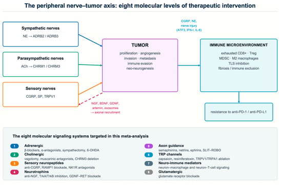
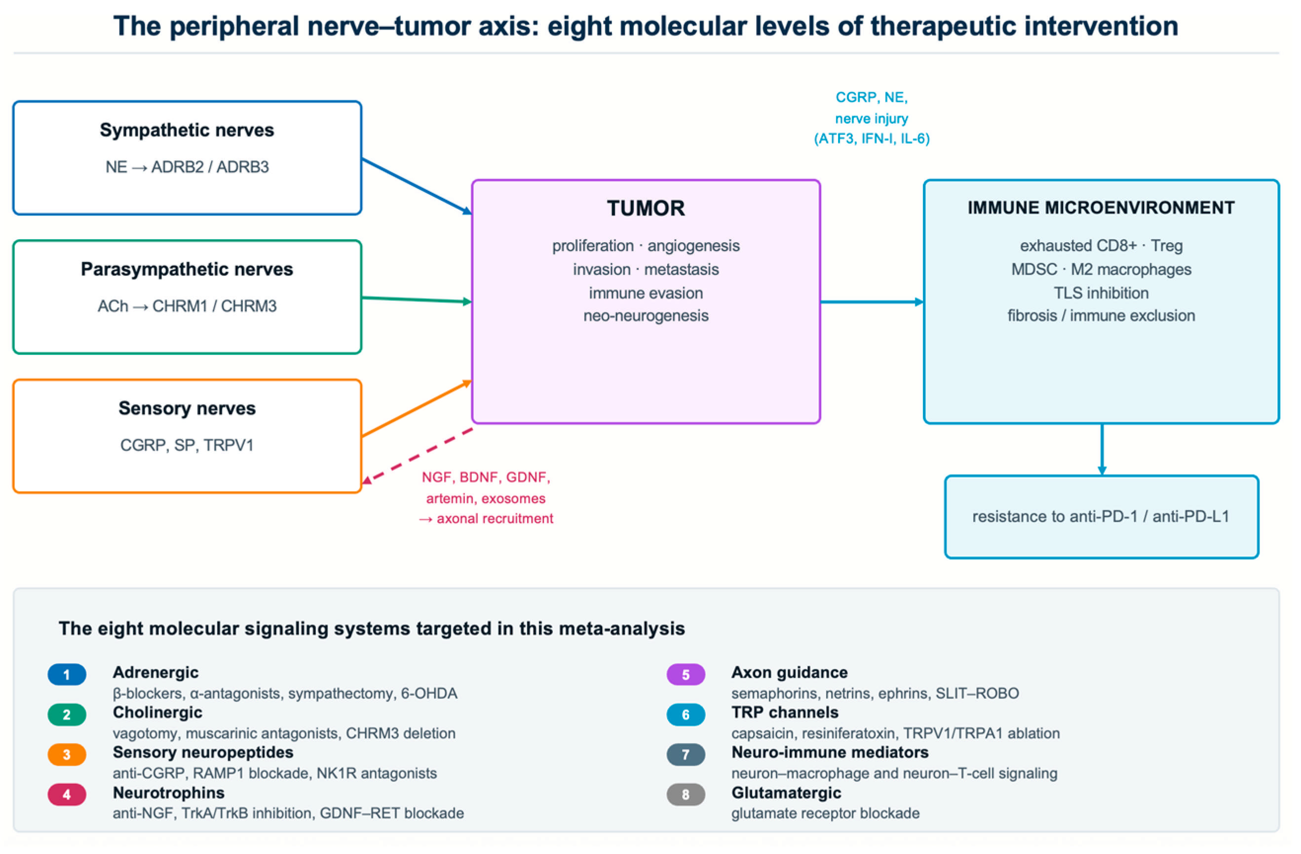
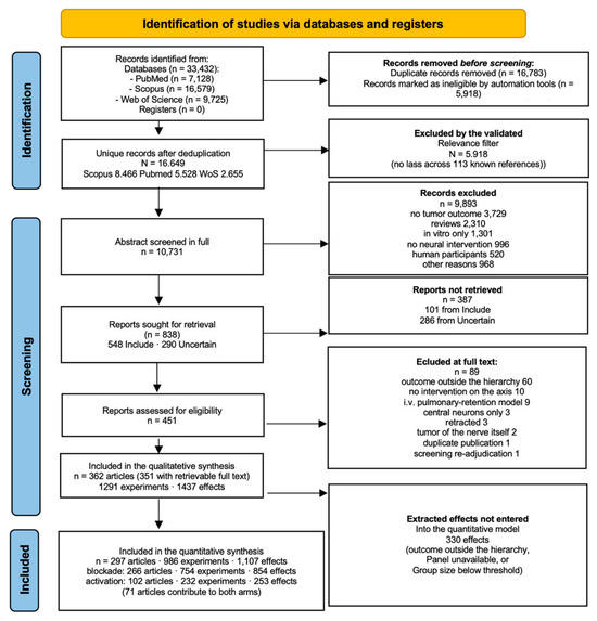
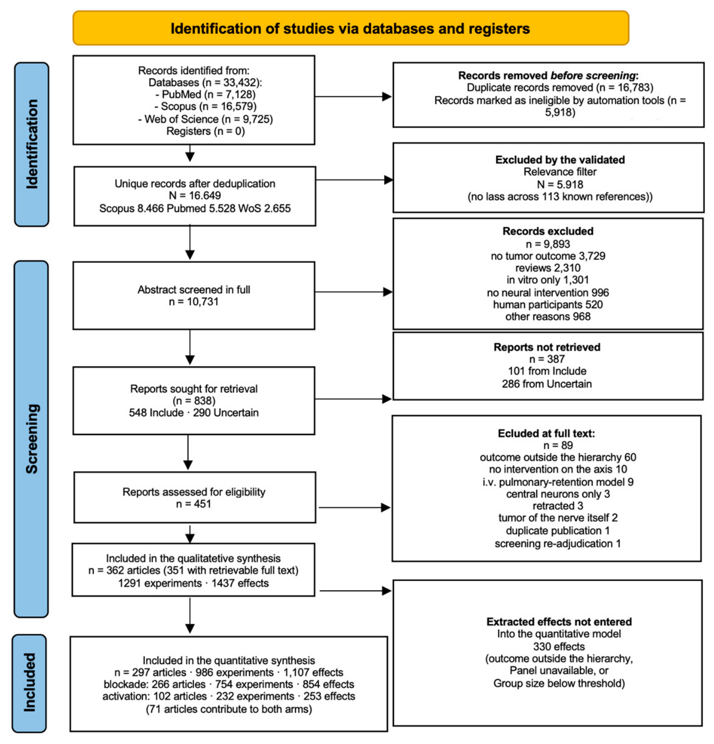
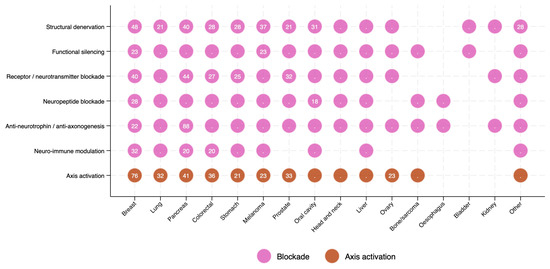
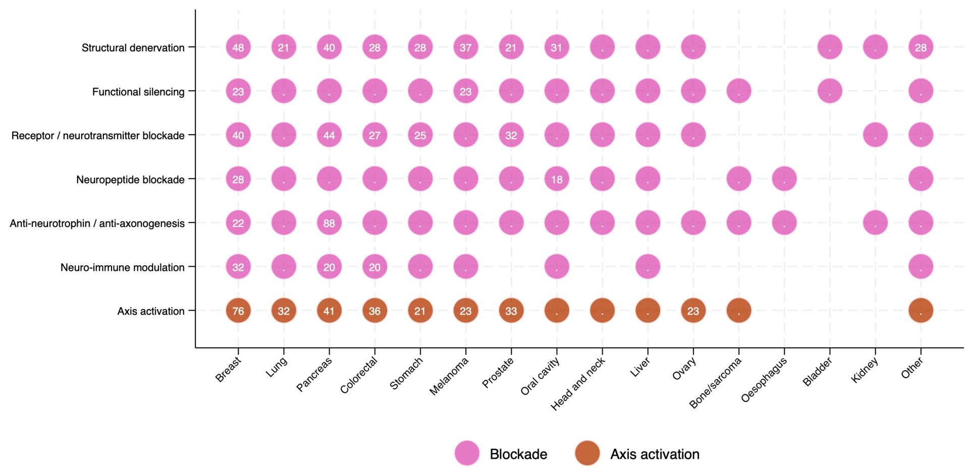
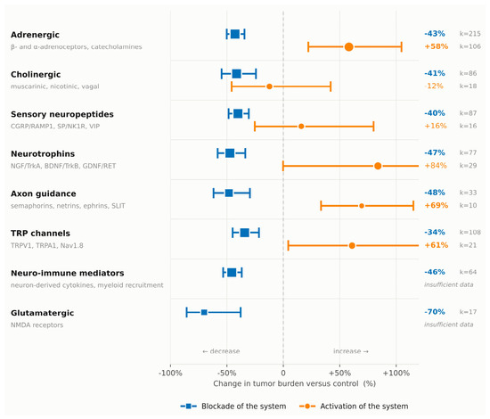
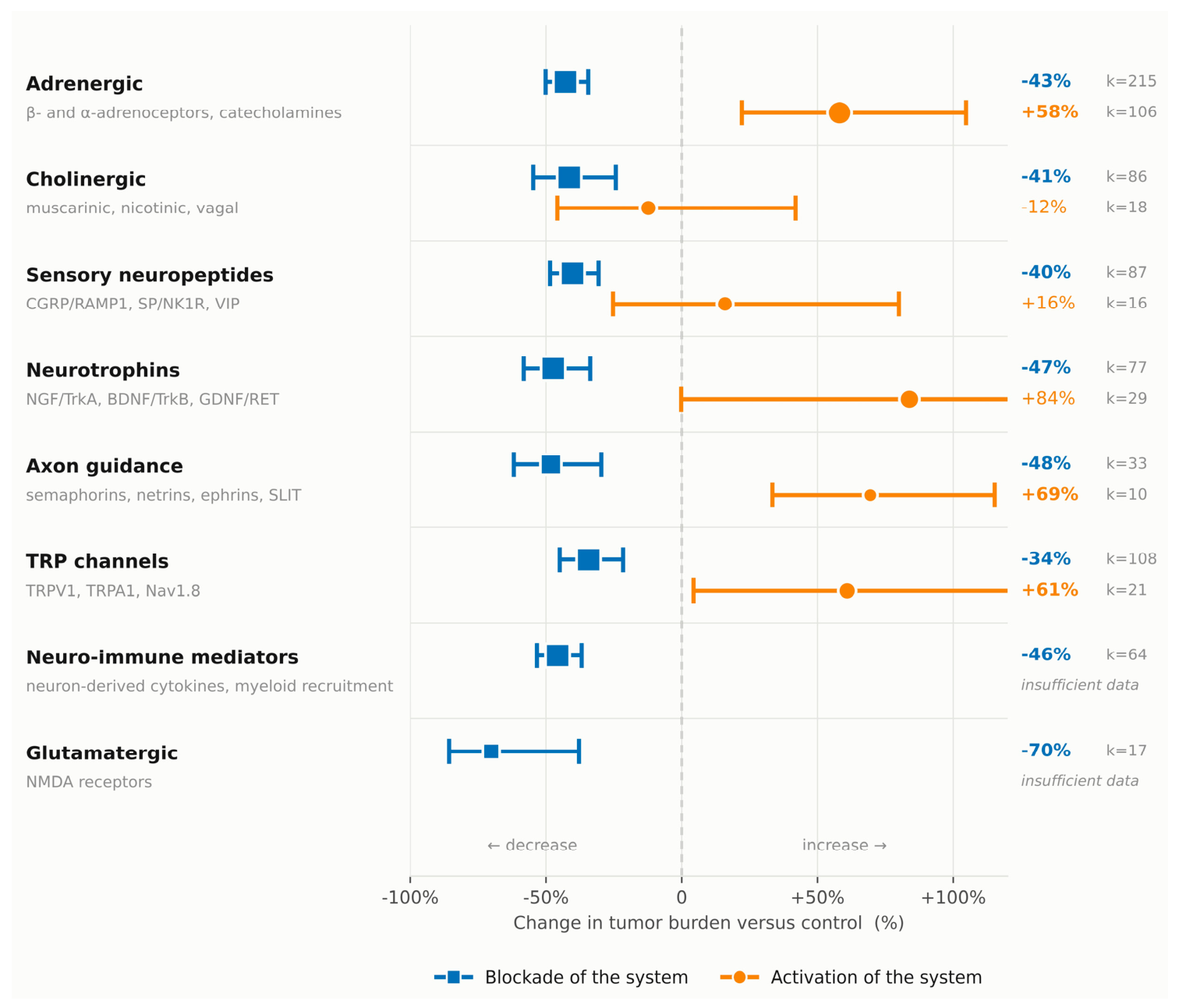
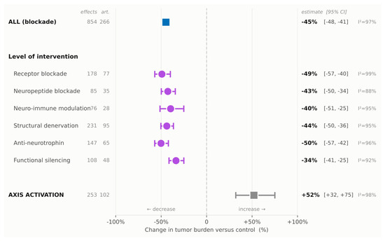
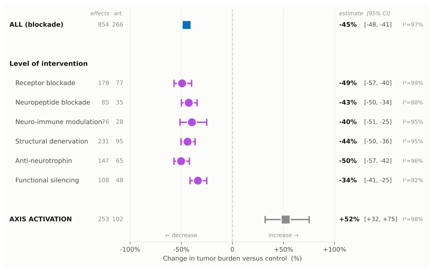
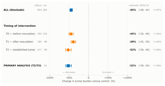
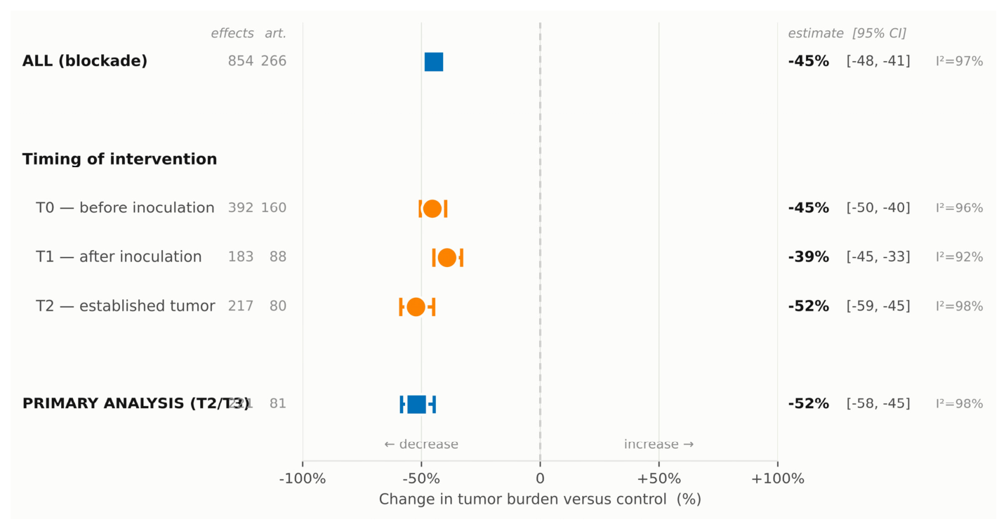
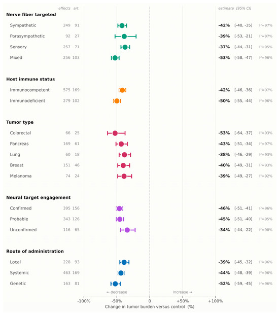
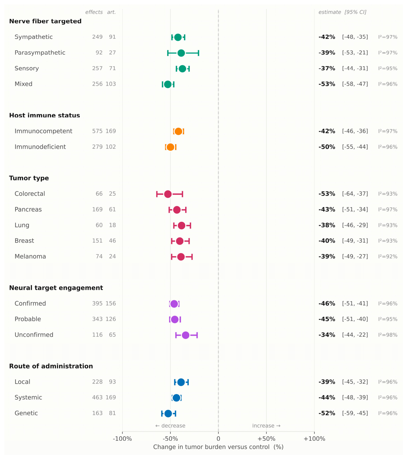
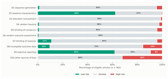
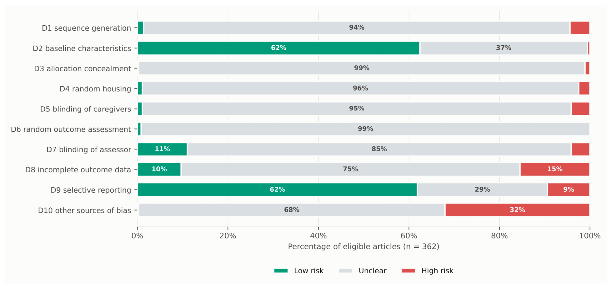
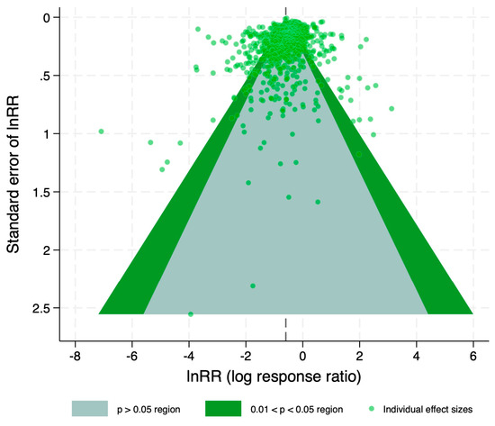
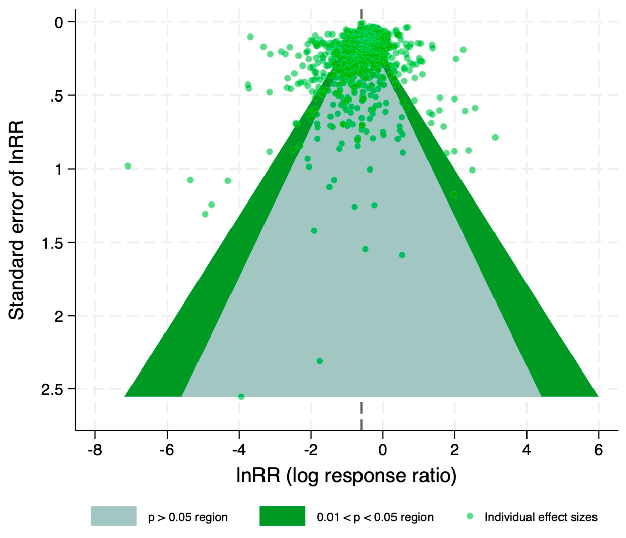

Disclaimer/Publisher’s Note: The statements, opinions and data contained in all publications are solely those of the individual author(s) and contributor(s) and not of MDPI and/or the editor(s). MDPI and/or the editor(s) disclaim responsibility for any injury to people or property resulting from any ideas, methods, instructions or products referred to in the content. |
© 2026 by the authors. Licensee MDPI, Basel, Switzerland. This article is an open access article distributed under the terms and conditions of the Creative Commons Attribution (CC BY) license.