


Abstract
T-cell immunoglobulin and mucin domain 3 (TIM-3), a well-known immune checkpoint molecule, is increasingly recognized for its regulatory functions beyond T cell exhaustion, particularly in macrophages. Recent advances have revealed the important role of this molecule in various pathological and physiological conditions. The demand for a comprehensive study of TIM-3 is increasing, particularly as a result of ongoing clinical trials targeting TIM-3 in oncology. This review is devoted to the role of TIM-3 in macrophage biology, focusing on associations between TIM-3 expression and macrophage polarization states and functional activity, as well as its involvement in the pathogenesis of different diseases and reproductive immunology. The review examines known effects and molecular mechanisms by which TIM-3 regulates macrophage functional phenotype and the contribution of TIM-3-expressing macrophages to cancer, pregnancy, inflammation, infectious and autoimmune diseases, and fibrosis. Findings highlight the controversial role of TIM-3 in the regulatory function of macrophages and suggest that TIM-3 functions differently depending on the context. The review also touches on gaps and unexplored parts of the topic. A summary of current data allows us to conclude that TIM-3 is an important modulator of macrophage functions and can be considered a potential therapeutic target in various pathological conditions.
1. Introduction
T-cell immunoglobulin and mucin domain 3 (TIM-3) are immune checkpoint receptors that are important for modulation of immune reactions. Checkpoint receptors are important players in the immune response that regulate T-cell activation. Their biological function is to downregulate signaling pathways, resulting in decreased production of pro-inflammatory cytokines and increased production of regulatory and anti-inflammatory cytokines by immune cells, as well as inhibition of proliferation and activation of effector T cells. All this contributes to the involvement of checkpoint receptors in immune tolerance, autoimmunity, and antitumor or antiviral immune evasion. The crucial role of these molecules in tumor immune evasion has led to the development of anti-programmed cell death protein 1 (PD-1) and anti-cytotoxic T-lymphocyte-associated protein 4 (CTLA-4) therapies for cancer. Currently, anti-TIM-3 drugs are being developed and are in clinical trials [1], since numerous preclinical studies show that TIM-3 expression on tumor cells leads to the development of resistance to anti-PD-1 or anti-CTLA-4 antibodies, and TIM-3 blocking induces an anti-tumor immune response [2,3,4,5]. Some authors think that targeting TIM-3 could be a good way to get around resistance to anti-PD-1 therapy.
Studies have also demonstrated a significant role of TIM-3 in autoimmune diseases. The TIM-3-ligand axis plays a key role in the pathogenesis of a number of autoimmune diseases and chronic inflammatory conditions, such as systemic lupus erythematosus, rheumatoid arthritis, inflammatory bowel disease, multiple sclerosis, vasculitis, and asthma [6]. Moreover, TIM-3 is an important molecule for infectious immunity and regulation of the inflammatory response [7,8,9,10].
With regard to T cells, the role of TIM-3 has been described in sufficient detail, and the scientific community has reached a certain consensus. Meanwhile, an increasing amount of evidence has emerged in recent years showing that TIM-3 is also important for the activity of myeloid cells. It was found that TIM-3 regulates the functions of monocytes, macrophages, and dendritic cells. Moreover, some studies provide information that TIM-3 expression on myeloid cells is higher than on T cells in some conditions [11,12,13].
TIM-3 is believed to have great significance in macrophage activity modulation. Since macrophages are multifunctional cells regulating various processes in the body, the issues of TIM-3 expression and signaling pathways triggered by the interaction of this molecule with ligands have become important. Macrophages are very flexible cells that are found in most tissues. They help control inflammation, repair, the strength of the immune response, and other normal and abnormal processes [14]. The role of tumor-associated macrophages is also well-known [15]. Therefore, clarifying the mechanisms of macrophage regulation is of particular interest since it increases the potential for using macrophages to regulate the above-mentioned processes in the body.
Thus, given the above, elucidating the role of TIM-3 in macrophage regulation is of particular interest, as it may open new therapeutic avenues for modulating immune responses in cancer and inflammatory diseases. Although TIM-3 is considered crucial for macrophage polarization and functional phenotype formation [16], current data on its role are controversial and fragmented. The precise mechanisms by which TIM-3 signaling regulates macrophage polarization and functions, and how this contributes to specific pathologies, remain uncertain and sometimes a subject of conflicting data. This review summarizes studies devoted to the importance of TIM-3 in the modulation of macrophage function and the role of TIM-3+ macrophages in various pathologies.
2. TIM-3 Structure and Ligands
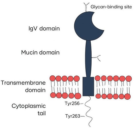
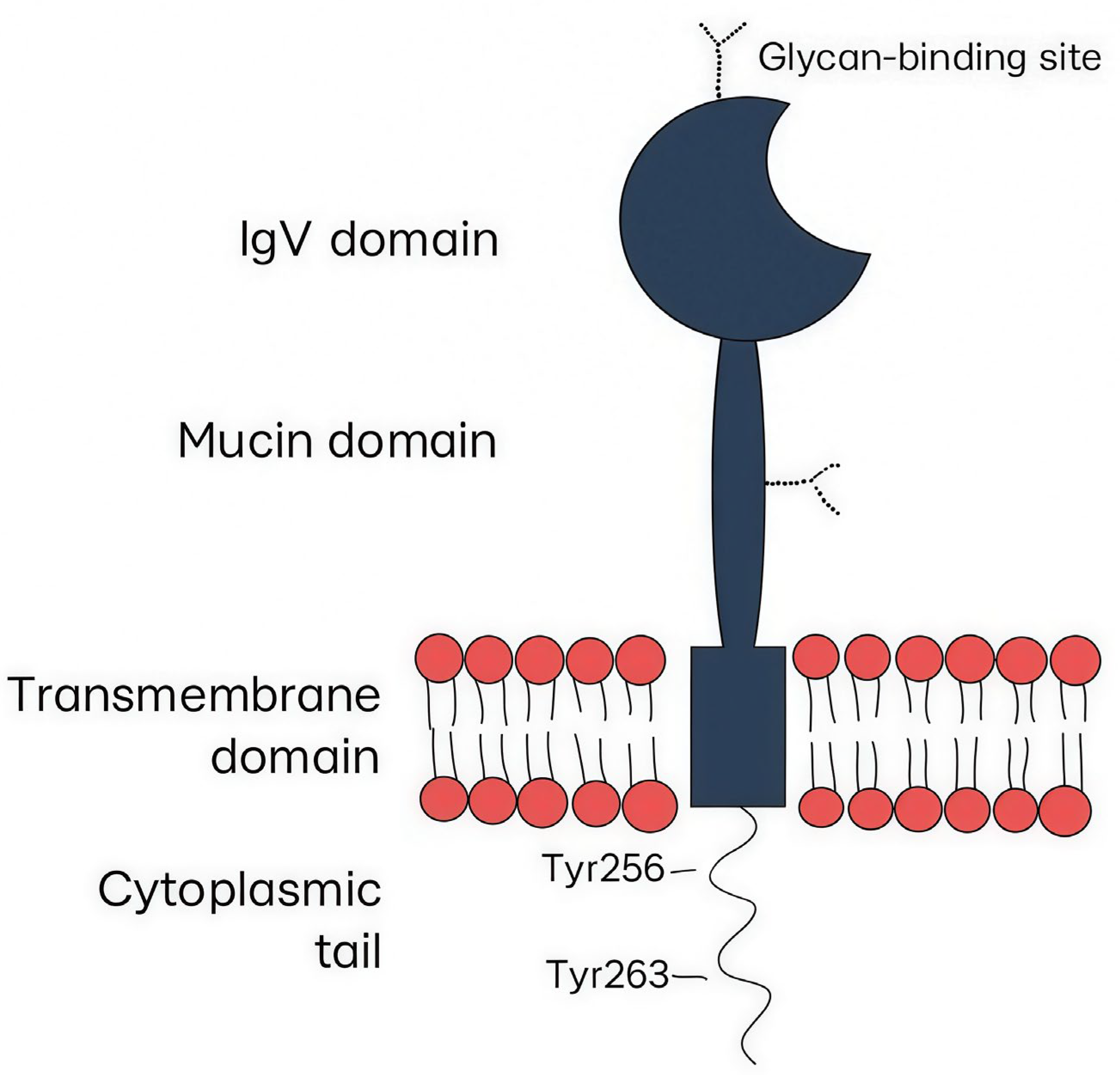
TIM-3 belongs to the immunoglobulin superfamily and is widely expressed on the surfaces of adoptive and innate immunity cells. The TIM-3 structure includes a membrane-distal N-terminal immunoglobulin (IgV) domain and a mucin domain with N-linked glycosylation, which account for the extracellular part of the receptor, a transmembrane domain, and a cytoplasmic tail (Figure 1) [17]. The IgV domain has a specific structure because of extra disulfide bonds between noncanonical cysteines, which is a feature of the TIM superfamily. Engagement of the IgV domain with appropriate ligands is considered to provide the immunoregulatory role of TIM-3 [17].
Figure 1.
The TIM-3 structure includes a membrane-distal N-terminal immunoglobulin (IgV) domain, a mucin domain with N-linked glycosylation, a transmembrane domain, and a cytoplasmic tail.
TIM-3 exists in membrane-bound form and soluble form, which lacks the mucin and transmembrane domains. The precise mechanism of releasing soluble TIM-3 is still unknown; however, soluble TIM-3 is most likely the result of shedding by disintegrin and metalloprotease (ADAM)-10 and ADAM-17 [18,19].
Several ligands for TIM-3 are known at present, including galectin-9 (Gal-9) [20], phosphatidylserine (PtdSer) [6,21,22], high-mobility group protein B1 (HMGB1, amphoterin) [23,24] and carcinoembryonic antigen-related cell adhesive molecule 1 (CEACAM1) [25]. There are still many unresolved questions regarding the interaction between ligands and TIM-3. First, it is unclear whether all of the above-mentioned molecules are true TIM-3 ligands, since some studies have not linked TIM-3 function to a particular TIM-3 ligand [26]. Besides, Gal-9 and CEACAM-1 can regulate T cells and macrophages independently of TIM-3 [27,28,29,30,31,32]. Moreover, De Sousa Linhares et al. found that CEACAM1 does not bind to TIM-3 [32]. The second question is the exact binding sites of TIM-3 and the ligands, and whether there is any competition between TIM-3 ligands. Finally, some authors believe that TIM-3 can bind to several ligands at the same time. Thus, engagement with TIM-3 and Gal-9 does not exclude binding to other ligands at different binding sites [33]. Moreover, it is likely that the binding of Gal-9 to TIM-3 can lead to a change in the receptor configuration, resulting in the cleft framed by the FG and CC′ loops becoming more available to other ligands [33].
3. The Role of TIM-3 in Macrophages
3.1. TIM-3 Expression on Different Macrophage Subtypes
The TIM-3 plays a key role in regulating macrophage function, affecting macrophage polarization, cytokine profile, and effector functions. It acts as an immunological ‘switch’, and its effect depends on the microenvironmental context, the type of ligand, and the state of cells. This regulation is particularly important in light of the existence of different functional states in macrophages, the extreme points of which are pro- and anti-inflammatory phenotypes.
Macrophages are cells representing a spectrum of functional states, the opposite of which are M1 and M2, based on their expression of certain surface receptors and the secretion of specific molecules [34]. M1 cells are characterized by a pro-inflammatory phenotype; they express CD80, CD86, CD40, and inducible nitric oxide synthase (iNOS), produce pro-inflammatory cytokines (IL-1β, TNFα), and provide anti-infective and anti-tumor activity [34]. In contrast, M2 are characterized by anti-inflammatory activity, expressing arginase-1, CD206, CD163, and CD204, producing anti-inflammatory cytokines (IL-10, transforming growth factor TGF-β); they limit inflammatory response and promote tissue repair and wound healing [34]. Researchers are trying to compare TIM-3 expression and macrophage functional phenotypes in order to determine whether TIM-3 is linked to any specific phenotype and whether it might serve as a marker, and also identify the signaling pathways that are responsible for this.
Most studies assume a link between TIM-3 and the M2 polarization state of macrophages. Thus, an increase in TIM-3 expression was detected on M2 macrophages in mice [35,36]. Ocaña-Guzman et al. demonstrated that TIM-3 expression is higher on human monocyte-derived macrophages polarized with IL-4 (M2) compared to cells polarized with lipopolysaccharide (LPS)+IFNγ (M1) [37]. The association between TIM-3 levels and M2 polarization was also shown in the study of Sun J et al., in which mouse adipose tissue macrophages were biased toward the M2 phenotype with increased TIM-3 expression and, conversely, toward the M1 phenotype with decreased TIM-3 expression [38]. TIM-3 expression was increased on promonocytic THP-1 cells, which exhibit an M2-like phenotype when cultured with secreted factors from anaplastic thyroid cancer cells [39]. Zhang et al. found increased expression of TIM-3 on CD68+CD163+ macrophages (M2 phenotype) in rats [40]. Finally, IL-4/signal transducer and activator of transcription (STAT) 6 signals responsible for M2 polarization may promote TIM-3 activation [41]. Furthermore, Cheng et al. recently showed that the addition of Gal-9 led to a significant increase in the proportion of TIM-3+M1 and TIM-3+M2 macrophages and a decrease in M1 cell proportions and M1/M2 ratio [42].
On the other hand, Yu et al. indicated an association between TIM-3 and M1 polarization of macrophages during intracerebral hemorrhage. The authors demonstrated that intracerebral hemorrhage promoted TIM-3 expression and M1 polarization in the perihematomal region in mice. In contrast, TIM-3 knockout attenuated M1 polarization [43]. In addition, when studying decidual macrophages, the authors demonstrated that neither TIM-3+ nor TIM-3− cells belonged exactly to either the M1 or M2 phenotype [44]. Thus, the link between TIM-3 and M1/M2 polarization probably depends on the localization of cells and the microenvironment, and it is not possible to unambiguously associate TIM-3 with a pro- or anti-inflammatory macrophage phenotype.
3.2. TIM-3 Signaling Pathways in Macrophages
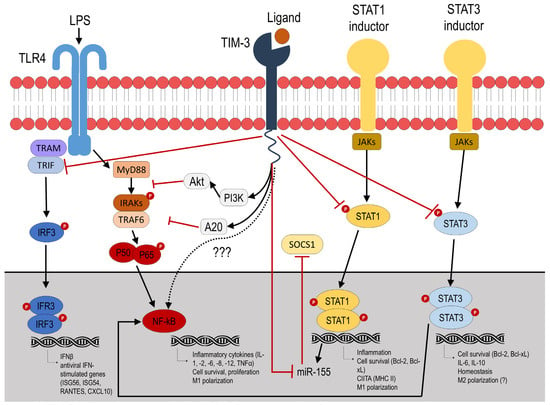
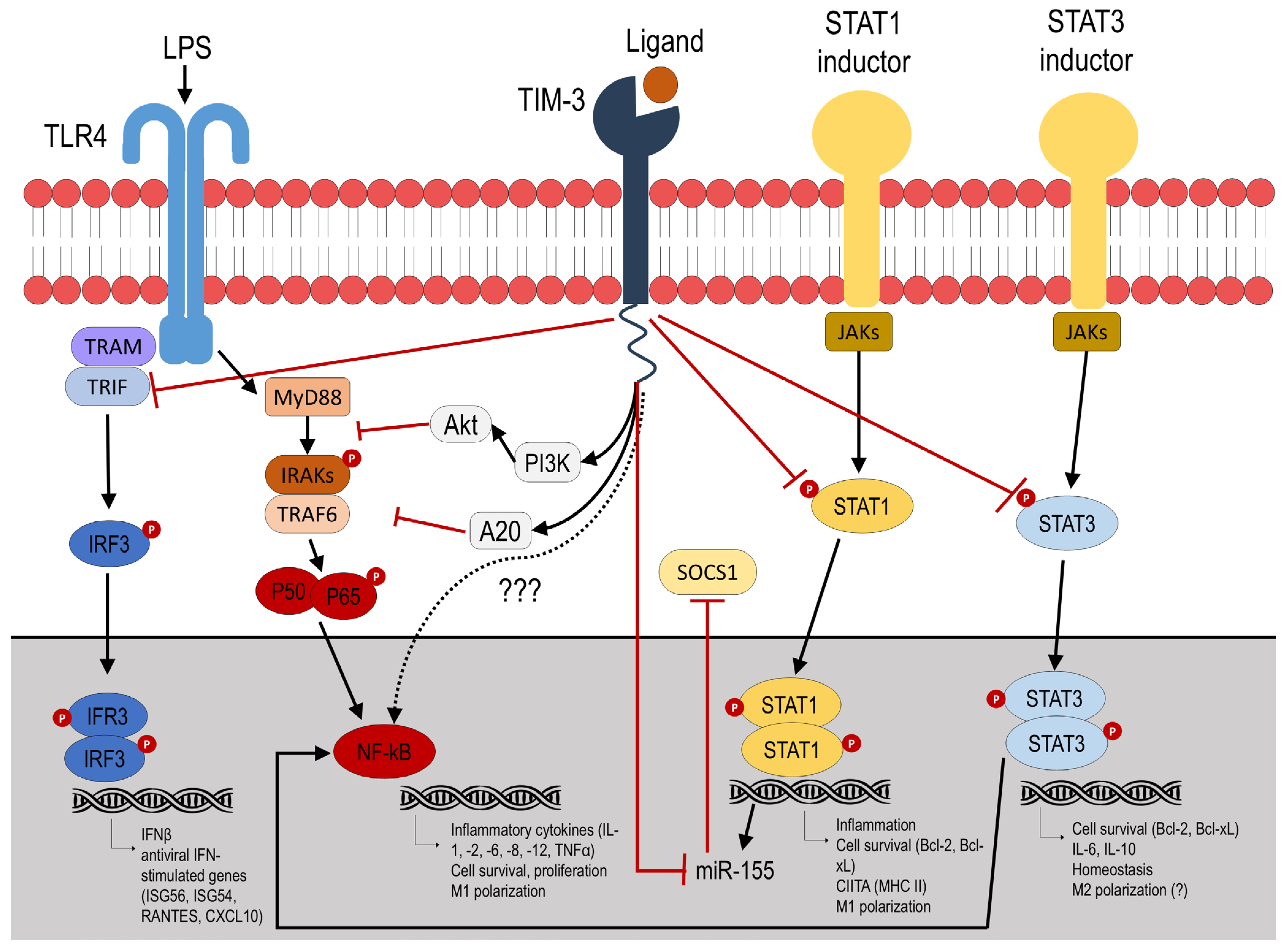
The exact mechanisms of TIM-3 effects are not yet fully understood. Due to non-canonical signaling, multiple ligands, and widespread expression, the TIM-3 pathway is very intricate and precisely regulated. It is still unknown exactly how TIM-3 regulates the pro- and anti-inflammatory activity of macrophages, although several studies have shown possible signaling cascades. TIM-3 acts through several intracellular pathways that can have both suppressive and activating effects on the inflammatory response (Figure 2).
Figure 2.
Described TIM-3 signaling mechanisms in macrophages. Question marks indicate that there is insufficient data on the given issue. Black arrows indicate the activating effect, red arrows indicate the inhibitory effect.
One such mechanism is LPS/toll-like receptor (TLR) 4 signaling inhibition. TLR4 is known to transmit signals through two distinct pathways: the MyD88-dependent pathway, which mediates classical NF-κB activation, and the TRIF (TIR (Toll/interleukin-1 receptor) domain-containing adaptor protein inducing interferon beta)-dependent one, which is associated with interferon regulatory factor 3 (IRF3) [45]. The available evidence suggests that TIM-3 activation in macrophages is capable of inhibiting both of these signaling cascades.
Thus, it has been demonstrated that TIM-3 signaling inhibited LPS–TLR-4-mediated NF-κB activation, presumably by increasing PI3K–AKT phosphorylation and A20 activation [46]. Another recent study confirmed this fact and showed that TIM-3 suppressed inflammatory signaling pathway of p65, a protein involved in NF-κB heterodimer formation, nuclear translocation, and activation [47].
Additionally, TIM-3 signaling inhibited the phosphorylation of IRF3 [48], whereas TIM-3 knockout attenuated M1 polarization, presumably through stimulation of TRIF and IRF3 transcription factors [42]. Given that TRIF/IRF3 is associated with TLR-4, this may provide further support for the hypothesis that TIM-3 activation in macrophages suppresses TLR-4 pathway.
On the other hand, several studies produced opposite results. Under certain conditions, ligand-dependent activation of TIM-3 may, on the contrary, enhance TLR-4-mediated signaling and activation of the NF-κB/TNFα pathway, promoting a proinflammatory response. Thus, the activating effect of TIM-3 on TLR-4 signaling was found for intestinal macrophages [48]. The study demonstrated that TIM-3 blockade did not stimulate macrophage polarization in the M1 direction in TLR-4 knockout mice, unlike in wild-type mice. Moreover, the authors found the same effect on RAW264.7 mouse cell line macrophages. Another study showed that TIM-3-expressing renal macrophages aggravate the progression of diabetic nephropathy through activation of the NF-κB/TNFα pathway [49]. These facts partially contradict previous studies showing that TIM-3 negatively regulates TLR-4 and NF-κB signaling and require further research.
TIM-3 is also able to modulate macrophage polarization by influencing STAT proteins. Jiang et al. found that TIM-3 polarizes macrophages into M2 cells by directly binding to STAT1, which is associated with the M1 phenotype, and inhibiting the STAT1-miR-155-SOCS1 signaling axis [50]. The association of TIM-3 expression with STAT1 was also demonstrated in the study of Tang et al. The authors reported that TIM-3 can suppress major histocompatibility complex (MHC) II expression in macrophages through the STAT1/CIITA pathway [51]. In another study, TIM-3 suppression by T. gondii infection resulted in inhibition of PI3K-AKT phosphorylation, which mediates the transcription factors C/EBPβ and SOCS1. This led to increased proinflammatory markers and cytokines in decidual macrophages [52]. Axis PI3K-AKT is also associated with NF-κB pathway [53]; therefore, it may be an additional mechanism for NF-κB inhibition described above.
Ma et al. demonstrated that the cis-association of Gal-9 and TIM-3 inhibited STAT3 phosphorylation, which was accompanied by a decrease in IL-23p19 and an increase in IL-12p35 [54]. In turn, there is a close interaction between STAT3 and the NF-κB signaling [53]; therefore, these data may support the theory of NF-κB inhibition mediated by LPS/TLR-4 downregulation via TIM-3 activation.
At the same time, the relationship between TIM-3 and LPS (TLR4 inducer) is likely interdependent, and in addition to the effect of TIM-3 expression on TLR-dependent cytokine production, LPS and the LPS/TLR4 pathway can directly influence TIM-3 expression. Thus, it has been shown that TLR stimulation leads to a decrease in TIM-3 expression on CD14+ monocytes [55]. Interestingly, the effect of LPS on bone marrow-derived macrophages polarization via the TIM-3/Gal-9 pathway may be time-dependent. In the study by Zhang et al., short-term LPS stimulation activated the TIM-3/Gal-9 signaling pathway, which led to the inhibition of the proinflammatory M1 phenotype, whereas long-term LPS stimulation was accompanied by inhibition of the TIM-3/Gal-9 signaling pathway, ultimately leading to stimulation of M1 polarization [56]. The Gal-9/TIM-3 pathway partially activates cytokine production IL-6, IL-8, and IL-10, but not IL-1β, IL-12p70, or TNFα in response to LPS in gastric cancer tissue-derived tumor-associated macrophages, TAMs [57]. Taken together, this indicates ambiguous regulation of TIM-3 expression by LPS and may partially explain the differences described above.
In addition, TIM-3 expression may also depend on other TLRs. For example, Vidyarthi et al. showed that stimulation of macrophages by a TLR-3 ligand reduced the expression of TIM-3 on M2 macrophages and increased the release of proinflammatory cytokines [58].
In summary, TIM-3 signaling in macrophages is nonlinear and context-dependent. Its final effect—inhibitory or activating—is determined by a combination of factors: ligand type, the involved intracellular pathway, and the microenvironment (e.g., the presence of LPS, cytokine background). This plasticity allows TIM-3 to act as a fine regulator of the immune response, participating both in limiting excessive inflammation and, under certain conditions, in maintaining a proinflammatory environment. Resolving apparent discrepancies in the data requires consideration of the specific biological context in which TIM-3 function is studied.
3.3. Association Between TIM-3 and Macrophage Functions
TIM-3 has a complex effect on the condition and function of macrophages. First of all, by modulating the repertoire and balance of secreted cytokines, TIM-3 directs the functional outcome of macrophage activation.
The TLR-4/NF-κB pathway is known to be responsible for the secretion of proinflammatory cytokines, including IL-1, -2, -6, -8, -12, and TNFα [53]. Since TIM-3 likely acts as an inhibitor of the TLR-4/NF-κB axis, its regulatory effect on macrophage polarization is mediated, at least in part, through the direct suppression of this cytokine program. Indeed, blocking anti-TIM-3 antibodies and TIM-3 gene silencing resulted in increased production of IL-6, IL-12, TNFα, and IL-1β by macrophages and a decrease in M2 polarization, while overexpression of TIM-3 suppressed the production of these cytokines [46,47,59,60,61]. At the same time, Wang et al. showed that the interaction of Gal-9 with TIM-3 significantly stimulated the secretion of IL-6, IL-8, and IL-10, but not IL-1β, IL-12p70, or TNFα, in the presence of LPS [57], indicating an ambiguous influence of the Gal-9/TIM-3 pathway.
In turn, the LPS/TLR-4 pathway can also drive the production of IL-10, the powerful anti-inflammatory cytokine, as well [57,62,63]. Therefore, suppression of TLR-4 pathway by TIM-3 may explain increased IL-10 production after the TIM-3 suppression [46,49]. This mechanism may determine some of the pro-inflammatory signals of TIM-3, for example, that were found in the study by Anderson et al., where TIM-3 exerted a proinflammatory effect in the presence of a TIM-3 monoclonal agonist [64].
In addition to modulating the cytokine profile, TIM-3 is also likely involved in the phagocytic activity of macrophages. Thus, treatment with anti-TIM-3 antibodies inhibited the phagocytic activity of alveolar macrophages in vivo [65]. DeKruyff et al. also concluded that TIM-3 may partially mediate macrophage phagocytosis due to the ability to bind apoptotic cells [66]. In contrast, Liu et al. found that TIM-3 activation inhibits the phagocytic and cytotoxic effects of macrophages [47], and Hou et al. demonstrated that blocking anti-TIM-3 antibodies enhanced phagocytosis of red blood cells infected with the malarial pathogen by murine splenic macrophages [67].
Furthermore, TIM-3 is involved in the regulation of T cell functions by macrophages. For example, co-cultivation of TIM-3-overexpressing macrophages with intestinal lymphocytes reduced the proinflammatory response in a mouse model of colitis [48], and blocking TIM-3 on peritoneal macrophages resulted in a decrease in Tregs but an increase in Th1 and Th17 CD4+ T cells [51]. Li et al. showed that TIM-3+ macrophages caused a Th2 and Treg bias in CD4+ T cells in contrast to TIM-3− macrophages and TIM-3 blockade [44].
Together, these findings suggest that TIM-3 exerts a complex effect on macrophage function by modulating cytokine secretion, phagocytic activity, and T-cell response. The contextual nature of these effects explains the dual and sometimes contradictory role of TIM-3, which must be considered when assessing its potential as a therapeutic target.
4. TIM-3 Expression on Macrophages in Cancer and Inflammation
4.1. Oncology
TIM-3 expression on macrophages and monocytes represents a significant mechanism of tumor evasion from immune surveillance, as supported by data obtained in various types of cancer. Macrophages in the tumor microenvironment (tumor-associated macrophages, TAMs) are able to express TIM-3. Thus, an increased level of TIM-3 expression on peripheral blood mononuclear cells and/or TAMs compared to healthy people was found in patients with hepatocellular carcinoma [68], colorectal cancer [11,69], non-small cell lung cancer [70], and gastric cancer [57]. Higher TIM-3 expression on macrophages was found in a patient who lost the sensitivity of spindle cell lung cancer to anti-PD-1 therapy after a good response [5]. TIM-3 was expressed on the surface of macrophages and microglia in patients with diffuse intrinsic pontine glioma (54%), whereas TIM-3 was not expressed in the brain of healthy individuals [71]. Vanmeerbeek et al. detected a niche of tumor-associated macrophages that co-expressed TIM-3 and V-domain immunoglobulin suppressor of T-cell activation (VISTA) [72].
TIM-3 expression on monocytes and macrophages may be one of the indicators of immune surveillance evasion of tumor. Hu et al. also demonstrated that high-level TIM-3 expression on the CD68+ cells (macrophage marker) indicates poor survival in glioma [73]. High TIM-3 expression in TAM was an independent predictor of poor prognosis in patients with non-small cell lung cancer and negatively correlated with overall survival [70]. In addition, increased TIM-3 expression on peripheral monocytes was associated with deeper tumor invasion, metastasis of tumor in the lymph nodes, and advanced clinical stage of cancer in patients with gastric cancer [57].
So, TIM-3-expressing macrophages are proposed as a potential target for anti-tumor therapy, and understanding their role and mechanisms of their activity in tumor pathology is crucial.
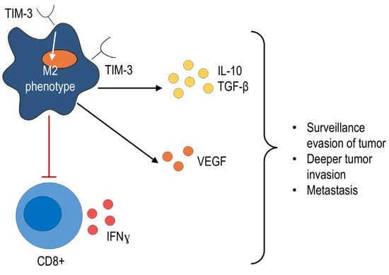
The negative effects of TIM-3-expressing macrophages are most often associated with the polarization of macrophages towards M2 and the occurrence of tumor-stimulating activity [39,50,68,69,74,75]. This process ensures a decrease in the production of proinflammatory cytokines (IL-12, TNFα, IL-6) and an increase in the production of IL-10 and TGF-β [50,74]. Additionally, as demonstrated for monocytes, TIM-3 expression on the cell surface can decrease the generation of IFNγ by activated CD8+ T cells [76].
Ni X. et al. showed that the interaction of Gal-9 and TIM-3 on macrophages leads to increased secretion of M2-associated proangiogenic factors such as vascular endothelial growth factor VEGF-A, while blockade of Gal-9/TIM-3 signaling inhibits M2 polarization and suppresses tumor growth in glioblastoma models [75].
Thus, TIM-3 is associated with M2 polarization, production of anti-inflammatory cytokines and proangiogenic factors, and suppression of CD8+ T cell activity (Figure 3). These findings suggest that TIM-3 blockade could be a promising strategy for overcoming resistance to the available checkpoint inhibitor treatments and explain the interest of scientists in developing new checkpoint inhibitors.
Figure 3.
Described mechanisms of immunosuppression of TIM-3-expressing macrophages in oncology. Black arrows indicate the activating effect, red arrows indicate the inhibitory effect.
At the same time, as demonstrated in Section 3.2 and Section 3.3, TIM-3 signaling in macrophages has a complex, contradictory biology, modulating both pro- and anti-inflammatory pathways. This duality suggests that TIM-3 blockade may have unintended consequences, highlighting the necessity for comprehensive preclinical and clinical studies to accurately predict its effects and understand the full implications of this therapy.
4.2. Non-Infectious Inflammation
TIM-3 plays an important role in the regulation of inflammation [76,77], but data on TIM-3 in myeloid cells are conflicting.
The anti-inflammatory effect of TIM-3 was described in a mouse model of acute inflammation. Wang et al. demonstrated that TIM-3-knockout mice showed increased susceptibility to colitis as well as more severe colitis and increased expression of proinflammatory mediators compared with controls [59]. Jiang et al. also showed that TIM-3 inhibits the polarization of pathogenic proinflammatory M1 macrophages in colitis, whereas TIM-3 down-regulation or blockade leads to an increase in the M1 response [48]. Moreover, adoptive transfer of TIM-3-silenced macrophages aggravates DSS (dextran sulfate sodium)-colitis and increases inflammation, while co-culture of TIM-3-overexpressing macrophages with intestinal lymphocytes reduces the pro-inflammatory response [48].
TIM-3 may also be involved in the pathogenesis of chronic diseases. For example, TIM-3 exerted a protective effect in steatohepatitis in mice by limiting the secretion of reactive oxygen species and inflammatory cytokines [46].
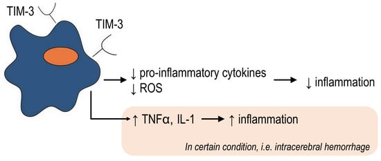
On the other hand, TIM-3 expression in microglia/macrophages was found to enhance inflammation after intracerebral hemorrhage and was positively correlated with pro-inflammatory cytokine (TNFα, IL-1β) concentrations and brain water content [43,77].
In another study, TIM-3 overexpression on myeloid cells led to the development of excessive lung inflammation in a mouse model, which was associated with galectin-3 production. TIM-3 blockade or galectin-3 inhibition was associated with a reduced inflammatory response in TIM-3 overexpressing mice. However, TIM-3 knockout resulted in lower body weight and shorter lifespan than WT mice, partly suggesting a positive role for TIM-3 in inflammation [78].
Taken together, the obtained data indicate a context-dependent function of TIM-3 during inflammation (Figure 4). On the one hand, TIM-3+ macrophages are associated with the suppression of acute and chronic inflammation; on the other hand, some studies have demonstrated that TIM-3+ macrophages can support the inflammatory response. These differences may be due to the macrophage origin, niche, and the cause of the inflammatory response.
Figure 4.
The dual and context-dependent role of TIM-3+ macrophages in inflammation. ↑ symbol indicates up-regulation, ↓ symbol indicates down-regulation.
4.3. Fibrosis as a Result of Unresolved Inflammation
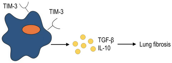
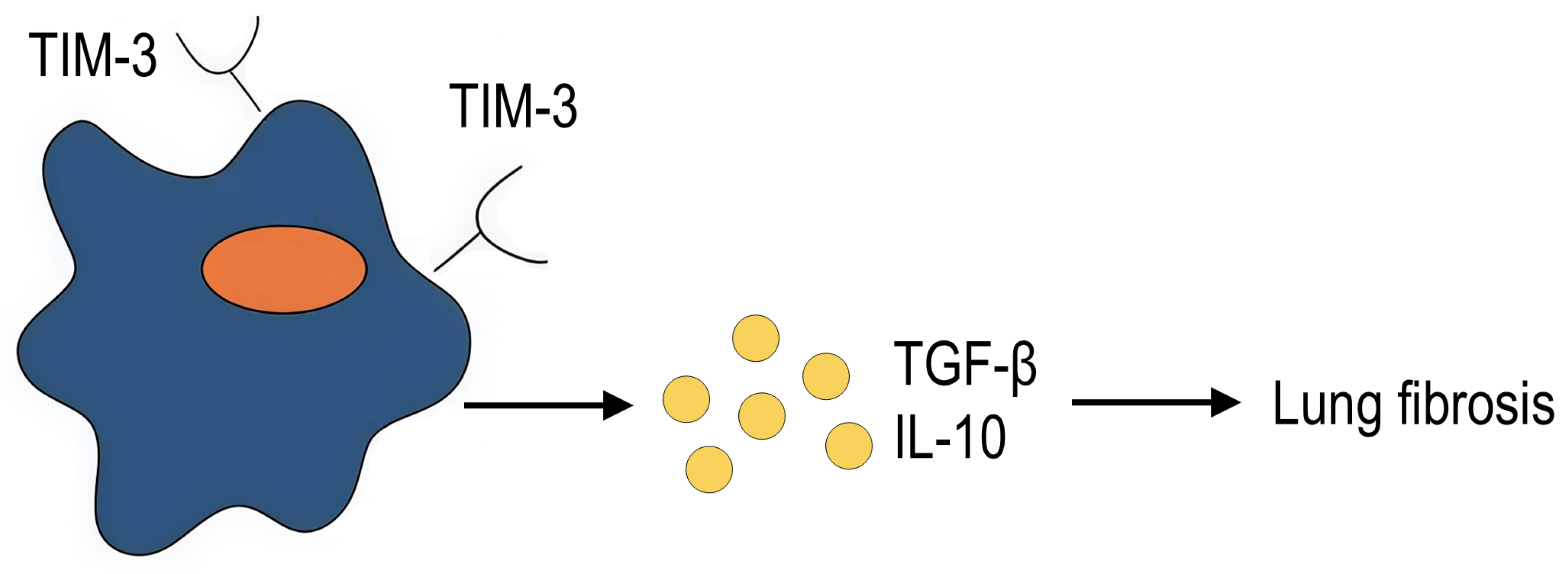
Inflammation is a complex process that, when dysregulated, can drive fibrosis. Given the crucial role of TIM-3-expressing macrophages in modulating inflammation, they are likely implicated in the fibrotic process as well. However, direct evidence linking TIM-3+ macrophages to fibrogenesis remains limited. To date, there is only one study examining the role of TIM-3+ macrophages in fibrosis. Wang et al. showed that TIM-3 aggravates pulmonary fibrosis. TIM-3 transgenic mice developed more severe pulmonary fibrosis compared to wild-type mice, but after macrophage depletion, the fibrosis score was significantly reduced in both wild-type and TIM-3 transgenic mice. Adoptive transfer of macrophages derived from TIM-3 transgenic mice resulted in more severe fibrosis compared to transfer of macrophages derived from wild-type mice. In light of these observations, the authors conclude that TIM-3-expressing macrophages exacerbate pulmonary fibrosis, probably due to increased production of profibrogenic markers of pulmonary fibrosis, IL-10 and TGF-β [79] (Figure 5). In turn, these factors can exert both profibrogenic and anti-inflammatory effects, which confirms the context-dependent involvement of TIM+ macrophages in the inflammatory process.
Figure 5.
TIM-3-expressing macrophages aggravate pulmonary fibrosis (summary of the study by Wang et al. [79]).
4.4. Autoimmune Inflammation
Previous studies have shown that TIM-3 dysfunction influences various immune cells and contributes to the pathogenesis of autoimmune disorders [6]. Studies conducted on T cells have shown that reduced TIM-3 expression in peripheral blood mononuclear cells may serve as an unfavorable prognostic factor in multiple sclerosis [80]. Anti-TIM-3 antibodies promote IFNγ and MCP-1 secretion by synovial mononuclear cells in patients with rheumatoid arthritis [81].
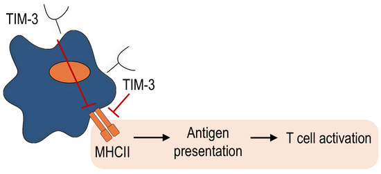
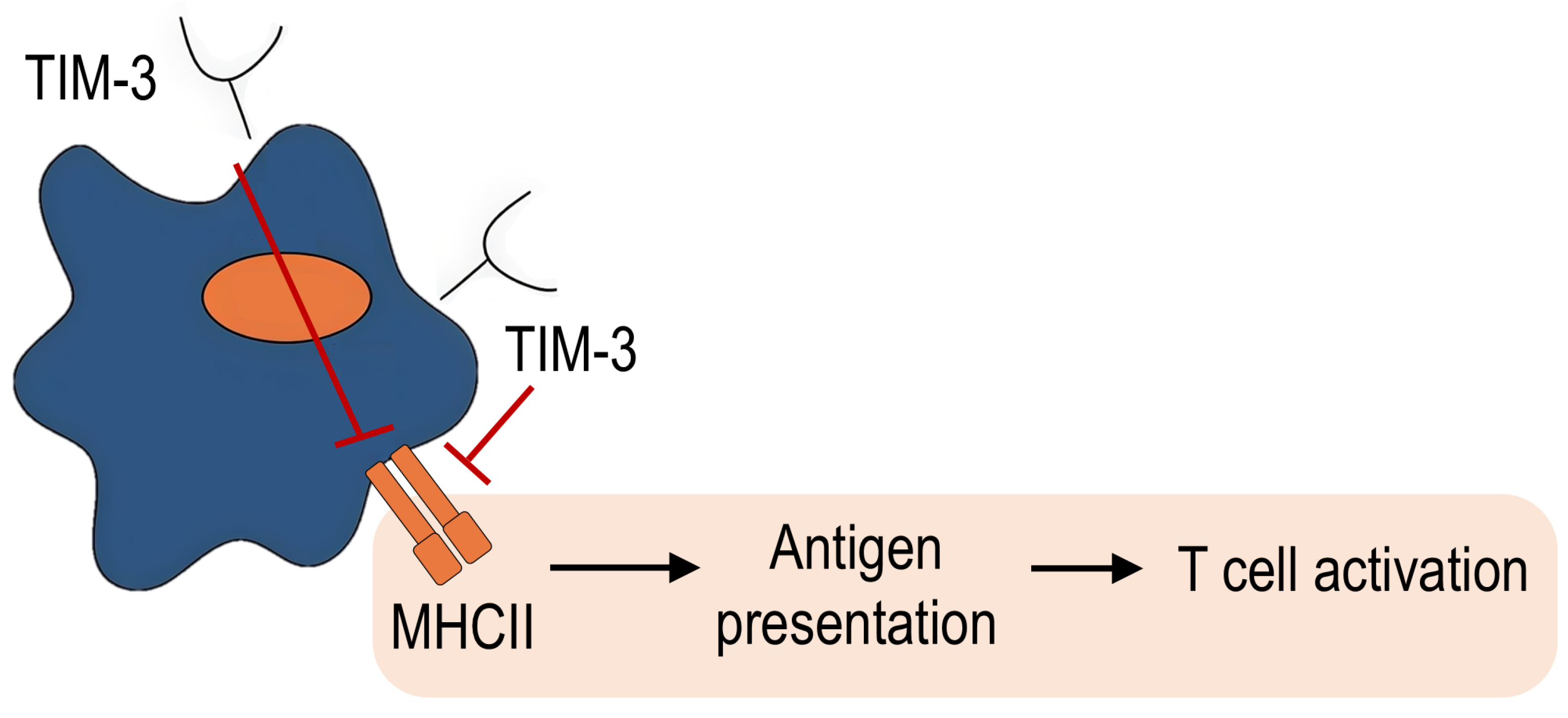
However, the role of TIM-3 expression on macrophages in autoimmune diseases is not clearly known, which is probably associated with the more important role of T cells in the pathogenesis of these diseases. Only one study investigating the role of TIM-3 expression on macrophages in autoimmune encephalomyelitis in mice showed that TIM-3 overexpression led to a decrease in inflammation, demyelination areas, and disease score in mice. At the same time, TIM-3 blocking, on the contrary, led to a significant increase in the severity of encephalomyelitis. These effects were associated with the ability of TIM-3 to suppress MHC-II expression in macrophages, which led to a decrease in antigen presentation and subsequent T cell activation (Figure 6) [51]. These findings suggest that TIM-3+ macrophages apparently play a positive role in autoimmune inflammation. However, further investigation is required to draw definitive conclusions, including studies of other autoimmune diseases such as systemic scleroderma, rheumatoid arthritis, ankylosing spondylitis, and systemic lupus erythematosus.
Figure 6.
The possible mechanism of involvement of TIM-3+ macrophages in autoimmune inflammation pathogenesis.
4.5. Infections
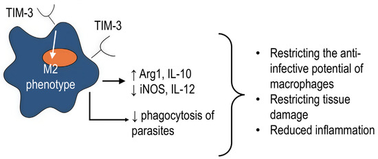
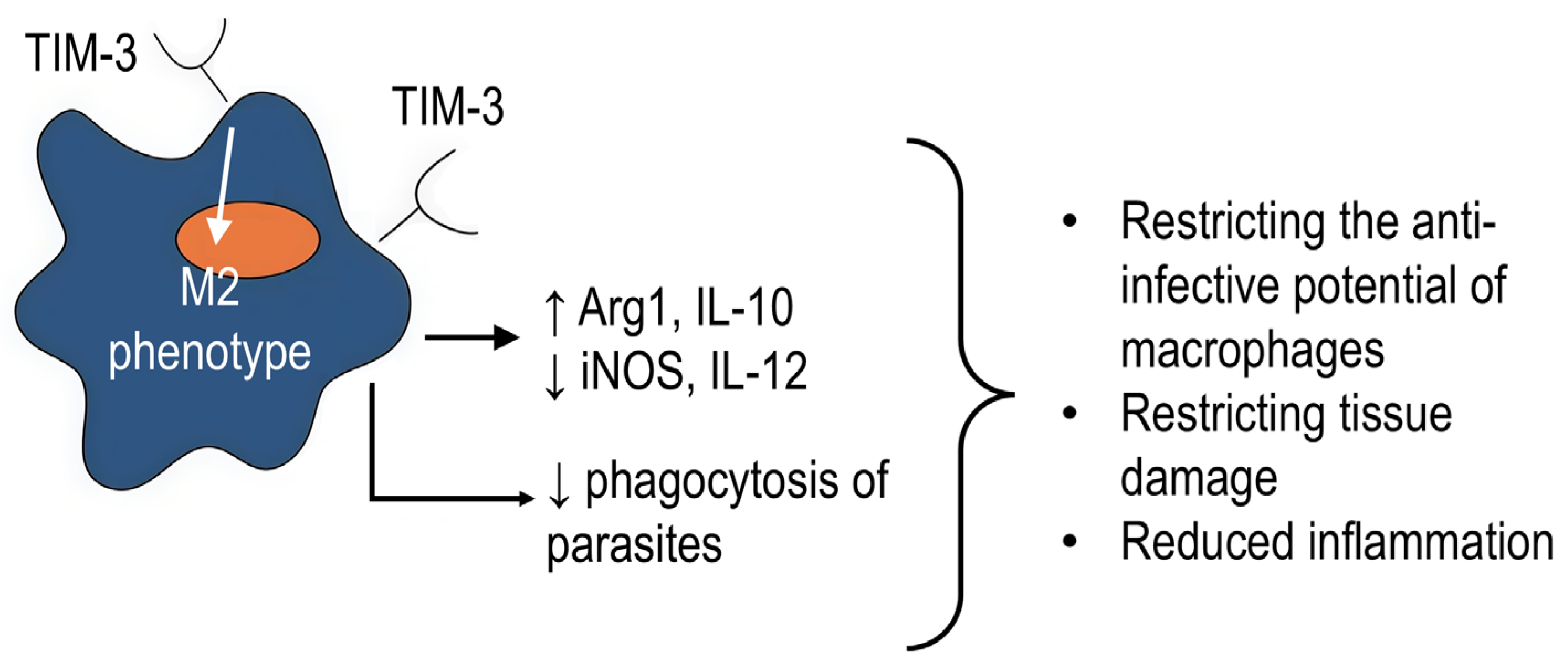
TIM-3 is involved in the regulation of anti-infective immune response [82]. Thus, after TIM-3 and PD-1 blocking, macrophages had a higher ability to limit the growth of M. tuberculosis in HIV+ (human immunodeficiency virus) patients [83]. In addition, some infectious agents induce infectious tolerance by increasing TIM-3 expression on macrophages, such as Schistosomes or Plasmodium. Moreover, blockade of TIM-3 during Schistosome infection led to polarization of macrophages in the antibacterial M1 direction, accompanied by increased expression of iNOS and IL-12 as well as a decrease in Arg1 and IL-10 [84]. Similarly, blockade of the TIM-3 signaling with anti-TIM-3 antibodies during Plasmodium infection enhanced phagocytosis and production of antiparasitic mediators by macrophages [67]. TIM-3 signaling is shown to suppress the phagocytic and cytotoxic functions of macrophages against C. albicans [47]. In addition, Tim-3 negatively regulates the secretion of IL-12 by monocytes in Hepacivirus C infection [85].
On the other hand, the anti-inflammatory action of TIM-3 appears to help restrict tissue damage in acute and chronic inflammation. Overexpression of TIM-3 in murine RAW264.7 macrophages reduced H. pylori-associated inflammation by blocking TLR4 and secretion of proinflammatory cytokines [86]. On monocytes, the findings are consistent with the broader hypothesis that TIM-3 helps suppress chronic infection, such as herpesvirus infection [87].
Yang et al. demonstrated that TIM-3 plays an important role in limiting sepsis-induced inflammation in both humans and mice. Downregulation and/or blockade of TIM-3 correlated with the severity of sepsis. Blockade and/or reduction in TIM-3 expression resulted in increased macrophage activation, which contributes to the systemic inflammatory response in sepsis. TIM-3 overexpression in macrophages significantly suppressed the production of proinflammatory cytokines [46].
On the other hand, TIM-3 expression may, under certain conditions, enhance the proinflammatory activity of myeloid cells. Thus, activation of TIM-3 in monocytes from patients with chronic hepatitis B increased the production of TNFα, IL-1β, and IL-6, while blocking TIM-3 reduced the ability of monocytes to induce a Th17 response [88].
Therefore, the cumulative evidence strongly suggests that TIM-3 on monocytes/macrophages predominantly plays a negative role in infection by restricting the anti-infective potential of macrophages (Figure 7). This negative regulation manifests likely through the promotion of M2-like phenotype, the attenuation of pro-inflammatory signaling pathways (e.g., TLR4), and further direct suppression of macrophage effector functions, such as phagocytosis and the production of antimicrobial mediators. While it might be beneficial in preventing persistent inflammation during certain chronic infections, it facilitates survival and persistence of various infectious agents within the host. Besides, TIM-3 in some infections may have a stimulatory role in inflammatory cytokine production and Th17 cell response.
Figure 7.
Mechanisms and effects of TIM-3 in anti-infective immunity of macrophages. ↑ symbol indicates up-regulation, ↓ symbol indicates down-regulation.
4.6. Diseases Associated with Inflammation in Pregnancy
Pregnancy is a unique condition in which the balance of pro- and anti-inflammatory agents is crucial. Disbalance leads to pathological processes such as preeclampsia and recurrent miscarriage [89]. It is noteworthy that TIM-3 is a critical immune checkpoint and multifunctional immunoregulatory molecule during pregnancy, inducing maternal-fetal tolerance and harmonizing the interactions between different cells in the process of maternal-fetal tolerance [90].
TIM-3 is constitutively expressed on decidual macrophages [52], and its expression is also increased on monocytes in the peripheral blood during pregnancy [41,44]. It is believed to be an important checkpoint that induces fetomaternal tolerance during pregnancy. TIM-3 expression is crucial for preventing pregnancy loss. A decrease in TIM-3 on CD14+ cells in the peripheral blood and decidual cells is associated with an imbalance in the anti- and pro-inflammatory cytokine profile, dysfunction of fetomaternal tolerance, and miscarriage [44,91].
In mouse models, adoptive transfer of TIM-3+ macrophages, but not TIM-3− macrophages, reduced macrophage depletion-induced abortion in mouse embryos [44,89], which may be associated with the production of angiogenic growth factors by TIM-3+ cells [92].
Preeclampsia-like disorders in rats are attenuated via the TIM-3/Gal-9 pathway, which reduces inflammatory cytokine levels [93].
On the other hand, other studies have suggested that abnormal TIM-3 expression levels may be associated with pregnancy loss [41]. One recent study found that significantly elevated TIM-3 levels on maternal macrophages in the decidua may be associated with the development of preeclampsia. However, the authors found this association for the female fetus but did not confirm it in the overall sample. In addition, the differences were on the very fringes of significance (p = 0.049) [94].
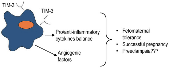
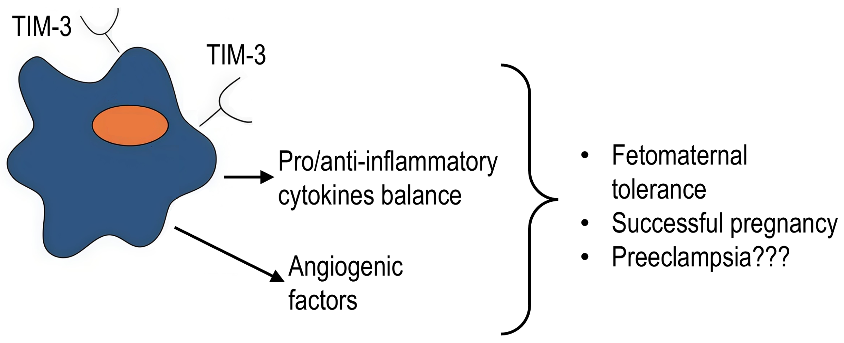
Therefore, TIM-3 expression on macrophages is an important regulator of successful pregnancy, fine-tuning the critical balance between pro- and anti-inflammatory states at the fetomaternal interface. These protective effects are likely mediated by angiogenic factor secretion and suppression of inflammatory cytokines (Figure 8); however, distinct mechanisms remain to be seen. Future research must focus on defining the optimal “therapeutic window” of TIM-3 activity and the distinct signaling pathways it engages in different pathologies of pregnancy.
Figure 8.
TIM-3 regulates the balance of pro- and anti-inflammatory cytokines during pregnancy. Its role in preeclampsia remains poorly understood. Question marks indicate that there is insufficient data on the given issue.
5. Conclusions
There is still much to learn about such a unique molecule as TIM-3. However, its expression on myeloid cells and, in particular, macrophages, plays a critical role in the regulation of the functional activity of these cells.
Despite a large amount of data demonstrating the anti-inflammatory effect of TIM-3 signaling and its association with the M2 phenotype of macrophages, some studies have found a link between TIM-3 expression and the proinflammatory properties of macrophages. Therefore, TIM-3 cannot be classified definitely as an inhibitory molecule since it rather plays a regulatory role.
The issue of ligand interactions with TIM-3 and TIM-3 signaling cascades remains unresolved. The exact pathways of TIM-3 signaling in macrophages require determination, but it is most likely that it is associated with TLRs and, in particular, TLR-4. Some of the probable mechanisms include the NF-κB, IRF3, STAT1, and STAT3 pathways. Further research in this area should clarify the discrepancies found and establish the exact pathways of activation of the TIM-3.
TIM-3 expression on macrophages is important for cancer and infectious diseases, acute and chronic inflammatory processes, as well as some physiological conditions, such as pregnancy. Given the ongoing clinical trials of anti-TIM-3 monoclonal antibodies for different diseases, the study of TIM-3 expression in macrophages, signaling pathways, and effects on macrophage functions is still relevant and necessary.
Current evidence indicates that TIM-3+ macrophages apparently play a negative role in oncological diseases, whereas their function in inflammation is context-dependent, and TIM-3 can either limit or promote the inflammatory response. These insights indicate that TIM-3+ macrophages represent a promising target for the development of further therapies aimed at illnesses such as cancer and both acute and chronic inflammation. Furthermore, TIM-3 may be a potential target in infectious diseases, as it appears to suppress anti-infective immunity. During pregnancy, the effect of TIM-3-expressing macrophages is likely positive; however, studies demonstrating the negative effect of TIM-3+ macrophages require further research to elucidate the role of TIM-3 during pregnancy.
The involvement of TIM-3+ macrophages in autoimmune inflammation and in defective inflammation resolution, particularly in fibrosis, is poorly understood and necessitates additional investigation. The staging of inflammation and different pathogenesis may be key factors in the divergence of TIM-3+ macrophage functions.
A deeper understanding of the physiopathological role of the TIM-3 pathway in innate immunity will shed new light on the pathogenesis of clinical diseases such as autoimmune diseases, chronic viral infections, and cancer, and suggest new approaches to intervention. The TIM-3 ligand axis and downstream signaling pathway molecules may become pivotal targets for future treatments.
Author Contributions
Conceptualization, A.M.; investigation, A.M.; writing—original draft preparation, A.M.; writing—review and editing, T.T. and E.C.; visualization, A.M.; supervision, T.T. and E.C. All authors have read and agreed to the published version of the manuscript.
Funding
This work was financed from the budget of RIFCI (ID 124112900022-0) and supported by a state-budgeted project (registration ID 125012300675-6).
Institutional Review Board Statement
Not applicable.
Informed Consent Statement
Not applicable.
Data Availability Statement
No new data were created or analyzed in this study. Data sharing is not applicable to this article.
Conflicts of Interest
The authors declare no conflicts of interest.
Abbreviations
The following abbreviations are used in this manuscript:
| TIM-3 | T-cell immunoglobulin and mucin domain 3 |
| PD-1 | Programmed cell death protein 1 |
| CTLA-4 | Cytotoxic T-lymphocyte-associated protein 4 |
| Ig | Immunoglobulin |
| ADAM A | disintegrin and metalloprotease |
| Gal-9 | galectin-9 |
| PtdSer | Phosphatidylserine |
| HMGB1 | High-mobility group protein B1 |
| CEACAM1 | Carcinoembryonic Antigen-Related Cell Adhesive Molecule 1 |
| STAT | Signal transducer and activator of transcription |
| LPS | Lipopolysaccharide |
| CD | Cluster of differentiation |
| iNOS | Inducible nitric oxide synthase |
| IL | Interleukin |
| TNFα | Tumor necrosis factor α |
| TGF-β | Transforming growth factor β |
| IFNγ | Interferon gamma |
| THP-1 | Human pro-monocytic cell line (acute monocytic leukemia) |
| TLR | Toll-like receptor |
| NF-κB | Nuclear factor kappa-light-chain-enhancer of activated B cells |
| IRF | Interferon regulatory factor |
| TRIF | TIR (Toll/interleukin-1 receptor) domain-containing adaptor protein inducing interferon beta |
| SOCS | Suppressor of cytokine signaling proteins |
| VEGF | Vascular endothelial growth factor |
| RAW264.7 | Mouse macrophage cell line (tumor tissue induced by Abelson murine leukemia virus) |
| TAM | Tumor-associated macrophages |
| DSS | Dextran Sulfate Sodium |
| MHC | Major histocompatibility complex |
| HIV | human immunodeficiency virus |
References
- Friedlaender, A.; Addeo, A.; Banna, G. New emerging targets in cancer immunotherapy: The role of TIM3. ESMO Open 2020, 4, e000629. [Google Scholar] [CrossRef] [Scilit] [PubMed]
- Fourcade, J.; Sun, Z.; Benallaoua, M.; Guillaume, P.; Luescher, I.F.; Sander, C.; Kirkwood, J.M.; Kuchroo, V.; Zarour, H.M. Upregulation of Tim-3 and PD-1 expression is associated with tumor antigen-specific CD8+ T cell dysfunction in melanoma patients. J. Exp. Med. 2010, 207, 2175–2186. [Google Scholar] [CrossRef] [Scilit] [PubMed]
- Koyama, S.; Akbay, E.A.; Li, Y.Y.; Herter-Sprie, G.S.; Buczkowski, K.A.; Richards, W.G.; Gandhi, L.; Redig, A.J.; Rodig, S.J.; Asahina, H.; et al. Adaptive resistance to therapeutic PD-1 blockade is associated with upregulation of alternative immune checkpoints. Nat. Commun. 2016, 7, 10501. [Google Scholar] [CrossRef] [Scilit] [PubMed]
- Oweida, A.; Hararah, M.K.; Phan, A.; Binder, D.; Bhatia, S.; Lennon, S.; Bukkapatnam, S.; Van Court, B.; Uyanga, N.; Darragh, L.; et al. Resistance to radiotherapy and PD-L1 blockade is mediated by TIM-3 upregulation and regulatory T-cell infiltration. Clin. Cancer Res. 2018, 24, 5368–5380. [Google Scholar] [CrossRef] [Scilit]
- Hayashi, F.; Akagi, K.; Taniguchi, H.; Matsutake, T.; Kawahara, H.; Sekine, I.; Gyotoku, H.; Takemoto, S.; Soda, H.; Ashizawa, K.; et al. TIM-3 expression induces resistance to PD-1 inhibitor in G-CSF-producing lung spindle cell carcinoma: A case report. Thorac. Cancer 2023, 14, 3556–3560. [Google Scholar] [CrossRef] [Scilit]
- Chen, H.; Zha, J.; Tang, R.; Chen, G. T-cell immunoglobulin and mucin-domain containing-3 (TIM-3): Solving a key puzzle in autoimmune diseases. Int. Immunopharmacol. 2023, 121, 110418. [Google Scholar] [CrossRef] [Scilit]
- Yang, J.; Chang, T.; Tang, L.; Deng, H.; Chen, D.; Luo, J.; Wu, H.; Tang, T.; Zhang, C.; Li, Z.; et al. Increased Expression of Tim-3 Is Associated with Depletion of NKT Cells In SARS-CoV-2 Infection. Front. Immunol. 2022, 13, 796682. [Google Scholar] [CrossRef] [Scilit]
- Sehrawat, S.; Reddy, P.B.; Rajasagi, N.; Suryawanshi, A.; Hirashima, M.; Rouse, B.T. Galectin-9/TIM-3 interaction regulates virus-specific primary and memory CD8 T cell response. PLoS Pathog. 2010, 6, e1000882. [Google Scholar] [CrossRef] [Scilit]
- Shi, Q.; Li, G.; Dou, S.; Tang, L.; Hou, C.; Wang, Z.; Gao, Y.; Gao, Z.; Hao, Y.; Mo, R.; et al. Negative Regulation of RIG-I by Tim-3 Promotes H1N1 Infection. Immunol. Investig. 2023, 52, 1–19. [Google Scholar] [CrossRef] [Scilit]
- Liu, Y.; Gao, L.F.; Liang, X.H.; Ma, C.H. Role of Tim-3 in hepatitis B virus infection: An overview. World J. Gastroenterol. 2016, 22, 2294–2303. [Google Scholar] [CrossRef] [Scilit]
- Khalaf, S.; Toor, S.M.; Murshed, K.; Kurer, M.A.; Ahmed, A.A.; Abu Nada, M.; Elkord, E. Differential expression of TIM-3 in circulation and tumor microenvironment of colorectal cancer patients. Clin. Immunol. 2020, 215, 108429. [Google Scholar] [CrossRef] [Scilit] [PubMed]
- Foks, A.C.; Ran, I.A.; Wasserman, L.; Frodermann, V.; Ter Borg, M.N.; de Jager, S.C.; van Santbrink, P.J.; Yagita, H.; Akiba, H.; Bot, I.; et al. T-cell immunoglobulin and mucin domain 3 acts as a negative regulator of atherosclerosis. Arterioscler. Thromb. Vasc. Biol. 2013, 33, 2558–2565. [Google Scholar] [CrossRef] [Scilit] [PubMed]
- De Mingo Pulido, Á.; Gardner, A.; Hiebler, S.; Soliman, H.; Rugo, H.S.; Krummel, M.F.; Coussens, L.M.; Ruffell, B. TIM-3 Regulates CD103+ Dendritic Cell Function and Response to Chemotherapy in Breast Cancer. Cancer Cell 2018, 33, 60–74.e6. [Google Scholar] [CrossRef] [Scilit] [PubMed]
- Shapouri-Moghaddam, A.; Mohammadian, S.; Vazini, H.; Taghadosi, M.; Esmaeili, S.A.; Mardani, F.; Seifi, B.; Mohammadi, A.; Afshari, J.T.; Sahebkar, A. Macrophage plasticity, polarization, and function in health and disease. J. Cell Physiol. 2018, 233, 6425–6440. [Google Scholar] [CrossRef] [Scilit]
- Pan, Y.; Yu, Y.; Wang, X.; Zhang, T. Tumor-Associated Macrophages in Tumor Immunity. Front. Immunol. 2020, 11, 583084. [Google Scholar] [CrossRef] [Scilit]
- Zhang, J.; Hou, C.; Dou, S.; Li, G.; Wang, Z.; Liu, Y.; Zhang, Y.; Wang, R.; Shen, B.; Han, G. T cell immunoglobulin and mucin domain protein 3 inhibits glycolysis in RAW 264.7 macrophages through Hexokinase 2. Scand. J. Immunol. 2021, 93, e12981. [Google Scholar] [CrossRef] [Scilit]
- Das, M.; Zhu, C.; Kuchroo, V.K. Tim-3 and its role in regulating anti-tumor immunity. Immunol. Rev. 2017, 276, 97–111. [Google Scholar] [CrossRef] [Scilit]
- Möller-Hackbarth, K.; Dewitz, C.; Schweigert, O.; Trad, A.; Garbers, C.; Rose-John, S.; Scheller, J. A disintegrin and metalloprotease (ADAM) 10 and ADAM17 are major sheddases of T cell immunoglobulin and mucin domain 3 (Tim-3). J. Biol. Chem. 2013, 288, 34529–34544. [Google Scholar] [CrossRef] [Scilit]
- Hansen, J.A.; Hanash, S.M.; Tabellini, L.; Baik, C.; Lawler, R.L.; Grogan, B.M.; Storer, B.; Chin, A.; Johnson, M.; Wong, C.H.; et al. A novel soluble form of Tim-3 associated with severe graft-versus-host disease. Biol. Blood Marrow Transplant. 2013, 19, 1323–1330. [Google Scholar] [CrossRef] [Scilit]
- Dardalhon, V.; Anderson, A.C.; Karman, J.; Apetoh, L.; Chandwaskar, R.; Lee, D.H.; Cornejo, M.; Nishi, N.; Yamauchi, A.; Quintana, F.J.; et al. Tim-3/galectin-9 pathway: Regulation of Th1 immunity through promotion of CD11b+Ly-6G+ myeloid cells. J. Immunol. 2010, 185, 1383–1392. [Google Scholar] [CrossRef] [Scilit]
- Nakayama, M.; Akiba, H.; Takeda, K.; Kojima, Y.; Hashiguchi, M.; Azuma, M.; Yagita, H.; Okumura, K. Tim-3 mediates phagocytosis of apoptotic cells and cross-presentation. Blood 2009, 113, 3821–3830. [Google Scholar] [CrossRef] [Scilit] [PubMed]
- Smith, C.M.; Li, A.; Krishnamurthy, N.; Lemmon, M.A. Phosphatidylserine binding directly regulates TIM-3 function. Biochem. J. 2021, 478, 3331–3349. [Google Scholar] [CrossRef] [Scilit] [PubMed]
- Chiba, S.; Baghdadi, M.; Akiba, H.; Yoshiyama, H.; Kinoshita, I.; Dosaka-Akita, H.; Fujioka, Y.; Ohba, Y.; Gorman, J.V.; Colgan, J.D.; et al. Tumor-infiltrating DCs suppress nucleic acid-mediated innate immune responses through interactions between the receptor TIM-3 and the alarmin HMGB1. Nat. Immunol. 2012, 13, 832–842. [Google Scholar] [CrossRef] [Scilit] [PubMed]
- Dolina, J.S.; Braciale, T.J.; Hahn, Y.S. Liver-primed CD8+ T cells suppress antiviral adaptive immunity through galectin-9-independent T-cell immunoglobulin and mucin 3 engagement of high-mobility group box 1 in mice. Hepatology 2014, 59, 1351–1365. [Google Scholar] [CrossRef] [Scilit]
- Huang, Y.H.; Zhu, C.; Kondo, Y.; Anderson, A.C.; Gandhi, A.; Russell, A.; Dougan, S.K.; Petersen, B.S.; Melum, E.; Pertel, T.; et al. CEACAM1 regulates TIM-3-mediated tolerance and exhaustion. Nature 2015, 517, 386–390, Correction in Nature 2016, 536, 359. https://doi.org/10.1038/nature17421. [Google Scholar] [CrossRef] [Scilit]
- Leitner, J.; Rieger, A.; Pickl, W.F.; Zlabinger, G.; Grabmeier-Pfistershammer, K.; Steinberger, P. TIM-3 does not act as a receptor for galectin-9. PLoS Pathog. 2013, 9, e1003253. [Google Scholar] [CrossRef] [Scilit]
- Su, E.W.; Bi, S.; Kane, L.P. Galectin-9 regulates T helper cell function independently of Tim-3. Glycobiology 2011, 21, 1258–1265. [Google Scholar] [CrossRef] [Scilit]
- Oomizu, S.; Arikawa, T.; Niki, T.; Kadowaki, T.; Ueno, M.; Nishi, N.; Yamauchi, A.; Hirashima, M. Galectin-9 suppresses Th17 cell development in an IL-2-dependent but Tim-3-independent manner. Clin. Immunol. 2012, 43, 51–58. [Google Scholar] [CrossRef] [Scilit]
- Chen, C.J.; Shively, J.E. The cell-cell adhesion molecule carcinoembryonic antigen-related cellular adhesion molecule 1 inhibits IL-2 production and proliferation in human T cells by association with Src homology protein-1 and down-regulates IL-2 receptor. J. Immunol. 2004, 172, 3544–3552. [Google Scholar] [CrossRef] [Scilit]
- Li, Y.; Shively, J.E. CEACAM1 regulates Fas-mediated apoptosis in Jurkat T-cells via its interaction with beta-catenin. Exp. Cell Res. 2013, 319, 1061–1072. [Google Scholar] [CrossRef] [Scilit]
- Kadowaki, T.; Arikawa, T.; Shinonaga, R.; Oomizu, S.; Inagawa, H.; Soma, G.; Niki, T.; Hirashima, M. Galectin-9 signaling prolongs survival in murine lung cancer by inducing macrophages to differentiate into plasmacytoid dendritic cell-like macrophages. Clin. Immunol. 2012, 142, 296–307. [Google Scholar] [CrossRef] [Scilit] [PubMed]
- De Sousa Linhares, A.; Kellner, F.; Jutz, S.; Zlabinger, G.J.; Gabius, H.J.; Huppa, J.B.; Leitner, J.; Steinberger, P. TIM-3 and CEACAM1 do not interact in cis and in trans. Eur. J. Immunol. 2020, 50, 1126–1141. [Google Scholar] [CrossRef] [Scilit] [PubMed]
- Wolf, Y.; Anderson, A.C.; Kuchroo, V.K. TIM3 comes of age as an inhibitory receptor. Nat. Rev. Immunol. 2020, 20, 173–185. [Google Scholar] [CrossRef] [Scilit] [PubMed]
- Chen, S.; Saeed, A.F.U.H.; Liu, Q.; Jiang, Q.; Xu, H.; Xiao, G.G.; Rao, L.; Duo, Y. Macrophages in immunoregulation and therapeutics. Signal Transduct. Target. Ther. 2023, 8, 207. [Google Scholar] [CrossRef] [Scilit]
- Frisancho-Kiss, S.; Coronado, M.J.; Frisancho, J.A.; Lau, V.M.; Rose, N.R.; Klein, S.L.; Fairweather, D. Gonadectomy of male BALB/c mice increases Tim-3(+) alternatively activated M2 macrophages, Tim-3(+) T cells, Th2 cells, and Treg in the heart during acute coxsackievirus-induced myocarditis. Brain Behav. Immun. 2009, 23, 649–657. [Google Scholar] [CrossRef] [Scilit]
- Zhang, Y.; Zhang, W. Tim-3 regulates the ability of macrophages to counter lipopolysaccharide-induced pulmonary epithelial barrier dysfunction via the PI3K/Akt pathway in epithelial cells. Mol. Med. Rep. 2020, 22, 534–542. [Google Scholar] [CrossRef] [Scilit]
- Ocaña-Guzman, R.; Ramon-Luing, L.A.; Vazquez-Bolaños, L.A.; Rodríguez-Alvarado, M.; Bulhusen-Rodriguez, F.; Torres-Hatem, A.; Gonzalez-Torres, K.; de Alba-Alvarado, M.C.; Sada-Ovalle, I. Tim-3 Is Differentially Expressed during Cell Activation and Interacts with the LSP-1 Protein in Human Macrophages. J. Immunol. Res. 2023, 2023, 3577334. [Google Scholar] [CrossRef] [Scilit]
- Sun, J.; Huang, Q.; Li, S.; Meng, F.; Li, X.; Gong, X. miR-330-5p/Tim-3 axis regulates macrophage M2 polarization and insulin resistance in diabetes mice. Mol. Immunol. 2018, 95, 107–113. [Google Scholar] [CrossRef] [Scilit]
- Stempin, C.C.; Geysels, R.C.; Park, S.; Palacios, L.M.; Volpini, X.; Motran, C.C.; Acosta Rodríguez, E.V.; Nicola, J.P.; Cheng, S.Y.; Pellizas, C.G.; et al. Secreted Factors by Anaplastic Thyroid Cancer Cells Induce Tumor-Promoting M2-like Macrophage Polarization through a TIM3-Dependent Mechanism. Cancers 2021, 13, 4821. [Google Scholar] [CrossRef] [Scilit]
- Zhang, Z.Y.; Schluesener, H.J.; Zhang, Z. Distinct expression of Tim-3 during different stages of rat experimental autoimmune neuritis. Brain Res. Bull. 2011, 86, 229–234. [Google Scholar] [CrossRef] [Scilit]
- Zhao, J.; Lei, Z.; Liu, Y.; Li, B.; Zhang, L.; Fang, H.; Song, C.; Wang, X.; Zhang, G.M.; Feng, Z.H.; et al. Human pregnancy up-regulates Tim-3 in innate immune cells for systemic immunity. J. Immunol. 2009, 182, 6618–6624. [Google Scholar] [CrossRef] [Scilit] [PubMed]
- Cheng, L.J.; Dong, S.Y.; Ni, X.F.; Mei, L.; He, J.G.; Mamat, A.; Gong, Z.C. Effect of the TIM-3/Gal-9 signaling pathway on macrophage polarization in peri-implantitis. PLoS ONE 2025, 20, e0328258. [Google Scholar] [CrossRef] [Scilit] [PubMed]
- Yu, A.; Zhang, X.; Li, M.; Ye, P.; Duan, H.; Zhang, T.; Yang, Z. Tim-3 enhances brain inflammation by promoting M1 macrophage polarization following intracerebral hemorrhage in mice. Int. Immunopharmacol. 2017, 53, 143–148. [Google Scholar] [CrossRef] [Scilit] [PubMed]
- Li, M.; Sun, F.; Xu, Y.; Chen, L.; Chen, C.; Cui, L.; Qian, J.; Li, D.; Wang, S.; Du, M. Tim-3+ decidual Mφs induced Th2 and Treg bias in decidual CD4+T cells and promoted pregnancy maintenance via CD132. Cell Death Dis. 2022, 13, 454. [Google Scholar] [CrossRef] [Scilit]
- Basak, B.; Akashi-Takamura, S. IRF3 function and immunological gaps in sepsis. Front. Immunol. 2024, 15, 1336813. [Google Scholar] [CrossRef] [Scilit]
- Yang, X.; Jiang, X.; Chen, G.; Xiao, Y.; Geng, S.; Kang, C.; Zhou, T.; Li, Y.; Guo, X.; Xiao, H.; et al. T cell Ig mucin-3 promotes homeostasis of sepsis by negatively regulating the TLR response. J. Immunol. 2013, 190, 2068–2079. [Google Scholar] [CrossRef] [Scilit]
- Liu, S.; Lin, K.; Tian, Y.; He, Y.; Zhang, Z.; Li, C.; Luo, Y.; Liang, Y.; Ma, Y.; Deng, W. Candidalysin induces Tim-3 expression in macrophages to suppress antifungal immunity in oropharyngeal candidiasis. Int. Immunopharmacol. 2025, 156, 114672. [Google Scholar] [CrossRef] [Scilit]
- Jiang, X.; Yu, J.; Shi, Q.; Xiao, Y.; Wang, W.; Chen, G.; Zhao, Z.; Wang, R.; Xiao, H.; Hou, C.; et al. Tim-3 promotes intestinal homeostasis in DSS colitis by inhibiting M1 polarization of macrophages. Clin. Immunol. 2015, 160, 328–335. [Google Scholar] [CrossRef] [Scilit]
- Yang, H.; Xie, T.; Li, D.; Du, X.; Wang, T.; Li, C.; Song, X.; Xu, L.; Yi, F.; Liang, X.; et al. Tim-3 aggravates podocyte injury in diabetic nephropathy by promoting macrophage activation via the NF-κB/TNF-α pathway. Mol. Metab. 2019, 23, 24–36. [Google Scholar] [CrossRef] [Scilit]
- Jiang, X.; Zhou, T.; Xiao, Y.; Yu, J.; Dou, S.; Chen, G.; Wang, R.; Xiao, H.; Hou, C.; Wang, W.; et al. Tim-3 promotes tumor-promoting M2 macrophage polarization by binding to STAT1 and suppressing the STAT1-miR-155 signaling axis. Oncoimmunology 2016, 5, e1211219. [Google Scholar] [CrossRef] [Scilit]
- Tang, L.; Li, G.; Zheng, Y.; Hou, C.; Gao, Y.; Hao, Y.; Gao, Z.; Mo, R.; Li, Y.; Shen, B.; et al. Tim-3 Relieves Experimental Autoimmune Encephalomyelitis by Suppressing MHC-II. Front. Immunol. 2022, 12, 770402. [Google Scholar] [CrossRef] [Scilit]
- Zhang, D.; Ren, L.; Zhao, M.; Yang, C.; Liu, X.; Zhang, H.; Jiang, Y.; Sun, X.; Li, T.; Cui, L.; et al. Role of Tim-3 in Decidual Macrophage Functional Polarization During Abnormal Pregnancy with Toxoplasma gondii Infection. Front. Immunol. 2019, 10, 1550. [Google Scholar] [CrossRef] [Scilit] [PubMed]
- Guo, Q.; Jin, Y.; Chen, X.; Ye, X.; Shen, X.; Lin, M.; Zeng, C.; Zhou, T.; Zhang, J. NF-κB in biology and targeted therapy: New insights and translational implications. Signal Transduct. Target. Ther. 2024, 9, 53. [Google Scholar] [CrossRef] [Scilit] [PubMed]
- Ma, C.J.; Li, G.Y.; Cheng, Y.Q.; Wang, J.M.; Ying, R.S.; Shi, L.; Wu, X.Y.; Niki, T.; Hirashima, M.; Li, C.F.; et al. Cis association of galectin-9 with Tim-3 differentially regulates IL-12/IL-23 expressions in monocytes via TLR signaling. PLoS ONE 2013, 8, e72488. [Google Scholar] [CrossRef] [Scilit] [PubMed]
- Zhang, Y.; Ma, C.J.; Wang, J.M.; Ji, X.J.; Wu, X.Y.; Moorman, J.P.; Yao, Z.Q. Tim-3 regulates pro- and anti-inflammatory cytokine expression in human CD14+ monocytes. J. Leukoc. Biol. 2012, 91, 189–196. [Google Scholar] [CrossRef] [Scilit]
- Zhang, W.; Zhang, Y.; He, Y.; Wang, X.; Fang, Q. Lipopolysaccharide mediates time-dependent macrophage M1/M2 polarization through the Tim-3/Galectin-9 signalling pathway. Exp. Cell Res. 2019, 376, 124–132. [Google Scholar] [CrossRef] [Scilit]
- Wang, Z.; Yin, N.; Zhang, Z.; Zhang, Y.; Zhang, G.; Chen, W. Upregulation of T-cell Immunoglobulin and Mucin-Domain Containing-3 (Tim-3) in Monocytes/Macrophages Associates with Gastric Cancer Progression. Immunol. Investig. 2017, 46, 134–148. [Google Scholar] [CrossRef] [Scilit]
- Vidyarthi, A.; Khan, N.; Agnihotri, T.; Negi, S.; Das, D.K.; Aqdas, M.; Chatterjee, D.; Colegio, O.R.; Tewari, M.K.; Agrewala, J.N. TLR-3 Stimulation Skews M2 Macrophages to M1 Through IFN-αβ Signaling and Restricts Tumor Progression. Front. Immunol. 2018, 9, 1650. [Google Scholar] [CrossRef] [Scilit]
- Wang, F.; Zhou, F.; Peng, J.; Chen, H.; Xie, J.; Liu, C.; Xiong, H.; Chen, S.; Xue, G.; Zhou, X.; et al. Macrophage Tim-3 maintains intestinal homeostasis in DSS-induced colitis by suppressing neutrophil necroptosis. Redox Biol. 2024, 70, 103072. [Google Scholar] [CrossRef] [Scilit]
- Liang, S.; Cai, J.; Li, Y.; Yang, R. 1,25-Dihydroxy-Vitamin D3 induces macrophage polarization to M2 by upregulating T-cell Ig-mucin-3 expression. Mol. Med. Rep. 2019, 19, 3707–3713. [Google Scholar] [CrossRef] [Scilit]
- Du, X.; Wu, Z.; Xu, Y.; Liu, Y.; Liu, W.; Wang, T.; Li, C.; Zhang, C.; Yi, F.; Gao, L.; et al. Increased Tim-3 expression alleviates liver injury by regulating macrophage activation in MCD-induced NASH mice. Cell Mol. Immunol. 2019, 16, 878–886. [Google Scholar] [CrossRef] [Scilit] [PubMed]
- Chanteux, H.; Guisset, A.C.; Pilette, C.; Sibille, Y. LPS induces IL-10 production by human alveolar macrophages via MAPKinases- and Sp1-dependent mechanisms. Respir. Res. 2007, 8, 71. [Google Scholar] [CrossRef] [Scilit] [PubMed]
- Van der Plas, M.J.; van Dissel, J.T.; Nibbering, P.H. Maggot secretions skew monocyte-macrophage differentiation away from a pro-inflammatory to a pro-angiogenic type. PLoS ONE 2009, 4, e8071. [Google Scholar] [CrossRef] [Scilit] [PubMed]
- Anderson, A.C.; Anderson, D.E.; Bregoli, L.; Hastings, W.D.; Kassam, N.; Lei, C.; Chandwaskar, R.; Karman, J.; Su, E.W.; Hirashima, M.; et al. Promotion of tissue inflammation by the immune receptor Tim-3 expressed on innate immune cells. Science 2007, 318, 1141–1143. [Google Scholar] [CrossRef] [Scilit]
- Isshiki, T.; Akiba, H.; Nakayama, M.; Harada, N.; Okumura, K.; Homma, S.; Miyake, S. Cutting Edge: Anti-TIM-3 Treatment Exacerbates Pulmonary Inflammation and Fibrosis in Mice. J. Immunol. 2017, 199, 3733–3737. [Google Scholar] [CrossRef] [Scilit]
- DeKruyff, R.H.; Bu, X.; Ballesteros, A.; Santiago, C.; Chim, Y.L.; Lee, H.H.; Karisola, P.; Pichavant, M.; Kaplan, G.G.; Umetsu, D.T.; et al. T cell/transmembrane, Ig, and mucin-3 allelic variants differentially recognize phosphatidylserine and mediate phagocytosis of apoptotic cells. J. Immunol. 2010, 184, 1918–1930. [Google Scholar] [CrossRef] [Scilit]
- Hou, N.; Jiang, N.; Zou, Y.; Piao, X.; Liu, S.; Li, S.; Chen, Q. Down-Regulation of Tim-3 in Monocytes and Macrophages in Plasmodium Infection and Its Association with Parasite Clearance. Front. Microbiol. 2017, 8, 1431. [Google Scholar] [CrossRef] [Scilit]
- Yan, W.; Liu, X.; Ma, H.; Zhang, H.; Song, X.; Gao, L.; Liang, X.; Ma, C. Tim-3 fosters HCC development by enhancing TGF-β-mediated alternative activation of macrophages. Gut 2015, 64, 1593–1604. [Google Scholar] [CrossRef] [Scilit]
- Katagata, M.; Okayama, H.; Nakajima, S.; Saito, K.; Sato, T.; Sakuma, M.; Fukai, S.; Endo, E.; Sakamoto, W.; Saito, M.; et al. TIM-3 Expression and M2 Polarization of Macrophages in the TGFβ-Activated Tumor Microenvironment in Colorectal Cancer. Cancers 2023, 15, 4943. [Google Scholar] [CrossRef] [Scilit]
- Zhang, C.; Xu, L.; Ma, Y.; Huang, Y.; Zhou, L.; Le, H.; Chen, Z. Increased TIM-3 expression in tumor-associated macrophages predicts a poorer prognosis in non-small cell lung cancer: A retrospective cohort study. J. Thorac. Dis. 2023, 15, 1433–1444. [Google Scholar] [CrossRef] [Scilit]
- Ausejo-Mauleon, I.; Labiano, S.; de la Nava, D.; Laspidea, V.; Zalacain, M.; Marrodán, L.; García-Moure, M.; González-Huarriz, M.; Hervás-Corpión, I.; Dhandapani, L.; et al. TIM-3 blockade in diffuse intrinsic pontine glioma models promotes tumor regression and antitumor immune memory. Cancer Cell 2023, 41, 1911–1926.e8. [Google Scholar] [CrossRef] [Scilit] [PubMed]
- Vanmeerbeek, I.; Naulaerts, S.; Sprooten, J.; Laureano, R.S.; Govaerts, J.; Trotta, R.; Pretto, S.; Zhao, S.; Cafarello, S.T.; Verelst, J.; et al. Targeting conserved TIM3+VISTA+ tumor-associated macrophages overcomes resistance to cancer immunotherapy. Sci. Adv. 2024, 10, eadm8660. [Google Scholar] [CrossRef] [Scilit] [PubMed]
- Hu, W.; Li, D.; Yang, Y.; Zheng, Y.; Zeng, J.; Sai, K. TIM-3/CD68 double-high expression in Glioma: Prognostic characteristics and potential therapeutic approaches. Int. Immunopharmacol. 2024, 139, 112665. [Google Scholar] [CrossRef] [Scilit] [PubMed]
- Ning, Q.; Jian, T.; Cui, S.; Shi, L.; Jian, X.; He, X.; Zhang, X.; Li, X. Tim-3 facilitates immune escape in benzene-induced acute myeloid leukemia mouse model by promoting macrophage M2 polarization. Ecotoxicol. Environ. Saf. 2023, 266, 115532. [Google Scholar] [CrossRef] [Scilit]
- Ni, X.; Wu, W.; Sun, X.; Ma, J.; Yu, Z.; He, X.; Cheng, J.; Xu, P.; Liu, H.; Shang, T.; et al. Interrogating glioma-M2 macrophage interactions identifies Gal-9/Tim-3 as a viable target against PTEN-null glioblastoma. Sci. Adv. 2022, 8, eabl5165. [Google Scholar] [CrossRef] [Scilit]
- Dong, J.; Cheng, L.; Zhao, M.; Pan, X.; Feng, Z.; Wang, D. Tim-3-expressing macrophages are functionally suppressed and expanded in oral squamous cell carcinoma due to virus-induced Gal-9 expression. Tumour Biol. 2017, 39, 1010428317701651. [Google Scholar] [CrossRef] [Scilit]
- Xu, C.; Wang, T.; Cheng, S.; Liu, Y. Increased expression of T cell immunoglobulin and mucin domain 3 aggravates brain inflammation via regulation of the function of microglia/macrophages after intracerebral hemorrhage in mice. J. Neuroinflammation 2013, 10, 141. [Google Scholar] [CrossRef] [Scilit]
- Kim, K.S.; Lee, C.; Kim, H.S.; Gu, S.J.; Yoon, H.J.; Won, S.B.; Lee, H.; Lee, Y.S.; Kim, S.S.; Kane, L.P.; et al. TIM-3 on myeloid cells promotes pulmonary inflammation through increased production of galectin-3. Commun. Biol. 2024, 7, 1090. [Google Scholar] [CrossRef] [Scilit]
- Wang, Y.; Kuai, Q.; Gao, F.; Wang, Y.; He, M.; Zhou, H.; Han, G.; Jiang, X.; Ren, S.; Yu, Q. Overexpression of TIM-3 in Macrophages Aggravates Pathogenesis of Pulmonary Fibrosis in Mice. Am. J. Respir. Cell Mol. Biol. 2019, 61, 727–736. [Google Scholar] [CrossRef] [Scilit]
- Lavon, I.; Heli, C.; Brill, L.; Charbit, H.; Vaknin-Dembinsky, A. Blood Levels of Co-inhibitory-Receptors: A Biomarker of Disease Prognosis in Multiple Sclerosis. Front. Immunol. 2019, 10, 835. [Google Scholar] [CrossRef] [Scilit]
- Skejoe, C.; Hansen, A.S.; Stengaard-Pedersen, K.; Junker, P.; Hoerslev-Pedersen, K.; Hetland, M.L.; Oestergaard, M.; Greisen, S.; Hvid, M.; Deleuran, M.; et al. T-cell immunoglobulin and mucin domain 3 is upregulated in rheumatoid arthritis, but insufficient in controlling inflammation. Am. J. Clin. Exp. Immunol. 2022, 11, 34–44. [Google Scholar] [PubMed]
- Wang, P.H.; Wu, M.F.; Hsu, C.Y.; Pan, S.W.; Shu, C.C.; Cheng, S.L. The Trend of TIM3 Expression on T Cells in Patients With Nontuberculous Mycobacterial Lung Disease: From Immune Cell Dysfunction to Clinical Severity. Front. Immunol. 2021, 12, 738056. [Google Scholar] [CrossRef] [Scilit] [PubMed]
- Sada-Ovalle, I.; Ocaña-Guzman, R.; Pérez-Patrigeón, S.; Chávez-Galán, L.; Sierra-Madero, J.; Torre-Bouscoulet, L.; Addo, M.M. Tim-3 blocking rescue macrophage and T cell function against Mycobacterium tuberculosis infection in HIV+ patients. J. Int. AIDS Soc. 2015, 18, 20078. [Google Scholar] [CrossRef] [Scilit] [PubMed]
- Hou, N.; Piao, X.; Liu, S.; Wu, C.; Chen, Q. Tim-3 induces Th2-biased immunity and alternative macrophage activation during Schistosoma japonicum infection. Infect. Immun. 2015, 83, 3074–3082. [Google Scholar] [CrossRef] [Scilit]
- Zhang, Y.; Ma, C.J.; Wang, J.M.; Ji, X.J.; Wu, X.Y.; Jia, Z.S.; Yao, Z.Q. Tim-3 negatively regulates IL-12 expression by monocytes in HCV infection. PLoS ONE 2011, 6, e19664. [Google Scholar] [CrossRef] [Scilit]
- Wang, F.; Mao, Z.; Liu, D.; Yu, J.; Wang, Y.; Ye, W.; Lin, D.; Zhou, N.; Xie, Y. Overexpression of Tim-3 reduces Helicobacter pylori-associated inflammation through TLR4/NFκB signaling in vitro. Mol. Med. Rep. 2017, 15, 3252–3258. [Google Scholar] [CrossRef] [Scilit]
- Rashchupkin, I.M.; Meledina, I.V.; Kotova, M.A.; Zheltova, O.I. PD-1 and Tim-3 expression on different subpopulations of monocytes in chronic often recurrent herpesvirus infection (In Rus). Russ. J. Infect. Immun. Infektsiya I Immun. 2024, 14, 575–580. [Google Scholar] [CrossRef] [Scilit]
- Wang, J.; Li, C.; Fu, J.; Wang, X.; Feng, X.; Pan, X. Tim-3 regulates inflammatory cytokine expression and Th17 cell response induced by monocytes from patients with chronic hepatitis B. Scand. J. Immunol. 2019, 89, e12755. [Google Scholar] [CrossRef] [Scilit]
- Negishi, Y.; Shima, Y.; Takeshita, T.; Morita, R. Harmful and beneficial effects of inflammatory response on reproduction: Sterile and pathogen-associated inflammation. Immunol. Med. 2021, 44, 98–115. [Google Scholar] [CrossRef] [Scilit]
- Meng, X.; Luo, Y.; Cui, L.; Wang, S. Involvement of Tim-3 in Maternal-fetal Tolerance: A Review of Current Understanding. Int. J. Biol. Sci. 2025, 21, 789–801. [Google Scholar] [CrossRef] [Scilit]
- Chabtini, L.; Mfarrej, B.; Mounayar, M.; Zhu, B.; Batal, I.; Dakle, P.J.; Smith, B.D.; Boenisch, O.; Najafian, N.; Akiba, H.; et al. TIM-3 regulates innate immune cells to induce fetomaternal tolerance. J. Immunol. 2013, 190, 88–96. [Google Scholar] [CrossRef] [Scilit]
- Cui, L.; Sun, F.; Xu, Y.; Li, M.; Chen, L.; Chen, C.; Qian, J.; Li, D.; Du, M.; Wang, S. Tim-3 Coordinates Macrophage-Trophoblast Crosstalk via Angiogenic Growth Factors to Promote Pregnancy Maintenance. Int. J. Mol. Sci. 2023, 24, 1538. [Google Scholar] [CrossRef] [Scilit]
- Li, Z.H.; Wang, L.L.; Liu, H.; Muyayalo, K.P.; Huang, X.B.; Mor, G.; Liao, A.H. Galectin-9 Alleviates LPS-Induced Preeclampsia-Like Impairment in Rats via Switching Decidual Macrophage Polarization to M2 Subtype. Front. Immunol. 2019, 9, 3142. [Google Scholar] [CrossRef] [Scilit]
- Mittelberger, J.; Seefried, M.; Löb, S.; Kuhn, C.; Franitza, M.; Garrido, F.; Ditsch, N.; Jeschke, U.; Dannecker, C. The expression of TIM-3 and Gal-9 on macrophages and Hofbauer cells in the placenta of preeclampsia patients. J. Reprod. Immunol. 2024, 164, 104296. [Google Scholar] [CrossRef] [Scilit]
Disclaimer/Publisher’s Note: The statements, opinions and data contained in all publications are solely those of the individual author(s) and contributor(s) and not of MDPI and/or the editor(s). MDPI and/or the editor(s) disclaim responsibility for any injury to people or property resulting from any ideas, methods, instructions or products referred to in the content. |
© 2026 by the authors. Licensee MDPI, Basel, Switzerland. This article is an open access article distributed under the terms and conditions of the Creative Commons Attribution (CC BY) license.