


Abstract
Pathogenic germline variants in the ATM gene are associated with a 20–30% lifetime risk of breast cancer. Crucially, a relevant fraction of loss-of-function variants in breast cancer susceptibility genes disrupts pre-mRNA splicing. We aimed to perform splicing analysis of ATM splice-site variants identified in the large-scale sequencing project BRIDGES (Breast Cancer After Diagnostic Gene Sequencing). To this end, we bioinformatically selected 47 splice-site variants across 17 exons that were genetically engineered into three minigenes and assayed in MCF-7 cells. Aberrant splicing was observed in 38 variants. Of these, 30 variants, including 7 missense, yielded no or negligible expression of the minigene full-length (mgFL) transcript. A total of 69 different transcripts were characterized, 48 of which harboured a premature termination codon. Some variants, such as c.2922-1G>A, generated complex patterns with up to 10 different transcripts. Alternative 3′ or 5′ splice-site usage was the predominant event. Integration of ATM minigene read-outs into the ACMG/AMP (American College of Medical Genetics and Genomics/Association for Molecular Pathology)-based specifications for the ATM gene enabled the classification of 30 ATM variants as pathogenic or likely pathogenic and 9 as likely benign. Overall, splicing assays provide key information for variant interpretation and the clinical management of patients.
1. Introduction
The ataxia–telangiectasia-mutated (ATM) gene (MIM# 607585) encodes a protein essential for maintaining genomic integrity, acting as a master regulator of the DNA damage response (DDR) through the phosphorylation of hundreds of downstream targets [1]. DDR dysregulation is known to cause multiple human genetic disorders [2], including chromosome instability disorders and an increased susceptibility to cancer [3]. Indeed, Ataxia–Telangiectasia (MIM#208900) is a rare inherited neurological disorder caused by bi-allelic loss-of-function (LOF) variants in ATM [4,5]. Heterozygous protein-truncating variants (PTVs) in ATM are associated with a roughly twofold increased risk of breast cancer (BC) and markedly elevated risks for other malignancies, including pancreatic cancer (~6.5-fold), gastric cancer (~3.0-fold), and prostate cancer (~2.6-fold) [6,7,8,9,10,11,12,13]. Moreover, ATM PTVs show a stronger association with estrogen receptor (ER)–positive breast cancer than with ER-negative breast cancer [10,11]. The National Comprehensive Cancer Network Guidelines (https://www.nccn.org/guidelines/guidelines-detail?category=2&id=1545, accessed on 12 May 2025) recommend annual mammography starting at age 40 and breast magnetic resonance imaging to be initiated between ages 30 and 35 for ATM pathogenic variant carriers. This strategy has been estimated to reduce BC mortality by more than 60% [14].
RNA splicing is a crucial and highly regulated step in gene expression, orchestrated by the spliceosome and controlled by a large array of recognition and regulatory sequences [15,16,17]. Splicing dysregulation induced by genetic variants at the 5′ and 3′ splice-sites (5′ss and 3′ss, respectively) is a well-established mechanism of pathogenicity [18,19,20,21]. Recent estimates suggest that approximately one-third of all disease-causing variants disrupt splicing [22]. This is exemplified by the ATM gene, where previous studies found that roughly half of pathogenic variants cause mis-splicing [8,23,24]. While variants at the ±1 and ±2 positions of the splice-sites are considered strong indicators of pathogenicity [25], predicting the splicing impact of variants at these and other splice-site positions remains challenging. Regardless of the specific nucleotide or motif affected, experimental splicing data are crucial for the clinical interpretation of variants. In this regard, splicing reporter minigenes represent valuable tools to test variants in any disease-responsible gene [26,27,28].
This work expands upon our previous splicing analyses of the breast cancer susceptibility genes RAD51C, RAD51D, PALB2, ATM, and CHEK2, conducted within the Breast Cancer After Diagnostic Gene Sequencing (BRIDGES) project (https://cordis.europa.eu/project/id/634935, accessed on 12 May 2025), where 34 known or suspected BC susceptibility genes were sequenced in 60,466 BC cases and 53,461 controls [10]. We conducted extensive splicing analyses of 259 variants in the five BC susceptibility genes described above using minigene assays [29,30,31,32,33]. A total of 124 were classified as (likely) pathogenic (LP/P) based on the guidelines of the American College of Medical Genetics and Genomics and the Association for Molecular Pathology (ACMG/AMP).
ATM is a very large gene comprising 63 exons, which complicates the splicing analysis of candidate variants distributed throughout its sequence. Building upon our initial investigation of 56 ATM splice-site BRIDGES variants across 22 exons [32], we have extended our analysis to another set of 47 preselected BRIDGES variants with a view to providing a more comprehensive functional evaluation of ATM splicing alterations. These variants were assessed using three new minigenes covering 17 additional exons. The resulting splicing data have ultimately contributed to a tentative clinical classification based on the ClinGen Hereditary Breast, Ovarian and Pancreatic Cancer Variant Curation Expert Panel (HBOP VCEP) Specifications for ATM variants [34].
2. Results
2.1. Wild Type Minigene Assays
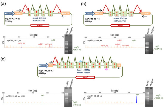
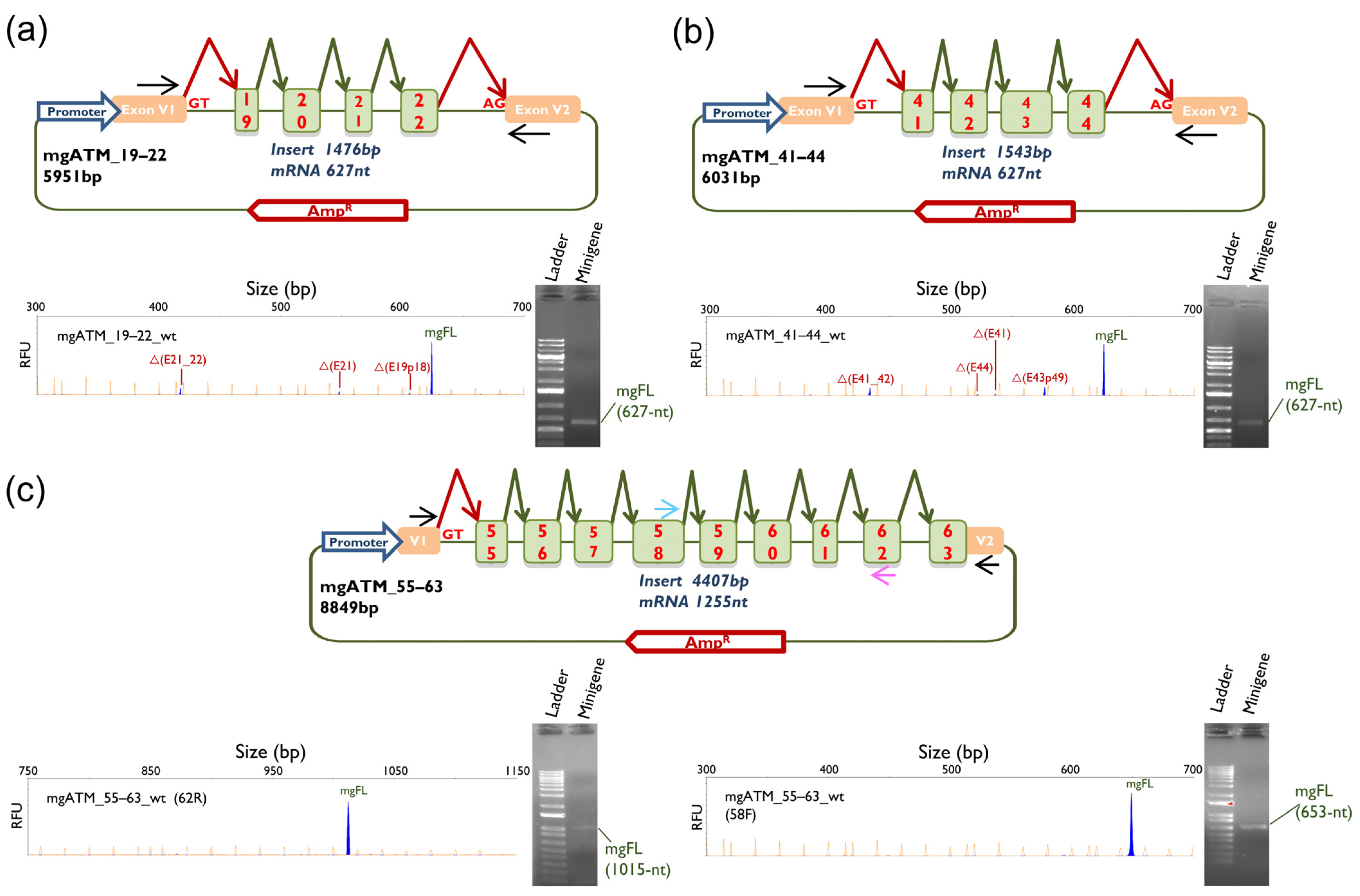
The three ATM wild type (wt) minigenes (mgATM_19–22, mgATM_41–44 and mgATM_55–63) were functionally assayed in MCF-7 cells. All three constructs predominantly yielded the expected minigene full-length (mgFL) transcripts (83.5%, 76.2% and 100%, respectively). In addition, several naturally occurring alternative transcripts were identified: Δ(E19p18), Δ(E21) and Δ(E21_E22) in mgATM19–22; Δ(E41), Δ(E41_E42), Δ(E43p49) and Δ(E44) in mgATM_41–44 (Table 1 and Figure 1). These alternative splicing events have been reported in the reference 300K-RNA dataset, with SpliceVault detection rates ranging from 0.2% for Δ(E41) to 16.1% for Δ(E43p49) [35].
Table 1.
Splicing outcomes of 47 ATM variants.
Figure 1.
Schematic representation and splicing assay of the wt minigenes: (a) mgATM_19–22; (b) mgATM_41–44; (c) mgATM_55–63. Exons are indicated by boxes; green and red elbow arrows indicate the expected splicing reactions and black arrows locate specific vector RT-PCR primers. Blue and pink arrows in mgATM_55–63 locate primers RT-ATM-58F and RT-ATM-62R, respectively. RT-PCR products were analyzed by agarose gel electrophoresis (right) and fluorescent fragment electrophoresis (left). mgFL, expected minigene full-length transcript. Transcript descriptors: Δ, skipping of exonic sequences; E, exon; p, acceptor shift (the exact number of nucleotides is indicated). The x-axis indicates size in base pairs (bp) (electropherograms on the top), and the y-axis represents Relative Fluorescence Units (RFU).
2.2. ATM Variants Assays
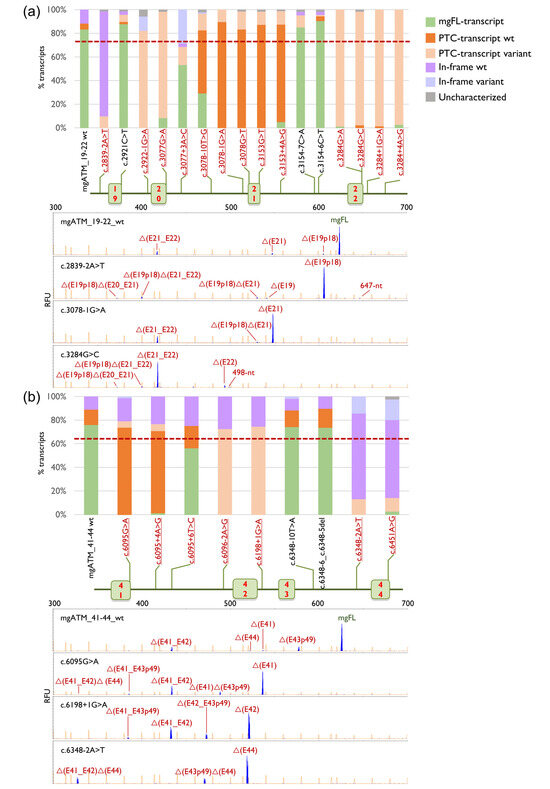
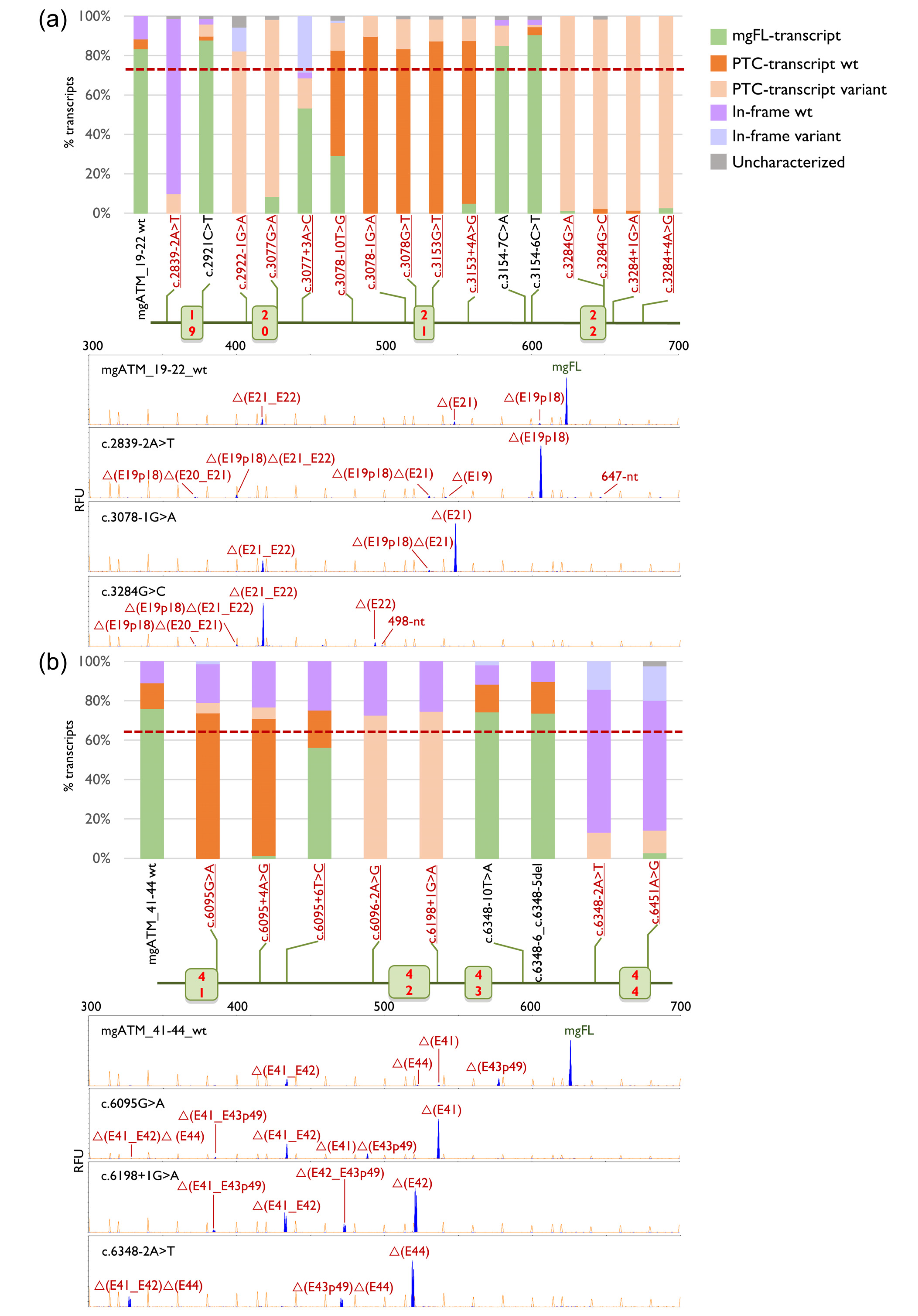
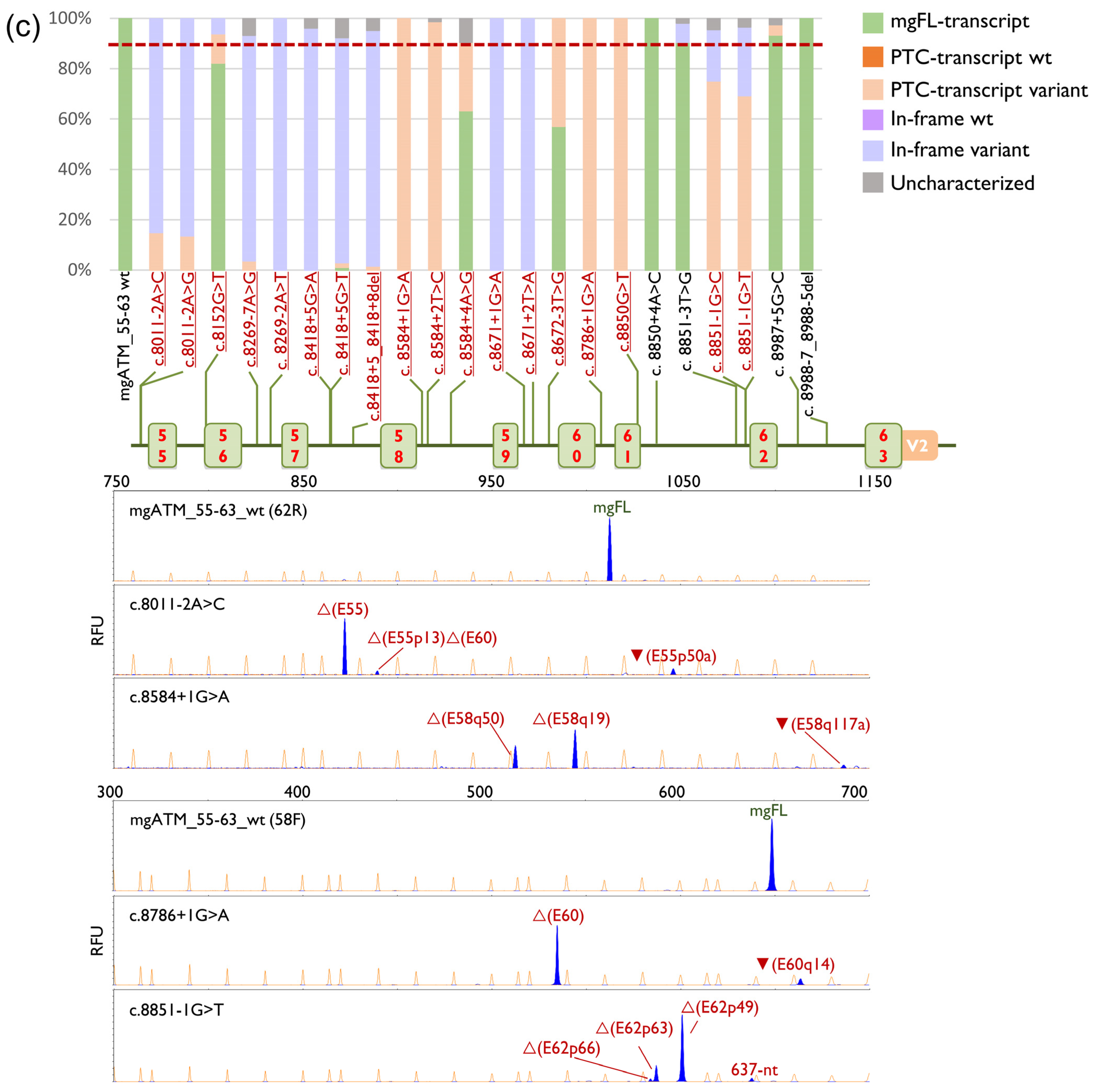
Wild type minigenes were used as template to genetically engineer the 47 candidate variants: 16 in mgATM_19–22, 9 in mgATM_41–44 and 22 in mgATM_55–63. Consistent with our previous report [32], a 10% reduction in the mgFL transcript was adopted as a conservative operational threshold for spliceogenic variants. This threshold serves to exclude weak impacts on splicing or inter-experimental variability that may result in minor fluctuations in transcript ratios. Analysis of results revealed that 38 out of 47 (81%) variants disrupted splicing, 30 of which exhibited a severe impact on splicing, resulting in neglible (<5%) or no detectable expression of mgFL-transcript (Table 1, Figure 2, Supplementary Figure S1). To check splicing reproducibility, two variants (c.3284G>A and c.3154-6C>T) and the wild type minigene mgATM_19–22 were also assayed in MDA-MD-231 cells, showing very similar outcomes (Supplementary Figure S2).
Figure 2.
Splicing outcomes of selected ATM variants in minigenes: (a) mgATM_19–22; (b) mgATM_41–44; (c) mgATM_55–63. Graphical representation of the relative contribution of each type of transcript (top), map of tested variants (middle) and fluorescent fragment electrophoresis of wt and two representative variants (bottom). Spliceogenic variants are highlighted in red and underlined. Bar graph legend: The dashed red line indicates the threshold above which a variant is considered spliceogenic. PTC-transcript wt and In-frame wt, alternative transcripts produced by the wt minigene; PTC-transcript variant and In-frame variant, transcripts induced by the corresponding variants.
Seventeen spliceogenic variants affected the canonical ±1 and ±2 positions, each resulting in the production of 100% aberrant transcripts. The remaining 21 variants altered other exonic or intronic nucleotides within the splice-site consensus sequence, producing splicing defects ranging from weak–moderate perturbation to complete loss of normal splicing. (Table 1, Figure 2, Supplementary Figure S1). Interestingly, one exon 19 variant (c.2921C>T, p.(Ser974Phe)) and two intron 21 variants (c.3154-7C>A, c.3154-6C>T) improved splicing efficiency, producing higher levels of mgFL compared to the wild type, (Table 1 and Figure 2a). Additionally, seven predicted missense variants (c.3078G>T, p.(Trp1026cys); c.3153G>T, p.(Glu1051Asp); c.3284G>A, p.(arg1095lys); c.3284G>C p. (Arg1095Thr); c.6095G>A p.(Arg2032lys); c.6451A>G, p.(Arg2151Gly); c.8850G>T p.(Glu2950Asp)) also induced severe splicing anomalies. These variants resulted in very low or undetectable levels of mgFL-transcripts. Notably, the nonsense variant (c.3077G>A, p.(Trp1026*)) generated only 8.5% mgFL-transcripts. Forty-one variants generated two or more transcripts, which illustrate the complexity of variant-induced splicing outcomes.
A total of 69 different transcripts were characterized by fluorescent-fragment analysis, including five mgFL-transcript carrying exonic variants (r.2921c>u, r.3077g>a, r.3284g>a, r.6451a>g and r.8152g>u) (Supplementary Table S1). Remarkably, variant c.3077G>A, which introduces, a stop codon [p.(Trp1026*)], produced 90% of transcripts with premature termination codon (PTC) (Figure 2a and Supplementary Figure S2). Twelve transcripts could not be characterized (1–9.4% of the overall expression) (Table 1 and Figure 2). Fourteen transcripts preserved the reading frame, whereas 48 transcripts were predicted to introduce a PTC, including mgFL-c.3077G>A, of which 46 were predicted to elicit the Nonsense-Mediated Decay (NMD) (Figure 2, Supplementary Table S1). The predominant splicing event (30 transcripts) was alternative usage of cryptic 5′ss or 3′ss, which was observed in 28 variants. Particularly, transcript ▼(E56p182) used a cryptic 5′ss strengthened by the fusion of two segments of a shortened intron 56 (a reduction from 7260 bp of the original intron to 567 bp) in the minigene (MaxEntScan (MES) natural 1.04 → MES chimeric ivs 4.04). Single/multiexon skipping was identified in 19 transcripts, while exon skipping combined with alternative splice-site usage was concomitantly detected in 11 different transcripts. Finally, one transcript contained full retention of intron 62 [▼(I62)] (Supplementary Table S1).
2.3. ACMG-AMP-Based Classification of Variants
According to the ACMG/AMP-based specifications of the ATM gene, 26 variants were classified as pathogenic (P), 4 as likely pathogenic (LP), 9 as likely benign (LB), and 8 as Variants of Uncertain Significance (VUS) (Table 2, Supplementary Tables S2 and S3). It is worth mentioning that seven predicted missense variants were first characterized as spliceogenic and subsequently classified as P. Of the 47 variants under investigation, 35 had been reported previously in ClinVar (https://www.ncbi.nlm.nih.gov/clinvar/?term=atm%5Bgene%5D&redir=gene; last accessed on 23 October 2025) with classification confidences ranging from conflicting/single submitter (N = 8) to expert panel review (N = 4) (Table 2, Supplementary Table S3). Our minigene-based classification showed full concordance with expert panel review. Specifically, it reclassified three out of eight VUSs as P (n = 1), LP (n = 1), or LB (n = 1). Furthermore, three conflicting variants (VUS/LB) were catalogued as LB, while one conflicting variant (LP/VUS) was upgraded to P. Further, the minigene-based classification reached a definite pathogenic status (≥ +10 points) for most variants reported as P/LP (11 out of 13).
Table 2.
ClinGen and minigene-based ACMG/AMP classification of 47 ATM variants.
3. Discussion
Massively parallel sequencing of breast and ovarian cancer susceptibility genes is a highly sensitive, cost-effective, and rapid approach for detecting risk-associated variants, ultimately improving the clinical management of patients [10,11,36,37]. Despite these advancements, a significant limitation is the frequent detection of VUS [38]. Accurately classifying these VUS poses a challenge for genetic counselling. Since splicing dysregulation is a prevalent etiopathogenic mechanism in BC susceptibility genes [39], the assessment of VUS via splicing studies is essential for proper classification and improved patient management.
Several in silico tools, such as MES or SpliceAI, predict variant impact with a certain degree of accuracy. However, functional studies are still necessary to verify these predictions. Key parameters, such as the level of splicing alteration for variants that do not affect the AG/GT dinucleotides or the number and type of transcripts generated by each variant, are beyond the current capabilities of these tools. For these reasons, experimental validation is key for assessing the potential pathogenicity of predicted splice-altering variants. When RNA from carriers is not available, hybrid minigenes are an effective tool for evaluating potential spliceogenic variants in virtually any human disease gene [40,41,42,43,44]. So far, we have built seven ATM minigenes: mgATM_4–9, mgATM_11–17, mgATM_19–22, mgATM_25–29, mgATM_41–44, mgATM_49–52 and mgATM_55–63. These clones cover 39 of the 63 ATM exons (62% of the coding sequence). According to the ClinVar database, 11500 ATM reported variants are located within the regions covered by our minigenes, illustrating their potential for large-scale splicing studies.
As artificial constructs, minigenes have inherent limitations. These include the shortening of long introns that may harbour internal regulatory sequences, potentially impacting splicing events. Furthermore, while minigene assays rely on a single insert with a specific haplotype (typically the reference sequence), the genetic background of each individual remains a critical factor, given that SNPs can modulate alternative splicing [45]. On the other hand, they also offer a number of advantages that we have previously underscored in other studies [46,47]. These can be summarized as: (1) analysis of a single mutant allele without the interference of the wild type allele; (2) use of disease-relevant cell lines; (3) NMD inhibition that improves the detection of all PTC-NMD transcripts; (4) high sensitivity and resolution of fluorescent capillary electrophoresis for the detection of transcripts; (5) high reproducibility of variant-induced outcomes [30,32]. Notably, the minigene results of eight variants from this study were supported by previous RNA studies in carriers (Supplementary Table S4): c.2922-1G>A (c.2922-2A>G carrier: Δ(E20p32) vs. Δ(E20p32) and Δ(E20p71) as main minigene transcripts) [38], c.3078-1G>A (Δ(E21)) [39], c.6095G>A (Δ(E41) in four independent studies) [17,40,41,42], c.6198+1G>A (Δ(E42) vs. Δ(E42) and Δ(E41_E42) as main minigene transcripts) [48], c.8418+5_8418+8del (Δ(E57)) [43], c.8418+5G>A (Δ(E57)) [44], c.8584+2T>C (Δ(E58q19)) [45], c.8786+1G>A (Δ(E60) and ▼(E60q14)) [46,47] and c.8851-1G>T (Δ(E62p63) vs. Δ(E62p49) and Δ(E62p63) in minigenes) [48]. The reliability of the minigene analysis was further supported by the strong concordance observed with SpliceAI-visual predictions [49] and SpliceVault top events [35], across the evaluated cases (Supplementary Table S4). The single exception was c.6095+6T>C for which SpliceAI predicted no spliceogenic effect, while mgATM_41–44 revealed a leaky splicing variant producing substantial levels of Δ(E41_E42) transcripts. This finding, together with the fact that SpliceAI-visual does not predict multi-exon skipping -as observed for exon 22 donor site, exon 42 acceptor site, and exon 42 donor site variants- underscores the critical relevance of experimental splicing assays to complement in silico predictions.
We formerly carried out exhaustive studies of 56 ATM splice-site variants and 62 ATM SRE (Splicing Regulatory Element)-variants by minigene assays [32,47,50]. Here, 47 new ATM splice-site variants were tested using three different minigenes with 17 exons, which were designed to closely replicate the genomic context of the ATM gene. The selection strategy for splice site variants was successful in our two studies of the ATM gene [32], with 84% of the selected variants disrupting splicing. Most spliceogenic variants of the present study (21/38, 55%) involved changes at conserved positions of the 5′ss and 3′ss beyond the canonical ±1,2 dinucleotides, demonstrating that any DNA change can be deleterious and may contribute to cancer risk. In addition to the intronic changes, seven missense variants severely disrupted splicing with no traces or negligible amounts of mgFL-transcripts (Table 1). Specifically, these variants primarily affected the last exon nucleotide (5 variants), but also the first and penultimate nucleotides.
Minigene analysis further revealed the high complexity of ATM splicing patterns, with a total of 69 different transcripts detected. This intricate landscape poses a challenge for clinical interpretation, particularly for variants like c.2922-1G>A, which generated up to 10 distinct transcripts.
Clinical Interpretation of Variants
Overall, minigene data made a major contribution to the classification of most variants, as illustrated in Table 2. In keeping with that, the minigene readouts for four variants were considered inconclusive (i.e., not contributing to the final classification) and were ultimately classified as VUS. In all four cases, minigene inconclusiveness was caused by the evidence of a partial effect on splicing (i.e., leaky variants). Interestingly, two of these four leaky variants have been assessed in a recent MAVE (Multiplexed Assays of Variant Effect) study [51] displaying different results. The c.8672-3T>G variant (with full-length transcripts contributing 57.1% of the expression in our minigene study) scored non-functional. By contrast, the c.8584+4A>G variant (full-length transcripts contributing 63.4% of the expression in our minigene study), scored as intermediate function. In both cases, MAVE data was reported with high confidence. Assuming that the leakiness level observed in our minigene assay mirrors that of the MAVE model (HCT116 cell line), the data supports a dosage sensitivity model in which ATM variant alleles expressing as much as 57% of full-length transcripts are fully pathogenic. These results sharply contrast with a previously proposed dosage-sensitive expression model that considered ATM alleles producing >30% full-length transcripts to be benign [32]. Such discrepancies underscore the challenge of developing robust dosage-sensitive expression models and reinforce the need for extreme caution when assigning the PVS1_(RNA) or BP7_(RNA) evidence to leaky variants.
In the future, robust dosage-sensitive expression models for ATM (or for other clinically relevant genes) could be developed by grouping variants with similar degrees of ‘leakiness’ for case–control burden analyses. Moreover, MAVEs that assess protein function and splicing profiles simultaneously should further improve model accuracy.
Among the 47 variants investigated, 17 affected canonical acceptor or donor positions (±1,2) and therefore carried a predictive PVS1 code of variable strength assigned by the HBOPC VCEP (Supplementary Table S2). In most cases, PVS1_(RNA) evidence from minigene readouts confirmed the VCEP predictions. The single discordant case was c.8269-2A>T. The VCEP recommended PVS1 at supporting strength for this exon 57 acceptor-site change because the conservative prediction favoured the short in-frame acceptor shift Δ(E57p18). Our minigene data, however, revealed that complete exon 57 skipping Δ(E57) contributed >70% of the overall signal, supporting PVS1_(RNA) at full strength.
Of note, variant c.2839-2A>T, reported as Pathogenic in ClinVar by a single submitter, was classified as VUS in our study. This conservative classification is supported by minigene data demonstrating an acceptor shift Δ(E19p18) caused by the activation of an exonic cryptic acceptor site. This acceptor shift results in an in-frame deletion, p.(Tyr947_Lys952del), which, when assessed against the ATM-specific PVS1 decision tree, provides only supporting evidence for pathogenicity. The absence of a functional score for c.2839-2A>T in the ATM MAVE study coupled with benign-supporting scores reported for closely related substitutions at the same site further support our conservative assessment [51].
In summary, we have carried out the most comprehensive splicing studies of the ATM gene to date, testing a total 165 variants (103 splice-site and 62 SRE variants) by minigene assays [32,47,50]. Our results demonstrate that a significant proportion (60%) of these variants impair splicing, with 42% being classified as (likely) pathogenic based on ACMG/AMP-based specifications refined for ATM. Future burden analyses using case–control or family data will be essential to further refine the classification method, and to provide accurate risk estimations that inform the clinical management of BC patients.
From a therapeutic perspective, glucocorticoid treatment has shown promising results in Ataxia–Telangiectasia patients by alleviating neurological symptoms. This effect is mediated by the generation of shorter ATM transcripts (specifically exons 3–52, 4–53, and 2–52), which encode a (partially) functional protein termed mini-ATM [52]. P/LP variants associated with splicing defects in exons 19–22 and 41–44 would theoretically result in ‘mini-ATM’ transcripts (exons 3–52, 4–53, and 2–52) that still retain the underlying splicing anomalies. Consequently, it remains uncertain whether these specific isoforms could provide even a partial rescue of ATM function. Conversely, Ataxia–Telangiectasia patients carrying P/LP variants within exons 55–63, which are nor part of the mini-ATM structure, may represent a cohort that could benefit from glucocorticoid therapy.
Collectively, our findings provide compelling evidence that variant-induced splicing disruptions are a major disease mechanism for ATM and, more broadly, for other BC susceptibility genes [29,31,32,33,53,54,55,56]. The ClinGen specifications for the ATM gene offer a valuable framework for the clinical interpretation of variants when splicing data are available. This work provides a strong rationale for systematically incorporating splicing data, such as that generated by minigene assays, into variant classification pipelines and genetic diagnostics to enhance patient care.
4. Materials and Methods
Ethical approval for this study was obtained from the Ethics Committee of the Spanish National Research Council-CSIC (28 May 2018).
4.1. Annotation and Selection of Candidate Variants
Variants, transcripts and predicted protein products were annotated following the Human Genome Variation Society guidelines (HGVS: https://hgvs-nomenclature.org/stable/, accessed on 12 May 2025), using the MANE selected transcript of ATM (GenBank NM_000051.4; Ensembl ENST00000675843.1). To simplify, generated transcripts were described with abbreviated annotations as previously described [57].
We previously selected potentially spliceogenic variants at the intron-exon boundaries based on: (i) ≥20% decrease in MaxEntScan (MES) scores [58,59,60]; (ii) creation of putative de novo sites (MES cutoff ≥ 3.0); or (iii) changes at conserved positions (−3, −2, −1, exon 5′-3′-ends, +3, +4, +5, and +6) of the consensus splice-sites, regardless of MES predictions [33]. Of the 381 variants at the ATM exon/intron boundaries originally identified in the BRIDGES project, 136 had been previously selected, 56 of which had been tested [32]. Here, we focus on another 47 candidate variants located at the ATM exons 19–22, 41–44 and 55–63.
4.2. Minigenes Construction and Site-Directed Mutagenesis
Three inserts with the target exons and their flanking intronic sequences were designed in-house to obtain the minigenes mgATM_19–22, mgATM_41–44 and mgATM_55–63 (Figure 1; Supplementary Figure S3; Supplementary Table S5).
Minigene mgATM_19–22 (5951 bp) was created by deleting exons 17–18 from mgATM_17–22, which had not yielded a clean splicing pattern in a previous study [32]. The mgATM_41–44 construct (6031 bp) was assembled in three consecutive steps using overlap extension PCR [61], as previously described [62]. Similarly, minigene mgATM_55–63 was assembled in two steps. First, the insert with exons 55–63 (4407 bp) was synthesized (GeneArt, Thermo Fisher Scientific, Waltham, MA, USA) and cloned into the splicing plasmid pSAD between the EagI and SalI restriction sites. Then, the last 88 nt of ATM exon 63 and 455 nt of vector intron were deleted to generate a chimeric exon 63-V2, obtaining the final minigene mgATM_55–63 (8307 bp) (Figure 1c; Supplementary Figure S3c; Supplementary Table S5). The final constructions were confirmed by sequencing (Macrogen, Madrid, Spain) and functionally assayed in MCF-7 cells (estrogen receptor-positive human breast adenocarcinoma cell line; ATCC HTB-22 [63]; LGC Standards, Barcelona, Spain) [64,65]. Selected variants were introduced into the wt minigene by site-directed mutagenesis (QuikChange Lightning kit, Agilent, Santa Clara, CA, USA) (Supplementary Table S5). Mutant minigenes were confirmed by sequencing (Macrogen).
4.3. Splicing Assays
Around 2 × 10⁵ MCF-7 cells were transiently transfected with 1 µg of wt or mutant minigenes. Transfection, NMD inhibition with cycloheximide, RNA purification and retrotranscription were performed as previously described [50]. To assess the reproducibility of the splicing outcomes, several variants were also tested in MDA-MD-231 cells.
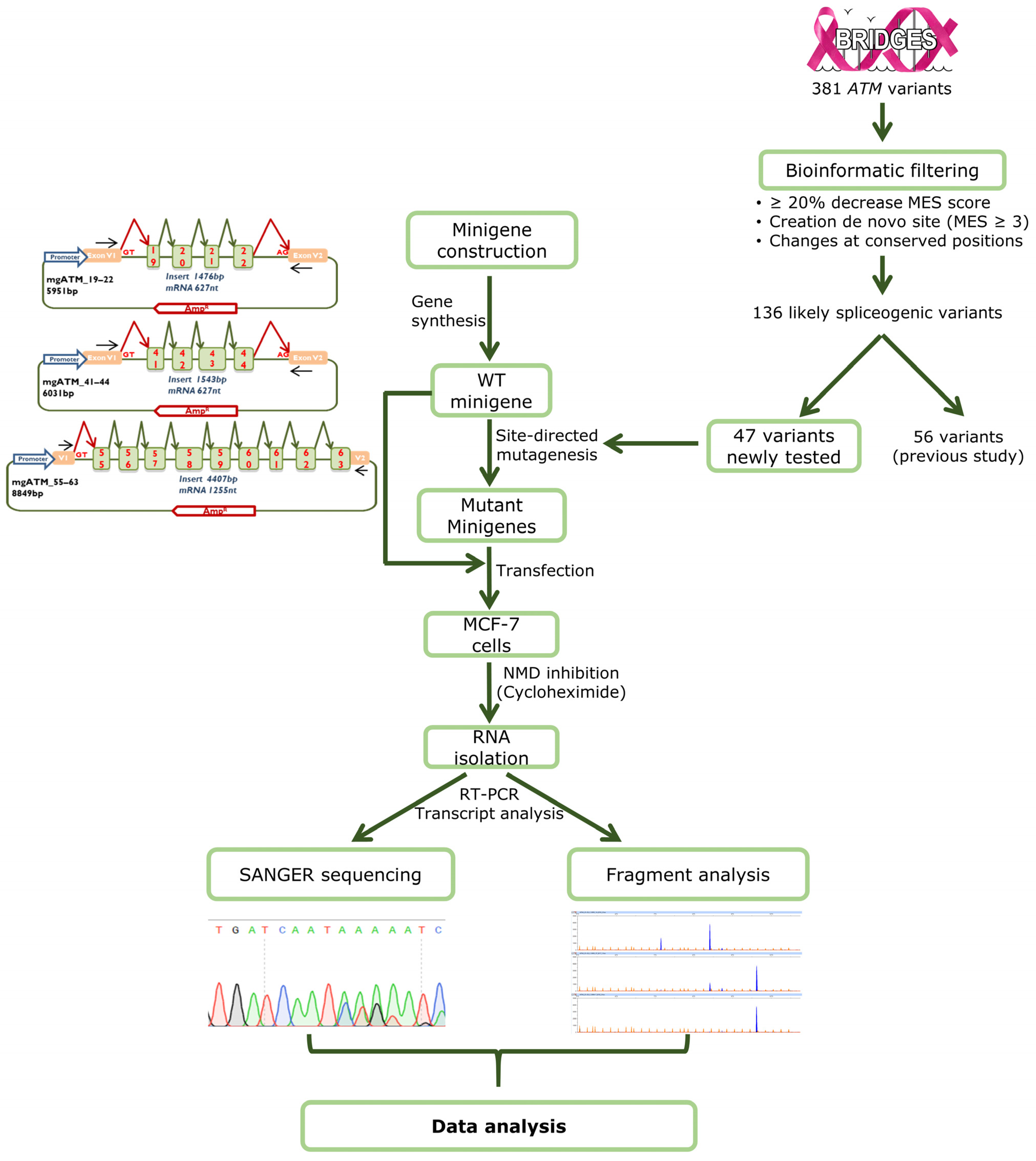
Forty ng of cDNA were amplified with Platinum Taq polymerase (Life Technologies, Carlsbad, CA, USA) as indicated in Supplementary Figure S4. To calculate the relative expression of each transcript, semiquantitative fluorescent RT-PCRs were performed in triplicate under the same conditions as indicated in Supplementary Figure S4, except that 26 cycles were applied. FAM-labelled PCR products were run with LIZ-1200 Size Standard (Life Technologies, Carlsbad, CA, USA) at the Macrogen facility and analyzed with the Peak Scanner software V1.0 (Life Technologies). Only peak heights > 50 RFU were considered to calculate the average relative proportions of each transcript and the corresponding standard deviations. Transcripts with a relative expression level below 1% were filtered out before downstream analysis. The complete workflow of minigene assays is shown in Figure 3.
Figure 3.
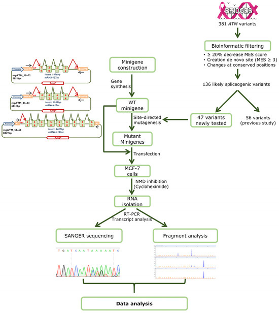
Workflow of the minigene strategy. The basic assay includes the following steps: (1) Minigene construction; (2) Bioinformatic filtering of variants; (3) Site-directed mutagenesis; (4) Transfection of the wild type and mutant minigenes; (5) Inhibition of Nonsense-mediated decay and RNA purification; (6) Transcript sequencing and fragment analysis by fluorescent capillary electrophoresis; (7) Data interpretation.
4.4. ACMG/AMP-Based Tentative Classification of ATM Genetic Variants
ATM variants were classified according to an ACMG/AMP point system, a Bayesian framework that allows for increased flexibility and accuracy in combining different ACMG/AMP criteria and strengths of evidence [63,66]. In this framework, point-based variant classification categories are defined as follows: Pathogenic (P) ≥ +10; Likely Pathogenic (LP) +6 to +9; Variant of Uncertain Significance (VUS) 0 to +5; Likely Benign (LB) −1 to −6; and Benign (B) ≤ −7. Points were assigned to ATM variants following the HBOP VCEP Guidelines for ATM (ATM version 1.4.0) [34].
To assign a PVS1_Variable (RNA)/BP7_Variable (RNA) evidence strength to mgATM read-outs, each transcript was evaluated against the ATM-specific PVS1 decision tree developed by the HBOP VCEP [34]. For complex mgATM readouts involving two or more transcripts, we followed ClinGen Sequence Variation Interpretation Splicing Subgroup recommendations [67]. Accordingly, each transcript was evaluated against the ATM-specific PVS1 decision tree, and a final pathogenic or benign evidence strength was assigned based on the relative contributions of pathogenic and benign-supporting transcripts to total minigene expression. The full methodological details are reported elsewhere [32]. For most read-outs, either pathogenic or benign supporting signal accounted for more than 80% of the total signal, making the final assignment of PVS1_RNA or BP7_RNA straightforward. Four variants (c.3078-10T>G, c.6095+6T>C, c.8584+4A>G, and c.8672-3T>G) did not exhibit a predominant signal, as both pathogenic and benign evidence accounted for less than 70% of the total signal. Consequently, the minigene readouts were deemed inconclusive, and neither PVS1_(RNA) nor BPT_(RNA) classifications were assigned (see Supplementary Table S2 for further details).
Variant classification combined the PVS1_Variable (RNA)/BP7_Variable (RNA) evidence derived from minigene readouts with other applicable ACMG/AMP criteria. SpliceAI-based PP3/BP4 evidence was not used in the final classification because it is not applicable alongside PVS1_Variable[RNA] or BP7_Variable[RNA]. REVEL (Rare Exome Variant Ensembl Learner)-based PP3/BP4 was applied for full-length transcripts encoding missense variants. REVEL results were integrated into the classification of complex minigene readouts and contributed to calls as PVS1_S[RNA] or BP7_S[RNA] (Supplementary Table S2).
Functional pathogenicity or benignity scores (PS3_Variable/BS3_Variable) were derived from a recent MAVE study on ATM [51] that was considered to match the HBOP VCEP PS3_Variable/BS3_Variable rubric for ATM (failure-to-rescue studies in ATM null cell lines). Variants were assigned functional evidence points by the study’s reported score confidence: ±2 points for high confidence, ±1 point for medium-high confidence, and 0 points for medium confidence. Functional evidence was not applied to variants with intermediate functional scores or to scores annotated only with computational confidence (Supplementary Table S2). PS4 was not applied because BRIDGES case–control data showed no statistically significant, variant-level association for any of the variants under review. The HBOPC VCEP does not endorse the following codes for ATM and they were therefore excluded from evaluation: PS2, PM1, PM6, PP2, PP4, PP5, BS2, BS4, BP1, BP3, BP5, BP6. The overall classification process is summarized in Supplementary Tables S2 and S3. This ACMG/AMP–based classification does not constitute a ClinGen-endorsed final clinical interpretation for any variant. On the contrary, it is intended only to illustrate the challenges of incorporating complex minigene readouts into an ACMG/AMP classification framework.
Supplementary Materials
The following supporting information can be downloaded at: https://www.mdpi.com/article/10.3390/ijms27020765/s1.
Author Contributions
Conceptualization, M.d.l.H. and E.A.V.-S.; data curation, I.L.-B., M.d.l.H. and E.A.V.-S.; formal analysis, I.L.-B., L.S.-M., E.B.-M., A.E.-S., L.R.-M., M.d.l.H. and E.A.V.-S.; funding acquisition, P.D., D.F.E., M.P.G.V., M.d.l.H. and E.A.V.-S.; investigation, I.L.-B., L.S.-M., D.R.-M., A.E.-S., E.B.-M., L.R.-M., P.P.-S., A.G.-Á., M.P.G.V., M.d.l.H. and E.A.V.-S.; methodology, I.L.-B., L.S.-M., D.R.-M., A.G.-Á., E.B.-M., M.d.l.H. and E.A.V.-S.; supervision, E.A.V.-S.; writing—original draft, I.L.-B., M.d.l.H. and E.A.V.-S.; writing—review and editing, I.L.-B., D.F.E., M.d.l.H. and E.A.V.-S. All authors have read and agreed to the published version of the manuscript.
Funding
P. Devilee, M.P.G. Vreeswijk, D.F. Easton, M. de la Hoya, and E.A. Velasco-Sampedro have received funding from the European Union’s Horizon 2020 research and innovation programme under grant agreement no. 634935. E.A. Velasco-Sampedro, lab is supported by grants from the Spanish Ministry of Science and Innovation, Plan Nacional de I + D + I 2023, ISCIII (PI23/00047), co-funded by FEDER from Regional Development European Funds (European Union). MdlH lab is supported by grants from the Spanish Ministry of Science and Innovation, Acción Estratégica en Salud 2024, ISCIII (PI24/00267) co-funded by FEDER from Regional Development European Funds (European Union). I. Llinares-Burguet is supported by a predoctoral fellowship from the Consejería de Educación, Junta de Castilla y León (2022–2025). Programa Estratégico Instituto de Biomedicina y Genética Molecular (IBGM) de Valladolid, Escalera de Excelencia, Junta de Castilla y León.
Institutional Review Board Statement
Ethical approval for this study was obtained from the Ethics Committee of the Spanish National Research Council-CSIC (28 May 2018).
Informed Consent Statement
Not applicable.
Data Availability Statement
All sequencing and fragment analysis data are available at Digital. CSIC: http://hdl.handle.net/10261/403423, accessed on 8 January 2026; http://doi.org/10.20350/DIGITALCSIC/17662, accessed on 8 January 2026.
Conflicts of Interest
The authors declare no conflicts of interest.
Abbreviations
The following abbreviations are used in this manuscript:
| ACMG/AMP | American College of Medical Genetics and Genomics/Association for Molecular Pathology |
| ATM | Ataxia–telangiectasia-mutated |
| BC | Breast Cancer |
| bp | Base pairs |
| BRIDGES | Breast Cancer After Diagnostic Gene Sequencing |
| DDR | DNA damage response |
| ER | estrogen receptor |
| HBOP VCEP | Hereditary Breast, Ovarian and Pancreatic Cancer Variant Curation Expert Panel |
| LB | Likely Benign |
| LP | Likely pathogenic |
| LoF | Loss-of-function |
| MAVE | Multiplexed assays of variant effects |
| MCF-7 | Michigan Cancer Foundation-7 |
| MDA-MB-231 | M D Anderson-Metastatic Breast-231 |
| MES | MaxEntScan |
| mgFL | minigene full-length |
| NMD | nonsense-mediated decay |
| P | Pathogenic |
| PTC | premature termination codon |
| PTV | protein-truncating variants |
| REVEL | Rare exome variant ensemble learner |
| RFU | Relative Fluorescence Units |
| RT-PCR | Reverse transcription polymerase chain reaction |
| SRE | Splicing Regulatory Elements |
| ss | Splice-site |
| VUS | Variants of Uncertain Significance |
| wt | Wild type |
References
- Lee, J.H.; Paull, T.T. Cellular Functions of the Protein Kinase ATM and Their Relevance to Human Disease. Nat. Rev. Mol. Cell Biol. 2021, 22, 796–814. [Google Scholar] [CrossRef] [Scilit]
- McKinnon, P.J.; Caldecott, K.W. DNA Strand Break Repair and Human Genetic Disease. Annu. Rev. Genom. Hum. Genet. 2007, 8, 37–55. [Google Scholar] [CrossRef] [Scilit]
- Taylor, A.M.R.; Rothblum-Oviatt, C.; Ellis, N.A.; Hickson, I.D.; Meyer, S.; Crawford, T.O.; Smogorzewska, A.; Pietrucha, B.; Weemaes, C.; Stewart, G.S. Chromosome Instability Syndromes. Nat. Rev. Dis. Primers 2019, 5, 64. [Google Scholar] [CrossRef] [Scilit]
- Choy, K.R.; Watters, D.J. Neurodegeneration in Ataxia-Telangiectasia: Multiple Roles of ATM Kinase in Cellular Homeostasis. Dev. Dyn. 2018, 247, 33–46. [Google Scholar] [CrossRef] [Scilit]
- Suarez, F.; Mahlaoui, N.; Canioni, D.; Andriamanga, C.; D’Enghien, C.D.; Brousse, N.; Jais, J.P.; Fischer, A.; Hermine, O.; Stoppa-Lyonnet, D. Incidence, Presentation, and Prognosis of Malignancies in Ataxia-Telangiectasia: A Report from the French National Registry of Primary Immune Deficiencies. J. Clin. Oncol. 2015, 33, 202–208. [Google Scholar] [CrossRef] [Scilit] [PubMed]
- Hall, M.J.; Bernhisel, R.; Hughes, E.; Larson, K.; Rosenthal, E.T.; Singh, N.A.; Lancaster, J.M.; Kurian, A.W. Germline Pathogenic Variants in the Ataxia Telangiectasia Mutated (ATM) Gene Are Associated with High and Moderate Risks for Multiple Cancers. Cancer Prev. Res. 2021, 14, 433–440. [Google Scholar] [CrossRef] [Scilit] [PubMed]
- Hsu, F.C.; Roberts, N.J.; Childs, E.; Porter, N.; Rabe, K.G.; Borgida, A.; Ukaegbu, C.; Goggins, M.G.; Hruban, R.H.; Zogopoulos, G.; et al. Risk of Pancreatic Cancer among Individuals with Pathogenic Variants in the ATM Gene. JAMA Oncol. 2021, 7, 1664–1668. [Google Scholar] [CrossRef] [Scilit] [PubMed]
- Renwick, A.; Thompson, D.; Seal, S.; Kelly, P.; Chagtai, T.; Ahmed, M.; North, B.; Jayatilake, H.; Barfoot, R.; Spanova, K.; et al. ATM Mutations That Cause Ataxia-Telangiectasia Are Breast Cancer Susceptibility. Nat. Genet. 2006, 38, 873–875. [Google Scholar] [CrossRef] [Scilit]
- Couch, F.J.; Shimelis, H.; Hu, C.; Hart, S.N.; Polley, E.C.; Na, J.; Hallberg, E.; Moore, R.; Thomas, A.; Lilyquist, J.; et al. Associations between Cancer Predisposition Testing Panel Genes and Breast Cancer. JAMA Oncol. 2017, 3, 1190–1196. [Google Scholar] [CrossRef] [Scilit]
- Dorling, L.; Carvalho, S.; Allen, J.; González-Neira, A.; Luccarini, C.; Wahlström, C.; Pooley, K.A.; Parsons, M.T.; Fortuno, C.; Wang, Q.; et al. Breast Cancer Risk Genes—Association Analysis in More than 113,000 Women. N. Engl. J. Med. 2021, 384, 428–439. [Google Scholar] [CrossRef] [Scilit]
- Hu, C.; Hart, S.N.; Gnanaolivu, R.; Huang, H.; Lee, K.Y.; Na, J.; Gao, C.; Lilyquist, J.; Yadav, S.; Boddicker, N.J.; et al. A Population-Based Study of Genes Previously Implicated in Breast Cancer. N. Engl. J. Med. 2021, 384, 440–451. [Google Scholar] [CrossRef] [Scilit]
- Narod, S.A. Which Genes for Hereditary Breast Cancer? N. Engl. J. Med. 2021, 384, 471–473. [Google Scholar] [CrossRef] [Scilit]
- Foulkes, W.D. The Ten Genes for Breast (and Ovarian) Cancer Susceptibility. Nat. Rev. Clin. Oncol. 2021, 18, 259–260. [Google Scholar] [CrossRef] [Scilit]
- Lowry, K.P.; Geuzinge, H.A.; Stout, N.K.; Alagoz, O.; Hampton, J.; Kerlikowske, K.; Koning, H.J.D.; Miglioretti, D.L.; Ravesteyn, N.T.V.; Schechter, C.; et al. Breast Cancer Screening Strategies for Women with ATM, CHEK2, and PALB2 Pathogenic Variants: A Comparative Modeling Analysis. JAMA Oncol. 2022, 8, 587–596. [Google Scholar] [CrossRef] [Scilit] [PubMed]
- Marasco, L.E.; Kornblihtt, A.R. The Physiology of Alternative Splicing. Nat. Rev. Mol. Cell Biol. 2023, 24, 242–254. [Google Scholar] [CrossRef] [Scilit] [PubMed]
- Bradley, R.K.; Anczuków, O. RNA Splicing Dysregulation and the Hallmarks of Cancer. Nat. Revi Cancer 2023, 23, 135–155. [Google Scholar] [CrossRef] [Scilit]
- Hertel, K.J. Combinatorial Control of Exon Recognition. J. Biol. Chem. 2008, 283, 1211–1215. [Google Scholar] [CrossRef] [Scilit] [PubMed]
- Buratti, E.; Baralle, M.; Baralle, F.E. Defective Splicing, Disease and Therapy: Searching for Master Checkpoints in Exon Definition. Nucleic Acids Res. 2006, 34, 3494–3510. [Google Scholar] [CrossRef] [Scilit]
- Baralle, D.; Lucassen, A.; Buratti, E. Missed Threads. The Impact of Pre-mRNA Splicing Defects on Clinical Practice. EMBO Rep. 2009, 10, 810–816. [Google Scholar] [CrossRef] [Scilit]
- Cooper, T.A.; Wan, L.; Dreyfuss, G. RNA and Disease. Cell 2009, 136, 777–793. [Google Scholar] [CrossRef] [Scilit]
- Bonnal, S.C.; López-Oreja, I.; Valcárcel, J. Roles and Mechanisms of Alternative Splicing in Cancer—Implications for Care. Nat. Rev. Clin. Oncol. 2020, 17, 457–474. [Google Scholar] [CrossRef] [Scilit] [PubMed]
- Sullivan, P.J.; Quinn, J.M.W.; Wu, W.; Pinese, M.; Cowley, M.J. SpliceVarDB: A Comprehensive Database of Experimentally Validated Human Splicing Variants. Am. J. Hum. Genet. 2024, 111, 2164–2175. [Google Scholar] [CrossRef] [Scilit]
- Teraoka, S.N.; Telatar, M.; Becker-Catania, S.; Liang, T.; Önengüt, S.; Tolun, A.; Chessa, L.; Sanal, Ö.; Bernatowska, E.; Gatti, R.A.; et al. Splicing Defects the Ataxia-Telangiectasia Gene, ATM: Underlying Mutations and Consequences. Am. J. Human. Genet. 1999, 64, 1617–1631. [Google Scholar] [CrossRef] [Scilit]
- Phan, L.M.; Rezaeian, A.-H. ATM: Main Features, Signaling Pathways, and Its Diverse Roles in DNA Damage Response, Tumor Suppression, and Cancer Development. Genes 2021, 12, 845. [Google Scholar] [CrossRef] [Scilit]
- Richards, S.; Aziz, N.; Bale, S.; Bick, D.; Das, S.; Gastier-Foster, J.; Grody, W.W.; Hegde, M.; Lyon, E.; Spector, E.; et al. Standards and Guidelines for the Interpretation of Sequence Variants: A Joint Consensus Recommendation of the American College of Medical Genetics and Genomics and the Association for Molecular Pathology. Genet. Med. 2015, 17, 405–424. [Google Scholar] [CrossRef] [Scilit] [PubMed]
- Cooper, T. A Use of Minigene Systems to Dissect Alternative Splicing Elements. Methods 2005, 37, 331–340. [Google Scholar] [CrossRef] [Scilit]
- Danilchenko, V.Y.; Panina, E.A.; Zytzar, M.V.; Orishchenko, K.E.; Posukh, O.L. Assessing the Functional Significance of Novel and Rare Variants of the SLC26A4 Gene Found in Patients with Hearing Loss by Minigene Assay. Int. J. Mol. Sci. 2025, 26, 10732. [Google Scholar] [CrossRef] [Scilit]
- Nekrasov, A.; Shchukina, E.; Rimskaya, B.; Zakharova, E. Functional Characterization of HGD Gene Variants by Minigene Splicing Assay. Int. J. Mol. Sci. 2025, 26, 10639. [Google Scholar] [CrossRef] [Scilit]
- Sanoguera-Miralles, L.; Valenzuela-Palomo, A.; Bueno-Martínez, E.; Llovet, P.; Díez-Gómez, B.; Caloca, M.J.; Pérez-Segura, P.; Fraile-Bethencourt, E.; Colmena, M.; Carvalho, S.; et al. Comprehensive Functional Characterization and Clinical Interpretation of 20 Splice-Site Variants of the RAD51C Gene. Cancers 2020, 12, 3771. [Google Scholar] [CrossRef] [Scilit]
- Sanoguera-Miralles, L.; Valenzuela-Palomo, A.; Bueno-Martínez, E.; Esteban-Sánchez, A.; Lorca, V.; Llinares-Burguet, I.; García-Álvarez, A.; Pérez-Segura, P.; Infante, M.; Easton, D.F.; et al. Systematic Minigene-Based Splicing Analysis and Tentative Clinical Classification of 52 CHEK2 Splice-Site Variants. Clin. Chem. 2024, 70, 319–338. [Google Scholar] [CrossRef] [Scilit] [PubMed]
- Bueno-Martínez, E.; Sanoguera-Miralles, L.; Valenzuela-Palomo, A.; Lorca, V.; Gómez-Sanz, A.; Carvalho, S.; Allen, J.; Infante, M.; Pérez-Segura, P.; Lázaro, C.; et al. RAD51D Aberrant Splicing in Breast Cancer: Identification of Splicing Regulatory Elements and Minigene-Based Evaluation of 53 DNA Variants. Cancers 2021, 13, 2845. [Google Scholar] [CrossRef] [Scilit]
- Bueno-Martínez, E.; Sanoguera-Miralles, L.; Valenzuela-Palomo, A.; Esteban-Sánchez, A.; Lorca, V.; Llinares-Burguet, I.; Allen, J.; García-Álvarez, A.; Pérez-Segura, P.; Durán, M.; et al. Minigene-Based Splicing Analysis and ACMG/AMP-Based Tentative Classification of 56 ATM Variants. J. Pathol. 2022, 258, 83–101. [Google Scholar] [CrossRef] [Scilit]
- Valenzuela-Palomo, A.; Bueno-Martínez, E.; Sanoguera-Miralles, L.; Lorca, V.; Fraile-Bethencourt, E.; Esteban-Sánchez, A.; Gómez-Barrero, S.; Carvalho, S.; Allen, J.; García-Álvarez, A.; et al. Splicing Predictions, Minigene Analyses, and ACMG-AMP Clinical Classification of 42 Germline PALB2 Splice-Site Variants. J. Pathol. 2022, 256, 321–334, Erratum in J. Pathol. 2023, 261, 372–373. [Google Scholar] [CrossRef] [Scilit]
- Richardson, M.E.; Holdren, M.; Brannan, T.; de la Hoya, M.; Spurdle, A.B.; Tavtigian, S.V.; Young, C.C.; Zec, L.; Hiraki, S.; Anderson, M.J.; et al. Specifications of the ACMG/AMP Variant Curation Guidelines for the Analysis of Germline ATM Sequence Variants. Am. J. Hum. Genet. 2024, 111, 2411–2426. [Google Scholar] [CrossRef] [Scilit] [PubMed]
- Dawes, R.; Bournazos, A.M.; Bryen, S.J.; Bommireddipalli, S.; Marchant, R.G.; Joshi, H.; Cooper, S.T. SpliceVault Predicts the Precise Nature of Variant-Associated Mis-Splicing. Nat. Genet. 2023, 55, 324–332. [Google Scholar] [CrossRef] [Scilit] [PubMed]
- Laduca, H.; Stuenkel, A.J.; Dolinsky, J.S.; Keiles, S.; Tandy, S.; Pesaran, T.; Chen, E.; Gau, C.L.; Palmaer, E.; Shoaepour, K.; et al. Utilization of Multigene Panels in Hereditary Cancer Predisposition Testing: Analysis of More than 2,000 Patients. Genet. Med. 2014, 16, 830–837. [Google Scholar] [CrossRef] [Scilit]
- Easton, D.F.; Pharoah, P.D.P.; Antoniou, A.C.; Tischkowitz, M.; Tavtigian, S.V.; Nathanson, K.L.; Devilee, P.; Meindl, A.; Couch, F.J.; Southey, M.; et al. Gene-Panel Sequencing and the Prediction of Breast-Cancer Risk. N. Engl. J. Med. 2015, 372, 2243–2257. [Google Scholar] [CrossRef] [Scilit]
- Castéra, L.; Krieger, S.; Rousselin, A.; Legros, A.; Baumann, J.-J.; Bruet, O.; Brault, B.; Fouillet, R.; Goardon, N.; Letac, O.; et al. Next-Generation Sequencing for the Diagnosis of Hereditary Breast and Ovarian Cancer Using Genomic Capture Targeting Multiple Candidate Genes. Eur. J. Hum. Genet. EJHG 2014, 22, 1305–1313. [Google Scholar] [CrossRef] [Scilit]
- Rhine, C.L.; Cygan, K.J.; Soemedi, R.; Maguire, S.; Murray, M.F.; Monaghan, S.F.; Fairbrother, W.G. Hereditary Cancer Genes Are Highly Susceptible to Splicing Mutations. PLoS Genet. 2018, 14, e1007231. [Google Scholar] [CrossRef] [Scilit] [PubMed]
- Tamayo, A.; Núñez-Moreno, G.; Ruiz, C.; Plaisancie, J.; Damian, A.; Moya, J.; Chassaing, N.; Calvas, P.; Ayuso, C.; Minguez, P.; et al. Minigene Splicing Assays and Long-Read Sequencing to Unravel Pathogenic Deep-Intronic Variants in PAX6 in Congenital Aniridia. Int. J. Mol. Sci. 2023, 24, 1562. [Google Scholar] [CrossRef] [Scilit]
- Carriço, J.N.; Gonçalves, C.I.; Aragüés, J.M.; Lemos, M.C. Kallmann Syndrome: Functional Analysis of a CHD7 Missense Variant Shows Aberrant RNA Splicing. Int. J. Mol. Sci. 2024, 25, 12061. [Google Scholar] [CrossRef] [Scilit]
- Rosso, L.E.; Pianigiani, G.; Morgan, A.; Rubinato, E.; Paccagnella, E.; Lenarduzzi, S.; Wischmeijer, A.; Spedicati, B.; Girotto, G. Unraveling the Functional Impact of Splicing Variants in Inherited Hearing Disorders Through Minigene Splicing Assays. Biomedicines 2025, 13, 2245. [Google Scholar] [CrossRef] [Scilit]
- Diallo, M.; Courdier, C.; Mercier, E.; Sequeira, A.; Defay-Stinat, A.; Plaisant, C.; Mesdaghi, S.; Rigden, D.; Javerzat, S.; Lasseaux, E.; et al. Functional Characterization of Splice Variants in the Diagnosis of Albinism. Int. J. Mol. Sci. 2024, 25, 8657. [Google Scholar] [CrossRef] [Scilit]
- Forte, G.; Buonadonna, A.L.; Fasano, C.; Sanese, P.; Cariola, F.; Manghisi, A.; Guglielmi, A.F.; Lepore Signorile, M.; De Marco, K.; Grossi, V.; et al. Clinical and Molecular Characterization of SMAD4 Splicing Variants in Patients with Juvenile Polyposis Syndrome. Int. J. Mol. Sci. 2024, 25, 7939. [Google Scholar] [CrossRef] [Scilit]
- Graveley, B.R. The Haplo-Spliceo-Transcriptome: Common Variations in Alternative Splicing in the Human Population. Trends Genet. 2008, 24, 5–7. [Google Scholar] [CrossRef] [Scilit] [PubMed]
- Acedo, A.; Hernández-Moro, C.; Curiel-García, Á.; Díez-Gómez, B.; Velasco, E.A. Functional Classification of BRCA2 DNA Variants by Splicing Assays in a Large Minigene with 9 Exons. Human. Mutat. 2015, 36, 210–221. [Google Scholar] [CrossRef] [Scilit]
- Llinares-Burguet, I.; Sanoguera-Miralles, L.; García-Álvarez, A.; Esteban-Sánchez, A.; Caloca, M.J.; de la Hoya, M.; Bueno-Martínez, E.; Velasco-Sampedro, E.A. Unproductive Alternative Splicing of ATM Exon 7: Mapping of Critical Regulatory Elements and Identification of 34 Spliceogenic Variants. J. Mol. Med. 2025, 103, 1447–1460. [Google Scholar] [CrossRef] [Scilit]
- Stankovic, T.; Kidd, A.M.J.; Sutcliffe, A.; McGuire, G.M.; Robinson, P.; Weber, P.; Bedenham, T.; Bradwell, A.R.; Easton, D.F.; Lennox, G.G.; et al. ATM Mutations and Phenotypes in Ataxia-Telangiectasia Families in the British Isles: Expression of Mutant ATM and the Risk of Leukemia, Lymphoma, and Breast Cancer. Am. J. Human. Genet. 1998, 62, 334–345. [Google Scholar] [CrossRef] [Scilit] [PubMed]
- de Sainte Agathe, J.-M.; Filser, M.; Isidor, B.; Besnard, T.; Gueguen, P.; Perrin, A.; Van Goethem, C.; Verebi, C.; Masingue, M.; Rendu, J.; et al. SpliceAI-Visual: A Free Online Tool to Improve SpliceAI Splicing Variant Interpretation. Hum. Genom. 2023, 17, 7. [Google Scholar] [CrossRef] [Scilit]
- Llinares-Burguet, I.; Sanoguera-Miralles, L.; Valenzuela-Palomo, A.; García-Álvarez, A.; Bueno-Martínez, E.; Velasco-Sampedro, E.A. Splicing Dysregulation of Non-Canonical GC-5′ Splice Sites of Breast Cancer Susceptibility Genes ATM and PALB2. Cancers 2024, 16, 3562. [Google Scholar] [CrossRef] [Scilit] [PubMed]
- Lee, K.S.; Min, J.-G.; Cheong, Y.; Oh, H.-C.; Jung, S.-Y.; Park, J.-I.; Song, M.; Seo, J.H.; Cho, S.-R.; Kim, H.H. Functional Assessment of All ATM SNVs Using Prime Editing and Deep Learning. Cell 2025, 188, 5081–5099.e27. [Google Scholar] [CrossRef] [Scilit]
- Ricci, A.; Biancucci, F.; Morganti, G.; Magnani, M.; Menotta, M. New Human ATM Variants Are Able to Regain ATM Functions in Ataxia Telangiectasia Disease. Cell Mol. Life Sci. 2022, 79, 601. [Google Scholar] [CrossRef] [Scilit]
- Sanz, D.J.; Acedo, A.; Infante, M.; Durán, M.; Pérez-Cabornero, L.; Esteban-Cardeñosa, E.; Lastra, E.; Pagani, F.; Miner, C.; Velasco, E.A. A High Proportion of DNA Variants of BRCA1 and BRCA2 Is Associated with Aberrant Splicing in Breast/Ovarian Cancer Patients. Clin. Cancer Res. 2010, 16, 1957–1967. [Google Scholar] [CrossRef] [Scilit]
- Fraile-Bethencourt, E.; Valenzuela-Palomo, A.; Díez-Gómez, B.; Goina, E.; Acedo, A.; Buratti, E.; Velasco, E.A. Mis-Splicing in Breast Cancer: Identification of Pathogenic BRCA2 Variants by Systematic Minigene Assays. J. Pathol. 2019, 248, 409–420. [Google Scholar] [CrossRef] [Scilit]
- Sanoguera-Miralles, L.; Bueno-Martínez, E.; Valenzuela-Palomo, A.; Esteban-Sánchez, A.; Llinares-Burguet, I.; Pérez-Segura, P.; García-Álvarez, A.; de la Hoya, M.; Velasco-Sampedro, E.A. Minigene Splicing Assays Identify 20 Spliceogenic Variants of the Breast/Ovarian Cancer Susceptibility Gene RAD51C. Cancers 2022, 14, 2960. [Google Scholar] [CrossRef] [Scilit] [PubMed]
- Valenzuela-Palomo, A.; Sanoguera-Miralles, L.; Bueno-Martínez, E.; Esteban-Sánchez, A.; Llinares-Burguet, I.; García-Álvarez, A.; Pérez-Segura, P.; Gómez-Barrero, S.; de la Hoya, M.; Velasco-Sampedro, E.A. Splicing Analysis of 16 PALB2 ClinVar Variants by Minigene Assays: Identification of Six Likely Pathogenic Variants. Cancers 2022, 14, 4541. [Google Scholar] [CrossRef] [Scilit]
- Lopez-Perolio, I.; Leman, R.; Behar, R.; Lattimore, V.; Pearson, J.F.; Castéra, L.; Martins, A.; Vaur, D.; Goardon, N.; Davy, G.; et al. Alternative Splicing and ACMG-AMP-2015-Based Classification of PALB2 Genetic Variants: An ENIGMA Report. J. Med. Genet. 2019, 56, 453–460. [Google Scholar] [CrossRef] [Scilit] [PubMed]
- Yeo, G.; Burge, C.B. Maximum Entropy Modeling of Short Sequence Motifs with Applications to RNA Splicing Signals. J. Comput. Biol. 2004, 11, 377–394. [Google Scholar] [CrossRef] [Scilit] [PubMed]
- Houdayer, C.; Caux-Moncoutier, V.; Krieger, S.; Barrois, M.; Bonnet, F.; Bourdon, V.; Bronner, M.; Buisson, M.; Coulet, F.; Gaildrat, P.; et al. Guidelines for Splicing Analysis in Molecular Diagnosis Derived from a Set of 327 Combined in Silico/in Vitro Studies on BRCA1 and BRCA2 Variants. Human. Mutat. 2012, 33, 1228–1238. [Google Scholar] [CrossRef] [Scilit]
- Moles-Fernández, A.; Duran-Lozano, L.; Montalban, G.; Bonache, S.; López-Perolio, I.; Menéndez, M.; Santamariña, M.; Behar, R.; Blanco, A.; Carrasco, E.; et al. Computational Tools for Splicing Defect Prediction in Breast/Ovarian Cancer Genes: How Efficient Are They at Predicting RNA Alterations? Front. Genet. 2018, 9, 366. [Google Scholar] [CrossRef] [Scilit]
- Bryksin, A.V.; Matsumura, I. Overlap Extension PCR Cloning: A Simple and Reliable Way to Create Recombinant Plasmids. BioTechniques 2010, 48, 463–465. [Google Scholar] [CrossRef] [Scilit] [PubMed]
- Llinares-Burguet, I.; Sanoguera-Miralles, L.; Bueno-Martínez, E.; García-Álvarez, A.; Valenzuela-Palomo, A.; Pérez-Segura, P.; de la Hoya, M.; Velasco-Sampedro, E.A. Generation of Functional Non-Canonical Donor Splice Sites by +2T Variants in Breast Cancer Susceptibility Genes: Impact on Clinical Interpretation. J. Pathol. 2025. [Google Scholar] [CrossRef] [Scilit] [PubMed]
- Tavtigian, S.V.; Harrison, S.M.; Boucher, K.M.; Biesecker, L.G. Fitting a Naturally Scaled Point System to the ACMG/AMP Variant Classification Guidelines. Human. Mutat. 2020, 41, 1734–1737. [Google Scholar] [CrossRef] [Scilit] [PubMed]
- Soule, H.D.; Vazquez, J.; Long, A.; Albert, S.; Brennan, M. A Human Cell Line from a Pleural Effusion Derived from a Breast Carcinoma2. J. Natl. Cancer Inst. 1973, 51, 1409–1416. [Google Scholar] [CrossRef] [Scilit]
- ATCC HTB-22; MCF7 [MCF-7] (ATCC® HTB-22TM), Human Breast Adenocarcinoma. American Type Culture Collection: Manassas, VA, USA, 2026.
- Tavtigian, S.V.; Greenblatt, M.S.; Harrison, S.M.; Nussbaum, R.L.; Prabhu, S.A.; Boucher, K.M.; Biesecker, L.G. Modeling the ACMG/AMP Variant Classification Guidelines as a Bayesian Classification Framework. Genet. Med. 2018, 20, 1054–1060. [Google Scholar] [CrossRef] [Scilit]
- Walker, L.C.; de la Hoya, M.; Wiggins, G.A.R.; Lindy, A.; Vincent, L.M.; Parsons, M.T.; Canson, D.M.; Bis-Brewer, D.; Cass, A.; Tchourbanov, A.; et al. Using the ACMG/AMP Framework to Capture Evidence Related to Predicted and Observed Impact on Splicing: Recommendations from the ClinGen SVI Splicing Subgroup. Am. J. Hum. Genet. 2023, 110, 1046–1067. [Google Scholar] [CrossRef] [Scilit]

Disclaimer/Publisher’s Note: The statements, opinions and data contained in all publications are solely those of the individual author(s) and contributor(s) and not of MDPI and/or the editor(s). MDPI and/or the editor(s) disclaim responsibility for any injury to people or property resulting from any ideas, methods, instructions or products referred to in the content. |
© 2026 by the authors. Licensee MDPI, Basel, Switzerland. This article is an open access article distributed under the terms and conditions of the Creative Commons Attribution (CC BY) license.