


Abstract
Radiotherapy can simultaneously promote tumor immunogenicity and damage the systemic immune compartments required to sustain antitumor responses. In breast cancer, radiation-induced lymphopenia is influenced by treatment geometry, irradiated volume, low-dose exposure, marrow and lymphoid irradiation, fractionation, and delivery characteristics, while nanoparticle radioenhancement can amplify tumor response through physical, redox, DNA-repair, and immune mechanisms. This hypothesis-driven translational review examines whether nanoparticle-generated biological amplification could be redirected from tumor intensification toward immune-dose shaping while preserving tumor control and immunogenicity. The evidence supports seven interacting dimensions of potential exchange: physical dose, volume/spatial exposure, distributed low-dose exposure, marrow dose, geometry/modality, fractionation, and temporal/delivery exposure. Formal nanoparticle-enabled dose exchange remains limited, with the clearest breast-specific evidence confined to in vitro isoeffect analysis; no study has yet demonstrated preserved tumor isoeffect and immunogenicity together with reduced multicompartment immune injury after nanoparticle-enabled exposure reduction. We, therefore, propose a dual-endpoint framework integrating tumor control, immunogenicity, multicompartment immune preservation, dosimetry, and nanoparticle biodistribution, together with a staged translational pathway from quantitative preclinical exchange studies to comparative clinical testing. This strategy remains hypothesis-generating, with triple-negative breast cancer representing a particularly informative initial setting.
1. Introduction
Radiotherapy (RT) is a cornerstone of breast cancer treatment, but its effects extend beyond direct tumor-cell killing. Ionizing radiation can promote immunogenic cell death (ICD), expose calreticulin, release ATP and HMGB1, generate cytosolic double-stranded DNA, and activate the cGAS–STING/type I interferon axis, supporting dendritic-cell maturation, antigen presentation, and recruitment of tumor-reactive T cells [1,2,3,4]. These responses depend strongly on dose, fractionation, and tumor biology [3,5,6,7,8].
Nanoparticle radioenhancement can amplify radiation effects through increased energy deposition, reactive oxygen species generation, hypoxia/redox modulation, DNA-repair interference, ferroptosis, or direct immune activation [9,10,11,12]. Hafnium oxide NBTXR3 increases DNA damage and cGAS–STING signaling and can broaden tumor immunogenicity and T-cell receptor repertoire [13,14]. Breast-specific platforms using manganese, platinum, gadolinium, lutetium, and gold nanomaterials similarly enhance ICD, dendritic-cell maturation, macrophage polarization, CD8+ recruitment, and, in selected models, distant tumor control and immune memory [10,12,15,16,17,18,19,20,21].
The same radiation exposure, however, can damage the immune system required to sustain these effects. Radiation-induced lymphopenia (RIL) in breast cancer is associated with treatment volume, regional nodal irradiation, low-dose exposure, effective dose to immune cells (EDIC), fractionation, technique, and marrow exposure [22,23,24,25,26]. Severe or persistent lymphopenia has been associated with poorer outcomes, although whether RIL is causal or reflects treatment intensity and baseline vulnerability remains unresolved [27,28,29,30,31]. RIL is also unlikely to arise from circulating-blood irradiation alone: dynamic models support repeated blood-pool exposure across fractions, clinical studies implicate hematopoietic marrow, and mechanistic evidence identifies tumor-draining lymph nodes as reservoirs contributing activated CD8+ T cells to irradiated tumors [25,26,32,33,34,35,36,37]. The immune system is, therefore, better considered a distributed organ at risk encompassing blood, lymphoid tissue, marrow, and tumor-associated immune cells [38,39,40].
This paradox may be particularly relevant in triple-negative breast cancer (TNBC), where tumor immune infiltration is prognostically important [41] and higher EDIC has been associated with poorer event-free survival in an exploratory cohort [42]. TNBC is also disproportionately represented in preclinical nanoparticle–radioimmunotherapy models, making it a logical initial testing context without implying exclusivity [21].
Importantly, immune-relevant radiation exposure is modifiable. Proton therapy, breath hold, and positioning can reduce modeled EDIC; fractionation and restricted-volume approaches can reduce lymphocyte exposure or depletion [29,43,44,45]; and reduced-volume irradiation can preserve lymphocytes while retaining cGAS–STING activation and tumor control [46,47]. FLASH adds a temporal dimension: ex vivo data show reduced chromosomal injury at ultra-high dose rates, while preclinical and computational studies demonstrate treatment-dependent immune effects that remain compartment-dependent [48,49,50,51]. Thus, immune injury reflects the spatial and temporal distribution of exposure, not prescription dose alone.
This creates an opportunity for nanoparticle radioenhancement distinct from tumor intensification. Greater tumor effect at an unchanged dose demonstrates radioenhancement, not dose exchange. True exchange requires a lower-dose NP + RT regimen to reproduce a predefined tumor effect achieved with higher-dose RT alone. Breast-specific in vitro dose–response and isoeffect studies show that a quantifiable exchange margin is possible, and computational and cross-tumor studies provide additional proof of concept, but clean in vivo demonstration in breast cancer remains lacking [11,52,53,54]. Clinical NBTXR3 studies establish a meaningful biological amplification margin, but this has so far been used to intensify tumor response rather than reduce physical exposure [55,56,57].
Dose reduction alone may also provide limited immune benefit if irradiated volume, distributed low-dose exposure, marrow or nodal exposure, and fraction number remain unchanged. We, therefore, propose nanoparticle-enabled immune-dose shaping: using nanoparticle-generated tumor biological amplification as a potentially exchangeable margin to reduce selected components of physical, spatial, or temporal radiation exposure while preserving tumor control and immunogenicity.
The evidence gap is specific. Nanoparticle studies generally assess tumor radiosensitization and immunogenicity without measuring systemic immune preservation, whereas breast RIL and dosimetry studies quantify lymphocyte depletion or immune exposure without establishing preservation of tumor immunogenicity. No study identified in this evidence set has combined nanoparticle-enabled reductions in immune-relevant radiation exposure, preserved tumor isoeffect, maintained immunogenicity, and reduced multicompartment immune injury within the same breast-cancer experiment or cohort.
This review, therefore, integrates clinical breast RIL and dosimetry evidence with nanoparticle radioenhancement, immunogenic RT, and geometric/temporal sparing; distinguishes same-dose enhancement from genuine dose exchange; and proposes a dual tumor–immune framework evaluating tumor control and immunogenicity alongside injury to circulating, lymphoid, and hematopoietic compartments. The result is not an established therapeutic strategy but a falsifiable translational hypothesis. Although individual components are supported by clinical, preclinical, and computational evidence, the integrated concept of nanoparticle-enabled immune-dose shaping has not yet been experimentally demonstrated. This review, therefore, proposes a proof-of-concept research framework for investigating whether nanoparticle-generated biological amplification can be redistributed to reduce immune-relevant radiation exposure while preserving tumor control and immunogenicity.
Figure 1 summarizes the radiotherapy immune paradox and the proposed concept.
Figure 1.
The radiotherapy immune paradox and the concept of nanoparticle-enabled immune-dose shaping. Radiotherapy can simultaneously enhance tumor immunogenicity through DNA damage, damage-associated molecular pattern release, cGAS–STING/type I interferon signaling, dendritic-cell activation, and T-cell recruitment, while also injuring circulating lymphocytes, lymphoid organs, and hematopoietic marrow. The resulting immune cost is influenced by treated volume and spatial extent, distributed low-dose exposure, marrow dose, treatment geometry and modality, fractionation, and temporal delivery. Nanoparticle radioenhancement may create a biological amplification margin that could be exploited to reduce physical, spatial, or temporal immune exposure while preserving tumor control and immunogenicity. These approaches must remain within oncologic target-coverage constraints. The dual objective is to maintain tumor efficacy and immunogenicity while minimizing radiation injury to the immune system. RT, radiotherapy; DAMPs, damage-associated molecular patterns; IFN, interferon; DC, dendritic cell; TdLN, tumor-draining lymph node; ALC, absolute lymphocyte count; ICD, immunogenic cell death; NK, natural killer; ROS, reactive oxygen species; cGAS–STING, cyclic GMP–AMP synthase–stimulator of interferon genes. Conceptual figure created using Canva Pty Ltd. (https://www.canva.com/magic-design/, accessed on 26 August 2026).
2. Review Methodology
This review is a structured, hypothesis-driven narrative synthesis rather than a systematic review or meta-analysis because the central question spans several partially independent fields: breast radiation-induced lymphopenia (RIL), immune dosimetry, nanoparticle radioenhancement, radiation immunogenicity, and fractionation/spatial/temporal dose modification. The objective was to determine whether these evidence streams can be integrated into a biologically coherent and experimentally testable framework. No PRISMA-style selection, formal risk-of-bias assessment, or meta-analysis was undertaken.
2.1. Literature Search and Evidence Domains
The evidence base was assembled through iterative, topic-targeted searches of PubMed/MEDLINE, Embase, Web of Science, and Scopus, together with citation tracking and retrieval of high-relevance articles. In total, 164 articles were retained, retrieved in full text, and individually reviewed, with no publication-date restriction. Studies were organized in a structured evidence map by disease/model, evidence setting, breast specificity, translational evidence level, nanoparticle or RT strategy, dose-exchange category, immune-dose-shaping dimension(s), principal tumor and immune endpoints, key contribution, and major limitation (Supplementary Table S1). The map served as an evidence inventory rather than a formal risk-of-bias or certainty assessment. Supplementary Table S1 represents the full evidence inventory identified and reviewed for this synthesis. The main-text reference list contains a curated subset selected for direct relevance to the specific claims developed in the manuscript, incremental evidentiary contribution, avoidance of redundant citation of concordant studies, and representative coverage across the predefined evidence domains. Inclusion in the evidence map, therefore, does not imply that every study was cited individually in the narrative synthesis. Additional details of the literature identification approach and representative search concepts are provided in Supplementary Methods S1.
The literature was grouped into five overlapping domains: breast RIL and immune dosimetry; nanoparticle-enhanced breast RT mechanisms; nanoparticle-enabled dose-exchange evidence; complementary immune-sparing strategies, including volume restriction, particle therapy, positioning, breath hold, fractionation, and FLASH; and indirect cross-tumor evidence used when breast-specific data were insufficient. Full-text assessment emphasized whether lower-dose NP + RT had been directly compared with higher-dose RT alone rather than whether dose reduction could merely be inferred from enhancement metrics.
2.2. Translational Evidence Hierarchy
Studies were interpreted using a four-level hierarchy of translational relevance rather than methodological quality. Level 1 comprised direct clinical breast-cancer evidence; Level 2, direct preclinical breast-cancer evidence; Level 3, indirect clinical or translational evidence from other malignancies; and Level 4, indirect preclinical, mechanistic, computational, methodological, review, or conceptual evidence. The hierarchy, therefore, reflects proximity to the clinical breast-cancer question rather than methodological rigor; indirect evidence was used primarily to define mechanisms, candidate endpoints, and testable hypotheses rather than breast-specific recommendations.
2.3. Classification of Nanoparticle Dose-Exchange Evidence
Because radiosensitization, radioenhancement, and dose reduction are used inconsistently across the nanoparticle literature [58,59,60,61], studies were classified according to the form of dose-exchange evidence provided. Category A denoted same-dose enhancement only. Category B denoted direct comparison of lower-dose NP + RT with higher-dose RT alone without a sufficiently rigorous tumor-isoeffect endpoint. Category C denoted formal dose–response or isoeffect evidence, including clonogenic dose–response curves, D50 or other isoeffect shifts, and sensitizer enhancement ratio/dose enhancement factor (SER/DEF) metrics derived from formal dose–response analysis [59,61,62,63]. Category D denoted claimed or implied dose reduction/low-dose efficacy without an appropriate higher-dose RT-alone comparator. Category E denoted insufficient information to classify. These categories describe evidence type rather than methodological quality; where experimental levels differed, in vitro and in vivo classifications were recorded separately.
2.4. Dual Interpretation of Tumor and Immune Outcomes
Tumor efficacy and immune preservation were not interpreted independently. Nanoparticle studies were evaluated for tumor-control and immunogenicity endpoints, whereas immune-sparing studies were evaluated for ALC, lymphocyte subsets, EDIC, distributed low-dose exposure, nodal/lymphoid and marrow exposure, fractionation, and functional outcomes. This reflects the principle that improvement on one side of the tumor–immune paradox does not establish improvement on the other. The review, therefore, distinguishes three non-equivalent quantities—physical radiation exposure, tumor biological effect, and immune biological effect—and treats the absence of their joint measurement as a central finding.
2.5. Cross-Tumor Extrapolation and Interpretation of Emerging Evidence
Evidence from other malignancies was included selectively when direct breast evidence was insufficient, including blood-irradiation modeling, marrow dose-volume effects, tumor-draining lymph-node biology, and FLASH-related immune effects [32,33,35,37,51]. Such evidence was not used to derive breast-specific dose constraints or justify omission of clinically indicated target volumes. Exploratory dosimetric thresholds, including EDIC, sternum dose, thymic dose, marrow V3–V20, and lung V1, were treated as hypothesis-generating pending independent validation.
To prevent the conflation of demonstrated evidence with translational extrapolation, Table 1 summarizes the principal claim boundaries underlying the proposed immune-dose-shaping framework.
Table 1.
Claim boundaries for nanoparticle-enabled immune-dose shaping in breast cancer radiotherapy.
3. The Two Immunological Consequences of Breast Radiotherapy
Radiotherapy exerts divergent effects on antitumor immunity: it can increase antigen availability and promote adaptive activation within the tumor while depleting radiosensitive lymphocytes across circulating, lymphoid, and hematopoietic compartments [1,39,64]. These effects depend on dose, fractionation, volume, geometry, baseline immune status, and tumor biology [6,24,47]. The relevant question is, therefore, not whether RT is immunostimulatory or immunosuppressive but which compartments are activated or injured, to what extent, and over what time scale.
3.1. Tumor Local Immunogenicity: From Radiation Damage to Adaptive Immune Activation
DNA damage can generate cytosolic double-stranded DNA and activate cGAS–STING/type I interferon signaling, while immunogenic cell death promotes calreticulin exposure and DAMP release, facilitating dendritic-cell maturation and CD8+ T-cell priming [1,3]. Breast-specific evidence supports several steps: cytoplasmic dsDNA and cGAS–STING signaling occur after photon and carbon-ion irradiation [3], and irradiated-tumor-cell products can increase effector-memory CD8+ responses that, combined with local RT and PD-L1 blockade, improve control of irradiated and nonirradiated tumors in a 4T1 model [4], although additional immune modulation was required for a robust response.
Radiation-induced immune activation is not monotonically dose-dependent. Excessive damage can trigger TREX1-mediated degradation of cytosolic DNA and attenuate cGAS–STING signaling [1], while dose per fraction and biologically effective dose can differentially regulate CD8+, NK-cell, and Treg responses in mammary tumor models [6]. In the randomized SIGNAL study, three-fraction neoadjuvant SBRT produced broader immune transcriptional and cellular changes than a single fraction [7], showing that larger dose per fraction cannot automatically be equated with greater immunogenicity. Sequence also matters: comparable cumulative exposures delivered in different high/low-dose orders produced different effector-to-regulatory balances [8]. Together, these findings support an immunogenic dose landscape shaped by fraction size, number, sequence, spatial distribution, and tumor context rather than a universal numerical “immunogenic dose” [65,66]. Partial-tumor irradiation generated STING-associated responses differing from full-tumor irradiation [46,47], while spatially fractionated irradiation produced distinct local and systemic antitumor immune effects in breast-tumor models [67], supporting spatial exposure as another variable for experimental exchange.
Human data complicate a purely activating interpretation. In the longitudinal PRADA/Neo-RT analysis, stromal tumor-infiltrating lymphocytes decreased significantly and had not recovered by surgery, with parallel peripheral-blood lymphodepletion [64], indicating that local RT effects can include depletion as well as activation. In 30 patients undergoing SBRT for bone metastasis, circulating PD-1 + CD8+ T cells were activated but so were Tregs, with greater Treg activation associated with poorer progression-free survival [68]. Immunogenic RT should, therefore, be evaluated using complementary molecular, cellular, and functional endpoints rather than a single favorable readout.
3.2. Systemic Immune-Cell Depletion: A Multicompartment Process
Only a small fraction of the total lymphocyte pool circulates at any time, with substantial populations residing in nodes, spleen, marrow, and tumor; clinically observed lymphopenia, therefore, likely integrates injury across dynamically connected compartments [32,33,39]. B cells appear particularly radiosensitive, T-cell subsets heterogeneous, and NK cells comparatively radioresistant, although intrinsic radiosensitivity alone does not fully explain treatment-related composition changes [39]. In 96 patients receiving adjuvant 3D-conformal RT, B-, T-, and NK-cell populations all fell significantly, regardless of prior chemotherapy [40]. Complementary longitudinal flow-cytometry data showed RT-associated shifts in circulating immune-cell composition, including increased CD4+ memory and regulatory T-cell subsets [69], supporting assessment of lymphocyte phenotype and function beyond total ALC.
Dosimetric evidence supports a graded exposure–response relationship. EDIC values corresponding to a 50% probability of grade ≥1, ≥2, and ≥3 lymphopenia were approximately 1.2, 2.1, and 3.7 Gy, respectively [22]. Distributed low-dose exposure also matters: full-DVH analysis identified volumes receiving as little as 1 Gy as predictors of lymphopenia, with ipsilateral-lung V1 among the most informative parameters [23]; earlier data linked greater lymphocyte decline to technique and broader low-dose lung exposure [70]; and interpretable machine learning likewise identified irradiated volume as an important determinant beyond prescription dose [71].
Hematopoietic and lymphoid structures add further exposure pathways. Sternum irradiation has been associated with clinically relevant lymphopenia using different dose-volume metrics [25,72], while thymic mean dose and low-dose exposure were incorporated into a predictive model for severe RIL [26]. Cross-tumor clinical evidence further supports a marrow contribution: in soft-tissue sarcoma, mean body and bone dose and low-dose bone-volume metrics, particularly bone V10 and V20, were associated with lower ALC nadir [35], while in chemotherapy-naïve patients receiving extended-field radiotherapy for prostate cancer, larger marrow V3–V10 predicted greater and persistent ALC decline, whereas a body-volume ≥ 2 Gy surrogate for circulating-blood exposure was not significantly associated [36]. Regional nodes are more complex because they are both potential disease sites and immune-priming structures; disease control remains the priority when nodal RT is indicated. Preclinical evidence shows that TdLN-derived effector CD8+ T cells migrate into irradiated tumors and contribute to repopulation after intratumoral depletion, with type I interferon signaling involved [37], reinforcing tumor–lymphatic–nodal–circulatory communication without justifying omission of indicated nodal irradiation. Systemic injury may also persist, with incomplete or prolonged lymphocyte recovery documented across cohorts [24,28,73,74], arguing against defining immune preservation from a single end-of-treatment count.
3.3. Why Tumor Immunogenicity and Lymphocyte Preservation Must Be Assessed Together
Neither intratumoral lymphocyte abundance nor peripheral ALC can independently represent radiation-induced immunity. RT may deplete and subsequently permit repopulation of intratumoral lymphocytes [37], activate tumor-reactive CD8+ T cells and Tregs simultaneously [6,68], and generate cGAS–STING activation without establishing functional systemic immunity. The relevant system is, therefore, better conceptualized as a tumor–lymphoid–blood–marrow network with potentially opposing effects across compartments [32,33,64,75]. PRADA illustrates this directly, with local and systemic immune depletion occurring within the same treatment pathway [64].
This distinction is particularly important for nanoparticle radioenhancement. Most nanoparticle studies assess tumor growth inhibition, radiosensitization, immunogenic cell death, or distant control without quantifying immune-compartment exposure, whereas the RIL/dosimetry literature measures depletion and physical exposure without establishing whether tumor immunogenicity is preserved. These studies address opposite sides of the same problem without testing their reconciliation. A successful immune-sparing strategy should, therefore, not be defined simply by a higher post-treatment lymphocyte count, any more than successful nanoparticle radioenhancement should be defined solely by greater tumor-cell killing. The goal is preservation of tumor control and immunogenic signaling together with reduced injury to the circulating, lymphoid, and hematopoietic compartments required to translate that signal into an effective antitumor response—the biological foundation for the framework developed below.
4. Clinical Significance of Radiation-Induced Lymphopenia in Breast Cancer
RIL is increasingly recognized as clinically relevant rather than a transient laboratory abnormality [39,40]. Its incidence varies with disease stage, chemotherapy, nodal irradiation, fractionation, and threshold definition, but contemporary cohorts consistently demonstrate measurable lymphocyte reduction [24,74,76]. Key uncertainties are how much depletion is attributable to radiation itself, which dosimetric features matter most, whether RIL is causally related to outcome, and whether it becomes especially relevant when subsequent treatment depends on intact antitumor immunity.
4.1. Incidence, Severity, and Kinetics
In a prospective multicenter pooled analysis of 607 patients receiving hypofractionated postmastectomy RT, mean nadir lymphocyte count was 0.75 ± 0.33 × 10⁹/L, 19.9% developed grade ≥3 RIL, and recovery to near-baseline required approximately one year [24]. Prior systemic therapy substantially influences magnitude: among 812 patients, grade ≥2 lymphocytopenia occurred in 19.4% without chemotherapy versus 45.1% with chemotherapy, with baseline lymphocytopenia and nodal irradiation important in both groups [76]. Pre-RT lymphopenia was present in 15.2% of 1012 patients, with prior chemotherapy the strongest determinant [77], indicating that nadir counts reflect both baseline vulnerability and additional radiation injury, and that studies ignoring pretreatment status may overattribute lymphopenia to radiation. In PRADA, the largest single decline occurred during the neoadjuvant RT interval itself (ALC 1.3→0.9 × 10⁹/L), although 93% received nodal irradiation [64].
Fractionation also influences kinetics. Comparing 50 Gy/25 fractions with 40.3 Gy/13 fractions after mastectomy, both produced substantial acute depletion, but six-month recovery favored the shorter schedule [78]. A post hoc analysis of 598 randomized postmastectomy patients likewise found hypofractionation associated with less RIL and higher nadir counts than conventional fractionation [29]. Persistent depletion occurs in selected populations: 26% of patients with residual nodal disease after neoadjuvant chemotherapy remained lymphopenic one year after RT [28], with other series similarly documenting sustained reductions [73,74,79]. These findings support longitudinal rather than single-timepoint assessment.
4.2. Dosimetric and Treatment Determinants
Whole-breast/chest-wall treatment, nodal irradiation, laterality, beam arrangement, intensity modulation, breath hold, particle therapy, and fractionation each alter blood, lung, heart, marrow, and lymphoid exposure differently; prescription dose alone, therefore, poorly represents relevant immune dose [43,45,80,81]. In 735 patients, modeled EDIC values associated with a 50% probability of grade ≥1/≥2/≥3 lymphopenia were approximately 1.2/2.1/3.7 Gy [22], although these remain exploratory research parameters. In 918 patients, full-DVH deep-learning analysis showed that volumes receiving even 1 Gy contributed to lymphopenia prediction, with ipsilateral-lung V1 among the most influential features [23]. Interpretable machine learning similarly suggested that irradiated volume may be more informative than incremental dose alone [71], indicating that reducing high-dose tumor exposure may be insufficient if broad low-dose distribution persists.
Treatment extent and technique are also important. Regional nodal irradiation is associated with greater depletion [31], and multi-region irradiation independently predicts lower nadir [24]. In a 163-patient locoregional breast-radiotherapy cohort, median EDIC was 4.23 Gy; right-sided treatment and regional nodal irradiation were associated with higher EDIC, with liver dose contributing to the laterality effect and integral dose to the nodal-volume effect [82]. Integrated photon-based RT was associated with lower nadir and slower recovery than a hybrid photon-electron approach [24]. Earlier data likewise linked RapidArc with greater decline and larger low-dose lung exposure [70], illustrating that improved high-dose conformity may coexist with increased low-dose volume [70,83].
Hematopoietic and lymphoid exposure adds further complexity. High mean sternum and heart doses were associated with grade 3 RIL, which occurred in 15.4% of a 156-patient cohort [72]; sternum D80 independently predicted clinically relevant lymphopenia in whole-breast RT without nodal treatment [25]; and thymic mean dose, V5, and V10, together with baseline ALC, technique, and internal mammary irradiation, predicted severe RIL with an AUC of 0.869 in 93 patients, 37.6% of whom developed grade ≥3 RIL [26]. These convergent but exploratory findings support a multicompartment organ-at-risk concept [32,34,39]; no single metric currently integrates baseline reserve, prior chemotherapy, treated volume, low-dose spread, nodal exposure, fraction number, technique, and hematopoietic/lymphoid exposure.
4.3. Prognostic Significance: Association Does Not Establish Causality
Associations between lymphopenia and inferior outcome are reproducible but do not establish causality [29,31,42,84,85,86,87,88]. Greater treatment-related lymphocyte reduction was associated with poorer disease-free survival in a randomized postmastectomy post hoc analysis [29]; greater RT-related decline predicted poorer recurrence-free survival after breast-conserving therapy [31]; persistent lymphopenia one year after RT predicted greater failure and death in residual node-positive disease [28]; and grade 3 RIL predicted substantially poorer DFS in a separate postmastectomy cohort [72].
More recent evidence introduces an important corrective. In 628 patients from a phase III randomized trial with 110.8-month median follow-up, only pretreatment peripheral lymphocyte count—not nadir count or recovery kinetics—remained independently associated with survival after multivariable adjustment [30]. In PRADA, baseline stromal TILs were not associated with relapse-free survival, whereas higher sTILs levels in residual tumor at surgery were [64], suggesting that post-treatment tissue immune composition may carry information distinct from peripheral counts. Takeda et al. likewise found lymphocytopenia prognostic only among chemotherapy-treated patients [76]. These findings suggest that part of the RIL–survival association may reflect baseline immune vulnerability rather than a causal effect of radiation-induced depletion. The most defensible conclusion is, therefore, that RIL is a reproducible prognostic marker without current proof of causality; the immune-dose-shaping hypothesis requires this relationship to be tested, not assumed.
4.4. RIL and Immunotherapy Efficacy: Indirect Evidence from Other Malignancies
Direct breast-specific evidence linking RIL to reduced immunotherapy efficacy remains insufficient. The strongest indirect evidence comes from stage III NSCLC, where severe lymphopenia at the start of consolidative immunotherapy after chemoradiation has repeatedly predicted inferior outcomes [89,90]. Modeled immune exposure adds further support: EDIC combined with baseline ALC stratified lymphopenia risk and treatment outcome after concurrent chemoradiation [91], while a pretreatment RIL-risk model showed durvalumab effectiveness varying by predicted lymphopenia risk [92].
These cross-tumor findings are mechanistically relevant given checkpoint inhibition’s growing role in TNBC, but there is currently no direct evidence that reducing EDIC, low-dose exposure, marrow dose, or lymphopenia during breast RT improves checkpoint-inhibitor efficacy. Breast prescriptions or target volumes should, therefore, not be modified on this basis. Instead, future breast radioimmunotherapy studies should prospectively assess immune preservation using baseline reserve, lymphocyte subsets, recovery kinetics, and tumor immune endpoints rather than ALC nadir alone. The central question is whether preventing avoidable immune injury improves therapeutic effectiveness, not simply whether lymphopenic patients fare worse. The principal breast-specific clinical evidence is summarized in Table 2.
Table 2.
Clinical breast-cancer evidence linking radiotherapy exposure, lymphopenia, immune-compartment dose, and outcome.
5. Nanoparticle Platforms That Amplify Immunogenic Radiotherapy
Nanoparticle-mediated radioenhancement in breast cancer is mechanistically heterogeneous—some platforms increase energy deposition, while others alter redox/hypoxic state, interfere with DNA-repair, or deliver immune-active cargo—but share the property of increasing tumor biological response for a given physical exposure [5,9,10,11,12,15,16,18]. Most studies are designed around same-dose enhancement: greater killing, ROS, ICD, or STING signaling at the same dose establishes radioenhancement, not dose exchange, and the key question is whether this margin can instead reshape the exposure responsible for systemic immune injury [60,61].
5.1. High-Z Physical Radioenhancement
High-Z materials increase secondary-electron generation, concentrating energy deposition and increasing ROS, DNA double-strand breaks, and cell death [9,58,59]. Gold nanoparticles encapsulated in PEGylated liposomes produced a dose enhancement factor at 2 Gy of 2.56 ± 0.31 versus 1.79 ± 0.57 for commercial AuroVist in 4T1 cells, with increased ROS, DNA damage, and improved in vivo efficacy; however, the irradiated in vivo groups received the same physical dose, constituting Category A enhancement rather than dose exchange [9]. AS1411-functionalized gold nanoparticles also showed enhancement, with strong photon-energy dependence—approximately 1.33–1.34 at 100–190 kV versus 1.14 at 6 MV in MDA-MB-231 cells [52,53]—so kilovoltage effects cannot be assumed to persist under clinical megavoltage conditions.
Hafnium oxide NBTXR3 provides the most advanced clinical proof of concept. In the randomized Act.In.Sarc trial, intratumoral NBTXR3 with 50 Gy/25 fractions was compared with the same prescription without nanoparticles, demonstrating enhanced effect rather than dose reduction [55]. Mechanistically, NBTXR3 enhances DNA damage, micronucleus formation, and cGAS–STING signaling [13], with later evidence of immunopeptidome and T-cell receptor repertoire changes [14]. These non-breast data show that a physical radioenhancer can amplify immunologically relevant events beyond nonspecific killing. Clinical programs have thus far principally spent this margin on greater tumor response at the prescribed dose rather than exchanging it for a lower physical dose, a distinction central to the present hypothesis.
5.2. Redox and Tumor-Microenvironment Modulation
A second class amplifies radiation by modifying hypoxia, antioxidant capacity, or mitochondrial metabolism [10,17,93,94,95,96,97]. In 4T1 tumors, a folate-targeted ruthenium(II)/MnO2 liposomal construct (RMLF) used MnO2 to catalyze endogenous H2O2 and relieve hypoxia, Mn2+ to enhance cGAS–STING signaling, and ruthenium to promote mitochondrial dysfunction and mtDNA release [10]. The effect extended beyond radiosensitization to M1 macrophage polarization, dendritic-cell maturation, and increased memory T-cell populations after rechallenge, but all irradiated groups received the same dose, making this Category A evidence. Related manganese-, iron-, platinum-, or nanozyme-based platforms are attractive for immune-dose shaping because their amplification is tumor-context dependent [15,98,99], but this mechanistic complexity cannot be reduced to a single-dose-equivalent quantity without formal dose–response testing.
5.3. DNA-Damage and Repair Modulation
Nanoparticle-mediated interference with DNA-damage repair can shift radiation response and potentially reduce the dose required for a predefined effect [11,100]. The strongest breast-specific example is RAD50-siRNA nanoparticles in MDA-MB-231 cells: clonogenic assays across 2, 4, and 6 Gy shifted the dose for 50% clonogenic survival from approximately 5.5 Gy with RT alone to 2.0 Gy with NP + RT, an approximately 2.75-fold reduction in the dose associated with 50% clonogenic survival [11]. This is the clearest breast-specific demonstration of nanoparticle-enabled isoeffect dose exchange in the evidence set. However, the in vivo component used the same 10 Gy dose in both arms, so this study provides formal dose-exchange evidence in vitro but same-dose enhancement only in vivo, showing exactly where translational evidence currently stops.
PARP inhibition provides another route. A lutetium-based nanoplatform combined with olaparib in 4T1 tumors produced an SER of ~1.55 at 6 Gy, increased ROS, ICD signaling, cGAS–STING activity, dendritic-cell maturation, M1 polarization, and long-term immune memory after rechallenge [12]. The in vitro dose–response analysis provides Category C quantitative radiosensitization evidence, with SER derived from the 50% viability doses; in vivo, however, the same 6 Gy fractions were used across irradiated groups. The study, therefore, does not demonstrate nanoparticle-enabled in vivo dose exchange despite being framed as low-dose radioimmunotherapy [12].
5.4. STING Activation and Immune-Cargo Nanoplatforms
A fourth group engineers immune activation directly rather than solely as a downstream consequence of tumor killing [5,12,15,18,98,101,102,103]. Beyond RMLF-mediated endogenous STING amplification, R837/BMS@Au8 nanoparticles combining gold nanoclusters, TLR7 agonist R837, and PD-1/PD-L1 inhibitor BMS-1 enhanced dendritic-cell maturation and systemic responses at only 1 Gy in a bilateral 4T1 model [104]. Without a higher-dose comparator, however, this low nominal dose cannot be interpreted as a demonstrated dose reduction. Zeng et al.’s PARP/STING platform likewise combines radiosensitization with calreticulin exposure, p-TBK1/p-IRF3/IFN-β signaling, dendritic-cell maturation, and M1 polarization [12].
These platforms are especially relevant because they may preserve immunogenicity if physical exposure is subsequently reduced [5,12,15,18,98,105]. Existing studies, however, test whether NP + RT produces stronger immunity at the same or nominally low dose, not whether NP + reduced-exposure RT preserves tumor control and immunogenicity while reducing systemic immune injury, a sequence no breast study has yet demonstrated.
5.5. From Radiosensitization to Physical-Dose Exchange
An amplification margin has no immune-sparing consequence if the same dose, field, fractionation, and low-dose distribution are retained. Most in vivo breast nanoparticle studies [9,10,12,15,18,98,105,106,107,108], therefore, remain Category A, whereas several provide Category C evidence in vitro through formal dose–response, SER/DEF, or isoeffect analyses [5,11,19,52,53], which does not automatically mean clinically meaningful de-escalation. The clearest example is the D50 shift reported by Zetrini et al. [11] from approximately 5.5 to 2.0 Gy in vitro, contrasted with same-dose enhancement in vivo. This captures the current translational boundary: dose exchange is demonstrable at the cellular level, but in vivo breast evidence remains at the radiosensitization stage.
Cross-tumor evidence provides a complementary but non-substitutable precedent. In hypoxic head-and-neck cancer, modeled gold nanoparticle radiosensitization was translated into isoeffective exposures [54]. NBTXR3 at 2 Gy exceeded selected DNA-damage endpoints from 4 Gy RT alone, although this was a proximal damage endpoint rather than tumor control [13]. A multimodal glioblastoma platform combining gold nanoparticles, 5-ALA, and ultrasound at 2 Gy outperformed 10 Gy RT alone, but because multiple interventions were combined, the apparent 80% reduction cannot be attributed to nanoparticle enhancement specifically [109]. Clinical NBTXR3 studies demonstrate biological radioenhancement but not clinical dose exchange: Bonvalot et al. provide randomized same-dose evidence, whereas Le Tourneau et al. and Chajon et al. are noncomparative studies without RT-alone control arms [55,56,57].
Importantly, the amplification margin should not be considered transferable across nanoparticle classes. High-Z materials derive a substantial component of radioenhancement from physical energy-deposition effects and may, therefore, be strongly dependent on beam quality and radiation energy, whereas redox-, hypoxia-, DNA-repair-, metabolic-, or immune-modulating platforms rely more heavily on tumor biology, intracellular processing, pharmacokinetics, treatment timing, and microenvironmental state. Hybrid nanoplatforms may combine several of these mechanisms, further complicating attribution of the observed enhancement to a single pathway. Consequently, the magnitude, reproducibility, and most appropriate dimension of any putative dose exchange should be established separately for each nanoparticle–radiotherapy combination rather than extrapolated from one platform or mechanistic class to another.
Thus, nanoparticle-enabled dose exchange in breast cancer has been demonstrated most clearly in vitro and is supported by computational and cross-tumor precedents, but clean in vivo evidence that lower-dose NP + RT reproduces higher-dose RT-alone tumor control remains lacking. No breast study has further shown that such exchange simultaneously reduces systemic immune injury. The next experiment must, therefore, test whether a quantified nanoparticle-generated margin can be exchanged first for lower physical tumor dose and ultimately for reduced circulating, lymphoid, hematopoietic, geometric, or temporal exposure. The principal studies and strength of their dose-exchange evidence are summarized in Table 3.
Table 3.
Nanoparticle radioenhancement and strength of evidence for physical-dose exchange.
6. Complementary Lymphocyte-Sparing Radiotherapy Levers
Nanoparticle radioenhancement is only one means of altering the tumor-effect/immune-injury balance. Breast RT can also be modified through geometry, volume, fractionation, particle selection, positioning, breath hold, and delivery time. These exposure components are not interchangeable, and several already have breast-specific evidence of reduced immune exposure [43,45,46,78]. Their relevance lies in combining a nanoparticle-generated amplification margin with such strategies rather than substituting for them; importantly, modality and fractionation may themselves modify the magnitude and biological character of that amplification margin.
6.1. Pharmacological Amplification Beyond Nanoparticle Radioenhancement
Any intervention that increases radiosensitivity or tumor immunogenicity could theoretically create an exchangeable margin, although evidence specifically targeting lymphocyte preservation remains limited. In a syngeneic 4T1 model, cabozantinib combined with hypofractionated RT modified CD8+, CD4+, Tregs, macrophages, and myeloid-derived suppressor cells across tumor, spleen, and TdLN [110], but without a lower-dose comparator, this does not establish pharmacological dose exchange. Nanoparticles may offer an advantage through tumor-preferential localization or local administration, whereas systemic radiosensitizers can also modify normal- or immune-tissue radiation response. The relevant quantity is, therefore, a favorable tumor-to-immune-compartment enhancement ratio, which should be measured rather than inferred from tumor efficacy alone.
6.2. Geometric Restriction, Particle Therapy, and Lymphoid Sparing
Geometric strategies can reduce immune exposure without changing prescribed tumor dose. In murine breast cancer, irradiation of approximately 40% of tumor area produced tumor control comparable with conventional irradiation while better preserving circulating and peritumoral lymphocytes and retaining dsDNA formation, DAMP release, and cGAS–STING activation [46]. This is among the closest existing studies to the proposed framework, although it did not involve nanoparticles. Partial-tumor irradiation likewise produced context-dependent inflammatory and STING responses distinct from full-volume exposure [47], supporting spatial exposure as an immunological variable without justifying omission of clinically indicated target tissue.
Clinical planning studies show that geometry and modality can substantially alter normal-tissue and low-dose exposure while preserving target coverage [111,112], while breast-specific EDIC studies demonstrate that treatment-design changes can also modify modeled immune exposure [43,44,45,82]. Lateral-decubitus positioning approximately halved EDIC and markedly reduced incidental level-I axillary dose compared with supine whole-breast irradiation when nodal RT was not indicated [45]. Intensity-modulated proton therapy reduced median EDIC from 3.37 to 2.13 Gy relative to VMAT, principally through lower integral dose [43], while proton-arc planning similarly reduced modeled EDIC in postmastectomy irradiation [112,113]. DIBH produced a smaller but measurable reduction in EDIC [44]. Together, these data show that target extent, incidental lymphoid exposure, integral dose, low-dose spread, positioning, and particle modality can be modified independently of tumor prescription [81,111].
These modality-dependent differences are relevant to nanoparticle-enabled immune-dose shaping for two distinct reasons. First, modalities that reduce integral dose, low-dose spread, or incidental lymphoid and marrow exposure may lower the immune cost of treatment independently of any nanoparticle effect. Second, the magnitude of nanoparticle radioenhancement itself may vary with beam quality and radiation energy. This is particularly important for high-Z platforms, for which secondary-electron production and resulting dose enhancement are energy-dependent; in breast-cancer models, enhancement observed at kilovoltage energies was substantially attenuated at 6 MV irradiation [52,53]. Consequently, a biological amplification margin quantified under one beam quality or modality should not be assumed to transfer unchanged to another. Different photon beam qualities, proton or electron therapy, brachytherapy, and ultra-high-dose-rate delivery may, therefore, require separate characterization of both nanoparticle enhancement and immune-relevant exposure before any exchange strategy is tested.
6.3. Fractionation and Repeated Blood-Pool Exposure
Repeated fractions create repeated opportunities for circulating lymphocytes to traverse irradiated fields. In postmastectomy breast RT, both conventional and hypofractionated schedules caused substantial acute lymphocyte depletion, but recovery favored the shorter regimen [78], while a larger randomized-trial analysis likewise associated hypofractionation with less RIL and higher nadir counts [29].
Dynamic blood-flow modeling provides a mechanistic explanation: in proton-treated hepatocellular carcinoma, reducing fractions from 15 to 5 increased predicted end-of-treatment ALC by approximately 17%, shortening beam-on time to 1 s by approximately 27.5% and combining both by approximately 60% [33]. These findings indicate that fraction number and delivery duration can independently influence circulating-blood exposure.
Shorter schedules, however, cannot automatically be considered immunologically superior. The SIGNAL study found broader immune changes after three fractions than one [7]; dose per fraction influenced CD8+-mediated activity and anti-PD-1 synergy in a mammary tumor model, with Treg responses modifying the effect [6]; and fraction sequence altered effector–regulatory balance [8]. Fraction number, dose per fraction, and sequence should, therefore, be treated as biologically distinct exchange variables.
Fractionation introduces an additional interaction with nanoparticle radioenhancement. Reducing the number of fractions may decrease repeated irradiation of recirculating lymphocytes and shorten cumulative treatment exposure, but changing dose per fraction can also alter tumor immunogenicity, lymphocyte injury, and the biological effect generated by the nanoparticle–radiation combination. These effects need not move in parallel; a schedule that reduces cumulative immune exposure may not necessarily preserve the same balance of cGAS–STING activation, effector-cell recruitment, regulatory-cell responses, and tumor control. Moreover, for nanoparticles whose activity depends on tumor retention, intracellular processing, redox modulation, or immune signaling, the interval between nanoparticle administration and irradiation and the spacing between fractions may influence the magnitude and persistence of radioenhancement. The relevant amplification margin should, therefore, be established under the same dose-per-fraction, fraction number, sequence, and treatment timing used in the subsequent immune-sparing experiment rather than extrapolated from a different fractionation regimen. Thus, modality and fractionation should be regarded not only as independent determinants of immune exposure but also as experimental context variables that may modify the magnitude and biological character of the nanoparticle-generated amplification margin.
6.4. Temporal Compression and FLASH Radiotherapy
FLASH extends immune-dose shaping into the temporal domain. Shortening delivery can reduce the blood volume traversing the irradiated field independently of any tissue-specific “FLASH effect.” In ex vivo human peripheral blood, ultra-high-dose-rate electrons produced fewer dicentric chromosomes than conventional dose rate, with lower BAX expression at 8 Gy [48]—evidence of reduced lymphocyte injury ex vivo, not clinical prevention of RIL.
Breast-specific evidence remains limited. In a syngeneic 4T1 model, FLASH maintained antitumor efficacy while reducing hepatic injury [49], whereas MV FLASH and conventional irradiation produced similar tumor control and tumor CD8+ infiltration but different splenic immune effects [114]. These findings reinforce that normal-tissue protection does not necessarily imply systemic immune preservation.
Modeling and cross-tumor experiments further caution against a simple “faster is better” principle. LymphoDose modeling showed that lymphoid-structure irradiation remains a substantial component of total lymphocyte-pool exposure even with ultra-high-dose-rate delivery [51]. Qin et al. found less circulating T-cell depletion but greater splenic depletion at higher dose rates [50], while dynamic blood-flow modeling indicates that high-dose-rate benefit principally reflects reduced circulating-blood exposure rather than direct sparing of resident lymphocytes in nodes, spleen, or marrow [33]. Temporal benefit, therefore, depends on dose rate, beam-on time, fractionation, and which immune compartments intersect the field.
6.5. From Independent Sparing Levers to Immune-Dose Shaping
Seven interacting dimensions of immune-relevant radiation exposure can be modified: physical dose, volume/spatial exposure, distributed low-dose exposure, marrow dose, geometry/modality, fractionation, and temporal/delivery exposure. Breast-specific studies show that spatial restriction can preserve lymphocytes and immunogenic signaling [46], geometry and modality can reduce modeled immune exposure [43,44,45,113], and fractionation can alter depletion and recovery [29,78]. Cross-tumor modeling further shows that delivery duration, recirculation, fractionation, and lymphoid dose cannot be reduced to prescription dose alone [33,51].
These seven dimensions should not be interpreted as independent, additive, or mathematically interchangeable quantities. Rather, they represent partially coupled components of radiation exposure that can influence different immune compartments through distinct mechanisms. Modification of one dimension may alter several others; for example, changing modality can simultaneously affect low-dose spread, integral dose, beam-on time, and marrow or lymphoid exposure, whereas changing fractionation alters both dose per fraction and cumulative recirculation of circulating lymphocytes through the treatment field. In this framework, ‘exchange’, therefore, denotes a testable redistribution of treatment exposure made possible by a quantified nanoparticle-generated amplification margin under a prespecified tumor-isoeffect constraint, rather than conservation of a single physical or biological quantity.
This framework is conceptual and hypothesis-generating: it organizes potentially modifiable exposure dimensions and defines testable relationships between nanoparticle-mediated tumor amplification and immune preservation, but it does not represent a clinically validated dose reduction strategy or establish treatment-planning constraints.
If nanoparticle enhancement permits lower tumor dose while fields, fractionation, and low-dose spread remain unchanged, systemic immune sparing may be limited. Conversely, the amplification margin could be allocated across several dimensions—lower physical dose, smaller volume where oncologically permissible, reduced integral or low-dose exposure, fewer fractions, altered geometry, or shorter delivery. Nanoparticle-enabled immune-dose shaping is, therefore, broader than dose de-escalation: the objective is to redistribute a quantified biological amplification margin across selected exposure dimensions while preserving tumor control and immunogenicity and reducing injury to circulating, lymphoid, and hematopoietic compartments.
The key question is not simply whether nanoparticles permit less RT but which component of exposure should be exchanged to achieve the greatest immune preservation without sacrificing tumor effect or immunogenicity. Complementary strategies relevant to this question are summarized in Table 4, and the seven interacting and nonorthogonal exchange dimensions are shown in Figure 2.
Table 4.
Complementary radiotherapy strategies relevant to immune preservation and NP-enabled immune-dose shaping.
Figure 2.
Seven-dimensional framework for nanoparticle-enabled immune-dose shaping in breast cancer radiotherapy. Nanoparticle radioenhancement may create a biological amplification margin that can potentially be redistributed across seven interacting, nonorthogonal exposure dimensions: physical dose, volume/spatial exposure, distributed low-dose exposure, marrow dose, geometry/modality, fractionation, and temporal/delivery exposure. The dimensions are partially coupled rather than independent; modifying one may alter several others simultaneously, and no fixed conversion factor between dimensions is implied. These dimensions may differentially affect circulating blood, lymphoid organs, marrow, tumor-associated immune cells, and tumor response. Successful immune-dose shaping would require preserved tumor isoeffect and immunogenicity together with demonstrably reduced systemic or multicompartment immune injury. This framework is hypothesis-generating and has not yet been validated clinically. Conceptual figure created using Canva Pty Ltd. (https://www.canva.com/magic-design/, accessed on 26 August 2026).
7. Can Tumor Immunogenicity and Lymphocyte Preservation Be Jointly Optimized?
Tumor immunogenicity and systemic immune preservation should not be treated as inevitably opposing objectives: the central optimization problem is to identify an exposure pattern providing adequate tumor local immunogenic effect while minimizing avoidable immune-system injury. If nanoparticles increase tumor biological effect, the resulting margin can be spent conventionally (maintaining prescription, increasing tumor damage) or exchanged for modified dose, volume, fractionation, or delivery while preserving a predefined tumor effect, the second use underlying immune-dose shaping, contingent on three questions: whether a reproducible immunogenic dose window exists; whether nanoparticle enhancement can maintain treatment within that window at lower physical immune exposure; and whether physical dose, tumor effect, and immune effect can be measured separately rather than assumed to move in parallel.
7.1. The Immunogenic Dose Window Is a Landscape Rather than a Single Dose
The dose–immunogenicity relationship is not monotonic: TREX1 induction at high single-fraction doses can attenuate cGAS–STING signaling in select systems [1], and in the AT3-OVA model, dose per fraction rather than BED determined local CD8+ engagement and anti-PD-1 synergy, while NK-cell-dependent control depended more on BED, and high-dose-per-fraction schedules generated stronger Treg accumulation, demonstrating simultaneous activation of antitumor and immunoregulatory pathways [6]. Clinical data reinforce this: three-fraction SIGNAL schedules produced broader immune changes than single fraction [7], and reversing high/low-dose fraction order substantially alters effector-regulatory balance despite similar cumulative exposure [8]. Spatial exposure adds another dimension—partial-tumor irradiation produces distinct STING responses from full-tumor irradiation, and reduced-area irradiation preserves tumor control, dsDNA formation, DAMPs release, cGAS–STING activation, and circulating/peritumoral lymphocytes simultaneously [46,47]. The useful concept is, therefore, an immunogenic dose landscape rather than a fixed threshold, with different immune endpoints potentially having different optima. Reducing physical exposure does not necessarily move treatment away from this optimum and could, in principle, reduce excessive local immune suppression while decreasing systemic injury; indiscriminate reduction, however, could fall below the exposure required for adequate cytotoxicity or innate signaling. The objective is the lowest immune-cost exposure that preserves a predefined tumor-control and immunogenic effect, not simply the lowest dose.
7.2. From a Tumor Dose Multiplier to an Immune-Dose-Shaping Tool
Most nanoparticle studies implicitly treat the platform as a tumor dose multiplier, holding prescription constant while maximizing damage. Zetrini et al. provided the clearest breast-specific evidence that a quantifiable in vitro amplification margin can be exchanged, although this remains unconfirmed in vivo [11]. The critical next step is deciding what to exchange for that margin. Reducing nominal tumor dose alone may produce considerably less immune sparing than anticipated if low-dose volume, nodal exposure, and beam geometry remain unchanged, given the predictive importance of low-dose volumes such as V1 in breast RIL models [23]. Alternatively, the margin could support volume exchange, building on Chen et al.’s 2024’s non-nanoparticle proof of concept [46], or fractionation exchange, though local consequences would need independent verification given that changing dose per fraction or sequence can alter immunity in ways not predicted by total dose [6,7,8], or temporal/delivery exchange, since shortening beam-on time can reduce circulating-pool exposure with benefit depending on patient-specific lymphocyte kinetics [33]. An enhancement factor cannot be arithmetically converted into an equivalent clinical dose reduction—radiosensitization depends on energy, concentration, distribution, oxygenation, and endpoint, so any exchange must be demonstrated empirically for the relevant tumor endpoint, preferably in vivo [58,59,61].
7.3. Physical Dose, Tumor Biological Effect, and Immune Effect Are Distinct Quantities
These three quantities are frequently conflated but are not interchangeable. Physical dose describes deposited energy and its distribution (DVHs, integral dose, low-dose volumes, fractionation, beam-on time, dynamic blood-exposure estimates). Tumor biological effect describes what that exposure does to malignant tissue (clonogenic survival, growth delay, DNA damage, ICD, cGAS–STING, local control), the level at which nanoparticle radioenhancement primarily acts. Immune effect is a third, more distributed dimension (lymphocyte depletion/preservation, subset and functional state, lymphoid/hematopoietic injury, tumor and nodal immune composition, distant control, memory) that cannot be reliably inferred from either physical dose or tumor response alone. A geometrically smaller field can preserve peritumoral lymphocytes while retaining cGAS–STING activation without changing dose per irradiated region [46]; conversely, circulating lymphocytes and stromal TILs both declined during PRADA treatment, yet baseline sTILs retained predictive value for pathologic complete response despite this treatment-related depletion [64]—immune-cell abundance changes should not be equated with tumor immunogenicity. Similarly, SER/DEF/D50 metrics quantify tumor radiosensitivity but not systemic sparing: Zetrini et al. [11] provided strong tumor-level dose-exchange evidence but none that the lower in vitro isoeffective dose translates into less lymphocyte injury in vivo.
Success, therefore, requires the preservation of tumor effect together with a demonstrated—not assumed—favorable immune effect across relevant circulating, lymphoid, hematopoietic, and tumor local compartments. These requirements are operationalized as the five-component proof standard in Section 9.6. The goal is no longer to maximize the NP + RT-versus-RT-alone difference but to test whether nanoparticle radioenhancement can decouple tumor biological effect from systemic immune exposure—unproven in breast cancer but experimentally tractable given convergent existing evidence.
8. Breast Cancer Subtype and Clinical Setting Considerations
The rationale for immune-dose shaping is unlikely to be uniform across breast cancer; subtype, baseline infiltration, systemic therapy, extent of nodal irradiation, and treatment intent all influence its potential value, and this framework is not a universal argument for de-escalation but identifies contexts where the balance is particularly testable.
8.1. Triple-Negative Breast Cancer as the Most Immediate Testing Context
TNBC more frequently shows substantial lymphocytic infiltration with direct clinical relevance to antitumor immunity. CD8+ tumor infiltration carried prognostic significance in ER-negative but not ER-positive disease in a randomized cohort [41], and higher EDIC was independently associated with poorer event-free survival specifically in TNBC [42]—convergent tissue- and dosimetry-level evidence supporting TNBC as a priority testing context. Several evidence strands converge here: the disproportionate representation of TNBC models such as MDA-MB-231 and 4T1 in the breast nanoparticle-radiosensitization literature [21]; the clearest measurable breast-specific in vitro dose-exchange evidence [11]; and direct dose/fractionation–immunity investigation in mammary models, including the dose-per-fraction/BED dissociation reported by Sia et al. [6] and the human neoadjuvant schedule comparison reported by Spears et al. [7]. TNBC should be viewed as a priority experimental context rather than the exclusive beneficiary, since neither preferential clinical benefit nor enhanced immunotherapy response has been established. The rationale is strongest because immune-mediated biology is comparatively visible and measurable here.
8.2. Hormone Receptor-Positive and HER2-Positive Disease
The weaker prognostic contribution of CD8+ infiltration in ER-positive disease [41] suggests local immune activation may be a less dominant determinant, providing less direct rationale for introducing a nanoparticle specifically for immune preservation. However, RIL severity depends on geometry, volume, baseline reserve, and nodal irradiation regardless of subtype [22,24,71,76], so immune-dose reduction may retain value even where local activation is less prominent, shifting the objective toward minimizing avoidable systemic injury while preserving established local control. HER2-positive disease involves complex sequential cytotoxic/targeted/immune-modulating exposures for which current evidence is insufficient for subtype-specific recommendations; subtype should be prospectively recorded rather than treated as background.
8.3. Neoadjuvant Radiotherapy as a Mechanistically Informative Setting
Because tumor tissue remains available after irradiation, this setting permits direct peripheral-intratumoral comparison linked to pathological response [115]. The PRADA analysis illustrates this: circulating lymphocytes declined during chemotherapy and further during RT, with stromal TILs also decreasing [64]; paired blood/tissue measurement reveals dynamics invisible in postoperative-only designs, providing a practical setting for testing whether a reduced-exposure regimen preserves tumor response alongside ALC, TILs, and innate-signaling endpoints. Its value in this framework is primarily mechanistic rather than as a general standard-care platform.
8.4. Adjuvant Whole-Breast and Regional Nodal Radiotherapy
Here, tumor tissue is unavailable, but systemic exposure can be studied under standardized conditions, making this setting suitable for validating dosimetric/hematologic components. Whole-breast irradiation without nodal treatment offers relative control for relating positioning, breath-hold, particle therapy, and fractionation to immune-cell exposure, with planning studies demonstrating substantial EDIC modification without omitting indicated targets [43,44,45,113]. Regional nodal irradiation is more consequential: larger volumes and nodal treatment are consistent RIL determinants [31], and near-universal nodal irradiation accompanied substantial ALC decline in PRADA [64], findings that argue not for omitting indicated nodal irradiation but for reducing unnecessary exposure around required targets, distinguishing intentional treatment from incidental irradiation of adjacent structures. Immune-dose shaping must operate within oncologic target requirements, optimizing integral dose, low-dose spread, beam arrangement, fractionation, and delivery time without providing biological justification for compromising established coverage.
8.5. The Immunotherapy Context
Systemic lymphocyte preservation may matter most when RT is intended to cooperate with immunotherapy, since checkpoint inhibition requires functional immune populations, though direct breast evidence that reducing RIL improves immunotherapy efficacy is currently lacking—the argument remains inferential, supported by cross-tumor data [89,90,91,92]. Nanoparticle-enhanced ICD, STING signaling, and immune-cell recruitment do not establish that lowering systemic exposure will enhance checkpoint efficacy; the appropriate next step is measuring local immunogenicity, systemic preservation, and therapeutic response concurrently, particularly in TNBC where the engagement rationale is strongest. A treatment causing substantial immune depletion alongside increased inflammatory signaling may generate an incomplete response, just as a lymphocyte-sparing regimen weakening antigen release or innate sensing may preserve cells without adequate stimulus.
8.6. Prioritizing Clinical Settings for Translation
The available evidence prioritizes immune-responsive models, particularly TNBC, for initial experimental testing; neoadjuvant studies for tissue-based mechanistic validation; and adjuvant cohorts for multicompartment dosimetry and peripheral immune assessment. Subtype and setting do not change the central hypothesis but determine where it can be tested most convincingly. The sequence for translating these settings into progressively more clinical testing is developed in Section 10.
9. Proposed Dual-Endpoint Research Framework
The central hypothesis can only be tested by designs measuring tumor efficacy and systemic immune preservation within the same experiment. The essential comparison is not simply NP + RT versus RT alone but whether a nanoparticle-enhanced regimen delivered with less physical immune exposure can preserve tumor control and immunogenicity while reducing injury to circulating, lymphoid, and hematopoietic compartments.
9.1. Experimental Comparison Arms
A minimum design requires six groups: sham control; nanoparticle alone; standard RT; NP + standard RT; reduced-exposure RT alone; and NP + reduced-exposure RT. The NP + standard-dose arm confirms genuine radioenhancement; the reduced-exposure RT-alone arm rules out reduced radiation exposure alone as the explanation for preserved response. The nanoparticle-alone arm is equally necessary: many radioenhancers are not immunologically inert—platforms activating STING, redox biology, or carrying immune cargo can alter lymphocyte viability, trafficking, or cytokine signaling independent of irradiation [15,16,98]—so without this arm, improved immune preservation after NP + reduced-exposure RT cannot be attributed specifically to reduced radiation exposure rather than direct nanoparticle pharmacology. The reduced exposure should derive from prior dose–response experiments identifying an isoeffective dose [11], extended in vivo via dose-escalation/de-escalation series using durable tumor-growth delay, local control, and, where appropriate, survival rather than short-term volume alone. A parallel volume-exchange series should compare full-field RT with NP-enhanced restricted-volume RT, building on the non-nanoparticle proof of principle [46]. Dose and volume exchange should be studied separately before combining with fractionation or delivery-time modification, since combining variables in one experiment would make it impossible to attribute the source of any observed benefit.
9.2. Tumor-Control and Immunogenicity Endpoints
Tumor isoeffect should be established using clonogenic dose–response curves in vitro and durable tumor-growth delay or local-control endpoints in vivo, with survival incorporated where appropriate. Preserved tumor control alone is insufficient; the regimen must retain immunogenic effects across cytosolic dsDNA, cGAS–STING, type I interferon, DAMPs release, dendritic-cell activation, CD8+/Treg/NK composition, and, where feasible, abscopal response and memory. The choice of immune endpoint must account for fractionation, since dose per fraction and BED can differentially regulate CD8+, NK-cell, and Treg responses [6]—equal tumor control after two schedules cannot be assumed to imply equal immunogenicity. Success requires NP + reduced-exposure RT to achieve a prespecified equivalence margin relative to standard RT while preserving or enhancing immunogenic endpoints, with that margin defined before analysis rather than inferred from nonsignificant differences in small groups.
9.3. Systemic Immune Preservation
Peripheral ALC alone is insufficient because it obscures redistribution among subsets and cannot indicate functional competence, and circulating blood represents only a fraction of the total lymphocyte pool. Studies should distinguish cell number, phenotype, localization, and function, assessing serial subsets, activation/exhaustion/memory phenotypes, and functional assays before, during, at nadir, and through recovery, sampling TdLNs, spleen, thymus, and marrow in parallel. Human studies should extend beyond single post-RT measurements to longitudinal ALC, nadir, recovery kinetics, and functional assays where feasible, as illustrated by the value of PRADA’s serial paired measurements [64]; clinical outcome associations such as EDIC’s link to event-free survival in TNBC [42] support relevance without establishing causality.
9.4. Multicompartment Dosimetry
Quantifying “less immune exposure” requires multiple physical layers. Circulating-blood exposure is as follows: static EDIC is a useful approximation associated with lymphopenia severity in breast RT [22], but dynamic models treating circulating cells as an organ at risk across fractions are preferable for mechanistic testing [32], since fraction number and beam-on time can substantially alter predicted nadir independent of treatment intent [33]; blood-dose assessment could include mean blood dose, low-dose blood-volume metrics such as V0.1/V0.5/V1, and cumulative fractional exposure. Lymphoid-organ exposure (TdLNs, spleen, thymus) should distinguish intentional target dose from reducible incidental exposure rather than treating nodes as simple avoidance structures. Hematopoietic exposure should include the sternum and, where relevant to the irradiated field, other marrow-bearing structures; breast-specific associations between sternum dose and lymphopenia provide direct support for this component [25,72]. Temporal exposure (fractions, beam-on time, sequence, dose rate) matters because very short delivery may reduce circulating-blood irradiation without ensuring protection of resident lymphocytes in lymphoid or hematopoietic compartments; multicompartment modeling and preclinical dose-rate data demonstrate that benefit in circulating blood need not parallel effects in spleen or other tissues [50,51]. Emerging spatiotemporal blood-dose frameworks move beyond single EDIC estimates toward this integration [34,51], though no single model yet captures all components sufficiently for routine use.
9.5. Nanoparticle Biodistribution
The immune benefit of reduced radiation exposure will depend partly on whether the radioenhancer is preferentially localized to the tumor rather than immune compartments. Nanoparticle biodistribution can involve liver, spleen, marrow, and lymphoid tissues depending on platform and route of administration, and local nanoparticle concentration may modify the biological radiation effect beyond that predicted from physical dose alone [58]. Studies should quantify concentration in tumor, blood, spleen, nodes, marrow, and liver at the time of irradiation, ideally summarized, as proposed here, by a tumor-to-immune-compartment radioenhancer ratio, addressing whether the nanoparticle preferentially amplifies radiation’s biological effect in a tumor versus immune tissue. The nanoparticle-alone arm described in Section 9.1 is what makes distinguishing direct immunomodulation from reduced-exposure sparing experimentally possible.
9.6. Defining Experimental Success
As a conceptual experimental framework intended to guide proof-of-concept studies rather than clinical implementation, we propose that nanoparticle-enabled immune-dose shaping should be considered demonstrated only when five components are assessed within the same experimental framework: (1) tumor isoeffect, with tumor control maintained within a predefined equivalence range relative to standard RT; (2) preserved or enhanced tumor immunogenicity, rather than inferred from tumor response alone; (3) demonstrably reduced systemic or multicompartment immune injury, including circulating, lymphoid, and hematopoietic compartments; (4) immune-relevant physical or dynamic dosimetry, documenting the intended reduction in one or more exposure dimensions; and (5) nanoparticle biodistribution, confirming tumor-preferential or otherwise favorable localization relative to immune compartments. Reproducible tumor radioenhancement and a modified regimen that exploits the resulting amplification margin are prerequisites for testing this five-component proof standard rather than separate success endpoints. The most convincing result would show NP + reduced-exposure RT is tumor-isoeffective to standard RT, preserves or enhances local immunogenic signaling, and produces less injury to circulating, lymphoid, and hematopoietic compartments, integrating tumor-level exchange [11], spatial/fractionation-immunity evidence [6,46], and clinical EDIC–lymphopenia and outcome evidence [22,42]. Equally informative would be negative results: preserved control with weakened immunogenicity means the exchange is oncologically adequate but immunologically incomplete; preserved immunogenicity without reduced injury means immune-dose shaping has not been achieved; improved preservation with inferior tumor control is unacceptable regardless of magnitude. The reframed question is as follows: can the biological gain created by the nanoparticle be redistributed to reduce immune exposure while maintaining tumor control and immunogenicity? The decisive six-arm experimental design and five-component proof standard proposed for testing this hypothesis are summarized in Table 5 and Figure 3.
Table 5.
Proposed dual-endpoint experimental framework for nanoparticle-enabled immune-dose shaping.
Figure 3.
Decisive experimental design for testing nanoparticle-enabled immune-dose shaping in breast cancer radiotherapy. The six-arm framework separates nanoparticle effects, radiation effects, same-dose radioenhancement, reduced-exposure effects, and the combined strategy. The critical comparison is standard RT versus NP + reduced-exposure RT at predefined tumor isoeffect. The nanoparticle-alone arm distinguishes direct nanoparticle immunomodulation from radiation-related immune preservation, the NP + standard RT arm confirms genuine radioenhancement, and the reduced-exposure RT-alone arm determines whether lower exposure alone explains the observed effect. Demonstration requires the same five-component proof standard as Table 5, assessed within one experimental framework: (A) tumor isoeffect; (B) preserved/enhanced immunogenicity; (C) reduced multicompartment immune injury; (D) immune-relevant dosimetry documenting the intended exposure reduction; and (E) nanoparticle biodistribution confirming favorable tumor-to-immune-compartment localization. Reproducible radioenhancement and a quantifiable margin are prerequisites for testing this standard. The framework is evidence-informed and has not yet been validated clinically. Abbreviations: ALC, absolute lymphocyte count; DC, dendritic cell; EDIC, effective dose to immune cells; ICD, immunogenic cell death; LN, lymph node; NP, nanoparticle; RT, radiotherapy; Treg, regulatory T cell; cGAS–STING, cyclic GMP–AMP synthase–stimulator of interferon genes. Conceptual figure created using Canva Pty Ltd. (https://www.canva.com/magic-design/, accessed on 26 August 2026).
10. Staged Translational Development Pathway
Translation requires a staged sequence in which each step resolves a different uncertainty, with failure at an earlier stage preventing escalation. The stages outlined below are proposed research development steps rather than clinically validated criteria for nanoparticle-enabled radiation de-escalation.
Stage I: Quantifying the Amplification Margin. Formal dose–response experiments must precede immune-sparing studies. The D50 shift from 5.5 → 2.0 Gy is the clearest breast-specific in vitro dose-exchange example [11], but the immediate gap is independent replication of this isoeffect shift in additional models and in vivo confirmation, not another same-dose enhancement experiment. Progression beyond Stage I requires a reproducible, quantitatively defined in vivo margin demonstrated across independent experiments and, where feasible, more than one biologically relevant model, with prespecified uncertainty around the isoeffect estimate.
Stage II: Controlled Preclinical Immune-Dose-Shaping Studies. Once quantified, the margin should be tested in the six-arm design described in Section 9.1, modifying a single dimension at a time—physical dose first, then irradiated volume separately, before introducing fraction-number or delivery-time changes—since dose, volume, fractionation, and temporal compression can each modify immunity through different mechanisms. Volume and fractionation changes affect tumor and immune outcomes through distinct pathways [6,46], so an apparently favorable result from changing several variables simultaneously would be mechanistically uninterpretable. Multicompartment sampling (blood, tumor, nodes, spleen, thymus, marrow) and nanoparticle biodistribution should be measured longitudinally alongside radiation-delivery modeling. The strongest result would be an NP + reduced-exposure RT regimen tumor-isoeffective to standard RT, preserving or improving local immunogenic signaling, and reducing systemic injury across more than one compartment.
Stage III: Biomarker-Rich Early Clinical Translation. Human translation should emphasize pharmacodynamic and feasibility endpoints over efficacy. The neoadjuvant setting offers particular advantages because tumor tissue remains accessible, permitting simultaneous ALC kinetics, subset, TILs, and mechanistic-signaling assessment, as paired PRADA measurements illustrate [64], despite not evaluating mechanistic immunogenicity endpoints. A biomarker-rich study should first establish safety, tumor deposition, and pharmacokinetics with standard-of-care RT exposure before introducing a predefined reduced-exposure cohort; dose reduction should not be the first clinical question for an untested platform. Adjuvant breast RT provides a complementary standardized setting combining serial hematologic measurement with EDIC and marrow/lymphoid dosimetry, supported by demonstrated exposure–lymphopenia relationships [22,23,25] and EDIC’s link to event-free survival in TNBC [42], though these associations do not establish causal benefit from lowering EDIC. Stage III should be considered successful only if feasibility, safety, tumor delivery, reduced immune exposure, and preservation of the intended tumor-biological effect can all be demonstrated in the same clinical setting.
Stage IV: Prospective Comparative Testing. Only after biological feasibility is demonstrated should the strategy enter comparative testing, no longer justified by radiosensitization alone. An initial randomized comparison of standard RT with NP + reduced-exposure RT could use a co-primary or hierarchical endpoint incorporating treatment-plan immune exposure and longitudinal preservation alongside predefined oncologic non-inferiority criteria, with the biological isoeffect evidence established during stages I–II informing regimen selection, while the clinical non-inferiority margin should be prespecified independently using clinically justified criteria. TNBC may represent the most informative initial context, though selection should also consider baseline lymphocyte count, systemic treatment, nodal irradiation, and nanoparticle biodistribution. Trials combining this strategy with checkpoint inhibition represent a later rather than initial step, since direct evidence that reducing lymphopenia improves immunotherapy outcomes in breast cancer is currently absent.
Stage V: Personalizing the Exchange. If feasible, the eventual goal is not a single universal prescription. Tumor-side variables (uptake, oxygenation, radiosensitivity, subtype) and immune-side variables (baseline ALC, radiosensitivity, recovery kinetics, marrow reserve) both influence the amplification margin and immune cost. Modeled benefit from altered fractionation/beam-on time has been shown to vary with individual lymphocyte radiosensitivity and recovery kinetics [33]. The eventual question becomes not “how much can dose be reduced” but “how should the margin be allocated in this patient to achieve the greatest reduction in immune cost while preserving tumor effect?”—closer to personalized optimization than uniform de-escalation.
Progression Criteria. These progression criteria are proposed here as an evidence-informed framework, not an established regulatory standard. A candidate platform should not proceed from radiobiological characterization to immune-dose-shaping experiments without a quantified margin; should not proceed to human reduced-exposure studies without in vivo tumor isoeffect and measurable immune preservation; should not progress to comparative testing without nanoparticle safety and favorable tumor localization relative to immune compartments; and clinical claims of immune benefit should require direct biological evidence rather than inference from a lower prescription alone. This sequence—quantify the margin, demonstrate preclinical exchange, establish dual tumor–immune benefit, validate in humans, then test comparative efficacy—converts the hypothesis into discrete, falsifiable questions. The resulting staged translational pathway—from quantitative amplification margin definition to personalized exchange—is summarized in Figure 4.
Figure 4.
Staged translational development pathway for nanoparticle-enabled immune-dose shaping. Translation progresses from quantitative definition and in vivo confirmation of the amplification margin, through controlled preclinical exchange and biomarker-rich early clinical investigation, to prospective comparative testing and ultimately patient-specific allocation of the margin. Progression between stages requires preservation of tumor isoeffect and immunogenicity together with evidence of multicompartment immune preservation, relevant dosimetry, nanoparticle biodistribution, and safety. NP, nanoparticle; RIL, radiation-induced lymphopenia; RT, radiotherapy; TdLN, tumor-draining lymph node. Conceptual figure created using Canva Pty Ltd. (https://www.canva.com/magic-design/, accessed on 26 August 2026).
11. Safety and Translational Barriers
Nanoparticle-enabled immune-dose shaping introduces two distinct risks: those of the nanoparticle itself and those created by deliberate modification of radiation exposure. These should be evaluated separately before jointly, because a platform may be an effective radioenhancer yet unsuitable for immune-dose shaping because of unfavorable biodistribution, direct immune toxicity, heterogeneous uptake, or insufficiently reproducible enhancement.
Biodistribution and Immune-Compartment Exposure. Systemically administered nanoparticles may accumulate in liver, spleen, marrow, and lymphoid tissues according to size, charge, coating, administration route, and clearance kinetics [58]. For high-Z platforms, local secondary-electron generation could increase the biological radiation effect in these compartments, so a reduced nominal RT dose cannot itself establish immune sparing. Biodistribution should, therefore, be quantified at the time of irradiation across tumor and relevant immune compartments and, where possible, expressed as the tumor-to-immune-compartment radioenhancer ratio proposed in Section 9.5. Intratumoral NBTXR3 demonstrates the feasibility of localized delivery [55] but not systemic immune preservation; intravenously administered platforms require independent compartment-specific assessment.
Direct Nanoparticle Immune Effects. Many radioenhancers directly influence oxidative stress, STING signaling, macrophage polarization, or checkpoint biology [5,15,16,98], complicating causal attribution. Increased CD8+ infiltration or preserved ALC after NP + reduced-exposure RT could reflect direct nanoparticle immunomodulation, reduced radiation injury, or both; the nanoparticle-alone arm proposed in Section 9.1 is intended to distinguish these effects. Functional assessment should extend beyond routine hematology because unchanged cell counts may coexist with altered activation, exhaustion, or antigen presentation, whereas transient peripheral reductions may reflect redistribution rather than injury.
Heterogeneity and Reproducibility. Heterogeneous nanoparticle uptake is more consequential when radioenhancement is used to justify reduced radiation exposure than when it supplements an unchanged prescription. Poorly penetrated tumor regions may otherwise receive neither adequate amplification nor the original radiation effect. Future studies should, therefore, quantify spatial nanoparticle distribution relative to viable tumor rather than total concentration alone, with imaging-based surrogates validated against biological effect. The in vitro D50 shift reported by Zetrini et al. [11] should not be assumed reproducible across tumors, irradiation conditions, or nanoparticle concentrations without independent confirmation.
Radiation Energy and Platform Transferability. High-Z radioenhancement is strongly energy-dependent: in one breast-cancer study, dose enhancement of approximately 1.33–1.34 at 100–190 kV fell to approximately 1.14 at 6 MV [52]. An enhancement observed under orthovoltage conditions should, therefore, not be transferred directly to clinical megavoltage planning, while proton, electron, brachytherapy, and FLASH delivery may involve different determinants. Non-high-Z platforms depend more strongly on tumor biology and delivery timing. Radioenhancement should, therefore, be regarded as platform-, context-, and endpoint-specific rather than as a universal multiplicative factor.
Oncologic Safety of Exchange. The evidentiary threshold must be higher when an amplification margin is used to reduce radiation exposure than when it is used to increase effect at unchanged dose. Breast-specific formal dose-exchange evidence remains strongest in vitro, whereas an independent in vivo demonstration of tumor isoeffect between lower-exposure NP + RT and higher-exposure RT alone is still lacking. Restricted-volume studies provide mechanistic support [46,47] but do not justify omission of clinically indicated target tissue; specifically, any theoretical immune advantage from reduced incidental lymphoid dose must not be conflated with withholding indicated regional nodal irradiation.
Manufacturing and Regulatory Reproducibility. Once a nanoparticle is used to justify reduced radiation exposure, batch-to-batch variation in size, charge, loading, and stability become clinically consequential. Physicochemical specifications should, therefore, be linked to retained radioenhancing activity, potentially through companion uptake or functional enhancement assays. The nanoparticle and radiation regimen should ultimately be evaluated as an interdependent therapeutic strategy rather than as independent components.
Avoiding False Equivalence. Lower prescription dose is not synonymous with lower immune toxicity. A reduced tumor dose may yield limited systemic benefit if the same blood volume, low-dose spread, fraction number, or lymphoid/marrow exposure persists. Conversely, modest prescription reduction combined with substantial decreases in distributed low-dose or multicompartment exposure could produce greater immune benefit. The relevant endpoint is, therefore, a demonstrable reduction in biological immune-system exposure and injury; lower physical dose or improved EDIC remains an intermediate mechanistic endpoint rather than proof of preserved immune function.
Translational Boundary. This translational boundary is proposed here as an evidence-informed safety framework, not an established regulatory standard. Clinical NBTXR3 experience shows that meaningful nanoparticle-mediated biological amplification is achievable in patients [55], while breast preclinical studies establish radiosensitization, in vitro dose-exchange evidence, and immune activation, and clinical breast studies show that immune exposure and RIL are measurable and modifiable. The translational boundary defined in Section 9.6 should, therefore, be maintained: progression beyond proof of concept requires satisfaction of the proposed five-component proof standard rather than improvement in any single dosimetric, tumor, or immune endpoint. Until such integrated evidence is available, nanoparticle-enabled immune-dose shaping should remain a research strategy rather than a rationale for clinical de-escalation.
12. Evidence Gaps and Prioritized Research Agenda
The literature is sufficiently developed to support a testable hypothesis but not clinical implementation. The principal weakness is fragmentation across domains rather than absence of data; the priority is, therefore, to integrate tumor, immune, dosimetric, and nanoparticle measurements within the same experimental framework.
Independent Replication of Dose Exchange. Breast-specific formal dose-exchange evidence remains predominantly in vitro, with the clearest example provided by Zetrini et al. [11]; a clean in vivo comparison of lower-dose NP + RT with higher-dose RT alone has not been identified. Future studies should generate complete dose–response curves and test candidate platforms across more than one biologically relevant model differing in radiosensitivity, immune phenotype, and hypoxic burden.
Direct Demonstration of Immune Benefit. No study has yet shown that a nanoparticle-enabled reduction in physical exposure reduces systemic immune injury while preserving tumor isoeffect. This is the decisive experiment underlying the hypothesis, and the six-arm design proposed in Section 9.1 provides one approach for separating radioenhancement, direct nanoparticle immunomodulation, and reduced-exposure sparing.
Defining Tumor and Immunogenic Isoeffect. Tumor-control equivalence does not guarantee immune equivalence. Sia et al. [6] provided one of the clearest breast-specific demonstrations that fractionation can produce divergent immune consequences despite comparable antitumor effects, but this dissociation has not been independently replicated in a second breast model, itself a research gap rather than merely supporting evidence. Future studies should, therefore, define tumor-control and immunogenic-quality equivalence separately and map multidimensional immunogenic dose landscapes rather than assume a universal numerical target.
Functional Multicompartment Immunology. ALC alone cannot distinguish depletion from redistribution or numerical preservation from functional impairment. Future studies should, therefore, incorporate phenotype, localization, and function, with longitudinal sampling of circulating blood and relevant lymphoid and hematopoietic compartments, including TdLN, spleen, thymus, and marrow, since redistribution can mimic depletion while numerical preservation can coexist with functional impairment.
Integrated and Nanoparticle-Aware Immune Dosimetry. Static EDIC [22] and low-dose-volume metrics such as V1 [23] should be complemented by dynamic fraction-by-fraction models [32,33], with lymphoid and marrow exposure quantified separately. Multicompartment approaches such as the BLOOD framework and related models [34,51] are important methodological advances but are not yet validated breast-specific standards. Because local nanoparticle concentration can modify biological effect at the same physical dose, biodistribution should also be incorporated into immune-dose estimates, including the tumor-to-immune-compartment radioenhancer ratio proposed in Section 9.5.
Identifying the Optimal Exchange Dimension. The relative immune-sparing value of reducing physical dose, irradiated volume, distributed low-dose exposure, marrow dose, geometric spread, fraction number, or delivery time remains unknown. Factorial designs should identify which dimension—or combination—provides the greatest immune benefit per unit of preserved tumor effect. Nodal and lymphoid exposure should be mapped quantitatively where relevant, without implying omission of clinically indicated regional treatment.
Defining the Optimal Population and Treatment Context. TNBC provides a strong initial rationale but may not represent the population deriving the greatest eventual benefit. Effect modification by subtype, baseline ALC, systemic therapy, treatment geometry, nanoparticle biodistribution, and immune reserve should be studied prospectively. The association between EDIC and event-free survival in TNBC [42] remains clinically relevant but does not establish that lowering EDIC improves outcomes.
Integration With Immunotherapy. Whether immune preservation enhances the therapeutic value of RT-generated tumor immunogenicity remains unresolved and should be tested only after nanoparticle-enabled exchange and immune preservation are established. Later trials should incorporate immune-relevant dosimetry, longitudinal immune profiling, and tumor biomarkers rather than treating RT exposure as binary.
Reporting Standards. Future studies should report nanoparticle physicochemical characteristics, administration timing, quantitative biodistribution, radiation parameters, formal tumor-isoeffect methodology, and longitudinal functional immune endpoints. Same-dose radioenhancement, quantitative radiosensitization, formal dose exchange, and demonstrated immune-dose shaping should remain explicitly distinguished.
Prioritized Sequence. This sequence mirrors the staged translational pathway in Section 10: first reproduce nanoparticle-enabled dose exchange quantitatively in vivo and pair it with direct measurement of immune preservation; only then add spatial, fractionation, or temporal complexity, followed by refinement of dynamic dosimetry, biodistribution, and functional immune assays. Clinical translation and immunotherapy integration should follow only after these mechanistic requirements are satisfied. The central question is no longer whether nanoparticles can enhance breast RT but whether that amplification can be converted into a measurable reduction in immune cost without sacrificing tumor control or immunogenicity.
13. Limitations
This is a hypothesis-driven translational review rather than a systematic review or meta-analysis. The literature search was iterative and topic-targeted across PubMed/MEDLINE, Embase, Web of Science, Scopus, and citation tracking and was not designed to guarantee exhaustive capture; no PRISMA selection process, formal risk-of-bias assessment, or quantitative evidence grading was performed. The evidence base combines clinical breast cohorts, preclinical models, cross-tumor translational studies, computational dosimetry, and nanoparticle radiobiology that differ substantially in biological context, radiation energy, fractionation, and clinical relevance. The four-level evidence hierarchy makes this heterogeneity explicit but cannot eliminate uncertainty from extrapolation across models and disease sites.
The nanoparticle literature remains dominated by same-dose radioenhancement rather than formal dose-exchange designs, and breast-specific evidence supports the biological plausibility of exchange more strongly than its in vivo or translational reproducibility. The proposed immune-dose-shaping framework is, therefore, assembled from complementary studies rather than validated within a single integrated experiment. In addition, current immune dosimetry incompletely captures patient-specific blood flow, lymphoid and marrow exposure, temporal effects, and nanoparticle distribution, so neither reduced physical dose nor improved modeled immune exposure can yet be assumed to represent biological immune preservation.
Associations between RIL or modeled immune exposure and clinical outcome are predominantly observational and remain susceptible to confounding by disease burden, systemic therapy, and baseline immune reserve; whether immune preservation causally improves outcomes requires prospective testing. Likewise, the proposed translational pathway, progression criteria, success criteria, and safety boundaries are evidence-informed, author-derived constructs rather than established field or regulatory standards and are intended to generate falsifiable experimental questions rather than clinical guidance.
Finally, the framework emphasizes breast cancer, particularly TNBC, because nanoparticle-radioenhancement evidence is concentrated in TNBC models. Generalizability to hormone receptor-positive or HER2-positive disease may, therefore, be limited by differences in tumor immune context and systemic therapy. Conversely, several mechanistic and dosimetric principles derive partly from other malignancies and may not transfer quantitatively to breast radiotherapy. These limitations reinforce the central distinction that nanoparticle-enabled immune-dose shaping is a testable translational hypothesis, not an established clinical strategy.
14. Conclusions
Breast radiotherapy creates a fundamental immunological tension: radiation can promote DNA damage, immunogenic cell death, cGAS–STING activation, and adaptive antitumor immunity within the tumor, while exposure of circulating blood, lymphoid tissue, and marrow can produce radiation-induced lymphopenia and weaken the systemic immune resources required to sustain those effects. Nanoparticle radioenhancement may help reconcile these opposing consequences by creating a biological amplification margin that could be exchanged, at least in part, for reductions in immune-relevant radiation exposure. This concept extends beyond physical-dose reduction to volume/spatial exposure, distributed low-dose exposure, marrow dose, geometry/modality, fractionation, and temporal delivery, provided oncologic target requirements are preserved.
The available evidence supports biological plausibility rather than clinical implementation. Breast studies demonstrate that immune exposure is measurable and treatment-dependent, while nanoparticle studies establish substantial radioenhancement and limited formal dose-exchange evidence. The critical next step is, therefore, integrated validation of the proposed framework in vivo before any clinical reduction in radiation exposure is considered.
The central question is, therefore, no longer simply whether nanoparticles can make radiotherapy more effective but whether the biological gain they generate can be redistributed to make radiotherapy more selective in immunological terms, preserving or enhancing tumor local immune effects while reducing avoidable systemic immune injury. If demonstrated experimentally, nanoparticle-enabled immune-dose shaping could represent a distinct translational strategy for reconciling radiotherapy-induced tumor immunogenicity with systemic immune preservation in breast cancer.
Supplementary Materials
The following supporting information can be downloaded at https://www.mdpi.com/article/10.3390/ijms27198928/s1.
Author Contributions
Conceptualization, S.L., S.M. and G.M.D.; methodology, S.L. and S.M.; formal analysis, S.L. and S.M.; investigation, S.L. and S.M.; data curation, S.L.; writing—original draft preparation, S.L. and S.M.; writing—review and editing, S.L. and G.M.D.; visualization, S.M.; supervision, S.L. and G.M.D.; project administration, G.M.D. All authors have read and agreed to the published version of the manuscript.
Funding
This research and APC were funded by Dr Laser Grant “Aplicații medicale ale laserilor de mare putere”, SMIS 326475, Universitatea de Medicină și Farmacie “Grigore T. Popa” Iași.”.
Data Availability Statement
No new data were created or analyzed in this study. Data sharing is not applicable to this article.
Acknowledgments
During the preparation of this manuscript, the authors used Canva Magic Design (Canva Pty Ltd., Sydney, Australia) for the visual design and graphical organization of Figure 1, Figure 2, Figure 3 and Figure 4. The figures are conceptual scientific schematics; the scientific content, terminology, relationships between elements, and interpretation presented in the figures were defined and verified by the authors. No experimental data, research results, or primary scientific images were generated or modified using this tool. The authors reviewed and edited all graphical outputs and take full responsibility for their final content.
Conflicts of Interest
The authors declare no conflicts of interest.
Abbreviations
The following abbreviations are used in this manuscript:
| ALC | Absolute lymphocyte count |
| ATP | Adenosine triphosphate |
| AUC | Area under the curve |
| AuNP | Gold nanoparticle |
| bDVH | Blood dose–volume histogram |
| BED | Biologically effective dose |
| BMI | Body mass index |
| cGAS | Cyclic GMP–AMP synthase |
| cGAS–STING | Cyclic GMP–AMP synthase–stimulator of interferon genes |
| DAMPs | Damage-associated molecular patterns |
| DC | Dendritic cell |
| DEF | Dose enhancement factor |
| DFS | Disease-free survival |
| DIBH | Deep-inspiration breath hold |
| DNA | Deoxyribonucleic acid |
| DSB | DNA double-strand break |
| DVH | Dose–volume histogram |
| EDIC | Effective dose to immune cells |
| EGCG | Epigallocatechin gallate |
| ER | Estrogen receptor |
| FLASH | Ultra-high-dose-rate radiotherapy |
| HCC | Hepatocellular carcinoma |
| HMGB1 | High-mobility group box 1 |
| ICD | Immunogenic cell death |
| IFN | Interferon |
| IMPT | Intensity-modulated proton therapy |
| NK | Natural killer |
| NP | Nanoparticle |
| NSCLC | Non-small-cell lung cancer |
| PARP | Poly(ADP-ribose) polymerase |
| PD-1 | Programmed cell death protein 1 |
| PD-L1 | Programmed death-ligand 1 |
| PLC | Peripheral lymphocyte count |
| RIL | Radiation-induced lymphopenia |
| ROS | Reactive oxygen species |
| RT | Radiotherapy |
| SBRT | Stereotactic body radiotherapy |
| SER | Sensitizer enhancement ratio |
| STING | Stimulator of interferon genes |
| sTILs | Stromal tumor-infiltrating lymphocytes |
| TdLN | Tumor-draining lymph node |
| TILs | Tumor-infiltrating lymphocytes |
| TLR7 | Toll-like receptor 7 |
| TNBC | Triple-negative breast cancer |
| Treg | Regulatory T cell |
| UHDR | Ultra-high dose rate |
| VMAT | Volumetric-modulated arc therapy |
References
- Vanpouille-Box, C.; Alard, A.; Aryankalayil, M.J.; Sarfraz, Y.; Diamond, J.M.; Schneider, R.J.; Inghirami, G.; Coleman, C.N.; Formenti, S.C.; Demaria, S. DNA exonuclease Trex1 regulates radiotherapy-induced tumour immunogenicity. Nat. Commun. 2017, 8, 15618. [Google Scholar] [CrossRef] [Scilit]
- Poleszczuk, J.; Enderling, H. The Optimal Radiation Dose to Induce Robust Systemic Anti-Tumor Immunity. Int. J. Mol. Sci. 2018, 19, 3377. [Google Scholar] [CrossRef] [Scilit]
- Totis, C.; Averbeck, N.B.; Jakob, B.; Schork, M.; Volpi, G.; Hintze, D.F.; Durante, M.; Fournier, C.; Helm, A. Induction of Cytoplasmic dsDNA and cGAS-STING Immune Signaling After Exposure of Breast Cancer Cells to X-ray or High-Energetic Carbon Ions. Adv. Radiat. Oncol. 2025, 10, 101783. [Google Scholar] [CrossRef] [Scilit]
- Kim, Y.; Jeon, S.H.; Kim, S.; Kang, M.H.; Han, M.G.; Lee, S.Y.; Kim, I.A. In vitro-irradiated cancer vaccine enhances anti-tumor efficacy of radiotherapy and PD-L1 blockade in a syngeneic murine breast cancer model. Radiother. Oncol. 2024, 200, 110480. [Google Scholar] [CrossRef] [Scilit]
- Yu, B.; Lu, X.; Feng, X.; Zhao, T.; Li, J.; Lu, Y.; Ye, F.; Liu, X.; Zheng, X.; Shen, Z.; et al. Gadolinium Oxide Nanoparticles Reinforce the Fractionated Radiotherapy-Induced Immune Response in Tri-Negative Breast Cancer via cGAS-STING Pathway. Int. J. Nanomed. 2023, 18, 7713–7728. [Google Scholar] [CrossRef] [Scilit]
- Sia, J.; Hagekyriakou, J.; Chindris, I.; Albarakati, H.; Leong, T.; Schlenker, R.; Keam, S.P.; Williams, S.G.; Neeson, P.J.; Johnstone, R.W.; et al. Regulatory T Cells Shape the Differential Impact of Radiation Dose-Fractionation Schedules on Host Innate and Adaptive Antitumor Immune Defenses. Int. J. Radiat. Oncol. Biol. Phys. 2021, 111, 502–514. [Google Scholar] [CrossRef] [Scilit]
- Spears, M.; Lock, M.; Yaremko, B.; Talebian, V.; Kerhoulas, Z.; Lynn, K.S.; Tran, W.T.; Gelman, N.; Mouawad, M.; Gaede, S.; et al. Determining Optimal Fractionation of Neoadjuvant Radiation in Low-Risk, Early-Stage Breast Cancer—Randomized SIGNAL Clinical Trial. Cancers 2026, 18, 1867. [Google Scholar] [CrossRef] [Scilit]
- Vetrugno, I.; Telarovic, I.; Sanchez-Fernandez, A.; Reichl, S.; Torelli, N.; Unkelbach, J.; Pruschy, M. Opposing Two-Fraction Regimens Combining Low and High Doses of Ionizing Radiation Elicit Differential Immune Responses. Int. J. Radiat. Oncol. Biol. Phys. 2026, 126, 196–207. [Google Scholar] [CrossRef] [Scilit]
- Choi, J.; Kim, G.; Park, B.; Park, J.; Li, S.; Lee, W.; Jeon, M.; Oh, C.; Lee, S.; Ye, S.J.; et al. Theranostic Gold Nanoparticles Encapsulated in a PEGylated Liposome as an Effective Radiosensitizer for Cancer Radiation Therapy. ACS Appl. Bio Mater. 2025, 8, 7877–7888. [Google Scholar] [CrossRef] [Scilit]
- Peng, J.; Quan, D.L.; Yang, G.; Wei, L.T.; Yang, Z.; Dong, Z.Y.; Zou, Y.M.; Hou, Y.K.; Chen, J.X.; Lv, L.; et al. Multifunctional nanocomposites utilizing ruthenium (II) complex/manganese (IV) dioxide nanoparticle for synergistic reinforcing radioimmunotherapy. J. Nanobiotechnol. 2024, 22, 735. [Google Scholar] [CrossRef] [Scilit]
- Zetrini, A.E.; Abbasi, A.Z.; He, C.; Lip, H.; Alradwan, I.; Rauth, A.M.; Henderson, J.T.; Wu, X.Y. Targeting DNA damage repair mechanism by using RAD50-silencing siRNA nanoparticles to enhance radiotherapy in triple negative breast cancer. Mater. Today Bio 2024, 28, 101206. [Google Scholar] [CrossRef] [Scilit]
- Zeng, B.; Ling, K.; Yuan, Q.; Chen, Z.; Zhang, G.; Kang, W.; Zheng, X.; Liao, C.; Mai, Y.; Huang, Z.; et al. An organolutetium nanosensitizer synergizes with PARP inhibition to unleash STING-mediated immunity for low-dose radioimmunotherapy. Theranostics 2026, 16, 1720–1739. [Google Scholar] [CrossRef] [Scilit]
- Marill, J.; Mohamed Anesary, N.; Paris, S. DNA damage enhancement by radiotherapy-activated hafnium oxide nanoparticles improves cGAS-STING pathway activation in human colorectal cancer cells. Radiother. Oncol. 2019, 141, 262–266. [Google Scholar] [CrossRef] [Scilit]
- Darmon, A.; Zhang, P.; Marill, J.; Mohamed Anesary, N.; Da Silva, J.; Paris, S. Radiotherapy-activated NBTXR3 nanoparticles modulate cancer cell immunogenicity and TCR repertoire. Cancer Cell Int. 2022, 22, 208. [Google Scholar] [CrossRef] [Scilit]
- Hu, H.; Zheng, S.; He, C.; Zheng, Y.; Wei, Q.; Chen, S.; Wu, Z.; Xu, Y.; Zhao, B.; Yan, C. Radiotherapy-sensitized cancer immunotherapy via cGAS-STING immune pathway by activatable nanocascade reaction. J. Nanobiotechnol. 2024, 22, 234. [Google Scholar] [CrossRef] [Scilit]
- Wang, D.; Lin, S.; Li, T.; Yang, X.; Zhong, X.; Chen, Q.; Jiang, G.; Li, C. Cancer cell membrane-coated siRNA-Decorated Au/MnO2 nanosensitizers for synergistically enhanced radio-immunotherapy of breast cancer. Mater. Today Bio 2024, 29, 101275. [Google Scholar] [CrossRef] [Scilit]
- Zhang, H.; Liu, Y.; Gao, X.; Ji, G.; Zheng, Y.; Luo, F.; Qi, X.; Zhao, S.; Guan, S.; Wang, C.; et al. Dual metabolic targeting liposomes potentiate triple-negative breast cancer radiosensitivity via glucose and glutathione starvation. Nanomedicine 2025, 70, 102870. [Google Scholar] [CrossRef] [Scilit]
- Li, J.; Tan, C.; Yang, J.; Xiang, Z.; Wang, Y.; Shen, M.; Zhu, S.; He, T.; Liang, X.; Shao, B.; et al. Radiotherapy-immunomodulated nanoplatform triggers both hypoxic and normoxic tumor associated antigens generation for robust abscopal effect and sustained immune memory. Biomaterials 2025, 316, 123005. [Google Scholar] [CrossRef] [Scilit]
- Lip, H.; Zetrini, A.; Park, E.; Cai, P.; Abbasi, A.Z.; Huyan, T.; Alradwan, I.; Rauth, A.M.; Wu, X.Y. Mitigating radioresistance mechanisms by polymer-lipid manganese dioxide nanoparticles enhances immunogenic cell death and antitumor immune response to facilitate abscopal effect in breast tumor models. Drug Deliv. Transl. Res. 2026, 16, 180–194. [Google Scholar] [CrossRef] [Scilit]
- Xu, R.; Han, X.; Sun, Y.; Ma, C.; Liao, S.; Yang, L.; Tang, R.; Zou, Z.; Zheng, X.; Luo, H.; et al. Boosting Radioimmunotherapy by Functionalized Self-Assembled EGCG Nanoparticles Enhances Antitumor Effect for FLASH-RT. Int. J. Nanomed. 2026, 21, 571116. [Google Scholar] [CrossRef] [Scilit]
- Lunca, S.; Morarasu, S.; Dimofte, G.M. Nanoparticle-Mediated Radiosensitization in Breast Cancer: A Systematic Review of Preclinical Evidence and Translational Challenges. Int. J. Mol. Sci. 2026, 27, 6522. [Google Scholar] [CrossRef] [Scilit]
- Chen, F.; Jin, J.Y.; Hui, T.S.K.; Jing, H.; Zhang, H.; Nong, Y.; Han, Y.; Wang, W.; Ma, L.; Yi, F.; et al. Radiation Induced Lymphopenia Is Associated with the Effective Dose to the Circulating Immune Cells in Breast Cancer. Front. Oncol. 2022, 12, 768956. [Google Scholar] [CrossRef] [Scilit]
- Chen, F.; Zhou, P.; Ren, G.; Lee, E.K.W.; Liu, Q.; Shen, Y.; Wang, Y.; El Helali, A.; Jin, J.Y.; Fu, P.; et al. Interpretable deep learning insights: Unveiling the role of 1 Gy volume on lymphopenia after radiotherapy in breast cancer. Radiother. Oncol. 2024, 197, 110333. [Google Scholar] [CrossRef] [Scilit]
- Zhao, X.R.; Fang, H.; Jing, H.; Zhong, Q.Z.; Wu, H.F.; Hou, X.R.; Dong, L.H.; Zhong, Y.H.; Jin, J.; Zhao, L.N.; et al. Longitudinal Analyses and Predictive Factors of Radiation-Induced Lymphopenia After Postmastectomy Hypofractionated Radiation Therapy for Breast Cancer: A Pooled Cohort Study of 2 Prospective Trials. Adv. Radiat. Oncol. 2025, 10, 101750. [Google Scholar] [CrossRef] [Scilit]
- Sun, Y.; Chen, Y.; Liu, T.; Pan, P.; Gu, Y.; Liang, X.; Shen, D.; Xu, L. Influencing factors of lymphopenia after conventional fractionated whole-breast radiotherapy following breast-conserving surgery for early breast cancer: A retrospective study. Front. Oncol. 2026, 15, 1669347. [Google Scholar] [CrossRef] [Scilit]
- Xia, T.; Zheng, S.; Qian, J.; Yang, Y.; Jin, L.; Jiang, G.; Zhao, P.; Hu, Y.; Li, S.; Zhang, L.; et al. Dosimetric Effect of Thymus on Radiation-Induced Lymphopenia in Breast Cancer Patients Who Received Adjuvant Radiotherapy. Dose Response 2025, 23, 15593258251406031. [Google Scholar] [CrossRef] [Scilit]
- Venkatesulu, B.P.; Mallick, S.; Lin, S.H.; Krishnan, S. A systematic review of the influence of radiation-induced lymphopenia on survival outcomes in solid tumors. Crit. Rev. Oncol. Hematol. 2018, 123, 42–51. [Google Scholar] [CrossRef] [Scilit]
- Gutkin, P.M.; Kozak, M.M.; von Eyben, R.; Horst, K.C. Lymphopenia and clinical outcomes in patients with residual nodal disease after neoadjuvant chemotherapy for breast cancer. Cancer Causes Control 2020, 31, 1021–1026. [Google Scholar] [CrossRef] [Scilit]
- Sun, G.Y.; Wang, S.L.; Song, Y.W.; Jin, J.; Wang, W.H.; Liu, Y.P.; Ren, H.; Fang, H.; Tang, Y.; Zhao, X.R.; et al. Radiation-Induced Lymphopenia Predicts Poorer Prognosis in Patients with Breast Cancer: A Post Hoc Analysis of a Randomized Controlled Trial of Postmastectomy Hypofractionated Radiation Therapy. Int. J. Radiat. Oncol. Biol. Phys. 2020, 108, 277–285. [Google Scholar] [CrossRef] [Scilit]
- Song, Y.C.; Chen, S.Y.; Zhao, X.R.; Jing, H.; Fang, H.; Tang, Y.; Hu, S.Y.; Song, Y.W.; Jin, J.; Liu, Y.P.; et al. Prognostic value of lymphocytes in patients with breast cancer receiving radiotherapy after breast-conserving surgery: A post hoc analysis of a phase III randomized trial. Radiother. Oncol. 2024, 199, 110390. [Google Scholar] [CrossRef] [Scilit]
- Yoon, C.I.; Hwang, J.; Kim, D.; Ji, J.H.; Lee, J.; Bae, S.J.; Jeong, J.; Chang, J.S.; Cho, Y.; Lee, H.S.; et al. Prognostic impact of radiotherapy-induced-lymphopenia in patients treated with breast-conservative surgery. Sci. Rep. 2023, 13, 14372. [Google Scholar] [CrossRef] [Scilit]
- Jin, J.Y.; Mereniuk, T.; Yalamanchali, A.; Wang, W.; Machtay, M.; Kong, F.M.S.; Ellsworth, S. A framework for modeling radiation induced lymphopenia in radiotherapy. Radiother. Oncol. 2020, 144, 105–113. [Google Scholar] [CrossRef] [Scilit]
- McCullum, L.; Shin, J.; Xing, S.; Beekman, C.; Schuemann, J.; Hong, T.; Duda, D.; Mohan, R.; Lin, S.H.; Correa-Alfonso, C.M.; et al. Predicting Severity of Radiation Induced Lymphopenia in Individual Proton Therapy Patients for Varying Dose Rate and Fractionation Using Dynamic 4-Dimensional Blood Flow Simulations. Int. J. Radiat. Oncol. Biol. Phys. 2023, 116, 1226–1233. [Google Scholar] [CrossRef] [Scilit]
- López-Martínez, I.N.; Espinoza, I.; Muñoz-Hernández, I.; Muñoz-Schuffenegger, P.; Sánchez-Nieto, B. BLOOD: A fast, customizable, and patient-specific computational framework for assessing whole-body lymphocyte dose, survival, and replenishment after radiotherapy treatments. Phys. Med. 2026, 145, 105771. [Google Scholar] [CrossRef] [Scilit]
- Ku, E.; Harada, G.; Chiao, E.; Rao, P.; Hosseinian, S.; Seyedin, S.; Healy, E.; Maxim, P.; Chow, W.; Stitzlein, R.; et al. The Correlation Between Lymphocyte Nadir and Radiation Therapy for Soft Tissue Sarcoma: Defining Key Dosimetric Parameters and Outlining Clinical Significance. Adv. Radiat. Oncol. 2024, 9, 101309. [Google Scholar] [CrossRef] [Scilit]
- den Hartog, J.; De Meerleer, G.; Laenen, A.; De Roover, R.; Joniau, S.; Slabbaert, K.; Janssens, A.; Dumez, H.; Giesen, A.; Rans, K. Hematological Toxicity and Bone Marrow Dose-Volume Effects in Chemotherapy-Naïve Patients Treated with Whole Pelvic and Para-aortic Lymph Node Radiation Therapy For Pathologic Pelvic Lymph Node-Positive Prostate Cancer. Adv. Radiat. Oncol. 2026, 11, 102072. [Google Scholar] [CrossRef] [Scilit]
- Friedman, D.; Kramer, G.; Alice, A.F.; Baird, J.; Ramin, S.; Medler, T.; Young, K.H.; Kaiser, M.G.; Grassberger, C.; Crittenden, M.R.; et al. Fluorescence tracking CD8 T cells from tumor-draining lymph nodes to irradiated tumors and their activation in the tumor environment. J. Immunol. 2026, 215, vkag142. [Google Scholar] [CrossRef] [Scilit]
- Lambin, P.; Lieverse, R.I.Y.; Eckert, F.; Marcus, D.; Oberije, C.; van der Wiel, A.M.A.; Guha, C.; Dubois, L.J.; Deasy, J.O. Lymphocyte-Sparing Radiotherapy: The Rationale for Protecting Lymphocyte-rich Organs When Combining Radiotherapy with Immunotherapy. Semin. Radiat. Oncol. 2020, 30, 187–193. [Google Scholar] [CrossRef] [Scilit]
- Paganetti, H. A review on lymphocyte radiosensitivity and its impact on radiotherapy. Front. Oncol. 2023, 13, 1201500. [Google Scholar] [CrossRef] [Scilit]
- Kobzeva, I.; Astrelina, T.; Suchkova, Y.; Malivanova, T.; Usupzhanova, D.; Brunchukov, V.; Rastorgueva, A.; Nikitina, V.; Lubaeva, E.; Sukhova, M.; et al. Effect of Radiation Therapy on Composition of Lymphocyte Populations in Patients with Primary Breast Cancer. J. Pers. Med. 2023, 13, 1399. [Google Scholar] [CrossRef] [Scilit]
- Özcan, D.; Switten Nielsen, P.; Alsner, J.; Nielsen, M.H.; Maae, E.; Milo, M.L.H.; Overgaard, J.; Offersen, B.V.; Tramm, T. Associations between tumor immune response and prognosis in node-negative breast cancer patients in the randomized DBCG HYPO trial. BMC Cancer 2026, 26, 480. [Google Scholar] [CrossRef] [Scilit]
- Loap, P.; Fouillade, C.; De Marzi, L.; Verginadis, I.I.; Kirova, Y. Effective Dose to Immune Cells (EDIC) Predicts Event-free Survival in Triple-Negative Breast Cancer Treated with Helical Tomotherapy. Clin. Oncol. (R. Coll. Radiol.) 2026, 55, 104187. [Google Scholar] [CrossRef] [Scilit]
- Loap, P.; Vu-Bezin, J.; De Marzi, L.; Kirova, Y. Proton therapy reduces the effective dose to immune cells in breast cancer patients. Strahlenther. Onkol. 2024, 200, 1074–1079. [Google Scholar] [CrossRef] [Scilit]
- Loap, P.; Vu-Bezin, J.; De Marzi, L.; Kirova, Y. Impact of deep inspiration breath-hold and fractionation on immune system exposure in breast radiotherapy. Br. J. Radiol. 2025, 98, 1390–1393. [Google Scholar] [CrossRef] [Scilit]
- Cheptea, C.; Kirova, Y.; Loap, P. Immune-sparing potential of isocentric lateral decubitus positioning in adjuvant whole-breast radiotherapy. Strahlenther. Onkol. 2025, 201, 1185–1190. [Google Scholar] [CrossRef] [Scilit]
- Chen, H.; Li, Y.; Shen, Q.; Guo, G.; Wang, Z.; Pan, H.; Wu, M.; Yan, X.; Yang, G. Reduced irradiation exposure areas enhanced anti-tumor effect by inducing DNA damage and preserving lymphocytes. Mol. Med. 2024, 30, 284, Erratum in Mol. Med. 2025, 31, 327. https://doi.org/10.1186/s10020-025-01382-4. [Google Scholar] [CrossRef] [Scilit]
- Mathieu, M.; Budhu, S.; Nepali, P.R.; Russell, J.; Powell, S.N.; Humm, J.; Deasy, J.O.; Haimovitz-Friedman, A. Activation of STING in Response to Partial-Tumor Radiation Exposure. Int. J. Radiat. Oncol. Biol. Phys. 2023, 117, 955–965. [Google Scholar] [CrossRef] [Scilit]
- Ryan, T.L.; Escalona, M.B.; O’Brien, K.; Tan, Y.; Kanagaraj, K.; Taveras, M.; Nemzow, L.; Phillippi, M.; Deoli, N.; Wang, E.; et al. Biological effects of conventional and ultra high dose rate radiation in human cells. Sci. Rep. 2026, 16, 3713. [Google Scholar] [CrossRef] [Scilit]
- Lu, X.; Xie, D.; Cui, L.; Shang, H.; Zhou, L. FLASH radiotherapy preserves hepatic function and maintains metabolic homeostasis in a murine breast cancer model: An experimental preclinical study. Radiat. Oncol. 2026, 21, 91. [Google Scholar] [CrossRef] [Scilit]
- Qin, X.; Li, Y.; You, P.; Zhu, Y.; Wang, J.; Wang, G.; Tian, C.; Kong, F.M.S.; Zha, H.; Ma, T.; et al. Local FLASH radiation exhibits a double-edged effect on the mouse immune system. Radiat. Oncol. 2026, 21, 53. [Google Scholar] [CrossRef] [Scilit]
- de Kermenguy, F.; Satragno, C.; El-Aichi, M.; Diallo, I.; Veres, C.; Talebi, F.; Schott, C.; Deutsch, E.; Robert, C. Influence of Fractionation and Beam Sequencing on Absorbed Dose to Circulating Lymphocytes During Ultra-High Dose Rate and Conventional Radiotherapy: An In Silico Study. JCO Clin. Cancer Inf. 2026, 10, e2600091. [Google Scholar] [CrossRef] [Scilit]
- Tudda, A.; Donzelli, E.; Nicolini, G.; Semperboni, S.; Bossi, M.; Cavaletti, G.; Castriconi, R.; Mangili, P.; Del Vecchio, A.; Sarno, A.; et al. Breast radiotherapy with kilovoltage photons and gold nanoparticles as radiosensitizer: An in vitro study. Med. Phys. 2022, 49, 568–578. [Google Scholar] [CrossRef] [Scilit]
- Mehrnia, S.S.; Hashemi, B.; Mowla, S.J.; Nikkhah, M.; Arbabi, A. Radiosensitization of breast cancer cells using AS1411 aptamer-conjugated gold nanoparticles. Radiat. Oncol. 2021, 16, 33. [Google Scholar] [CrossRef] [Scilit]
- Huynh, M.; Kempson, I.; Bezak, E.; Phillips, W. Predictive modeling of hypoxic head and neck cancers during fractionated radiotherapy with gold nanoparticle radiosensitization. Med. Phys. 2021, 48, 3120–3133. [Google Scholar] [CrossRef] [Scilit]
- Bonvalot, S.; Rutkowski, P.L.; Thariat, J.; Carrère, S.; Ducassou, A.; Sunyach, M.P.; Agoston, P.; Hong, A.; Mervoyer, A.; Rastrelli, M.; et al. NBTXR3, a first-in-class radioenhancer hafnium oxide nanoparticle, plus radiotherapy versus radiotherapy alone in patients with locally advanced soft-tissue sarcoma (Act.In.Sarc): A multicentre, phase 2-3, randomised, controlled trial. Lancet Oncol. 2019, 20, 1148–1159, Erratum in Lancet Oncol. 2019, 20, e468. https://doi.org/10.1016/S1470-2045(19)30529-7. [Google Scholar] [CrossRef] [Scilit]
- Le Tourneau, C.; Liem, X.; Nguyen, F.; Deraedt, S.; Salas, S.; Calugaru, V.; Plana Serrahima, M.; Wong Hee Kam, S.; Hoffmann, C.; Lesnik, M.; et al. Intratumoral Radioenhancer Nanoparticle NBTXR3 Followed by Radiotherapy in Head and Neck Cancer: A Phase 1 Dose-Expansion Nonrandomized Clinical Trial. JAMA Otolaryngol. Head. Neck Surg. 2026, 152, 284–293. [Google Scholar]
- Chajon, E.; Pracht, M.; Rolland, Y.; Nguyen, F.; Bronowicki, J.P.; Durand-Labrunie, J.; Vendrely, V.; Sa Cunha, A.; Laurent, V.; Rio, E.; et al. Radioenhancing Hafnium Oxide Nanoparticles (NBTXR3) Followed by Stereotactic Body Radiation Therapy in Patients with Hepatocellular Carcinoma and Liver Metastases (NBTXR3-103): Phase 1 Dose-Escalation Trial. Adv. Radiat. Oncol. 2025, 11, 101937. [Google Scholar] [CrossRef] [Scilit]
- Retif, P.; Pinel, S.; Toussaint, M.; Frochot, C.; Chouikrat, R.; Bastogne, T.; Barberi-Heyob, M. Nanoparticles for Radiation Therapy Enhancement: The Key Parameters. Theranostics 2015, 5, 1030–1044. [Google Scholar] [CrossRef] [Scilit]
- Subiel, A.; Ashmore, R.; Schettino, G. Standards and Methodologies for Characterizing Radiobiological Impact of High-Z Nanoparticles. Theranostics 2016, 6, 1651–1671. [Google Scholar] [CrossRef] [Scilit]
- Gerken, L.R.H.; Gerdes, M.E.; Pruschy, M.; Herrmann, I.K. Prospects of nanoparticle-based radioenhancement for radiotherapy. Mater. Horiz. 2023, 10, 4059–4082. [Google Scholar] [CrossRef] [Scilit]
- Luo, Y. Radiobiological perspective on metrics for quantifying dose enhancement effects of High-Z nanoparticles. Front. Nanotechnol. 2025, 7, 1603334. [Google Scholar] [CrossRef] [Scilit]
- Franken, N.A.P.; Rodermond, H.M.; Stap, J.; Haveman, J.; van Bree, C. Clonogenic assay of cells in vitro. Nat. Protoc. 2006, 1, 2315–2319. [Google Scholar] [CrossRef] [Scilit]
- Brix, N.; Samaga, D.; Belka, C.; Zitzelsberger, H.; Lauber, K. Analysis of clonogenic growth in vitro. Nat. Protoc. 2021, 16, 4963–4991. [Google Scholar] [CrossRef] [Scilit]
- Yoneyama, M.; Zormpas-Petridis, K.; Robinson, R.; Sobhani, F.; Provenzano, E.; Steel, H.; Lightowlers, S.; Towns, C.; Castillo, S.P.; Anbalagan, S.; et al. Longitudinal Assessment of Tumor-Infiltrating Lymphocytes in Primary Breast Cancer Following Neoadjuvant Radiation Therapy. Int. J. Radiat. Oncol. Biol. Phys. 2024, 120, 862–874. [Google Scholar] [CrossRef] [Scilit]
- Dewan, M.Z.; Galloway, A.E.; Kawashima, N.; Dewyngaert, J.K.; Babb, J.S.; Formenti, S.C.; Demaria, S. Fractionated but not single-dose radiotherapy induces an immune-mediated abscopal effect when combined with anti-CTLA-4 antibody. Clin. Cancer Res. 2009, 15, 5379–5388. [Google Scholar] [CrossRef] [Scilit]
- Oba, T.; Kajihara, R.; Yokoi, T.; Repasky, E.A.; Ito, F. Neoadjuvant In Situ Immunomodulation Enhances Systemic Antitumor Immunity against Highly Metastatic Tumors. Cancer Res. 2021, 81, 6183–6195. [Google Scholar] [CrossRef] [Scilit]
- Johnsrud, A.J.; Jenkins, S.V.; Jamshidi-Parsian, A.; Quick, C.M.; Galhardo, E.P.; Dings, R.P.M.; Vang, K.B.; Narayanasamy, G.; Makhoul, I.; Griffin, R.J. Evidence for Early Stage Anti-Tumor Immunity Elicited by Spatially Fractionated Radiotherapy-Immunotherapy Combinations. Radiat. Res. 2020, 194, 688–697. [Google Scholar] [CrossRef] [Scilit]
- Jeon, S.H.; Jang, B.S.; Kim, D.Y.; Kim, J.H.; Shin, E.C.; Kim, I.A. Dynamic Responses of Circulating T Cells After Stereotactic Body Radiation Therapy for Bone Metastasis in Patients with Breast Cancer. Int. J. Radiat. Oncol. Biol. Phys. 2024, 118, 790–800. [Google Scholar] [CrossRef] [Scilit]
- Cattin, S.; Fellay, B.; Calderoni, A.; Christinat, A.; Negretti, L.; Biggiogero, M.; Badellino, A.; Schneider, A.-L.; Tsoutsou, P.; Franzetti Pellanda, A.; et al. Circulating immune cell populations related to primary breast cancer, surgical removal, and radiotherapy revealed by flow cytometry analysis. Breast Cancer Res. 2021, 23, 64. [Google Scholar] [CrossRef] [Scilit]
- Chen, F.; Yu, H.; Zhang, H.; Nong, Y.; Wang, Q.; Jing, H.; Han, Y.; Wu, J.; Zhou, Z.; Yang, L.; et al. Risk factors for radiation induced lymphopenia in patients with breast cancer receiving adjuvant radiotherapy. Ann. Transl. Med. 2021, 9, 1288. [Google Scholar] [CrossRef] [Scilit]
- Yu, H.; Chen, F.; Lam, K.O.; Yang, L.; Wang, Y.; Jin, J.Y.; Ei Helali, A.; Kong, F.M.S. Potential Determinants for Radiation-Induced Lymphopenia in Patients with Breast Cancer Using Interpretable Machine Learning Approach. Front. Immunol. 2022, 13, 768811. [Google Scholar] [CrossRef] [Scilit]
- Ni, W.; Wang, X.; Wang, Q.; Ge, Y.; Mu, X. Radiation-Induced Lymphopenia Prognosis and Risk Factors in Postmastectomy Radiotherapy Patients. Cancer Manag. Res. 2025, 17, 1047–1058. [Google Scholar] [CrossRef] [Scilit]
- Athale, J.; Broderick, K.; Wu, X.J.; Grossman, S. Evidence for Persistent Radiation-Related Injury to the Immune System in Patients with Local Breast Cancer. Int. J. Radiat. Oncol. Biol. Phys. 2023, 115, 1192–1196. [Google Scholar] [CrossRef] [Scilit]
- Dixon-Douglas, J.; Virassamy, B.; Clarke, K.; Hun, M.; Luen, S.J.; Savas, P.; van Geelen, C.T.; David, S.; Francis, P.A.; Salgado, R.; et al. Sustained lymphocyte decreases after treatment for early breast cancer. npj Breast Cancer 2024, 10, 94. [Google Scholar] [CrossRef] [Scilit]
- Zhu, L.; Hu, S.; Chen, Q.; Zhang, H.; Fu, J.; Zhou, Y.; Bai, Y.; Pan, Y.; Shao, C. Macrophage contributes to radiation-induced anti-tumor abscopal effect on transplanted breast cancer by HMGB1/TNF-α signaling factors. Int. J. Biol. Sci. 2021, 17, 926–941. [Google Scholar] [CrossRef] [Scilit]
- Takeda, K.; Umezawa, R.; Yamamoto, T.; Takahashi, N.; Suzuki, Y.; Kishida, K.; Omata, S.; Jingu, K. Lymphocytopenia following adjuvant radiotherapy for breast cancer. Precis. Radiat. Oncol. 2024, 8, 22–29. [Google Scholar] [CrossRef] [Scilit]
- Chen, F.; Ma, L.; Wang, Q.; Zhou, M.; Nong, Y.; Jing, H.; Han, Y.; Liu, Y.; Hu, Y.; Yu, H.; et al. Chemotherapy is a risk factor of lymphopenia before adjuvant radiotherapy in breast cancer. Cancer Rep. 2022, 5, e1525. [Google Scholar] [CrossRef] [Scilit]
- Yuan, C.; Wang, Q. Comparative analysis of the effect of different radiotherapy regimes on lymphocyte and its subpopulations in breast cancer patients. Clin. Transl. Oncol. 2018, 20, 1219–1225. [Google Scholar] [CrossRef] [Scilit]
- Wersal, C.; Keller, A.; Weiss, C.; Giordano, F.A.; Abo-Madyan, Y.; Tuschy, B.; Sütterlin, M.; Wenz, F.; Sperk, E. Long-term changes in blood counts after intraoperative radiotherapy for breast cancer—Single center experience and review of the literature. Transl. Cancer Res. 2019, 8, 1882–1903. [Google Scholar] [CrossRef] [Scilit]
- Dayal, R.; Ramadan, L.; Veldeman, L.; Murillo, C.; Vandenputte, M.; De Pauw, T.; Płódowska, M.; Lisowska, H.; Węgierek-Ciuk, A.; Braziewicz, J.; et al. Chromosomal damage in breast cancer patients undergoing different radiotherapy schemes. Int. J. Radiat. Biol. 2026, 1–12. [Google Scholar] [CrossRef] [Scilit]
- Sirak, I.; Pohanková, D.; Kašaová, L.; Hodek, M.; Motyčka, P.; Asqar, A.; Grepl, J.; Paluska, P.; Novotná, V.; Vosmik, M.; et al. Cardiac doses with deep inspiration breath hold in breast cancer radiotherapy: Direct comparison between WBI, PBI, and interstitial APBI. Rep. Pract. Oncol. Radiother. 2024, 29, 155–163. [Google Scholar] [CrossRef] [Scilit]
- Loap, P.; Vu Bezin, J.; De Marzi, L.; Kirova, Y. Determinants of radiation dose to immune cells during breast radiotherapy. Strahlenther. Onkol. 2025, 201, 106–114. [Google Scholar] [CrossRef] [Scilit]
- Johansson, G.; Kojoj, R.; Surdashi, S.; Olin, M.; Fredén, E.; Sen, P.; Dahl, E.; Wendt, C. Evaluation of 3D-CRT vs. VMAT for radiotherapy of whole breast with simultaneous integrated boost. Acta Oncol. 2026, 65, 543–552. [Google Scholar] [CrossRef] [Scilit]
- Afghahi, A.; Purington, N.; Han, S.S.; Desai, M.; Pierson, E.; Mathur, M.B.; Seto, T.; Thompson, C.A.; Rigdon, J.; Telli, M.L.; et al. Higher Absolute Lymphocyte Counts Predict Lower Mortality from Early-Stage Triple-Negative Breast Cancer. Clin. Cancer Res. 2018, 24, 2851–2858. [Google Scholar] [CrossRef] [Scilit]
- Liu, J.; Ma, F.; Sun, B.; Cong, Y.; Xuan, L.; Wang, Q.; Wu, S. Predictive Value of Lymphocyte-Related Blood Parameters at the Time Point of Lymphocyte Nadir During Radiotherapy in Breast Cancer. Onco Targets Ther. 2020, 13, 151–161. [Google Scholar] [CrossRef] [Scilit]
- Kim, D.; Bae, S.J.; Ahn, S.G.; Jeong, J.; Park, S.; Park, W.-C.; Park, G.; Lee, S.; Yoon, C.I. RT-induced dynamic changes in the lymphocyte-to-monocyte ratio in patients with breast cancer indicate poor prognosis. Breast Cancer Res. Treat. 2022, 193, 637–647. [Google Scholar] [CrossRef] [Scilit]
- Rafat, M.; Aguilera, T.A.; Vilalta, M.; Bronsart, L.L.; Soto, L.A.; von Eyben, R.; Golla, M.A.; Ahrari, Y.; Melemenidis, S.; Afghahi, A.; et al. Macrophages Promote Circulating Tumor Cell-Mediated Local Recurrence Following Radiation Therapy in Immunosuppressed Patients. Cancer Res. 2018, 78, 4241–4252. [Google Scholar] [CrossRef] [Scilit]
- Sherry, A.D.; von Eyben, R.; Newman, N.B.; Gutkin, P.; Mayer, I.; Horst, K.; Chakravarthy, A.B.; Rafat, M. Systemic Inflammation After Radiation Predicts Locoregional Recurrence, Progression, and Mortality in Stage II-III Triple-Negative Breast Cancer. Int. J. Radiat. Oncol. Biol. Phys. 2020, 108, 268–276. [Google Scholar] [CrossRef] [Scilit]
- Cho, Y.; Kim, Y.; Chamseddine, I.; Lee, W.H.; Kim, H.R.; Lee, I.J.; Hong, M.H.; Cho, B.C.; Lee, C.G.; Cho, S.; et al. Lymphocyte dynamics during and after chemo-radiation correlate to dose and outcome in stage III NSCLC patients undergoing maintenance immunotherapy. Radiother. Oncol. 2022, 168, 1–7. [Google Scholar] [CrossRef] [Scilit]
- Thor, M.; Shepherd, A.F.; Preeshagul, I.; Offin, M.; Gelblum, D.Y.; Wu, A.J.; Apte, A.; Simone, C.B., II; Hellmann, M.D.; Rimner, A.; et al. Pre-treatment immune status predicts disease control in NSCLCs treated with chemoradiation and durvalumab. Radiother. Oncol. 2022, 167, 158–164. [Google Scholar] [CrossRef] [Scilit]
- Yang, G.; Yoon, H.I.; Lee, J.; Kim, J.; Kim, H.; Cho, J.; Lee, C.G.; Chang, J.S.; Cho, Y.; Kim, J.S.; et al. Risk of on-treatment lymphopenia is associated with treatment outcome and efficacy of consolidation immunotherapy in patients with non-small cell lung cancer treated with concurrent chemoradiotherapy. Radiother. Oncol. 2023, 189, 109934. [Google Scholar] [CrossRef] [Scilit]
- van Rossum, P.S.N.; Damen, P.J.J.; Cortiula, F.; Hobbs, B.P.; De Marchi, L.; Mohan, R.; Peters, M.; Schneiders, F.L.; De Ruysscher, D.; Wijsman, R.; et al. Pretreatment Risk Model for Radiation-Induced Lymphopenia Is Associated with Adjuvant Durvalumab Efficacy in Patients with Unresectable Stage III NSCLC. Int. J. Radiat. Oncol. Biol. Phys. 2026, 125, 1434–1444. [Google Scholar] [CrossRef] [Scilit]
- Shao, L.; Liu, D.; Liu, X.; Wang, X.; Yang, X.; Niu, R.; Yin, S.; Xu, P.; Mao, Y.; Du, X.; et al. Glucose oxidase and MnO2 functionalized liposome for catalytic radiosensitization enhanced synergistic breast cancer therapy. Biomed. Pharmacother. 2024, 179, 117402. [Google Scholar] [CrossRef] [Scilit]
- Yang, Z.; Shao, B.; Wang, D.; Chen, H.; Zhao, X.; Zeng, M.; Liu, Y.; Li, Q.; Bi, Z. A TNBC-targeting nano-radiosensitizer reverses radioresistance by synergizing GSH scavenging with dual inhibition of Trop2 and lncRNA MNX1-AS1. Biomaterials 2026, 327, 123787. [Google Scholar] [CrossRef] [Scilit]
- Tian, Y.; Wang, L.; Chen, R.; Miao, Y.; Liu, Y.; Huang, W.; Fang, L.; Liu, S.; Luo, J.; Sun, X.; et al. Self-Assembled Triple-Targeted Radiosensitizer Enhances Hypoxic Tumor Targeting and Radio-Immunotherapy Efficacy. Angew. Chem. Int. Ed. Engl. 2025, 64, e202500645. [Google Scholar] [CrossRef] [Scilit]
- Wu, T.; Yang, N.; Wang, Q.; Song, J.; Yan, W.; Liu, Y.; Liu, S.; Zheng, N.; Zhang, Y. Hydrogen Peroxide Responsive Hafnium-Based Nanomaterials for Enhanced Tumor Radiosensitization. ACS Appl. Bio Mater. 2026, 9, 6140–6153. [Google Scholar] [CrossRef] [Scilit]
- Zheng, S.; Zheng, Y.; Hu, H.; Liu, M.; He, C.; Wu, Z.; Hou, M.; Liu, G.; Xu, Y.; Yan, C.; et al. Ion-interference amplifier nano-system enhances radiotherapy via mitochondrial dysfunction and efferocytosis inhibition. J. Nanobiotechnol. 2025, 23, 737. [Google Scholar] [CrossRef] [Scilit]
- Chen, S.F.; Ng, P.L.; Lai, C.W.; Wang, F.J.; Wang, Y.C.; Chen, M.H.; Tung, F.I.; Liu, T.Y. Bacterial membrane-modified cerium oxide nanoboosters enhance systemic antitumor effects of radiotherapy in metastatic triple-negative breast cancer. J. Nanobiotechnol. 2025, 23, 105. [Google Scholar] [CrossRef] [Scilit]
- Zhang, W.; Guo, Y.; Xu, G.; Liu, L.; Chen, L.; Zhang, C.; Li, Y.; Li, J.; Zhao, Y. Engineering MnPt Bimetallic Nanozymes for Cascade Enzymatic Therapy and Enhanced Radio-Immunotherapy. Adv. Sci. 2026, 13, e19300. [Google Scholar] [CrossRef] [Scilit]
- Atkinson, J.; Chopin, J.; Bezak, E.; Le, H.; Kempson, I. Gold Nanoparticles Cause Radiosensitization in 4T1 Cells by Inhibiting DNA Double Strand Break Repair: Single Cell Comparisons of DSB Formation and γH2AX Expression. ChemPhysChem 2025, 26, e202400764. [Google Scholar] [CrossRef] [Scilit]
- Pan, P.; Dong, X.; Chen, Y.; Ye, J.-J.; Sun, Y.-X.; Zhang, X.-Z. A heterogenic membrane-based biomimetic hybrid nanoplatform for combining radiotherapy and immunotherapy against breast cancer. Biomaterials 2022, 289, 121810. [Google Scholar] [CrossRef] [Scilit]
- Yeh, Y.; Huang, Y.; Shan, Y.; Wu, S.; Wang, H.; Hou, P.; Chang, Y.; Su, W.; Chen, H.H.; Yen, C.; et al. Intra-arterial delivery of FePt nanoparticles induces ferroptosis and immune infiltration to enhance radiotherapy. Mater. Today Bio 2026, 39, 103461. [Google Scholar] [CrossRef] [Scilit]
- Zhang, F.; Feng, L.; Jia, C.; Wu, Y.; Liu, J.; Shuai, X.; Cao, Z. Mixed-valence Pt(0)/Pt2+ nanoassemblies as high-Z radiosensitizers and metallo-immune regulators for potent radiotherapy of breast cancer. Nano Today 2023, 48, 101708. [Google Scholar] [CrossRef] [Scilit]
- Zhang, R.; Jia, M.; Lv, H.; Li, M.; Ding, G.; Cheng, G.; Li, J. Assembling Au8 clusters on surfaces of bifunctional nanoimmunomodulators for synergistically enhanced low dose radiotherapy of metastatic tumor. J. Nanobiotechnol. 2024, 22, 20. [Google Scholar] [CrossRef] [Scilit]
- Shin, S.; Bae, G.H.; Park, J.D.; Koh, E.Y.; Ko, S.; Han, J.; Park, C.G.; Kim, D.H.; Na, K.; Park, W. Transforming lipid nanoparticles into radio-activatable therapeutics through synergistic ferroptosis for enhanced cancer radiotherapy. Biomaterials 2026, 330, 124002. [Google Scholar] [CrossRef] [Scilit]
- Okaiyeto, K.; Klebowski, B.; Zara, S.; Gigliobianco, M.R.; Di Martino, P. Biogenic ZnO-CuO Nanocomposites Synthesised Using Salvia africana Luteus Increased the Radiosensitising Effect of Proton Irradiation in MCF7 Breast Cancer Cells. Nanomaterials 2026, 16, 789. [Google Scholar] [CrossRef] [Scilit]
- Pustovalova, M.V.; Nekrasov, V.D.; Kontareva, E.G.; Nofal, Z.; Smirnova, A.V.; Chigasova, A.K.; Andreev, E.V.; Fadeikina, I.N.; Nechaev, A.N.; Merkher, Y.; et al. Cyclodextrin-stabilized silver and gold nanoparticles induce radiosensitization of MDA-MB-231 and MCF7 breast cancer cells. Nanomedicine 2026, 75, 102992. [Google Scholar] [CrossRef] [Scilit]
- Sisin, N.N.T.; Che Mat, N.F.; Ab Rashid, R.; Dollah, N.; Abdul Razak, K.; Geso, M.; Algethami, M.; Rahman, W.N. Natural Baicalein-Rich Fraction as Radiosensitizer in Combination with Bismuth Oxide Nanoparticles and Cisplatin for Clinical Radiotherapy. Int. J. Nanomed. 2022, 17, 3853–3874. [Google Scholar] [CrossRef] [Scilit]
- Chiang, C.S.; Shih, I.J.; Shueng, P.W.; Kao, M.; Zhang, L.W.; Chen, S.F.; Chen, M.H.; Liu, T.Y. Tumor cell-targeting radiotherapy in the treatment of glioblastoma multiforme using linear accelerators. Acta Biomater. 2021, 125, 300–311. [Google Scholar] [CrossRef] [Scilit]
- Keshavarz, F.; Kouchaki, A.; Soltanshahi, M.; Jaafari, M.R.; Mafi, A.R.; Shabani, M.; Jalali, S.A. Pre-treatment of cabozantinib prior to hypofractionated radiotherapy increases immunomodulatory effects and tumor size reduction in a 4T1 breast cancer murine model. Sci. Rep. 2026, 16, 22312. [Google Scholar] [CrossRef] [Scilit]
- Hjelstuen, M.H.B.; Mjaaland, I.; Vikström, J.; Dybvik, K.I. Radiation during deep inspiration allows loco-regional treatment of left breast and axillary-, supraclavicular- and internal mammary lymph nodes without compromising target coverage or dose restrictions to organs at risk. Acta Oncol. 2012, 51, 333–344. [Google Scholar] [CrossRef] [Scilit]
- Saksornchai, K.; Tawonwong, T.; Oonsiri, P.; Vimolnoch, M.; Sarsitthithum, T.; Lertbutsayanukul, C.; Nantavithya, C. Dosimetric comparison study of ultrahypofractionated photon versus proton treatment plans in post breast-conserving surgery breast cancer. PLoS ONE 2026, 21, e0344699. [Google Scholar] [CrossRef] [Scilit]
- Zhou, L.; He, Y.; Shen, S.; Wang, X.; Jia, S.; Feng, H.; Yang, X.; Zeng, X. A study on the application of range shifter and bolus in spot-scanning proton arc (SPArc) therapy after modified radical mastectomy for left-sided breast cancer. Front. Public Health 2026, 14, 1773124. [Google Scholar] [CrossRef] [Scilit]
- Zhu, H.; Xie, D.; Wang, Y.; Huang, R.; Chen, X.; Yang, Y.; Wang, B.; Peng, Y.; Wang, J.; Xiao, D.; et al. Comparison of intratumor and local immune response between MV X-ray FLASH and conventional radiotherapies. Clin. Transl. Radiat. Oncol. 2023, 38, 138–146. [Google Scholar] [CrossRef] [Scilit]
- Gunster, J.L.B.; Groen, E.J.; Salgado, R.; Marijnen, C.A.M.; Scholten, A.N. Tumor-Infiltrating Lymphocytes and Preoperative Partial Breast Irradiation in Early-Stage Breast Cancer: Insights From the Prospective PAPBI Cohort. Int. J. Radiat. Oncol. Biol. Phys. 2026, 124, 387–394. [Google Scholar] [CrossRef] [Scilit]
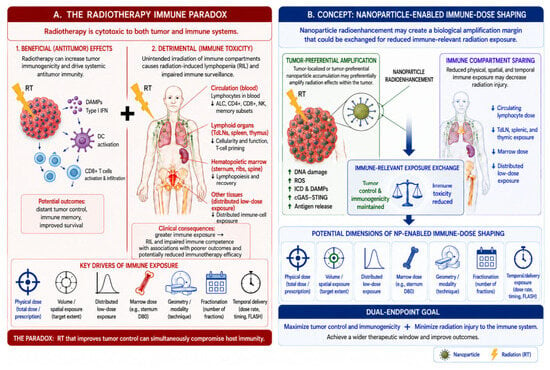
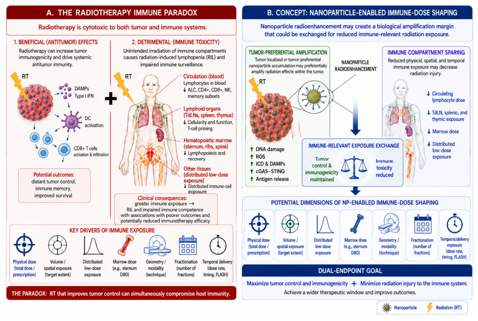
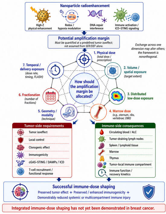
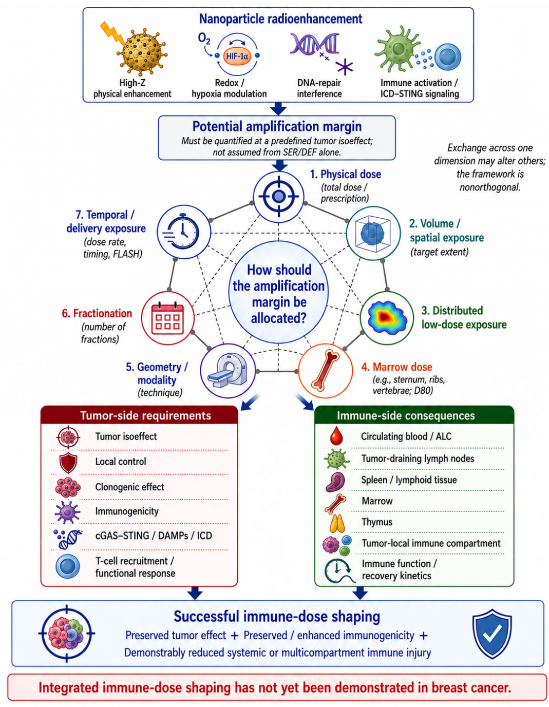
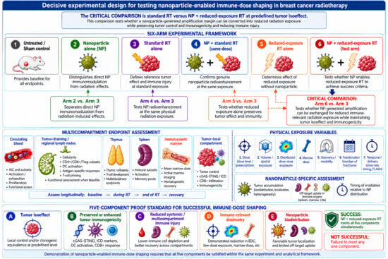
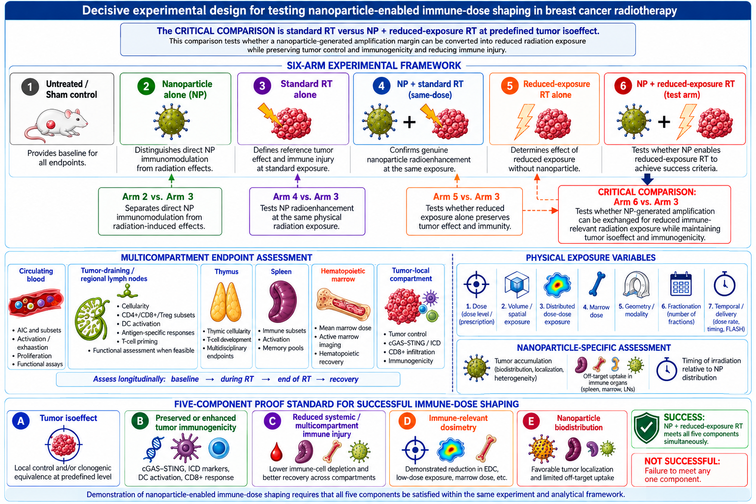
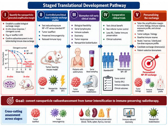
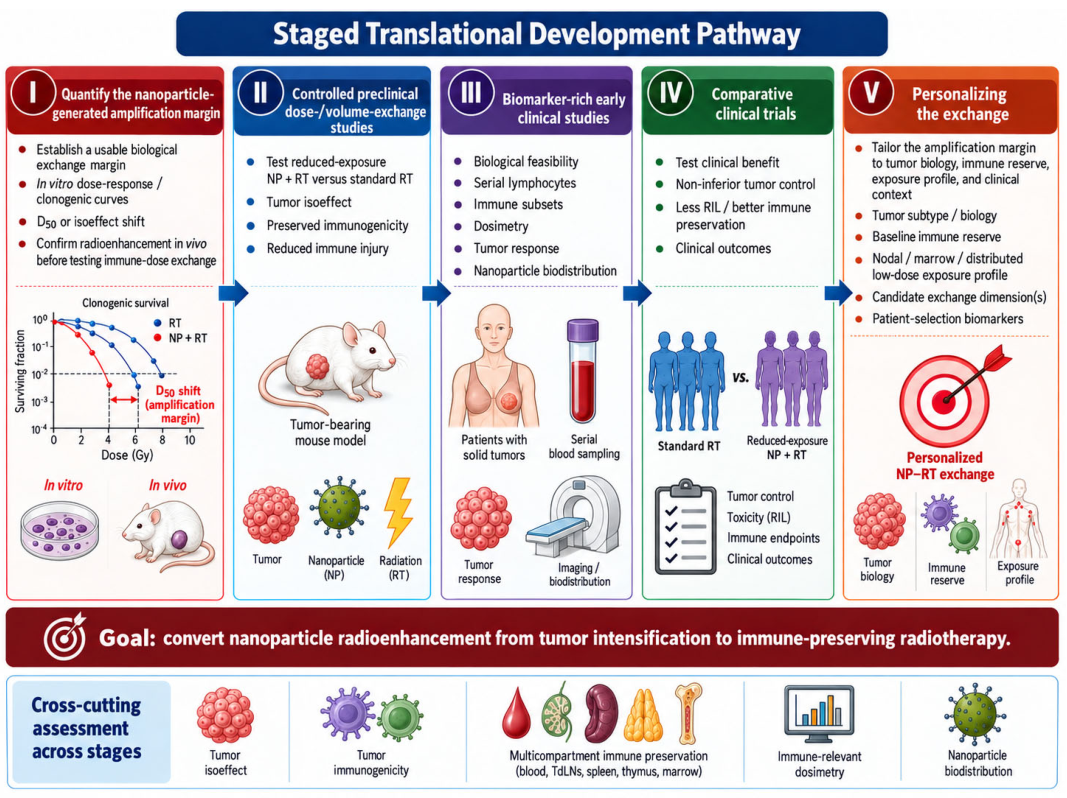

Disclaimer/Publisher’s Note: The statements, opinions and data contained in all publications are solely those of the individual author(s) and contributor(s) and not of MDPI and/or the editor(s). MDPI and/or the editor(s) disclaim responsibility for any injury to people or property resulting from any ideas, methods, instructions or products referred to in the content. |
© 2026 by the authors. Licensee MDPI, Basel, Switzerland. This article is an open access article distributed under the terms and conditions of the Creative Commons Attribution (CC BY) license.