


Abstract
Chronic rhinosinusitis (CRS) is a heterogeneous inflammatory disease imposing a substantial health and economic burden. Artificial intelligence (AI) has emerged as a promising tool to address CRS complexity in both clinical and research settings. We conducted a systematic review following PRISMA guidelines, searching PubMed, Web of Science, and Embase for peer-reviewed articles utilizing machine learning, deep learning, and natural language processing, with a focus on molecular-level or immunologic mechanisms in CRS. Thirty-one studies were included in the qualitative synthesis. Our results demonstrate that while AI-driven analysis helps stratify patient endotypes, the identified schemes exhibit substantial heterogeneity in cluster numbers and features, raising concerns about their reproducibility. AI applications also included histopathological image interpretation, drug discovery, biologic therapy selection, and prognostic modeling to evaluate revision surgery risks and olfactory dysfunction. However, using the PROBAST+AI framework, we found that 87.1% of the assessed studies carry a high overall risk of bias, mainly due to the reliance on apparent performance or internal validation and limited independent evaluation of model performance, indicating that many reported performance estimates may be optimistic. Future large-scale, multicenter collaborations integrating AI into clinical workflows will be essential to establish reliable and generalizable AI applications for personalized CRS management.
1. Introduction
Artificial Intelligence (AI) brings about many benefits as it allows machines to perform tasks that traditionally require human intelligence, including reasoning, pattern recognition, decision-making, and language comprehension. Recent breakthroughs in machine learning (ML), deep learning (DL), and natural language processing (NLP) have enabled AI systems to process complex datasets, perform tasks automatically, and generate personalized responses, which will open the door to revolutionary applications across a range of medical areas, such as disease diagnosis, clinical treatment, and medical research, among others [1,2]. In the early 1970s, MYCIN was designed to assist physicians in diagnosing and treating bacterial infections, marking a milestone in the AI application of medicine [3]. After that, the rapid advancement of computer science has led to the development of various AI technologies. Research has shown potential applications of AI in cancer screening and research, detecting diabetic retinopathy, EKG reading, and chest X-ray interpretation [4,5,6,7]. Regarding disease management, AI holds promise in the precision medicine of chronic disease, the prediction of individual response to cancer treatment, and rational drug dosage and monitoring in clinical practice [8,9,10,11]. The relationship between these AI techniques is demonstrated in Figure 1.
Figure 1.
Relationship between machine learning (ML), deep learning (DL), and natural language processing (NLP). ML is algorithms in AI that learn patterns from data. DL is a subfield of ML based on artificial neural networks (ANNs). NLP applies ML and DL techniques to analyze and represent human language.
Table 1 shows a comparison of ML, DL, and NLP. ML provides the foundational approaches, DL advances automated feature extraction for complex data, and NLP applies these methods to human language understanding. A rapidly evolving application of NLP with DL is the development of large language models, among which ChatGPT is one of the most prominent examples [12].
Table 1.
Comparison of machine learning (ML), deep learning (DL), and natural language processing (NLP) [12,13,14,15,16].
Chronic rhinosinusitis (CRS) is a significant health issue, impacting approximately 5–12% of the general population [26]. The risk factors of CRS include genetic predisposition, environmental factors, and comorbid conditions such as asthma and allergies [27]. The nasal and facial symptoms are more prominent than systemic or oropharyngeal symptoms [28]. The chronicity leads to substantial medical resource utilization and a global economic burden, driven mainly by increased office visits and prescription costs, with national healthcare expenditures in the U.S. being estimated at $8.6 billion annually [29], and €1501 per patient based on a Dutch cohort [30]. CRS is a heterogeneous disease known by its diverse underlying pathophysiology and classification. By phenotypic classification, CRS can be categorized based on endoscopic features, clinical courses, and comorbidities. For example, CRS has been traditionally divided into CRS with nasal polyps (CRSwNP) and CRS without nasal polyps (CRSsNP) according to the presence of nasal polyps [26]. Recent research has shifted focus to endotyping, which categorizes CRS based on the underlying pathophysiological mechanisms [31]. The T helper cell responses and their downstream effects in CRS have led to the identification of three distinct endotypes, namely the Type 1, Type 2, and Type 3 endotypes, which can be broadly categorized into Type 2 and non-Type 2 endotypes based on phenotype prevalence, clinical comorbidities, therapeutic targets, and response to treatment, where various biologics such as dupilumab, omalizumab, and mepolizumab have been developed in recent years [32,33,34]. The European Position Paper on Rhinosinusitis and Nasal Polyps (EPOS) 2020 introduced a revised classification system for CRS, with a combination of clinical features, localized or diffuse presentations, endotype, and phenotype [26,35]. Table 2 summarizes the common classification frameworks of CRS.
Table 2.
Phenotypic and endotypic classifications of chronic rhinosinusitis (CRS) based on EPOS2020 [26].
Clinical management of CRS has faced multiple challenges due to its marked disease heterogeneity, complicated classification frameworks, diverse treatment modalities, and varied patient profiles [9,36]. These make CRS a strong candidate for AI integration, as AI offers a powerful solution by integrating and analyzing large-scale clinical and molecular datasets. This review summarizes the current application and the future perspectives of AI in the field of CRS.
2. Methods
We performed a structured systematic review according to PRISMA guidelines [37] (the completed PRISMA checklist is available in Supplementary Table S1). The protocol for this review was not registered. A systematic literature search in PubMed, Web of Science, and Embase databases from 1 January 2015 to 30 May 2025 was conducted to identify peer-reviewed articles that had employed AI approaches in CRS diagnosis, classification, imaging, pharmacology, surgery, and prognostication. The search strategy applied keywords and MeSH terms related to “chronic rhinosinusitis,” “rhinosinusitis,” and “chronic sinusitis.” AI-related terms included “artificial intelligence,” “AI,” “machine learning,” “deep learning,” “data mining,” “neural network,” “supervised,” “unsupervised,” “cluster,” “natural language processing,” “ChatGPT,” “large language model,” and “predictive model.” The full search strategies for all databases are provided in Supplementary Table S2. Boolean operators (“AND,” “OR”) were used to combine disease-related and AI-related terms. The final literature search was conducted on 10 June 2025.
All identified records were imported into EndNote 2025 (Clarivate Analytics, Philadelphia, PA, USA) for reference management. We utilized both an automation tool on EndNote and manual checking to remove duplicate records. Two independent reviewers screened the titles and abstracts during the initial screening phase. We excluded records that did not focus on CRS, did not utilize AI-based approaches, lacked a molecular-level or immunologic mechanistic focus, were non-English language publications, or were review articles or commentaries. Full-text articles of the remaining records were assessed for eligibility by the same reviewers. Reports were further excluded from the final synthesis if they demonstrated a lack of AI model performance or validation metrics, presented duplicate data or cohort overlap, or contained insufficient AI methodological details. Any discrepancies between the two reviewers during the screening and selection process were resolved through discussion and consensus, or by consulting a third senior reviewer.
Data extraction was performed independently by two reviewers. The extracted data items included: first author, publication year, specific clinical task, data modality and size, type of AI or machine learning algorithms, validation methods, evaluation metrics, and main outcomes. The primary outcomes sought were the predictive performance of the AI models and the identified clinical or biological features. When multiple models were evaluated within a single study, the results of the best-performing model were prioritized for extraction. Regarding missing or unclear information, no assumptions were made, and only available data were extracted and synthesized.
The methodological quality and risk of bias of the included studies were appraised using the newly updated Prediction model Risk Of Bias assessment Tool for AI (PROBAST+AI) framework [38]. In accordance with the updated framework, two reviewers independently assessed each study across two distinct phases: the methodological quality of the model development process, and the risk of bias in the evaluation of model performance. Both phases were assessed across four signaling domains: participants and data sources, predictors, outcomes, and analysis. Applicability was assessed separately for participants and data sources, predictors, and outcomes. Disagreements in the quality assessment were resolved by consensus. PROBAST+AI distinguishes apparent, internal, and external validation, noting that the absence of external performance evaluation may lead to a high Domain 4 rating. However, in accordance with the PROBAST+AI framework, an overall high risk of bias may be reclassified to low when the model was developed on a large dataset, appropriately internally validated, and the other domains showed low concerns.
In accordance with PRISMA 2020 guidelines (Item 4) and the PROBAST+AI framework, our review objective and applicability assessments were anchored to the following PICOTS criteria: Population (P): Adult patients diagnosed with chronic rhinosinusitis (CRSwNP or CRSsNP). Index model (I): Artificial intelligence or machine learning algorithms investigating molecular-level or immunologic mechanisms through the analysis of molecular, immunological, digital pathology, or related clinical surrogate data. Comparator (C): Traditional statistical predictive models or no comparative model. Outcome (O): Identification of disease endotypes, histopathological classifications, and prediction of clinical outcomes. Timing & Setting (T, S): preoperative, intraoperative, or postoperative diagnostic and prognostic settings in tertiary or general clinical environments. Given the nascent and rapidly evolving nature of AI applications in rhinology, our PICOTS criteria were intentionally designed to be comprehensive rather than narrowly restricted. This approach enables us to capture the full landscape of how machine learning is currently being deployed across different biological modalities (e.g., transcriptomics, proteomics, and digital pathology) to address the clinical heterogeneity of CRS. Consequently, due to the significant heterogeneity in the clinical tasks, data modalities, and performance metrics across the included studies, a quantitative meta-analysis was not feasible. Instead, a narrative synthesis was performed in adherence to the Synthesis Without Meta-analysis (SWiM) reporting guidelines to ensure a transparent evaluation [39]. The characteristics and main outcomes of the individual studies were tabulated to facilitate visual comparison, and the extracted data were systematically categorized and presented according to their specific clinical applications in CRS.
3. Results
3.1. Study Selection
Figure 2 illustrates the detailed selection process. A total of 962 records were identified, including 254 from PubMed, 317 from Web of Science, and 391 from Embase, and imported into EndNote for reference management. 427 records were removed, consisting of 261 duplicates identified using an automation tool in EndNote, 165 duplicates removed manually, and 1 retracted record. 535 records were screened by title and abstract. Among them, 484 were excluded for not meeting the following inclusion criteria: not focusing on CRS (n = 296), not using AI-based approaches (n = 102), lacking a molecular-level or immunologic mechanistic focus (n = 65), being review articles or commentaries (n = 11), and being non-English language publications (n = 10). The remaining 51 reports were sought for retrieval and underwent full-text assessment for eligibility, while 2 reports were not retrieved due to unavailable full texts or access restrictions. The remaining 49 full-text articles were assessed for eligibility, and 18 reports were further excluded due to duplicate data or cohort overlap (n = 10), a lack of AI model performance or validation metrics (n = 4), and insufficient AI methodological details (n = 4). No additional records were identified through citation searching or other methods. Finally, 31 studies were included in the qualitative synthesis.
Figure 2.
PRISMA 2020 flow diagram illustrating the selection process of studies for the systematic review, including records identification, screening, and exclusion criteria.
3.2. Study Characteristics
Table 3 summarizes the characteristics and AI applications of included studies. The 31 included studies consisted of 11 (35.5%) primary prospective studies, 9 (29.0%) cross-sectional studies, 5 (16.1%) retrospective analyses, 4 (12.9%) in silico database analyses, and 2 (6.5%) observational/case–control studies. Because several studies incorporated more than one data modality, the following categories were not mutually exclusive. Thirteen studies (41.9%) incorporated clinical features, including structured clinical records, olfactory test results, and symptom scoring systems such as the SNOT-22. Twelve studies (38.7%) analyzed non-invasive biomarkers, utilizing inflammatory mediators derived from nasal mucus, secretions, serum, urine, and nasal mucus-derived exosomes (NMDEs). Six studies (19.4%) used transcriptomic and multi-omics data, containing single-cell RNA sequencing, bulk RNA sequencing, and microarray profiling from public databases. Three studies (9.7%) applied automated digital pathology, utilizing whole slide images (WSIs) and H&E-stained sections for immune cell quantification.
Table 3.
Summary of characteristics and artificial intelligence applications of included studies.
3.3. Risk of Bias and Applicability Assessment
The model development and model evaluation of the 31 included studies were systematically appraised using the PROBAST+AI tool in Table 4.
3.3.1. Model Development (Methodological Quality)
During the model development phase, the majority of studies demonstrated low concerns in Domain 1 (Participants and Data Sources), Domain 2 (Predictors), and Domain 3 (Outcomes). Clinical data sources, diagnostic criteria, and predictor measurements were generally clearly defined. Notably, Sireci et al. [60] evaluated the performance of a pre-trained ChatGPT model without model development or retraining; therefore, the PROBAST+AI development phase was considered not applicable for this study.
Quality concerns primarily emerged in Domain 4 (Analysis). Recognizing the distinct nature of unsupervised models, exploratory clustering and machine-learning studies were not penalized merely because they did not follow conventional supervised prediction-modelling pipelines. Studies using relatively adequate sample sizes together with clearly described and appropriate analytical strategies, such as Dorismond et al. [47], Liao et al. [53], and Wang Z. et al. [68], were therefore rated as having low concerns in the analysis domain.
In contrast, high concerns were assigned when explicit structural analytical limitations were identified. The most frequent concern involved a very small effective sample size combined with high-dimensional exploratory network or clustering analyses, or sequential data-driven feature selection, as observed in studies such as Divekar et al. [45], Ishino et al. [50], Wang H. et al. [66], Wang X. et al. [67], and Zhou et al. [70]. These features may lead to unstable analytical solutions and limited reproducibility. Other concerns included outcome-driven univariable predictor selection prior to model fitting, which introduces predictor selection bias and a high risk of overfitting (Guo et al. [48]), and highly restricted effective development units in deep-learning pipelines, such as Hsu et al. [49], in which model training was based on only 10 whole-slide images.
3.3.2. Model Evaluation (Risk of Bias, ROB)
During the model evaluation phase, Domain 1 (Participants and Data Sources) was assessed primarily according to participant selection, exclusions, and attrition, rather than simply according to whether an independent external validation cohort was available. Most studies had evaluation populations that were appropriate for their prespecified target populations and were therefore rated as having low concerns in Domain 1. Chowdhury et al. [44] was the exception because only 62 of 147 enrolled participants had longitudinal postoperative follow-up, resulting in a substantially restricted evaluation subset and a potential selection/attrition concern.
Overall, 27 of 31 studies (87.1%) were rated as having high overall risk of bias in the model evaluation phase, primarily because of concerns in Domain 4 (Analysis and Performance Evaluation). Under our conservative operational rule, studies without independent external performance evaluation were generally assigned a high ROB in Domain 4 when model performance relied solely on apparent performance or internal validation procedures, including data splitting, cross-validation, or bootstrap resampling. Studies incorporating independent participant data for model evaluation, including Hsu et al. [49] and Nakayama et al. [58], generally demonstrated lower concerns regarding evaluation bias. In contrast, independent biomarker verification without applying and evaluating the complete prediction model in an independent dataset, as in Chen J. et al. [42], was not considered equivalent to external validation of a prediction model. Wang H. et al. [66] performed external validation using an independent GEO dataset; however, the limited validation sample size raised high concern in evaluation Domain 4, contributing to a high overall risk of bias judgement.
Notably, in accordance with the PROBAST+AI guidance, Lou et al. [55] was assigned a low overall ROB despite the absence of a strict external cohort. Although Domain 4 raised concerns under our conservative operational rule, the study used a relatively large dataset (N = 366), appropriate internal cross-validation, and showed no material concerns in the other evaluation domains. The overall ROB judgment therefore remained low, reflecting the consideration of the study’s evaluation methods as a whole rather than a mechanical aggregation of individual domain judgments.
3.3.3. Applicability
Applicability concerns were generally low across the included studies for both the model development and evaluation phases. The participant populations, predictors, outcomes, and clinical tasks were considered sufficiently aligned with the intended scope of the present systematic review. No material applicability concerns were identified that would limit interpretation of the included studies within the scope of this review.
Table 4.
Risk of bias and applicability assessment based on the PROBAST+AI guidelines.
3.4. AI and Machine Learning Applications
The AI applications in the 31 included studies were categorized into four main areas, according to their primary application. Fifteen studies (48.4%) focused on CRS classification and phenotyping. Nine studies (29.0%) involved prognostication and patient outcomes. Four studies (12.9%) were categorized under pharmacology and therapeutic optimization. Three studies (9.7%) focused on imaging and diagnostic analysis. As detailed in our PROBAST+AI evaluation (Section 3.3), 27 of the 31 included studies (87.1%) were assessed as having a high risk of bias in the model evaluation phase, primarily due to concerns in Domain 4 (Analysis and Performance Evaluation), particularly reliance on apparent performance or internal validation and limited independent evaluation of model performance. Consequently, throughout the following subsections, most reported performance metrics (e.g., high AUCs and prediction accuracies) reflect internally validated or apparent estimates. These headline figures should be interpreted with caution, as they may represent optimistic estimates of model performance.
3.4.1. CRS Classification
While some deep learning algorithms applied to digital pathology also contribute to phenotypic classification (discussed separately in Section 3.4.2), the 15 studies detailed in this section mostly employed clustering algorithms on biomarker and clinical data to uncover inflammatory endotypes.
In 2016, a landmark study by Tomassen et al. identified 10 inflammatory endotype clusters of CRS through an unsupervised cluster analysis based on 14 cytokine and immune biomarkers in a phenotype-free approach. The results revealed a spectrum of immune responses, ranging from non-inflammatory profiles to high type 2 inflammation presentation, and were further matched with clinical phenotypes. They identified four clusters that exhibited low or undetectable levels of IL-5, ECP, IgE, and albumin, which are markers associated with type 2 inflammation, and corresponded with the CRSsNP phenotype and a low asthma prevalence. Among them, Cluster 1 showed no significant inflammatory elevation. Cluster 2 demonstrated a TNF-α/IL-22 profile. Cluster 3 was marked by IFN-γ and MPO/IL-8 elevation. Cluster 4 exhibited Th17-neutrophilic inflammation. The remaining six clusters showed high IL-5 and eosinophilic signatures, with further categorization based on IL-5 levels and comorbidity of asthma. Clusters 5 to 7 showed moderate IL-5 levels and mixed CRSsNP/CRSwNP features, while Clusters 8 to 10 displayed very high IL-5, IgE, and SE-IgE levels and were composed almost exclusively of CRSwNP patients with a high asthma prevalence. Clusters 9 and 10, particularly, had the highest IgE levels and universally expressed SE-IgE, indicating a strong superantigen-driven Th2 inflammation [63]. Romano et al. validated tissue cytokine profiles in a Brazilian multicenter cross-sectional study, where hierarchical clustering and principal component analysis revealed two main clusters of high and low inflammatory patterns. They determined that a tissue eosinophilia cut-off of 43 eosinophils per high-power field distinguished these two distinct inflammatory profiles [59].
Further studies expanded on tissue networks and remodeling factors. Given that a laboratory-based approach may not be practical in routine clinical settings, Divekar et al. [46] applied an unbiased network-mapping approach using a commercially available immunoassay panel for the endotyping of sinonasal tissue obtained from CRS patients and control subjects, which revealed three distinct cytokine–chemokine clusters in CRS patients. Cluster 1 was enriched for Th1/Th17-type markers. Cluster 2 was dominated by Th2-associated cytokines. Cluster 3 was characterized by elevated levels of chemokines and growth factors [46]. To study the association between endotypes and remodeling of CRS, Wang X. et al. identified 5 endotypes of CRS by integrating inflammatory and remodeling biomarkers, including cytokines, neutrophil factors, remodeling factors, MMP and fibronectin. Notably, Cluster 3 was characterized by low type 2 inflammation but exhibited high expression of neutrophil-associated factors, such as IL-8, G-CSF, MPO, and extracellular matrix proteins such as fibronectin, suggesting a neutrophil-dominant remodeling endotype. Cluster 5 demonstrated pronounced type 2 inflammation, accompanied by comparatively elevated levels of neutrophil-associated and tissue remodeling markers. Across Clusters 1 to 5, there was a progressive increase in the prevalence of nasal polyps, asthma, allergy, anosmia, aspirin sensitivity, and CRS recurrence [67]. Similarly, Viksne et al. utilized hierarchical cluster analysis to evaluate cytokines and antimicrobial peptides, classifying five specific inflammatory endotypes and observing that endotypes associated with neutrophilic inflammation or a mixed neutrophilic/type 2 pattern were predominant in their study population [65].
In addition to inflammatory pathways, machine learning has also been used in analyzing public transcriptomic databases and molecular subtypes. Chen Z. et al. applied machine learning algorithms to datasets from the Gene Expression Omnibus and classified CRSwNP patients into two distinct anoikis-related molecular subtypes characterized by different immune microenvironments. The models further identified four key anoikis-related genes, CDH3, PTHLH, PDCD4, and androgen receptor, with high diagnostic accuracy. Among these, androgen receptor and PTHLH were recognized as cluster-specific biomarkers [43]. Nakayama et al. investigated transcriptomic differences using whole-transcriptome and single-cell RNA sequencing (scRNA-seq). Their unbiased unsupervised clustering revealed that White and Japanese patients harbor the same major type 2 and non-type 2 endotypes, though with a statistically significant difference in the proportion of these endotypes between the two populations. There was a markedly higher prevalence of the type 2 endotype in White patients compared to Japanese patients. Furthermore, using droplet-based scRNA-seq, they mapped the cellular origins of newly identified signature transcripts, discovering that type 2-driving chemokines such as CCL13 and CCL18 were upregulated within CD163-high M2 macrophages, whereas cystatin SN (CST1) and CCL26 were expressed in basal, suprabasal, and secretory epithelial cells. Their random forest classifier further established that a select panel of these novel transcripts, particularly CCL13, CST1, and CCL18, could predict type 2 endotypes with an internally resampled AUC of 0.956; although the transcriptomic signatures were externally validated via an independent qPCR cohort, this specific model performance metric relies on internal bootstrapping [58]. Ishino et al. performed transcriptomic analysis using bulk RNA barcoding and sequencing. The hierarchical cluster analysis revealed that a cluster featuring severe type 2 inflammation is associated with upregulated platelet-activating factor (PAF) synthesis—the enzyme LPCAT1—and downregulated PAF degradation—the enzyme PAFAH2 [50].
Since the tissue-based assessment involves an invasive nature, Turner et al. defined CRS endotypes in a U.S. population entirely based on analysis of mucus collected via a minimally invasive approach. Similar to tissue-based results from a European population [63] but differing methodologically, they identified 6 inflammatory endotypes that differ substantially with respect to phenotype and disease behavior [64]. Similarly, Miyake et al. utilized nasal mucus-derived exosomes (NMDEs) to perform a liquid biopsy as an efficient alternative to traditional tissue sampling. Through cluster analysis, they demonstrate that a high Cystatin-2 expression level was associated with CRS comorbidities, and therefore, the cystatin level in NMDEs can predict CRS phenotype and disease severity, suggesting potential utility for treatment stratification [56]. Moreover, Becker et al. demonstrated the potential of machine learning for the discovery of non-invasive biomarkers in CRS phenotyping and endotyping. In a study on nasal mucus from 103 CRS patients, the authors applied complex algorithms, such as t-SNE, AdaBoost, and XGBoost, to a panel of 12 inflammatory proteins. Their models showed that mucus-based biomarkers had predictive performance for CRS phenotypes similar to conventional tissue-based methods. They discovered that IL-5 acted as the major driver for patient clustering, whereas periostin and cystatin-SA were important markers in epithelial- and tissue-derived signatures. Additionally, a four-biomarker panel of IL-5, IgE, IL-17, and periostin had the best predictive performance. These findings support the potential utility of AI-based analysis of non-invasive samples for CRS classification [41]. Wang Z. et al. established a pre-pruned decision tree model based on the levels of IL-5 in nasal secretion, blood eosinophil counts, and the asthma status of CRSwNP patients, yielding an internally validated AUC of 0.90 for predicting high levels of type 2 biomarkers [68].
Systemic and mixed clinical indicators were explored for CRS classification. Divekar et al. [47] applied unsupervised network analysis to assess serum biomarkers during acute exacerbations of CRSwNP, revealing a systemic increase in growth factors, specifically VEGF and GM-CSF [45]. Many studies have focused on identifying biomarkers for eosinophilic chronic rhinosinusitis (eCRS), but none have employed methods from the field of AI. Thorwarth et al. used supervised machine learning techniques, specifically logistic regression and artificial neural networks (ANN), to predict eCRS of patients who underwent endoscopic sinus surgery (ESS) for the treatment of CRS, based on 3 input variables, which are peripheral eosinophil count, urinary leukotriene E4 (uLTE4) level, and polyp status. The ANN models using the surgeon-specific dataset demonstrated predictive ability with a sensitivity of 92.3% (95% CI: 65.0–99.8) and a specificity of 85.7% (95% CI: 42.1–99.6) for the eCRS field [62].
Across clustering studies, the number of identified endotypes ranged from 2 to 10, with substantial variation in input features, clustering approaches, and criteria used to assess cluster stability or separation. Although several studies identified broadly overlapping inflammatory patterns, such as type 2, neutrophilic, and mixed inflammatory profiles, the proposed endotype structures were not directly comparable because they were derived from largely non-overlapping biological variables and heterogeneous analytical frameworks. Consequently, the available evidence demonstrates substantial molecular and inflammatory heterogeneity in CRS but does not yet establish a single reproducible AI-derived endotype classification framework across independent populations.
3.4.2. Histopathological Image Analysis
To study the cellular phenotyping diagnosis of nasal polyps by whole-slide imaging (WSI), Wu et al. established an AI chronic rhinosinusitis evaluation platform 2.0 (AICEP 2.0) by applying a convolutional neural network (CNN)-based deep learning model. As the first AI for cellular phenotyping, AICEP 2.0 analyzed the proportion of inflammatory cells for each image and discovered four phenotypes of nasal polyps with different clinical characteristics. They also demonstrated that the percentage of eosinophils in peripheral blood was positively correlated with that in polyp tissue on WSI [69]. Similarly, Hsu et al. developed a CNN with query-driven multiple instance learning (MIL)-based automated eosinophil detector and achieved a sensitivity of 100.00% and a specificity of 90.48% [49].
For the purpose of examining the relationship between angiogenesis and the immune microenvironment, Liu et al. utilized a fully convolutional network (FCN) to quantify microvessels of tissues from CRS patients and revealed that angiogenesis was associated with type 2 inflammation. Additionally, they discovered a negative correlation between angiogenesis and local tissue expression of TNF-α and TGF-β [54].
3.4.3. Pharmacology and Therapeutic Optimization
Four studies applied artificial intelligence and bioinformatics to identify novel molecular therapeutic targets and optimize pharmacologic treatments for CRSwNP.
Wang H. et al. applied multiple machine learning algorithms, including weighted gene co-expression network analysis (WGCNA), least absolute shrinkage and selection operator (LASSO) regression, random forest (RF), and SVM-RFE algorithms to identify hub genes associated with the pathogenesis of CRSwNP. They recognized gene BTBD10, ERAP1, GIPC1, and PEX6 as key hub genes and potential novel therapeutic targets [66]. Similarly, Zhou et al. focused on macrophage polarization and metabolism and utilized WGCNA, LASSO, and RF algorithms to screen for candidate genes. They identified ALOX5, HMOX1, and PLA2G7 as core biomarkers of CRSwNP. Additionally, using a drug–biomarker interaction network, they further predicted that selenium and the transcription factor FOXC1 are potential regulatory targets [70].
Exploring post-transcriptional regulation, Chen J. et al. constructed a competitive endogenous RNA (ceRNA) network using data mining and experimental verification. They identified a novel MIAT/miR-125a/IRF4 regulatory axis, demonstrating that the long noncoding RNA MIAT acts as a sponge to regulate IRF4 expression. This ceRNA axis was correlated with immune cell infiltration, specifically dendritic cells and M2 macrophages, offering further insight into novel molecular therapeutic targets for CRSwNP [42].
At the clinical decision-making level, Sireci et al. performed a novel application of large language models by using ChatGPT to guide the selection of biological therapies, including dupilumab, mepolizumab, and omalizumab, for patients with CRSwNP. The model showed a concordance percentage of 68% with the recommendations of the multidisciplinary Rhinology Board, indicating its potential to assist otolaryngologists in selecting biological therapy. However, they stated that it requires further training with large databases and integration with clinical data and image processing to enhance its diagnostic ability [60].
3.4.4. Prognostication and Surgical Outcomes
Nine studies addressed prognostication and surgical outcomes, applying predictive models to forecast postoperative symptom improvement, long-term disease recurrence, and revision surgery risks.
Adnane et al. aimed to employ an unsupervised clustering method to identify which patient groups achieve the greatest improvement in QOL following ESS. They classified three distinct clusters of patients with CRS using unsupervised cluster analysis. All clusters showed significant clinical improvement, but there were significant differences in QOL outcomes between clusters, with factors like nasal polyps and mucosal eosinophilia endotype negatively impacting QOL improvement [40]. Similarly, Chowdhury et al. investigated the utility of baseline mucus cytokines in predicting postoperative 22-item Sino-Nasal Outcome Test (SNOT-22) scores using random forest and stepwise multivariate linear regression algorithms. They found that mucus IL-5 levels independently predicted postoperative SNOT-22 improvement, whereas IL-2 and TNF-α predicted postoperative worsening [44]. Focusing specifically on smell loss, Morse et al. applied machine learning algorithms, including hierarchical cluster analysis and random forest, to identify patterns of olfactory dysfunction in CRS patients, finding that anosmia is associated with a specific endotype characterized by severe nasal polyposis, tissue eosinophilia, and aspirin-exacerbated respiratory disease (AERD). Furthermore, IL-2, CT scores, and AERD were independently predictive of olfactory function. These findings indicated that a complex integration of inflammatory, clinical, and demographic factors contributes to olfactory loss in CRS patients [57].
Other models have been designed to predict disease trajectory and the need for revision surgery. Lou et al. identified five cellular phenotypes of CRSwNP and developed a clinical algorithm to predict recurrence, reporting an internally validated 98.5% polyp recurrence rate for a marked tissue eosinophilia cluster; although this model was assessed as having a low overall risk of bias due to its large sample size and robust internal procedures, the estimate still requires independent external confirmation for broader generalizability [55]. Similarly, Dorismond et al. utilized hierarchical clustering to analyze the cytokine profiles of 269 CRSwNP patients to predict time to recurrence. They identified six disease clusters and revealed a correlation between inflammatory endotypes and the time to recurrence. Two clusters, one exhibiting a mixed Type 1 and Type 3 inflammatory profile, including IFN-γ, IL-4, and IL-17A, and the other with elevated IL-12 and IL-21, showed the shortest time to recurrence. On the other hand, a cluster featuring innate and proinflammatory markers, including IL-1β, IL-6, and IL-8, showed the longest recurrence-free interval. These findings suggested that difficult-to-treat cases may require novel therapeutic strategies beyond Type 2 inflammation targets to prevent disease recurrence [47].
Identifying patients at high risk for difficult-to-treat or refractory CRS has also been a major focus of predictive modeling. Liao et al. integrated both clinical and molecular variables into a cluster analysis, identifying seven endotypes. They demonstrated that both severe eosinophilic and neutrophilic inflammation can lead to poor treatment outcomes, while a high IL-10 level cluster had no difficult-to-treat cases, suggesting the role of IL-10 in limiting inflammation [53]. To predict with a non-invasive method, Guo et al. constructed a prediction model using classification and regression tree (CART) and random forest algorithms. They found that the CCL17 levels of nasal secretion, hyposmia scores, allergic rhinitis comorbidity, and MIP-1β levels demonstrated an internally validated accuracy of 94% for predicting difficult-to-treat CRS [48]. For specific histologic subtypes, Kim et al. employed decision tree and random forest models to demonstrate that subepithelial neutrophil infiltration (human neutrophil elastase-positive cells), along with Lund–Mackay scores and endotype, acts as a cellular biomarker for predicting poor surgical outcomes of CRSwNP patients [51]. For non-eosinophilic CRSwNP, Li et al. utilized a random forest algorithm and stepwise logistic regression to evaluate clinical and biological variables. Their integrated model achieved an internally validated AUC of 0.89, identifying the biomarkers PDGF-β, PD-L1, MIP-3b, and PDGF-α as independent predictors of postoperative uncontrolled disease status in patients without significant eosinophilic infiltration [52].
4. Discussion
4.1. Summary of Principal Findings
This systematic review summarizes the current applications and potential of AI in advancing the understanding and clinical evaluation of CRS. Across the 31 included studies, AI-based approaches were used to characterize molecular and inflammatory endotypes and to analyze multidimensional data, including tissue transcriptomics, non-invasive biomarkers, digital pathology, and clinical variables. These applications identified heterogeneous inflammatory patterns, candidate therapeutic targets, and factors associated with postoperative outcomes, highlighting the potential of AI to support more detailed CRS phenotyping and risk stratification.
4.2. CRS Endotyping and Non-Invasive Diagnostics
Endotypic classification is based on underlying immunologic mechanisms, allowing a more precise identification of disease variants and their associated biomarkers [33]; also, understanding the cellular diversity in CRSwNP can help in diagnosis and the selection of therapeutic approaches, including the use of biologics [71]. The AI-driven clustering models reviewed in this study reveal a much more complex and heterogeneous spectrum [46,59,63,65,67]. These findings demonstrate the potential of biomarker-based clustering to identify immunologic profiles with potential clinical relevance and may provide a basis for future treatment stratification [33]. However, a critical examination of these models highlights a lack of consensus that must be confronted. The number of defined endotypes ranged from 2 to 10. This variation was accompanied by substantial differences in input features, clustering strategies, and criteria used to assess cluster separation or stability, limiting direct comparison and reproducibility across studies. The current literature presents a fragmented landscape of localized models rather than a universal CRS endotyping scheme. Therefore, while these models demonstrate the potential of AI in revealing immunologic profiles, their immediate clinical utility is hindered by this lack of standardization. Reconciling these divergent endotype schemes requires future large-scale multicenter studies to validate unified biomarker panels using standardized mathematical clustering metrics.
AI-based analyses of non-invasive specimens, including nasal secretions, mucus-derived exosomes, and serum, suggest the potential for the transition from invasive tissue biopsies, which are often limited by spatial heterogeneity across the sinonasal cavity [72,73], to non-invasive diagnostic strategies. Multiple studies applied complex algorithms, such as AdaBoost, XGBoost, and decision trees, to nasal secretions, mucus-derived exosomes, and serum to predict CRS endotypes [41,45,56,62,64,68]. By identifying biomarker panels, ML models have potential in disease endotyping in the outpatient setting, bypassing the need for invasive tissue sampling.
4.3. Accelerating Pharmacological Research and Therapeutic Optimization
AI can be applied in therapeutics for a dual purpose: accelerating drug discovery and optimizing clinical decision-making. The current treatment landscape for CRS is rapidly expanding toward targeted immunotherapies [26]. In this study, we reveal that machine learning frameworks have the ability to process high-dimensional transcriptomic data, searching for novel molecular targets and investigating complex regulatory networks [42,66,70]. From a translational perspective, these approaches may help prioritize candidate molecular targets and generate hypotheses for subsequent experimental validation and drug development [74,75].
Currently, using generative AI to guide the selection of monoclonal antibodies [60,76] shows potential to become an interactive bedside tool for clinicians. However, while large language models are promising for optimizing treatments, we must approach them with caution. Because these algorithms can still misinterpret data or generate “hallucinations,” their recommendations require strict verification by medical specialists before they can be safely used in real-world clinical practice [60].
4.4. Redefining Prognostication and Surgical Risk Assessment
FESS remains a solution for CRS refractory to medical therapy, but anticipating which patients will achieve disease control or suffer recurrent olfactory dysfunction, along with long-term disease trajectory, has relied on static clinical parameters [26,77]. By capturing complex, non-linear relationships among diverse variables, the reviewed approaches explored whether combinations of clinical, inflammatory, and molecular variables could improve risk stratification beyond conventional clinical indicators. [40,44,47,48,51,52,53,55,57]. A key pathophysiological insight synthesized from these models is the concern with the traditional Type 2-centric view of recurrence. While severe Type 2 inflammation is a well-established driver of surgical failure [55], AI models have identified mixed Type 1/3 or neutrophilic inflammatory profiles as associated with shorter time to recurrence or poorer postoperative outcomes [47,51]. This finding suggests that focusing exclusively on Type 2 pathways may be insufficient for a subset of patients.
In principle, validated prediction models could support individualized risk stratification and inform postoperative surveillance or treatment planning, changing the medical approach from simply reacting to problems to actively preventing them. However, the current evidence base does not yet establish that these models can reliably support such decisions in routine clinical practice [48,53,57].
Figure 3 demonstrates the potential role of AI in the clinical management of CRS for clinicians.
Figure 3.
Integration of artificial intelligence (AI) into the clinical management of chronic rhinosinusitis (CRS). This figure illustrates the role of AI frameworks, including machine learning (ML), deep learning (DL), and natural language processing (NLP), in processing multi-modal data sources (histopathological images, biofluids, tissues, and clinical data) across the clinical management of CRS. AI has the potential to assist the diagnosis workup through clinical phenotyping, molecular endotyping, non-invasive biomarker analysis, and histopathological image analysis. In treatment, AI contributes to biologic therapy selection, drug discovery, therapeutic target identification, and surgical planning. During follow-up and prognosis, AI enables the prediction of disease recurrence and revision surgery, prognostic modeling of quality of life (QoL), and risk assessment for olfactory dysfunction.
4.5. Challenges and Limitations of AI in CRS
Although AI has potential applications in medicine, some challenges and limitations remain. Current studies are often constrained by small sample size, single-center design, and various biases that limit the generalizability of the results. Data-related challenges, including limited quality of datasets, lack of standardization, and privacy or legal barriers to gathering the data, have further hindered model development and the reliability of generated results. At the technical level, many complex AI models remain difficult to interpret because their learned representations, parameters, and feature interactions are not readily understandable in human terms. Limited interpretability may complicate error analysis and assessment of model generalizability. Separately, overfitting remains a methodological concern in studies with small effective sample sizes and high-dimensional predictors, while generative AI systems may additionally produce hallucinated or unsupported outputs, still struggle with misinterpretation of information and data privacy issues, and need verification from specialists for those AI-generated findings [1,78,79].
Several limitations must be acknowledged, both at the review level and the study level. First, regarding our review methodology, the protocol for this systematic review was not prospectively registered in PROSPERO or a similar registry, which constitutes a methodological limitation. Furthermore, our search strategy was restricted to English-language, peer-reviewed articles across three major databases (PubMed, Web of Science, Embase), excluding grey literature, preprints, and trial registries. These restrictions introduce a potential language and publication bias. Because of the substantial heterogeneity in study designs, clinical tasks, and performance metrics across the included studies, a formal quantitative assessment of publication or reporting bias was not feasible; therefore, the risk of publication bias remains unquantified and must be acknowledged. Additionally, during data extraction, we primarily reported the metrics of the best-performing models from each study. Extracting only the best-performing models introduces selective reporting or selection bias at the review level and may lead to an overemphasis on the highest reported performance estimates, which might not be fully reproducible in prospective applications.
Second, at the study level, several included studies face potential limitations regarding model overfitting and cross-dataset confounding. For instance, some models were developed using sample sizes that were relatively small compared to the large number of analyzed features (e.g., evaluating a ~39-plex biomarker panel on fewer than 20 individuals [45]). In the absence of robust dimensionality reduction or independent external validation, these models may be susceptible to overfitting. The lack of reproducibility and comparability across the clustering schemes constitutes a major scientific limitation. Because studies utilize disparate input variables and validate their clusters using fundamentally non-comparable mathematical criteria, it remains unclear whether any of the proposed endotype schemes (which vary from 2 to 10 clusters) are reproducible across different populations. This central heterogeneity currently prevents the establishment of a consensus AI-driven CRS classification system. Additionally, for studies integrating independent microarray datasets (e.g., merging multiple GEO series), the management of batch effects was not consistently detailed. Unmitigated cross-dataset confounding could influence the reproducibility of the identified molecular targets.
Finally, the results of our PROBAST+AI assessment indicate a notable contrast between applicability and risk of bias in the current literature. As detailed in Table 4, the included studies consistently demonstrated low applicability concerns. This indicates that the populations, predictors, outcomes, and clinical tasks evaluated in the included studies were generally aligned with the prespecified scope of the review. However, 27 of the 31 studies (87.1%) were rated as having high overall risk of bias in the model evaluation phase, with concerns most frequently arising in Domain 4 (Analysis and Performance Evaluation). The common reliance on internal validation methodologies and the limited use of independent evaluation of model performance suggest that the reported performance metrics (e.g., high AUCs or accuracies) may be optimistic. Clinically, these concerns suggest that the reported performance estimates may be sensitive to the characteristics of the original development cohorts and may not generalize well to other populations or clinical settings. If these algorithms were deployed in different real-world healthcare settings, they could experience significant performance degradation. Therefore, while these AI models provide valuable translational insights into CRS, their generalizability remains uncertain, emphasizing the need for external validation before they can be integrated into routine clinical decision-making.
4.6. Future Directions
Current studies face several limitations, including dataset constraints, data privacy and ethical issues, and the variable accuracy of AI models. Future research should prioritize large, multicenter, and prospectively characterized cohorts with standardized clinical, molecular, and imaging data collection. For AI-derived endotypes, standardized input features and clustering methods, along with independent replication, are needed to assess reproducibility across populations. Predictive models require strict validation, independent external evaluation, calibration, and transparent reporting before clinical implementation. Prospective evaluation of multimodal AI systems in real-world settings is also needed to determine their clinical utility.
Looking forward, we envision the development of a well-established, comprehensive AI system for CRS clinical management that integrates personal profiles, clinical presentations, laboratory biomarkers, and macroscopic imaging data. This AI system could employ a nested architecture, with different AI algorithms being trained for different purposes within each dataset. These algorithms would then be integrated into a unified AI-assisted framework, with the potential to support rhinologists in daily practice by enhancing diagnostic accuracy, assisting in treatment decision-making, and optimizing patient outcomes through a data-driven, individualized approach.
5. Conclusions
This systematic review provides an overview of the current research findings, applications, and future potential of artificial intelligence (AI) in the clinical management of chronic rhinosinusitis (CRS). AI technologies, particularly machine learning (ML), deep learning (DL), and natural language processing (NLP), have been applied to CRS endotyping, histopathological image analysis, therapeutic target discovery, treatment selection, and outcome prediction.
AI-based clustering studies have revealed substantial inflammatory heterogeneity in CRS; however, the number of proposed endotypes, input features, and validation methods varied considerably across studies, limiting direct comparison and reproducibility. Future studies should prioritize standardized multicenter cohorts and independent replication to determine whether these endotype schemes can be reliably reproduced across populations. AI has also shown potential in digital pathology, molecular target discovery, and prognostic modeling, but the clinical utility of these applications remains to be established.
Importantly, 27 of 31 included studies (87.1%) were rated as having high overall risk of bias in the model evaluation phase, mainly because of limited independent evaluation of model performance. Therefore, reported performance estimates should be interpreted cautiously, and rigorous external validation in standardized, adequately sized, multicenter cohorts is needed before widespread clinical implementation.
Supplementary Materials
The following supporting information can be downloaded at: https://www.mdpi.com/article/10.3390/ijms27198812/s1.
Author Contributions
Conceptualization, C.-S.H., D.-L.H. and C.-F.L.; methodology, C.-S.H., J.-Y.L. and C.-F.L.; software, J.-Y.L.; validation, C.-S.H., J.-Y.L. and C.-F.L.; formal analysis, C.-S.H. and J.-Y.L.; investigation, C.-S.H.; resources, C.-S.H. and C.-F.L.; data curation, C.-S.H., M.S. and C.-F.L.; writing—original draft preparation, C.-S.H. and C.-F.L.; writing—review and editing, C.-S.H., T.-H.Y. and C.-F.L.; visualization, D.-L.H. and C.-F.L.; supervision, T.-H.Y. and C.-F.L.; project administration, M.S.; funding acquisition, T.-H.Y. and C.-F.L. All authors have read and agreed to the published version of the manuscript.
Funding
This research is supported by Taiwan National Science and Technology Council grants 114-2320-B-002-024, 115-2813-C-002-276-B and 115-2320-B-002-067-MY3.
Institutional Review Board Statement
Not applicable.
Informed Consent Statement
Not applicable.
Data Availability Statement
No new data were created or analyzed in this study. Data sharing is not applicable to this article.
Acknowledgments
The authors thanked the staff of the Eighth Core Lab and integrative Medical Data Center (NTUH-iMD), staff of Department of Medical Research, for technical support of this study. The authors have reviewed and edited the output and take full responsibility for the content of this publication.
Conflicts of Interest
The authors declare no conflicts of interest.
Abbreviations
The following abbreviations are used in this manuscript:
| AECRS | Acute Exacerbation of Chronic Rhinosinusitis |
| AERD | Aspirin-Exacerbated Respiratory Disease |
| AFRS | Allergic Fungal Rhinosinusitis |
| AI | Artificial Intelligence |
| ANN | Artificial Neural Network |
| AR | Acute Rhinitis |
| AUC | Area Under the Curve |
| AUROC | Area Under the Receiver Operating Characteristic curve |
| CART | Classification And Regression Tree |
| ceRNA | Competing Endogenous RNA |
| CNN | Convolutional Neural Network |
| CRS | Chronic Rhinosinusitis |
| CRSsNP | Chronic Rhinosinusitis without Nasal Polyps |
| CRSwNP | Chronic Rhinosinusitis with Nasal Polyps |
| DL | Deep Learning |
| eCRS | Eosinophilic Chronic Rhinosinusitis |
| EPOS | European Position Paper on Rhinosinusitis and Nasal Polyps |
| ESS | Endoscopic Sinus Surgery |
| FCN | Fully Convolutional Network |
| FESS | Functional Endoscopic Sinus Surgery |
| GEO | Gene Expression Omnibus |
| GM-CSF | Granulocyte-Macrophage Colony-Stimulating Factor |
| IFN-γ | Interferon-gamma |
| IgE | Immunoglobulin E |
| IL | Interleukin |
| LASSO | Least Absolute Shrinkage and Selection Operator |
| LLM | Large Language Model |
| MIL | Multiple Instance Learning |
| ML | Machine Learning |
| MPO | Myeloperoxidase |
| NLP | Natural Language Processing |
| NMDEs | Nasal mucus-derived exosomes |
| NSAIDs | Non-Steroidal Anti-Inflammatory Drugs |
| PAF | Platelet-Activating Factor |
| PCA | Principal Component Analysis |
| PRISMA | Preferred Reporting Items for Systematic Reviews and Meta-Analyses |
| PROBAST+AI | Prediction model Risk Of Bias Assessment Tool for AI |
| QOL | Quality Of Life |
| RF | Random Forest |
| RL | Reinforcement Learning |
| ROC | Receiver Operating Characteristic |
| scRNA-seq | Single-cell RNA sequencing |
| SNOT-22 | 22-item Sino-Nasal Outcome Test |
| SVM-RFE | Support Vector Machine-Recursive Feature Elimination |
| TNF-α | Tumor Necrosis Factor-alpha |
| t-SNE | t-distributed Stochastic Neighbor Embedding |
| VEGF | Vascular Endothelial Growth Factor |
| WGCNA | Weighted Gene Co-expression Network Analysis |
| WSI | Whole Slide Image |
References
- Alowais, S.A.; Alghamdi, S.S.; Alsuhebany, N.; Alqahtani, T.; Alshaya, A.I.; Almohareb, S.N.; Aldairem, A.; Alrashed, M.; Bin Saleh, K.; Badreldin, H.A.; et al. Revolutionizing healthcare: The role of artificial intelligence in clinical practice. BMC Med. Educ. 2023, 23, 689. [Google Scholar] [CrossRef] [Scilit] [PubMed]
- Liu, Y.Y.; Jiang, S.P.; Wang, Y.B. Artificial intelligence optimizes the standardized diagnosis and treatment of chronic sinusitis. Front. Physiol. 2025, 16, 1522090. [Google Scholar] [CrossRef] [Scilit] [PubMed]
- Shortliffe, E.H.; Davis, R.; Axline, S.G.; Buchanan, B.G.; Green, C.C.; Cohen, S.N. Computer-based consultations in clinical therapeutics: Explanation and rule acquisition capabilities of the MYCIN system. Comput. Biomed. Res. 1975, 8, 303–320. [Google Scholar] [CrossRef] [Scilit] [PubMed]
- Perez-Lopez, R.; Ghaffari Laleh, N.; Mahmood, F.; Kather, J.N. A guide to artificial intelligence for cancer researchers. Nat. Rev. Cancer 2024, 24, 427–441. [Google Scholar] [CrossRef] [Scilit] [PubMed]
- Lim, J.I.; Regillo, C.D.; Sadda, S.R.; Ipp, E.; Bhaskaranand, M.; Ramachandra, C.; Solanki, K. Artificial Intelligence Detection of Diabetic Retinopathy: Subgroup Comparison of the EyeArt System with Ophthalmologists’ Dilated Examinations. Ophthalmol. Sci. 2023, 3, 100228. [Google Scholar] [CrossRef] [Scilit] [PubMed]
- Raghunath, S.; Pfeifer, J.M.; Ulloa-Cerna, A.E.; Nemani, A.; Carbonati, T.; Jing, L.; vanMaanen, D.P.; Hartzel, D.N.; Ruhl, J.A.; Lagerman, B.F.; et al. Deep Neural Networks Can Predict New-Onset Atrial Fibrillation From the 12-Lead ECG and Help Identify Those at Risk of Atrial Fibrillation-Related Stroke. Circulation 2021, 143, 1287–1298. [Google Scholar] [CrossRef] [Scilit] [PubMed]
- Becker, J.; Decker, J.A.; Römmele, C.; Kahn, M.; Messmann, H.; Wehler, M.; Schwarz, F.; Kroencke, T.; Scheurig-Muenkler, C. Artificial Intelligence-Based Detection of Pneumonia in Chest Radiographs. Diagnostics 2022, 12, 1465. [Google Scholar] [CrossRef] [Scilit] [PubMed]
- Huang, C.; Clayton, E.A.; Matyunina, L.V.; McDonald, L.D.; Benigno, B.B.; Vannberg, F.; McDonald, J.F. Machine learning predicts individual cancer patient responses to therapeutic drugs with high accuracy. Sci. Rep. 2018, 8, 16444. [Google Scholar] [CrossRef] [Scilit] [PubMed]
- Subramanian, M.; Wojtusciszyn, A.; Favre, L.; Boughorbel, S.; Shan, J.; Letaief, K.B.; Pitteloud, N.; Chouchane, L. Precision medicine in the era of artificial intelligence: Implications in chronic disease management. J. Transl. Med. 2020, 18, 472. [Google Scholar] [CrossRef] [Scilit] [PubMed]
- Han, K.; Cao, P.; Wang, Y.; Xie, F.; Ma, J.; Yu, M.; Wang, J.; Xu, Y.; Zhang, Y.; Wan, J. A Review of Approaches for Predicting Drug-Drug Interactions Based on Machine Learning. Front. Pharmacol. 2021, 12, 814858. [Google Scholar] [CrossRef] [Scilit] [PubMed]
- Poweleit, E.A.; Vinks, A.A.; Mizuno, T. Artificial Intelligence and Machine Learning Approaches to Facilitate Therapeutic Drug Management and Model-Informed Precision Dosing. Ther. Drug Monit. 2023, 45, 143–150. [Google Scholar] [CrossRef] [Scilit] [PubMed]
- Chowdhary, K.R. Fundamentals of Artificial Intelligence; Springer: New Delhi, India, 2020; p. 716. [Google Scholar]
- Liu, P.R.; Lu, L.; Zhang, J.Y.; Huo, T.T.; Liu, S.X.; Ye, Z.W. Application of Artificial Intelligence in Medicine: An Overview. Curr. Med. Sci. 2021, 41, 1105–1115. [Google Scholar] [CrossRef] [Scilit] [PubMed]
- Rahmani, A.M.; Yousefpoor, E.; Yousefpoor, M.S.; Mehmood, Z.; Haider, A.; Hosseinzadeh, M.; Ali Naqvi, R. Machine Learning (ML) in Medicine: Review, Applications, and Challenges. Mathematics 2021, 9, 2970. [Google Scholar] [CrossRef] [Scilit]
- Mohammed, M.; Khan, M.; Bashier, E. Machine Learning: Algorithms and Applications; CRC Press: Boca Raton, FL, USA, 2016. [Google Scholar]
- Gheisari, M.; Ebrahimzadeh, F.; Rahimi, M.; Moazzamigodarzi, M.; Liu, Y.; Pramanik, P.; Heravi, M.; Mehbodniya, A.; Ghaderzadeh, M.; Feylizadeh, M.; et al. Deep learning: Applications, architectures, models, tools, and frameworks: A comprehensive survey. CAAI Trans. Intell. Technol. 2023, 8, 581–606. [Google Scholar] [CrossRef] [Scilit]
- Hamed, A.; Sobhy, A.; Nassar, H. Accurate Classification of COVID-19 Based on Incomplete Heterogeneous Data using a KNN Variant Algorithm. Arab. J. Sci. Eng. 2021, 46, 8261–8272. [Google Scholar] [CrossRef] [Scilit] [PubMed]
- Huynh, T.; Nibali, A.; He, Z. Semi-supervised learning for medical image classification using imbalanced training data. Comput. Methods Programs Biomed. 2022, 216, 106628. [Google Scholar] [CrossRef] [Scilit] [PubMed]
- Gong, J.; Liu, J.Y.; Jiang, Y.J.; Sun, X.W.; Zheng, B.; Nie, S.D. Fusion of quantitative imaging features and serum biomarkers to improve performance of computer-aided diagnosis scheme for lung cancer: A preliminary study. Med. Phys. 2018, 45, 5472–5481. [Google Scholar] [CrossRef] [Scilit] [PubMed]
- Komaru, Y.; Yoshida, T.; Hamasaki, Y.; Nangaku, M.; Doi, K. Hierarchical Clustering Analysis for Predicting 1-Year Mortality After Starting Hemodialysis. Kidney Int. Rep. 2020, 5, 1188–1195. [Google Scholar] [CrossRef] [Scilit] [PubMed]
- Yang, C.Y.; Shiranthika, C.; Wang, C.Y.; Chen, K.W.; Sumathipala, S. Reinforcement learning strategies in cancer chemotherapy treatments: A review. Comput. Methods Programs Biomed. 2023, 229, 107280. [Google Scholar] [CrossRef] [Scilit] [PubMed]
- Khalilpourazari, S.; Hashemi Doulabi, H. Designing a hybrid reinforcement learning based algorithm with application in prediction of the COVID-19 pandemic in Quebec. Ann. Oper. Res. 2022, 312, 1261–1305. [Google Scholar] [CrossRef] [Scilit] [PubMed]
- Bai, L.; Zhang, Y.; Wang, P.; Zhu, X.; Xiong, J.-W.; Cui, L. Improved diagnosis of rheumatoid arthritis using an artificial neural network. Sci. Rep. 2022, 12, 9810. [Google Scholar] [CrossRef] [Scilit] [PubMed]
- Hossain, E.; Rana, R.; Higgins, N.; Soar, J.; Barua, P.D.; Pisani, A.R.; Turner, K. Natural Language Processing in Electronic Health Records in relation to healthcare decision-making: A systematic review. Comput. Biol. Med. 2023, 155, 106649. [Google Scholar] [CrossRef] [Scilit] [PubMed]
- Meystre, S.M.; Heider, P.M.; Cates, A.; Bastian, G.; Pittman, T.; Gentilin, S.; Kelechi, T.J. Piloting an automated clinical trial eligibility surveillance and provider alert system based on artificial intelligence and standard data models. BMC Med. Res. Methodol. 2023, 23, 88. [Google Scholar] [CrossRef] [Scilit] [PubMed]
- Fokkens, W.J.; Lund, V.J.; Hopkins, C.; Hellings, P.W.; Kern, R.; Reitsma, S.; Toppila-Salmi, S.; Bernal-Sprekelsen, M.; Mullol, J.; Alobid, I.; et al. European Position Paper on Rhinosinusitis and Nasal Polyps 2020. Rhinology 2020, 58, 1–464. [Google Scholar] [CrossRef] [Scilit] [PubMed]
- Sedaghat, A.R.; Kuan, E.C.; Scadding, G.K. Epidemiology of Chronic Rhinosinusitis: Prevalence and Risk Factors. J. Allergy Clin. Immunol. Pract. 2022, 10, 1395–1403. [Google Scholar] [CrossRef] [Scilit] [PubMed]
- Bhattacharyya, N. The economic burden and symptom manifestations of chronic rhinosinusitis. AM. J. Rhinol. 2003, 17, 27–32. [Google Scholar] [CrossRef] [Scilit]
- Bhattacharyya, N. Incremental health care utilization and expenditures for chronic rhinosinusitis in the United States. Ann. Otol. Rhinol. Laryngol. 2011, 120, 423–427. [Google Scholar] [CrossRef] [Scilit] [PubMed]
- Lourijsen, E.S.; Fokkens, W.J.; Reitsma, S. Direct and indirect costs of adult patients with chronic rhinosinusitis with nasal polyps. Rhinology 2020, 58, 213–217. [Google Scholar] [CrossRef] [Scilit] [PubMed]
- Akdis, C.A.; Bachert, C.; Cingi, C.; Dykewicz, M.S.; Hellings, P.W.; Naclerio, R.M.; Schleimer, R.P.; Ledford, D. Endotypes and phenotypes of chronic rhinosinusitis: A PRACTALL document of the European Academy of Allergy and Clinical Immunology and the American Academy of Allergy, Asthma & Immunology. J. Allergy Clin. Immunol. 2013, 131, 1479–1490. [Google Scholar] [CrossRef] [Scilit] [PubMed]
- Bachert, C.; Akdis, C.A. Phenotypes and Emerging Endotypes of Chronic Rhinosinusitis. J. Allergy Clin. Immunol. Pr. 2016, 4, 621–628. [Google Scholar] [CrossRef] [Scilit] [PubMed]
- Kato, A.; Peters, A.T.; Stevens, W.W.; Schleimer, R.P.; Tan, B.K.; Kern, R.C. Endotypes of chronic rhinosinusitis: Relationships to disease phenotypes, pathogenesis, clinical findings, and treatment approaches. Allergy 2022, 77, 812–826. [Google Scholar] [CrossRef] [Scilit] [PubMed]
- Tai, J.; Han, M.; Kim, T.H. Therapeutic Strategies of Biologics in Chronic Rhinosinusitis: Current Options and Future Targets. Int. J. Mol. Sci. 2022, 23, 5523. [Google Scholar] [CrossRef] [Scilit] [PubMed]
- Grayson, J.W.; Hopkins, C.; Mori, E.; Senior, B.; Harvey, R.J. Contemporary Classification of Chronic Rhinosinusitis Beyond Polyps vs No Polyps: A Review. JAMA Otolaryngol. Head Neck Surg. 2020, 146, 831–838, Erratum in JAMA Otolaryngol. Head Neck Surg. 2020, 146, 876. [Google Scholar] [CrossRef] [Scilit] [PubMed]
- Minzoni, A.; Gallo, O. Artificial intelligence’s potential in tailoring prescription of biologic therapy for chronic rhinosinusitis. J. Allergy Clin. Immunol. Pract. 2023, 11, 3285–3286. [Google Scholar] [CrossRef] [Scilit] [PubMed]
- Page, M.J.; McKenzie, J.E.; Bossuyt, P.M.; Boutron, I.; Hoffmann, T.C.; Mulrow, C.D.; Shamseer, L.; Tetzlaff, J.M.; Akl, E.A.; Brennan, S.E.; et al. The PRISMA 2020 statement: An updated guideline for reporting systematic reviews. BMJ 2021, 372, n71. [Google Scholar] [CrossRef] [Scilit] [PubMed]
- Moons, K.G.M.; Damen, J.A.A.; Kaul, T.; Hooft, L.; Andaur Navarro, C.; Dhiman, P.; Beam, A.L.; Van Calster, B.; Celi, L.A.; Denaxas, S.; et al. PROBAST+AI: An updated quality, risk of bias, and applicability assessment tool for prediction models using regression or artificial intelligence methods. BMJ 2025, 388, e082505. [Google Scholar] [CrossRef] [Scilit] [PubMed]
- Campbell, M.; McKenzie, J.E.; Sowden, A.; Katikireddi, S.V.; Brennan, S.E.; Ellis, S.; Hartmann-Boyce, J.; Ryan, R.; Shepperd, S.; Thomas, J.; et al. Synthesis without meta-analysis (SWiM) in systematic reviews: Reporting guideline. BMJ 2020, 368, l6890. [Google Scholar] [CrossRef] [Scilit] [PubMed]
- Adnane, C.; Adouly, T.; Khallouk, A.; Rouadi, S.; Abada, R.; Roubal, M.; Mahtar, M. Using preoperative unsupervised cluster analysis of chronic rhinosinusitis to inform patient decision and endoscopic sinus surgery outcome. Eur. Arch. Otorhinolaryngol. 2017, 274, 879–885. [Google Scholar] [CrossRef] [Scilit] [PubMed]
- Becker, M.; Kist, A.M.; Wendler, O.; Pesold, V.V.; Bleier, B.S.; Mueller, S.K. Prediction of phenotypes by secretory biomarkers and machine learning in patients with chronic rhinosinusitis. Eur. Rev. Med. Pharmacol. Sci. 2025, 29, 1–11. [Google Scholar] [CrossRef] [Scilit] [PubMed]
- Chen, J.; Xing, Q.; Yang, H.; Yang, F.; Luo, Y.; Kong, W.; Wang, Y. Construction and analysis of a ceRNA network and patterns of immune infiltration in chronic rhinosinusitis with nasal polyps: Based on data mining and experimental verification. Sci. Rep. 2022, 12, 17. [Google Scholar] [CrossRef] [Scilit] [PubMed]
- Chen, Z.; Qu, L.; Hao, Q.; Teng, S.; Liu, S.; Wu, Q.; Yi, H.; Shen, X.; Li, L.; Xu, Z.; et al. Identification of anoikis-related genes classification patterns and immune infiltration characterization in chronic rhinosinusitis with nasal polyps based on machine learning. Front. Mol. Biosci. 2025, 12, 1624300. [Google Scholar] [CrossRef] [Scilit] [PubMed]
- Chowdhury, N.; Li, P.; Chandra, R.; Turner, J. Baseline mucus cytokines predict 22-item Sino-Nasal Outcome Test results after endoscopic sinus surgery. Int. Forum Allergy Rhinol. 2020, 10, 15–22. [Google Scholar] [CrossRef] [Scilit] [PubMed]
- Divekar, R.; Samant, S.; Rank, M.; Hagan, J.; Lal, D.; O’Brien, E.; Kita, H. Immunological profiling in chronic rhinosinusitis with nasal polyps reveals distinct VEGF and GM-CSF signatures during symptomatic exacerbations. Clin. Exp. Allergy 2015, 45, 767–778. [Google Scholar] [CrossRef] [Scilit] [PubMed]
- Divekar, R.; Rank, M.; Squillace, D.; Kita, H.; Lal, D. Unsupervised network mapping of commercially available immunoassay yields three distinct chronic rhinosinusitis endotypes. Int. Forum Allergy Rhinol. 2017, 7, 373–379. [Google Scholar] [CrossRef] [Scilit] [PubMed]
- Dorismond, C.; Trivedi, Y.; Krysinski, M.R.; Lubner, R.J.; Huang, L.C.; Goswami, S.; Sheng, Q.; Chandra, R.K.; Chowdhury, N.I.; Turner, J.H. Effects of inflammatory endotypes on disease trajectory in chronic rhinosinusitis with nasal polyps. J. Allergy Clin. Immunol. 2025, 156, 139–149.e134. [Google Scholar] [CrossRef] [Scilit] [PubMed]
- Guo, C.; Liao, B.; Liu, J.; Pan, L.; Liu, Z. Predicting difficult-to-treat chronic rhinosinusitis by noninvasive biological markers. Rhinology 2021, 59, 15. [Google Scholar] [CrossRef] [Scilit] [PubMed]
- Hsu, Y.; Lin, K.; Lee, M.; Shen, L.; Yeh, T.; Lin, Y. Multiple instance learning for eosinophil quantification of sinonasal histopathology images: A hierarchical determination on whole slide images. Int. Forum Allergy Rhinol. 2024, 14, 1513–1516. [Google Scholar] [CrossRef] [Scilit] [PubMed]
- Ishino, T.; Oda, T.; Kawasumi, T.; Takemoto, K.; Nishida, M.; Horibe, Y.; Chikuie, N.; Taruya, T.; Hamamoto, T.; Ueda, T.; et al. Severe Type 2 Inflammation Leads to High Platelet-Activating-Factor-Associated Pathology in Chronic Rhinosinusitis with Nasal Polyps-A Hierarchical Cluster Analysis Using Bulk RNA Barcoding and Sequencing. Int. J. Mol. Sci. 2024, 25, 2113. [Google Scholar] [CrossRef] [Scilit] [PubMed]
- Kim, D.; Lim, H.; Eun, K.; Seo, Y.; Kim, J.; Kim, Y.; Kim, M.; Jin, S.; Han, S.; Kim, D. Subepithelial neutrophil infiltration as a predictor of the surgical outcome of chronic rhinosinusitis with nasal polyps. Rhinology 2021, 59, 173–180. [Google Scholar] [CrossRef] [Scilit] [PubMed]
- Li, Z.; Lu, T.; Sun, L.; Hou, Y.; Chen, C.; Lai, S.; Yan, Y.; Yu, L.; Liu, S.; Huang, W.; et al. Factors for predicting the outcome of surgery for non-eosinophilic chronic rhinosinusitis with nasal polyps. Ann. Allergy Asthma Immunol. 2024, 133, 559–567.e553. [Google Scholar] [CrossRef] [Scilit] [PubMed]
- Liao, B.; Liu, J.; Li, Z.; Zhen, Z.; Cao, P.; Yao, Y.; Long, X.; Wang, H.; Wang, Y.; Schleimer, R.; et al. Multidimensional endotypes of chronic rhinosinusitis and their association with treatment outcomes. Allergy 2018, 73, 1459–1469. [Google Scholar] [CrossRef] [Scilit] [PubMed]
- Liu, W.; Liu, X.; Zhang, N.; Li, J.; Wen, Y.; Wei, Y.; Li, Z.; Lu, T.; Wen, W. Microvessel quantification by fully convolutional neural networks associated with type 2 inflammation in chronic rhinosinusitis. Ann. Allergy Asthma Immunol. 2022, 128, 697–704.e691. [Google Scholar] [CrossRef] [Scilit] [PubMed]
- Lou, H.; Meng, Y.; Piao, Y.; Zhang, N.; Bachert, C.; Wang, C.; Zhang, L. Cellular phenotyping of chronic rhinosinusitis with nasal polyps. Rhinology 2016, 54, 150–159. [Google Scholar] [CrossRef] [Scilit]
- Miyake, M.; Workman, A.; Nocera, A.; Wu, D.; Mueller, S.; Finn, K.; Amiji, M.; Bleier, B. Discriminant analysis followed by unsupervised cluster analysis including exosomal cystatins predict presence of chronic rhinosinusitis, phenotype, and disease severity. Int. Forum Allergy Rhinol. 2019, 9, 1069–1076. [Google Scholar] [CrossRef] [Scilit] [PubMed]
- Morse, J.; Shilts, M.; Ely, K.; Li, P.; Sheng, Q.; Huang, L.; Wannemuehler, T.; Chowdhury, N.; Chandra, R.; Das, S.; et al. Patterns of olfactory dysfunction in chronic rhinosinusitis identified by hierarchical cluster analysis and machine learning algorithms. Int. Forum Allergy Rhinol. 2019, 9, 255–264. [Google Scholar] [CrossRef] [Scilit] [PubMed]
- Nakayama, T.; Lee, I.; Le, W.; Tsunemi, Y.; Borchard, N.; Zarabanda, D.; Dholakia, S.; Gall, P.; Yang, A.; Kim, D.; et al. Inflammatory molecular endotypes of nasal polyps derived from White and Japanese populations. J. Allergy Clin. Immunol. 2022, 149, 1296–1308.E6. [Google Scholar] [CrossRef] [Scilit] [PubMed]
- Romano, F.; Valera, F.; Fornazieri, M.; Lopes, N.; Miyake, M.; Dolci, R.; Nakanishi, M.; Freire, G.; Sakano, E.; Toro, M.; et al. Inflammatory Profile of Chronic Rhinosinusitis With Nasal Polyp Patients in Brazil: Multicenter Study. Otolaryngol. Head Neck Surg. 2024, 171, 1552–1561. [Google Scholar] [CrossRef] [Scilit] [PubMed]
- Sireci, F.; Lorusso, F.; Immordino, A.; Centineo, M.; Gerardi, I.; Patti, G.; Rusignuolo, S.; Manzella, R.; Gallina, S.; Dispenza, F. ChatGPT as a New Tool to Select a Biological for Chronic Rhino Sinusitis with Polyps, “Caution Advised” or “Distant Reality”? J. Pers. Med. 2024, 14, 563. [Google Scholar] [CrossRef] [Scilit] [PubMed]
- Soler, Z.; Schlosser, R.; Bodner, T.; Alt, J.; Ramakrishnan, V.; Mattos, J.; Mulligan, J.; Mace, J.; Smith, T. Endotyping chronic rhinosinusitis based on olfactory cleft mucus biomarkers. J. Allergy Clin. Immunol. 2021, 147, 1732–1741. [Google Scholar] [CrossRef] [Scilit] [PubMed]
- Thorwarth, R.; Scott, D.; Lal, D.; Marino, M. Machine learning of biomarkers and clinical observation to predict eosinophilic chronic rhinosinusitis: A pilot study. Int. Forum Allergy Rhinol. 2021, 11, 8–15. [Google Scholar] [CrossRef] [Scilit] [PubMed]
- Tomassen, P.; Vandeplas, G.; Van Zele, T.; Cardell, L.; Arebro, J.; Olze, H.; Förster-Ruhrmann, U.; Kowalski, M.; Olszewska-Ziaber, A.; Holtappels, G.; et al. Inflammatory endotypes of chronic rhinosinusitis based on cluster analysis of biomarkers. J. Allergy Clin. Immunol. 2016, 137, 1449–1456. [Google Scholar] [CrossRef] [Scilit] [PubMed]
- Turner, J.; Chandra, R.; Li, P.; Bonnet, K.; Schlundt, D. Identification of clinically relevant chronic rhinosinusitis endotypes using cluster analysis of mucus cytokines. J. Allergy Clin. Immunol. 2018, 141, 1895–1897.E7. [Google Scholar] [CrossRef] [Scilit] [PubMed]
- Viksne, R.; Sumeraga, G.; Pilmane, M. Endotypes of Chronic Rhinosinusitis with Primary and Recurring Nasal Polyps in the Latvian Population. Int. J. Mol. Sci. 2024, 25, 5159. [Google Scholar] [CrossRef] [Scilit] [PubMed]
- Wang, H.; Xu, X.; Lu, H.; Zheng, Y.; Shao, L.; Lu, Z.; Zhang, Y.; Song, X. Identification of Potential Feature Genes in CRSwNP Using Bioinformatics Analysis and Machine Learning Strategies. J. Inflamm. Res. 2024, 17, 7573–7590. [Google Scholar] [CrossRef] [Scilit] [PubMed]
- Wang, X.; Sima, Y.; Zhao, Y.; Zhang, N.; Zheng, M.; Du, K.; Wang, M.; Wang, Y.; Hao, Y.; Li, Y.; et al. Endotypes of chronic rhinosinusitis based on inflammatory and remodeling factors. J. Allergy Clin. Immunol. 2023, 151, 458–468. [Google Scholar] [CrossRef] [Scilit] [PubMed]
- Wang, Z.; Wang, Q.; Duan, S.; Zhang, Y.; Zhao, L.; Zhang, S.; Hao, L.; Li, Y.; Wang, X.; Wang, C.; et al. A diagnostic model for predicting type 2 nasal polyps using biomarkers in nasal secretion. Front. Immunol. 2022, 13, 1054201. [Google Scholar] [CrossRef] [Scilit] [PubMed]
- Wu, Q.; Chen, J.; Ren, Y.; Qiu, H.; Yuan, L.; Deng, H.; Zhang, Y.; Zheng, R.; Hong, H.; Sun, Y.; et al. Artificial intelligence for cellular phenotyping diagnosis of nasal polyps by whole-slide imaging. eBioMedicine 2021, 66, 103336. [Google Scholar] [CrossRef] [Scilit] [PubMed]
- Zhou, J.; Wang, H.; Wang, J.; Zhou, F. Discovering biomarkers for chronic sinusitis with nasal polyps: A study integrating bioinformatics analysis and experimental validation of macrophage polarization and metabolism-related genes. Front. Bioinform. 2025, 5, 1613136. [Google Scholar] [CrossRef] [Scilit] [PubMed]
- Bachert, C.; Marple, B.; Hosemann, W.; Cavaliere, C.; Wen, W.; Zhang, N. Endotypes of Chronic Rhinosinusitis with Nasal Polyps: Pathology and Possible Therapeutic Implications. J. Allergy Clin. Immunol. Pract. 2020, 8, 1514–1519. [Google Scholar] [CrossRef] [Scilit] [PubMed]
- Lopez, E.M.; Stepp, W.H.; Ebert, C.S., Jr.; Thorp, B.D.; Senior, B.A.; Jaspers, I.; Kimple, A.; Rebuli, M.E. Site-specific detection and differential levels of immune mediators in the sinonasal mucosa. Int. Forum Allergy Rhinol. 2023, 13, 80–84. [Google Scholar] [CrossRef] [Scilit] [PubMed]
- Jiang, N.; Kern, R.C.; Altman, K.W. Histopathological evaluation of chronic rhinosinusitis: A critical review. Am. J. Rhinol. Allergy 2013, 27, 396–402. [Google Scholar] [CrossRef] [Scilit] [PubMed]
- Paul, D.; Sanap, G.; Shenoy, S.; Kalyane, D.; Kalia, K.; Tekade, R.K. Artificial intelligence in drug discovery and development. Drug Discov. Today 2021, 26, 80–93. [Google Scholar] [CrossRef] [Scilit] [PubMed]
- Wilman, W.; Wróbel, S.; Bielska, W.; Deszynski, P.; Dudzic, P.; Jaszczyszyn, I.; Kaniewski, J.; Mlokosiewicz, J.; Rouyan, A.; Satlawa, T.; et al. Machine-designed biotherapeutics: Opportunities, feasibility and advantages of deep learning in computational antibody discovery. Brief. Bioinform. 2022, 23, 20. [Google Scholar] [CrossRef] [Scilit] [PubMed]
- Manzella, R.; Immordino, A.; Galletti, C.; Giammona Indaco, F.; Stilo, G.; Messina, G.; Lorusso, F.; Gargano, R.; Frangipane, S.; Giunta, G.; et al. ChatGPT in the Management of Chronic Rhinosinusitis with Nasal Polyps: Promising Support or Digital Illusion? Insights from a Multicenter Observational Study. J. Clin. Med. 2025, 14, 4501. [Google Scholar] [CrossRef] [Scilit] [PubMed]
- Bear, V.D. Functional endoscopic sinus surgery. Med. J. Aust. 1991, 155, 243–245. [Google Scholar] [CrossRef] [Scilit] [PubMed]
- Goktas, P.; Karakaya, G.; Kalyoncu, A.F.; Damadoglu, E. Artificial Intelligence Chatbots in Allergy and Immunology Practice: Where Have We Been and Where Are We Going? J. Allergy Clin. Immunol. Pract. 2023, 11, 2697–2700. [Google Scholar] [CrossRef] [Scilit] [PubMed]
- Ching, T.; Himmelstein, D.S.; Beaulieu-Jones, B.K.; Kalinin, A.A.; Do, B.T.; Way, G.P.; Ferrero, E.; Agapow, P.M.; Zietz, M.; Hoffman, M.M.; et al. Opportunities and obstacles for deep learning in biology and medicine. J. R. Soc. Interface 2018, 15, 20170387. [Google Scholar] [CrossRef] [Scilit] [PubMed]
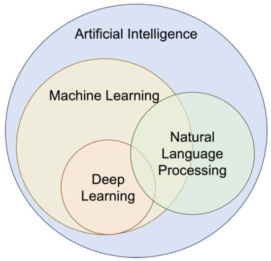
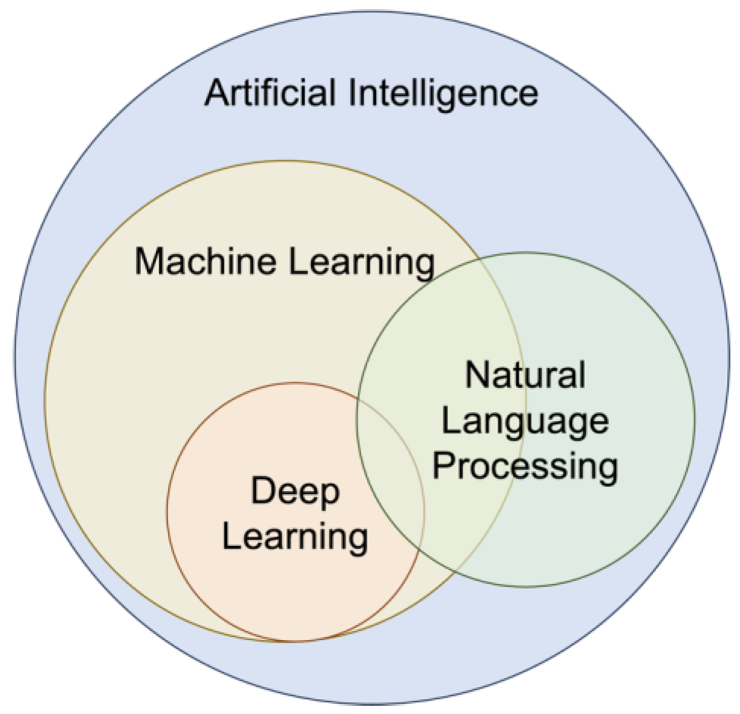
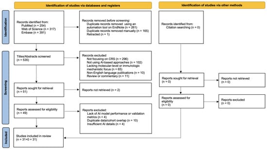
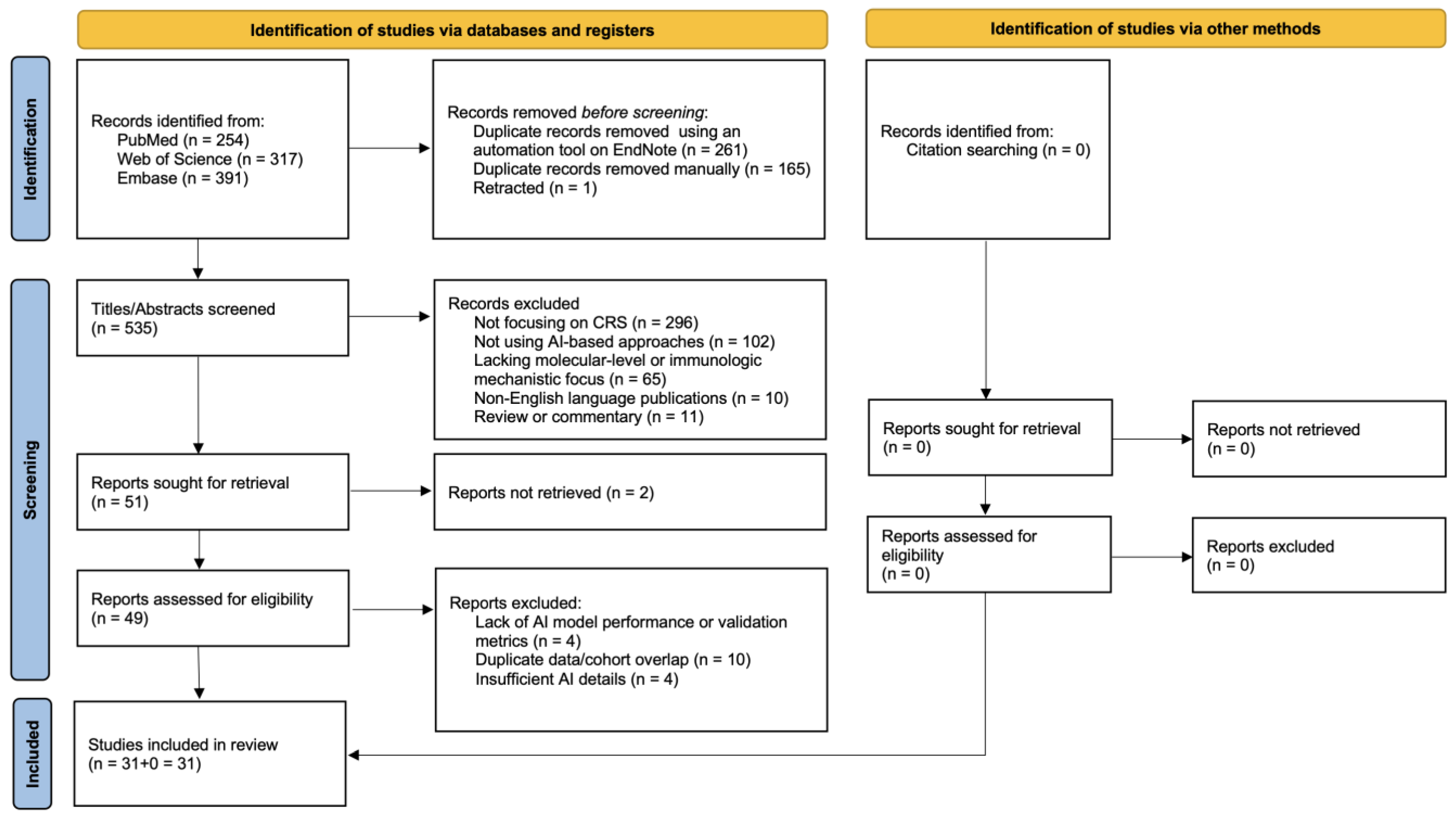
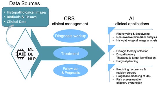
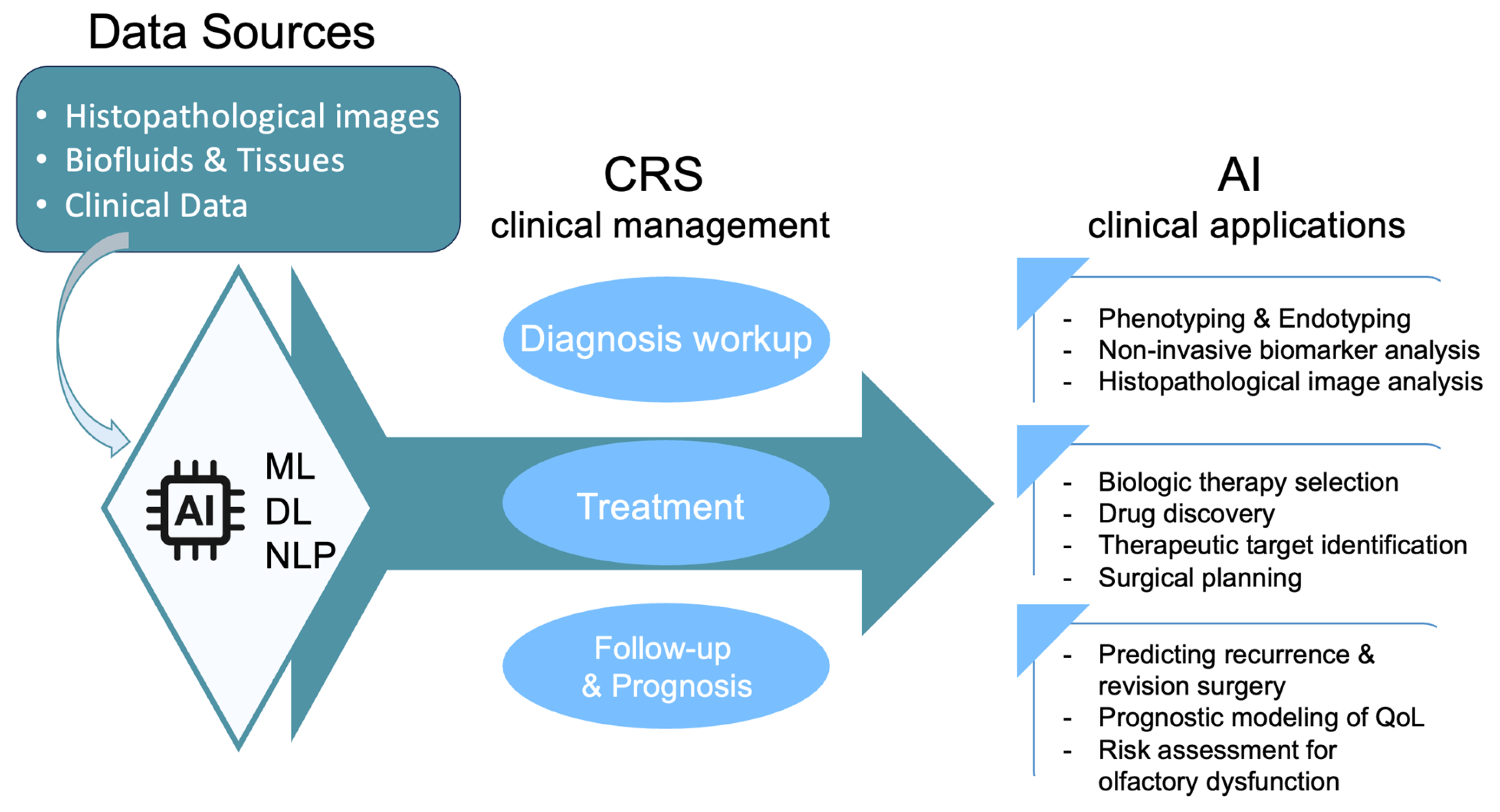

Disclaimer/Publisher’s Note: The statements, opinions and data contained in all publications are solely those of the individual author(s) and contributor(s) and not of MDPI and/or the editor(s). MDPI and/or the editor(s) disclaim responsibility for any injury to people or property resulting from any ideas, methods, instructions or products referred to in the content. |
© 2026 by the authors. Licensee MDPI, Basel, Switzerland. This article is an open access article distributed under the terms and conditions of the Creative Commons Attribution (CC BY) license.