


Abstract
Encoding genetic information involves both structure and sequence. Non-protein-coding (NCR) RNAs modulate host immune responses by guiding and assembling stress-specific protein effectors, with flipons enabling dynamic transitions between states. Small NCRs transcribed by both RNA Polymerases 2 and 3 normally modulate these pathways. The miRNAs, tRNA fragments, mitrons, piRNAs, and endo-small-interfering RNA products target the guide effector complexes to their targets. These NCRs filter the subset of RNAs that are translated, a process modulated by other NCRs that fine-tune RNA stability, localization, and kinetics through the modifications they direct. When these RNAs, or their targets, are dysregulated, host interferon responses are activated through the accumulation of uncapped triphosphorylated RNAs, dsRNAs, or flipon-encoded Z-RNAs. These responses are initially suppressed to avoid attacks on self-RNAs, but above a threshold, highly immunogenic pathways are unleashed. These same responses resist viral and other infections without pathogen-specific effectors or prior exposure. They also eliminate dysfunctional cells. These universal responses, when unresolved, contribute to a range of pathologies, increasing cancer, inflammatory diseases, and senescence. The review focuses on recent findings showing how these integrated host responses are driven by, and exploit, the retrotransposon invasion of animal genomes.
Keywords:
flipons; endogenous retroelements; noncoding RNA; dsRNA; Z-RNA; argonaute; reverse transcription; infection; immunity; interferon; cancer; lupus 1. Introduction
Over the years, many different roles for noncoding RNAs in biology have been proposed, both good and bad, but no consensus exists. This contrasts with the coding genome, where the protein made from a transcript can be isolated, its properties determined, and its modifications mapped. Many regard this form of encoding, a triplet nucleotide cipher embedded in a DNA double helix capable of templating its own replication, as “the secret of life”. Yet many noncoding RNAs (NCRs) exist, and life could not exist without them. Examples include ribosomal RNAs, which catalyze the polymerization of amino acids into proteins, and small RNAs that regulate DNA readout, transcript processing, stability, and mRNA translatability. Other NCRs, such as 7SK, regulate RNA polymerase processivity, while 7SL promotes protein export across membranes. The classical view overlooks the essential role of NCRs in encoding genetic information and defending the genome from threats within.
2. Flipons
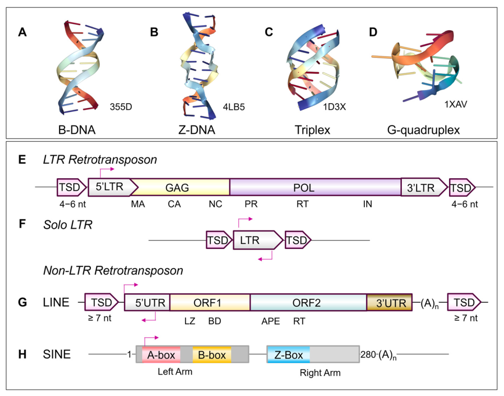
This different view of NCRs emerges from the idea that nucleic acids also encode genetic information by their structure. By adopting alternative conformations, NCRs can assemble distinct complexes with different functions. Such outcomes, for example, regulate immune responses that discriminate self from foe. Here, we explore this perspective. Sequences that encode alternative structures are called flipons [1]. They can transition from one conformation to another without a change in sequence or cleavage of the phosphate backbone. The alternative flipon DNA and RNA structures include left-handed Z-DNA and Z-RNA (referred here collectively as ZNAs), 3-stranded triplexes, and 4-stranded G-quadruplexes (GQ) (Figure 1A–D). Formation of each depends on particular sequence motifs, with ZNAs favored in particular by alternating pyrimidine–purine repeats, such as (dCdG:dCdG)n, triplexes by polypurine sequences, and G-quadruplexes by guanosine motifs capable of forming tetrads that stack on each other to form a quadruplex four-stranded helix [1]. ZNA and GQ motifs are enriched in promoters and endogenous retroelements (EREs) that are the focus of this discussion [2,3,4,5,6,7,8,9,10,11,12,13]. Of all these flipons, the evidence for biological function is strongest for ZNAs. The structure-specific Zα domain has provided the tools necessary to show this [14,15,16]. Through the work of many, the dependence of many biological pathways on ZNAs is supported by Mendelian genetic, biophysical, biochemical, and cellular biology evidence, as described throughout the manuscript.
Many factors determine when the flip from one structure to another will occur. These include structure-specific proteins; epigenetic modifications of both the nucleic acid and the proteins involved; topological stress generated by processive polymerases or helicases, or the ejection of nucleosomes from underwound DNA; and the stretching or twisting of RNA and DNA duplexes by tangles formed by pairing of complementary single-stranded RNA (ssRNA) sequences. The RNAs involved often affect multiple outcomes and are challenging to classify. Whether NCRs are long or short, repeat or non-repeat ssRNA, double-stranded RNA (dsRNA), or structured one way or another, they make no sense when evaluated by sequence alone (pun intended). However, flipons, as dynamic structural elements of the genome, are important to the evolutionary transition from self-replicating tinkers to cells [17].
Figure 1.
Sequence elements in the genome include flipons and retrotransposons. Flipons encode genetic information by adopting different DNA conformations as the context changes. The transition requires no sequence modification or backbone cleavage. Each structure helps seed different protein complexes. The alternative folds include: (A) Right-handed B-DNA. (B) Left-handed Z-DNA. (C) Three-stranded triplexes. (D) Four-stranded quadruplexes. The retrotransposons include: those with long terminal repeats (LTRs) (E), and solo LTRs derived from recombination of the 5′ and 3′ LTRs (F). Long terminal repeat (LTR) retrotransposons are composed of a group antigen (GAG) with matrix (MA), capsid (CA), and nucleic acid (NA) domains, a polymerase (POL) with protease (PR), reverse transcriptase (RT), and an integrase (IN) domain, but lack the envelope protein that is found in retroviruses. The terminal sequence duplication (TSD) is a duplication of the genomic sequence at the insertion site. Another class includes long interspersed nuclear elements (LINEs) (G) and short interspersed nuclear elements (SINEs) (H). A full-length LINE has two open reading frames (ORFs). ORF1 has a leucine zipper (LZ) and a C-terminal nucleic acid-binding domain (BD). The RNA polymerase 3 (RPOLIII) promoter binding sites (A- and B-box) are shown, along with the experimentally confirmed Z-box [7,18]. Only LTRs and LINEs encode the reverse transcriptase required to integrate their RNAs at new sites. SINEs compete for the LINE reverse transcriptases (modified from [19]).
To survive long enough, genomes require a sense of self, enabling them to defend against invasive replicants such as DNA and RNA transposons and viruses. These processes rely on NCRs, and their origins predate the emergence of sequence-specific nucleic acid-binding proteins and the embellishments they enabled, both in defense and in conquering new environs. Here, we describe how flipons encoded by NCRs enable real-time survival against threats and perturbations through a layered meshwork of protections.
3. Endogenous Retroelements and the Threat from Within
Here, the focus is not on viral and bacterial pathogens that attack from the outside. Instead, we will discuss the transposons that attack from within. Two classes of transposons exist: DNA and RNA (referred to here as EREs). They serve no purpose, but by spreading through a host genome, they disrupt active genes and pose an existential threat to a species. These threats have driven many adaptations that ensure host survival. The ideal solution is to eliminate transposons from a genome, but in some cases, these elements confer survival advantages. Even in bacteria with their lean genomes, transposons enable the horizontal transfer of advantageous traits from one individual to another. Here, the focus is on animal genomes where other solutions have left genomes filled with vast expanses of ERE-derived sequences.
DNA transposons and ERE did not always share the same evolutionary fate. For example, the human genome largely extinguished the DNA transposons about 37 million years ago [20]. These elements are maintained by a “cut-and-paste” mechanism. Although they comprise about 3% of the current sequence space, they are mostly incapable of transposition. Even so, the remnants of these elements are still targeted by Krüppel-associated box zinc finger proteins (KRAB-ZFPs) [21]. Over 74% of the sequences are classified as miniature inverted-repeat transposable elements (MITEs), which do not encode proteins. They retain the terminal inverted repeats that engage the transposases necessary for transposition. By doing so, they have in the past “cheated” active DNA transposons of the enzymes needed for their persistence, contributing to their mutual demise [20].
In contrast, ERE-derived sequences now occupy over 50% of the human genome. Their RNAs can capture the reverse transcriptases required for “copy and paste” retrotransposition immediately after the ribosome produces them. EREs use two distinct strategies to prime DNA synthesis. Long terminal repeat (LTR) retrotransposons use host tRNAs to prime the reverse transcriptase (RT). In contrast, the long interspersed nuclear elements (LINEs) cleave the host DNA and use the resulting ends as primers [22,23,24]. The latest estimate is that the human genome still contains 68 copies of active LINE L1 retrotransposases [25].
In both cases, other classes of ERE emerge that do not code for proteins. Solo LTRs arise by recombination between the 5′ and 3′ LTR ends, leading to loss of protein-coding sequences but retention of regulatory sequences. They can act as bidirectional promoters, driving transcription of adjacent regions. Like solo LTRs, short interspersed nuclear elements (SINEs) are noncoding (Figure 1E–H). They compete with LINEs for the RT as the protein emerges from the ribosome, ensuring that both persist. SINEs incorporate regulatory sequences from the host genome to ensure their own transcription, and disperse these throughout the genome [26,27]. Both solo LTRs and SINEs are enriched in transcription (TF) binding sites and in sequences, called flipons, that encode genetic information by the structures they form. As we will discuss, these ERE non-protein-coding sequences play key roles in host evolution.
Retrotransposon insertion into the genome is haphazard. In general, SINEs target active genes, while LINEs and LTRs accumulate in intergenic regions. These attacks trace back to the earliest eukaryotic progenitors. Immune responses were originally shaped to suppress LTRs, LINEs, and SINEs. These pathways now protect the host against a wide range of emerging threats. The invasive elements now defend the genome they previously ravaged. The focus is strictly on the role these elements play in host survival, a prerequisite for transmitting genetic information from one generation to the next. When these processes are dysregulated, they contribute to a range of pathologies, increasing cancer, inflammatory diseases, and senescence. Rather than classifying each retrotransposon or each noncoding RNA into a type and subtype that maps to a particular teleological function, I take a biological perspective. The text covers a wide range of topics and includes detailed supporting experimental evidence from the referenced publications. I begin with the NCRs produced by RPOL3, as their biology is quite impactful.
3.1. RPOL3 and Transcription
RPOL3 transcripts depend on A and B promoter sequences within a transcription Unit (TU), such as in SINEs (Figure 1H) [28]. Transcription relies on the RPOL3 TFIIIB and TFIIIC complexes. Importantly, the TFIIIC complex does not turn over. It acts as an anchor that ensures rapid RPOL3 turnover, enabling high-output states [29,30]. RPOL3 transcription is terminated by a poly(thymine) segment, usually 4–6 bases long, with the SENTAXIN helicase enforcing the stop. The size of the RPOL3 TU that lies between the transcription start site (TSS) and the TTS varies from ~70 to 90 nucleotides (nt) for tRNAs, ~100 to 300 nt for SINEs, ~300 nt for 7SL RNA, and ~100–200 nt for Vault RNA (vtRNA) and BC200 [31].
RPOL3 transcripts have an uncapped 5′ end with a triphosphate terminus. These ends are usually masked by the signal recognition particle SRP9/14 subunits [32]. This masking prevents binding of the retinoic acid-inducible gene I (RIG-I)-like receptor (RLR) gene products, which would otherwise activate an interferon response. The 3′ end is generally protected by that La protein that prevents digestion of the transcript by exonucleases [33].
3.2. Processing of RPOL3 Transcripts
The NCRs produced by RPOL3 are essential for normal cell function. They modulate the transcription and translation of many metazoan RNAs through a variety of NCR fragments [34,35,36,37]. Here, we first focus on tRNAs, then on SINEs, and their various roles in immunity.
tRNAs and their fragments: These NCRs include tRNA fragments that represent an ancient regulatory system dating back to the origin of eukaryotes (Figure 2A). A variety of tRNA fragments (tRFs) are involved in these pathways, including tRNA halves, quarters, and end pieces. These products have been extensively reviewed [36,38,39,40,41,42,43]. They arise in both Dicer-dependent and Dicer-independent ways through enzymes such as RNase-P and RNase-Z that act during pre-tRNA maturation. The tRFs have been assigned different functions depending on their single-stranded RNA sequence (reviewed in [41,43,44]). We focus on how the TRF duplex structure and length determine the effector pathways used, and how this impacts immune responses.
Figure 2.
The L-shaped tRNA structure that is processed to generate fragments loaded onto Argonaute (AGO) by the AGO maturation complex (AMC) and the RNA-induced silencing complex (RISC) loading complex (RLC). (A) The tRNA L-shaped fold (4YNA). The TψC-acceptor double-stranded RNA (dsRNA) duplex is long enough to engage the AMC, but the D-loop anticodon-stem duplex is too short. The inset shows the tRNA backbone with cleavage sites for Dicer and Angiogenin. (B) The human AMC loaded with the let-7a-1 miRNA (9W51). All coordinates were obtained from the Protein Data Bank [45]. (C) The human RLC is composed of Dicer and the TAR RNA-binding protein 2 (TARBP2) with a pre-miRNA loaded. (D) The RISC with the guide strand (fuchsia) bound to a target RNA (teal).
Small tRNA fragments that are duplex in character can be loaded into an RNA-induced silencing complex (RISC) by the Argonaute (AGO) maturation complex (AMC, Figure 2B) [40,46,47]. The AMC consists of the heat shock protein 90 beta (HSP90β), AGO, and the p23 cochaperone (encoded by the PTGES3 gene). All the components, including HSP90β, are abundantly expressed under normal conditions [48]. The geometry of tRNA fragments dictates their loading. Even after nicking, the tRFs remain duplex [49]. Structurally, they retain the tRNA L-shape, with the TψC stem stacked on the acceptor stem and the D-stem on the anticodon stem. Only the TψC-acceptor stem is of sufficient minimum length of 11–12 bases to fulfill the loading threshold of the AMC (Figure 2). Together with the single-stranded 3′-CCA tail overhang, the stem provides a template that resembles the 19- to 22-nucleotide microRNA duplex. Two nicks are required to free this stem from the tRNA and arm the AGO effector with an ssRNA guide. The tRFs preferentially associate with AGO1, 3, and 4 [50]. These argonautes are less stringent than AGO2 in their loading requirements and can accept guide RNAs with 5′ G and 5′ C ends, among other differences in their binding preferences [51]. The particular AGO loaded can have important biological consequences, affecting both the stability and translation of targeted RNAs through the complexes they form. For example, experimental studies reveal that tRF5-GluCTC induced by respiratory syncytial virus impairs post-transcriptional gene silencing in cells deficient in AGO1 or 4, whereas deletion of AGO2 and AGO3 has no effect [52].
The Dicer-generated fragments can also be loaded onto the RISC loading complex (RLC). This complex evolved later than the AMC and incorporates either the TAR RNA-binding protein (encoded by TARBP2) or the PKR-associated protein X (PACT, encoded by PRKRA) (Figure 2C,D) [53,54,55]. It was predominantly characterized by its association with microRNAs (miRNAs) [56]. In vitro, it is sufficient to load miRNAs onto AGO2 [57] but TARDP1 is not required in vivo [58]. The role of PACT is also challenging to analyze, as it suppresses PKR activation and the induction of inflammatory responses [59]. Recent evidence suggests that the RLC now cooperates with the AMC to increase the loading efficiency of various NCRs inside cells [47]. Another potential path to loading tRNA fragments onto Ago proteins would use pre-mRNAs as substrates for the classical pathway. However, the binding of La to the 3′ RNA end inhibits this outcome, with the exception of the pre-tRNA-Ile (Ile-TAT-2-3) in mice, which has low affinity for the LA protein. Processing of this pre-tRNA can then generate either the tRNA or mmu-miR-1983 [60].
In contrast, the 8- to 9-base-pair D-Anticodon stem is structurally too short for high-efficiency AMC loading [47]. Instead, modifications that shield them from exonuclease digestion protect the short TRFs they produce, allowing them to serve other roles. For example, TRFs that incorporate internal tRNA fragments can inhibit tRNAArg and tRNAAla deamination by Adenosine deaminase acting on transfer RNA (ADAT) [61]. The fragments produced can inhibit protein binding to their targets. An example is the 39-base-pair trf3e fragment produced from the 3′ end of tRNAGlu. The TψC stem–loop incorporates a 9-nucleotide sequence that binds nucleolin with high affinity (120 nM). The interaction prevents nucleolin-mediated inhibition of p53 protein translation [62]. In contrast, tRNAval fragments directly bind the chaperone molecule eukaryotic translation elongation factor 1 alpha 1 (EEF1A1) to promote nuclear translocation. The subsequent activation of the mouse double minute 2 homolog (MDM2) ligase, which ubiquitinates p53, leads to its destruction [63]. Fragments can act in other ways by directly binding to the ribosome, as shown for the 5′ end of archaeal valine tRNA (Val-tRF) from Haloferax volcanii [64].
The tRNAs can contribute to cellular somatic defenses against ERE in somatic tissues. They inhibit the somatic spread of LTR retrotransposons, which depend upon tRNAs to prime reverse transcription. Interestingly, a 22 nt TRF3 can bind AGO and suppress translation of canonical transcripts. However, an 18 bp sequence that lacks the 3′ CCA tail cannot, as it is too short to engage AGO2 [47]. Notably, the 18 bp fragment that binds directly to primer sites inhibits LTR retrotransposition [38].
4. Cellular Stress and tRNA Fragment Structures
During stress, tRF production increases: angiogenin is released from inhibition by the Ribonuclease inhibitor RNH1, while RNase L is activated by the 2′,5′-oligoadenylate (2–5A) produced by interferon-induced oligoadenylate synthetases. The cleavage susceptibility and overall stability of NCRs depend heavily on the RNA modifications added during pre-tRNA processing. The outcome varies by tissue, reflecting the different tRNA isoforms and modification machinery expressed [65]. Experimentally, the long tRFs produced by angiogenin cleavage certainly increase during stress, but no new fragments are loaded onto AGO [66].
tRFs can form alternative structures, such as G-quadruplexes (GQ) and dumbbell dimers, which affect their persistence and function. For example, the GQ formed by tRNAAla and tRNACys 5′ halves also inhibits translation and promotes stress granule formation [67,68]. Instead, the 5′ halves from tRNAGly and tRNAGlu produced during stress have the potential to facilitate the translocation from the cytoplasm to the nucleus of factors that modulate alternative splicing and promote cell survival [69,70,71]. Notably, dimer formation masks the UUA sequence present in the Glu-5′tsRNA-CTC that drives mitochondrial import by leucyl-tRNA synthetase 2. In the mitochondria, the fragment inhibits the charging of mitochondrial leucine tRNA, disrupting mitochondrial translation and contributing to age-related neuronal degeneration [72]. Dimer formation also stabilizes the 5′ tiRNA-Gly-GCC. Dissociation of the dimer during stress protects neurons against excitotoxic necrosis through an antisense-mediated mechanism [73].
Cellular Stress and AGO Loading
During the heat shock response, AGO proteins localize to stress granules, where AGO2 is inactivated by ADP-ribosylation, thereby decreasing tRNA loading and RNA interference in human cells [74,75]. Also, despite increased production, HSP90β interactions with folded proteins reduce its availability, decreasing NCR loading through the AMC. These changes relieve the AGO-dependent suppression of the mRNAs encoding proteins that mediate stress responses in normal cells. A subset of tRFs may still load onto AGO during stress due to the increased tRNA levels, as occurs when tRNAs are overexpressed in normal cells [40].
Alternatively, the tRFs arising from enzymatic cleavage of tRNA may themselves directly reprogram translation and other cellular pathways [76]. A wide range of nucleases produce tRNA fragments, with angiogenin generating cyclic 3′ phosphates. This modification does not inhibit binding to AGO proteins but may instead be preferred for loading onto AGO2 [77]. In contrast, miRNA subsets are incorporated into the RISC during stress, including miR-210 (a hypoxia switch), miR-34a (DNA damage response), and mitrons, some of which form GQs that may modulate protein interactions [78,79,80,81,82,83].
5. RPOL3, RPOL2 and SINE Transcripts
SINE transcripts illustrate many other roles NCRs play in immunity. SINEs are transcribed by both RNA polymerase 3 (RPOL3) and RNA polymerase 2 (RPOL2), with the outcomes depending on the enzyme used. The RPOL3 readout echoes their origins. They are descended from highly expressed, RPOL3-transcribed 7SL RNA, which scaffolds the signal recognition particle (SRP) anchor essential for tethering ribosomes to the endoplasmic reticulum. SINE transcription by RPOL3 is strongly suppressed in differentiated cells, but increases dramatically under stress.
Most SINE transcripts in unstressed cells are instead synthesized by RPOL2, reflecting the preferential retrotransposition of SINEs into active genes over millions of years [84]. Often, insertion occurs in the reverse orientation relative to an existing family member, generating an inverted SINE repeat. During transcription, the repeats fold back on themselves to form dsRNA. Inverted repeats are most frequent in introns and the untranslated regions (UTRs) of mRNAs. These folds impact intron splicing, protein recoding, mRNA stability, and translation. They are enriched at the 3′ ends of genes, many lying just beyond transcription termination sites (TTS).
5.1. RPOL3 Regulates SINE Transcription Under Stress Conditions via Alu Elements
Surprisingly, RPOL3 mostly activates SINE transcription only under stress, reversing the suppression of EREs in normal cells that prevents their retrotransposition. This selective silencing of SINEs by facultative heterochromatin depends on the reversible histone marks written by the histone-lysine N-methyltransferase SUV39H1. Those SINEs that undergo DNA methylation are instead permanently consigned to constitutive heterochromatin. Their expression is not restored by 5-azacytidine inhibition of DNA methylation [85]. SINE repression in normal cells is highly efficient. Of the ~1.3 million Alu copies bound by the RPOL3 subunit TFIII3C, only ~1300 RPOL3 transcripts were detectable in an in vitro study of cell lines, with only 120 expressed in at least three cell types [86].
Epigenetic regulation renders SINE transcription by RPOL3 highly inducible. Over time, SINEs have acquired TF binding sites through mutation and deamination of methylated cytosines to thymine. One study reported that 34 TFs, out of the 1084 tested, bound to greater than 20% of Alu SINEs [26,27]. The SINEs also encode flipons that form ZNA, triplexes, and G-quadruplexes [7,18,87,88,89]. Cellular stresses, such as heat shock, also trigger expression from RPOL3-dependent Alu elements [90]. Similarly, activation is also initiated by adenovirus type 5 and herpes simplex type 1 infection [91,92]. The initial unpacking driven by heat shock factors (HSF) depends on a 13-base motif centered on position 221 of the Alu right arm that differs slightly from the consensus HSF1 and HSF2 GAA repeat sequences [93,94]. High levels of uncapped RNAs then trigger the RIG-I-directed interferon response to defend the cell.
5.2. RPOL3 Regulation of RPOL2 Transcription Under Stress Conditions
The activation of Alu transcription by RPOL3 by TFs, and during cell stress, also inhibits the RPOL2 transcription of many genes. This interaction is direct, with the Alu RNA blocking by disrupting contacts between RPOL2 and promoter DNA in preformed complexes [95,96]. Cryo-EM studies reveal that the Alu right arm structurally mimics the RPOL2 elongation complex. The interaction is of high affinity, with a Kd of ~20 nM [97]. During stress, Alus are rapidly expressed many hours before the transcription and translation of heat shock proteins, thereby protecting the cell in the interim from the activation of EREs that ectopically activate suppressed developmental pathways [90,98].
In other cases, RPOL3 instead promotes RPOL2 transcription. Overall, ~20% of active Alu elements are within 2 kb of an active divergently transcribed RPOL2, with 60% belonging to the Alu-S family [99,100,101]. Of these, ~2% to 3% are actively induced by RA, with the RA receptor binding to a direct repeat motif with a 2-base spacer (DR2) on the Alu left-arm. Around 100,000–200,000 SINEs have DR2 elements and are located close to RPOL2-transcribed genes, with 95% of RA-activated elements from the Alu-S subfamily of repeats. Over 18,000 of these Alu-DR2 SINEs are situated directly within a regulatory window of ±10 kilobases (kb) of the 5′ ends of more than 10,000 RPOL2-transcribed genes [102]. Activation of RPOL3 transcription likely turns these elements into enhancers that promote RPOL2 transcription of the associated gene [99,103,104]. The best-characterized genes responding to RA are those involved in stem cell maintenance, such as teratocarcinoma-derived growth factor 1 (TDGF1) and the homeobox protein NANOG [105]. RA also enhances expression of interferon-induced proteins involved in antiviral defenses [106].
After expression, DR2-induced Alu transcripts can be processed through a Dicer-dependent pathway, producing fragments of ~28–65 nt. After export to the cytoplasm, AGO3 binds the RNAs, which lack the strict length restrictions of other Argonaute family members [105]. Evidence suggests that AGO3-bound repeat-induced RNAs scaffold assembly of the Enhancer of Decapping Complex 4 (EDC4, also known as RCD8) to downregulate RPOL2 transcripts from the neighboring gene. The complex formed promotes mRNA degradation by Exoribonuclease 1 (encoded by XRN1), creating a feedback loop in which RPOL3 transcripts control RPOL2 mRNA levels. The AGO3-bound RPOL3 Alu transcripts engage complementary sequences in the 3′ UTR of TDGF1 and NANOG, terminating stem cell pluripotency. As reported in early studies of vitamin A, they also limit interferon responses [107].
6. RPOL3-Mediated Retrotransposon Defense and RNA:DNA Flipons
RPOL3 transcripts produced under stress conditions also enable another form of defense against EREs based on RNA:DNA hybrids. These RNAs serve as substrates for ERE reverse transcriptases, which use their presence to produce RNA:DNA hybrids (RDHs) that activate immune responses. Notably, cytoplasmic DRHs have been reported to be RPOL3-dependent [108]. They are not associated with the RPOL2-generated damage arising from R-loop formation [109]. They are likely generated by an active LINE-1 reverse transcriptase recruited by RPOL3 transcripts via their Poly(dA) segment, a repechage of how SINEs were first generated from 7SL RNAs. Indeed, RT inhibitors prevent the accumulation of cytoplasmic RDHs in granulocytes from patients with systemic lupus erythematosus [110]. The LINE-1 RT is efficiently primed by cytoplasmic tRNAs, short RNAs, and hairpins. The RDH produced are then thought to activate the DNA sensor cyclic GMP−AMP synthase (cGAS), leading to interferon production [111,112]. However, immunofluorescence studies do not co-localize cytoplasmic LINE-1 proteins with cGAS, but instead with the ZNA-binding protein 1 (ZBP1) sensor [108]. The findings suggest a novel mechanism in which increased RPOL3 transcription of SINEs can defend the host by triggering cell death pathways. When the execution is thwarted, either in cancer, infection, or senescent cells, chronic inflammation is instead triggered. Indeed, high levels of SINE expression characterize many of these diseases [19,113,114,115,116,117].
7. RPOL3 Transcripts and DNA Flipons
While RNAs can form a variety of structures and sequence-specific interactions, transcription also exploits the cycling of flipons from one conformation to another (Figure 3). Once triggered, high transcription rates drive Z-DNA formation, which the RPOL3 subunit RNAP3C likely recognizes. The interaction reinitiates assembly of the transcription complex anchored by TFIIIB, and establishes a chromatin structure permissive to high RNA expression [118].
Figure 3.
The potential for RNA Polymerase 3 (RPOL3)-transcribed short interspersed nuclear elements (SINE) repeats to regulate nucleosome placement, transcription rate, and flipon conformation. The arrows indicate the direction of transcription, and the “−” and “+” signs represent regions of negative and positive supercoiling generated by RPOL3 according to the twin domain model of Liu and Wang [119]. The “An” indicates the variable Alu poly(dA) 3′ tail present just before the oligo-dT transcription termination site. An Alu is a SINE family member characterized by the fragment sizes produced by digestion with the AluI restriction enzyme. Nucleosomes are shown wrapped in negatively supercoiled DNA. (A) Closely spaced direct repeats can be transcribed by two polymerases acting in tandem to increase upstream local negative supercoiling, or by a single RPOL3 if the oligo-dT termination signal on the first is weak, or the downstream transcription (TFIIIC) binding site has been lost. (B) Direct repeats spaced further apart can influence the site of nucleosome insertion. The upstream transcription unit (TU) can generate negative supercoiling to promote nucleosome insertion upstream of that element, and positive supercoiling to displace the nucleosome downstream. Alternatively, the downstream TU can unwind the DNA immediately upstream to promote nucleosome docking between the two TUs. This nucleosome phases the upstream nucleosome array. Nucleosome placement over the upstream TU promoter then prevents RPOL3 from docking and transcribing this region. (C) Divergent transcription of inverted repeats, or nucleosome ejection, greatly increases local negative supercoiling between the TUs. The energy stored in the negative supercoils then powers the transition of promoter flipons to an alternative state. The formation of Z-DNA and G-quadruplexes then helps localize transcription and RNA processing complexes to these sites [120,121]. (D) Convergent transcription of inverted repeats promotes nucleosome undocking by increasing positive supercoiling, promoting replacement of canonical histones with others, such as Centromere Protein A (CENP-A), that structure chromatin differently [122].
In the human genome, pairs of RPOL3-transcribed SINEs occur in close proximity, with two-thirds being direct repeats. Around a quarter of these direct repeats are within 20 bases of each other, while ~60% are 100 or more bases apart. In contrast, around ~80% of inverted repeats are separated by at least 100 base pairs. Most direct and inverted Alu pairs share between 65% and 85% sequence identity [123]. The repeats allow RPOL3 to modulate several outcomes, depending on the local supercoiling produced as the polymerase plows through the DNA helix. The underwound DNA generated upstream of RPOL3 (indicated by “−” in Figure 3) creates a site that facilitates the wrapping of DNA around nucleosomes as they dock. The downstream positive supercoiling (indicated by “+” in Figure 3) can negate the negative supercoiling required for stable nucleosome insertion, leading to ejection of the nucleosome (Figure 3B,C).
With closely spaced direct repeats, the TUs can form an RPOL3 train, in which the downstream TU induces negative supercoiling that promotes engagement of the upstream RPOL3, thereby generating high levels f unwinding in the upstream region (Figure 3A). Readthrough of a weak termination signal or loss of the TFIIIC-binding site in the downstream TU can produce a loner RPOL3 transcript. With direct repeats farther apart, the TUs can fire independently, creating a switch that keeps only one TU active by changing nucleosome placement (Figure 3B). In one case, negative supercoiling arising just 5′ to the downstream TU can rephase a nucleosome array to mask the upstream TSS, thereby suppressing that TU. In another situation, transcriptional interference produced by readthrough of the downstream TSS by the upstream TU will disrupt readout from the downstream TU (Figure 3B). When RPOL3 TUs are convergent, the region of intense positive supercoiling can phase nucleosome arrays by displacing nucleosomes from this region, or by replacing canonical histones with those that can dock to positively supercoiled DNA, such as Centromere Protein A (CENPA) [122,124]. The underwound DNA released by the nucleosome counters the increased tension without requiring topoisomerases (Figure 3C). When RPOL3 TUs are divergent, higher levels of negative supercoiling are generated in the intervening segment (Figure 3D). If flipons are encoded in this region, the negative supercoiling generated can power the flip to an alternative conformation, such as ZNA, G-quadruplex, or a hairpin cruciform. Indeed, EREs are enriched for flipons, with both Z-DNA and G-quadruplex structures experimentally confirmed for SINEs, LINEs, and LTRs. These play roles in regulating their expression and that of surrounding genes [5,18,125,126,127,128,129].
The underwound, RNA-free regions between divergent TUs (Figure 3D) also provide landing spots for RNAs acting in trans to modulate local flipon conformations. The RNAs can dock to make triplexes. Alternatively, the process can generate R-loops, in which an RNA hybridizes to one DNA strand. The displaced DNA can then fold as a GQ or an i-motif, depending on its sequence. R-loop formation has been proposed to facilitate processes like splicing by bridging TUs with nuclear speckles [130]. Alu transcripts have also been reported to promote the assembly of enhancer–promoter condensates through sequence-specific interactions between different chromosomal regions [131]. How the production of the Alu pairs at each site is coordinately regulated remains unknown. The short length of the RPOL3 TUs makes trans sense–antisense interactions more favorable than those formed in cis. Unlike convergent RPOL2 transcripts, RPOL3 transcripts from adjacent TUs have little sequence overlap [132].
Collectively, these different pairings enable the chromatin remodeling necessary to mount genome-wide responses to infections induced by stress-activated RPOL3 transcription. The alternative conformations flipons adopt help maintain an open DNA state. The complexes they assemble then seed state-specific condensates necessary to protect the genome. A subset of these complexes modulate RPOL2 activity and pre-mRNA processing.
8. RPOL2-Produced NCRs in Host Defenses
RPOL2 transcripts play a complex role in host protection through the proteins they encode. Here, the focus is on NCRs that RPOL2 produces from EREs or targets. These include antisense RNAs, SINE dsRNAs, circular RNAs (circRNAs), and RNAs that regulate RPOL3 transcription.
8.1. RPOL2 and RNA Defenses Against Active Endogenous Retroelements
RPOL2 transcripts work in concert with RPOL3 to defend the host. These ancient NCRs actively target EREs expressed in the germline, where DNA is open, reflecting the nucleosome loss in sperm and the rapid cell proliferation in the embryo. They include piRNAs incorporated into a piRISC composed of PIWI proteins, which are another member of the AGO clade. In humans, short ~20-nucleotide piRNAs guide PIWIL3-containing complexes to newly evolving non-LTR retrotransposons. These short piRNAs are generated by a ping-pong mechanism, in which one piRNA directs the generation of another from a complementary RNA strand. The most highly expressed ERE strand then generates the most piRNAs. Longer ~26–30-nucleotide piRNAs are generated by phased processing, where a single strand undergoes inchworm cleavage starting at the 5′ end to generate 3′ piRNAs that target PIWIL1 to mostly LTR transcripts. The pathways can work cooperatively, with remnant RNAs from the ping-pong pathway then undergoing phased processing to yield the longer piRNAs [133]. The system is not typically expressed in normal tissues because it can potentially target host mRNAs, causing off-target effects [46,134]. DICER can also process the dsRNAs produced from RPOL2-transcribed EREs into endogenous small interfering RNAs (endo-siRNAs) [135,136,137]. This pathway later evolved into the AGO RISC loading complex (RLC) (Figure 2C,D) [53,55]. It is not thought to play a major role in normal cells, as it depends on forming long dsRNAs that can trigger interferon responses. Instead, AGO proteins bound to ERE-derived sequences have evolved other roles, particularly in early development (reviewed in [138]). Somatic cells now use KRAB-ZFP proteins to suppress transcription of both LTR and non-LTR retrotransposons [139].
8.2. RPOL2 Transcript Modulation of RPOL3 Transcription
RPOL2 transcripts derived from EREs also regulate RPOL3 transcripts. They target the long poly(dA) segments that drive SINE retrotransposition by capturing LINE RTs. The duplex poly(dA) tracts are susceptible to triplex formation with the NCR produced by RPOL2 docking as the third strand (Figure 1C). The poly(dA) tracts are longest in the youngest members of the Alu family, extending up to 97 nucleotides in primate-specific AluY [140]. Long noncoding RNAs (lncRNAs) exploit this vulnerability by forming triplexes that suppress SINE expression. The lncRNA KCNQ1OT1, for example, contains an extended triplet-forming poly(rA) sequence that targets SINEs genome-wide [141].
8.3. RPOL2 and Antisense Transcripts
Experimental evidence shows that RPOL2 can transcribe the single-stranded DNA of R-loops in the antisense direction [116], suggesting that their biology is more complicated than first imagined. Previously, ~20% of human genes were identified as having overlapping antisense transcripts [142]. Convergent transcription was thought to produce transcriptional interference, providing a means for regulating gene expression [143,144,145]. A recent study has confirmed the high frequency of convergent transcription at nearby promoters, with a median separation of 413 bp (range: 2 to 2301 bp). These promoters did not exhibit transcriptional interference [132]. Instead, a coordinating command center (CCC) that lies between them synchronizes promoter activity. The CCC prevents clashes between the RPOL2 polymerases transcribing each TU by gating their activity (Figure 4A). The design also enables regulation via small RNAs that target a specific transcript, thereby suppressing or activating a particular promoter. The RISCs formed can also cleave nascent transcripts, leading to the dynamic ejection of RPOL2 [146]. Both NCR and CCC sequences can undergo selection to optimize cell survival and phenotype. These NCRs can also modulate alternative flipon folds, as previously proposed [147], including GQs, intercalated cytosine-rich i-motifs, hairpins, ZNA, and slipped-strand structures [5,148,149]. Notably, CCCs between convergent promoters show an increased propensity to form both GQs and R-loops [132]. In addition, lncRNAs, including those derived from ERE, can also form triplexes (as extensively reviewed in [150]).
Figure 4.
Promiscuous gene transcription modulated by noncoding RNAs (NCRs) acting in cis and in trans. (A) Cis regulation of gene expression. Recent evidence reveals that most promoters undergo bidirectional transcription, with ~20% converging with another promoter. The coordinating command center (CCC) lies between them, ensuring that the transcription of each TU is timed to avoid collisions between RNA polymerase 2 (RPOL2) in normal cells. (B) Trans regulation of gene expression. Trans-acting small RNAs and lncRNAs target the transcripts and modulate their production. When the timing is disrupted during stress, antisense transcripts may arise. The dsRNAs formed are captured by the integrator complex and passed into the RNA-induced silencing complex (RISC) pathway. Other processing pathways also produce RISCs with guides processed from miRNAs and from other NCRs, such as tRNAs and other RNA polymerase 3 (RPOL3) transcripts. RNA guides can modulate flipon conformation through direct interactions with DNA that promote or prevent conformational change, or indirectly through the negative supercoiling generated by processive enzymes such as polymerases and helicases.
8.4. RPOL2 and SINE Transcripts
Alu inverted repeats (AIRs) transcribed by RPOL2 within the same TU can also regulate immune responses by forming dsRNAs (Figure 5A). Viruses also produce immunogenic dsRNAs, which are countered by defenses evolved against EREs [129,151,152,153,154,155]. Many reviews explore how dsRNAs impact RNA stability, render miRNA target sites inaccessible, and serve as substrates for the RNA-editing enzymes double-strand-specific adenosine-to-inosine deaminases ADAR1 (encoded by ADAR) and ADAR2 (encoded by ADARB2) [156,157,158] (Figure 5B). Here, the focus is on how ERE-generated dsRNAs now encode a universal host-based defense [118,129]. The design contrasts with that found in classic genetic organisms like flies and roundworms. In these organisms, RNA interference pathways specifically target pathogen sequences. RTs are required to amplify responses. Hosts under retrotransposition attack select against such enzymes, as they favor the attacker rather than the defender [118]. Notably, the piRNA amplification schemes do not depend on such enzymes. In contrast to the pathogen-specific RNA defenses of flies and worms, the host-based defenses do not depend on RTs to stop invasive threats.
In the other case, host antiviral defenses are activated by viral transcription factors (TFs). The strategy fails when pathogens can mutate the TFs or their motifs faster than a host can mirror the changes. The host must instead control threat detection and response independently of pathogen sequence variations. It also potentially fails when host TF motifs used in host-based defenses arise from ERE sequences. The TFs driving the response also activate transposon expression [159]. This holds for EREs like SINEs, which do not encode proteins and instead depend on host-encoded factors for expression. RNA- and DNA-based sequence-specific defenses are further complicated by the incorporation of ERE sequences into host genes, making it harder to distinguish host and ERE-derived RNAs. An elegant solution is provided by flipons, which direct responses by their structure rather than their sequence [1,87]. In short, the host uses the flipons within EREs to regulate the host defensive response. The level of Z-RNAs generated determines the outcome. Figure 5 and Figure 6 illustrate the design.
Figure 5.
Helicases induce the flip to Z-RNA by tensioning dsRNA. (A) The Melanoma differentiation-associated protein 5 (MDA5) protein (encoded by the interferon-induced helicase C domain-containing protein 1 [IFIH1] gene) assembles on dsRNA stochastically and forms a filament that scaffolds the interferon response. During the process, the dsRNA is tensioned. This stretch is sufficient to flip an Alu inverted-repeat sequence to Z-RNA, as it contains a conserved Z-prone sequence called a Z-Box. In one model, the transition relaxes the filament, causing the MDA5 filament to collapse, with ATP hydrolysis completing the cycle. Z-RNA formation engages double-stranded RNA-specific adenosine deaminase (ADAR1) p150 through its Zα domains. The enzyme then deaminates adenosine to form inosine, promoting disposal of the dsRNA and terminating interferon induction by this substrate. (B) ADAR1 has two isoforms. Only the interferon-induced p150 isoform recognizes Z-RNA through the Zα domain. Zβ does not recognize Z-RNA but, like Zα, will bind GQ with high affinity. The p110 isoform is constitutively expressed and mostly nuclear, whereas p150 is mainly cytoplasmic. (Adapted from [160]).
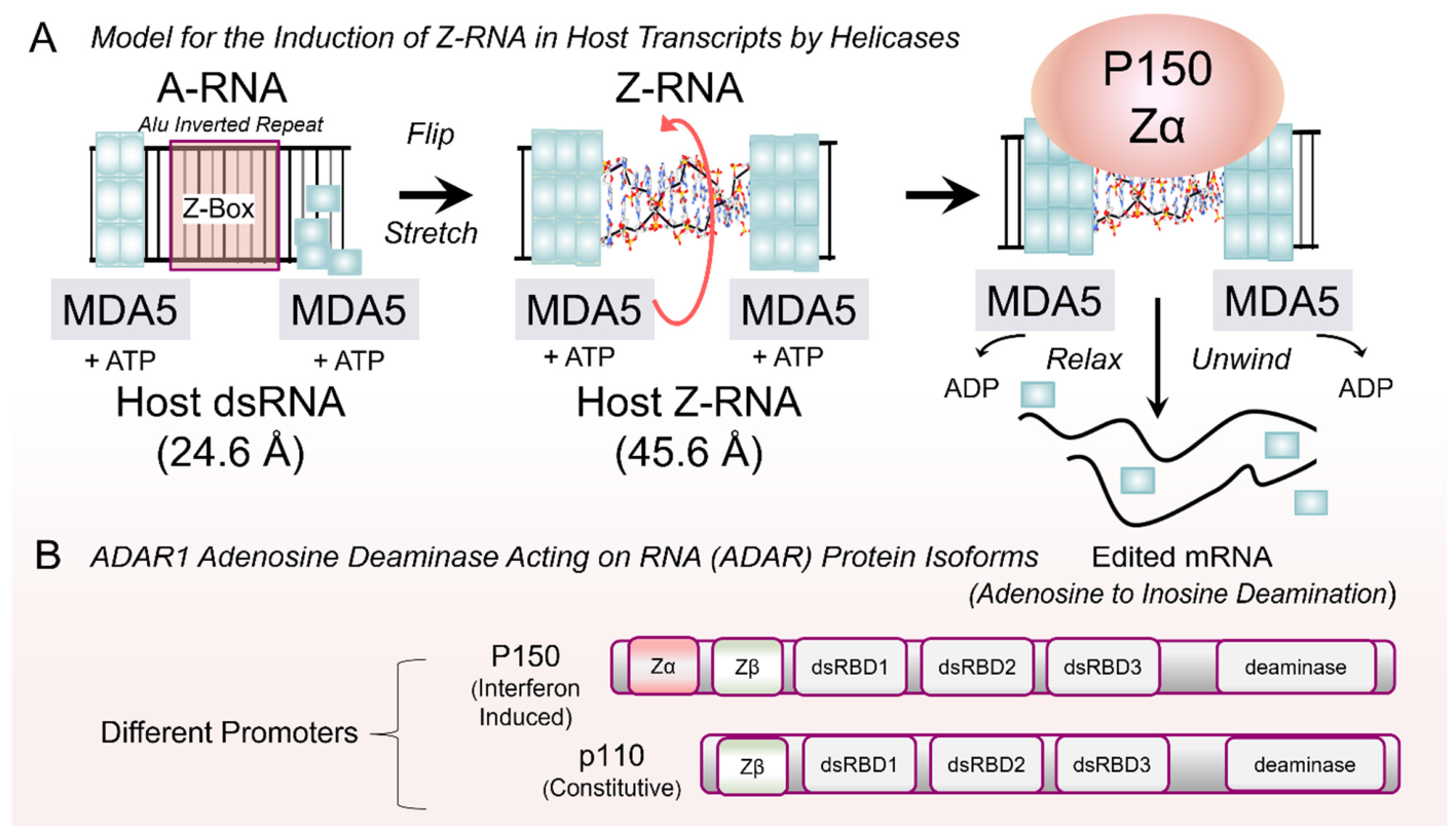
Figure 6.
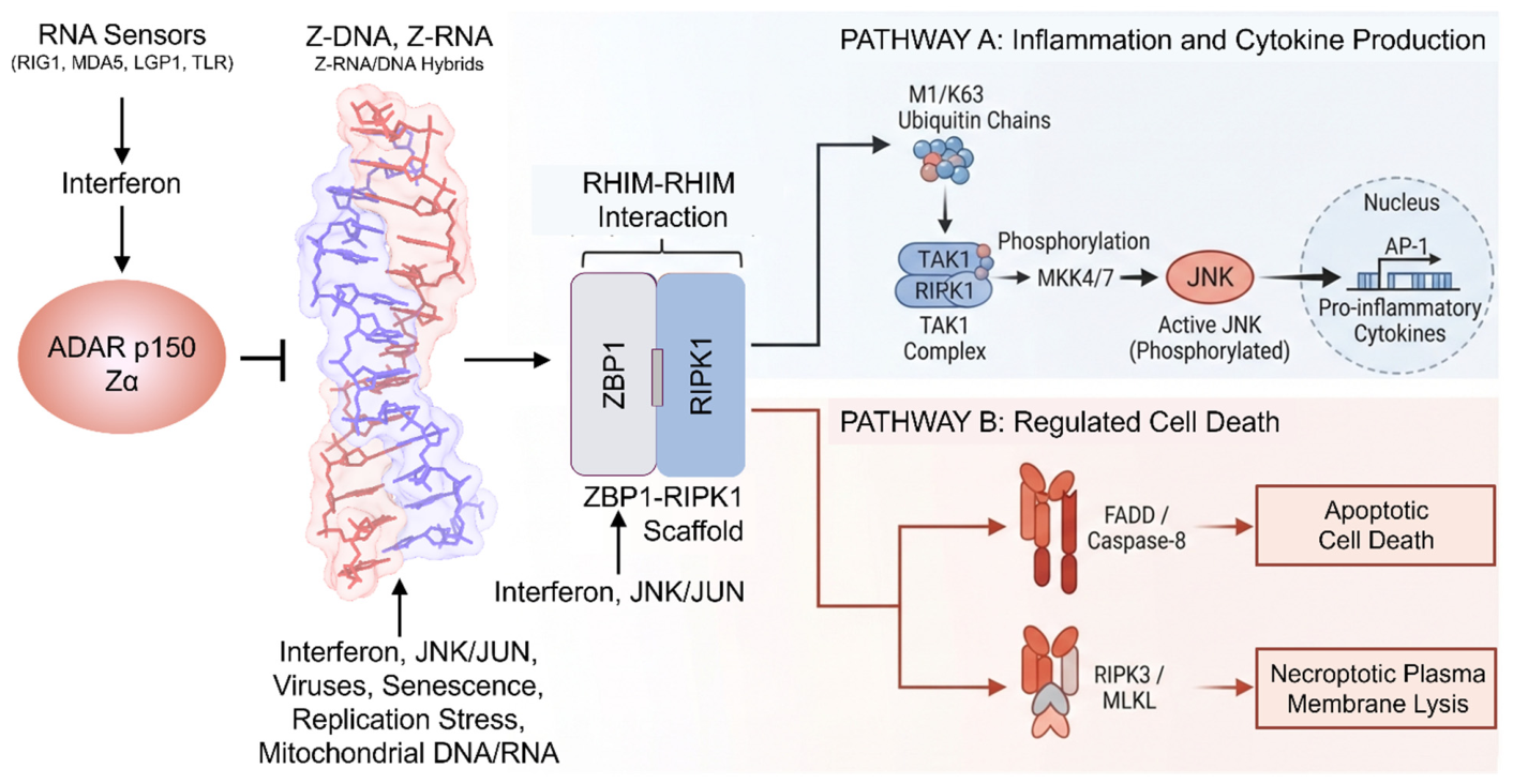
Double-stranded RNA-specific adenosine deaminase (ADAR1) p150 negatively regulates ZNA binding protein (ZBP1) activation. ADAR1 p150 is induced when RNA sensors initiate interferon production. In humans, ZBP1 signals through receptor-interacting serine/threonine-protein kinase 1 (RIPK1) to activate downstream cytokine responses and induce cell death via apoptosis or necroptosis. The pathway activated is context-specific. Interferon strongly induces ZBP1 during stress and infection, to levels that exceed the capacity of ADAR1 p150 to prevent its activation and thereby silence the responses it induces.
It is based on AIRs inserted into normal host transcripts. The default host response to these dsRNAs is to stochastically assemble filaments of melanoma differentiation-associated protein 5 (MDA5, encoded by the interferon-induced helicase C domain-containing protein 1 IFIH1 gene) [161]. If left unchecked, the filaments activate an interferon response [162,163]. However, those host-encoded dsRNAs derived from SINEs incorporate flipons. They contain Z-boxes that flip to Z-RNA when the filament is stretched (from 24.6 to 45.6 Å). One model suggests that the longer Z-RNA relaxes the tension and triggers MDA5 to dissociate and hydrolyze ATP (Figure 5A) [7,18,160]. The Z-RNA then localizes the p150 isoform of ADAR1 to the dsRNA via its Z-RNA-specific Zα domain (Figure 5B). Subsequent deamination of the dsRNA prevents further MDA5 filament formation, thereby preventing an immune response against self. Inosine-specific RNases then clear the modified RNAs. Interferon induces the p150 isoform as part of the initial response. In the absence of interferon, expression of an ADAR1 p110 isoform is driven from a different promoter (Figure 5B) [164].
Genetic proof that ADAR1 p150 suppresses interferon activation comes from individuals from families that express p110 normally but transmit one p150 allele with either an N173S or a P193A loss-of-function variant that no longer binds ZNA, combined with a p150 null allele. Because a normal allele no longer masks the loss-of-function allele, the variant maps directly to the phenotype. The individuals with the unmasked loss-of-function haplotype develop an Aicardi–Goutières type 6 interferonopathy [165].
AIRs inserted into normal host transcripts allow these RNAs to be distinguished from viral RNAs, which lack this feature. Furthermore, the many insertions into normal host messages make it difficult for the virus to suppress each one individually. Viruses such as vaccinia have a Zα ortholog that directly binds Z-RNA, thereby masking it [166]. Viruses like influenza and herpes simplex try to prevent host responses by trapping host polymerases on host genome DNA. Their proteins disrupt the normal termination of host transcription, causing the host polymerase to continue elongating along the chromosome. This process shuts down host RNA production. The host counters this by enriching Z-RNA-forming EREs just past the normal transcription termination site. High ERE levels are expressed when viral proteins disrupt termination, causing Z-RNA to accumulate as well. The volume of Z-RNAs produced overwhelms ADAR1 p150’s ability to squelch them [127,129,167,168,169].
The Z-RNAs then activate ZBP1, the only other Zα-containing protein in the human genome. This protein is also interferon-induced and often expressed at much higher levels than ADAR p150. ZBP1, along with Z-RNA-forming transcripts, is also induced by the Janus kinase (JNK) signaling pathway [170,171] (Figure 6). ZBP1 can then trigger scaffold assembly through its receptor-interacting protein homotypic interaction motifs (RHIMs) [172,173,174]. Depending on the cell context, the RIPK1-dependent processes in humans can lead to an inflammatory form of cell death called necroptosis, a silent apoptotic cell death, or a chronic inflammatory response [175]. Other downstream processes triggered by ADAR1 p150 loss also have adverse outcomes. The neurological damage in mice with an Aicardi–Goutières syndrome-like phenotype caused by a strongly penetrant loss-of-function W195A Zα allele [176] is not rescued by deletion of either ZBP1 or the dsRNA Protein Kinase R (PKR) sensor that induces interferon via MDA5.
The host-based strategy depends on multiple sensors of a cell’s well-being that activate interferon responses. These sensors warn of an imminent threat by detecting bacterial metabolites, viral transcripts, cytoplasmic DNA, and host DNA damage. They initiate a host-based defensive response. Both interferon and stress kinase pathways are triggered. These pathways also induce transcription of EREs that are otherwise suppressed in normal cells [129,177,178,179]. The dsRNAs formed further stimulate the interferon response. The outcome depends on how much Z-RNA accumulates. If Z-RNA levels exceed the ADAR p150 threshold, the response amplifies to eliminate the threat.
9. Circular RNAs and ADAR1 p150
SINEs can also generate circular RNAs (circRNAs) through aberrant splicing of AIRs (Figure 7). The circRNAs are formed by back-splicing of a downstream splice donor to an upstream splice acceptor site. Back-splicing is promoted by Alus with complementary sequences. They bring the splice sites into close proximity by hybridizing to form loops. A subset of circRNAs is exported to the cytoplasm. Many contain repeat sequences complementary to the UTR sequences of cytoplasmic mRNA (Figure 7C) [180,181]. When they form hybrids, the circRNAs can activate Staufen 1 (STAU1)-mediated messenger RNA decay (SMD) [180]. In other cases, circRNAs incorporate exons and modulate the stability of mRNAs that have complementary exons. Several such exon–exon pairs have been identified. These circRNAs often retain exon-junction complexes that induce the nonsense-mediated decay of the mRNAs they bind. The interactions can prevent the translation of cellular proteins necessary for survival [181]. Examples include the circRNAs: hsa_circ_0002082 and circ hsa_circ_0008496 that downregulate the pro-apoptotic BIM protein (Bcl-2-like protein 11 encoded by BCL2L11) [181]. Another set of circRNAs protects cells against inflammation. They retain sequences that form 12–16 bp dsRNA. These stems are not long enough to activate interferon responses but instead inhibit PKR-mediated inflammatory responses [182] (Figure 7B). A different set of circRNAs is reported to inhibit cyclic GMP−AMP synthase (cGAS) activation of the interferon response [183]. Overall, these mechanisms are anti-inflammatory.
Figure 7.
Double-stranded RNA-specific adenosine deaminase (ADAR1) p150 can also suppress back-splicing of RNA promoted by Alu sequences near splice junctions. When folded into dsRNA, Alu sequences can pair downstream splice donors with upstream splice acceptors to yield circular RNAs (circRNAs). (A) Back-splicing is promoted by RNA pausing that can result from oxidative DNA damage or the alternative flipon structures and repair complexes that result. (B) The circRNAs contain short dsRNAs that can inhibit Protein Kinase R (PKR) and cyclic GMP−AMP synthase (cGAS) activation, preventing interferon activation. (C) The circRNA sequences can promote the triage of pro-apoptotic messages by pairing with sequences in their 3′ UTRs and activating RNA decay pathways. (D) ADAR1 p150 and DEAH box protein 9 (DHX9) deficiency leads to the accumulation of circRNAs that pair to form dsRNA tangles that trigger massive cell death.
Back-splicing is promoted by processes that cause RPOL2 pausing and serve as sensors for oxidative DNA damage and replication stress, both of which can induce Z-DNA and GQ (Figure 7A) [184,185,186,187,188,189]. Loss of either ADAR1 p150, which engages both conformations, or the helicase ATP-dependent RNA helicase A (RHA encoded by DHX9) greatly increases accumulation of circRNA and of the dsRNAs formed by circRNA hybrids. Interferon-induced RNase L cleaves the single-stranded circRNA to relieve their suppressive effects, but not the dsRNA hybrids they form. Subsequent cell death can be triggered through several cell death pathways besides ZBP1 [181,190,191] (Figure 7D). This circRNA accumulation is proposed to exacerbate several neurodegenerative diseases [191] and may explain the neurological phenotype of the loss-of-function ADAR1 p150 W195A Zα allele [179].
10. RNA Polymerase Pausing, Oxidative Damage and DNA Repair
Oxidative DNA damage in tissues with high mitochondrial activity, such as the nervous system, contributes to circRNA formation by enhancing polymerase pausing, allowing more time for Alu RNA hybrids to form and for back-splicing to occur. Increased levels of reactive oxygen species (ROS) promote ZNA formation by oxidizing guanosines. These adducts impair B-DNA reformation by Z-flipons during the transcription cycle [184,185,186,187,188]. The repair enzyme 8-oxoguanine DNA glycosylase (OGG1) cannot excise the oxidized base in the Z-DNA conformation. In this situation, localization of the DHX9 helicase by ZNA-bound ADAR1 p150 may be essential for recruiting the repair complex (Figure 7). Subsequently, DHX9’s interaction with poly[ADP-ribose] polymerase 1 (PARP1) recruits OGG1 to the lesion. Z-DNA thereby facilitates the rapid genome scanning needed to localize OGG1 to lesions in active genes [192,193].
10.1. Mitochondrial DNA-Activated Immunity
In addition to causing oxidative damage, damaged mitochondria release their oxidized nucleic acids into the cytoplasm, also leading to ZBP1-dependent outcomes. Notably, ZBP1 can enhance cGAS activity by docking to Z-DNA formed within the covalently closed circular oxidized mitochondrial genome, while engaging the N-terminus of cGAS through its RHIM domains. Interferon induced by 2′3′-cGAMP-mediated activation of the stimulator of interferon genes (STING) then further increases ZBP1 expression, creating a positive feedback loop that promotes cell death [187] (Figure 6). 2′3′-cGAMP also activates STING and induces ADAR1-mediated autophagy in bystander cells, promoting their death after interferon-induced upregulation of ZBP1, as seen in SARS-CoV-2 infection [194,195,196,197]. Cell death is further enhanced by the DNA double-strand break sensor Meiotic recombination 11 (MRE11) during DNA damage repair. The enzyme releases STING from the acidic nucleosome patch that sequesters the protein in an inactive state [198]. ZBP1 potentially promotes this release by directly interacting with MRE11 and inhibiting its repair activity [199]. The cGAS-ZBPI interaction can also be triggered by extracellular neutrophil nets that play a role in acute inflammatory pancreatitis and harbor both Z- and G-flipons [200], as well as in systemic lupus erythematosus [201].
10.2. RNA Polymerase Pausing and Replicative DNA Damage
Persistent Z-DNA from oxidative damage inhibits polymerase elongation, increasing negative supercoiling as trailing polymerases arrive, stall, and eject from the DNA. This process generates high levels of positive supercoiling ahead of the lesion, destabilizing chromatin and causing extensive upstream DNA unwinding. The 5′ negative supercoiling promotes R-loop formation, in which the displaced single-stranded DNA is particularly prone to further oxidative damage, as well as the flip to Z-DNA and the folding of GQ by both RNA and DNA. The repair complexes recruited further exacerbate the stalling of other trailing RNA polymerases by these alternative structures [185]. If unresolved, this process leads to DNA damage, particularly during cell division, when collisions between RNA and DNA polymerase at stalled replication forks amplify the negative outcomes. These collisions are frequently associated with RPOL3 stalls at inverted repeats [202].
Replicative stress also results in cytoplasmic DNA fragments and the accumulation of sub-telomeric RNAs that can activate ZBP1 [203]. These outcomes are augmented by the recent discovery that both the Zα domains of ADAR1 and ZBP1 can dock to both GQ DNA and RNA, with the GQ terra RNA activating ZBP1 [88,89,204,205]. ADAR1 Zβ (Figure 7) also has a strong preference for telomeric GQ [89]. Previously, it was known that Zβ does not bind Z-DNA or Z-RNA because the structure-specific tyrosine residue in Zα is replaced by isoleucine in Zβ. Unlike Z-RNA, which forms from dsRNA, GQ can form in ssRNA. Potentially, ADAR1 p150 is optimized to bind Z-RNA formed in the cytoplasm, while Zβ recognizes the GQ formed by Alu elements in pre-mRNAs and can offset the risks they pose associated with mis-splicing or R-loop-induced DNA damage (Figure 8) [88].
Figure 8.
Double-stranded RNA-specific adenosine deaminase (ADAR1) p150 and p110 isoforms modulate distinct cellular outcomes. The Zα domain can localize ADAR1 to Z-RNA formed by Alu inverted-repeat dsRNA and promote editing of that substrate, thereby turning off interferon responses against self-RNAs and preventing ZBP1 activation. The Zα domain can also localize the enzyme to non-host Z-DNA or Z-RNA, disrupting viral transcript processing and translation through editing and dsRNA binding. The ADAR1 Zβ domain in the nucleus can disrupt ssRNA Alu transcripts by binding to the G-quadruplexes (GQ) they form. This inhibits the mis-splicing and DNA damage they produce (adapted from [89]).
11. Emerging Themes
The protection of hosts by the mechanism described here and summarized in Table 1 represents a series of ad hoc adaptations that enabled hosts to survive and transmit their genomes to progeny. While we tend to look for well-engineered systems and imagine Nature can likewise order its catalog, reality is far different. As the separate evolution of the AMC and RLC machinery shows, solutions often overlap, creating redundancy and resilience. Retrotransposons are both targeted and exploited, with some adaptations limiting ERE spread in a sequence-dependent manner. At the same time, the alternative folds they adopt, such as ZNA, G-quadruplexes, and triplexes, undergo exaptation to program cell responses. Of these structures, ZNA sensors enable host-encoded immune defenses that do not depend on reverse transcriptases for amplification while protecting against a wide range of pathogens [118,129].
Table 1.
A myriad of different ncRNA pathways protect the host against pathogens and itself.
This regulation of pathways by alternative structures dates back to the early eukaryotic progenitors, and likely preceded the use of sequence-specific TFs. These structures also enabled condensate assembly that defined spaces for different chemistries to evolve, including RNA catalysts, while protecting their interior from external hazards [206]. The structures formed depend on the flipon motif involved. As extensive studies of Z-DNA show, the transition to an alternative fold can depend on salt, metal, temperature, and pH conditions [207]. Sequence-specific targeting of the motifs provides another way to modulate their conformation. Nature likely exploited nucleic acids to control flipon conformation early on in protocells. Trial and error improved on earlier iterations. Sequence-specific proteins arrived later, building on proven RNA adaptations, as evidenced by small RNAs that now guide protein effectors and catalyze protein synthesis in the ribosome. The protein tools evolved were more robust and came with modular domains that could be assembled, duplicated, or otherwise modified to improve a species’ survival and reproductive success. A key imperative was to protect the genome from invasive replicants.
Eukaryotic RNA polymerases have adapted to defend the host. The three human polymerases stem from the same ancestral enzyme. Out of the 14 subunits in RPOL1, 12 subunits in RPOL2, and 17 subunits in RPOL3, five core subunits (RPB5, RPB6, RPB8, RPB10, and RPB12) are completely identical and physically shared between them [208]. Yet each protects cells against existential threats differently. RPOL3 transcribes SINEs to enable defenses based on small NCRs that regulate mRNA expression, translation, and protein function in normal cells. When stressed, RPOL3-dependent SINE transcription shifts cells from a relatively quiescent state to one primed to defend itself. In this process, RPOL3 modulates RPOL2 transcription, selectively activating pathways that robustly repel threats from within.
ERE-derived sequences regulate RPOL2 transcription during development [209]. NCRs can also target these elements to regulate chromatin conformation and the flipon structures that modulate gene expression. These pathways so far have proven challenging to exploit therapeutically. Explicitly targeting regulatory flipons offers a new opportunity.
Funding
This research received no external funding.
Institutional Review Board Statement
Not applicable.
Informed Consent Statement
Not applicable.
Data Availability Statement
No new data were created or analyzed in this study. Data sharing is not applicable to this article.
Acknowledgments
Conflicts of Interest
Alan Herbert is the founder of InsideOutBio. The author declares no conflicts of interest. InsideOutBio has no intellectual property relating to flipon-based concepts discussed in the review.
References
- Herbert, A. A Genetic Instruction Code Based on DNA Conformation. Trends Genet. 2019, 35, 887–890. [Google Scholar] [CrossRef] [Scilit] [PubMed]
- Schroth, G.P.; Chou, P.J.; Ho, P.S. Mapping Z-DNA in the human genome. Computer-aided mapping reveals a nonrandom distribution of potential Z-DNA-forming sequences in human genes. J. Biol. Chem. 1992, 267, 11846–11855. [Google Scholar] [CrossRef] [Scilit]
- Huppert, J.L.; Balasubramanian, S. Prevalence of quadruplexes in the human genome. Nucleic Acids Res. 2005, 33, 2908–2916. [Google Scholar] [CrossRef] [Scilit] [PubMed]
- Cer, R.Z.; Bruce, K.H.; Mudunuri, U.S.; Yi, M.; Volfovsky, N.; Luke, B.T.; Bacolla, A.; Collins, J.R.; Stephens, R.M. Non-B DB: A database of predicted non-B DNA-forming motifs in mammalian genomes. Nucleic Acids Res. 2011, 39, D383–D391. [Google Scholar] [CrossRef] [Scilit] [PubMed]
- Biffi, G.; Tannahill, D.; McCafferty, J.; Balasubramanian, S. Quantitative visualization of DNA G-quadruplex structures in human cells. Nat. Chem. 2013, 5, 182–186. [Google Scholar] [CrossRef] [Scilit] [PubMed]
- Senturk Cetin, N.; Kuo, C.C.; Ribarska, T.; Li, R.; Costa, I.G.; Grummt, I. Isolation and genome-wide characterization of cellular DNA:RNA triplex structures. Nucleic Acids Res. 2019, 47, 2306–2321. [Google Scholar] [CrossRef] [Scilit] [PubMed]
- Herbert, A. Z-DNA and Z-RNA in human disease. Commun. Biol. 2019, 2, 7. [Google Scholar] [CrossRef] [Scilit] [PubMed]
- Beknazarov, N.; Jin, S.; Poptsova, M. Deep learning approach for predicting functional Z-DNA regions using omics data. Sci. Rep. 2020, 10, 19134. [Google Scholar] [CrossRef] [Scilit] [PubMed]
- Guiblet, W.M.; Cremona, M.A.; Harris, R.S.; Chen, D.; Eckert, K.A.; Chiaromonte, F.; Huang, Y.F.; Makova, K.D. Non-B DNA: A major contributor to small- and large-scale variation in nucleotide substitution frequencies across the genome. Nucleic Acids Res. 2021, 49, 1497–1516. [Google Scholar] [CrossRef] [Scilit] [PubMed]
- Georgakopoulos-Soares, I.; Victorino, J.; Parada, G.E.; Agarwal, V.; Zhao, J.; Wong, H.Y.; Umar, M.I.; Elor, O.; Muhwezi, A.; An, J.Y.; et al. High-throughput characterization of the role of non-B DNA motifs on promoter function. Cell Genom. 2022, 2, 100111. [Google Scholar] [CrossRef] [Scilit] [PubMed]
- Umerenkov, D.; Herbert, A.; Konovalov, D.; Danilova, A.; Beknazarov, N.; Kokh, V.; Fedorov, A.; Poptsova, M. Z-flipon variants reveal the many roles of Z-DNA and Z-RNA in health and disease. Life Sci. Alliance 2023, 6, e202301962. [Google Scholar] [CrossRef] [Scilit] [PubMed]
- Bashkatov, A.; Andreasyan, A.; Konovalov, D.; Herbert, A.; Poptsova, M. Deep learning deciphers the related role of master regulators and G-quadruplexes in tissue specification. Sci. Rep. 2025, 15, 23119. [Google Scholar] [CrossRef] [Scilit] [PubMed]
- Konovalov, D.; Umerenkov, D.; Herbert, A.; Poptsova, M. GQ-DNABERT reveals GQ proximal enhancer-promoter interactions associated with tissue-specific transcription. Nucleic Acids Res. 2025, 53, gkaf1007. [Google Scholar] [CrossRef] [Scilit] [PubMed]
- Herbert, A.G.; Spitzner, J.R.; Lowenhaupt, K.; Rich, A. Z-DNA binding protein from chicken blood nuclei. Proc. Natl. Acad. Sci. USA 1993, 90, 3339–3342. [Google Scholar] [CrossRef] [Scilit] [PubMed]
- Herbert, A.; Lowenhaupt, K.; Spitzner, J.; Rich, A. Chicken double-stranded RNA adenosine deaminase has apparent specificity for Z-DNA. Proc. Natl. Acad. Sci. USA 1995, 92, 7550–7554. [Google Scholar] [CrossRef] [Scilit] [PubMed]
- Herbert, A.; Alfken, J.; Kim, Y.G.; Mian, I.S.; Nishikura, K.; Rich, A. A Z-DNA binding domain present in the human editing enzyme, double-stranded RNA adenosine deaminase. Proc. Natl. Acad. Sci. USA 1997, 94, 8421–8426. [Google Scholar] [CrossRef] [Scilit] [PubMed]
- Herbert, A. Flipons and the origin of the genetic code. Biol. Lett. 2025, 21, 20240635. [Google Scholar] [CrossRef] [Scilit] [PubMed]
- Nichols, P.J.; Bevers, S.; Henen, M.; Kieft, J.S.; Vicens, Q.; Vogeli, B. Recognition of non-CpG repeats in Alu and ribosomal RNAs by the Z-RNA binding domain of ADAR1 induces A-Z junctions. Nat. Commun. 2021, 12, 793. [Google Scholar] [CrossRef] [Scilit] [PubMed]
- Herbert, A. Neurodegenerative diseases reflect the reciprocal roles played by retroelements in regulating memory and immunity. Front. Neurosci. 2024, 18, 1445540. [Google Scholar] [CrossRef] [Scilit] [PubMed]
- Pace, J.K., II; Feschotte, C. The evolutionary history of human DNA transposons: Evidence for intense activity in the primate lineage. Genome Res. 2007, 17, 422–432. [Google Scholar] [CrossRef] [Scilit] [PubMed]
- de Tribolet-Hardy, J.; Thorball, C.W.; Forey, R.; Planet, E.; Duc, J.; Coudray, A.; Khubieh, B.; Offner, S.; Pulver, C.; Fellay, J.; et al. Genetic features and genomic targets of human KRAB-zinc finger proteins. Genome Res. 2023, 33, 1409–1423. [Google Scholar] [CrossRef] [Scilit] [PubMed]
- Cordaux, R.; Batzer, M.A. The impact of retrotransposons on human genome evolution. Nat. Rev. Genet. 2009, 10, 691–703. [Google Scholar] [CrossRef] [Scilit] [PubMed]
- Chuong, E.B.; Elde, N.C.; Feschotte, C. Regulatory activities of transposable elements: From conflicts to benefits. Nat. Rev. Genet. 2017, 18, 71–86. [Google Scholar] [CrossRef] [Scilit] [PubMed]
- Ghanim, G.E.; Hu, H.; Boulanger, J.; Nguyen, T.H.D. Structural mechanism of LINE-1 target-primed reverse transcription. Science 2025, 388, eads8412. [Google Scholar] [CrossRef] [Scilit] [PubMed]
- Beck, C.R.; Collier, P.; Macfarlane, C.; Malig, M.; Kidd, J.M.; Eichler, E.E.; Badge, R.M.; Moran, J.V. LINE-1 retrotransposition activity in human genomes. Cell 2010, 141, 1159–1170. [Google Scholar] [CrossRef] [Scilit] [PubMed]
- Shrestha, S.; Sewell, J.A.; Santoso, C.S.; Forchielli, E.; Pro, S.C.; Martinez, M.; Bass, J.I.F. Discovering human transcription factor physical interactions with genetic variants, novel DNA motifs, and repetitive elements using enhanced yeast one-hybrid assays. Genome Res. 2019, 29, 1533–1544. [Google Scholar] [CrossRef] [Scilit] [PubMed]
- Du, A.Y.; Chobirko, J.D.; Zhuo, X.; Feschotte, C.; Wang, T. Regulatory transposable elements in the encyclopedia of DNA elements. Nat. Commun. 2024, 15, 7594. [Google Scholar] [CrossRef] [Scilit] [PubMed]
- Turowski, T.W.; Tollervey, D. Transcription by RNA polymerase III: Insights into mechanism and regulation. Biochem. Soc. Trans. 2016, 44, 1367–1375. [Google Scholar] [CrossRef] [Scilit] [PubMed]
- Abascal-Palacios, G.; Ramsay, E.P.; Beuron, F.; Morris, E.; Vannini, A. Structural basis of RNA polymerase III transcription initiation. Nature 2018, 553, 301–306. [Google Scholar] [CrossRef] [Scilit] [PubMed]
- Ferrari, R.; de Llobet Cucalon, L.I.; Di Vona, C.; Le Dilly, F.; Vidal, E.; Lioutas, A.; Oliete, J.Q.; Jochem, L.; Cutts, E.; Dieci, G.; et al. TFIIIC Binding to Alu Elements Controls Gene Expression via Chromatin Looping and Histone Acetylation. Mol. Cell 2020, 77, 475–487.e11. [Google Scholar] [CrossRef] [Scilit] [PubMed]
- Arimbasseri, A.G.; Rijal, K.; Maraia, R.J. Comparative overview of RNA polymerase II and III transcription cycles, with focus on RNA polymerase III termination and reinitiation. Transcription 2014, 5, e27639. [Google Scholar] [CrossRef] [Scilit] [PubMed]
- Naesens, L.; Haerynck, F.; Gack, M.U. The RNA polymerase III-RIG-I axis in antiviral immunity and inflammation. Trends Immunol. 2023, 44, 435–449. [Google Scholar] [CrossRef] [Scilit] [PubMed]
- Xie, J.; Aiello, U.; Clement, Y.; Haidara, N.; Girbig, M.; Schmitzova, J.; Pena, V.; Muller, C.W.; Libri, D.; Porrua, O. An integrated model for termination of RNA polymerase III transcription. Sci. Adv. 2022, 8, eabm9875. [Google Scholar] [CrossRef] [Scilit] [PubMed]
- Kawaji, H.; Nakamura, M.; Takahashi, Y.; Sandelin, A.; Katayama, S.; Fukuda, S.; Daub, C.O.; Kai, C.; Kawai, J.; Yasuda, J.; et al. Hidden layers of human small RNAs. BMC Genom. 2008, 9, 157. [Google Scholar] [CrossRef] [Scilit] [PubMed]
- Li, Z.; Ender, C.; Meister, G.; Moore, P.S.; Chang, Y.; John, B. Extensive terminal and asymmetric processing of small RNAs from rRNAs, snoRNAs, snRNAs, and tRNAs. Nucleic Acids Res. 2012, 40, 6787–6799. [Google Scholar] [CrossRef] [Scilit] [PubMed]
- Su, Z.; Wilson, B.; Kumar, P.; Dutta, A. Noncanonical Roles of tRNAs: TRNA Fragments and Beyond. Annu. Rev. Genet. 2020, 54, 47–69. [Google Scholar] [CrossRef] [Scilit] [PubMed]
- Wang, Y.; Song, X.; Ma, G.; Liu, X.; Gan, C.; Chen, A.; Tian, M.; Han, X. tRNA-derived small RNAs in pulmonary diseases: Regulatory functions and clinical prospects. Pharmacol. Res. 2026, 226, 108153. [Google Scholar] [CrossRef] [Scilit] [PubMed]
- Schorn, A.J.; Gutbrod, M.J.; LeBlanc, C.; Martienssen, R. LTR-Retrotransposon Control by tRNA-Derived Small RNAs. Cell 2017, 170, 61–71.e11. [Google Scholar] [CrossRef] [Scilit] [PubMed]
- Zhang, Y.; Shi, J.; Chen, Q. TsRNAs: New players in mammalian retrotransposon control. Cell Res. 2017, 27, 1307–1308. [Google Scholar] [CrossRef] [Scilit] [PubMed]
- Kuscu, C.; Kumar, P.; Kiran, M.; Su, Z.; Malik, A.; Dutta, A. TRNA fragments (tRFs) guide Ago to regulate gene expression post-transcriptionally in a Dicer-independent manner. RNA 2018, 24, 1093–1105. [Google Scholar] [CrossRef] [Scilit] [PubMed]
- Ganser, L.R.; Kelly, M.L.; Herschlag, D.; Al-Hashimi, H.M. The roles of structural dynamics in the cellular functions of RNAs. Nat. Rev. Mol. Cell Biol. 2019, 20, 474–489. [Google Scholar] [CrossRef] [Scilit] [PubMed]
- Pfleiderer, M.M.; Galej, W.P. Emerging insights into the function and structure of the Integrator complex. Transcription 2021, 12, 251–265. [Google Scholar] [CrossRef] [Scilit] [PubMed]
- Di Fazio, A.; Gullerova, M. An old friend with a new face: TRNA-derived small RNAs with big regulatory potential in cancer biology. Br. J. Cancer 2023, 128, 1625–1635. [Google Scholar] [CrossRef] [Scilit] [PubMed]
- Velazquez-Flores, M.; Ruiz Esparza-Garrido, R. Fragments derived from non-coding RNAs: How complex is genome regulation? Genome 2024, 67, 292–306. [Google Scholar] [CrossRef] [Scilit] [PubMed]
- Berman, H.M.; Westbrook, J.; Feng, Z.; Gilliland, G.; Bhat, T.N.; Weissig, H.; Shindyalov, I.N.; Bourne, P.E. The Protein Data Bank. Nucleic Acids Res. 2000, 28, 235–242. [Google Scholar] [CrossRef] [Scilit] [PubMed]
- Swarts, D.C.; Makarova, K.; Wang, Y.; Nakanishi, K.; Ketting, R.F.; Koonin, E.V.; Patel, D.J.; van der Oost, J. The evolutionary journey of Argonaute proteins. Nat. Struct. Mol. Biol. 2014, 21, 743–753. [Google Scholar] [CrossRef] [Scilit] [PubMed]
- Lee, Y.Y.; Jeong, M.; Lee, H.; Lee, D.; Lee, J.; Park, J.; Kim, V.N.; Roh, S.H. Structural basis for chaperone-guided assembly of RNA-induced silencing complex. Nature 2026, 655, 1339–1348. [Google Scholar] [CrossRef] [Scilit] [PubMed]
- Schopf, F.H.; Biebl, M.M.; Buchner, J. The HSP90 chaperone machinery. Nat. Rev. Mol. Cell Biol. 2017, 18, 345–360. [Google Scholar] [CrossRef] [Scilit] [PubMed]
- Costa, B.; Li Calzi, M.; Castellano, M.; Blanco, V.; Cuevasanta, E.; Litvan, I.; Ivanov, P.; Witwer, K.; Cayota, A.; Tosar, J.P. Nicked tRNAs are stable reservoirs of tRNA halves in cells and biofluids. Proc. Natl. Acad. Sci. USA 2023, 120, e2216330120. [Google Scholar] [CrossRef] [Scilit] [PubMed]
- Kumar, P.; Anaya, J.; Mudunuri, S.B.; Dutta, A. Meta-analysis of tRNA derived RNA fragments reveals that they are evolutionarily conserved and associate with AGO proteins to recognize specific RNA targets. BMC Biol. 2014, 12, 78. [Google Scholar] [CrossRef] [Scilit]
- Nakanishi, K. Anatomy of four human Argonaute proteins. Nucleic Acids Res. 2022, 50, 6618–6638. [Google Scholar] [CrossRef] [Scilit] [PubMed]
- Choi, E.J.; Ren, J.; Zhang, K.; Wu, W.; Lee, Y.S.; Lee, I.; Bao, X. The Importance of AGO 1 and 4 in Post-Transcriptional Gene Regulatory Function of tRF5-GluCTC, an Respiratory Syncytial Virus-Induced tRNA-Derived RNA Fragment. Int. J. Mol. Sci. 2020, 21, 8766. [Google Scholar] [CrossRef] [Scilit] [PubMed]
- Piriyapongsa, J.; Marino-Ramirez, L.; Jordan, I.K. Origin and evolution of human microRNAs from transposable elements. Genetics 2007, 176, 1323–1337. [Google Scholar] [CrossRef] [Scilit] [PubMed]
- Lee, H.Y.; Zhou, K.; Smith, A.M.; Noland, C.L.; Doudna, J.A. Differential roles of human Dicer-binding proteins TRBP and PACT in small RNA processing. Nucleic Acids Res. 2013, 41, 6568–6576. [Google Scholar] [CrossRef] [Scilit] [PubMed]
- Rogato, A.; Richard, H.; Sarazin, A.; Voss, B.; Cheminant Navarro, S.; Champeimont, R.; Navarro, L.; Carbone, A.; Hess, W.R.; Falciatore, A. The diversity of small non-coding RNAs in the diatom Phaeodactylum tricornutum. BMC Genom. 2014, 15, 698. [Google Scholar] [CrossRef] [Scilit] [PubMed]
- Bartel, D.P. Metazoan MicroRNAs. Cell 2018, 173, 20–51. [Google Scholar] [CrossRef] [Scilit] [PubMed]
- MacRae, I.J.; Ma, E.; Zhou, M.; Robinson, C.V.; Doudna, J.A. In vitro reconstitution of the human RISC-loading complex. Proc. Natl. Acad. Sci. USA 2008, 105, 512–517. [Google Scholar] [CrossRef] [Scilit] [PubMed]
- Kim, Y.; Yeo, J.; Lee, J.H.; Cho, J.; Seo, D.; Kim, J.S.; Kim, V.N. Deletion of human tarbp2 reveals cellular microRNA targets and cell-cycle function of TRBP. Cell Rep. 2014, 9, 1061–1074. [Google Scholar] [CrossRef] [Scilit] [PubMed]
- Acosta, F.M.; Pfaller, C.K. From activator to suppressor: PACT is joining the company of PKR negative regulators. RNA 2025, 31, 1539–1541. [Google Scholar] [CrossRef] [Scilit] [PubMed]
- Hasler, D.; Lehmann, G.; Murakawa, Y.; Klironomos, F.; Jakob, L.; Grasser, F.A.; Rajewsky, N.; Landthaler, M.; Meister, G. The Lupus Autoantigen La Prevents Mis-channeling of tRNA Fragments into the Human MicroRNA Pathway. Mol. Cell 2016, 63, 110–124. [Google Scholar] [CrossRef] [Scilit] [PubMed]
- Roura Frigole, H.; Camacho, N.; Castellvi Coma, M.; Fernandez-Lozano, C.; Garcia-Lema, J.; Rafels-Ybern, A.; Canals, A.; Coll, M.; Ribas de Pouplana, L. TRNA deamination by ADAT requires substrate-specific recognition mechanisms and can be inhibited by tRFs. RNA 2019, 25, 607–619. [Google Scholar] [CrossRef] [Scilit] [PubMed]
- Falconi, M.; Giangrossi, M.; Zabaleta, M.E.; Wang, J.; Gambini, V.; Tilio, M.; Bencardino, D.; Occhipinti, S.; Belletti, B.; Laudadio, E.; et al. A novel 3′-tRNA(Glu)-derived fragment acts as a tumor suppressor in breast cancer by targeting nucleolin. FASEB J. 2019, 33, 13228–13240. [Google Scholar] [CrossRef] [Scilit] [PubMed]
- Cui, H.; Li, H.; Wu, H.; Du, F.; Xie, X.; Zeng, S.; Zhang, Z.; Dong, K.; Shang, L.; Jing, C.; et al. A novel 3′tRNA-derived fragment tRF-Val promotes proliferation and inhibits apoptosis by targeting EEF1A1 in gastric cancer. Cell Death Dis. 2022, 13, 471. [Google Scholar] [CrossRef] [Scilit] [PubMed]
- Wu, Y.; Ni, M.T.; Wang, Y.H.; Wang, C.; Hou, H.; Zhang, X.; Zhou, J. Structural basis of translation inhibition by a valine tRNA-derived fragment. Life Sci. Alliance 2024, 7, e202302488. [Google Scholar] [CrossRef] [Scilit] [PubMed]
- Ando, D.; Rashad, S.; Begley, T.J.; Endo, H.; Aoki, M.; Dedon, P.C.; Niizuma, K. Decoding Codon Bias: The Role of tRNA Modifications in Tissue-Specific Translation. Int. J. Mol. Sci. 2025, 26, 706. [Google Scholar] [CrossRef] [Scilit] [PubMed]
- Su, Z.; Kuscu, C.; Malik, A.; Shibata, E.; Dutta, A. Angiogenin generates specific stress-induced tRNA halves and is not involved in tRF-3-mediated gene silencing. J. Biol. Chem. 2019, 294, 16930–16941. [Google Scholar] [CrossRef] [Scilit] [PubMed]
- Ivanov, P.; O’Day, E.; Emara, M.M.; Wagner, G.; Lieberman, J.; Anderson, P. G-quadruplex structures contribute to the neuroprotective effects of angiogenin-induced tRNA fragments. Proc. Natl. Acad. Sci. USA 2014, 111, 18201–18206. [Google Scholar] [CrossRef] [Scilit] [PubMed]
- Lyons, S.M.; Gudanis, D.; Coyne, S.M.; Gdaniec, Z.; Ivanov, P. Identification of functional tetramolecular RNA G-quadruplexes derived from transfer RNAs. Nat. Commun. 2017, 8, 1127, Erratum in Nat Commun. 2017, 8, 2020. https://doi.org/10.1038/s41467-017-02140-9. PMID: 29209051. [Google Scholar] [CrossRef] [Scilit] [PubMed]
- Tosar, J.P.; Gambaro, F.; Darre, L.; Pantano, S.; Westhof, E.; Cayota, A. Dimerization confers increased stability to nucleases in 5′ halves from glycine and glutamic acid tRNAs. Nucleic Acids Res. 2018, 46, 9081–9093. [Google Scholar] [CrossRef] [Scilit] [PubMed]
- Han, L.; Lai, H.; Yang, Y.; Hu, J.; Li, Z.; Ma, B.; Xu, W.; Liu, W.; Wei, W.; Li, D.; et al. A 5′-tRNA halve, tiRNA-Gly promotes cell proliferation and migration via binding to RBM17 and inducing alternative splicing in papillary thyroid cancer. J. Exp. Clin. Cancer Res. 2021, 40, 222. [Google Scholar] [CrossRef] [Scilit] [PubMed]
- Jin, H.; Yeom, J.H.; Shin, E.; Ha, Y.; Liu, H.; Kim, D.; Joo, M.; Kim, Y.H.; Kim, H.K.; Ryu, M.; et al. 5′-tRNAGly(GCC) halves generated by IRE1α are linked to the ER stress response. Nat. Commun. 2024, 15, 9273. [Google Scholar] [CrossRef] [Scilit] [PubMed]
- Li, D.; Gao, X.; Ma, X.; Wang, M.; Cheng, C.; Xue, T.; Gao, F.; Shen, Y.; Zhang, J.; Liu, Q. Aging-induced tRNA(Glu)-derived fragment impairs glutamate biosynthesis by targeting mitochondrial translation-dependent cristae organization. Cell Metab. 2024, 36, 1059–1075.e9. [Google Scholar] [CrossRef] [Scilit] [PubMed]
- Jirstrom, E.; Matveeva, A.; Baindoor, S.; Donovan, P.; Ma, Q.; Morrissey, E.P.; Arijs, I.; Boeckx, B.; Lambrechts, D.; Garcia-Munoz, A.; et al. Effects of ALS-associated 5′tiRNA(Gly-GCC) on the transcriptomic and proteomic profile of primary neurons in vitro. Exp. Neurol. 2025, 385, 115128. [Google Scholar] [CrossRef] [Scilit] [PubMed]
- Detzer, A.; Engel, C.; Wunsche, W.; Sczakiel, G. Cell stress is related to re-localization of Argonaute 2 and to decreased RNA interference in human cells. Nucleic Acids Res. 2011, 39, 2727–2741. [Google Scholar] [CrossRef] [Scilit] [PubMed]
- Leung, A.K.; Vyas, S.; Rood, J.E.; Bhutkar, A.; Sharp, P.A.; Chang, P. Poly(ADP-ribose) regulates stress responses and microRNA activity in the cytoplasm. Mol. Cell 2011, 42, 489–499. [Google Scholar] [CrossRef] [Scilit] [PubMed]
- Saikia, M.; Krokowski, D.; Guan, B.J.; Ivanov, P.; Parisien, M.; Hu, G.F.; Anderson, P.; Pan, T.; Hatzoglou, M. Genome-wide identification and quantitative analysis of cleaved tRNA fragments induced by cellular stress. J. Biol. Chem. 2012, 287, 42708–42725. [Google Scholar] [CrossRef] [Scilit] [PubMed]
- Lai, H.; Feng, N.; Zhai, Q. Discovery of the major 15-30 nt mammalian small RNAs, their biogenesis and function. Nat. Commun. 2023, 14, 5796. [Google Scholar] [CrossRef] [Scilit] [PubMed]
- Ruby, J.G.; Jan, C.H.; Bartel, D.P. Intronic microRNA precursors that bypass Drosha processing. Nature 2007, 448, 83–86. [Google Scholar] [CrossRef] [Scilit] [PubMed]
- He, L.; He, X.; Lim, L.P.; de Stanchina, E.; Xuan, Z.; Liang, Y.; Xue, W.; Zender, L.; Magnus, J.; Ridzon, D.; et al. A microRNA component of the p53 tumour suppressor network. Nature 2007, 447, 1130–1134. [Google Scholar] [CrossRef] [Scilit] [PubMed]
- Chan, S.Y.; Zhang, Y.Y.; Hemann, C.; Mahoney, C.E.; Zweier, J.L.; Loscalzo, J. MicroRNA-210 controls mitochondrial metabolism during hypoxia by repressing the iron-sulfur cluster assembly proteins ISCU1/2. Cell Metab. 2009, 10, 273–284. [Google Scholar] [CrossRef] [Scilit] [PubMed]
- Leung, A.K.; Sharp, P.A. MicroRNA functions in stress responses. Mol. Cell 2010, 40, 205–215. [Google Scholar] [CrossRef] [Scilit] [PubMed]
- Olejniczak, M.; Kotowska-Zimmer, A.; Krzyzosiak, W. Stress-induced changes in miRNA biogenesis and functioning. Cell Mol. Life Sci. 2018, 75, 177–191. [Google Scholar] [CrossRef] [Scilit] [PubMed]
- Salim, U.; Menon, M.B.; Dhamija, S.; Vivekanandan, P. RNA G-quadruplexes regulate mammalian mirtron biogenesis. J. Biol. Chem. 2025, 301, 108276. [Google Scholar] [CrossRef] [Scilit] [PubMed]
- Batzer, M.A.; Deininger, P.L. Alu repeats and human genomic diversity. Nat. Rev. Genet. 2002, 3, 370–379. [Google Scholar] [CrossRef] [Scilit] [PubMed]
- Varshney, D.; Vavrova-Anderson, J.; Oler, A.J.; Cowling, V.H.; Cairns, B.R.; White, R.J. SINE transcription by RNA polymerase III is suppressed by histone methylation but not by DNA methylation. Nat. Commun. 2015, 6, 6569. [Google Scholar] [CrossRef] [Scilit] [PubMed]
- Conti, A.; Carnevali, D.; Bollati, V.; Fustinoni, S.; Pellegrini, M.; Dieci, G. Identification of RNA polymerase III-transcribed Alu loci by computational screening of RNA-Seq data. Nucleic Acids Res. 2015, 43, 817–835. [Google Scholar] [CrossRef] [Scilit] [PubMed]
- Herbert, A. ALU non-B-DNA conformations, flipons, binary codes and evolution. R. Soc. Open Sci. 2020, 7, 200222. [Google Scholar] [CrossRef] [Scilit] [PubMed]
- Herbert, A.; Cherednichenko, O.; Lybrand, T.P.; Egli, M.; Poptsova, M. Zα and Zβ Localize ADAR1 to Flipons That Modulate Innate Immunity, Alternative Splicing, and Nonsynonymous RNA Editing. Int. J. Mol. Sci. 2025, 26, 2422. [Google Scholar] [CrossRef] [Scilit] [PubMed]
- Kroft, C.W.; Krall, J.B.; Warchol, M.; Welty, R.; Herbert, A.; Henen, M.A.; Vogeli, B. Zα and Zβ domains of ADAR1 and ZBP1 bind G-quadruplexes with nanomolar affinities, establishing Zβ as a G-quadruplex-specific domain. Nucleic Acids Res. 2026, 54, gkag419. [Google Scholar] [CrossRef] [Scilit] [PubMed]
- Liu, W.M.; Chu, W.M.; Choudary, P.V.; Schmid, C.W. Cell stress and translational inhibitors transiently increase the abundance of mammalian SINE transcripts. Nucleic Acids Res. 1995, 23, 1758–1765. [Google Scholar] [CrossRef] [Scilit] [PubMed]
- Panning, B.; Smiley, J.R. Activation of expression of multiple subfamilies of human Alu elements by adenovirus type 5 and herpes simplex virus type 1. J. Mol. Biol. 1995, 248, 513–524. [Google Scholar] [CrossRef] [Scilit] [PubMed]
- Cantarella, S.; Vezzoli, M.; Carnevali, D.; Morselli, M.; Zemke, N.R.; Montanini, B.; Daussy, C.F.; Wodrich, H.; Teichmann, M.; Pellegrini, M.; et al. Adenovirus small E1A directs activation of Alu transcription at YAP/TEAD- and AP-1-bound enhancers through interactions with the EP400 chromatin remodeler. Nucleic Acids Res. 2024, 52, 9481–9500. [Google Scholar] [CrossRef] [Scilit] [PubMed]
- Pandey, R.; Mandal, A.K.; Jha, V.; Mukerji, M. Heat shock factor binding in Alu repeats expands its involvement in stress through an antisense mechanism. Genome Biol. 2011, 12, R117. [Google Scholar] [CrossRef] [Scilit] [PubMed]
- Feng, N.; Feng, H.; Wang, S.; Punekar, A.S.; Ladenstein, R.; Wang, D.C.; Zhang, Q.; Ding, J.; Liu, W. Structures of heat shock factor trimers bound to DNA. iScience 2021, 24, 102951. [Google Scholar] [CrossRef] [Scilit] [PubMed]
- Mariner, P.D.; Walters, R.D.; Espinoza, C.A.; Drullinger, L.F.; Wagner, S.D.; Kugel, J.F.; Goodrich, J.A. Human Alu RNA is a modular transacting repressor of mRNA transcription during heat shock. Mol. Cell 2008, 29, 499–509. [Google Scholar] [CrossRef] [Scilit] [PubMed]
- Yakovchuk, P.; Goodrich, J.A.; Kugel, J.F. B2 RNA and Alu RNA repress transcription by disrupting contacts between RNA polymerase II and promoter DNA within assembled complexes. Proc. Natl. Acad. Sci. USA 2009, 106, 5569–5574. [Google Scholar] [CrossRef] [Scilit] [PubMed]
- Tluckova, K.; Kaczmarek, B.; Salmazo, A.; Bernecky, C. Mechanism of mammalian transcriptional repression by noncoding RNA. Nat. Struct. Mol. Biol. 2025, 32, 607–612. [Google Scholar] [CrossRef] [Scilit] [PubMed]
- Queitsch, C.; Sangster, T.A.; Lindquist, S. Hsp90 as a capacitor of phenotypic variation. Nature 2002, 417, 618–624. [Google Scholar] [CrossRef] [Scilit] [PubMed]
- Zhang, X.O.; Gingeras, T.R.; Weng, Z. Genome-wide analysis of polymerase III-transcribed Alu elements suggests cell-type-specific enhancer function. Genome Res. 2019, 29, 1402–1414. [Google Scholar] [CrossRef] [Scilit] [PubMed]
- Oler, A.J.; Alla, R.K.; Roberts, D.N.; Wong, A.; Hollenhorst, P.C.; Chandler, K.J.; Cassiday, P.A.; Nelson, C.A.; Hagedorn, C.H.; Graves, B.J.; et al. Human RNA polymerase III transcriptomes and relationships to Pol II promoter chromatin and enhancer-binding factors. Nat. Struct. Mol. Biol. 2010, 17, 620–628. [Google Scholar] [CrossRef] [Scilit] [PubMed]
- van Breugel, M.E.; Gerber, A.; van Leeuwen, F. The choreography of chromatin in RNA polymerase III regulation. Biochem. Soc. Trans. 2024, 52, 1173–1189. [Google Scholar] [CrossRef] [Scilit] [PubMed]
- Laperriere, D.; Wang, T.T.; White, J.H.; Mader, S. Widespread Alu repeat-driven expansion of consensus DR2 retinoic acid response elements during primate evolution. BMC Genom. 2007, 8, 23. [Google Scholar] [CrossRef] [Scilit] [PubMed]
- Su, M.; Han, D.; Boyd-Kirkup, J.; Yu, X.; Han, J.-D.J. Evolution of Alu Elements toward Enhancers. Cell Rep. 2014, 7, 376–385. [Google Scholar] [CrossRef] [Scilit] [PubMed]
- Policarpi, C.; Crepaldi, L.; Brookes, E.; Nitarska, J.; French, S.M.; Coatti, A.; Riccio, A. Enhancer SINEs Link Pol III to Pol II Transcription in Neurons. Cell Rep. 2017, 21, 2879–2894. [Google Scholar] [CrossRef] [Scilit] [PubMed]
- Hu, Q.; Tanasa, B.; Trabucchi, M.; Li, W.; Zhang, J.; Ohgi, K.A.; Rose, D.W.; Glass, C.K.; Rosenfeld, M.G. DICER- and AGO3-dependent generation of retinoic acid-induced DR2 Alu RNAs regulates human stem cell proliferation. Nat. Struct. Mol. Biol. 2012, 19, 1168–1175. [Google Scholar] [CrossRef] [Scilit] [PubMed]
- Pelicano, L.; Li, F.; Schindler, C.; Chelbi-Alix, M.K. Retinoic acid enhances the expression of interferon-induced proteins: Evidence for multiple mechanisms of action. Oncogene 1997, 15, 2349–2359. [Google Scholar] [CrossRef] [Scilit] [PubMed]
- Blalock, J.E.; Gifford, G.E. Retinoic acid (vitamin A acid) induced transcriptional control of interferon production. Proc. Natl. Acad. Sci. USA 1977, 74, 5382–5386. [Google Scholar] [CrossRef] [Scilit] [PubMed]
- Koo, C.X.; Kobiyama, K.; Shen, Y.J.; LeBert, N.; Ahmad, S.; Khatoo, M.; Aoshi, T.; Gasser, S.; Ishii, K.J. RNA polymerase III regulates cytosolic RNA:DNA hybrids and intracellular microRNA expression. J. Biol. Chem. 2015, 290, 7463–7473. [Google Scholar] [CrossRef] [Scilit] [PubMed]
- Crossley, M.P.; Song, C.; Bocek, M.J.; Choi, J.H.; Kousouros, J.N.; Sathirachinda, A.; Lin, C.; Brickner, J.R.; Bai, G.; Lans, H.; et al. R-loop-derived cytoplasmic RNA-DNA hybrids activate an immune response. Nature 2023, 613, 187–194, Erratum in Nature 2024, 626, E6. https://doi.org/10.1038/s41586-024-07064-1. PMID: 38263519. [Google Scholar] [CrossRef] [Scilit] [PubMed]
- Moadab, F.; Sohrabi, S.; Wang, X.; Najjar, R.; Wolters, J.C.; Jiang, H.; Miao, W.; Romero, D.; Zaller, D.M.; Tran, M.; et al. Subcellular location of L1 retrotransposon-encoded ORF1p, reverse transcription products, and DNA sensors in lupus granulocytes. Mob. DNA 2024, 15, 14. [Google Scholar] [CrossRef] [Scilit] [PubMed]
- Mankan, A.K.; Schmidt, T.; Chauhan, D.; Goldeck, M.; Honing, K.; Gaidt, M.; Kubarenko, A.V.; Andreeva, L.; Hopfner, K.P.; Hornung, V. Cytosolic RNA:DNA hybrids activate the cGAS-STING axis. EMBO J. 2014, 33, 2937–2946. [Google Scholar] [CrossRef] [Scilit] [PubMed]
- Baldwin, E.T.; van Eeuwen, T.; Hoyos, D.; Zalevsky, A.; Tchesnokov, E.P.; Sanchez, R.; Miller, B.D.; Di Stefano, L.H.; Ruiz, F.X.; Hancock, M.; et al. Structures, functions and adaptations of the human LINE-1 ORF2 protein. Nature 2024, 626, 194–206. [Google Scholar] [CrossRef] [Scilit] [PubMed]
- Jang, K.L.; Latchman, D.S. HSV infection induces increased transcription of Alu repeated sequences by RNA polymerase III. FEBS Lett. 1989, 258, 255–258. [Google Scholar] [CrossRef] [Scilit] [PubMed]
- Hung, T.; Pratt, G.A.; Sundararaman, B.; Townsend, M.J.; Chaivorapol, C.; Bhangale, T.; Graham, R.R.; Ortmann, W.; Criswell, L.A.; Yeo, G.W.; et al. The Ro60 autoantigen binds endogenous retroelements and regulates inflammatory gene expression. Science 2015, 350, 455–459. [Google Scholar] [CrossRef] [Scilit] [PubMed]
- Larsen, P.A.; Hunnicutt, K.E.; Larsen, R.J.; Yoder, A.D.; Saunders, A.M. Warning SINEs: Alu elements, evolution of the human brain, and the spectrum of neurological disease. Chromosome Res. 2018, 26, 93–111. [Google Scholar] [CrossRef] [Scilit] [PubMed]
- Aune, T.M.; Tossberg, J.T.; Heinrich, R.M.; Porter, K.P.; Crooke, P.S., III. Alu RNA Structural Features Modulate Immune Cell Activation and A-to-I Editing of Alu RNAs Is Diminished in Human Inflammatory Bowel Disease. Front. Immunol. 2022, 13, 818023. [Google Scholar] [CrossRef] [Scilit] [PubMed]
- Frost, B.; Dubnau, J. The Role of Retrotransposons and Endogenous Retroviruses in Age-Dependent Neurodegenerative Disorders. Annu. Rev. Neurosci. 2024, 47, 123–143. [Google Scholar] [CrossRef] [Scilit] [PubMed]
- Herbert, A. The ancient Z-DNA and Z-RNA specific Zα fold has evolved modern roles in immunity and transcription through the natural selection of flipons. R. Soc. Open Sci. 2024, 11, 240080. [Google Scholar] [CrossRef] [Scilit] [PubMed]
- Liu, L.F.; Wang, J.C. Supercoiling of the DNA template during transcription. Proc. Natl. Acad. Sci. USA 1987, 84, 7024–7027. [Google Scholar] [CrossRef] [Scilit] [PubMed]
- Herbert, A. The evolutionary entanglement of flipons with zinc fingers and retroelements has engendered a large family of Z-DNA and G-quadruplex binding proteins. Open Biol. 2025, 15, 250171. [Google Scholar] [CrossRef] [Scilit] [PubMed]
- Herbert, A. Control of Gene Expression by Proteins That Bind Many Alternative Nucleic Acid Structures Through the Same Domain. Int. J. Mol. Sci. 2025, 27, 272. [Google Scholar] [CrossRef] [Scilit] [PubMed]
- Furuyama, T.; Henikoff, S. Centromeric nucleosomes induce positive DNA supercoils. Cell 2009, 138, 104–113. [Google Scholar] [CrossRef] [Scilit] [PubMed]
- Stenger, J.E.; Lobachev, K.S.; Gordenin, D.; Darden, T.A.; Jurka, J.; Resnick, M.A. Biased distribution of inverted and direct Alus in the human genome: Implications for insertion, exclusion, and genome stability. Genome Res. 2001, 11, 12–27. [Google Scholar] [CrossRef] [Scilit] [PubMed]
- Herbert, A. Nucleosomes and flipons exchange energy to alter chromatin conformation, the readout of genomic information, and cell fate. Bioessays 2022, 44, e2200166. [Google Scholar] [CrossRef] [Scilit] [PubMed]
- Lexa, M.; Steflova, P.; Martinek, T.; Vorlickova, M.; Vyskot, B.; Kejnovsky, E. Guanine quadruplexes are formed by specific regions of human transposable elements. BMC Genom. 2014, 15, 1032. [Google Scholar] [CrossRef] [Scilit] [PubMed]
- de Reuver, R.; Dierick, E.; Wiernicki, B.; Staes, K.; Seys, L.; De Meester, E.; Muyldermans, T.; Botzki, A.; Lambrecht, B.N.; Van Nieuwerburgh, F.; et al. ADAR1 interaction with Z-RNA promotes editing of endogenous double-stranded RNA and prevents MDA5-dependent immune activation. Cell Rep. 2021, 36, 109500. [Google Scholar] [CrossRef] [Scilit] [PubMed]
- Zhang, T.; Yin, C.; Fedorov, A.; Qiao, L.; Bao, H.; Beknazarov, N.; Wang, S.; Gautam, A.; Williams, R.M.; Crawford, J.C.; et al. ADAR1 masks the cancer immunotherapeutic promise of ZBP1-driven necroptosis. Nature 2022, 606, 594–602. [Google Scholar] [CrossRef] [Scilit] [PubMed]
- Lee, D.H.; Bae, W.H.; Ha, H.; Park, E.G.; Lee, Y.J.; Kim, W.R.; Kim, H.S. Z-DNA-Containing Long Terminal Repeats of Human Endogenous Retrovirus Families Provide Alternative Promoters for Human Functional Genes. Mol. Cells 2022, 45, 522–530. [Google Scholar] [CrossRef] [Scilit] [PubMed]
- Yin, C.; Fedorov, A.; Guo, H.; Crawford, J.C.; Rousseau, C.; Zhong, X.; Williams, R.M.; Gautam, A.; Koehler, H.S.; Whisnant, A.W.; et al. Host cell Z-RNAs activate ZBP1 during virus infections. Nature 2025, 648, 707–716. [Google Scholar] [CrossRef] [Scilit] [PubMed]
- Liu, S.; Chen, Z.; Shen, X.; Gao, J.; Guo, J.; Rong, Y.; Li, T.; Zhou, F.; Si, Y.; Zhao, H.; et al. Alu repeat-containing RNAs spatially organize actively transcribed genomic regions around nuclear speckles. Mol. Cell 2026, 86, 1723–1741.e11. [Google Scholar] [CrossRef] [Scilit] [PubMed]
- Liang, L.; Cao, C.; Ji, L.; Cai, Z.; Wang, D.; Ye, R.; Chen, J.; Yu, X.; Zhou, J.; Bai, Z.; et al. Complementary Alu sequences mediate enhancer-promoter selectivity. Nature 2023, 619, 868–875, Erratum in Nature 2023, 620, E26. https://doi.org/10.1038/s41586-023-06475-w. PMID: 37524778. [Google Scholar] [CrossRef] [Scilit] [PubMed]
- Wiechens, E.; Vigliotti, F.; Siniuk, K.; Schwarz, R.; Schwab, K.; Riege, K.; van Bommel, A.; Gorlich, I.; Bens, M.; Sahm, A.; et al. Gene regulation by convergent promoters. Nat. Genet. 2025, 57, 206–217. [Google Scholar] [CrossRef] [Scilit] [PubMed]
- Zhang, F.; Zhang, H.; Xiao, Y.; Liu, M.; Ren, A.; Liu, S.; Wu, L. Integrated small and long RNA sequencing reveals piRNA mediated transposon repression during human oogenesis. Nat. Commun. 2026, 17, 3804. [Google Scholar] [CrossRef] [Scilit] [PubMed]
- Gainetdinov, I.; Vega-Badillo, J.; Cecchini, K.; Bagci, A.; Colpan, C.; De, D.; Bailey, S.; Arif, A.; Wu, P.H.; MacRae, I.J.; et al. Relaxed targeting rules help PIWI proteins silence transposons. Nature 2023, 619, 394–402. [Google Scholar] [CrossRef] [Scilit] [PubMed]
- Suh, N.; Baehner, L.; Moltzahn, F.; Melton, C.; Shenoy, A.; Chen, J.; Blelloch, R. MicroRNA function is globally suppressed in mouse oocytes and early embryos. Curr. Biol. 2010, 20, 271–277. [Google Scholar] [CrossRef] [Scilit] [PubMed]
- Flemr, M.; Malik, R.; Franke, V.; Nejepinska, J.; Sedlacek, R.; Vlahovicek, K.; Svoboda, P. A retrotransposon-driven dicer isoform directs endogenous small interfering RNA production in mouse oocytes. Cell 2013, 155, 807–816. [Google Scholar] [CrossRef] [Scilit] [PubMed]
- Stein, P.; Rozhkov, N.V.; Li, F.; Cardenas, F.L.; Davydenko, O.; Vandivier, L.E.; Gregory, B.D.; Hannon, G.J.; Schultz, R.M. Essential Role for endogenous siRNAs during meiosis in mouse oocytes. PLoS Genet. 2015, 11, e1005013, Erratum in PLoS Genet. 2015, 11, e1005129. https://doi.org/10.1371/journal.pgen.1005129. eCollection 2015 Apr. PMID: 25860788. [Google Scholar] [CrossRef] [Scilit] [PubMed]
- Nazer, E.; Gomez Acuna, L.; Kornblihtt, A.R. Seeking the truth behind the myth: Argonaute tales from “nuclearland”. Mol. Cell 2022, 82, 503–513. [Google Scholar] [CrossRef] [Scilit] [PubMed]
- Yang, P.; Wang, Y.; Macfarlan, T.S. The Role of KRAB-ZFPs in Transposable Element Repression and Mammalian Evolution. Trends Genet. 2017, 33, 871–881. [Google Scholar] [CrossRef] [Scilit] [PubMed]
- Roy-Engel, A.M.; Salem, A.H.; Oyeniran, O.O.; Deininger, L.; Hedges, D.J.; Kilroy, G.E.; Batzer, M.A.; Deininger, P.L. Active Alu element “A-tails”: Size does matter. Genome Res. 2002, 12, 1333–1344. [Google Scholar] [CrossRef] [Scilit] [PubMed]
- Zhang, X.; Jiang, Q.; Li, J.; Zhang, S.; Cao, Y.; Xia, X.; Cai, D.; Tan, J.; Chen, J.; Han, J.J. KCNQ1OT1 promotes genome-wide transposon repression by guiding RNA-DNA triplexes and HP1 binding. Nat. Cell Biol. 2022, 24, 1617–1629. [Google Scholar] [CrossRef] [Scilit] [PubMed]
- Chen, J.; Sun, M.; Kent, W.J.; Huang, X.; Xie, H.; Wang, W.; Zhou, G.; Shi, R.Z.; Rowley, J.D. Over 20% of human transcripts might form sense-antisense pairs. Nucleic Acids Res. 2004, 32, 4812–4820. [Google Scholar] [CrossRef] [Scilit] [PubMed]
- Cowley, M.; Wood, A.J.; Bohm, S.; Schulz, R.; Oakey, R.J. Epigenetic control of alternative mRNA processing at the imprinted Herc3/Nap1l5 locus. Nucleic Acids Res. 2012, 40, 8917–8926. [Google Scholar] [CrossRef] [Scilit] [PubMed]
- Palmer, A.C.; Egan, J.B.; Shearwin, K.E. Transcriptional interference by RNA polymerase pausing and dislodgement of transcription factors. Transcription 2011, 2, 9–14. [Google Scholar] [CrossRef] [Scilit] [PubMed]
- Osato, N.; Suzuki, Y.; Ikeo, K.; Gojobori, T. Transcriptional interferences in cis natural antisense transcripts of humans and mice. Genetics 2007, 176, 1299–1306. [Google Scholar] [CrossRef] [Scilit] [PubMed]
- Lykke-Andersen, S.; Zumer, K.; Molska, E.S.; Rouviere, J.O.; Wu, G.; Demel, C.; Schwalb, B.; Schmid, M.; Cramer, P.; Jensen, T.H. Integrator is a genome-wide attenuator of non-productive transcription. Mol. Cell 2021, 81, 514–529.e6. [Google Scholar] [CrossRef] [Scilit] [PubMed]
- Herbert, A.; Pavlov, F.; Konovalov, D.; Poptsova, M. Conserved microRNAs and Flipons Shape Gene Expression during Development by Altering Promoter Conformations. Int. J. Mol. Sci. 2023, 24, 4884. [Google Scholar] [CrossRef] [Scilit] [PubMed]
- Gehring, K.; Leroy, J.L.; Gueron, M. A tetrameric DNA structure with protonated cytosine-cytosine base pairs. Nature 1993, 363, 561–565. [Google Scholar] [CrossRef] [Scilit] [PubMed]
- Edwards, S.F.; Sirito, M.; Krahe, R.; Sinden, R.R. A Z-DNA sequence reduces slipped-strand structure formation in the myotonic dystrophy type 2 (CCTG)·(CAGG) repeat. Proc. Natl. Acad. Sci. USA 2009, 106, 3270–3275. [Google Scholar] [CrossRef] [Scilit] [PubMed]
- Herbert, A. The Chromaverse Is Colored by Triplexes Formed Through the Interactions of Noncoding RNAs with HNPRNPU, TP53, AGO, REL Proteins, Intrinsically-Disordered Regions, and Flipons. Int. J. Mol. Sci. 2026, 27, 1482. [Google Scholar] [CrossRef] [Scilit] [PubMed]
- Levanon, E.Y.; Eisenberg, E.; Yelin, R.; Nemzer, S.; Hallegger, M.; Shemesh, R.; Fligelman, Z.Y.; Shoshan, A.; Pollock, S.R.; Sztybel, D.; et al. Systematic identification of abundant A-to-I editing sites in the human transcriptome. Nat. Biotechnol. 2004, 22, 1001–1005. [Google Scholar] [CrossRef] [Scilit] [PubMed]
- Kim, D.D.; Kim, T.T.; Walsh, T.; Kobayashi, Y.; Matise, T.C.; Buyske, S.; Gabriel, A. Widespread RNA editing of embedded alu elements in the human transcriptome. Genome Res. 2004, 14, 1719–1725. [Google Scholar] [CrossRef] [Scilit] [PubMed]
- Blow, M.; Futreal, P.A.; Wooster, R.; Stratton, M.R. A survey of RNA editing in human brain. Genome Res. 2004, 14, 2379–2387. [Google Scholar] [CrossRef] [Scilit] [PubMed]
- Athanasiadis, A.; Rich, A.; Maas, S. Widespread A-to-I RNA editing of Alu-containing mRNAs in the human transcriptome. PLoS Biol. 2004, 2, e391. [Google Scholar] [CrossRef] [Scilit] [PubMed]
- Solomon, O.; Di Segni, A.; Cesarkas, K.; Porath, H.T.; Marcu-Malina, V.; Mizrahi, O.; Stern-Ginossar, N.; Kol, N.; Farage-Barhom, S.; Glick-Saar, E.; et al. RNA editing by ADAR1 leads to context-dependent transcriptome-wide changes in RNA secondary structure. Nat. Commun. 2017, 8, 1440. [Google Scholar] [CrossRef] [Scilit] [PubMed]
- Reich, D.P.; Bass, B.L. Mapping the dsRNA World. Cold Spring Harb. Perspect. Biol. 2019, 11, a035352. [Google Scholar] [CrossRef] [Scilit] [PubMed]
- Nishikura, K. A-to-I editing of coding and non-coding RNAs by ADARs. Nat. Rev. Mol. Cell Biol. 2016, 17, 83–96. [Google Scholar] [CrossRef] [Scilit] [PubMed]
- Tan, M.H.; Li, Q.; Shanmugam, R.; Piskol, R.; Kohler, J.; Young, A.N.; Liu, K.I.; Zhang, R.; Ramaswami, G.; Ariyoshi, K.; et al. Dynamic landscape and regulation of RNA editing in mammals. Nature 2017, 550, 249–254. [Google Scholar] [CrossRef] [Scilit] [PubMed]
- Chuong, E.B.; Elde, N.C.; Feschotte, C. Regulatory evolution of innate immunity through co-option of endogenous retroviruses. Science 2016, 351, 1083–1087. [Google Scholar] [CrossRef] [Scilit] [PubMed]
- Herbert, A. To “Z” or not to “Z”: Z-RNA, self-recognition, and the MDA5 helicase. PLoS Genet. 2021, 17, e1009513. [Google Scholar] [CrossRef] [Scilit] [PubMed]
- Liddicoat, B.J.; Piskol, R.; Chalk, A.M.; Ramaswami, G.; Higuchi, M.; Hartner, J.C.; Li, J.B.; Seeburg, P.H.; Walkley, C.R. RNA editing by ADAR1 prevents MDA5 sensing of endogenous dsRNA as nonself. Science 2015, 349, 1115–1120. [Google Scholar] [CrossRef] [Scilit] [PubMed]
- Hartner, J.C.; Schmittwolf, C.; Kispert, A.; Muller, A.M.; Higuchi, M.; Seeburg, P.H. Liver disintegration in the mouse embryo caused by deficiency in the RNA-editing enzyme ADAR1. J. Biol. Chem. 2004, 279, 4894–4902. [Google Scholar] [CrossRef] [Scilit] [PubMed]
- Ward, S.V.; George, C.X.; Welch, M.J.; Liou, L.Y.; Hahm, B.; Lewicki, H.; de la Torre, J.C.; Samuel, C.E.; Oldstone, M.B. RNA editing enzyme adenosine deaminase is a restriction factor for controlling measles virus replication that also is required for embryogenesis. Proc. Natl. Acad. Sci. USA 2011, 108, 331–336. [Google Scholar] [CrossRef] [Scilit] [PubMed]
- Patterson, J.B.; Thomis, D.C.; Hans, S.L.; Samuel, C.E. Mechanism of interferon action: Double-stranded RNA-specific adenosine deaminase from human cells is inducible by alpha and gamma interferons. Virology 1995, 210, 508–511. [Google Scholar] [CrossRef] [Scilit] [PubMed]
- Herbert, A. Mendelian disease caused by variants affecting recognition of Z-DNA and Z-RNA by the Za domain of the double-stranded RNA editing enzyme ADAR. Eur. J. Hum. Genet. 2020, 28, 114–117. [Google Scholar] [CrossRef] [Scilit] [PubMed]
- Koehler, H.; Cotsmire, S.; Zhang, T.; Balachandran, S.; Upton, J.W.; Langland, J.; Kalman, D.; Jacobs, B.L.; Mocarski, E.S. Vaccinia virus E3 prevents sensing of Z-RNA to block ZBP1-dependent necroptosis. Cell Host Microbe 2021, 29, 1266–1276.e5. [Google Scholar] [CrossRef] [Scilit] [PubMed]
- de Reuver, R.; Verdonck, S.; Dierick, E.; Nemegeer, J.; Hessmann, E.; Ahmad, S.; Jans, M.; Blancke, G.; Van Nieuwerburgh, F.; Botzki, A.; et al. ADAR1 prevents autoinflammation by suppressing spontaneous ZBP1 activation. Nature 2022, 607, 784–789. [Google Scholar] [CrossRef] [Scilit] [PubMed]
- Hubbard, N.W.; Ames, J.M.; Maurano, M.; Chu, L.H.; Somfleth, K.Y.; Gokhale, N.S.; Werner, M.; Snyder, J.M.; Lichauco, K.; Savan, R.; et al. ADAR1 mutation causes ZBP1-dependent immunopathology. Nature 2022, 607, 769–775. [Google Scholar] [CrossRef] [Scilit] [PubMed]
- Jiao, H.; Wachsmuth, L.; Wolf, S.; Lohmann, J.; Nagata, M.; Kaya, G.G.; Oikonomou, N.; Kondylis, V.; Rogg, M.; Diebold, M.; et al. ADAR1 averts fatal type I interferon induction by ZBP1. Nature 2022, 607, 776–783. [Google Scholar] [CrossRef] [Scilit] [PubMed]
- Liu, R.; Cao, H.; Zhang, S.; Cai, M.; Zou, T.; Wang, G.; Zhang, D.; Wang, X.; Xu, J.; Deng, S.; et al. ZBP1-mediated apoptosis and inflammation exacerbate steatotic liver ischemia/reperfusion injury. J. Clin. Investig. 2024, 134, e180451. [Google Scholar] [CrossRef] [Scilit] [PubMed]
- Jang, Y.; Bae, H.; Oh, S.; Yu, G.; Bae, H.; Nguyen, M.Q.; Mall, R.; Fu, M.; Ghosh, A.; Lee, J.; et al. Innate immune sensing of dietary alcohol ignites inflammation to drive alcohol-related disease. Sci. Adv. 2026, 12, eaea3979. [Google Scholar] [CrossRef] [Scilit] [PubMed]
- Sun, X.; Yin, J.; Starovasnik, M.A.; Fairbrother, W.J.; Dixit, V.M. Identification of a novel homotypic interaction motif required for the phosphorylation of receptor-interacting protein (RIP) by RIP3. J. Biol. Chem. 2002, 277, 9505–9511. [Google Scholar] [CrossRef] [Scilit] [PubMed]
- Kaiser, W.J.; Upton, J.W.; Mocarski, E.S. Receptor-interacting protein homotypic interaction motif-dependent control of NF-kappa B activation via the DNA-dependent activator of IFN regulatory factors. J. Immunol. 2008, 181, 6427–6434. [Google Scholar] [CrossRef] [Scilit] [PubMed]
- Li, J.; McQuade, T.; Siemer, A.B.; Napetschnig, J.; Moriwaki, K.; Hsiao, Y.S.; Damko, E.; Moquin, D.; Walz, T.; McDermott, A.; et al. The RIP1/RIP3 necrosome forms a functional amyloid signaling complex required for programmed necrosis. Cell 2012, 150, 339–350. [Google Scholar] [CrossRef] [Scilit] [PubMed]
- Lu, F.; Tang, L.; Tian, Z.; Tian, J.; Fu, Y.; Gao, A.; Dong, H.; Chen, J.; Lu, H.; Kim, C.; et al. Human ZBP1 is a potent inducer of cell death through mechanisms divergent from mouse ZBP1. EMBO Rep. 2026, 27, 5062–5091. [Google Scholar] [CrossRef] [Scilit] [PubMed]
- Yoo, H.; Nakahama, T.; Sugihara, R.; Gao, Y.; Ishizawa, Y.; Yang, J.; Ni, Z.; Iwasaki, M.; Kato, Y.; Kawahara, Y. Aberrant multicellular interferon signaling underlies Adar1 mutation-driven Aicardi-Goutieres syndrome-like encephalopathy. Cell Rep. 2026, 45, 117113. [Google Scholar] [CrossRef] [Scilit] [PubMed]
- Mehdipour, P.; Marhon, S.A.; Ettayebi, I.; Chakravarthy, A.; Hosseini, A.; Wang, Y.; de Castro, F.A.; Loo Yau, H.; Ishak, C.; Abelson, S.; et al. Epigenetic therapy induces transcription of inverted SINEs and ADAR1 dependency. Nature 2020, 588, 169–173, Erratum in Nature 2021, 591, E20. https://doi.org/10.1038/s41586-021-03329-1. PMID: 33654322. [Google Scholar] [CrossRef] [Scilit] [PubMed]
- Chiappinelli, K.B.; Burns, K.H. Transposable Element Activation: A Hallmark of Cancer. Cancer Discov. 2026, 16, 1494–1509. [Google Scholar] [CrossRef] [Scilit] [PubMed]
- Wang, Y.; Daddi, A.A.; Hosseini, A.; Kato, K.; Khalili, E.; Lindholm, H.T.; Li, M.; Wang, Y.; Michael, D.C.; O’Brien, C.A.; et al. m6A modification suppresses innate anti-tumour immunity in colorectal cancer by limiting alu-derived dsRNA accumulation. Nat. Commun. 2026, 17, 6428. [Google Scholar] [CrossRef] [Scilit] [PubMed]
- Gong, C.; Maquat, L.E. LncRNAs transactivate STAU1-mediated mRNA decay by duplexing with 3′ UTRs via Alu elements. Nature 2011, 470, 284–288. [Google Scholar] [CrossRef] [Scilit] [PubMed]
- Boo, S.H.; Shin, M.K.; Hwang, H.J.; Hwang, H.; Chang, S.; Kim, T.; Baek, D.; Kim, Y.K. Circular RNAs trigger nonsense-mediated mRNA decay. Mol. Cell 2024, 84, 4862–4877.e7. [Google Scholar] [CrossRef] [Scilit] [PubMed]
- Liu, C.X.; Li, X.; Nan, F.; Jiang, S.; Gao, X.; Guo, S.K.; Xue, W.; Cui, Y.; Dong, K.; Ding, H.; et al. Structure and Degradation of Circular RNAs Regulate PKR Activation in Innate Immunity. Cell 2019, 177, 865–880.e21. [Google Scholar] [CrossRef] [Scilit] [PubMed]
- Xia, P.; Wang, S.; Ye, B.; Du, Y.; Li, C.; Xiong, Z.; Qu, Y.; Fan, Z. A Circular RNA Protects Dormant Hematopoietic Stem Cells from DNA Sensor cGAS-Mediated Exhaustion. Immunity 2018, 48, 688–701.e7. [Google Scholar] [CrossRef] [Scilit] [PubMed]
- Wang, J.; Wang, S.; Zhong, C.; Tian, T.; Zhou, X. Novel insights into a major DNA oxidative lesion: Its effects on Z-DNA stabilization. Org. Biomol. Chem. 2015, 13, 8996–8999. [Google Scholar] [CrossRef] [Scilit] [PubMed]
- Fleming, A.M.; Zhu, J.; Ding, Y.; Esders, S.; Burrows, C.J. Oxidative Modification of Guanine in a Potential Z-DNA-Forming Sequence of a Gene Promoter Impacts Gene Expression. Chem. Res. Toxicol. 2019, 32, 899–909. [Google Scholar] [CrossRef] [Scilit] [PubMed]
- Vongsutilers, V.; Shinohara, Y.; Kawai, G. Epigenetic TET-Catalyzed Oxidative Products of 5-Methylcytosine Impede Z-DNA Formation of CG Decamers. ACS Omega 2020, 5, 8056–8064. [Google Scholar] [CrossRef] [Scilit] [PubMed]
- Lei, Y.; VanPortfliet, J.J.; Chen, Y.F.; Bryant, J.D.; Li, Y.; Fails, D.; Torres-Odio, S.; Ragan, K.B.; Deng, J.; Mohan, A.; et al. Cooperative sensing of mitochondrial DNA by ZBP1 and cGAS promotes cardiotoxicity. Cell 2023, 186, 3013–3032.e22. [Google Scholar] [CrossRef] [Scilit] [PubMed]
- Herbert, A. Flipons and small RNAs accentuate the asymmetries of pervasive transcription by the reset and sequence-specific microcoding of promoter conformation. J. Biol. Chem. 2023, 299, 105140. [Google Scholar] [CrossRef] [Scilit] [PubMed]
- Wang, G.; Vasquez, K.M. Dynamic alternative DNA structures in biology and disease. Nat. Rev. Genet. 2023, 24, 211–234. [Google Scholar] [CrossRef] [Scilit] [PubMed]
- Aktas, T.; Avsar Ilik, I.; Maticzka, D.; Bhardwaj, V.; Pessoa Rodrigues, C.; Mittler, G.; Manke, T.; Backofen, R.; Akhtar, A. DHX9 suppresses RNA processing defects originating from the Alu invasion of the human genome. Nature 2017, 544, 115–119. [Google Scholar] [CrossRef] [Scilit] [PubMed]
- Medina, F.; Bellizzi, A. The emerging role of circular RNAs in neurodegenerative diseases and viral infections. J. Neurovirol. 2026, 32, 28. [Google Scholar] [CrossRef] [Scilit] [PubMed]
- Cristini, A.; Groh, M.; Kristiansen, M.S.; Gromak, N. RNA/DNA Hybrid Interactome Identifies DXH9 as a Molecular Player in Transcriptional Termination and R-Loop-Associated DNA Damage. Cell Rep. 2018, 23, 1891–1905. [Google Scholar] [CrossRef] [Scilit] [PubMed]
- D’Augustin, O.; Huet, S.; Campalans, A.; Radicella, J.P. Lost in the Crowd: How Does Human 8-Oxoguanine DNA Glycosylase 1 (OGG1) Find 8-Oxoguanine in the Genome? Int. J. Mol. Sci. 2020, 21, 8360. [Google Scholar] [CrossRef] [Scilit] [PubMed]
- Herbert, A.; Poptsova, M. Z-RNA and the Flipside of the SARS Nsp13 Helicase: Is There a Role for Flipons in Coronavirus-Induced Pathology? Front. Immunol. 2022, 13, 912717. [Google Scholar] [CrossRef] [Scilit] [PubMed]
- Mishra, S.; Jain, D.; Dey, A.A.; Nagaraja, S.; Srivastava, M.; Khatun, O.; Balamurugan, K.; Anand, M.; Ashok, A.K.; Tripathi, S.; et al. Bat RNA viruses employ viral RHIMs orchestrating species-specific cell death programs linked to Z-RNA sensing and ZBP1-RIPK3 signaling. iScience 2024, 27, 111444. [Google Scholar] [CrossRef] [Scilit] [PubMed]
- Yang, B.; Hu, A.; Wang, T.; Chen, X.; Ma, C.; Yang, X.; Deng, K. SARS-CoV-2 infection induces ZBP1-dependent PANoptosis in bystander cells. Proc. Natl. Acad. Sci. USA 2025, 122, e2500208122. [Google Scholar] [CrossRef] [Scilit] [PubMed]
- Wang, H.; Liang, M.; Zhang, J.; Tong, H.; Zhang, F.; Liu, Y.; Wang, P.; Chang, M.; Han, F.; Liu, S.; et al. The NSP5, ORF6 and NSP13 of SARS-CoV-2 Cooperate to Modulate Inflammatory Cell Death Activation. Adv. Sci. 2025, 12, e03977. [Google Scholar] [CrossRef] [Scilit] [PubMed]
- Cho, M.G.; Kumar, R.J.; Lin, C.C.; Boyer, J.A.; Shahir, J.A.; Fagan-Solis, K.; Simpson, D.A.; Fan, C.; Foster, C.E.; Goddard, A.M.; et al. MRE11 liberates cGAS from nucleosome sequestration during tumorigenesis. Nature 2024, 625, 585–592. [Google Scholar] [CrossRef] [Scilit] [PubMed]
- Shi, S.N.; Xu, Q.; Liao, Z.; Gong, W.; Cui, Y.; Liu, J.; Jiao, X.; Wu, Y.; Luo, M.; Zhang, Y.; et al. ZBP1 antagonizes MRE11-mediated DNA end resection and confers synthetic lethality to PARP inhibition in ovarian cancer. Drug Resist. Updates 2026, 84, 101319. [Google Scholar] [CrossRef] [Scilit] [PubMed]
- Zhang, H.; Wang, Z.; Lu, J.; Li, J.; Jia, Y.; Xie, X.; Ding, Y.; Cao, F.; Li, F. Neutrophil extracellular traps prime the ZBP1-cGAS sensor complex, triggering necroptosis and inflammatory injury in acute pancreatitis. Int. J. Biol. Sci. 2026, 22, 2398–2417. [Google Scholar] [CrossRef] [Scilit] [PubMed]
- Pisetsky, D.S.; Herbert, A. The role of DNA in the pathogenesis of SLE: DNA as a molecular chameleon. Ann. Rheum. Dis. 2024, 83, 830–837. [Google Scholar] [CrossRef] [Scilit] [PubMed]
- Shastri, N.; Tsai, Y.C.; Hile, S.; Jordan, D.; Powell, B.; Chen, J.; Maloney, D.; Dose, M.; Lo, Y.; Anastassiadis, T.; et al. Genome-wide Identification of Structure-Forming Repeats as Principal Sites of Fork Collapse upon ATR Inhibition. Mol. Cell 2018, 72, 222–238.e11. [Google Scholar] [CrossRef] [Scilit] [PubMed]
- Nassour, J.; Aguiar, L.G.; Correia, A.; Schmidt, T.T.; Mainz, L.; Przetocka, S.; Haggblom, C.; Tadepalle, N.; Williams, A.; Shokhirev, M.N.; et al. Telomere-to-mitochondria signalling by ZBP1 mediates replicative crisis. Nature 2023, 614, 767–773. [Google Scholar] [CrossRef] [Scilit] [PubMed]
- Kang, H.J.; Le, T.V.; Kim, K.; Hur, J.; Kim, K.K.; Park, H.J. Novel interaction of the Z-DNA binding domain of human ADAR1 with the oncogenic c-Myc promoter G-quadruplex. J. Mol. Biol. 2014, 426, 2594–2604. [Google Scholar] [CrossRef] [Scilit] [PubMed]
- Qin, G.; Zhao, C.; Gao, C.; Hu, W.; Zhang, N.; Ren, J.; Qu, X. Telomeric repeat-containing RNA G-quadruplexes trigger ZBP1-mediated cell death. Nat. Commun. 2026, 17, 3076. [Google Scholar] [CrossRef] [Scilit] [PubMed]
- Herbert, A. The Simple Biology of Flipons and Condensates Enhances the Evolution of Complexity. Molecules 2021, 26, 4881. [Google Scholar] [CrossRef] [Scilit] [PubMed]
- Rich, A.; Nordheim, A.; Wang, A.H.J. The Chemistry and Biology of Left-Handed Z-DNA. Annu. Rev. Biochem. 1984, 53, 791–846. [Google Scholar] [CrossRef] [PubMed]
- Barba-Aliaga, M.; Alepuz, P.; Perez-Ortin, J.E. Eukaryotic RNA Polymerases: The Many Ways to Transcribe a Gene. Front. Mol. Biosci. 2021, 8, 663209. [Google Scholar] [CrossRef] [Scilit] [PubMed]
- Oomen, M.E.; Rodriguez-Terrones, D.; Kurome, M.; Zakhartchenko, V.; Mottes, L.; Simmet, K.; Noll, C.; Nakatani, T.; Mourra-Diaz, C.M.; Aksoy, I.; et al. An atlas of transcription initiation reveals regulatory principles of gene and transposable element expression in early mammalian development. Cell 2025, 188, 1156–1174.e20. [Google Scholar] [CrossRef] [Scilit] [PubMed]
- Meng, E.C.; Goddard, T.D.; Pettersen, E.F.; Couch, G.S.; Pearson, Z.J.; Morris, J.H.; Ferrin, T.E. UCSF ChimeraX: Tools for structure building and analysis. Protein Sci. 2023, 32, e4792. [Google Scholar] [CrossRef] [Scilit] [PubMed]
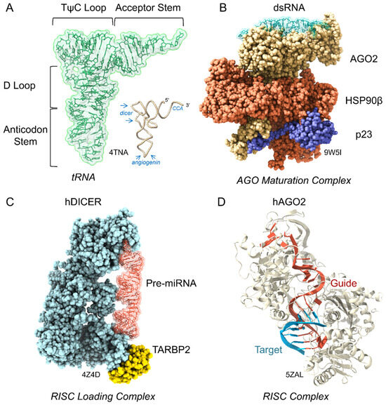
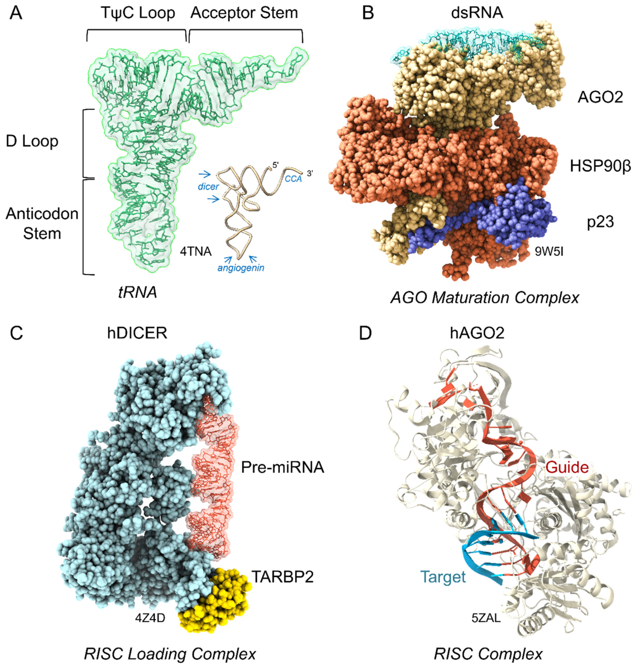
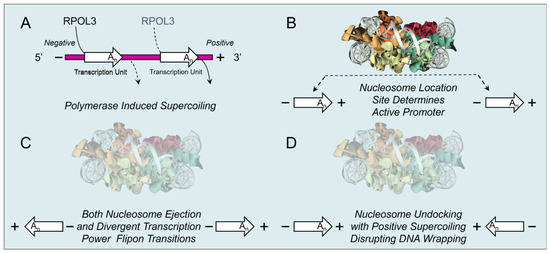
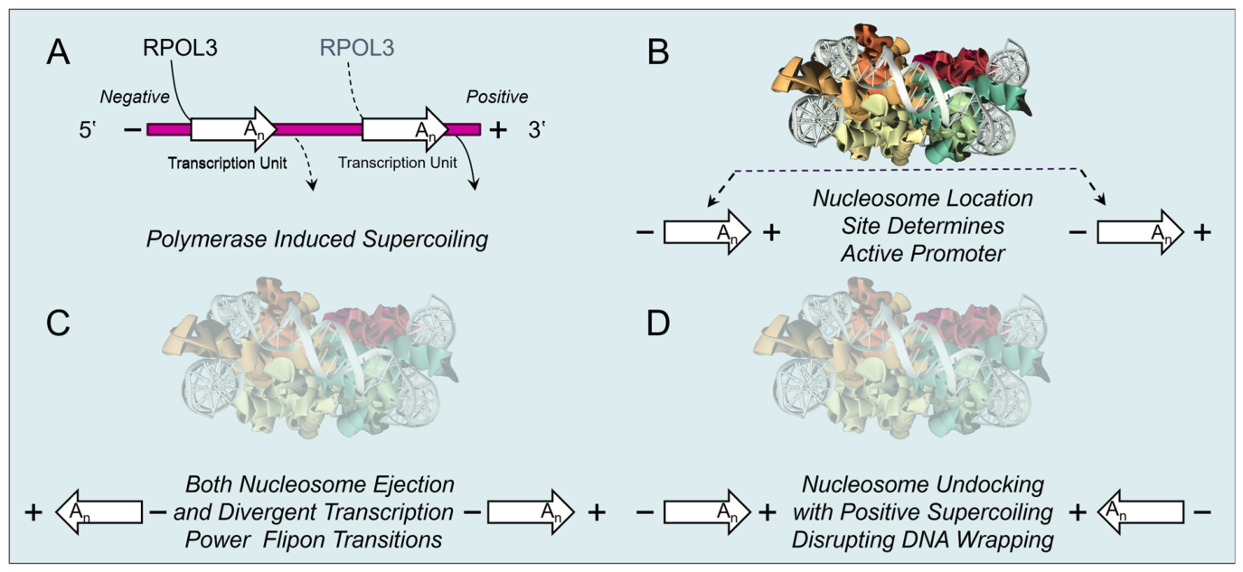
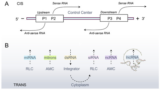
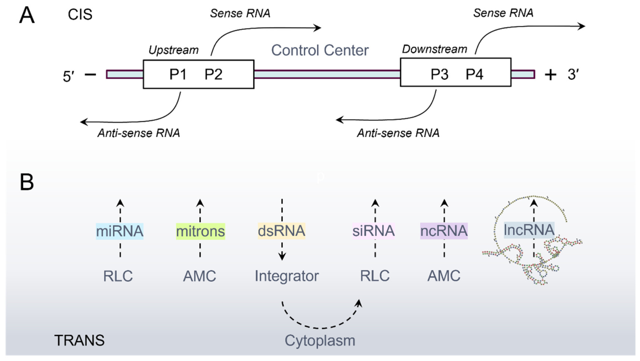
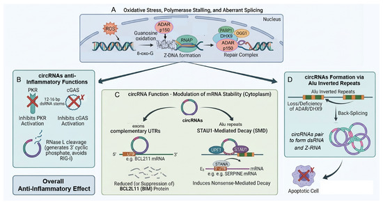
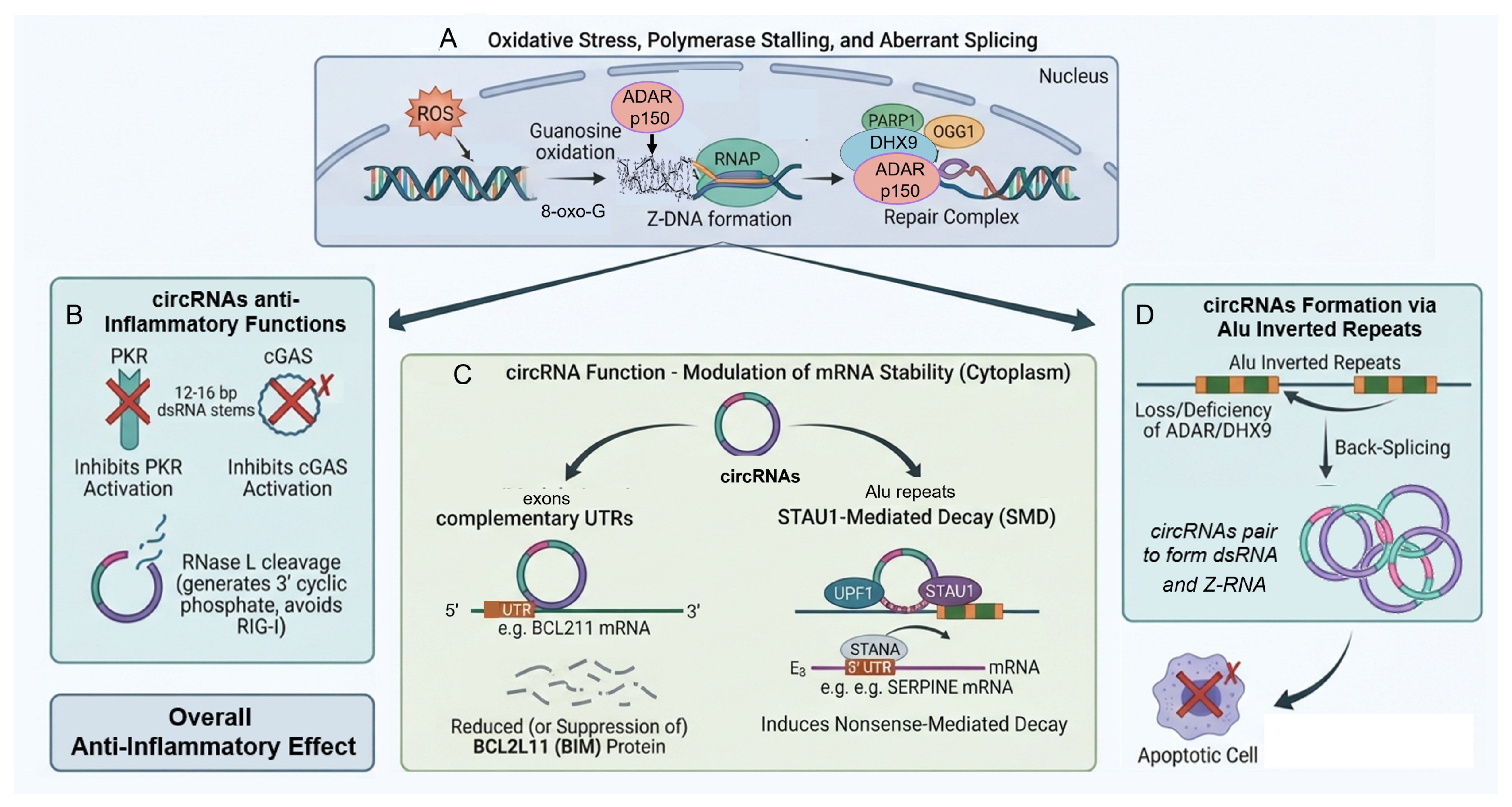
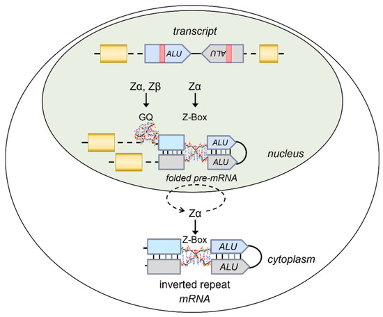
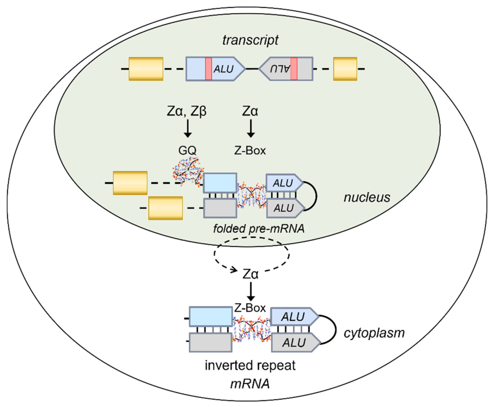

Disclaimer/Publisher’s Note: The statements, opinions and data contained in all publications are solely those of the individual author(s) and contributor(s) and not of MDPI and/or the editor(s). MDPI and/or the editor(s) disclaim responsibility for any injury to people or property resulting from any ideas, methods, instructions or products referred to in the content. |
© 2026 by the author. Licensee MDPI, Basel, Switzerland. This article is an open access article distributed under the terms and conditions of the Creative Commons Attribution (CC BY) license.