


Abstract
Bothrops marajoensis is a Brazilian pit viper whose venom remains poorly characterized compared with those of other medically important Bothrops species. In the present study, we performed a comparative biochemical characterization of B. marajoensis venom with six additional Bothrops venoms (B. alternatus, B. atrox, B. jararaca, B. jararacussu, B. moojeni, and B. neuwiedi). Venoms were analyzed by reversed-phase chromatography, electrophoresis, immunorecognition by commercial bothropic antivenom, phosphodiesterase activity, and their effects on the viability of differentiated C2C12 myotubes. Chromatographic and electrophoretic analyses revealed a largely conserved overall venom composition among species, with qualitative differences, although quantitative differences in protein relative abundance were evident. Bothropic antivenom recognized all venoms, indicating broad antigenic cross-reactivity despite venom variation. B. marajoensis exhibited intermediate phosphodiesterase (PDE) activity relative to other species evaluated, confirming functional variability among Bothrops venoms. In the cell viability assay, B. marajoensis displayed comparatively low apparent cytotoxicity toward differentiated C2C12 myotubes, maintaining higher cell viability than highly myotoxic venoms such as those of B. jararacussu and B. moojeni. These findings expand the biochemical characterization of B. marajoensis venom and demonstrate that, although venoms share the conserved toxin repertoire typical for Bothrops genus, quantitative differences influence its biological activities, preserving broad recognition by bothropic antivenom.
Keywords:
Bothrops marajoensis; snake venom; Bothrops; antivenom; phosphodiesterase; myotoxicity; C2C12 cells 1. Introduction
Snake venoms are complex mixtures of bioactive molecules that have evolved to immobilize prey and deter predators, often causing profound physiological effects on various systems [1,2]. Among the diverse group of venomous snakes, Bothrops marajoensis—a species endemic to Marajó Island in Brazil—has attracted growing scientific interest because of the unique composition and multifaceted biological actions of its venom [1].
Studies have revealed that B. marajoensis venom exhibits a wide range of pharmacological activities, including neurotoxic, myotoxic, cytolytic, cardiotoxic, nephrotoxic, antibacterial, and antiparasitic effects [3,4,5]. A case report of a male child (<10 years old) from Marajó Island, Brazil, shows a severe B. marajoensis envenomation to his right foot, with transport complications that delayed medical care by 25 h, during which the patient developed somnolence and a comatose state. Upon hospital admission, he presented extensive right lower-limb edema, ecchymosis, hematuria, and severe neurological impairment. Cranial computed tomography revealed a right frontal intracerebral hemorrhage, manifesting clinically with right-sided flaccid hemiplegia and leftward labial commissure deviation. Treatment included 12 vials of bothropic antivenom, conservative clinical management, and physical therapy. Following 19 days of hospitalization, the patient was discharged with permanent right flaccid hemiplegia, representing the first documented case of pediatric hemorrhagic stroke caused by B. marajoensis envenomation [6].
Regarding the venom’s biological functions, a study investigated the impact of B. marajoensis venom, focusing on its neurotoxic and myotoxic effects in isolated neuromuscular preparations from chick biventer cervicis (CBC) and mouse phrenic nerve-diaphragm (PND). The venom induces dose-dependent neuromuscular blockade: at lower doses (1 μg/mL in CBC and 5 μg/mL in PND), it inhibits neuromuscular activity without harming muscle tissue; at higher doses (20 μg/mL in PND), it causes both neuromuscular blockade and moderate muscle damage. These findings indicate that B. marajoensis venom can impair neuromuscular function, with blockade often leading to muscle edema [1].
To date, only a limited number of toxins have been isolated from B. marajoensis venom, including a thrombin-like serine protease (TLBm), Lys49 phospholipase A2 (PLA2) homologs, a metalloproteinase, and an L-amino acid oxidase [7,8,9,10]. Examples of Lys49 PLA2 are the BmjeTX-I and BmjeTX-II isoforms from B. marajoensis venom, which were characterized by biochemical and mass spectrometric approaches. Both are basic PLA2 myotoxins highly homologous to those found in other Bothrops species. Ex vivo, they blocked neuromuscular transmission, suggesting a presynaptic mechanism. In mice, both induced myonecrosis, increased systemic IL-6 levels, and caused moderate paw edema, indicating local vascular permeability [8].
In addition to their myotoxic effects, B. marajoensis toxins can interfere with hemostatic processes. The thrombin-like serine protease, TLBm was purified from B. marajoensis venom and characterized as a 33.3 kDa single-chain protein with a conserved catalytic triad. Functionally, TLBm acts as an α,β-fibrinogenase and induces serine protease-dependent platelet aggregation. Despite causing minimal local edema, its procoagulant activity may contribute to systemic hemostatic disturbances during envenomation [10].
Hemostatic alterations are also associated with the snake venom metalloproteinase (SVMP) component of the venom. BmHF-1 is a P-I class SVMP isolated, with 27.2 kDa containing the conserved zinc-binding motif and Met-turn characteristic of metzincins. BmHF-1 preferentially hydrolyzes the Aα-chain of fibrinogen and is inhibited by metal chelators, confirming its SVMP activity. Functionally, it induces weak hemorrhage and dose-dependent local edema, but no myotoxicity, suggesting a predominant role in local tissue inflammation during envenomation [9].
Beyond myotoxic, inflammatory, and hemostatic effects, B. marajoensis venom also contains toxins with potential antimicrobial and antiparasitic activities. BmarLAAO is a 67–72 kDa acidic L-amino acid oxidase (LAAO) purified from B. marajoensis venom. It exhibits dose-dependent antimicrobial activity against Gram-positive and Gram-negative bacteria and potent leishmanicidal effects against Leishmania spp. Notably, BmarLAAO shows low cytotoxicity toward murine macrophages, highlighting its potential as a natural lead for antimicrobial and antiparasitic drug development [11]. This highlights the still fragmentary understanding of its venom composition and its contribution to the observed toxicological and therapeutic properties. Given the emerging evidence of the venom’s diverse bioactivities and the identification of novel toxins with potential pharmacological value, this article aims to provide a comprehensive overview of the biochemical and biological properties of B. marajoensis venom, seeking to elucidate the molecular underpinnings of its toxic effects and to explore prospects for therapeutic development based on its unique constituents.
2. Results
2.1. Venom Fractionation and Comparision
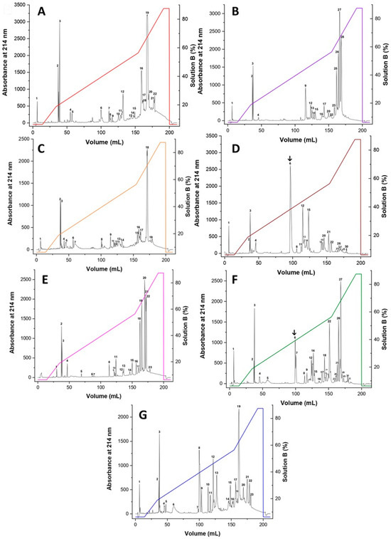
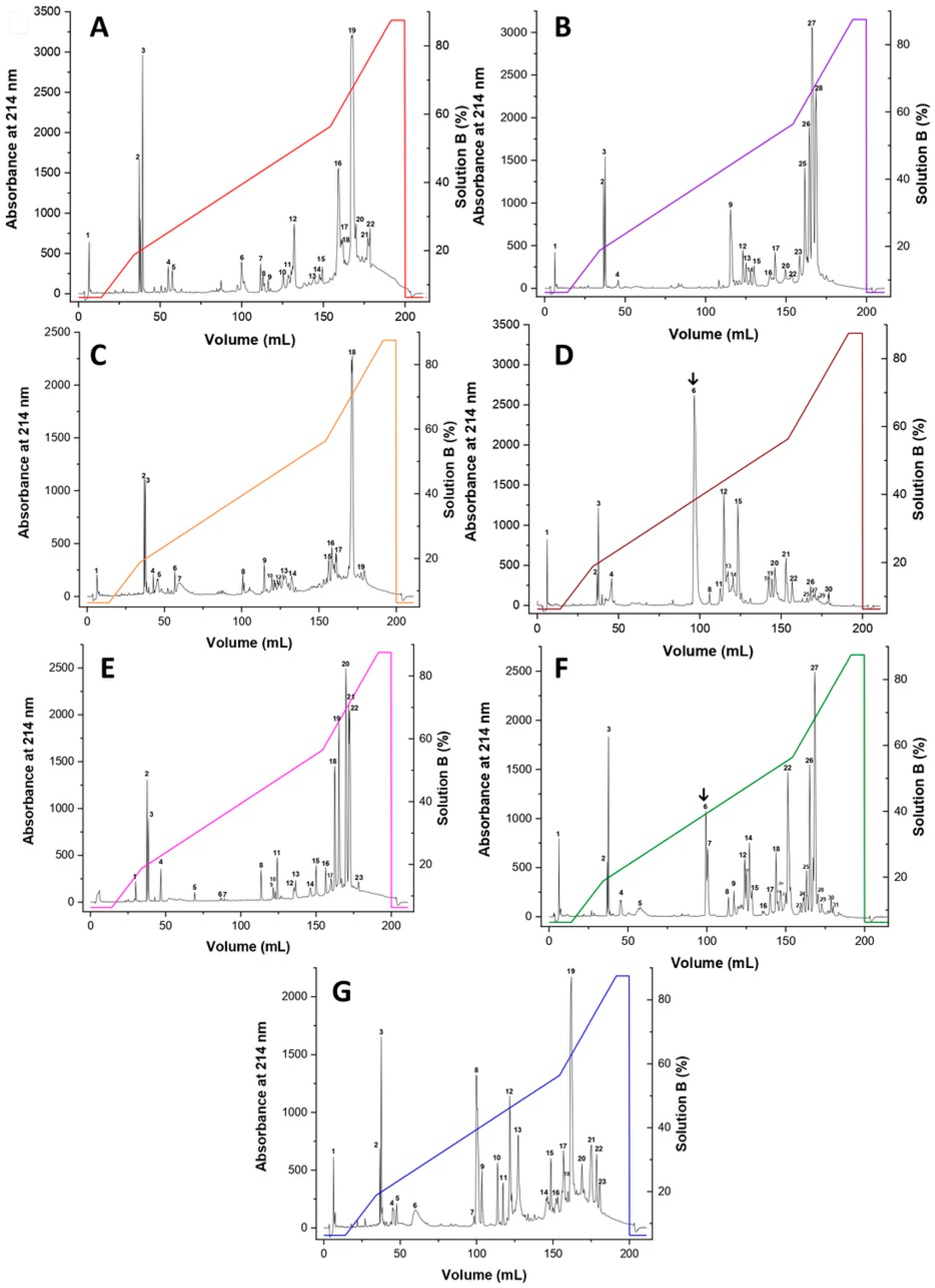
Venom fractionation from seven Bothrops species (B. alternatus, B. atrox, B. jararaca, B. jararacussu, B. marajoensis, B. moojeni, and B. neuwiedi) is presented in Figure 1A–G. All venoms exhibited complex and heterogeneous elution patterns, with multiple peaks distributed across the chromatographic gradient, consistent with the known biochemical complexity of Bothrops spp. venoms. A conserved overall profile was observed across species, with components eluting in early, intermediate, and late elution volumes. Early eluting fractions likely correspond to more hydrophilic and/or low-molecular-weight components, whereas late-eluting peaks suggest the presence of more hydrophobic and larger toxins. However, significant interspecific variation was evident in both peak distribution and relative abundance. For example, B. jararacussu and B. moojeni showed higher peaks in intermediate elution profile. As observed in Figure 1D,F, fraction 6 was more abundant and evident in these species.
Figure 1.
Reversed-phase chromatographic profiles of Bothrops spp. venoms. Crude venoms from Bothrops species were fractionated by reversed-phase liquid chromatography. Profiles correspond to: (A) B. alternatus (red), (B) B. atrox (purple), (C) B. jararaca (orange), (D) B. jararacussu (brown), (E) B. marajoensis (pink), (F) B. moojeni (green), and (G) B. neuwiedi (blue). Elution was monitored at 214 nm, using a C18 analytical column, and a segmented gradient of up to 100% solvent B (0.1% TFA in 80% MeCN), at a flow rate of 1.0 mL/min. The arrows indicate the intermediate peak 6.
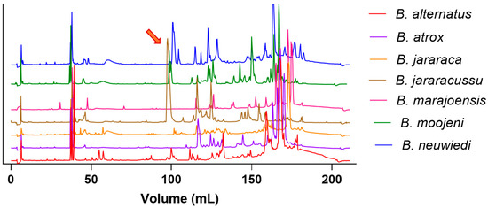
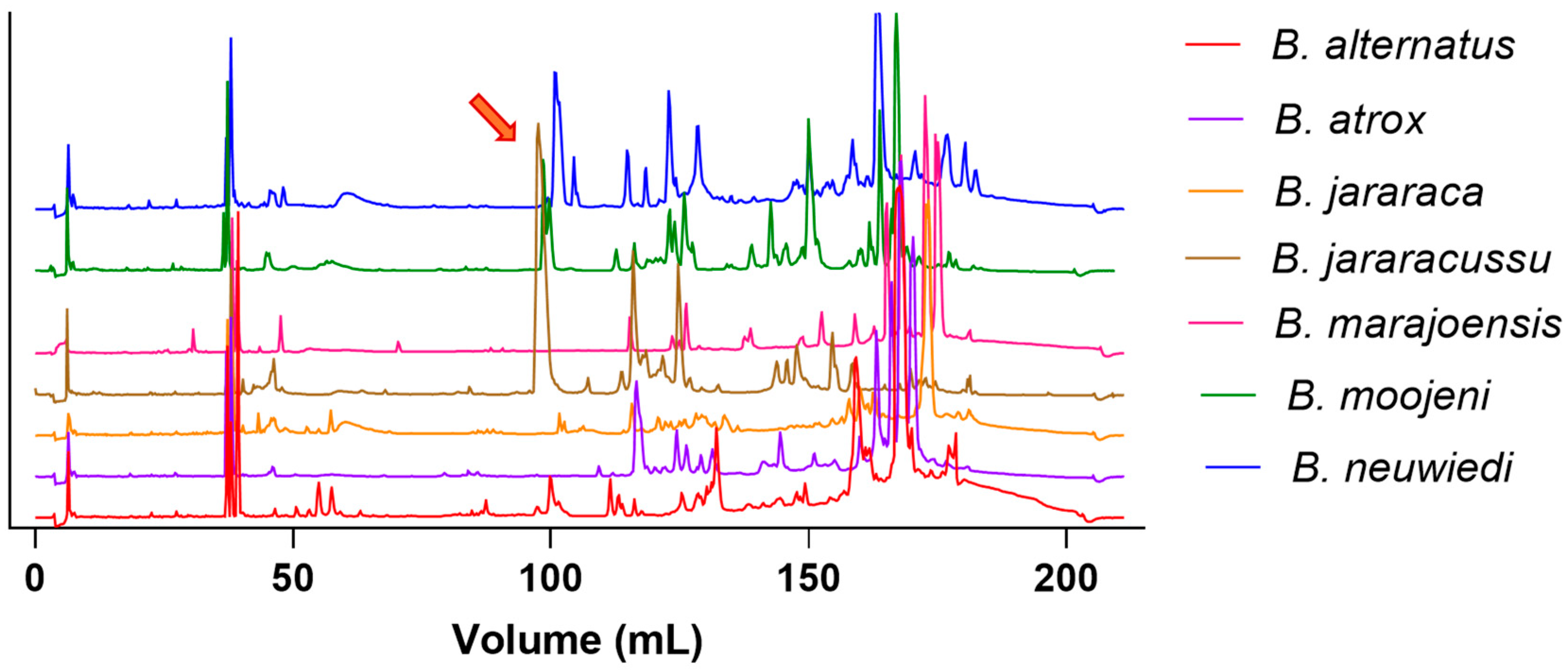
These similarities and differences in venom composition are evident when we overlay the chromatograms in Figure 2. The intermediate peak is even more apparent in the venoms of B. jararacussu, B. moojeni, and B. atrox, although the retention time differs slightly for the latter. A notable feature was observed in B. jararacussu, whose chromatogram showed a dominant peak in the middle region of the profile, in contrast to the more evenly distributed peaks in the other species.
Figure 2.
Comparative chromatographic profiles of crude venoms from seven Bothrops species obtained by reversed-phase chromatography. Chromatograms were normalized and vertically offset for clarity. Distinct elution profiles reflect differences in the molecular weight distribution and relative abundance of venom components among species. Elution volume (mL) is shown on the x-axis. The arrow indicates the predominant intermediate-eluting peak in this region.
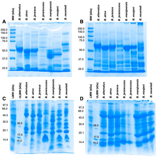
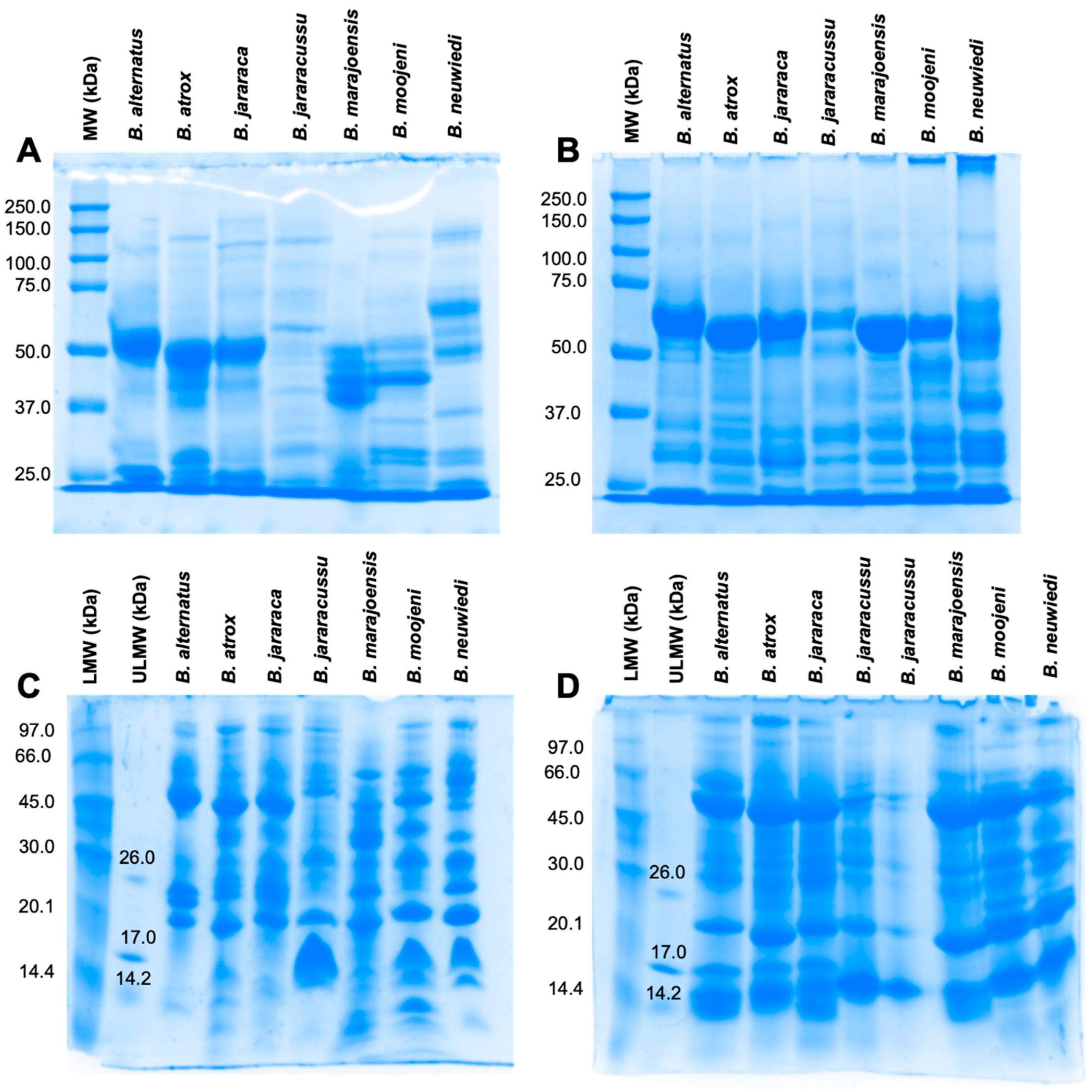
The electrophoretic profiles obtained by 13.5% SDS-PAGE of Bothrops spp. venoms indicate the presence of distinct components with different molecular masses (Figure 3A,B). Under non-reducing conditions, the electrophoretic profile showed a prominent band in the 50–60 kDa range in the venoms of B. alternatus, B. atrox, and B. jararaca, suggesting the presence of SVMPs. However, differences in band intensity among species suggest quantitative variation in SVMPs subclasses rather than the presence of unique proteins among these species. In contrast, the venoms of B. jararacussu, B. neuwiedi, B. moojeni, and B. marajoensis exhibited distinct electrophoretic profiles, differing in both their banding patterns and the distribution of proteins according to molecular weight (Figure 3A). Under reducing conditions, evident changes were observed, particularly for B. moojeni and B. marajoensis, whose banding patterns became more similar under these conditions (Figure 3B).
Figure 3.
Electrophoretic profiles of Bothrops spp. venoms. Venoms (50 μg) were analyzed by (A) Non-reduced 13.5% SDS-PAGE; (B) reduced 13.5% SDS-PAGE; (C) non-reduced 10% Tricine SDS-PAGE; (D) reduced 10% Tricine SDS-PAGE. MW: molecular weight marker (250–10 kDa); LMW: low-molecular-weight marker (97.0–14.4 kDa); ULMW: ultra-low-molecular-weight marker (26.6–1.06 kDa).
Tricine SDS-PAGE provides improved resolution of low-molecular-weight components present in the venom samples. Thus, under non-reducing conditions, a prominent band around 20 kDa was observed in all species (Figure 3C). Nevertheless, B. jararacussu, B. moojeni and B. neuwiedi venoms exhibited more pattern similarity between 14.2–17 kDa range. Thus, it corroborates with the pronounced intermediate peaks presented in chromatographic analysis. This range indicates the existence of PLA2 myotoxins, usually found between the 14–20 kDa. Furthermore, these species showed considerable similarity, particularly due to the prominent bands at approximately 26, and 45 kDa. Conversely, the more evenly distributed chromatographic profiles of B. marajoensis and B. alternatus suggest lower relative PLA2 abundance. In addition, the similarity among the electrophoretic profiles of B. alternatus, B. atrox, and B. jararaca remained evident, reinforcing, above all, the SVMP presence, whereas B. marajoensis exhibited a distinct electrophoretic profile.
Under reducing conditions, the banding patterns showed a high degree of similarity among all species in the molecular weight distribution of the protein bands. Prominent bands were observed at approximately 14.2, 20, 26, and 45 kDa, differing mainly in their staining intensity (Figure 3D). These bands can suggest snake venom serine proteinases (SVSPs), typically in the 25–35 kDa range, with B. jararacussu showing reduced intensity compared to B. atrox. Despite the overall similarity among the venoms, a greater resemblance was observed among B. alternatus, B. atrox, and B. jararaca, as well as between B. marajoensis and B. neuwiedi. These findings corroborate the idea that interspecific variation in the abundance of venom compounds underlies the functional results presented.
2.2. Antivenom Recognition
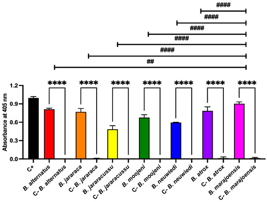
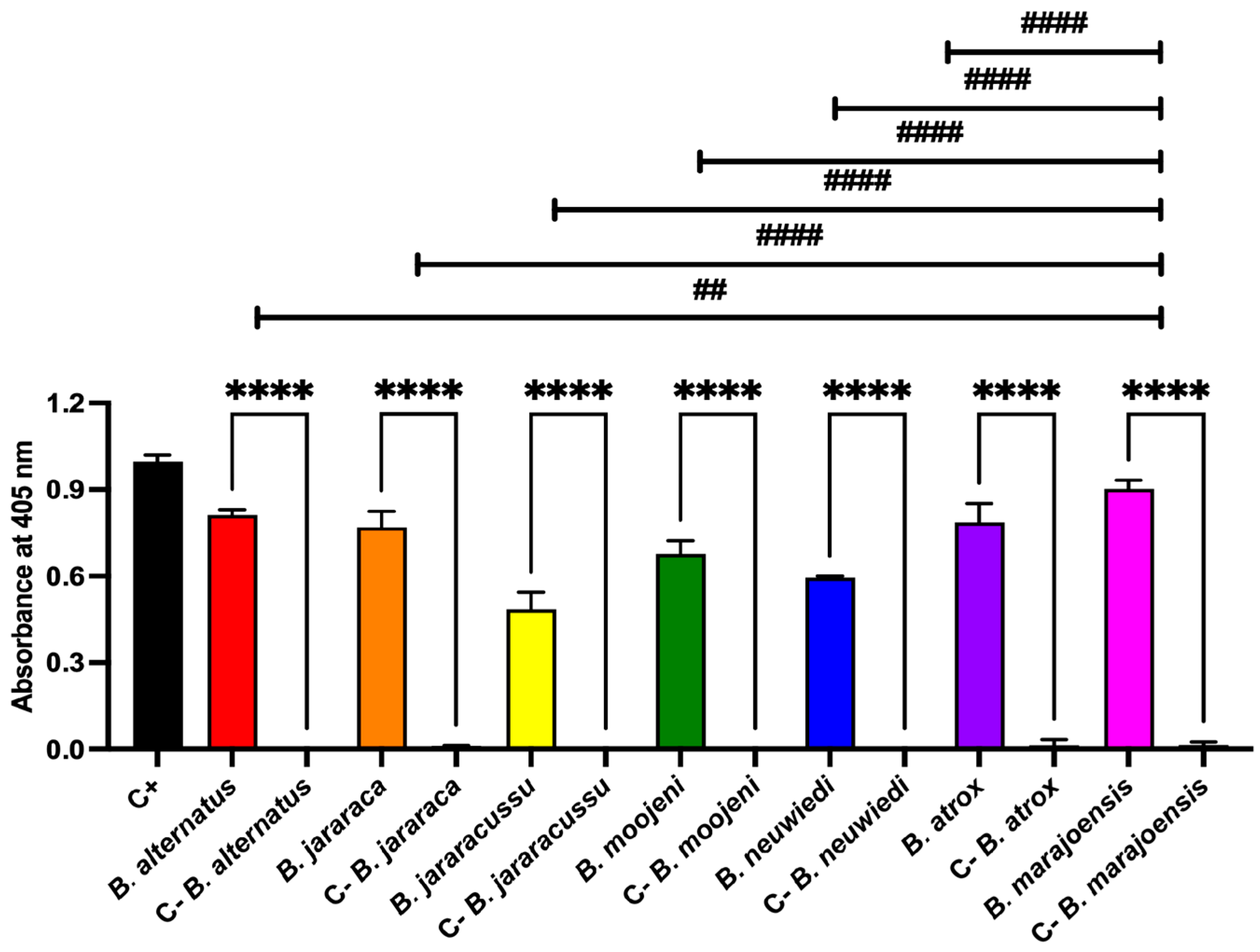
All snake venoms were evaluated for immunorecognition by bothropic antivenom, in order to determine whether the pentavalent antivenom exhibited cross-reactivity toward the tested venoms. All venoms showed significant immunoreactivity against the antivenom, demonstrating broad antigenic recognition (Figure 4). In contrast, non-immune horse serum exhibited no significant binding to the venoms, indicating the absence of nonspecific interactions and confirming the specificity of the observed antibody-mediated recognition.
Figure 4.
Bothrops spp. venoms recognition by bothropic antivenom. Through indirect ELISA, Bothrops spp. venom (2 μg each) were incubated with commercial bothropic antivenom from Instituto Butantan. Absorbance was measured at 405 nm. Data are presented as mean ± SD analyzed by one-way ANOVA and Tukey’s multiple comparison test (n = 4). C+: wells coated with non-immunized horse serum diluted 1:50; C− venom: venoms incubated with non-immunized horse serum diluted 1:100. **** p < 0.0001 when compared to its negative control. ## p < 0.003 and #### p < 0.0001 when compared to B. marajoensis.
Remarkably, B. marajoensis exhibited significantly higher recognition than all other Bothrops spp. venoms tested, suggesting a greater degree of antigenic similarity and/or a higher abundance of shared immunoreactive components among the venoms evaluated.
2.3. Phosphodiesterase Activity
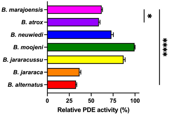
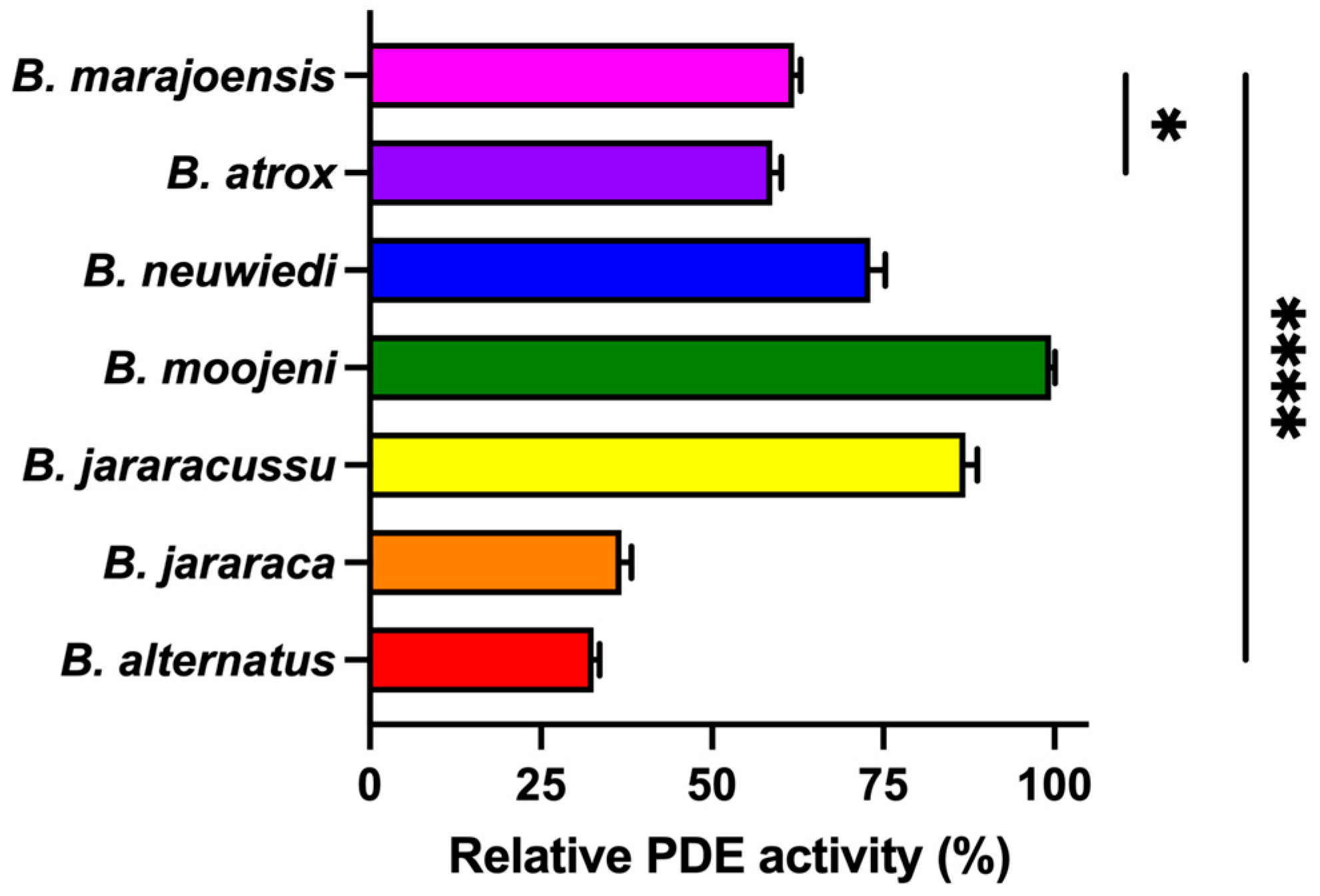
Snake venom phosphodiesterases (PDEs) play important synergistic roles in envenomation by modulating vascular responses and purinergic signaling [12,13]. To investigate this less explored enzymatic component, relative PDE activity was evaluated by the specific substrate use among the analyzed venoms (Figure 5). Notably, B. marajoensis venom exhibited higher PDE activity compared to B. atrox, B. jararaca and B. alternatus venoms. Regarding the other venoms, B. neuwiedi, B. moojeni, and B. jararacussu, B. marajoensis venom showed significantly lower PDE activity. These results indicate differences in venom composition and reveal interspecific biochemical variability among the analyzed venoms.
Figure 5.
PDE activity assay with Bothrops spp. venoms. A mixture (150 μL) of venoms (5 μg) and 0.1 M Tris-HCl, pH 8, was incubated for 10 min at 37 °C. The substrate bis(p-nitrophenyl) phosphate (1 mM) was added, and was incubated for 30 min at 37 °C. The reaction was stopped with 0.05 M sodium hydroxide (NaOH). Absorbance was measured at 400 nm. The enzymatic activity was expressed as a percentage of relative activity to that of highest absorbance. Bars represent the mean ± SD (n = 4). Data were analyzed by one-way ANOVA followed by Dunnett’s post hoc test, using B. marajoensis venom as the control/reference group. * p < 0.02 and **** p < 0.0001 when compared to B. marajoensis snake venom.
2.4. Effects of Bothrops Venoms on the Viability of Differentiated C2C12 Myotubes
Although isolated myotoxins have been identified in B. marajoensis venom, the overall myotoxic effect of the crude venom remains poorly understood. To address this, we evaluated the direct effects of Bothrops venoms on differentiated C2C12 myotubes.
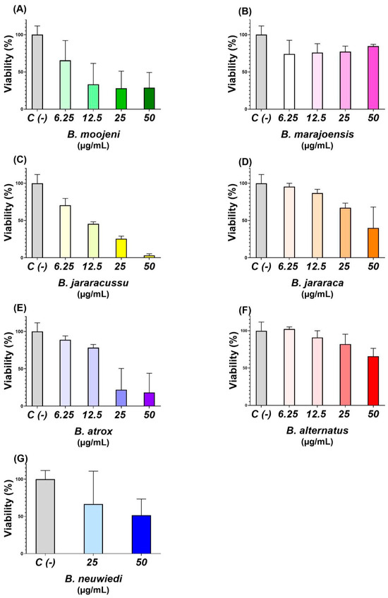
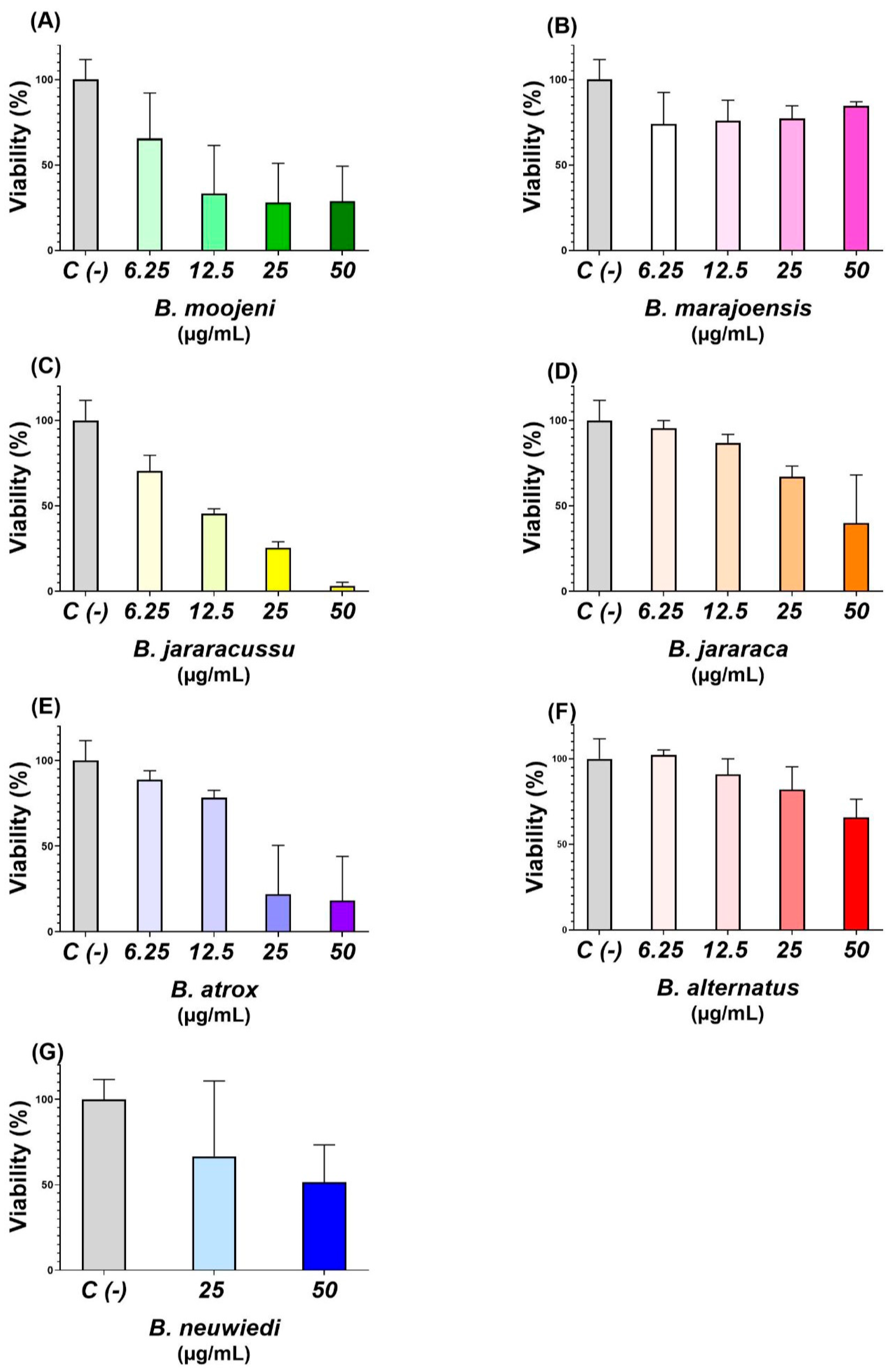
The evaluated Bothrops venoms exhibited distinct effects on the viability of differentiated C2C12 myotubes (Figure 6). The strongest reductions were observed for B. jararacussu and B. moojeni, while B. atrox also produced a marked effect, although with considerable inter-replicate variability. Conversely, B. alternatus and B. marajoensis maintained comparatively higher viability values, and B. jararaca exhibited an intermediate response. Owing to the limited number of concentrations tested, the effect of B. neuwiedi could not be fully defined.
Figure 6.
Effects of Bothrops spp. venoms on the viability of differentiated C2C12 myotubes. Cells were exposed to venoms (6.25–50 µg/mL, 1.25–10 µg/well, in 200 µL) from (A) B. moojeni, (B) B. marajoensis, (C) B. jararacussu, (D) B. jararaca, (E) B. atrox, (F) B. alternatus, and (G) B. neuwiedi. Cell viability was determined using an ATP-dependent luminescence assay and expressed as a percentage relative to the negative control (C (−), normalized to 100%). Bars represent the mean ± standard deviation (SD) (n = 3). Data analysis and graphical representation were performed using GraphPad Prism version 8.0.1 (GraphPad Software, San Diego, CA, USA).
Cell viability was differentially affected by the venom species and the amount applied per well. At the highest concentration, B. jararacussu venom induced the most pronounced reduction in cell viability, reaching 3.107 ± 2.187%, followed by B. atrox and B. moojeni, with viability values of 27.080 ± 29.472% and 40.530 ± 4.271%, respectively. In contrast, cells exposed to B. marajoensis venom maintained the highest viability at this concentration, at 84.603 ± 2.399%.
A progressive recovery of cell viability with decreasing venom amounts was particularly evident for B. jararacussu, increasing from 3.107 ± 2.187% at 50 µg/mL to 70.390 ± 9.212% at 6.25 µg/mL. A similar concentration-related pattern was observed for B. jararaca, for which viability increased from 56.090 ± 3.946% to 95.433 ± 4.425%, and for B. alternatus, which increased from 65.650 ± 10.698% to 102.200 ± 2.971%. The latter value was comparable to that of the negative control, indicating no apparent reduction in cell viability at the lowest tested amount.
For B. atrox, marked reductions in viability were observed at 50 and 25 µg/mL (10 and 5 µg/well); however, these conditions exhibited high standard deviations, indicating substantial variability among replicates. At lower venom amounts, viability increased to 78.173 ± 4.423% and 88.833 ± 5.277% at 12.5 and 6.25 µg/mL (2.5 and 1.25 µg/well), respectively. B. moojeni caused a persistent reduction in viability across the tested range, although partial recovery was observed at 6.25 µg/mL. Conversely, B. marajoensis maintained viability values between approximately 74% and 85%, without a clear concentration-dependent recovery. B. neuwiedi, evaluated only at 12.5 and 6.25 µg/mL (2.5 and 1.25 µg/well), produced viability values of 51.540 ± 21.846% and 66.700 ± 44.100%, respectively, with considerable experimental variability.
The venoms exhibited distinct concentration–response profiles, with the most pronounced reductions in ATP-dependent cell viability observed following exposure to B. jararacussu, B. moojeni, B. atrox, and B. jararaca. In contrast, cell viability remained above 50% throughout the evaluated concentration range for B. alternatus and B. marajoensis. For B. neuwiedi, only the two lowest concentrations were evaluated due to the limited amount of venom available, which prevented testing the complete concentration range used for the other venoms. Consequently, a reliable determination of the half-maximal inhibitory concentration (IC50) was not possible for this venom. For the remaining venoms, IC50 values were estimated from the concentration–response data and expressed in µg/mL, based on a final assay volume of 200 µL per well. The estimated IC50 values are presented in Table 1.
Table 1.
Estimated IC50 values of differentiated C2C12 myotubes after Bothrops venoms exposure.
Overall, the findings indicate that the effects of Bothrops venoms on differentiated C2C12 myotubes were species-dependent and, for some venoms, dose-dependent. Under the present experimental conditions, B. jararacussu and B. moojeni showed the strongest apparent effects on ATP-dependent viability and the lowest estimated IC50 values. These results are compatible with comparatively greater apparent cytotoxic or metabolically inhibitory activity toward differentiated skeletal muscle cells. Although B. atrox also produced substantial reductions in ATP-dependent viability, the high variability and non-monotonic response observed at the two highest concentrations prevent a definitive interpretation of its relative potency. Therefore, its estimated IC50 should be regarded as preliminary and interpreted with particular caution.
In contrast, B. jararaca, B. alternatus, and B. marajoensis did not reduce viability below 50% within the evaluated concentration range. This finding suggests lower apparent potency in this experimental model compared with B. jararacussu and B. moojeni. However, it does not demonstrate the absence of biological effects, as changes in cellular function may occur even when the ATP-dependent signal remains above 50%.
The results obtained for B. neuwiedi remain inconclusive because only two concentrations were tested and the responses exhibited substantial variability. Additional concentrations, particularly above 12.5 µg/mL, would be required to establish whether this venom induces a concentration-dependent reduction in C2C12 myotube viability.
It should also be emphasized that the ATP-based assay primarily reflects cellular metabolic activity. Consequently, a reduction in luminescence may represent a decrease in the number of viable cells, suppression of cellular metabolism, or a combination of both processes. The results should therefore not be interpreted exclusively as direct evidence of cell death. Complementary analyses of membrane integrity, cellular morphology, mitochondrial activity, and cell-death pathways would be necessary to better characterize the mechanisms underlying the observed effects.
The present findings provide preliminary evidence of marked interspecific differences in the effects of Bothrops venoms on differentiated C2C12 myotubes. B. jararacussu and B. moojeni produced the most consistent reductions in ATP-dependent viability and displayed the lowest estimated IC50 values among the venoms evaluated. B. atrox also showed a potentially relevant effect, although its concentration–response profile was affected by substantial experimental variability. In comparison, B. jararaca, B. alternatus, and B. marajoensis exhibited lower apparent potency within the tested concentration range, whereas the available data for B. neuwiedi were insufficient for a reliable assessment.
3. Discussion
Although the venoms of Bothrops species share a common repertoire of toxin families, including SVMPs, PLA2s, SVSPs, LAAOs, C-type lectins (CTLs), and cysteine-rich secretory proteins (CRISPs), substantial interspecific variation exists in the relative abundance of these components. Such compositional differences are considered of Bothrops venoms and have been associated with ecological adaptation, prey specialization, geographic distribution, and evolutionary divergence [14,15]. From a phylogenetic perspective, B. marajoensis belongs to the B. atrox species complex, sharing a close evolutionary lineage with B. atrox, B. moojeni, and B. jararacussu, whereas B. neuwiedi and B. jararaca represent the B. neuwiedi + B. jararaca clade, and B. alternatus represent distinct clade [16]. This evolutionary proximity is reflected in the overall chromatographic and electrophoretic similarities among members of the B. atrox complex observed in this study. However, the distinct quantitative variations and functional differences, such as the lower apparent myotoxicity of B. marajoensis compared to B. moojeni, highlight that venom diversification within Bothrops snakes is shaped not only by phylogenetic relationships but also by local evolutionary pressures, geographic isolation, as B. marajoensis in Marajó Island (Brazil), and quantitative regulation of toxin expression.
In the present study, the chromatographic profiles and SDS-PAGE revealed a high degree of similarity among the analyzed venoms, indicating that these species possess largely conserved venom arsenals. Nevertheless, distinct differences in peak intensities were observed, particularly in the central region of the chromatograms. Notably, B. jararacussu exhibited a marked increase in the intensity of components eluting in the intermediate portion of the profile, suggesting a higher abundance of specific toxin classes rather than the presence of exclusive proteins. This observation is consistent with previous proteomic studies demonstrating that venom diversification within the genus is driven primarily by quantitative shifts in toxin expression rather than by the emergence or disappearance of toxin families [14,15].
B. jararacussu represents an interesting example of quantitative venom specialization. Previous studies demonstrated that adult specimens express substantially higher levels of Lys49 PLA2 myotoxins, whereas juvenile venoms contain proportionally higher levels of proteolytic enzymes. This ontogenetic shift highlights the remarkable plasticity of venom composition within a single species and reinforces that differences in chromatographic peak intensity can reflect biologically meaningful changes in toxin abundance. The higher intensity observed in the intermediate chromatographic region of B. jararacussu in the present study is consistent with its well-known enrichment in PLA2 myotoxins, which contribute significantly to the pronounced myotoxic activity characteristic of this species [15].
The present study demonstrated that B. marajoensis venom shares the conserved biochemical architecture characteristic of the genus while exhibiting differences in protein composition and enzymatic activity. These findings are consistent with previous venomic studies showing that Bothrops venoms are predominantly composed of conserved toxin families, including SVMPs, PLA2s, and SVSPs, whose relative abundance varies among species and contributes to functional diversification rather than the emergence of novel toxin classes [17,18,19].
A major finding of the present study was the broad immunoreactivity of the commercial bothropic antivenom against all analyzed Bothrops venoms. Remarkably, B. marajoensis venom exhibited the highest recognition by the commercial antivenom, despite not being included in the pentavalent immunization mixture (which consists of B. alternatus, B. jararaca, B. jararacussu, B. moojeni and B. neuwiedi) [20]. Despite the biochemical differences revealed by chromatographic and electrophoretic analyses, ELISA demonstrated extensive antigenic recognition, indicating that the major immunogenic epitopes are highly conserved among these species, suggesting also that B. marajoensis shares highly conserved immunodominant epitopes with the species used for antivenom production. Furthermore, quantitative variations in venom composition, such as a higher relative proportion of major immunogens like SVMPs and PLA2s, may result in greater overall antibody binding capacity per microplate well during indirect ELISA. This observation agrees with previous antivenomic studies showing that SVMPs, SVSPs, and PLA2s are the principal immunogens during horse immunization and are largely responsible for the extensive cross-reactivity exhibited by Brazilian bothropic antivenoms [18,19]. The predominance of these conserved toxin families in B. marajoensis venom likely explains its efficient recognition by the commercial antivenom, despite quantitative variation among species [18]. Although ELISA does not directly predict toxin neutralization, broad antigen recognition represents an essential prerequisite for antivenom efficacy. Therefore, complementary antivenomic approaches combined with in vivo neutralization assays would further clarify the effectiveness of the commercial bothropic antivenom against B. marajoensis venom, correlating with functional protection against lethality and local tissue damage.
Another relevant finding was the marked interspecific variation in PDE activity. B. marajoensis exhibited intermediate PDE activity compared with the other Bothrops species analyzed, indicating that enzymatic activity does not necessarily correlate with the overall chromatographic or electrophoretic similarity among venoms. Although PDEs are minor venom components, they have been associated with nucleotide hydrolysis, adenosine generation, platelet dysfunction, vascular alterations, and modulation of inflammatory responses, acting synergistically with other toxin families during envenoming [12,13]. These findings support the concept that venom diversification involves not only variation in major toxin families but also modulation of accessory enzymes that contribute to the functional diversity of Bothrops venoms.
One of the most relevant functional findings of the present study was the marked interspecific variability in the effects of Bothrops venoms on differentiated C2C12 myotubes. Skeletal muscle represents one of the primary targets of local tissue injury following Bothrops envenoming, and differentiated C2C12 myotubes constitute a well-established in vitro model for investigating the direct effects of venom toxins on skeletal muscle because they reproduce several structural and metabolic characteristics of mature muscle fibers [21]. Under the experimental conditions employed, B. jararacussu and B. moojeni induced the greatest reductions in ATP-dependent viability, whereas B. marajoensis exhibited a lower impact on myotube viability, even at the highest venom concentration tested. These findings demonstrate that, although Bothrops venoms share the same major toxin families, they differ considerably in their biological activities, emphasizing that quantitative and qualitative variations in venom composition are critical determinants of toxicity [22].
The significant cytotoxicity seen in B. jararacussu aligns with its known high levels of myotoxic phospholipases A2, especially Lys49 PLA2 homologues, recognized as some of the most potent myotoxins in Bothrops venoms [23]. These Lys49 homologues, unlike catalytically active Asp49 PLA2s, cause toxicity via a mechanism that does not rely on catalysis. They disrupt plasma membrane integrity, leading to swift calcium influx, mitochondrial failure, ATP depletion, oxidative stress, and ultimately irreversible myonecrosis [21,23]. Likewise, the marked cytotoxicity induced by B. moojeni probably reflects the combined action of abundant PLA2 myotoxins together with metalloproteinases, which contribute to extracellular matrix degradation and potentiate muscle damage by facilitating toxin diffusion and amplifying local tissue injury [24].
In contrast, B. marajoensis exhibited comparatively low cytotoxicity, maintaining approximately 75–85% cell viability throughout the concentration range evaluated. Although previous studies have identified myotoxic Lys49 PLA2 homologues such as BmjeTX-I, BmjeTX-II and BmajPLA2-II in this venom [5]. The present findings suggest that differences in the relative abundance of these toxins, their intrinsic toxic potency, or synergistic interactions with other venom components may contribute to the reduced myotoxic profile observed in comparison with B. jararacussu and B. moojeni. Interestingly, this functional behavior is compatible with the RP-FPLC chromatographic profiles obtained in the present study, which revealed distinct distributions of venom components among the analyzed species. Together, these observations reinforce the concept that biological activity depends not only on the presence of particular toxin families but also on their relative abundance and interactions within the venom proteome [22].
It is important to note that the CellTiter-Glo assay detects intracellular ATP, indicating cellular metabolic activity rather than immediate cell death. Therefore, reduced luminescence could be due to mitochondrial issues, membrane damage, metabolic suppression, or activated cell death pathways. Conversely, maintained ATP levels do not always mean the cell is unharmed, as sublethal injury might still be present [25]. The cell viability assays demonstrate that the myotoxic potential of Bothrops venoms varies substantially among species despite the conservation of their principal toxin families. The comparatively low cytotoxicity exhibited by B. marajoensis highlights that relatively subtle differences in venom composition are sufficient to generate distinct biological responses. These findings complement the biochemical characterization presented herein and reinforce the importance of integrating proteomic and functional approaches to improve our understanding of venom variability and its pathological consequences, particularly for medically relevant but comparatively understudied species such as B. marajoensis.
Overall, this study expands the biochemical characterization of the poorly investigated B. marajoensis venom by integrating chromatographic, electrophoretic, immunological, and enzymatic analyses. Despite quantitative differences in toxin abundance and PDE activity, the conservation of major toxin families preserved broad antigenic recognition by the commercial bothropic antivenom, supporting its potential clinical applicability. Future LC-MS/MS-based proteomic studies associated with functional neutralization assays will be important to identify toxin isoforms responsible for the observed biochemical differences and to improve our understanding of venom evolution, antivenom efficacy, and the biotechnological potential of B. marajoensis venom.
4. Materials and Methods
4.1. Venoms
Venom from seven adults Bothrops species from captive bred, B. alternatus, B. atrox, B. jararaca, B. jararacussu, B. marajoensis, B. moojeni, and B. neuwiedi, were obtained from the Serpentarium of the University of São Paulo at Ribeirão Preto Medical School, which is accredited by the Instituto Brasileiro do Meio Ambiente e dos Recursos Naturais Renováveis (Brazilian Institute of Environment and Renewable Natural Resources), under the registration number 1506748. This research project is registered at the Sistema Nacional de Gestão do Patrimônio Genético e do Conhecimento Tradicional Associado (SISGEN, National System of Genetic Heritage and Associated Traditional Knowledge, No. A4A9FDD). Venoms were dried under vacuum at room temperature for 6 h and stored at −20 °C until their use.
4.2. Venom Fractionation
Dried venom (3 mg) was dissolved in 2 mL of deionized water and centrifuged at 13,000 rpm for 10 min, at 4 °C. The supernatant was then collected, and trifluoroacetic acid (TFA) and acetonitrile (MeCN) were added to achieve final concentrations of 0.1% (v/v) and 5% (v/v), respectively. Purification was performed by reversed-phase protein liquid chromatography (RP-HPLC), using a C18 analytical column (250 × 4.6 mm, 5 μm, Phenomenex, Torrance, CA, USA), in an ÄKTA Purifier UPC10 system (GE Healthcare, Uppsala, Sweden). The samples were fractionated according to the method described by Calvete et al. (2007) [17]. The column was pre-equilibrated with solution A (0.1% TFA), and the proteins were eluted with a segmented gradient up to 100% solvent B (0.1% TFA in 80% ACN), at a flow rate of 1.0 mL/min. Fraction elution was monitored at 214 nm.
4.3. Electrophoresis
Bothrops spp. species venom (50 μg) was subjected to 13.5% SDS-PAGE and to 10% Tricine-SDS-PAGE, according to the method described by Laemmli (1970) [26] and Schägger (2006) [27], respectively, both under reducing and non-reducing conditions. As markers, Full Range Molecular Weight (250–10 kDa, 161-0374, BioRad, Hercules, CA, USA), Low Molecular Weight (97.0–14.4 kDa, 17-0446-01, GE Healthcare, Uppsala, Sweden) and Ultra-Low Molecular Weight (26.6–1.06 kDa, M3546-1VL, Sigma-Aldrich, Saint Louis, MO, USA) were used. All gels were stained with Coomassie® Brilliant Blue G-250 (Sigma-Aldrich, St. Louis, MO, USA).
4.4. ELISA
Indirect enzyme-linked immunosorbent assay (ELISA) was performed according to Oliveira et al. (2018) [28]. 96-well microplate (Costar, Corning Incorporated, Corning, NY, USA) was coated with Bothrops spp. venoms (2 μg each) in 0.05 M carbonate/bicarbonate buffer, pH 9.6 (100 μL/well). Non-immunized horse serum was coated and used as positive control (1:50, H0146, lot SLBS7574, Sigma-Aldrich, St. Louis, MO, USA). The plate was incubated for 16 h at 4 °C, then washed three times with phosphate-buffered saline (PBS), pH 7.2, and blocked by adding 250 μL of PBS containing 2% (w/v) non-fat dry milk (ref. 1706404, BioRad, Lagoa Santa, Brazil; MPBS) and incubated for 2 h at 37 °C. The plate was then washed three times with PBS-0.05% Tween (PBS-T) and three times with PBS and incubated for 1 h at 37 °C with commercial bothropic antivenom (1:100 in 1% MPBS, batch 1305077, Instituto Butantan, São Paulo, Brazil). As a negative control, venoms were incubated with non-immunized horse serum diluted 1:100 in 1% MPBS. The plate was washed three times with PBS-T and PBS, respectively. After that, the plate was incubated with 100 μL of anti-horse polyclonal antibodies conjugated with peroxidase (IgG-HRP, A6917, Sigma-Aldrich, St. Louis, MO, USA) diluted 1:10,000 in 1% MPBS. After 1 h of incubation at 37 °C, the plate was washed three times with PBS-T and three times with PBS. To each well, 100 μL of 3,3′,5,5′-Tetramethylbenzidine (TMB, ref. T0440, Sigma-Aldrich, St. Louis, MO, USA), and incubated in a dark chamber at RT until color development. The reaction was stopped with 50 μL of 1 M H2SO4 (Merck, São Paulo, Brazil). Absorbance was measured at 405 nm on a 96-well plate reader (Sunrise-basic Tecan, Männedorf, Switzerland). The assay was performed in quadruplicates (n = 4).
4.5. PDE Activity
Phosphodiesterase (PDE) activity was determined in Bothrops spp. venoms according to Björk (1963) [29], modified by Valério et al. (2002) [30], in a 96-well microplate. Bothrops spp. venoms (5 μg/well) were incubated in 0.1 M Tris–HCl buffer, pH 8.0, 10 min at 37 °C. A solution containing 1 mM bis(p-nitrophenyl) phosphate (N3002, Sigma-Aldrich, St. Louis, MO, USA) substrate was added, and the plate was incubated for 30 min at 37 °C. The reaction was stopped with 0.05 M NaOH, and absorbance readings were performed in a microplate reader (Sunrise-basic, Tecan, Männedorf, Switzerland) at 400 nm. The highest absorbance was considered 100% of activity. The assay was carried out in quadruplicate (n = 4).
4.6. C2C12 Cell Culture and Myogenic Differentiation
Murine C2C12 myoblasts (ATCC CRL-1772), kindly provided by A.M.S. from Instituto Butantan (São Paulo, Brazil), were received cryopreserved and subsequently thawed and expanded under aseptic conditions. The cells used in the experiments were cultured for one passage after thawing. Since the cells were provided by another laboratory, their complete previous passage history was not available.
Cells were maintained in cell culture flasks in modified RPMI 1640 medium (Nova Biotecnologia Ltd.a., São Paulo, Brazil; BR30260-05) containing L-glutamine (2.05 mM), HEPES (25 mM), β-mercaptoethanol (50 µM), sodium bicarbonate (2.0 g/L), and 1% (v/v) penicillin/streptomycin, corresponding to final concentrations of approximately 100 U/mL penicillin and 100 µg/mL streptomycin. The medium was supplemented with 10% (v/v) fetal bovine serum (FBS; Nova Biotecnologia Ltd.a., Brazil), which was heat-inactivated in the laboratory before use. Cells were maintained at 37 °C in a humidified atmosphere containing 5% CO2 in a Panasonic MCO-19AIC-PA CO2 incubator (Panasonic, Mississauga, ON, Canada). Cell morphology and growth were routinely monitored by phase-contrast microscopy.
When cultures reached approximately 80% confluence, the culture medium was removed and the adherent cell monolayer was exposed to 5 mL of 0.25% trypsin-EDTA, 1× (Nova Biotecnologia Ltd.a., Brazil), for approximately 5 min or until complete cell detachment was observed. Trypsin activity was then neutralized by adding 10 mL of modified RPMI 1640 medium supplemented with 10% FBS. The resulting cell suspension was transferred to a centrifuge tube and centrifuged for 10 min at 2500 rpm using an Eppendorf Centrifuge 5810 R equipped with an A-4-44 rotor (Eppendorf, Hamburg, Germany). The supernatant was discarded, and the resulting cell pellet was resuspended in 1 mL of complete culture medium.
Cell concentration and viability were determined using a Neubauer counting chamber after staining with 0.4% Trypan Blue solution (Bio-Rad Laboratories, Hercules, CA, USA). Cells were then seeded into cell culture-treated 96-well plates at a density of 5 × 10⁴ cells/well and maintained under the same incubation conditions. Approximately three days after seeding, the cultures reached ~80% confluence.
Myogenic differentiation was initiated by replacing the growth medium with differentiation medium consisting of the same modified RPMI 1640 formulation containing L-glutamine (2.05 mM), HEPES (25 mM), β-mercaptoethanol (50 µM), sodium bicarbonate (2.0 g/L), and penicillin/streptomycin, but without FBS and supplemented instead with 2% (v/v) horse serum (Nova Biotecnologia Ltd.a., Brazil). Horse serum was also heat-inactivated in the laboratory before use. Cells were maintained under differentiation conditions for 72 h, during which progressive cell alignment, myoblast fusion, and the formation of elongated multinucleated myotubes were observed, confirming the establishment of a differentiated skeletal muscle cell model.
4.6.1. Treatment with Bothrops Venoms
After the differentiation period, C2C12-derived myotubes were exposed to crude venoms from all seven Bothrops species. Before treatment, the culture medium was completely removed from all wells, including the negative control wells, and replaced with fresh medium. Venom stock solutions were individually diluted immediately before use in the same modified RPMI 1640 medium used during differentiation, supplemented with 2% (v/v) horse serum. For six of the venoms, final concentrations of 6.25, 12.5, 25, and 50 µg/mL were tested in a final volume of 200 µL per well. For B. neuwiedi, only the two lowest concentrations (6.25 and 12.5 µg/mL) were evaluated due to the limited amount of venom available. The assay was performed in triplicate (n = 3). Negative control myotubes underwent the same complete medium replacement procedure and received 200 µL of fresh venom-free culture medium. Differentiated myotubes were exposed to the respective venom treatments for 24 h at 37 °C in a humidified atmosphere containing 5% CO2. At the end of the exposure period, cell viability was assessed using an ATP-based luminescence assay.
4.6.2. Cell Viability Assay
Cell viability was determined using the CellTiter-Glo® 2.0 Luminescent Cell Viability Assay (Promega, Madison, WI, USA), following the manufacturer’s recommendations. The assay is based on the quantification of intracellular adenosine triphosphate (ATP), which serves as an indicator of metabolically active and viable cells.
During the 24 h venom exposure period, cells were maintained in 200 µL of modified RPMI 1640 medium supplemented with 2% (v/v) horse serum. At the end of the exposure period, 150 µL of culture medium was carefully removed from each well, leaving 50 µL of medium containing the respective venom treatment. Subsequently, 50 µL of CellTiter-Glo® 2.0 reagent was added directly to each well of the original cell culture plate, resulting in a 1:1 ratio of assay reagent to the remaining culture medium and a final assay volume of 100 µL per well.
After reagent addition, the original culture plate was gently agitated manually by lightly tapping the sides of the plate to promote cell lysis, ATP release, and homogenization of the reaction mixture. The entire 100 µL content of each well was then transferred to a white 96-well plate (Thermo Scientific, Waltham, MA, USA) for luminescence measurement. Following signal stabilization, luminescence was measured using a GloMax® Explorer Multimode Microplate Reader (Promega, Madison, WI, USA). Signal acquisition was performed using the pre-programmed CellTiter-Glo/Luminescence protocol available on the instrument and corresponding to the CellTiter-Glo® assay. Cell viability was expressed as a percentage relative to the negative control, which was considered to represent 100% viability.
4.7. Statistical Analyses
Quantitative results are expressed as mean ± standard deviation (SD) values. For statistical comparisons involving three or more groups, one-way analysis of variance (ANOVA) was performed, followed by Tukey’s post hoc test for multiple pairwise comparisons or Dunnett’s test when comparing treatments against a control group. A value of p < 0.05 was considered statistically significant. All analyses were conducted using GraphPad Prism software (version 10.0, GraphPad Software, San Diego, CA, USA).
5. Conclusions
This study provides an expanded characterization of B. marajoensis venom through comparative analyses with six venoms from Bothrops species. Although they shared a conserved biochemical composition, chromatographic and electrophoretic analyses revealed quantitative differences in protein venoms, reflecting distinct enzymatic and biological profiles. Despite these biochemical differences, the antivenom recognized all venoms, supporting the conservation of major antigenic determinants across the genus. Together, these findings contribute to the understanding of venom variability in B. genus, reinforce the potential clinical applicability of the bothropic antivenom against B. marajoensis, and provide a framework for future proteomic studies, toxin isolation, and functional investigations aimed at identifying the molecular determinants responsible for the distinct biological activities of this poorly explored venom.
Author Contributions
Conceptualization, I.O. and E.A.; methodology, I.O., I.F., B.J., E.R., L.A., I.L., F.C. (Francielle Cordeiro), F.C. (Felipe Costa), E.F., M.P. and E.A.; investigation, I.O., I.F., B.J., E.R., L.A., I.L., F.C. (Francielle Cordeiro), F.C. (Felipe Costa) and E.F.; data curation, I.O.; writing—original draft preparation, I.O., I.F., B.J., E.R., L.A., I.L., F.C. (Francielle Cordeiro), F.C. (Felipe Costa) and E.F.; writing—review and editing, I.O., I.F., M.P. and E.A.; supervision, M.P. and E.A.; project administration, I.O.; funding acquisition, M.P. and E.A. All authors have read and agreed to the published version of the manuscript.
Funding
This research was funded by Fundação de Amparo à Pesquisa do Estado de São Paulo (FAPESP, São Paulo Research Foundation, scholarships to I.O. No. 2020/13176-3, I.F. No. 2023/11264-0, and No. 2024/14618-0, B.J. No. 2024/17849-3, F. Costa No. 2026/01115-6, I.L. No. 2025/05917-7, E.F. No. 2024/13258-0, and F.C. 2023/11311-9, grants to E.A. No. 2019/10173-6 and No. 2021/11936-3), Conselho Nacional de Desenvolvimento Científico e Tecnológico (CNPq, The National Council for Scientific and Technological Development, scholarship to M.P. No. 305778/2023-4 and E.A. No. 309399/2021-1), and Programa de Excelência Acadêmica da Coordenação de Aperfeiçoamento de Pessoal de Nível Superior (PROEX/CAPES, Program of Academic Excellence of Coordination for the Improvement of Higher Education Personnel, scholarships to E.R. No. 88887.245405/2026-00 and L.A. No. 88887.245289/2026-00).
Institutional Review Board Statement
Ethical review and approval were not required for this study because only previously collected venom samples were used, and no experiments, procedures, or interventions involving live animals were performed by the authors. Venom samples were obtained from the Serpentarium of the University of São Paulo at Ribeirão Preto Medical School, which is accredited by the Instituto Brasileiro do Meio Ambiente e dos Recursos Naturais Renováveis (IBAMA; Brazilian Institute of Environment and Renewable Natural Resources) under registration number 1506748. This research project is also registered at the Sistema Nacional de Gestão do Patrimônio Genético e do Conhecimento Tradicional Associado (SISGEN, National System of Genetic Heritage and Associated Traditional Knowledge, No. A4A9FDD).
Informed Consent Statement
Not applicable.
Data Availability Statement
The original contributions presented in this study are included in the article. Further inquiries can be directed to the corresponding author.
Conflicts of Interest
The authors declare no conflicts of interest. The funders had no role in the design of this study; in the collection, analyses, or interpretation of data; in the writing of the manuscript; or in the decision to publish the results.
Abbreviations
| ANOVA | Analysis of variance |
| CBC | Chick biventer cervicis |
| CRISPs | Cysteine-rich secretory proteins |
| CTLs | C-type lectins |
| ELISA | Enzyme-linked immunosorbent assay |
| IC50 | Half-maximal inhibitory concentration |
| LAAOs | L-amino acid oxidases |
| LC-MS/MS | Liquid Chromatography tandem Mass Spectrometry |
| LMW | Low Molecular Weight |
| MeCN | Acetonitrile |
| NaOH | Sodium hydroxide |
| PDE | Phosphodiesterase |
| PLA2 | Phospholipases A2 |
| PND | Phrenic nerve-diaphragm |
| RP-FPLC | Reversed-phase protein liquid chromatography |
| SD | Standard deviation |
| SDS-PAGE | Sodium Dodecyl Sulfate–Polyacrylamide Gel Electrophoresis |
| SVMPs | Snake venom metalloproteinases |
| SVSPs | Snake venom serine proteinases |
| TFA | Trifluoroacetic acid |
| TLBm | Thrombin-like serine protease from Bothrops marajoensis |
| ULMW | Ultra-Low Molecular Weight |
References
- Cavalcante, W.L.; Hernandez-Oliveira, S.; Galbiatti, C.; Randazzo-Moura, P.; Rocha, T.; Ponce-Soto, L.; Marangoni, S.; Pai-Silva, M.D.; Gallacci, M.; da Cruz-Höfling, M.A.; et al. Biological Characterization of Bothrops marajoensis Snake Venom. J. Venom. Res. 2011, 2, 37–41. [Google Scholar] [PubMed]
- Ministério da Saúde do Brasil. Manual de Diagnóstico e Tratamento de Acidentes Por Animais Peçonhentos; Fundação Nacional de Saúde: Brasília, Brazil, 2001. [Google Scholar]
- Evangelista, I.L.; Costa Martins, A.M.; Falcão Nascimento, N.R.; Havt, A.; Azul Monteiro Evangelista, J.S.; Sá De Norões, T.B.; Toyama, M.H.; Diz-Filho, E.B.; De Oliveira Toyama, D.; Fonteles, M.C.; et al. Renal and Cardiovascular Effects of Bothrops marajoensis Venom and Phospholipase A2. Toxicon 2010, 55, 1061–1070. [Google Scholar] [CrossRef] [Scilit] [PubMed]
- Grabner, A.N.; Alfonso, J.; Kayano, A.M.; Moreira-Dill, L.S.; Dos Santos, A.P.D.A.; Caldeira, C.A.S.; Sobrinho, J.C.; Gómez, A.; Grabner, F.P.; Cardoso, F.F.; et al. BmajPLA 2 -II, a Basic Lys49-Phospholipase A 2 Homologue from Bothrops marajoensis Snake Venom with Parasiticidal Potential. Int. J. Biol. Macromol. 2017, 102, 571–581. [Google Scholar] [CrossRef] [Scilit] [PubMed]
- Ponce-Soto, L.A.; Martins-de-Souza, D.; Marangoni, S. Neurotoxic, Myotoxic and Cytolytic Activities of the New Basic PLA2 Isoforms BmjeTX-I and BmjeTX-II Isolated from the Bothrops marajoensis (Marajó Lancehead) Snake Venom. Protein J. 2010, 29, 103–113. [Google Scholar] [CrossRef] [Scilit] [PubMed]
- de Oliveira Pardal, P.P.; Pinheiro, A.C.J.d.S.; Silva, C.T.C.; Santos, P.R.S.G.; Gadelha, M.A.d.C. Hemorrhagic Stroke in Children Caused by Bothrops marajoensis Envenoming: A Case Report. J. Venom. Anim. Toxins Incl. Trop. Dis. 2015, 21, 53. [Google Scholar] [CrossRef] [Scilit] [PubMed]
- Dantas, R.T.; Jorge, A.R.C.; Jorge, R.J.B.; Menezes, R.R.P.P.B.D.; Lima, D.B.; Torres, A.F.C.; Toyama, M.H.; Monteiro, H.S.A.; Martins, A.M.C. L-Amino Acid Oxidase from Bothrops marajoensis Causes Nephrotoxicity in Isolated Perfused Kidney and Cytotoxicity in MDCK Renal Cells. Toxicon 2015, 104, 52–56. [Google Scholar] [CrossRef] [Scilit] [PubMed]
- Lisboa, A.; Melaré, R.; Franco, J.R.B.; Bis, C.V.; Gracia, M.; Ponce-Soto, L.A.; Marangoni, S.; Rodrigues-Simioni, L.; Da Cruz-Höfling, M.A.; Rocha, T. Presynaptic Proteins as Markers of the Neurotoxic Activity of BmjeTX-I and BmjeTX-II Toxins from Bothrops marajoensis (Marajó Lancehead) Snake Venom. Biochem. Res. Int. 2016, 2016, 2053459. [Google Scholar] [CrossRef] [Scilit] [PubMed]
- Torres-Huaco, F.D.; Ponce-Soto, L.A.; Martins-de-Souza, D.; Marangoni, S. Purification and Characterization of a New Weak Hemorrhagic Metalloproteinase BmHF-1 from Bothrops marajoensis Snake Venom. Protein J. 2010, 29, 407–416. [Google Scholar] [CrossRef] [Scilit] [PubMed]
- Vilca-Quispe, A.; Ponce-Soto, L.A.; Winck, F.V.; Marangoni, S. Isolation and Characterization of a New Serine Protease with Thrombin-like Activity (TLBm) from the Venom of the Snake Bothrops marajoensis. Toxicon 2010, 55, 745–753. [Google Scholar] [CrossRef] [Scilit] [PubMed]
- Costa Torres, A.F.; Dantas, R.T.; Toyama, M.H.; Diz Filho, E.; Zara, F.J.; Rodrigues de Queiroz, M.G.; Pinto Nogueira, N.A.; Rosa de Oliveira, M.; de Oliveira Toyama, D.; Monteiro, H.S.A.; et al. Antibacterial and Antiparasitic Effects of Bothrops marajoensis Venom and Its Fractions: Phospholipase A2 and L-Amino Acid Oxidase. Toxicon 2010, 55, 795–804. [Google Scholar] [CrossRef] [Scilit] [PubMed]
- Dhananjaya, B.L.; D’souza, C.J.M. An Overview on Nucleases (DNase, RNase, and Phosphodiesterase) in Snake Venoms. Biochem. Mosc. 2010, 75, 1–6. [Google Scholar] [CrossRef] [Scilit] [PubMed]
- de Oliveira, I.S.; Pucca, M.B.; Ferreira, I.G.; Cerni, F.A.; Jacob, B.d.C.d.S.; Wiezel, G.A.; Pinheiro-Júnior, E.L.; Cordeiro, F.A.; Bordon, K.d.C.F.; Arantes, E.C. State-of-the-Art Review of Snake Venom Phosphodiesterases (svPDEs). Toxicon 2022, 217, 121–130. [Google Scholar] [CrossRef] [Scilit] [PubMed]
- Sanz, L.; Pérez, A.; Quesada-Bernat, S.; Diniz-Sousa, R.; Calderón, L.A.; Soares, A.M.; Calvete, J.J.; Caldeira, C.A.S. Venomics and Antivenomics of the Poorly Studied Brazil’s Lancehead, Bothrops Brazili (Hoge, 1954), from the Brazilian State of Pará. J. Venom. Anim. Toxins Incl. Trop. Dis. 2020, 26, e20190103. [Google Scholar] [CrossRef] [Scilit] [PubMed]
- Da Silva Aguiar, W.; Da Costa Galizio, N.; Sant’Anna, S.S.; Silveira, G.P.M.; De Souza Rodrigues, F.; Grego, K.F.; De Morais-Zani, K.; Tanaka-Azevedo, A.M. Ontogenetic Study of Bothrops jararacussu Venom Composition Reveals Distinct Profiles. Toxicon 2020, 186, 67–77. [Google Scholar] [CrossRef] [Scilit] [PubMed]
- Fenwick, A.M.; Gutberlet, R.L., Jr.; Evans, J.A.; Parkinson, C.L. Morphological and Molecular Evidence for Phylogeny and Classification of South American Pitvipers, Genera Bothrops, Bothriopsis, and Bothrocophias (Serpentes: Viperidae). Zool. J. Linn. Soc. 2009, 156, 617–640. [Google Scholar] [CrossRef] [Scilit]
- Calvete, J.J.; Juárez, P.; Sanz, L. Snake Venomics. Strategy and Applications. J. Mass Spectrom. 2007, 42, 1405–1414. [Google Scholar] [CrossRef] [Scilit] [PubMed]
- Godoy, T.A.D.; Lima, E.O.V.D.; Silveira, G.P.M.; Rodrigues, F.S.; Sant’anna, S.S.; Hatakeyama, D.M.; Grego, K.F.; Tanaka-Azevedo, A.M. Analysis of the Genus Bothrops Snake Venom: An Inter and Intraspecific Comparative Study. Heliyon 2024, 10, e37262. [Google Scholar] [CrossRef] [Scilit] [PubMed]
- Gutiérrez, J.M.; Lomonte, B.; León, G.; Alape-Girón, A.; Flores-Díaz, M.; Sanz, L.; Angulo, Y.; Calvete, J.J. Snake Venomics and Antivenomics: Proteomic Tools in the Design and Control of Antivenoms for the Treatment of Snakebite Envenoming. J. Proteom. 2009, 72, 165–182. [Google Scholar] [CrossRef] [Scilit] [PubMed]
- Instituto Butantan. Soro Antibotrópico (Pentavalente). Available online: https://butantan.gov.br/assets/arquivos/soros-e-vacinas/soros/Soro%20antibotrópico.pdf (accessed on 16 September 2026).
- Gutiérrez, J.M.; Ownby, C.L. Skeletal Muscle Degeneration Induced by Venom Phospholipases A2: Insights into the Mechanisms of Local and Systemic Myotoxicity. Toxicon 2003, 42, 915–931. [Google Scholar] [CrossRef] [Scilit] [PubMed]
- Tasoulis, T.; Isbister, G.K. A Review and Database of Snake Venom Proteomes. Toxins 2017, 9, 290. [Google Scholar] [CrossRef] [Scilit] [PubMed]
- Lomonte, B.; Rangel, J. Snake Venom Lys49 Myotoxins: From Phospholipases A2 to Non-Enzymatic Membrane Disruptors. Toxicon 2012, 60, 520–530. [Google Scholar] [CrossRef] [Scilit] [PubMed]
- Fox, J.W.; Serrano, S.M.T. Structural Considerations of the Snake Venom Metalloproteinases, Key Members of the M12 Reprolysin Family of Metalloproteinases. Toxicon 2005, 45, 969–985. [Google Scholar] [CrossRef] [Scilit] [PubMed]
- Riss, T.L.; Moravec, R.A.; Niles, A.L.; Duellman, S.; Benink, H.A.; Worzella, T.J.; Minor, L. Cell Viability Assays. In Assay Guidance Manual; Markossian, S., Grossman, A., Baskir, H., Arkin, M., Auld, D., Austin, C., Baell, J., Brimacombe, K., Chung, T.D.Y., Coussens, N.P., et al., Eds.; Eli Lilly & Company and the National Center for Advancing Translational Sciences: Bethesda, MD, USA, 2004. [Google Scholar]
- Laemmli, U.K. Cleavage of Structural Proteins during the Assembly of the Head of Bacteriophage T4. Nature 1970, 227, 680–685. [Google Scholar] [CrossRef] [Scilit] [PubMed]
- Schägger, H. Tricine–SDS-PAGE. Nat. Protoc. 2006, 1, 16–22. [Google Scholar] [CrossRef] [Scilit] [PubMed]
- de Oliveira, I.S.; Pucca, M.B.; Sampaio, S.V.; Arantes, E.C. Antivenomic Approach of Different Crotalus Durissus Collilineatus Venoms. J. Venom. Anim. Toxins Incl. Trop. Dis. 2018, 24, 34. [Google Scholar] [CrossRef] [Scilit] [PubMed]
- Björk, W. Purification of Phosphodiesterase from Bothrops atrox Venom, with Special Consideration of the Elimination of Monophosphatases. J. Biol. Chem. 1963, 238, 2487–2490. [Google Scholar] [CrossRef] [Scilit]
- Valério, A.A.; Corradini, A.C.; Panunto, P.C.; Mello, S.M.; Hyslop, S. Purification and Characterization of a Phosphodiesterase from Bothrops alternatus Snake Venom. J. Protein Chem. 2002, 21, 495–503. [Google Scholar] [CrossRef] [Scilit] [PubMed]

Disclaimer/Publisher’s Note: The statements, opinions and data contained in all publications are solely those of the individual author(s) and contributor(s) and not of MDPI and/or the editor(s). MDPI and/or the editor(s) disclaim responsibility for any injury to people or property resulting from any ideas, methods, instructions or products referred to in the content. |
© 2026 by the authors. Licensee MDPI, Basel, Switzerland. This article is an open access article distributed under the terms and conditions of the Creative Commons Attribution (CC BY) license.